Embed Size (px)
Citation preview
207 International Journal of Scientifi c Study | September 2015 | Vol 3 | Issue 6
Intralesional Sclerotherapy in Hemangiomas of the Glans PenisKshitij Manerikar1, Gurjit Singh2, Iqbal Ali3
1Post-graduate Student, Department of Surgery, Dr. D. Y. Patil Medical College, Hospital and Research Centre, Pimpri, Pune, Maharashtra, India, 2Professor and Head, Department of Surgery, Dr. D. Y. Patil Medical College, Hospital and Research Centre, Pimpri, Pune, Maharashtra, India, 3Professor, Department of Surgery, Dr. D. Y. Patil Medical College, Hospital and Research Centre, Pimpri, Pune, Maharashtra, India
over the glans penis since last 10 years. The patient had no urinary complaints or any diffi culty in erection. Patient gave no signifi cant family history.
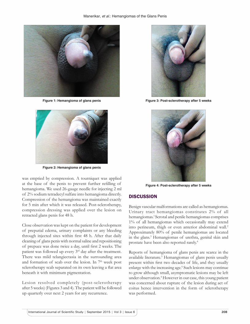
On examination, there was an elevated irregular bluish red lesion of size 2 cm × 1.5 cm on the left dorsolateral portion of the glans penis (hemangioma of the glans penis) (Figures 1 and 2). The lesion was compressible, painless and non-pulsatile. There was no local rise in temperature. Lesion did not increase on erection of penis. Both the testicles were normal in size and shape.
Clinical examination was consistent with the diagnosis of hemangioma on the glans penis. Color Doppler studies of lesion on the glans penis showed well-defi ned, hypoechoic lesion of approximately 1.4 cm × 0.8 cm on dorsal aspect of the glans penis, in the subcutaneous plane. It showed few feeding vessels traversing it and showing column fl ow on Doppler imaging. Lesion was abutting the tunica albuginea. Corpora cavernosa was normal. There was no infl ux or effl ux from the corpus spongiosum.
Treatment was performed under local anesthesia using 2% lidocaine for penile block. 2 ml of sclerosant sodium tetradecyl sulfate 2% was used. The fi rst hemangioma
INTRODUCTION
Benign vascular malformation is called as “hemangiomas.” They are classifi ed as capillary, cavernous, arteriovenous, venous, and mixed subtypes. Urinary tract hemangiomas constitutes 2% of all hemangiomas.1 Only 1% of all hemangiomas comprises scrotal and penile hemangiomas which can extend into perineum, thigh or even anterior abdominal wall.2 We present a case of young man with hemangioma on the glans penis which was treated with intralesional sclerotherapy.
CASE REPORT
The 22-year-old unmarried man came with complaints of gradually increasing painless swelling of insidious onset
Case Report
Abstract
Hemangiomas are vascular benign lesions which can occur on any part of the body. Urinary tract hemangiomas and specifi cally that of glans penis are extremely rare. Treatment modalities available for such lesions are surgical excision, cryotherapy, sclerotherapy, and electrofulguration. In view of the paucity of reported patients a gold standard for treatment is yet to be established. A 22-year-old male with hemangioma of glans penis of 10 years duration presented because of his concern about lesion rupturing during the act of coitus. The patient was successfully treated with sclerotherapy using 2 ml of 2% sodium tetradecyl sulfate. Lesion resolved completely 5 weeks post-sclerotherapy and there was no evidence of any recurrence in follow-up period of next 2 years.
Key words: Hemangiomas, Laser therapy, Penis, Sclerotherapy, Sodium tetradecyl sulfate
Access this article online
www.ijss-sn.com
Month of Submission : 07-2015Month of Peer Review : 08-2015Month of Acceptance : 08-2015Month of Publishing : 09-2015
Corresponding Author: Dr. Kshitij Manerikar, Department of Surgery, Dr. D. Y. Patil Vidyapeeth’s Padmashree Dr. D. Y. Patil Medical College, Hospital & Research Centre, Pimpri, Pune, Maharashtra, India. Phone: +91-9967979386. E-mail: [email protected]
DOI: 10.17354/ijss/2015/424
Manerikar, et al.: Hemangiomas of the Glans Penis
208International Journal of Scientifi c Study | September 2015 | Vol 3 | Issue 6
was emptied by compression. A tourniquet was applied at the base of the penis to prevent further refi lling of hemangioma. We used 26-gauge needle for injecting 2 ml of 2% sodium tetradecyl sulfate into hemangioma directly. Compression of the hemangioma was maintained exactly for 5 min after which it was released. Post-sclerotherapy, compression dressing was applied over the lesion on retracted glans penis for 48 h.
Close observation was kept on the patient for development of preputial edema, urinary complaints or any bleeding through injected sites within fi rst 48 h. After that daily cleaning of glans penis with normal saline and repositioning of prepuce was done twice a day, until fi rst 2 weeks. The patient was followed up every 3rd day after the treatment. There was mild telangiectasia in the surrounding area and formation of scab over the lesion. In 7th week post sclerotherapy scab separated on its own leaving a fl at area beneath it with minimum pigmentation.
Lesion resolved completely (post-sclerotherapy after 5 weeks) (Figures 3 and 4). The patient will be followed up quarterly over next 2 years for any recurrence.
DISCUSSION
Benign vascular malformations are called as hemangiomas. Urinary tract hemangiomas constitutes 2% of all hemangiomas.1 Scrotal and penile hemangiomas comprises 1% of all hemangiomas which occasionally may extend into perineum, thigh or even anterior abdominal wall.2
Approximately 80% of penile hemangiomas are located in the glans.3 Hemangiomas of urethra, genital skin and prostate have been also reported rarely.4
Reports of hemangioma of glans penis are scarce in the available literature.5 Hemangiomas of glans penis usually present within fi rst two decades of life, and they usually enlarge with the increasing age.6 Such lesions may continue to grow although small, asymptomatic lesions may be left under observation.4 However in our case, this young patient was concerned about rupture of the lesion during act of coitus hence intervention in the form of sclerotherapy was performed.
Figure 1: Hemangioma of glans penis
Figure 2: Hemangioma of glans penis
Figure 3: Post-sclerotherapy after 5 weeks
Figure 4: Post-sclerotherapy after 5 weeks

Manerikar, et al.: Hemangiomas of the Glans Penis
209 International Journal of Scientifi c Study | September 2015 | Vol 3 | Issue 6
The majority of lesions of hemangioma of glans penis do not require any treatment as they are asymptomatic. Treatment is only required in cases of the presence of symptoms such as pain, bleeding from lesion or for cosmetic purposes.
Due to less number of reported cases of glans penis hemangioma there is a lack of standard therapeutic protocol. Different types of available therapies for treatment of hemangioma of glans penis include cryotherapy, surgical excision, sclerotherapy, and electrofulguration.7
Jimenez-Cruz and Osca were the first to introduce neodymium:Yttrium-aluminum-garnet (Nd:YAG) laser in the treatment of hemangioma of glans penis. Nd:YAG laser has excellent tissue coagulation properties without causing any fi brosis, but its tissue penetration rate is deeper as it is poorly absorbed by body pigment.8 Potassium thiophosphate laser is preferable in children, mainly in large lesions as it is absorbed by hemoglobin and hence produces less scar. According to available literature laser treatment for such hemangiomas has shown good functional and cosmetic result.9 However, availability of laser therapy and cost-effectiveness is major issue.
Intralesional sclerotherapy is a cost-effective method which is easily available and is inexpensive. Prolonged compression over the injected site after injection of sclerosant is known to achieve better results with least morbidity.10 Compression causes direct contact of sclerosant agent with the endothelium which disrupts endothelium and causes edema in few minutes. This causes thrombus formation in lumen of vessel followed by subsequent fi brosis leading to endosclerosis.9
Amongst variety of sclerosants which are available such as 30% hypertonic saline, 2% or 3% sodium tetradecyl sulfate, ethanolamine, 2% hydroxypolyaethoxydodecan polidocanol, and polyiodide iodine, sodium tetradecyl sulfate 2%, 2 ml in quantity was used by us. We used
26-gauge needle for injecting sclerosant, but other gauge needles have also been used according to literature.3
CONCLUSION
Surgical excision has high risk of damaging underlying structures and causing deformity. Since hemangioma over glans grows beneath the epidermis so partial resection of corpus spongiosum is almost always necessary.3 Glans penis is devoid of subcutaneous tissue and tunica albuginea of corpus spongiosum is very poorly developed. This characteristic anatomy of glans penis makes it diffi cult to dissect skin from corpus spongiosum.
The rarity of hemangioma of glans penis and its poor documentation with respect to treatment makes it diffi cult to choose best method among available modalities. This case is presented for its rarity and satisfactory outcome of sclerotherapy.
REFERENCES
1. Amaro JL, Agostinho AD, Polido Júnior A, Costa RP, Trindade Filho JC, Trindade JC. Treatment of hemangioma of the glans penis using Nd:Yag laser. Apropos of a case. J Urol (Paris) 1997;103:62-3.
2. Lin CY, Sun GH, Yu DS, Wu CJ, Chen HI, Chang SY. Intrascrotal hemangioma. Arch Androl 2002;48:259-65.
3. Tsujii T, Iwai T, Inoue Y, Kubota T, Kihara K, Oshima H. Cutaneous hemangioma of the penis successfully treated with sclerotherapy and ligation. Int J Urol 1998;5:396-7.
4. Ulker V, Esen T. Hemangioma of the glans penis treated with Nd:YAG laser. Int Urol Nephrol 2005;37:95-6.
5. Sharma GR. Hemangioma of glans penis. Internet J Urol 2005;3:2.6. Rastogi R. Diffuse cavernous hemangioma of the penis, scrotum, perineum,
and rectum – A rare tumor. Saudi J Kidney Dis Transpl 2008;19:614-8.7. Savoca G, De Stefani S, Buttazzi L, Gattuccio I, Trombetta C, Belgrano E.
Sclerotherapy of hemangioma of the glans penis. Urology 2000;56:153.8. Jimenez-Cruz JF, Osca JM. Laser treatment of glans penis hemangioma.
Eur Urol 1993;24:81-3.9. Kumar A, Goyal NK, Trivedi S, Dwivedi US, Singh PB. Primary cavernous
hemangioma of the glans penis: Rare case report with a review of the literature. Aesthetic Plast Surg 2008;32:386-8.
10. Weiss RA, Goldman MP. Advances in sclerotherapy. Dermatol Clin 1995;13:431-45.
How to cite this article: Manerikar K, Singh G, Ali I. Intralesional Sclerotherapy in Hemangiomas of the Glans Penis. Int J Sci Stud 2015;3(6):207-209.
Source of Support: Nil, Confl ict of Interest: None declared.