Embed Size (px)
Citation preview
INTRAOCULAR LENS EXCHANGE WITH REMOVAL OF THE OPTIC ONLY IN THE LATE POSTOPERATIVE PERIOD
Charles M, Charles DE, Charles N, Jelusich G, Palacio A, Zompa T
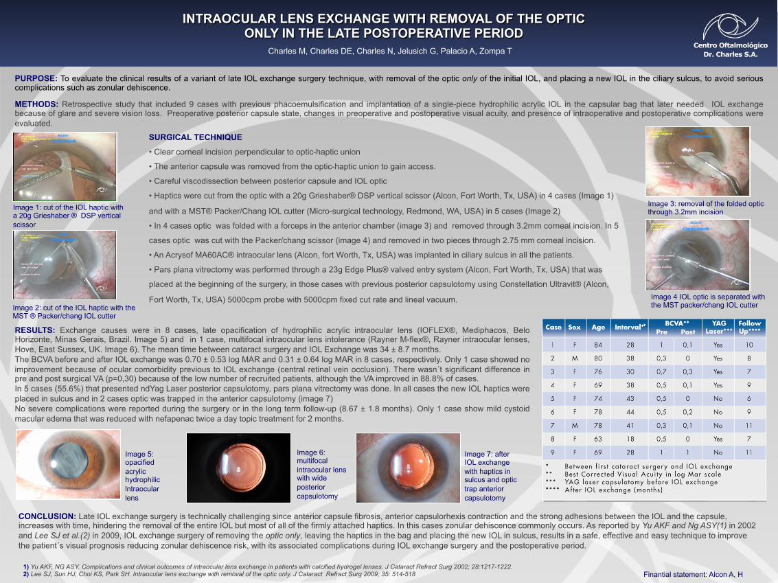
PURPOSE: To evaluate the clinical results of a variant of late IOL exchange surgery technique, with removal of the optic only of the initial IOL, and placing a new IOL in the ciliary sulcus, to avoid serious complications such as zonular dehiscence. METHODS: Retrospective study that included 9 cases with previous phacoemulsification and implantation of a single-piece hydrophilic acrylic IOL in the capsular bag that later needed IOL exchange because of glare and severe vision loss. Preoperative posterior capsule state, changes in preoperative and postoperative visual acuity, and presence of intraoperative and postoperative complications were evaluated.
RESULTS: Exchange causes were in 8 cases, late opacification of hydrophilic acrylic intraocular lens (IOFLEX®, Mediphacos, Belo Horizonte, Minas Gerais, Brazil. Image 5) and in 1 case, multifocal intraocular lens intolerance (Rayner M-flex®, Rayner intraocular lenses, Hove, East Sussex, UK. Image 6). The mean time between cataract surgery and IOL Exchange was 34 ± 8.7 months. The BCVA before and after IOL exchange was 0.70 ± 0.53 log MAR and 0.31 ± 0.64 log MAR in 8 cases, respectively. Only 1 case showed no improvement because of ocular comorbidity previous to IOL exchange (central retinal vein occlusion). There wasn´t significant difference in pre and post surgical VA (p=0,30) because of the low number of recruited patients, although the VA improved in 88.8% of cases. In 5 cases (55.6%) that presented ndYag Laser posterior capsulotomy, pars plana vitrectomy was done. In all cases the new IOL haptics were placed in sulcus and in 2 cases optic was trapped in the anterior capsulotomy (image 7) No severe complications were reported during the surgery or in the long term follow-up (8.67 ± 1.8 months). Only 1 case show mild cystoid macular edema that was reduced with nefapenac twice a day topic treatment for 2 months.
CONCLUSION: Late IOL exchange surgery is technically challenging since anterior capsule fibrosis, anterior capsulorhexis contraction and the strong adhesions between the IOL and the capsule, increases with time, hindering the removal of the entire IOL but most of all of the firmly attached haptics. In this cases zonular dehiscence commonly occurs. As reported by Yu AKF and Ng ASY(1) in 2002 and Lee SJ et al.(2) in 2009, IOL exchange surgery of removing the optic only, leaving the haptics in the bag and placing the new IOL in sulcus, results in a safe, effective and easy technique to improve the patient´s visual prognosis reducing zonular dehiscence risk, with its associated complications during IOL exchange surgery and the postoperative period.
SURGICAL TECHNIQUE
• Clear corneal incision perpendicular to optic-haptic union
• The anterior capsule was removed from the optic-haptic union to gain access.
• Careful viscodissection between posterior capsule and IOL optic
• Haptics were cut from the optic with a 20g Grieshaber® DSP vertical scissor (Alcon, Fort Worth, Tx, USA) in 4 cases (Image 1)
and with a MST® Packer/Chang IOL cutter (Micro-surgical technology, Redmond, WA, USA) in 5 cases (Image 2)
• In 4 cases optic was folded with a forceps in the anterior chamber (image 3) and removed through 3.2mm corneal incision. In 5
cases optic was cut with the Packer/chang scissor (image 4) and removed in two pieces through 2.75 mm corneal incision.
• An Acrysof MA60AC® intraocular lens (Alcon, fort Worth, Tx, USA) was implanted in ciliary sulcus in all the patients.
• Pars plana vitrectomy was performed through a 23g Edge Plus® valved entry system (Alcon, Fort Worth, Tx, USA) that was
placed at the beginning of the surgery, in those cases with previous posterior capsulotomy using Constellation Ultravit® (Alcon,
Fort Worth, Tx, USA) 5000cpm probe with 5000cpm fixed cut rate and lineal vacuum.
Image 1: cut of the IOL haptic with a 20g Grieshaber ® DSP vertical scissor
Image 2: cut of the IOL haptic with the MST ® Packer/chang IOL cutter
Image 4 IOL optic is separated with the MST packer/chang IOL cutter
Image 3: removal of the folded optic through 3.2mm incision
Image 5: opacified acrylic hydrophilic lntraocular lens
Image 7: after IOL exchange with haptics in sulcus and optic trap anterior capsulotomy
Image 6: multifocal intraocular lens with wide posterior capsulotomy
1) Yu AKF, NG ASY. Complications and clinical outcomes of intraocular lens exchange in patients with calcified hydrogel lenses. J Cataract Refract Surg 2002; 28:1217-1222. 2) Lee SJ, Sun HJ, Choi KS, Park SH. Intraocular lens exchange with removal of the optic only. J Cataract Refract Surg 2009; 35: 514-518 Finantial statement: Alcon A, H
Materials and methods
� Retrospective study that included 9 cases with previous phacoemulsification and implantation of a single-piece hydrophilic acrylic IOL in the capsular bag that later needed IOL exchange because of glare and severe vision loss. Preoperative posterior capsule state, changes in preoperative and postoperative visual acuity, and presence of intraoperative and postoperative complications were evaluated.
� The surgical technique consisted in cutting the optic from the haptics and its subsequent removal, leaving the haptics in the bag, and placing the new IOL haptics in sulcus. To see the surgical technique in HD: http://vimeo.com/47373617
Exchange causes were � in 8 cases late opacification of
hydrophilic acrylic intraocular lens (IOFLEX®, Mediphacos, Belo Horizonte, Minas Gerais, Brazil)
� In 1 case, multifocal intraocular lens intolerance (Rayner M-flex®, Rayner intraocular lenses, Hove, East Sussex, UK)
The mean time between cataract surgery and IOL Exchange was 34 ± 8.7 months.
Opacified acrylic hydrophilic lntraocular lens
Multifocal intraocular lens with wide posterior capsulotomy
Results
The BCVA before and after IOL exchange was 0.70 ± 0.53 log MAR and 0.31 ± 0.64 log MAR in 8 cases, respectively. Only 1 case showed no improvement because of ocular comorbidity previous to IOL exchange (central retinal vein occlusion). There wasn´t significant difference in pre and post surgical VA (p=0,30) because of the low number of recruited patients, although the VA improved in 88.8% of cases.
Results
Results
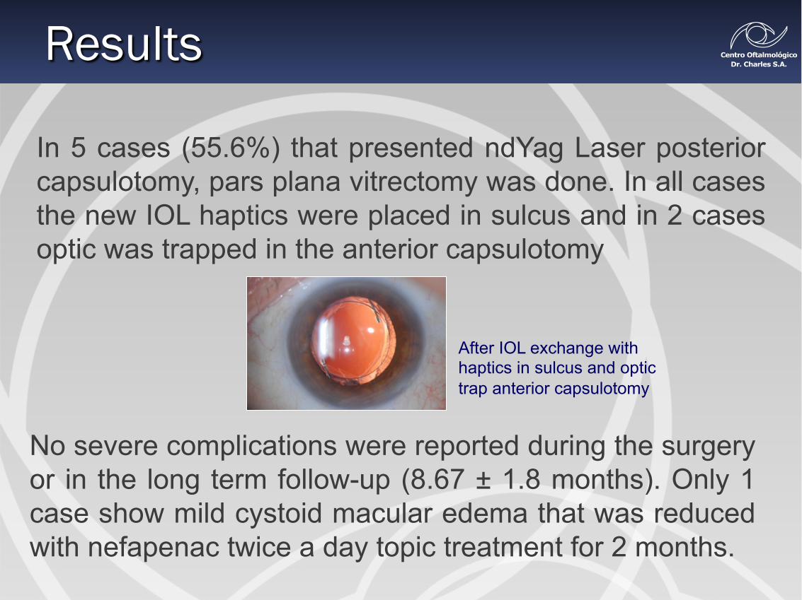
In 5 cases (55.6%) that presented ndYag Laser posterior capsulotomy, pars plana vitrectomy was done. In all cases the new IOL haptics were placed in sulcus and in 2 cases optic was trapped in the anterior capsulotomy
After IOL exchange with haptics in sulcus and optic trap anterior capsulotomy
No severe complications were reported during the surgery or in the long term follow-up (8.67 ± 1.8 months). Only 1 case show mild cystoid macular edema that was reduced with nefapenac twice a day topic treatment for 2 months.
Conclusion
Late IOL exchange surgery is technically challenging since anterior capsule fibrosis, anterior capsulorhexis contraction and the strong adhesions between the IOL and the capsule, increases with time, hindering the removal of the entire IOL but most of all of the firmly attached haptics. In this cases zonular dehiscence commonly occurs. As reported by Yu AKF and Ng ASY(1) in 2002 and Lee SJ et al.(2) in 2009, IOL exchange surgery of removing the optic only, leaving the haptics in the bag and placing the new IOL in sulcus, results in a safe, effective and easy technique to improve the patient´s visual prognosis reducing zonular dehiscence risk, with its associated complications during IOL exchange surgery and the postoperative period.
1) Yu AKF, NG ASY. Complications and clinical outcomes of intraocular lens exchange in patients with calcified hydrogel lenses. J Cataract Refract Surg 2002; 28:1217-1222.
2) Lee SJ, Sun HJ, Choi KS, Park SH. Intraocular lens exchange with removal of the optic only. J Cataract Refract Surg 2009; 35: 514-518