Embed Size (px)
Citation preview

Intraoperative assessment of tumor margins duringglioma resection by desorption electrosprayionization-mass spectrometryValentina Pirroa, Clint M. Alfaroa, Alan K. Jarmuscha, Eyas M. Hattabb,1, Aaron A. Cohen-Gadolc,1,and R. Graham Cooksa,1
aDepartment of Chemistry, Purdue University, West Lafayette, IN 47907; bDepartment of Pathology and Laboratory Medicine, Indiana University,Indianapolis, IN 46202; and cDepartment of Neurological Surgery, Indiana University School of Medicine, Indianapolis, IN 46202
Contributed by R. Graham Cooks, May 19, 2017 (sent for review April 19, 2017; reviewed by Leroy Hood and Richard N. Zare)
Intraoperative desorption electrospray ionization-mass spectrom-etry (DESI-MS) is used to characterize tissue smears by comparisonwith a library of DESI mass spectra of pathologically determinedtissue types. Measurements are performed in the operating roomwithin 3 min. These mass spectra provide direct information ontumor infiltration into white or gray brain matter based onN-acetylaspartate (NAA) and on membrane-derived complex lip-ids. The mass spectra also indicate the isocitrate dehydrogenasemutation status of the tumor via detection of 2-hydroxyglutarate,currently assessed postoperatively on biopsied tissue using immu-nohistochemistry. Intraoperative DESI-MS measurements made atsurgeon-defined positions enable assessment of relevant diseasestate of tissue within the tumor mass and examination of the re-section cavity walls for residual tumor. Results for 73 biopsies from10 surgical resection cases show that DESI-MS allows detection ofglioma and estimation of high tumor cell percentage (TCP) at sur-gical margins with 93% sensitivity and 83% specificity. TCP mea-surements from NAA are corroborated by indirect measurementsbased on lipid profiles. Notably, high percentages (>50%) of unre-sected tumor were found in one-half of the margin biopsy smears,even in cases where postoperative MRI suggested gross total tu-mor resection. Unresected tumor causes recurrence and malignantprogression, as observed within a year in one case examined in thisstudy. These results corroborate the utility of DESI-MS in assessingsurgical margins for maximal safe tumor resection. IntraoperativeDESI-MS analysis of tissue smears, ex vivo, can be inserted into thecurrent surgical workflow with no alterations. The data underscorethe complexity of glioma infiltration.
ambient ionization | neurological smears | tumor infiltration | lipids |glioma
We describe the rapid analysis of neurological tissue smearsby desorption electrospray ionization-mass spectrometry
(DESI-MS) in the operating room (OR) from 10 subjects whounderwent glioma resection. Biopsied tissue specimens fromsurgeon-defined positions in the tumor and the walls of the resectioncavity were smeared onto glass microscope slides and sprayed withcharged solvent droplets to extract molecules from the unprocessedtissue while the splashed secondary droplets were vacuumed into acustomized ion-trap mass spectrometer modified for use in the ORat Indianapolis IU (Indiana University) Health Methodist Hospital.Three separate items of information were sought from the DESImass spectra: (i) tissue type, specifically whether glioma, white brainmatter, gray brain matter, or mixtures of these types; (ii) isocitratedehydrogenase (IDH) status, i.e., whether or not this enzyme carriesa characteristic mutation, the presence of which is associated withmore favorable prognosis; and (iii) the tumor cell percentage (TCP)in the sampled biopsy as a measure of tumor infiltration (the latter isarguably the most actionable intraoperatively and the one for whichthe least information is currently available).The infiltrative nature of most gliomas, as well as visual and
textural similarities between infiltrative regions and normal brain
parenchyma, poses a substantial challenge to the neurosurgeonduring tumor resection. Microsurgical resection aims to maximizetumor excision while minimizing morbidity (1, 2) as the extent ofresection affects both overall and progression-free survival. Im-proved survival and quality of life correlates with maximal re-section of both low- and high-grade gliomas (3). Importantly,tumor recurrence and malignant progression is likely to occurfrom residual tumor within 0.5 cm of the resection (i.e., surgical)margin, viz., the point at which resection ceases (3). Stereotacticvolumetric resection is usually based on preoperative structuraland functional MRI used to determine tumor location, size, sur-gical approach, and to estimate the extent of resection achievable.Intraoperative MRI has been implemented to mitigate anatomicalshifts and improve resection; however, MRI costs and technicalhurdles, such as prolonged surgery time for image acquisition,limit its practical and routine implementation (1).The standard diagnostic approach of intraoperative consulta-
tion, pathology, provides information on tumor type and grade.Intraoperative histopathological evaluation of tissue resected fromsurgical margins to estimate residual tumor infiltration is un-common, partly because preparation and analysis of tissue sectionsor smears is laborious and time-consuming, and partly due to theinherent difficulty in assessing degree of infiltration in suboptimalpreparations. Therefore, the amount of residual tumor near the
Significance
Gliomas infiltrate into surrounding healthy brain tissue. Micro-surgical resection aims for maximal tumor resection while mini-mizing morbidity. Surgical margins are defined based on thesurgeon’s experience, visual observation, and neuronavigation.Surgical margin assessment is rarely undertaken intraoperativelydue to time constraints and unreliability of such evaluation.Routine, pathologic intraoperative examination provides no mo-lecular information. Molecular measurements using mass spec-trometry can be made rapidly on tissue during surgery to identifytissue types, estimate tumor infiltration, and recognize the pres-ence of prognostic mutations by monitoring oncometabolites andphospholipids. This intraoperative study demonstrates the powerof mass spectrometry in assessing diagnostic and prognostic in-formation on discrete surgeon-defined points along the resectionmargins to improve tumor resection, even in regions without MRIcontrast enhancement.
Author contributions: V.P., A.A.C.-G., and R.G.C. designed research; C.M.A., A.K.J., E.M.H.,and A.A.C.-G. performed research; V.P., C.M.A., A.K.J., and E.M.H. analyzed data;A.A.C.-G. performed surgery; and V.P., E.M.H., and R.G.C. wrote the paper.
Reviewers: L.H., Institute for Systems Biology; and R.N.Z., Stanford University.
The authors declare no conflict of interest.1To whom correspondence may be addressed. Email: [email protected],[email protected], or [email protected].
This article contains supporting information online at www.pnas.org/lookup/suppl/doi:10.1073/pnas.1706459114/-/DCSupplemental.
6700–6705 | PNAS | June 27, 2017 | vol. 114 | no. 26 www.pnas.org/cgi/doi/10.1073/pnas.1706459114
Dow
nloa
ded
by g
uest
on
Nov
embe
r 27
, 202
1
resection margins is not usually measured during surgery and isonly assessed postoperatively and indirectly by MRI (1, 4).Following the publication of the 2016 WHO classification of
central nervous system tumors (5), immunohistochemistry andmolecular testing are also performed on sampled neurologicaltissue to evaluate several diagnostic and prognostic markers (e.g.,IDH mutation and 1p/19q codeletion) but results again are avail-able only postoperatively. The need for near-real-time assessmentof tumor pathology, genetics, and margin status during surgicalresection in predicting patient outcome would suggest advantagesto a multimodal approach in which imaging and diagnostic tech-nologies are coupled.The analysis of neurological tissue smears using DESI-MS has
the potential to become an ex vivo diagnostic strategy that canprovide information on each of three points that we discuss in turn:(i) Tissue type, specifically glioma, white brain matter, gray brainmatter, or a mixture of these. The use of ambient ionization MSmethods, specifically DESI (6) and rapid evaporative ionizationmass spectrometry (REIMS) (7), to distinguish diseased fromhealthy tissue based on membrane-derived phospholipid signaturesis now well established. Note that an alternative method of mo-lecular diagnosis of neurological tissue in the form of Ramanspectroscopy is being developed concurrently (8, 9). Both DESI-MSand REIMS have been applied to different organs (7, 10–14),generally with excellent disease discrimination via comparison withspectra of pathology-defined reference material. REIMS requiresthe use of special surgical procedures but gives real-time in-formation. DESI gives results in a few minutes but is applicable toany surgical method, with no alteration in procedures, and thesame tissue smears can be stained after analysis and blindly eval-uated by an expert pathologist for validation. Whereas massspectral profiles (i.e., chemical signatures) of tissue sections allowaccurate differentiation between gliomas and normal brain pa-renchyma (15–20), we emphasize in this intraoperative study thecharacterization of tissue smears, especially the recognition ofthose of mixed compositions. (ii) IDH status. It has previously beenshown that the presence of IDH mutation, a prognostic markerin gliomas (21, 22), can be inferred by the MS detection of2-hydroxyglutarate (2HG) in tissue sections (20). The same dis-criminating information is sought here in tissue smears––which canbe prepared and analyzed inside the OR more easily and rapidlythan tissue sections (19, 23)––and this is the approach used in thisintraoperative study. (iii) Percentage of tumor cells. Encouragingpreliminary results on tissue sections indicated that it might bepossible to estimate TCP in tissue via measurements of N-acety-laspartate (NAA). However, this highly important objective neededto be tested in the relevant intraoperative environment, especiallyon tissue resected near surgical margins for identification of re-sidual tumor, and that is a significant aim of this study.
Results and DiscussionGeneral Observations. Intraoperative DESI-MS was performed insuch a way as to mimic one foreseeable implementation in which asingle mass spectrometer is moved into an OR setting just beforesurgery, powered on, and is ready for use within ∼30 min. Thechosen unit mass resolution linear ion-trap mass spectrometer used(17–19) performed well without significant electronic, vacuum, oreven mass calibration problems over the 15-mo duration of thestudy (SI Appendix, Table S1). Fresh tissue samples were smearedand analyzed by DESI-MS within 3 min, a timeframe that ad-dresses surgical needs. In the case of all surgeries, DESI-MSanalysis of multiple biopsies was completed long before anyintraoperative pathologic consultation findings were reported backto the neurosurgeon by the neuropathologist. The observed timingof in situ, quasi–real-time analysis by DESI-MS supports the factthat multiple measurements––made at discrete points selected bythe neurosurgeon––are reliable and have the potential to provide
the surgeon with additional information to guide resection ma-neuvers, particularly near critical anatomical structures.
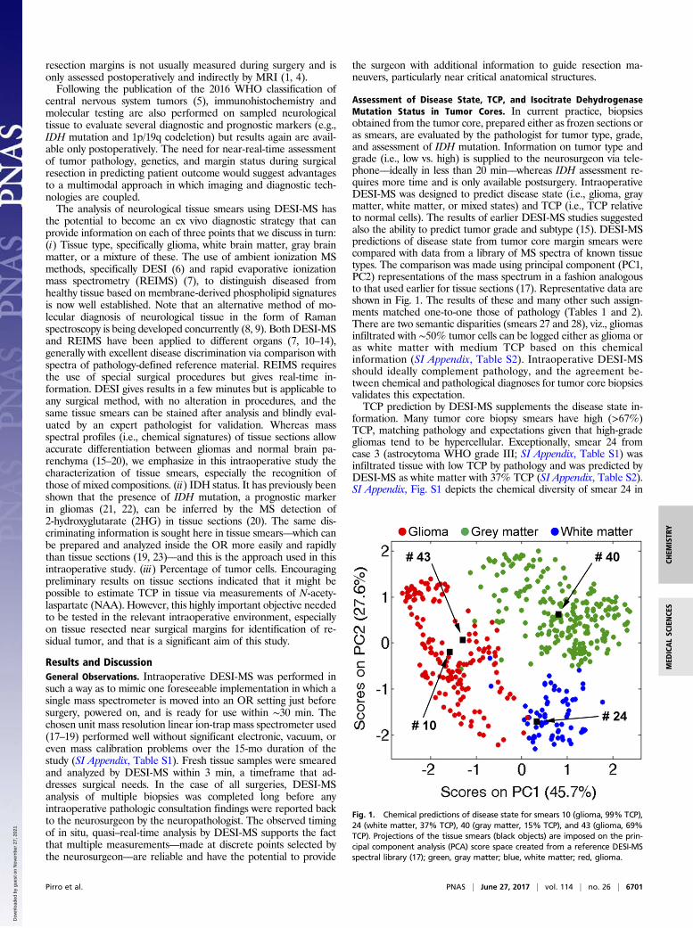
Assessment of Disease State, TCP, and Isocitrate DehydrogenaseMutation Status in Tumor Cores. In current practice, biopsiesobtained from the tumor core, prepared either as frozen sections oras smears, are evaluated by the pathologist for tumor type, grade,and assessment of IDH mutation. Information on tumor type andgrade (i.e., low vs. high) is supplied to the neurosurgeon via tele-phone––ideally in less than 20 min––whereas IDH assessment re-quires more time and is only available postsurgery. IntraoperativeDESI-MS was designed to predict disease state (i.e., glioma, graymatter, white matter, or mixed states) and TCP (i.e., TCP relativeto normal cells). The results of earlier DESI-MS studies suggestedalso the ability to predict tumor grade and subtype (15). DESI-MSpredictions of disease state from tumor core margin smears werecompared with data from a library of MS spectra of known tissuetypes. The comparison was made using principal component (PC1,PC2) representations of the mass spectrum in a fashion analogousto that used earlier for tissue sections (17). Representative data areshown in Fig. 1. The results of these and many other such assign-ments matched one-to-one those of pathology (Tables 1 and 2).There are two semantic disparities (smears 27 and 28), viz., gliomasinfiltrated with ∼50% tumor cells can be logged either as glioma oras white matter with medium TCP based on this chemicalinformation (SI Appendix, Table S2). Intraoperative DESI-MSshould ideally complement pathology, and the agreement be-tween chemical and pathological diagnoses for tumor core biopsiesvalidates this expectation.TCP prediction by DESI-MS supplements the disease state in-
formation. Many tumor core biopsy smears have high (>67%)TCP, matching pathology and expectations given that high-gradegliomas tend to be hypercellular. Exceptionally, smear 24 fromcase 3 (astrocytoma WHO grade III; SI Appendix, Table S1) wasinfiltrated tissue with low TCP by pathology and was predicted byDESI-MS as white matter with 37% TCP (SI Appendix, Table S2).SI Appendix, Fig. S1 depicts the chemical diversity of smear 24 in
Fig. 1. Chemical predictions of disease state for smears 10 (glioma, 99% TCP),24 (white matter, 37% TCP), 40 (gray matter, 15% TCP), and 43 (glioma, 69%TCP). Projections of the tissue smears (black objects) are imposed on the prin-cipal component analysis (PCA) score space created from a reference DESI-MSspectral library (17); green, gray matter; blue, white matter; red, glioma.
Pirro et al. PNAS | June 27, 2017 | vol. 114 | no. 26 | 6701
CHEM
ISTR
YMED
ICALSC
IENCE
S
Dow
nloa
ded
by g
uest
on
Nov
embe
r 27
, 202
1

comparison with another tumor core biopsy (smear 43) from aWHO grade IV tumor with high TCP. Two smears were preparedfrom nine tumor core biopsies by splitting the tissue in half. En-couragingly, the disease state was the same and the NAA-basedpredicted TCP agreed to within 15% (SI Appendix, Table S2).IDH mutation status was assessed intraoperatively via negative-
ion DESI-MS detection of 2HG (m/z 147) followed by frag-mentation using collision-induced dissociation for structuralconfirmation. IDH mutation status is currently only determined viaimmunohistochemistry and genetic testing postoperatively, eventhough its intraoperative assessment could have surgical utility.More aggressive resection of IDH-mutant tumors, which are as-sociated with better prognosis (21, 22), significantly improvesoverall and progression-free survival, whereas more aggressive re-section of wild-type tumors does not result in such benefits (24).The oncometabolite 2HG was detected in the tumor core biopsiesfrom cases 1, 3, 5, 7, and 9; these were determined postoperativelyto be IDH-mutant (SI Appendix, Table S1). In the mutant tumors,the 2HG signal intensity varied between smears, grossly correlatingwith the degree of tumor infiltration (Fig. 2), as previously ob-served by DESI-MS for tissue sections (20). Independent studiesconfirmed a positive correlation between 2HG concentration andtumor cellularity but showed no significant differences betweentumor types and grades (24). No 2HG was detected for wild-typetumors (SI Appendix, Fig. S2).
Assessment of Disease State and TCP at Discrete Points nearResection Margins. Forty-four of 80 biopsies were obtained nearsurgical margins (within 0.5 cm from where tumor resection ceased).The visualization of the pseudomargin of gliomas and their marginstatus can be difficult and unreliable. It is also highly unusual andinefficient to use intraoperative pathologic consultation to de-termine where to cease resection. Postoperative MRI normally as-sesses the approximate volume of residual tumor. The absence ofresidual contrast enhancement around the resection cavity is inter-preted as macroscopic or gross total resection (4). We have de-veloped two models for estimating residual tumor at discrete pointsresected at the discretion of the surgeon. One is based on NAAsignal intensity whereas the other is a more indirect measure basedon the lipid profile of the tissue. Note, although we provide pre-diction of disease state from smears of margin biopsies, the pa-thologist does not routinely or exclusively use such samples for thedetermination of tumor type, grade (such metrics are determinedusing tumor core biopsies), or margin status when applicable.N-acetylaspartate model for TCP predictions. A roughly exponentialdecrease of NAA signal intensity was observed vs. pathologicallymeasured increase in TCP in tissue sections. This observation hasnow been extended and confirmed in tissue smears analyzed intra-operatively (SI Appendix, Fig. S3). TCP predictions based on NAAof biopsied tissue near surgical margins ranged between 0% and100%. Notably, about one-half of the margin tissue smears wereinfiltrated with more than 50% tumor cells. Fig. 3 shows the fre-quency of low, medium, and high TCP, as well as of the disease statepredictions, both compared with histopathological results. Agree-ment is good. The absence of a signal for NAA in tissue––when adistinct glioma lipid profile is present––is specific for high-densitygliomas (SI Appendix, Table S3). Sensitivity is also high, as just threesmears (69, 77–79) showed low NAA signal (TCP estimates between
73–74%; SI Appendix, Table S2) when pathology determined lessthan 10% tumor cells. The presence of vascular proliferation wasnoted in these smears. The availability of an intraoperative tool foridentification of high (>50%) residual tumor infiltration at surgicalmargins is essential for neurosurgeons to refine surgical maneuvers.Safe removal of high tumor infiltration is a primary goal of neuro-surgery whereas areas of low infiltration are more likely the target ofcoadjuvant postsurgical therapies. Our current qualitative method-ology is less accurate in differentiating betweenmedium and low TCP(SI Appendix, Table S3). The literature suggests that NAA varies withage, gender, and other biological factors (25) that were not controlledin this small study. Biological variability affecting agreement betweenchemical and pathological estimations manifests itself when NAA ispresent in the tissue; i.e., in low- and medium-infiltrated tissue.Lipid profile deconvolution for evaluation of tumor density. The lipidprofile detected by DESI-MS can show the presence of tumor intissue and distinguishes the background into which the tumor in-filtrates; i.e., gray or white brain matter (17). The dynamic changesof DESI-MS lipid profiles acquired from tissue sections werepreviously interpreted in terms of three-component mixtures ofglioma, white matter, and gray matter (SI Appendix, Fig. S4). Thedynamics are reflective of the infiltrative nature of gliomas into thesurrounding brain parenchyma. A linear regression model wasdeveloped to determine composition of gray matter, white matter,and glioma based on a reference spectral library (see details in SIAppendix). We further speculate that the deconvolution of lipidprofiles into fractions of white matter, gray matter, and glioma canindicate tumor density and corroborate NAA predictions. In the44 margin smears analyzed, the lipid deconvolution underscoredthe complexity of gliomas identifying tumor infiltration into white-(e.g., smear 18) or gray matter (e.g., smear 44), into a mixture ofthose (e.g., smear 49), or even identifying mixtures of gray andwhite matter with no detectable presence of tumor cells (e.g.,smear 68). SI Appendix, Fig. S5 depicts the dynamic of the lipidprofiles reflecting such complexity. This determination is madeeven more difficult during pathological evaluation by the fact thattumor effaces normal parenchyma resulting in identification ofinfiltrated tissue (not otherwise specified) [(IT nos); Fig. 3]. Mosttumors infiltrate into white matter, so their lipid signatures expresshigh abundances of sulfatides related to myelination of neurons(17). Only a few smears showed infiltration into gray matter(smears 20, 26, 40, 42, 44, 45, 55, 67, and 68; SI Appendix, TableS2). In these cases, pathological evaluation confirmed the presenceof gray matter infiltrated with tumor cells or mixed with whitematter. As was the case for NAA, tissue near surgical marginsshowed high heterogeneity in lipid expression. The deconvolutionof lipid profiles of the smears into their constituent tissue typesaccurately identified low- (14 smears of 18) and high-density gli-omas (10 smears of 14), whereas less accuracy was obtained formedium-infiltrated tissue (SI Appendix, Table S4). A larger cohortof patients is necessary to develop a more accurate multifactorialregression model based on experimental results rather than usingthe assumption of linear combination with no interactions.Residual tumor near surgical margins. Tumor infiltration variedgreatly not only in biopsied tissue resected near surgical mar-gins but also within the epicenter of the tumor. The peculiar
Table 2. Association between chemical predictions ofTCP vs. pathology for tumor cores
Chemical evaluation of tumor cell percentage
Pathological evaluation Low (<33%) Medium (34–67%) High (>67%)
Low 0 2 2Medium 0 2 1High 0 0 21
Table 1. Association between chemical predictions of diseasestate and pathology for tumor cores
Chemical evaluation of disease state
Pathological evaluation Gray matter White matter Glioma
Infiltrated tissue 0 3 1Glioma 0 2 22
6702 | www.pnas.org/cgi/doi/10.1073/pnas.1706459114 Pirro et al.
Dow
nloa
ded
by g
uest
on
Nov
embe
r 27
, 202
1

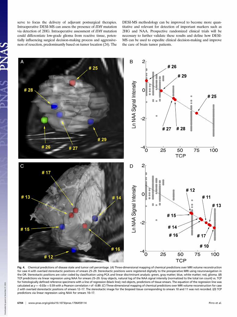
behavior of gliomas to infiltrate irregularly into brain matter,mostly following white-matter tracts, was observed in the mul-tiple measurements performed by DESI-MS. We found highTCP near the margins in both high- and low-grade gliomas,with only a weak relationship between TCP and biopsy locationwithin the tumor volume (i.e., decreasing TCP moving fromtumor core to margins). Marginal points sometimes showedTCP higher than those measured inside the tumor. As extremeexamples, we detail cases 2 and 4. For case 4, glioblastomaWHO grade IV, two biopsies (smears 25 and 26) obtained nearsurgical margins had TCP (predicted using NAA) of 75% and0%, respectively, matching pathology (high and low TCP). Twobiopsies resected inside the tumor mass showed TCP of about40% (SI Appendix, Table S2). For case 2, dysembryoplasticneuroepithelial tumor WHO grade I, discrete biopsied tissueresected near the surgical margins showed TCP from 65% to93% (Fig. 4). Again, marginal points showed TCP higher thandiscrete locations inside the tumor mass (SI Appendix, TableS2). Similar trends were found in the other cases as well (SIAppendix, Table S2). Remarkably, and unfortunately, post-operative MRI provided no evidence of residual tumor in anyof the cases except 1 and 2. Limitations in estimating extent ofresection by MRI exist and are largely due to lack of correlationbetween MRI contrast enhancement and histological compo-sition of the tissue, difficulties in calculating tumor volume, and
the occurrence of nonenhancing tumors (4). Also, the volumeof the resection cavity does not provide a true indication of theresidual microscopic tumor burden (3). Unresected tumor isthe primary cause of recurrence and malignant progression.This is shown in cases 1 and 7, which refer to the same subjectwho underwent glioma resection twice within one year. Theprimary tumor was diagnosed as oligodendroglioma WHOgrade II, using WHO 2007 terminology (SI Appendix, TableS1). The tumor was adherent to medial structures and thelateral thalamus; only partial tumor resection was performed astotal resection was determined to be unsafe. Residual tumorwas detected by DESI-MS in the posterior margin, with TCPranging between 74% and 88% into the biopsied tissue (smears6–8, case 1). A secondary tumor mass recurred from the pos-terior margin and progressed into glioblastoma WHO grade IV(SI Appendix, Fig. S6). Notably, the lipid signature of the re-current tumor showed higher intensity of phosphatidylinositol(38:4), m/z 885.5 (SI Appendix, Fig. S6), which has been notedas a discriminatory marker for glioblastoma in previous DESI-MSstudies (15). Also, DESI-MS identified the presence of 2HG in theprimary oligodendroglioma, indicative of IDHmutation, confirmedpostoperatively (SI Appendix, Table S1). Progression of oligo-dendrogliomas into glioblastomas occurs predominantly inIDH-mutant tissue (21, 22).
ConclusionsRapid DESI-MS analysis of tissue smears was performed inside theOR during glioma resection. The results from the first 10 patientsin a projected 50-patient study indicate that the DESI-MS meth-odology is simple, reliable, and can be inserted into the currentsurgical workflow without interference. Simplicity of the in-strumentation, a low-resolution ion-trap mass spectrometer, andthe methodology is desirable for robustness and ruggedness. DESI-MS faithfully recapitulates basic diagnostic information (diseasestate and TCP) in less time than is needed for pathological eval-uation. This enables multiple direct measurements on neurologicaltissue for examination of clinically relevant variants within the tu-mor and for the assessment of discrete points in the resection cavityfor residual tumor. The finding that biopsies taken near each otherand having similar tumor types can show vastly different TCPsspeaks to the complexity and known heterogeneity of glioma in-vasion into adjacent tissue. This heterogeneity partly accounts forfailure of single treatment paradigms. High percentages of tumorcells were found near surgical margins, even in cases where post-operative MRI showed gross total resection, corroborating theutility of DESI-MS coupled with neuronavigation to assist inmaximal tumor resection, an outcome associated with better pa-tient survival. In situations where tumor is detected but cannot besafely removed, intraoperative measurement of residual tumor can
Fig. 3. Margin smears (n = 44). (A) Frequency of DESI-MS prediction of disease state vs. pathology. G, glioma; GM, gray matter; IT (nos), infiltrated tissue (nototherwise specified); WM, white matter. DESI-MS has no IT (nos) assignments because all smears were assigned. (B) Frequency of DESI-MS prediction of TCP usingNAA vs. pathology. Percentages of tumor cells are categorized as low (<33%), medium (34–67%), and high (>67%).
Fig. 2. Signal intensity of 2HG (m/z 147) normalized to the total ion counts insmears 62–70 for case 9. Secondary axis shows TCP as estimated by pathologyin the same smears.
Pirro et al. PNAS | June 27, 2017 | vol. 114 | no. 26 | 6703
CHEM
ISTR
YMED
ICALSC
IENCE
S
Dow
nloa
ded
by g
uest
on
Nov
embe
r 27
, 202
1
serve to focus the delivery of adjuvant postsurgical therapies.Intraoperative DESI-MS can assess the presence of IDH mutationvia detection of 2HG. Intraoperative assessment of IDH mutationcould differentiate low-grade glioma from reactive tissue, poten-tially influencing surgical decision-making process and aggressive-ness of resection, predominantly based on tumor location (24). The
DESI-MS methodology can be improved to become more quan-titative and relevant for detection of important markers such as2HG and NAA. Prospective randomized clinical trials will benecessary to further validate these results and define how DESI-MS can be used to expedite clinical decision-making and improvethe care of brain tumor patients.
Fig. 4. Chemical predictions of disease state and tumor cell percentage. (A) Three-dimensional mapping of chemical predictions over MRI volume reconstructionfor case 4 with overlaid stereotactic positions of smears 25–29. Stereotactic positions were registered digitally to the preoperative MRI using neuronavigation inthe OR. Stereotactic positions are color coded by classification using PCA and linear discriminant analysis: green, gray matter; blue, white matter; red, glioma. (B)TCP predictions via linear regression using NAA for smears 25–29. Gray objects, natural log of the NAA signal intensity (normalized to the total ion count) vs. TCPfor histologically defined reference specimens with a line of regression (black line); red objects, predictions of tissue smears. The equation of the regression line wascalculated as y = –0.03x + 0.59 with a Pearson correlation r of –0.89. (C) Three-dimensional mapping of chemical predictions overMRI volume reconstruction for case2 with overlaid stereotactic positions of smears 12–17. The stereotactic image for the biopsied tissue corresponding to smears 10 and 11 was not recorded. (D) TCPpredictions via linear regression using NAA for smears 10–17.
6704 | www.pnas.org/cgi/doi/10.1073/pnas.1706459114 Pirro et al.
Dow
nloa
ded
by g
uest
on
Nov
embe
r 27
, 202
1

Materials and MethodsTen subjects who underwent glioma resection were recruited in this study[Indiana University Institutional Review Board (IRB) Protocol No. 1410342262].The Health Insurance Portability and Accountability Act authorization wasobtained from each subject. Details on the patient cohort are reported in SIAppendix, Table S1. Tissue biopsies (total of 73 smears; SI Appendix, Table S2)collected from stereotactically registered positions in either the tumor core ornear the surgical margins (within 0.5 cm from where tumor resection ceased)were obtained during surgical glioma resection and analyzed using DESI-MS inthe OR. The number and size of the biopsies were determined at the discretionof the neurosurgeon. Tissue specimens (5–50 mm3) were smeared on standardmicroscope glass slides (18). The act of smearing homogenizes the tissue andallows acquisition of representative data even when examining a small fractionof the surface of the smear, thereby limiting the analysis time to a few minutes(19, 23). Scans acquired orthogonally to the smearing direction average anyremaining heterogeneity of the tissue, a conclusion which is supported by DESI-MS images of tissue smears (SI Appendix, Fig. S7). DESI-MS was performed usingdimethylformamide-acetonitrile (1:1 vol/vol), a solvent that preserves tissuemorphology (26). MS measurements were performed using a linear ion-trapmass spectrometer (Finnigan LTQ, Thermo Scientific) modified for use in theOR; details in SI Appendix. Smears were subjected to sequential negative-mode DESI-MS (analysis time, 2.2 min) to obtain spectral signatures providing
information on the presence of tumor, the extent of tumor infiltration, and thepresence of IDH mutation via 2HG detection. The same smears analyzed byDESI-MS were stained and blindly evaluated by an expert neuropathologist,postoperatively, to provide information on overall diagnosis, TCP, and WHOgrading when feasible. Chemical data were analyzed offline to minimize thepotential influence on surgical decision-making (following the terms of ap-proved IRB); computations are rapid, simple, and feasible in situ. Smears werediagnosed as gray brain matter, white brain matter, or glioma via patternrecognition of their DESI-MS spectral signatures using a reference DESI-MSspectral library (17). Additional information can be found in SI Appendix.TCP was predicted using the relative intensity of NAA (17) as well as from thelipid profiles. Preoperative and postoperative MRI scans, radiology, operative,and pathology reports were obtained for each case within 2 wk of surgery.
ACKNOWLEDGMENTS. The authors acknowledge Dr. Zane Baird for construc-tion of the modified mass spectrometer; Adam Hollerbach for 3D-printing thesmear devices; clinical research nurses Jaala Hughes and Heather Cero for patientconsent, providing clinical data, and IRB monitoring; and the surgical teams ofA.A.C.-G. and Dr. Troy Payner for their keen cooperation. The research wassupported by the National Institute of Biomedical Imaging and Bioengineering,NIH Grant R21EB015722; and the Purdue University Center for Cancer Research.
1. Young RM, Jamshidi A, Davis G, Sherman JH (2015) Current trends in the surgicalmanagement and treatment of adult glioblastoma. Ann Transl Med 3:121.
2. Orringer D, et al. (2012) Extent of resection in patients with glioblastoma: Limitingfactors, perception of resectability, and effect on survival. J Neurosurg 117:851–859.
3. Hervey-Jumper SL, Berger MS (2016) Maximizing safe resection of low- and high-grade glioma. J Neurooncol 130:269–282.
4. Eidel O, et al. (2017) Tumor infiltration in enhancing and non-enhancing parts ofglioblastoma: A correlation with histopathology. PLoS One 12:e0169292.
5. Louis DN, et al. (2016) The 2016 World Health Organization Classification of Tumorsof the Central Nervous System: A summary. Acta Neuropathol 131:803–820.
6. Cooks RG, Ouyang Z, Takats Z, Wiseman JM (2006) Detection technologies. Ambientmass spectrometry. Science 311:1566–1570.
7. Balog J, et al. (2013) Intraoperative tissue identification using rapid evaporativeionization mass spectrometry. Sci Transl Med 5:194ra93.
8. Orringer DA, et al. (2017) Rapid intraoperative histology of unprocessed surgicalspecimens via fibre-laser-based stimulated Raman scattering microscopy. Nat BiomedEng 1:0027.
9. Jermyn M, et al. (2015) Intraoperative brain cancer detection with Raman spectros-copy in humans. Sci Transl Med 7:274ra19.
10. Eberlin LS, et al. (2014) Molecular assessment of surgical-resection margins of gastriccancer by mass-spectrometric imaging. Proc Natl Acad Sci USA 111:2436–2441.
11. Eberlin LS, et al. (2016) Pancreatic cancer surgical resection margins: Molecular as-sessment by mass spectrometry imaging. PLoS Med 13:e1002108.
12. Tata A, et al. (2016) Rapid detection of necrosis in breast cancer with desorptionelectrospray ionization mass spectrometry. Sci Rep 6:35374.
13. Calligaris D, et al. (2015) Molecular typing of meningiomas by desorption electrosprayionization mass spectrometry imaging for surgical decision-making. Int J Mass Spectrom377:690–698.
14. Ifa DR, Eberlin LS (2016) Ambient ionization mass spectrometry for cancer diagnosisand surgical margin evaluation. Clin Chem 62:111–123.
15. Eberlin LS, et al. (2012) Classifying human brain tumors by lipid imaging with mass
spectrometry. Cancer Res 72:645–654.16. Eberlin LS, et al. (2013) Ambient mass spectrometry for the intraoperative molecular
diagnosis of human brain tumors. Proc Natl Acad Sci USA 110:1611–1616.17. Jarmusch AK, et al. (2016) Lipid and metabolite profiles of human brain tumors by
desorption electrospray ionization-MS. Proc Natl Acad Sci USA 113:1486–1491.18. Jarmusch AK, et al. (2016) Differential lipid profiles of normal human brain matter
and gliomas by positive and negative mode desorption electrospray ionization - mass
spectrometry imaging. PLoS One 11:e0163180.19. Pirro V, et al. (2017) Utility of neurological smears for intrasurgical brain cancer di-
agnostics and tumour cell percentage by DESI-MS. Analyst (Lond) 142:449–454.20. Santagata S, et al. (2014) Intraoperative mass spectrometry mapping of an onco-
metabolite to guide brain tumor surgery. Proc Natl Acad Sci USA 111:11121–11126.21. Tietze A, et al. (March 3, 2017) Noninvasive assessment of isocitrate dehydrogenase
mutation status in cerebral gliomas by magnetic resonance spectroscopy in a clinical
setting. J Neurosurg, 10.3171/2016.10.JNS161793.22. Cohen AL, Holmen SL, Colman H (2013) IDH1 and IDH2 mutations in gliomas. Curr
Neurol Neurosci Rep 13:345.23. Woolman M, et al. (2017) An assessment of the utility of tissue smears in rapid cancer
profiling with desorption electrospray ionization mass spectrometry (DESI-MS). J Am
Soc Mass Spectrom 28:145–153.24. Beiko J, et al. (2014) IDH1 mutant malignant astrocytomas are more amenable to
surgical resection and have a survival benefit associated with maximal surgical re-
section. Neuro-oncol 16:81–91.25. Moffett JR, Ross B, Arun P, Madhavarao CN, Namboodiri AM (2007) N-Acetylaspartate
in the CNS: from neurodiagnostics to neurobiology. Prog Neurobiol 81:89–131.26. Eberlin LS, et al. (2011) Nondestructive, histologically compatible tissue imaging by
desorption electrospray ionization mass spectrometry. ChemBioChem 12:2129–2132.
Pirro et al. PNAS | June 27, 2017 | vol. 114 | no. 26 | 6705
CHEM
ISTR
YMED
ICALSC
IENCE
S
Dow
nloa
ded
by g
uest
on
Nov
embe
r 27
, 202
1