Embed Size (px)
Citation preview
Intraoperative Neural Monitoring in Pediatrics
Tod Sloan MD MBA PhD
I have no conflicts of interest or financial disclosures University of Colorado School of Medicine
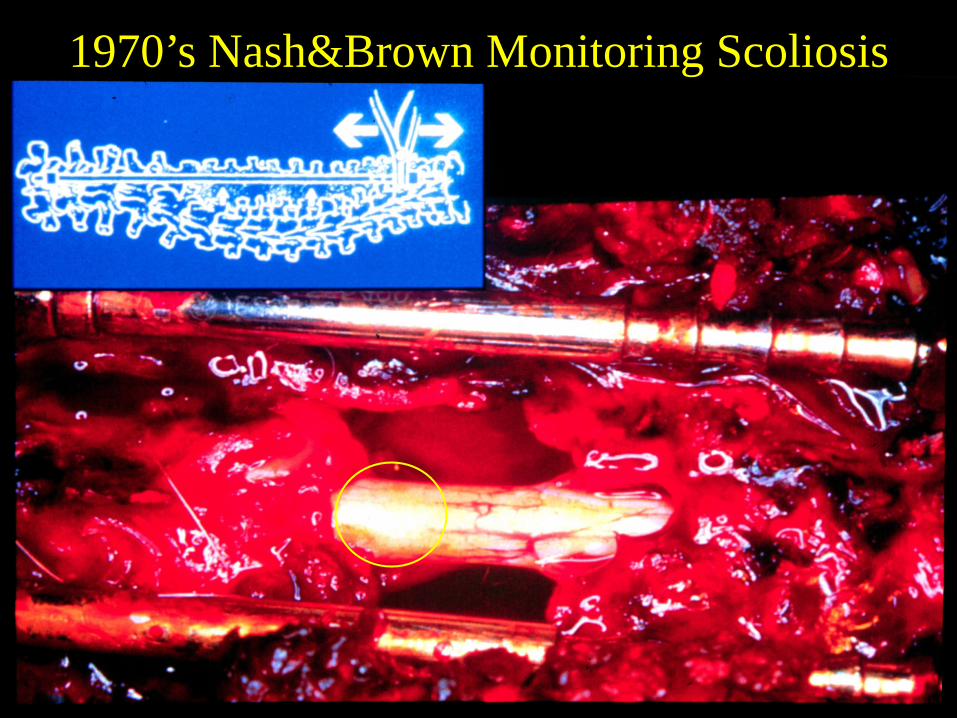
1970’s Nash&Brown Monitoring Scoliosis
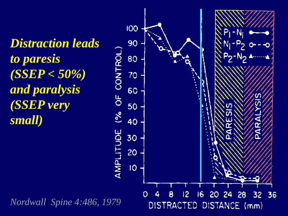
Distraction leads to paresis (SSEP < 50%) and paralysis (SSEP very small)
Nordwall Spine 4:486, 1979
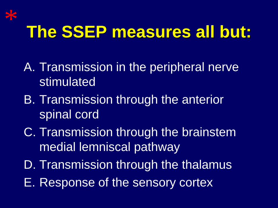
The SSEP measures all but:
A. Transmission in the peripheral nerve stimulated
B. Transmission through the anterior spinal cord
C. Transmission through the brainstem medial lemniscal pathway
D. Transmission through the thalamus E. Response of the sensory cortex
*
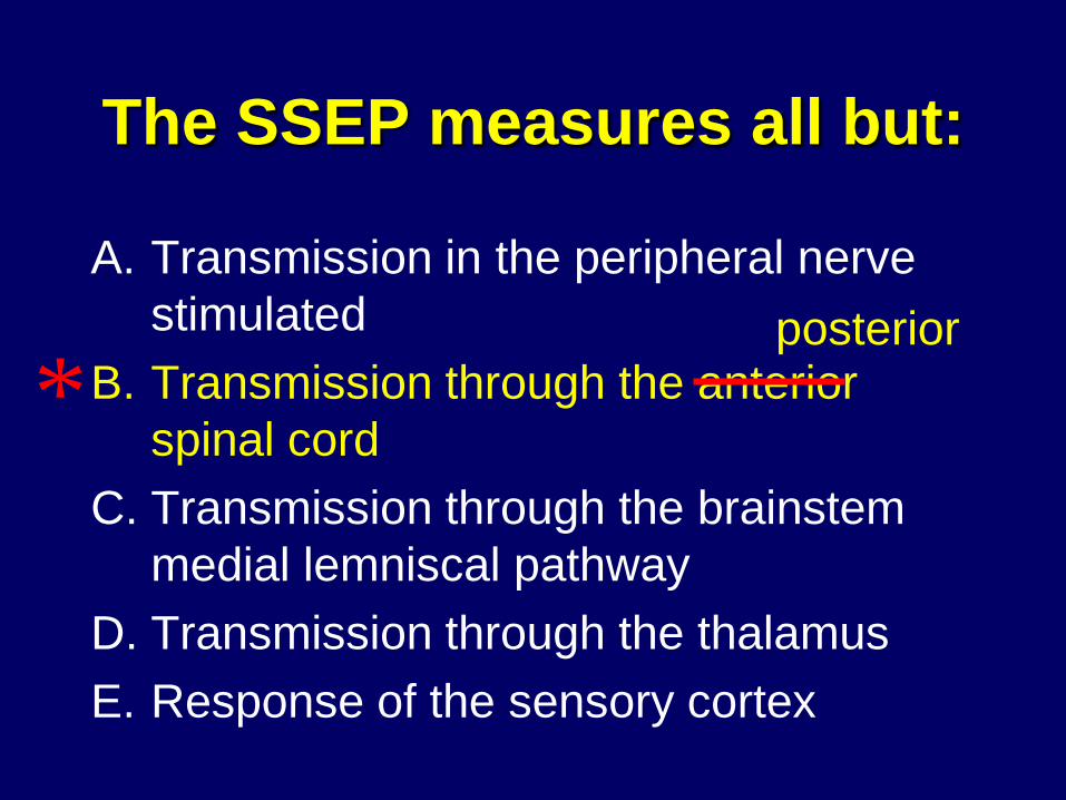
The SSEP measures all but:
A. Transmission in the peripheral nerve stimulated
B. Transmission through the anterior spinal cord
C. Transmission through the brainstem medial lemniscal pathway
D. Transmission through the thalamus E. Response of the sensory cortex
* posterior
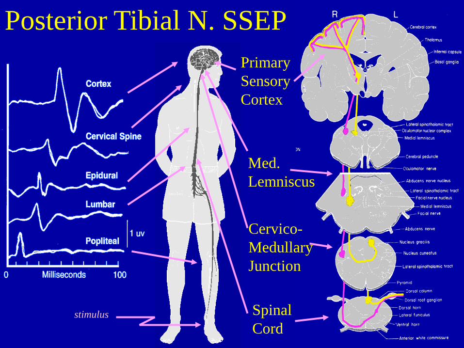
Primary Sensory Cortex
Med. Lemniscus
Cervico-Medullary Junction
Spinal Cord
Posterior Tibial N. SSEP
stimulus
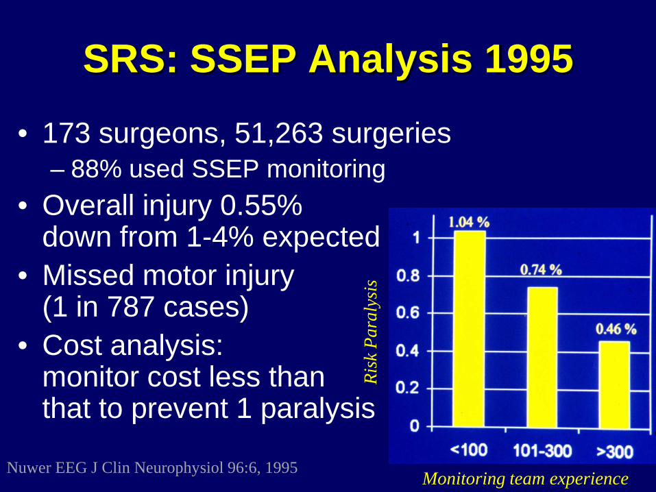
SRS: SSEP Analysis 1995
• 173 surgeons, 51,263 surgeries – 88% used SSEP monitoring
• Overall injury 0.55% down from 1-4% expected
• Missed motor injury (1 in 787 cases)
• Cost analysis: monitor cost less than that to prevent 1 paralysis
Nuwer EEG J Clin Neurophysiol 96:6, 1995 Monitoring team experience
Risk
Par
alys
is

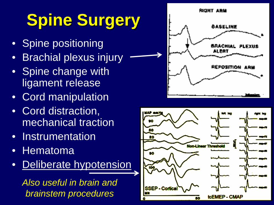
Spine Surgery • Spine positioning • Brachial plexus injury • Spine change with
ligament release • Cord manipulation • Cord distraction,
mechanical traction • Instrumentation • Hematoma • Deliberate hypotension
Also useful in brain and brainstem procedures
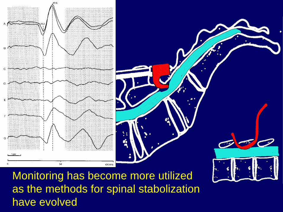
Monitoring has become more utilized as the methods for spinal stabolization have evolved
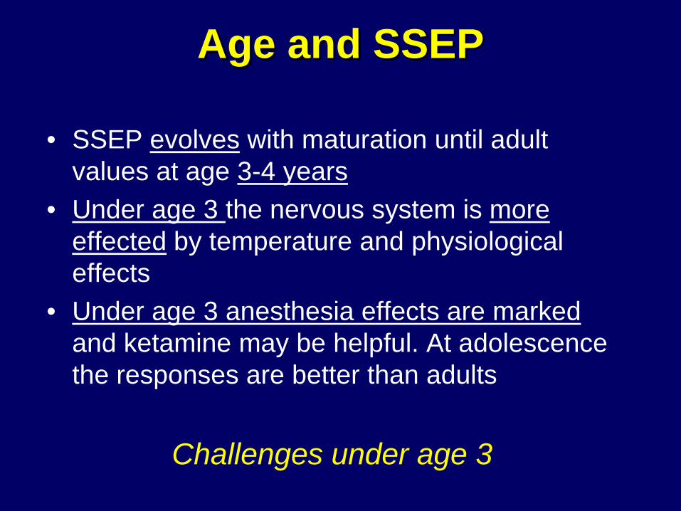
Age and SSEP
• SSEP evolves with maturation until adult values at age 3-4 years
• Under age 3 the nervous system is more effected by temperature and physiological effects
• Under age 3 anesthesia effects are marked and ketamine may be helpful. At adolescence the responses are better than adults
Challenges under age 3
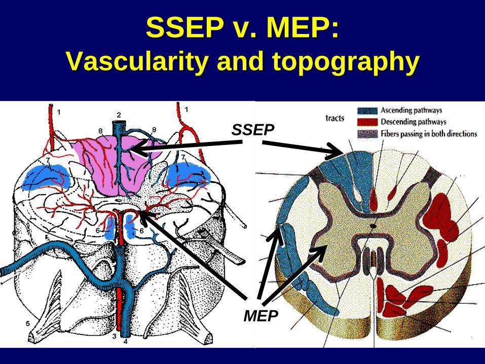
SSEP v. MEP: Vascularity and topography
SSEP
MEP
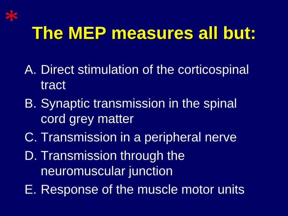
The MEP measures all but:
A. Direct stimulation of the corticospinal tract
B. Synaptic transmission in the spinal cord grey matter
C. Transmission in a peripheral nerve D. Transmission through the
neuromuscular junction E. Response of the muscle motor units
*
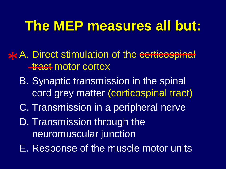
The MEP measures all but:
A. Direct stimulation of the corticospinal tract motor cortex
B. Synaptic transmission in the spinal cord grey matter (corticospinal tract)
C. Transmission in a peripheral nerve D. Transmission through the
neuromuscular junction E. Response of the muscle motor units
*
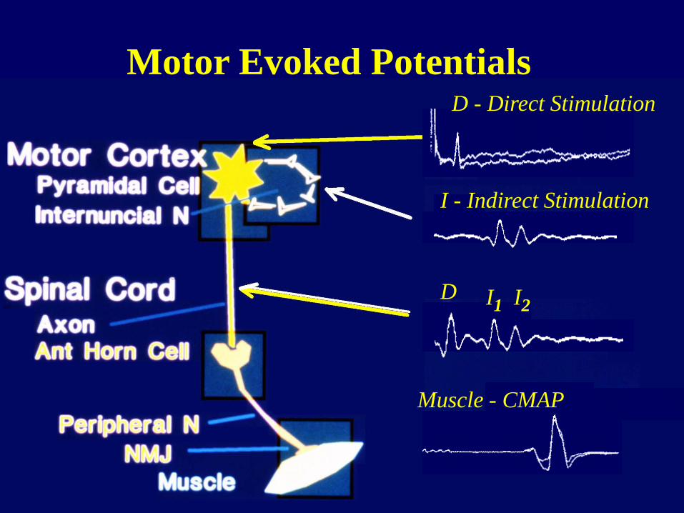
Motor Evoked Potentials
D I1 I2
Muscle - CMAP
I - Indirect Stimulation
D - Direct Stimulation
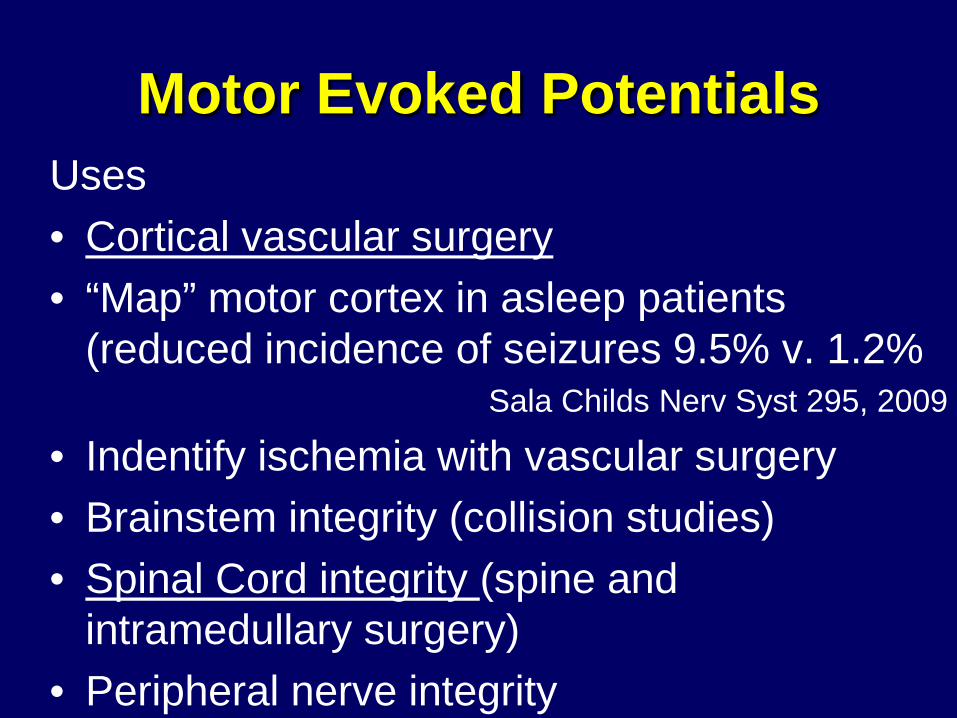
Motor Evoked Potentials Uses • Cortical vascular surgery • “Map” motor cortex in asleep patients
(reduced incidence of seizures 9.5% v. 1.2% Sala Childs Nerv Syst 295, 2009
• Indentify ischemia with vascular surgery • Brainstem integrity (collision studies) • Spinal Cord integrity (spine and
intramedullary surgery) • Peripheral nerve integrity
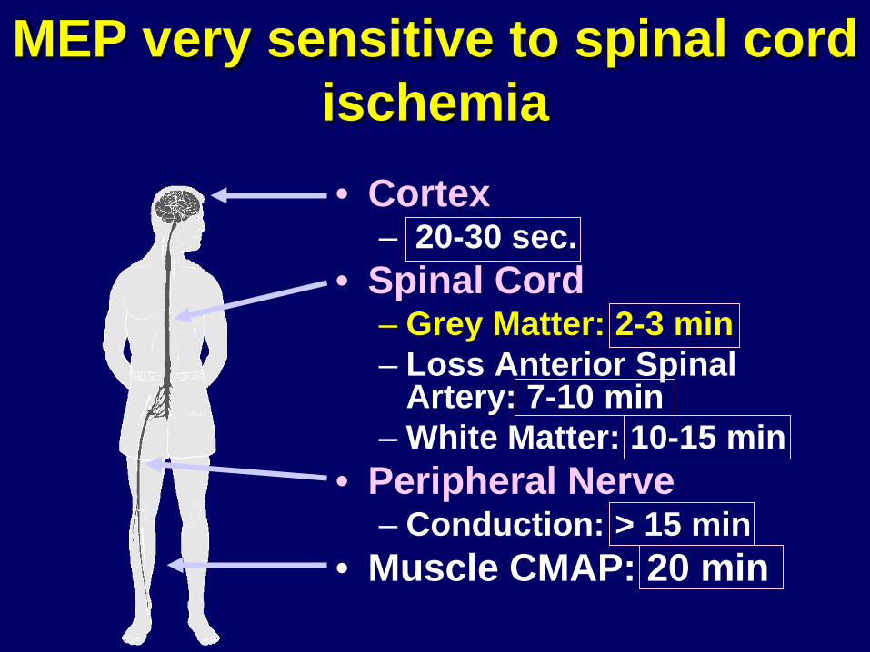
MEP very sensitive to spinal cord ischemia • Cortex
– 20-30 sec. • Spinal Cord
– Grey Matter: 2-3 min – Loss Anterior Spinal
Artery: 7-10 min – White Matter: 10-15 min
• Peripheral Nerve – Conduction: > 15 min
• Muscle CMAP: 20 min
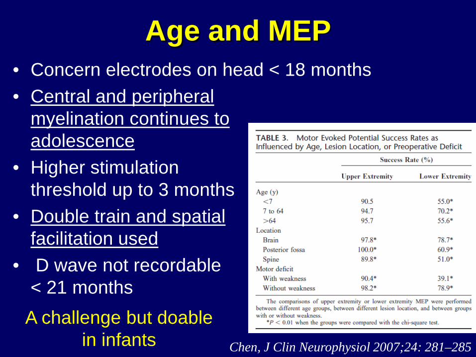
Age and MEP • Concern electrodes on head < 18 months • Central and peripheral
myelination continues to adolescence
• Higher stimulation threshold up to 3 months
• Double train and spatial facilitation used
• D wave not recordable < 21 months
Chen, J Clin Neurophysiol 2007;24: 281–285
A challenge but doable in infants
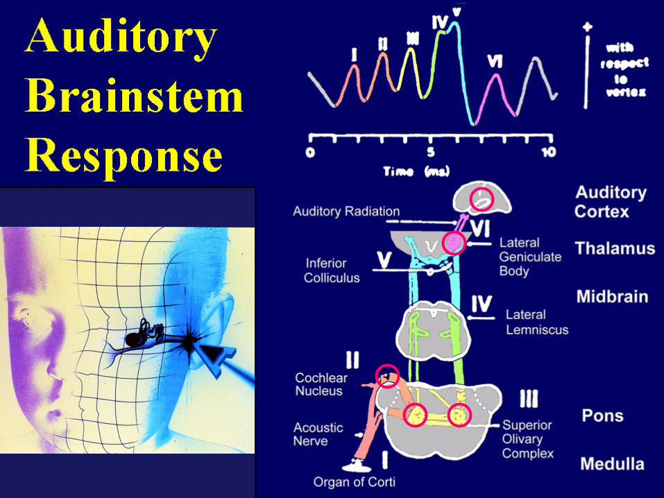
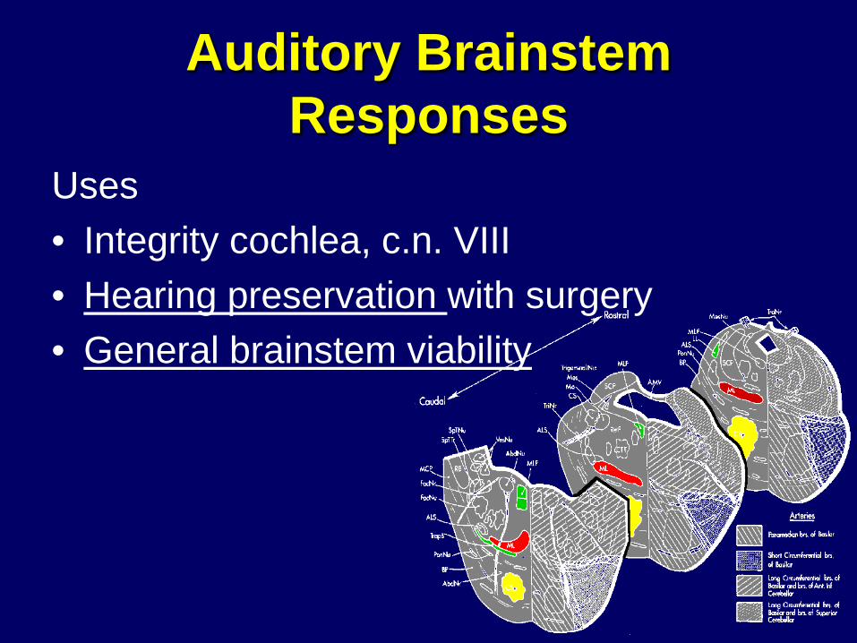
Auditory Brainstem Responses
Uses • Integrity cochlea, c.n. VIII • Hearing preservation with surgery • General brainstem viability
Age and ABR
• Useful to assess hearing in infants • Auditory nerve myelinated by birth – wave I
normal • Brainstem myelination proceeds caudal to
rostral (waves 1 to V) – progressive improvement later waves to adult values at age 3
Useful in infants and adult values by age 3
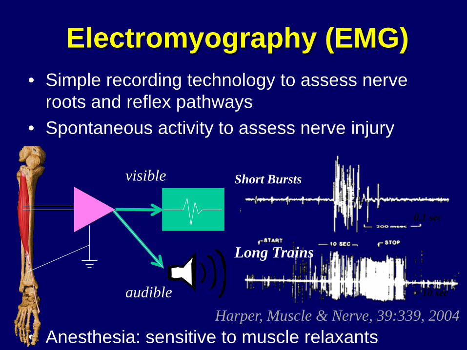
Electromyography (EMG) • Simple recording technology to assess nerve
roots and reflex pathways • Spontaneous activity to assess nerve injury
• Anesthesia: sensitive to muscle relaxants
Short Bursts Long Trains
0.1 sec
> 10 sec
Harper, Muscle & Nerve, 39:339, 2004
audible
visible
Which is true of EMG recording?:
A. EMG in the very young may resemble a myopathy
B. In infants and newborns neuromuscular transmission may suggest a transmission defect
C. Peripheral nerve myeliantion proceeds until age 3-5 with near adult conduction velocity by age 3
D. All of the above
*
Which is true of EMG recording?:
A. EMG in the very young may resemble a myopathy
B. In infants and newborns neuromuscular transmission may suggest a transmission defect
C. Peripheral nerve myelination proceeds until age 3-5 with near adult conduction velocity by age 3
D. All of the above *
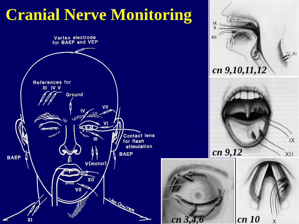
Cranial Nerve Monitoring
cn 3,4,6
cn 9,10,11,12
cn 10
cn 9,12
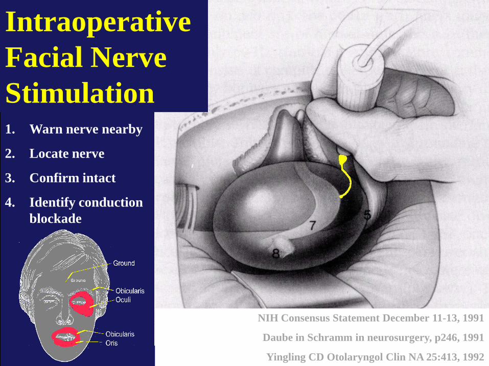
Intraoperative Facial Nerve Stimulation
NIH Consensus Statement December 11-13, 1991
Daube in Schramm in neurosurgery, p246, 1991
Yingling CD Otolaryngol Clin NA 25:413, 1992
1. Warn nerve nearby
2. Locate nerve
3. Confirm intact
4. Identify conduction blockade
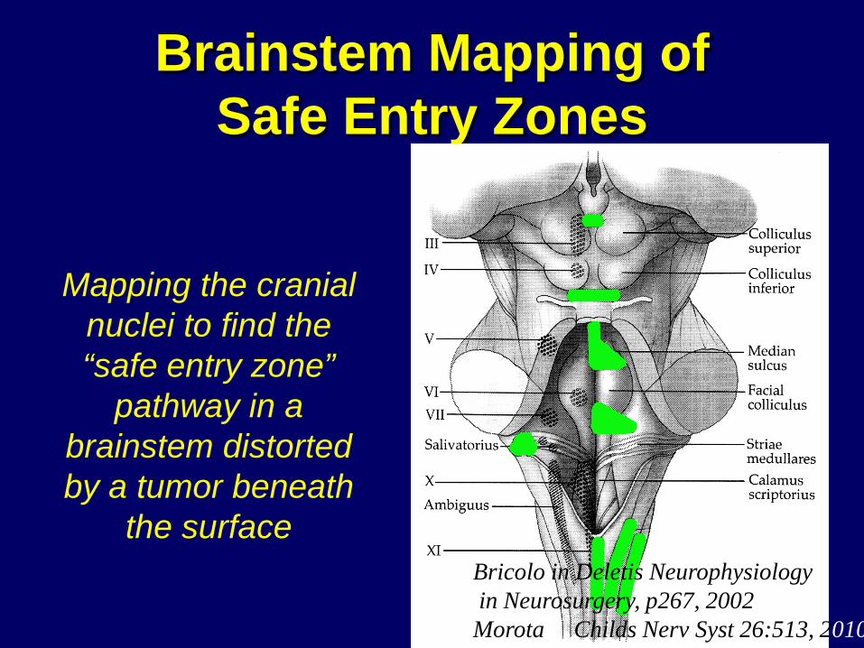
Brainstem Mapping of Safe Entry Zones
Bricolo in Deletis Neurophysiology in Neurosurgery, p267, 2002 Morota Childs Nerv Syst 26:513, 2010
Mapping the cranial nuclei to find the “safe entry zone”
pathway in a brainstem distorted by a tumor beneath
the surface
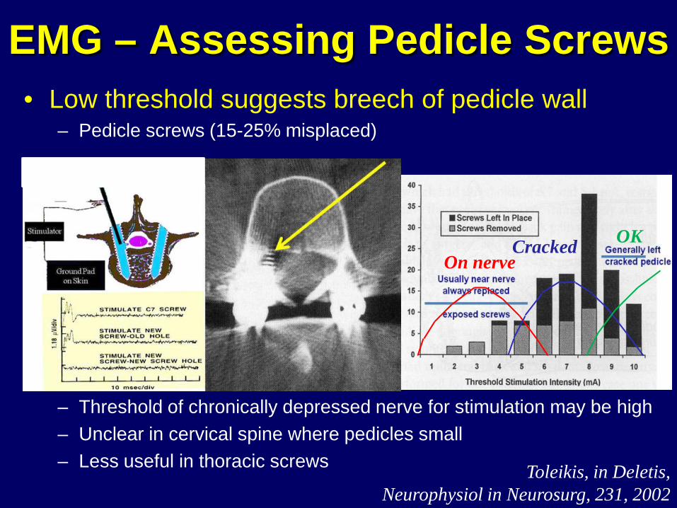
EMG – Assessing Pedicle Screws • Low threshold suggests breech of pedicle wall
– Pedicle screws (15-25% misplaced)
– Threshold of chronically depressed nerve for stimulation may be high – Unclear in cervical spine where pedicles small – Less useful in thoracic screws
On nerve Cracked OK
Toleikis, in Deletis, Neurophysiol in Neurosurg, 231, 2002
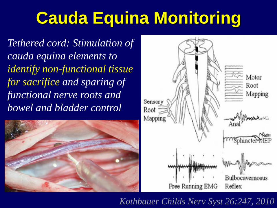
Cauda Equina Monitoring
Kothbauer Childs Nerv Syst 26:247, 2010
Tethered cord: Stimulation of cauda equina elements to identify non-functional tissue for sacrifice and sparing of functional nerve roots and bowel and bladder control
In general:
A. Maturation of the nervous system evolves after birth until adolescence with the majority of changes under age 3
B. SSEP recordings are challenging under age 3 and superb in adolescence
C. MEP stimulation may be risky under 18 months due to open fontanelle
D. Facilitation methods may be needed for MEP under age 3
E. ABR is not recordable until age 5 years
*
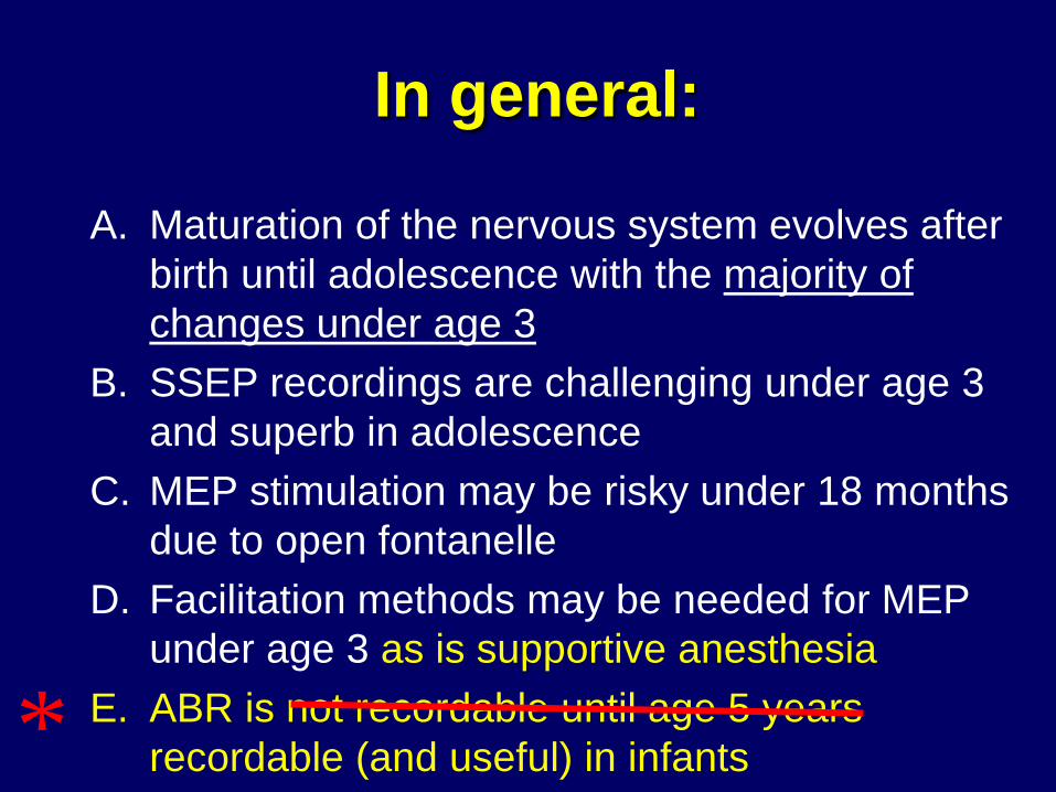
In general:
A. Maturation of the nervous system evolves after birth until adolescence with the majority of changes under age 3
B. SSEP recordings are challenging under age 3 and superb in adolescence
C. MEP stimulation may be risky under 18 months due to open fontanelle
D. Facilitation methods may be needed for MEP under age 3 as is supportive anesthesia
E. ABR is not recordable until age 5 years recordable (and useful) in infants
*
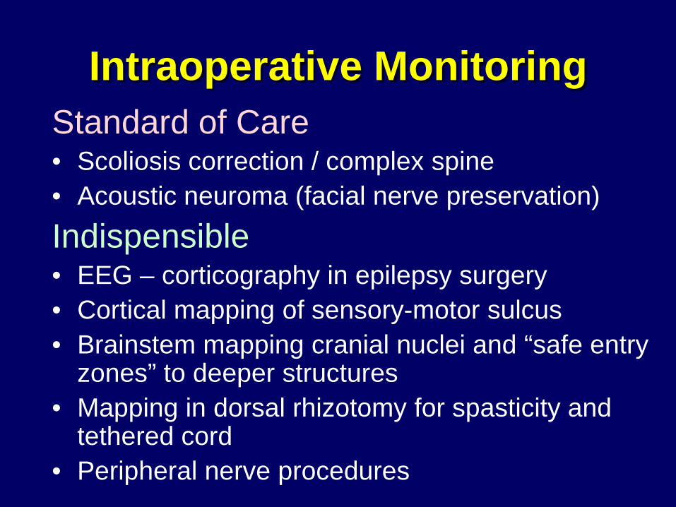
Intraoperative Monitoring Standard of Care • Scoliosis correction / complex spine • Acoustic neuroma (facial nerve preservation) Indispensible • EEG – corticography in epilepsy surgery • Cortical mapping of sensory-motor sulcus • Brainstem mapping cranial nuclei and “safe entry
zones” to deeper structures • Mapping in dorsal rhizotomy for spasticity and
tethered cord • Peripheral nerve procedures
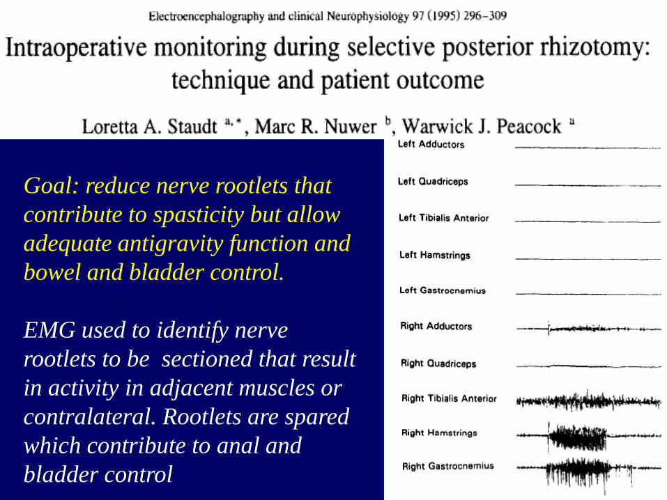
Goal: reduce nerve rootlets that contribute to spasticity but allow adequate antigravity function and bowel and bladder control. EMG used to identify nerve rootlets to be sectioned that result in activity in adjacent muscles or contralateral. Rootlets are spared which contribute to anal and bladder control
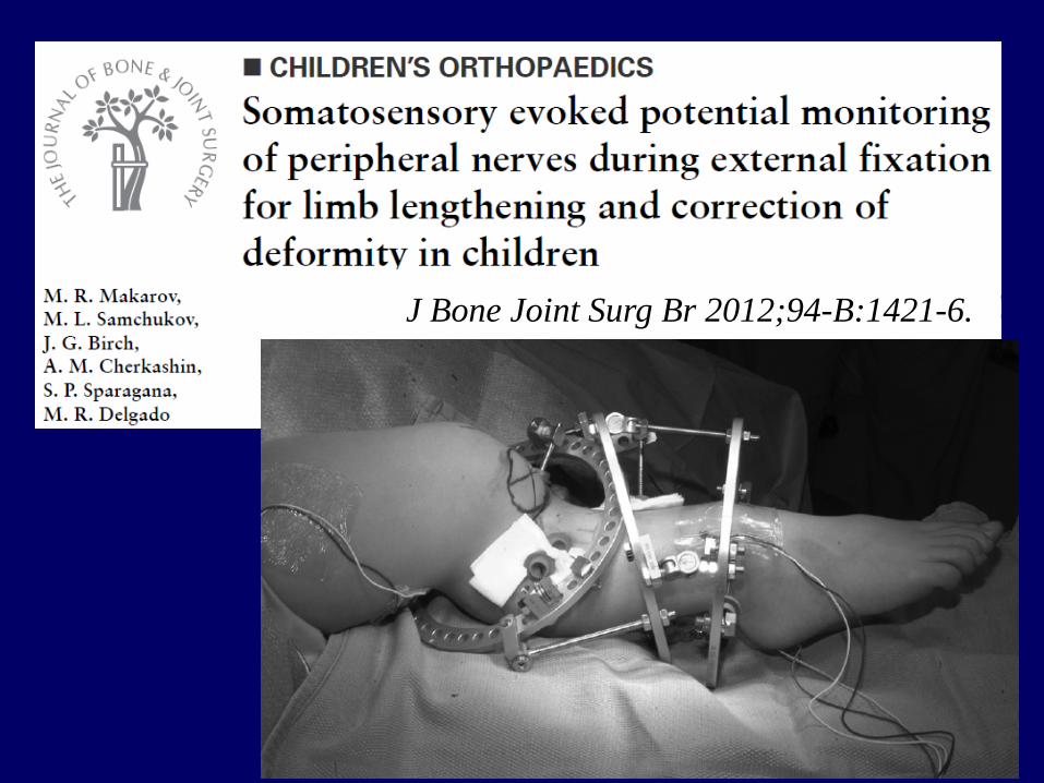
J Bone Joint Surg Br 2012;94-B:1421-6.
![Practical deep neural nets for detecting marine mammals ...danielnouri.org/docs/dclde2013-neural-nets.pdfConvolutional Neural Networks [Krizhevsky 2012] Improving neural networks by](https://img.pdfslide.net/doc/110x75/5fef2f54b164744e7046f536/practical-deep-neural-nets-for-detecting-marine-mammals-convolutional-neural.jpg)
![Chapter 2 Introduction to Neural networktomczak/PDF/[Grbic]Neural...Chapter 2 Introduction to Neural network 2.1 Introduction to Artiflcial Neural Net-work Artiflcial Neural Networks](https://img.pdfslide.net/doc/110x75/5f22a87bbf292e3b5d18b33c/chapter-2-introduction-to-neural-network-tomczakpdfgrbicneural-chapter-2.jpg)