Embed Size (px)
Citation preview
Intraoperative use of IndoCyaninE Green
fluorescence imaging to prevent
anastomotic leakage in colorectal surgery:
Systematic Review, Meta-Analysis
and Study Protocol for the ICEberG Trial
Author: Ruth Blanco Colino
Tutor: Eloy Espín Basany
Bachelor’s degree in Medicine Final Project – Class 2011-2017
Universitat Autònoma de Barcelona
Hospital Universitari Vall d’Hebron
R. Blanco Colino Intraoperative use of ICG fluorescence imaging to prevent AL in colorectal surgery
1
CONTENTS
1. ABSTRACT ........................................................................................................................... 2
2. BACKGROUND .................................................................................................................... 4
3. HYPOTHESIS AND OBJECTIVES ...................................................................................... 7
4. SYSTEMATIC REVIEW AND META-ANALYSIS ............................................................ 8
4.1. Material and methods ....................................................................................................... 8
4.2. Results ............................................................................................................................ 10
4.3. Discussion ...................................................................................................................... 13
5. STUDY PROTOCOL FOR THE ICEBERG TRIAL ........................................................... 16
5.1. Rationale for a RCT on ICG use in colorectal surgery .................................................. 16
5.2. Study design ................................................................................................................... 16
5.3. Outcome measures ......................................................................................................... 16
5.4. Eligibility criteria ........................................................................................................... 18
5.5. Site selection .................................................................................................................. 18
5.6. Randomisation ............................................................................................................... 18
5.7. Blinding.......................................................................................................................... 19
5.8. Interventions: experimental and control arm ................................................................. 19
5.9. Recruitment and trial timeline ....................................................................................... 20
5.10. Data management......................................................................................................... 21
5.11. Statistical methods ....................................................................................................... 21
5.12. Ethical considerations and trial registration ................................................................. 22
6. CONCLUSIONS................................................................................................................... 22
7. REFERENCES ..................................................................................................................... 23
R. Blanco Colino Intraoperative use of ICG fluorescence imaging to prevent AL in colorectal surgery
2
1. ABSTRACT
Intraoperative use of IndoCyaninE Green fluorescence imaging to prevent anastomotic
leakage in colorectal surgery: systematic review, meta-analysis and study protocol for
the ICEberG trial
Aim: ICG fluorescence imaging has been increasingly considered as a potential tool to assess
anastomosis perfusion. This study aims to validate its efficacy in reducing anastomotic
leakage (AL) rate in colorectal surgery.
Method: A systematic review and a meta-analysis of studies comparing fluorescence imaging
with standard care were conducted. Furthermore, a prospective randomised controlled trial
(RCT) was proposed.
Results: 1302 patients from 5 studies were included. Fluorescence imaging significantly
reduced AL risk in cancer surgery (OR:0.34; CI:0.16-0.74; P=0.006). Low AL rates were
shown in rectal surgery (ICG:1.1% vs non-ICG:6.1%; P=0.02). There was no significant AL
rate decrease when procedures for benign and malignant indication were combined. To date,
there are no published RCTs, though 3 ongoing trials were identified.
Conclusions: Fluorescence imaging seems to reduce AL rate following colorectal surgery for
cancer. However, large well-design RCTs are needed to provide evidence for its routine use.
The proposed ICEberG trial would address this need.

R. Blanco Colino Intraoperative use of ICG fluorescence imaging to prevent AL in colorectal surgery
3
Ús intraoperatori d’imatges per fluorescència amb verd d’indocianina per la prevenció
de la fuita anastomòtica en cirurgia colorectal: revisió sistemàtica, meta-anàlisi i
protocol per l’assaig clínic ICEberG
Objectiu: L’ús d’imatges per fluorescència amb verd d’indocianina s’està considerant cada
cop més com una potencial eina per l’avaluació de la perfusió de l’anastomosi. L’objectiu
d’aquest estudi és validar la seva eficàcia en la reducció de la taxa de fuita anastomòtica en la
cirurgia colorectal.
Mètodes: S’ha realitzat una revisió sistemàtica i un meta-anàlisi dels estudis que comparaven
l’ús d’imatges per fluorescència amb la pràctica habitual. A més, s’ha proposat un assaig
controlat aleatori (ACA) prospectiu.
Resultats: 1302 pacients de 5 estudis van ser inclosos a l’anàlisi. L’ús d’imatges per
fluorescència va reduir significativament el risc de fuita en cirurgia per càncer (OR:0.34;
CI:0.16-0.74; P=0.006). Es va veure una baixa taxa de fuita anastomòtica en cirurgia de recte
(ICG:1.1% vs no-ICG:6.1%; P=0.02). No es va trobar un descens significatiu en la taxa de
fuita anastomòtica quan es van combinar procediments amb indicació benigna i maligna.
Actualment no existeixen ACA publicats, tot i que s’han identificat 3 assaigs en curs.
Conclusions: L’ús d’imatges per fluorescència sembla reduir la taxa de fuita anastomòtica
després de cirurgia colorectal per càncer. No obstant, es necessiten grans ACA que
proporcionin evidència en l’ús d’aquesta tècnica a la pràctica habitual. El proposat ACA
ICEberG abordaria aquesta necessitat.
R. Blanco Colino Intraoperative use of ICG fluorescence imaging to prevent AL in colorectal surgery
4
2. BACKGROUND
Anastomotic leakage (AL) represents one of the most feared complications following
colorectal surgery; it has been associated with increased postoperative morbidity and
mortality rates. (1,2) Due to the lack of a standardized definition for AL, there is still
variability in studies reporting this condition. (3) In 2010, an attempt to define AL in anterior
rectal resections was made by the International Study Group of Rectal Cancer. The definition
proposed for AL was “a defect of the intestinal wall at the anastomotic site leading to a
communication between the intraluminal and extraluminal compartments” and they
recommended considering a pelvic abscess in the proximity of the anastomosis as AL. In
addition, different AL grades were established according to their clinical management: Grade
A when no change in patients’ management was required; Grade B if active therapeutic
intervention was needed without surgical intervention; Grade C when re-
laparotomy/laparoscopy was performed. (4) Kulu et al. (5) proved this AL definition and
severity grading to be a simple, easily applicable, and valid classification.
AL rate in colorectal surgery vary from 1% to 19% depending on the anatomic location of the
anastomosis: ileocolic (1% to 8%); colocolic (2% to 3%); ileorectal (3% to 7%); colorectal or
coloanal (5% to 19%). (3,6,7) In the Rectal Cancer Project of the Spanish Society of
Surgeons, the rate of AL for rectal cancer surgery was 10%. (8) The reduction of AL rates by
improving its prevention, diagnosis and management, continues being a challenge nowadays.
Finding new techniques to reduce AL has been highlighted as a research priority by the
Association of Coloproctology of Great Britain and Ireland (ACPGBI). (9)
Multiple conditions have been associated with a greater risk of AL: male sex, age,
comorbidities, high American Society of Anaesthesiologists (ASA) score, malnutrition,
obesity, smoking, immunosuppression, alcohol abuse, preoperative chemotherapy and
radiotherapy, advance tumour stage, diverticulitis, low anastomoses, prolonged operative
R. Blanco Colino Intraoperative use of ICG fluorescence imaging to prevent AL in colorectal surgery
5
time, inadequate anastomotic blood supply, blood loss or perioperative blood transfusion and
intraoperative septic conditions appearance. (3,10–12) Adequate perfusion of the anastomosis
is essential for an optimal healing and AL prevention. (13–15) Consequently, bowel ischemia
detected intraoperatively may reduce the risk of AL.
Different intraoperative techniques have been proposed to assess anastomotic integrity and
bowel viability in colorectal surgery. Traditionally, usual anastomotic assessment includes
direct visualisation of the anastomosis, integrity of doughnuts assessment and the air leak test.
Subjective signs indicating optimal anastomosis perfusion are evaluated including serosal -
mucosal colour and/or bleeding at the cut edge of the bowel and/or palpable pulsations of
mesenteric arteries. (12,16) However, surgeons’ predictive accuracy of AL risk has been
shown to be low and underestimated in the study of Karliczek et al. (17) They suggest the
need of a reliable predictive test that could be used intraoperatively. Although there is no
consensus on the use of air leak test in colorectal anastomosis, it is a widespread and well-
stablished technique. It consists in filling the pelvis with warm saline solution covering the
anastomosis, then air is insufflated on the rectum to identify any possible leak. (3) This is a
mechanical test, without any insight in the physiology of AL.
Other experimental techniques assessing blood supply and/or integrity of anastomosis have
been described. Some of them are intraoperative endoscopy, pulse oximetry, Doppler
ultrasound and Doppler flowmetry, intramucosal pH measurement, visible light oxygen
spectroscopy and near infrared oxygen spectroscopy. None of the mentioned techniques are
routinely used, mainly because of its complexity and its high variability of the measurements.
(16,18,19)
Fluorescence imaging with indocyanine green (ICG) has been increasingly considered as a
potential intraoperative tool that could be used in routine practice to ensure adequate
perfusion at the time of anastomosis formation. It allows surgeons to visualize bowel
R. Blanco Colino Intraoperative use of ICG fluorescence imaging to prevent AL in colorectal surgery
6
microperfusion at a real-time being a fast technique which is easy to perform. Recent
literature shows the potential benefit of fluorescence imaging with ICG in lowering AL rates
by changing the surgical plan. (20–26) Although it has been proven to be a safe and feasible
tool in colorectal surgery, (27–29) further research is needed to validate its efficacy in
reducing AL rate. (1)
ICG is a sterile, water-soluble, trycarbocyanine compound dye that absorbs near-infrared light
in the region of 800-810 nm and emits it at 830 nm. ICG is administered by intravenous
injection (1.25 – 3.75 mg, depending on the patient weight) and it is rapidly bounded to
albumin with minimal leakage into the interstitium. ICG is removed from the blood by the
liver with a half-life of 3-5 minutes and then it is excreted via the bile within 10-15 minutes
with no known metabolites. (30,31) Since 1956, when ICG was approved for clinical and
research use in humans by the Food and Drug Administration (FDA), it has been used in
different areas including plastic surgery, neurosurgery, ophthalmology, hepatobiliary and
transplant surgery. The ICG applications with greater interest in colorectal surgery are
anastomosis perfusion assessment and lymphatic road-mapping. Fluorescence imaging for
bowel perfusion assessment can be used immediately before and/or after anastomosis
formation once tissues are in their anatomic positions. (32) ICG clinical use has been proven
to be very safety and rare cases of anaphylaxis have been described. (33) However, it should
be used with caution in patients with iodine allergy (allergic reaction 1/300000). Moreover,
manufacturers recommend a maximum daily dose of less than 2 mg/kg. (30,32,34)
There are different platforms available to perform fluorescence imaging with ICG. SPY
EliteTM system from Novadaq® was designed to be used in open cases and it provides a
numeric assessment of perfusion. In laparoscopic surgery, the PINPOINTTM from Novadaq®
(Canada), IC-View® from Pulsion Medical Systems (Germany) and the D-Light from Storz®
(Germany) can be used. For robotic surgery, FIREFLYTM system integrated in surgical
R. Blanco Colino Intraoperative use of ICG fluorescence imaging to prevent AL in colorectal surgery
7
robotic platform da Vinci SITM (USA) is available. In white light mode, these systems provide
a standard laparoscopic view and it can be readily switched to near-infrared (NIR) mode, in
which ICG fluorescence is visualised. Also, PINPOINTTM has a dual display mode allowing
to superimpose green ICG fluorescence with white-light image. (30,35) These commercial
available systems lack to provide a quantification of tissue perfusion. (1)
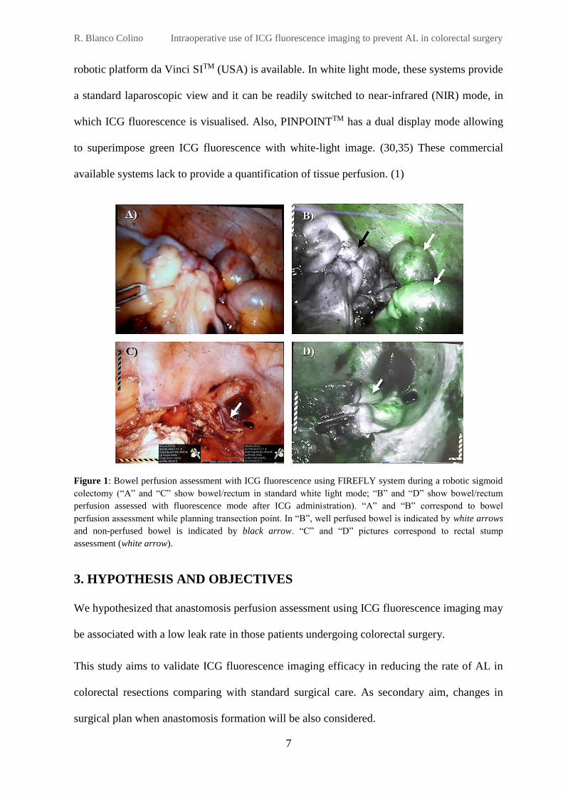
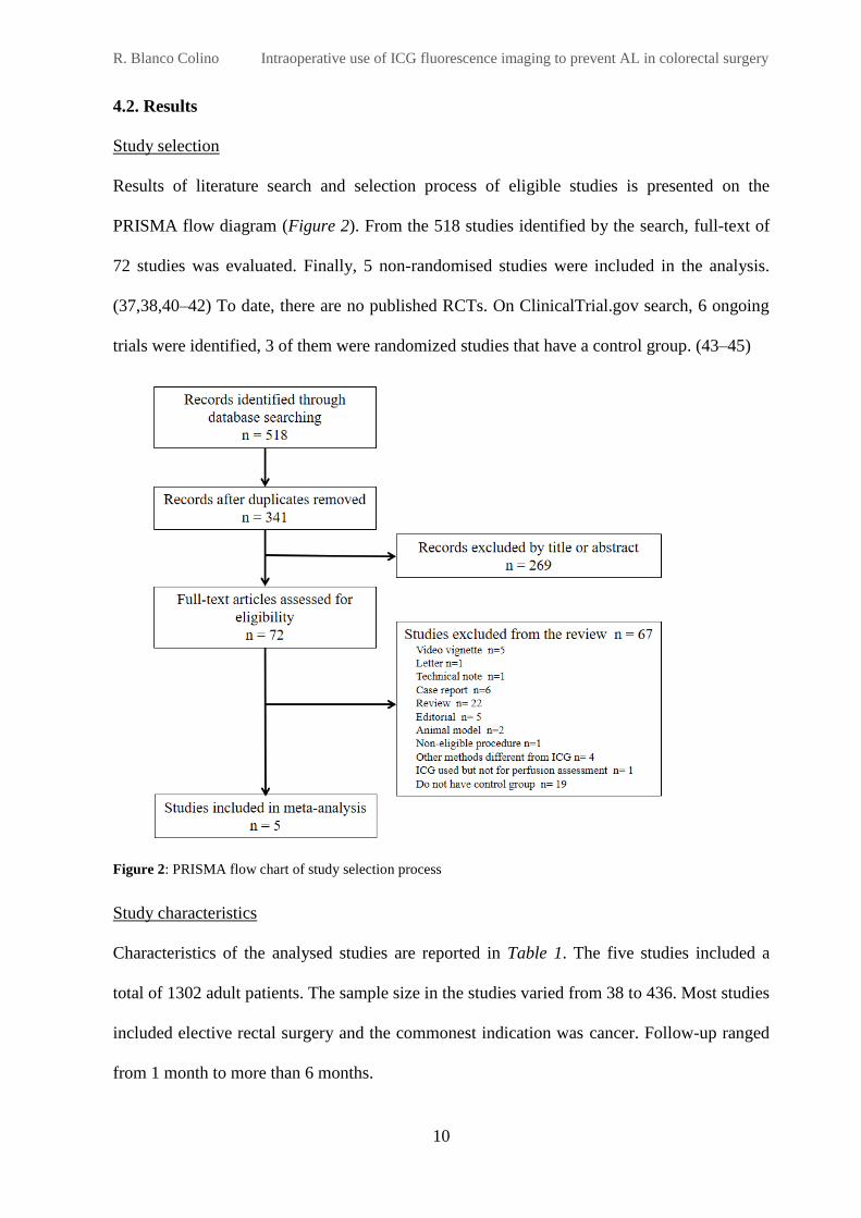
Figure 1: Bowel perfusion assessment with ICG fluorescence using FIREFLY system during a robotic sigmoid
colectomy (“A” and “C” show bowel/rectum in standard white light mode; “B” and “D” show bowel/rectum
perfusion assessed with fluorescence mode after ICG administration). “A” and “B” correspond to bowel
perfusion assessment while planning transection point. In “B”, well perfused bowel is indicated by white arrows
and non-perfused bowel is indicated by black arrow. “C” and “D” pictures correspond to rectal stump
assessment (white arrow).
3. HYPOTHESIS AND OBJECTIVES
We hypothesized that anastomosis perfusion assessment using ICG fluorescence imaging may
be associated with a low leak rate in those patients undergoing colorectal surgery.
This study aims to validate ICG fluorescence imaging efficacy in reducing the rate of AL in
colorectal resections comparing with standard surgical care. As secondary aim, changes in
surgical plan when anastomosis formation will be also considered.
R. Blanco Colino Intraoperative use of ICG fluorescence imaging to prevent AL in colorectal surgery
8
4. SYSTEMATIC REVIEW AND META-ANALYSIS
4.1. Material and methods
A systematic review was conducted in accordance with the Preferred Reporting Items for
Systematic Reviews and Meta-Analyses (PRISMA) statement. (36)
Eligibility criteria
Studies that compared intraoperative use of ICG fluorescence imaging with standard care for
the assessment of anastomosis perfusion or viability, were eligible for inclusion. Patients of
any age undergoing colon or rectal resection with anastomosis were included, regardless of
operative approach, urgency of surgery and surgical indication. The primary outcome
measure was the AL rate with at least 30 days follow-up. Randomized Controlled Trials
(RCTs), cohort studies, case-control studies and quasi-randomised studies were searched.
Case reports were excluded. Studies using ICG fluorescence for other purposes different from
perfusion assessment were excluded, as well as those studies based on animal models.
Search strategy
An electronic search was carried out using PubMed, Scopus, Web of Science, Google Scholar
databases and the Cochrane Library. The reference list of identified systematic reviews and
review articles were hand-searched for additional references. Furthermore, the register
ClinicalTrials.gov was searched to identify ongoing trials. A combination of medical subject
heading (MeSH) terms and keywords were searched: “indocyanine green”, “ICG”, “coloring
agents”, “fluorescence”, “fluorescein angiography”, “fluorescent dyes”, “anastomotic leak”,
“anastomotic leakage”, “anastomotic perfusion”, “anastomosis, surgical”, “bowel perfusion”,
“blood supply”, “perfusion assessment”, “colorectal surgery”, “colon surgery”, “rectal
surgery” “colorectal resection”, “bowel resection” using the Boolean operator “OR” for each
concept. Each concept was combined with “AND”. The complete search strategy is shown in

R. Blanco Colino Intraoperative use of ICG fluorescence imaging to prevent AL in colorectal surgery
9
the Appendix. No search limits were applied and all languages were included. The latest date
of this search was 24th January 2017.
Study selection and data extraction
Studies were screened by title and abstract; then full-text was obtained for those studies
identified as potentially eligible.
From each study, data were extracted on: study characteristics and year of publication, patient
inclusion period, sample size, surgical indication, surgical management (operative approach,
procedure and whether a change in surgical plan was made), fluorescence imaging system
used and anastomotic leakage rate. Authors were contacted to provide additional information
that was not available in the original studies; two authors could not be contacted or were not
able to provide the requested data. (37,38)
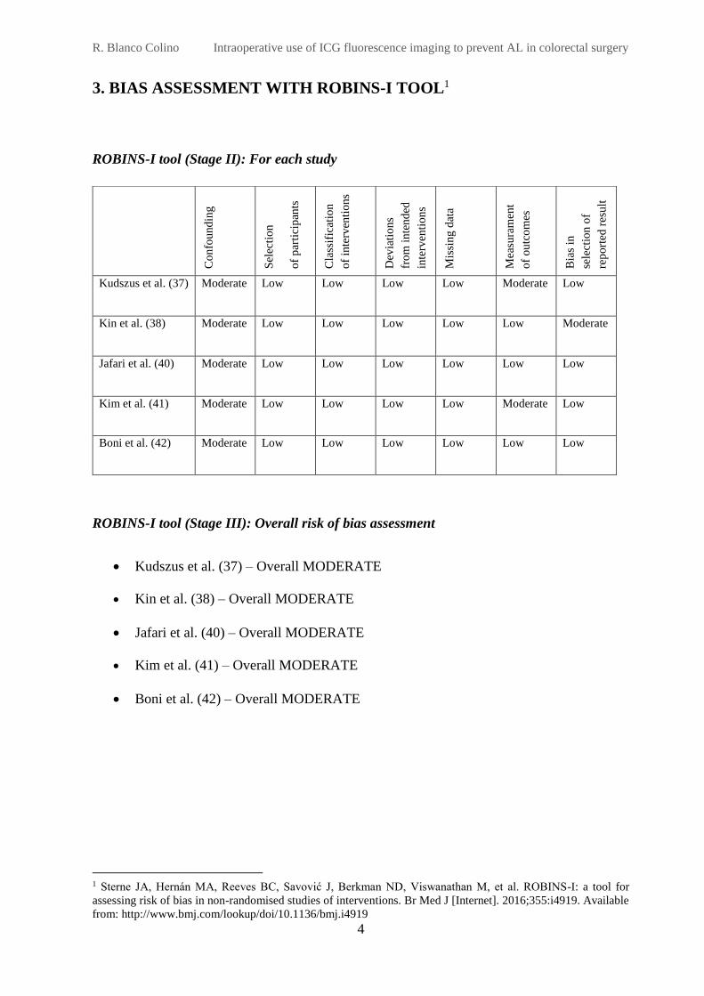
Risk of bias assessment
The quality of the included studies was evaluated using the ROBINS-I risk of bias assessment
tool for non-randomized studies of interventions. (39) Seven domains were covered including
confounding and selection of participants into the study, classification of interventions,
deviations from intended interventions, missing data, measurement of outcomes, and selection
of the reported result.
Statistical analysis
Analyses were performed using Review Manager (RevMan) Version 5.3 (Copenhagen: The
Nordic Cochrane Centre, The Cochrane Collaboration, 2014). The odds ratios (OR) were
calculated from the original data and were assessed as the summary statistic. Values were
reported with 95% confidence intervals (CI). As there was a substantial level of heterogeneity
expected across the included studies, Mantel-Haenszel (M-H) method and random-effects
models were employed for quantitative statistical analysis of dichotomous variables.
Statistical heterogeneity was assessed using I2 test and by visual inspection of forest plots.
R. Blanco Colino Intraoperative use of ICG fluorescence imaging to prevent AL in colorectal surgery
10
4.2. Results
Study selection
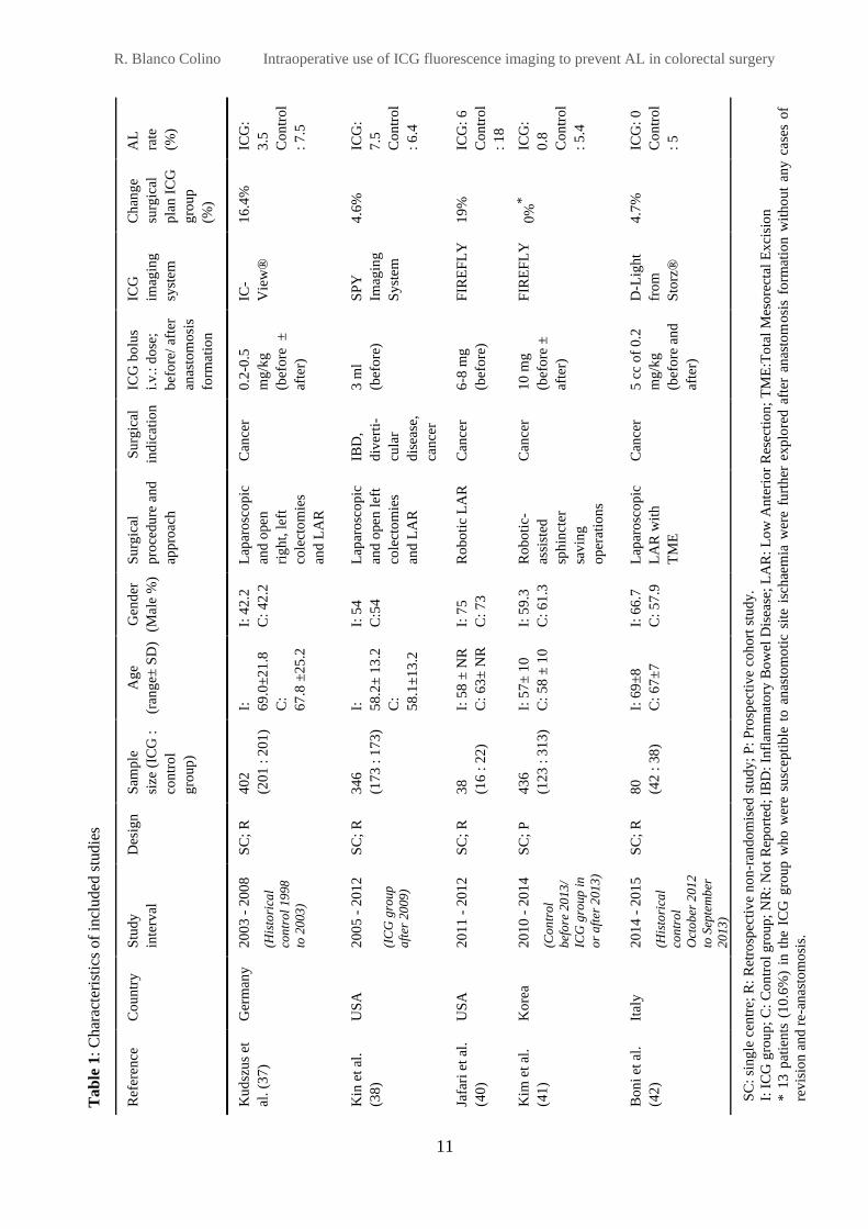
Results of literature search and selection process of eligible studies is presented on the
PRISMA flow diagram (Figure 2). From the 518 studies identified by the search, full-text of
72 studies was evaluated. Finally, 5 non-randomised studies were included in the analysis.
(37,38,40–42) To date, there are no published RCTs. On ClinicalTrial.gov search, 6 ongoing
trials were identified, 3 of them were randomized studies that have a control group. (43–45)
Figure 2: PRISMA flow chart of study selection process
Study characteristics
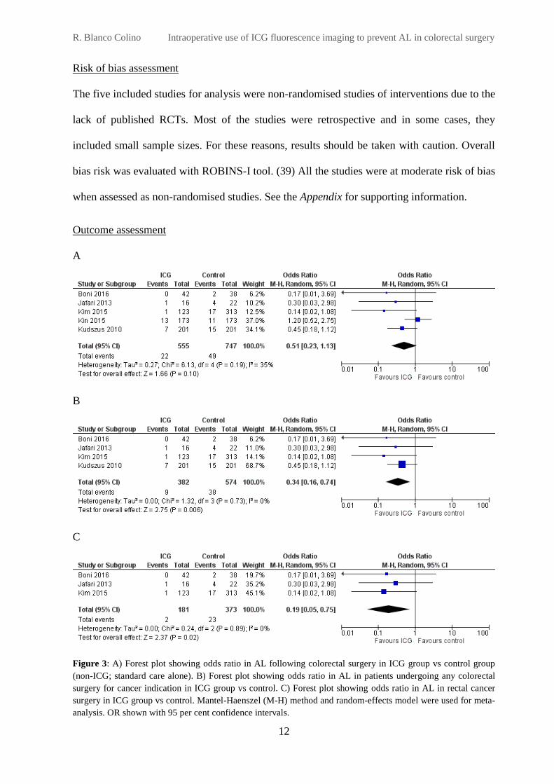
Characteristics of the analysed studies are reported in Table 1. The five studies included a
total of 1302 adult patients. The sample size in the studies varied from 38 to 436. Most studies
included elective rectal surgery and the commonest indication was cancer. Follow-up ranged
from 1 month to more than 6 months.
R. Blanco Colino Intraoperative use of ICG fluorescence imaging to prevent AL in colorectal surgery
11
AL
rate
(%)
ICG
:
3.5
Co
ntr
ol
: 7
.5
ICG
:
7.5
Co
ntr
ol
: 6
.4
ICG
: 6
Co
ntr
ol
: 1
8
ICG
:
0.8
Co
ntr
ol
: 5
.4
ICG
: 0
Co
ntr
ol
: 5
Ch
ang
e
surg
ical
pla
n I
CG
gro
up
(%)
16
.4%
4.6
%
19
%
0%
*
4.7
%
ICG
imag
ing
syst
em
IC-
Vie
w®
SP
Y
Imag
ing
Sy
stem
FIR
EF
LY
FIR
EF
LY
D-L
igh
t
fro
m
Sto
rz®
ICG
bo
lus
i.v
.: d
ose
;
bef
ore
/ af
ter
anas
tom
osi
s
form
atio
n
0.2
-0.5
mg
/kg
(bef
ore
±
afte
r)
3 m
l
(bef
ore
)
6-8
mg
(bef
ore
)
10
mg
(bef
ore
±
afte
r)
5 c
c o
f 0
.2
mg
/kg
(bef
ore
an
d
afte
r)
Su
rgic
al
ind
icat
ion
Can
cer
IBD
,
div
erti
-
cula
r
dis
ease
,
can
cer
Can
cer
Can
cer
Can
cer
Su
rgic
al
pro
cedu
re a
nd
app
roac
h
Lap
aro
sco
pic
and
open
rig
ht,
lef
t
cole
cto
mie
s
and
LA
R
Lap
aro
sco
pic
and
open
lef
t
cole
cto
mie
s
and
LA
R
Ro
bo
tic
LA
R
Ro
bo
tic-
assi
sted
sph
inct
er
sav
ing
op
erat
ion
s
Lap
aro
sco
pic
LA
R w
ith
TM
E
Gen
der
(Mal
e %
)
I: 4
2.2
C:
42
.2
I: 5
4
C:5
4
I: 7
5
C:
73
I: 5
9.3
C:
61
.3
I: 6
6.7
C:
57
.9
Ag
e
(ran
ge±
SD
)
I:
69
.0±
21
.8
C:
67
.8 ±
25
.2
I:
58
.2±
13
.2
C:
58
.1±
13
.2
I: 5
8 ±
NR
C:
63
± N
R
I: 5
7±
10
C:
58
± 1
0
I: 6
9±
8
C:
67
±7
Sam
ple
size
(IC
G :
con
tro
l
gro
up
)
40
2
(20
1 :
20
1)
34
6
(17
3 :
17
3)
38
(16
: 2
2)
43
6
(12
3 :
31
3)
80
(42
: 3
8)
Des
ign
SC
; R
SC
; R
SC
; R
SC
; P
SC
; R
Stu
dy
inte
rval
20
03
- 2
00
8
(His
tori
cal
con
tro
l 19
98
to 2
003
)
20
05
- 2
01
2
(IC
G g
roup
aft
er 2
009
)
20
11
- 2
01
2
20
10
- 2
01
4
(Co
ntr
ol
bef
ore
20
13
/
ICG
gro
up
in
or
aft
er 2
01
3)
20
14
- 2
01
5
(His
tori
cal
con
tro
l
Oct
ob
er 2
01
2
to S
epte
mb
er
20
13
)
Co
un
try
Ger
man
y
US
A
US
A
Ko
rea
Ital
y
Ref
eren
ce
Ku
dsz
us
et
al.
(37
)
Kin
et
al.
(38
)
Jafa
ri e
t al
.
(40
)
Kim
et
al.
(41
)
Bo
ni
et a
l.
(42
)
Tab
le 1
: C
har
acte
rist
ics
of
incl
ud
ed s
tudie
s
SC
: si
ng
le c
entr
e; R
: R
etro
spec
tiv
e n
on
-ran
do
mis
ed s
tud
y;
P:
Pro
spec
tiv
e co
hort
stu
dy
.
I: I
CG
gro
up
; C
: C
on
tro
l g
roup
; N
R:
No
t R
epo
rted
; IB
D:
Infl
amm
ato
ry B
ow
el D
isea
se;
LA
R:
Lo
w A
nte
rio
r R
esec
tio
n;
TM
E:T
ota
l M
eso
rect
al E
xci
sio
n
* 1
3 p
atie
nts
(1
0.6
%)
in t
he
ICG
gro
up
who
wer
e su
scep
tib
le t
o a
nas
tom
oti
c si
te i
sch
aem
ia w
ere
furt
her
exp
lore
d a
fter
an
asto
mo
sis
form
atio
n w
ith
ou
t an
y c
ase
s of
rev
isio
n a
nd r
e-a
nas
tom
osi
s.
R. Blanco Colino Intraoperative use of ICG fluorescence imaging to prevent AL in colorectal surgery
12
Risk of bias assessment
The five included studies for analysis were non-randomised studies of interventions due to the
lack of published RCTs. Most of the studies were retrospective and in some cases, they
included small sample sizes. For these reasons, results should be taken with caution. Overall
bias risk was evaluated with ROBINS-I tool. (39) All the studies were at moderate risk of bias
when assessed as non-randomised studies. See the Appendix for supporting information.
Outcome assessment
A
B
C
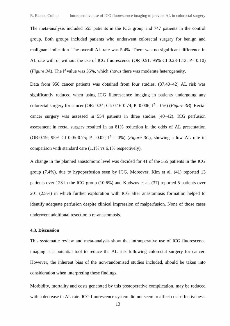
Figure 3: A) Forest plot showing odds ratio in AL following colorectal surgery in ICG group vs control group
(non-ICG; standard care alone). B) Forest plot showing odds ratio in AL in patients undergoing any colorectal
surgery for cancer indication in ICG group vs control. C) Forest plot showing odds ratio in AL in rectal cancer
surgery in ICG group vs control. Mantel-Haenszel (M-H) method and random-effects model were used for meta-
analysis. OR shown with 95 per cent confidence intervals.
R. Blanco Colino Intraoperative use of ICG fluorescence imaging to prevent AL in colorectal surgery
13
The meta-analysis included 555 patients in the ICG group and 747 patients in the control
group. Both groups included patients who underwent colorectal surgery for benign and
malignant indication. The overall AL rate was 5.4%. There was no significant difference in
AL rate with or without the use of ICG fluorescence (OR 0.51; 95% CI 0.23-1.13; P= 0.10)
(Figure 3A). The I2 value was 35%, which shows there was moderate heterogeneity.
Data from 956 cancer patients was obtained from four studies. (37,40–42) AL risk was
significantly reduced when using ICG fluorescence imaging in patients undergoing any
colorectal surgery for cancer (OR: 0.34; CI: 0.16-0.74; P=0.006; I2 = 0%) (Figure 3B). Rectal
cancer surgery was assessed in 554 patients in three studies (40–42). ICG perfusion
assessment in rectal surgery resulted in an 81% reduction in the odds of AL presentation
(OR:0.19; 95% CI 0.05-0.75; P= 0.02; I2 = 0%) (Figure 3C), showing a low AL rate in
comparison with standard care (1.1% vs 6.1% respectively).
A change in the planned anastomotic level was decided for 41 of the 555 patients in the ICG
group (7.4%), due to hypoperfusion seen by ICG. Moreover, Kim et al. (41) reported 13
patients over 123 in the ICG group (10.6%) and Kudszus et al. (37) reported 5 patients over
201 (2.5%) in which further exploration with ICG after anastomosis formation helped to
identify adequate perfusion despite clinical impression of malperfusion. None of those cases
underwent additional resection o re-anastomosis.
4.3. Discussion
This systematic review and meta-analysis show that intraoperative use of ICG fluorescence
imaging is a potential tool to reduce the AL risk following colorectal surgery for cancer.
However, the inherent bias of the non-randomised studies included, should be taken into
consideration when interpreting these findings.
Morbidity, mortality and costs generated by this postoperative complication, may be reduced
with a decrease in AL rate. ICG fluorescence system did not seem to affect cost-effectiveness.
R. Blanco Colino Intraoperative use of ICG fluorescence imaging to prevent AL in colorectal surgery
14
The initial burden of a NIR unit are 70.000€ and then the cost for ICG dye is 13€ per patient.
(41) In contrast, AL represents 1.6 to 5 million euros of the annual direct healthcare costs in
the UK and over 22.000€ per patient in the USA. (3) AL also increases the mortality risk
(from 1.9% without AL to 15.9% with AL) and, the length of stay (from 7 days without AL to
23 days with AL). (11) In colorectal cancer surgery, AL has been associated with reduced
long-term cancer specific survival and a greater risk for systemic and local recurrence. (46,47)
However, this association remains unclear when referring to rectal surgery. (48)
Several studies have assessed the use of ICG fluorescence in colorectal surgery, but most of
them correspond to case series with small sample sizes. Fluorescence imaging has been
described in surgical procedures for benign and malignant indication and different operative
approaches (22–25,49) including robotic colorectal surgery, (20,26) transanal rectal surgery,
(27) and minimally invasive surgery. (50)
One of the limitations of this meta-analysis is the lack of randomisation in the studies
included. In addition, four studies were retrospective (37,38,40,42) and results from ICG
fluorescence group were compared with a control group from a different period of time. At
present, there is no RCT published. Three ongoing RCTs were found on ClinicalTrial.gov
register. AL rate is the primary outcome measure in the three studies, two of them with 30
days follow-up (43,44) and one of them with 2 months follow-up. (45) One of the RCTs
would include low anterior resections for rectal cancer, (45) other would evaluate ICG use
during rectal or left colectomies (benign and malignant disease) (44) and the last one includes
robotic colorectal surgery for cancer, IBD or diverticular disease indication. (43)
Results of this study must be taken with caution due to the variability on AL definition, as
well as differences in the length of follow-up, surgical technique and application of ICG on
the experimental group. In all the included studies, ICG fluorescence was used before
intestinal resection to plan transection level. In some cases, it was also used after the
R. Blanco Colino Intraoperative use of ICG fluorescence imaging to prevent AL in colorectal surgery
15
anastomosis formation. (41,42) Moreover, the quantitative definition of an adequate or
inadequate preanastomotic perfusion is not well defined, mainly because most of the actual
imaging systems lack to quantify tissue perfusion. However, some experimental studies
assessing fluorescence quantification in animal models have been published. (51)
Additionally, Sherwinter et al. (27) used a fluorescence score in their study based on the
sequence of fluorescence uptake and time of maximal excitation.
ICG fluorescence seems to help in identifying the need for a change in the surgical plan,
extending resection margins or requiring revision and re-anastomosis. A change in the
planned anastomotic level was decided in 7.4% (41 over 555 patients in the ICG group).
Usually, a change is decided if a bowel hypoperfusion is detected by fluorescence, even if it
had seemed well-perfused by visual examination. In contrast, ICG fluorescence can also help
confirm a competent perfusion in those cases with a clinical impression of malperfusion, and
therefore not to extent the resection margins further.
In the present meta-analysis, Kin et al. (38) was the only study that have reported a non-
reduction in AL rate when using intraoperative fluorescence. However, this study presents
some limitations that could have influenced results. Only proximal bowel perfusion was
assessed, and therefore rectal stump perfusion was not confirmed. In contrast with the other
studies, which only included patients undergoing surgery for cancer indication, this study also
included patients with inflammatory bowel disease and diverticular disease.
Despite the limitation of the available studies, this systematic review and meta-analysis show
that ICG fluorescence imaging is a promising tool that could change usual practice. It may
reduce AL rate in patients undergoing colorectal resection for cancer indication. However, its
use for benign indications is uncertain. There is a need of larger, well-designed RCTs to
assess if AL rate can be reduced by incorporating ICG fluorescence imaging in routine
colorectal surgery for benign or malignant indication.
R. Blanco Colino Intraoperative use of ICG fluorescence imaging to prevent AL in colorectal surgery
16
5. STUDY PROTOCOL FOR THE ICEBERG TRIAL
5.1. Rationale for a RCT on ICG use in colorectal surgery
We propose a randomised controlled trial addressing the intraoperative use of ICG
fluorescence imaging to reduce AL rate in patients undergoing colorectal surgery.
The following issues justify the need of a RCT:
1. There is no RCT published comparing ICG fluorescence imaging with standard care.
2. There are 3 ongoing trials. PILLAR III, is a multicentre RCT that is including patients
undergoing low anterior resection for rectal o rectosigmoid neoplasm. (45) The other two
are single centre RCT; one is including patients undergoing robotic surgery (43) and the
other includes laparoscopic rectal resections and left colectomies, (44) both studies
include surgeries for benign and malignant indications. Therefore, there is no RCT
addressing ICG fluorescence use in routine practice for any condition (benign and
malignant) under laparoscopic or robotic approach, in total, right or left colectomies and
rectal resection procedures.
3. There is no current data assessing ICG dose, application, fluorescence diagnosis and
management if a change in surgical plan is decided.
5.2. Study design
This is a prospective, multicentre, randomised controlled trial comparing intraoperative use of
ICG fluorescence imaging with standard care alone in anastomosis formation.
5.3. Outcome measures
Primary outcome measure
AL rate within a 60 days follow-up. AL will be defined and graded as Rahbari et al. proposed
in their study. (4) Pelvic abscess in the proximity of the anastomosis will also be considered
as AL. Stricture of the anastomosis will not be considered as AL.
R. Blanco Colino Intraoperative use of ICG fluorescence imaging to prevent AL in colorectal surgery
17
C-reactive protein (CRP) as well as procalcitonin (PCT) have been suggested as useful
screening markers for early prediction of AL on postoperative days 3-5. (52) Their levels will
be used to determine if a CT scan for AL detection is necessary.
Secondary outcome measure
1. Number of cases in which a change in the surgical plan is decided. For example:
extension of the resection margins; colon/rectum preservation; protective ileostomy
(avoided or performed).
2. Major postoperative complications defined by the Clavien-Dindo classification (53) grade
III to V at 60 days of follow-up (see Appendix for more information).
3. Length of stay, including from day 0 (day of surgery) to day of discharge.
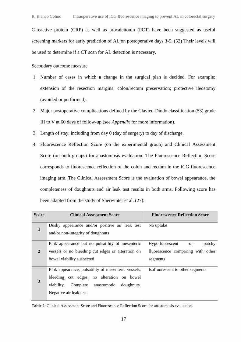
4. Fluorescence Reflection Score (on the experimental group) and Clinical Assessment
Score (on both groups) for anastomosis evaluation. The Fluorescence Reflection Score
corresponds to fluorescence reflection of the colon and rectum in the ICG fluorescence
imaging arm. The Clinical Assessment Score is the evaluation of bowel appearance, the
completeness of doughnuts and air leak test results in both arms. Following score has
been adapted from the study of Sherwinter et al. (27):
Score Clinical Assessment Score Fluorescence Reflection Score
1 Dusky appearance and/or positive air leak test
and/or non-integrity of doughnuts
No uptake
2
Pink appearance but no pulsatility of mesenteric
vessels or no bleeding cut edges or alteration on
bowel viability suspected
Hypofluorescent or patchy
fluorescence comparing with other
segments
3
Pink appearance, pulsatility of mesenteric vessels,
bleeding cut edges, no alteration on bowel
viability. Complete anastomotic doughnuts.
Negative air leak test.
Isofluorescent to other segments
Table 2: Clinical Assessment Score and Fluorescence Reflection Score for anastomosis evaluation.
R. Blanco Colino Intraoperative use of ICG fluorescence imaging to prevent AL in colorectal surgery
18
5.4. Eligibility criteria
Inclusion criteria
Adult patients (≥ 18 years) undergoing colorectal resection with a primary anastomosis
(including right or left colectomy, total colectomy, sigmoid and rectal resection) will be
included. Surgeries under laparoscopic or robotic approach for benign or malignant
indication will be eligible for inclusion.
Exclusion criteria
Patients with known allergy or history of adverse reaction to iodine or indocyanine green
Pregnant or breast-feeding patients
Patients undergoing emergency surgery
Patients undergoing colorectal resection without anastomosis
Patients undergoing palliative surgery for terminal illness
Not consenting patients
5.5. Site selection
This study is proposed by the general surgery department, colorectal surgery unit at Hospital
Universitari Vall d’Hebron. The ICEberG trial is a multicentre study and it will be performed
in European colorectal units. Moreover, it is opened to other international centres interested in
joining the trial. Sites will be eligible to participate based on fluorescence imaging system
availability at their units, case volumes and surgical experience.
5.6. Randomisation
After obtaining written consent from the patients, they will be randomised by a web-based
method on a 1:1 basis. Stratified randomisation will be performed by the planned surgical
procedure: total colectomy, right colectomy, left colectomy, sigmoid resection, and rectal
resection.
R. Blanco Colino Intraoperative use of ICG fluorescence imaging to prevent AL in colorectal surgery
19
5.7. Blinding
This trial is blinded to the patient. The trial design and the expected intervention on the
experimental group do not enable to blind the surgeon performing the procedure.
5.8. Interventions: experimental and control arm
Standard care for AL detection will be performed in both arms and it will include:
Visual examination of both segments (before and after anastomosis)
Integrity of doughnuts assessment if applicable
Air leak test (after anastomosis)
Intervention arm
The intervention group consists on using ICG fluorescence imaging intraoperatively.
Preoperative bowel preparation, as well as planned surgical procedure will be performed
according to surgeon’s standard practice until time of transection point. A bolus of 0.1 – 0.3
mg/kg of indocyanine green will be injected intravenously by the anaesthetist. If laparoscopic
approach, PINPOINTTM system will be used. If robotic approach, then FIREFLYTM will be
the system used.
Prior to fluorescence assessment, surgeon will visually examine anastomosis segments as it
would be done in standard care and will assign a score following the “Clinical Assessment
Score” presented on Table 2. Then, fluorescence of both segments of the anastomosis will be
assessed and a score will be assigned by the surgeon according to the “Fluorescence
Reflection Score” on Table 2 (1 no uptake to 3 maximal uptake). Once the anastomosis is
done, “Clinical Assessment Score” and “Fluorescence Reflection Score” will be used again to
assign a score after anastomosis performance. If a change on the surgical planned is decided,
this will be noted on case report form.
R. Blanco Colino Intraoperative use of ICG fluorescence imaging to prevent AL in colorectal surgery
20
Control arm – No intervention
In the control group, only standard care will be performed. Therefore, both segments will be
visually examined using the “Clinical Assessment Score” before and after anastomosis
formation. If a change on the surgical planned is decided, this will be noted on case report
form.
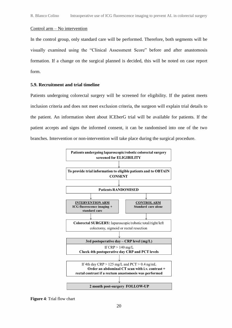
5.9. Recruitment and trial timeline
Patients undergoing colorectal surgery will be screened for eligibility. If the patient meets
inclusion criteria and does not meet exclusion criteria, the surgeon will explain trial details to
the patient. An information sheet about ICEberG trial will be available for patients. If the
patient accepts and signs the informed consent, it can be randomised into one of the two
branches. Intervention or non-intervention will take place during the surgical procedure.
Figure 4: Trial flow chart
R. Blanco Colino Intraoperative use of ICG fluorescence imaging to prevent AL in colorectal surgery
21
In order to detect AL postoperatively, all patients’ CRP levels will be evaluated on the 3rd
postoperative day. If CRP is over 140 mg/L, then 4th postoperative day CRP and PCT will be
evaluated. If at 4th day CRP is over 125 and PCT is over 0.4 ng/mL, then a CT scan with
intravenous contrast will be ordered, adding rectal contrast if a rectum anastomosis was
performed.
Requirement of radiological or surgical management of an AL will be recorded, as well as
major postoperative complications including death. Patients will be follow-up for at least two
months.
The end of the study is the date of the last follow-up for the last patient included in the study.
5.10. Data management
Data collection will include preoperative, intraoperative and postoperative details. Data will
be collected on predesigned paper forms (see Appendix – case report form) and then, it will be
entered into an electronic database system.
5.11. Statistical methods
Sample size
In previously cohort studies in Europe, AL rate has ranged among 6% to 8% in colonic
resections and among 10% to 12% for rectal resections. (7,8,54)
Assuming an average of AL following colorectal surgery of a 9% in the control group and a
3% in the experimental group based on published studies, accepting an alpha risk of 0.05 and
a beta risk of 0.2, a total of 512 patients (256 patients each arm) are the minimum required for
the study if a 10% of drop-out rate is assumed. The ARCSINUS approximation has been used.
Statistical analysis
Data analysis will be performed using R Foundation for Statistical Computing software (R
version 3.3.1). For baseline characteristics, descriptive statistics will be used. Chi squared test
R. Blanco Colino Intraoperative use of ICG fluorescence imaging to prevent AL in colorectal surgery
22
or Fisher’s Exact test will be used for categorical variables, including AL rate. For continuous
outcomes, Student’s t test will be applied for normal distribution and Mann-Whitney U test
for data that is not normally distributed. Univariate exploratory analysis and multiple logistic
regression will be used to test risk factors associated with AL (see Appendix – case report
form). P < 0.05 will be used as level of significance.
5.12. Ethical considerations and trial registration
The trial will be performed following the principles of the Declaration of Helsinki and Good
Clinical Practice. (55) Before the start of the trial, protocol, information sheet and informed
consent documents will be approved by the Ethical Committee at each participating centre.
Recruitment will not start in any individual centre until local approval is obtained.
Only patients who accept and sign the informed consent will be included. Participation in the
study is voluntary and patients can withdraw consent at any time.
The trial will be registered at the www.clinicaltrials.gov website.
6. CONCLUSIONS
Anastomotic leakage is still a common postoperative complication, which has been associated
with greater morbidity and mortality rates. The findings of the systematic review and meta-
analysis performed, suggest that ICG fluorescence imaging is a promising tool to prevent AL
in patients undergoing colorectal anastomosis. In order to stablish fluorescence imaging as
part of routine colorectal surgery, randomised controlled trials are needed to provide evidence
in its use. Therefore, ICEberG trial has been proposed in this work addressing this necessity.
Finally, European and international collaboration among surgeons is essential to run powered
studies which allow to address this issue together and, consequently, to get better outcomes
for our patients.
R. Blanco Colino Intraoperative use of ICG fluorescence imaging to prevent AL in colorectal surgery
23
7. REFERENCES
1. Vallance A, Wexner S, Berho M, Cahill R, Coleman M, Haboubi N, et al. A collaborative
review of the current concepts and challenges of anastomotic leaks in colorectal surgery.
Colorectal Dis. 2017; 19 (1): O1-O12.
2. Alves A, Panis Y, Trancart D, Regimbeau JM, Pocard M, Valleur P. Factors associated
with clinically significant anastomotic leakage after large bowel resection: Multivariate
analysis of 707 patients. World J Surg. 2002;26(4):499–502.
3. McDermott FD, Heeney A, Kelly ME, Steele RJ, Carlson GL, Winter DC. Systematic
review of preoperative, intraoperative and postoperative risk factors for colorectal
anastomotic leaks. Br J Surg. 2015;102(5):462–79.
4. Rahbari NN, Weitz J, Hohenberger W, Heald RJ, Moran B, Ulrich A, et al. Definition
and grading of anastomotic leakage following anterior resection of the rectum: A
proposal by the International Study Group of Rectal Cancer. Surgery. 2010;147(3):339–
51.
5. Kulu Y, Ulrich A, Bruckner T, Contin P, Welsch T, Rahbari NN, et al. Validation of the
International Study Group of Rectal Cancer definition and severity grading of
anastomotic leakage. Surgery. 2013;153(6):753–61.
6. Phitayakorn R, Delaney CP, Reynolds HL, Champagne BJ, Heriot AG, Neary P, et al.
Standardized algorithms for management of anastomotic leaks and related abdominal and
pelvic abscesses after colorectal surgery. World J Surg. 2008;32(6):1147–56.
7. European Society of Coloproctology collaborating group. The relationship between
method of anastomosis and anastomotic failure after right hemicolectomy and ileo-caecal
resection: an international snapshot audit. Colorectal Dis. 2016;38(1):42–9.
8. Ortiz H, Biondo S, Codina A, Ciga MÁ, Enríquez-Navascués J, Espín E, et al. Hospital
variation in anastomotic leakage after rectal cancer surgery in the Spanish Association of
Surgeons project: The contribution of hospital volume. Cir Esp. 2016;94(4):213–20.
R. Blanco Colino Intraoperative use of ICG fluorescence imaging to prevent AL in colorectal surgery
24
9. Tiernan J, Cook A, Geh I, George B, Magill L, Northover J, et al. Use of a modified
Delphi approach to develop research priorities for the association of coloproctology of
Great Britain and Ireland. Colorectal Dis. 2014;16(12):965–70.
10. Kingham TP, Pachter HL. Colonic Anastomotic Leak: Risk Factors, Diagnosis, and
Treatment. J Am Coll Surg. 2009;208(2):269–78.
11. Frasson M, Flor-Lorente B, Ramos Rodríguez JL, Granero-Castro P, Hervás D, Alvarez
Rico MA, et al. Risk Factors for Anastomotic Leak After Colon Resection for Cancer:
Multivariate Analysis and Nomogram From a Multicentric, Prospective, National Study
With 3193 Patients. Ann Surg. 2015;262(2):321–330.
12. Chadi SA, Fingerhut A, Berho M, DeMeester SR, Fleshman JW, Hyman NH, et al.
Emerging trends in the etiology, prevention, and treatment of gastrointestinal anastomotic
leakage. J Gastrointest Surg. 2016;20(12):2035–51.
13. Vignali A, Gianotti L, Braga M, Radaelli G, Malvezzi L, Di Carlo V. Altered
microperfusion at the rectal stump is predictive for rectal anastomotic leak. Dis Colon
Rectum. 2000;43(1):76–82.
14. Rutegård M. Anastomotic leakage in rectal cancer surgery: The role of blood perfusion.
World J Gastrointest Surg. 2015;7(11):289-292.
15. Sparreboom CL, Wu ZQ, Ji JF, Lange JF. Integrated approach to colorectal anastomotic
leakage: Communication, infection and healing disturbances. World J Gastroenterol.
2016;22(32):7226–35.
16. Hirst NA, Tiernan JP, Millner PA, Jayne DG. Systematic review of methods to predict
and detect anastomotic leakage in colorectal surgery. Colorectal Dis. 2014;16(2):95–109.
17. Karliczek A, Harlaar N, Zeebregts C, Wiggers T, Baas P, van Dam G. Surgeons lack
predictive accuracy for anastomotic leakage in gastrointestinal surgery. Int J Color Dis.
2009; 24 (5):569-576.
R. Blanco Colino Intraoperative use of ICG fluorescence imaging to prevent AL in colorectal surgery
25
18. Nachiappan S, Askari A, Currie A, Kennedy RH, Faiz O. Intraoperative assessment of
colorectal anastomotic integrity: a systematic review. Surg Endosc. 2014;28(9):2513–30.
19. Karliczek A, Benaron DA, Baas PC, Zeebregts CJ, Van Der Stoel A, Wiggers T, et al.
Intraoperative assessment of microperfusion with visible light spectroscopy in esophageal
and colorectal anastomoses. Eur Surg Res. 2008;41(3):303–11.
20. Bae SU, Min BS, Kim NK. Robotic low ligation of the inferior mesenteric artery for
rectal cancer using the firefly technique. Yonsei Med J. 2015;56(4):1028–35.
21. Boni L, David G, Dionigi G, Rausei S, Cassinotti E, Fingerhut A. Indocyanine green-
enhanced fluorescence to assess bowel perfusion during laparoscopic colorectal resection.
Surg Endosc. 2016;30(7):2736–42.
22. Foppa C, Denoya PI, Tarta C, Bergamaschi R. Indocyanine green fluorescent dye during
bowel surgery: Are the blood supply “guessing days” over? Tech Coloproctol.
2014;18(8):753–8.
23. Gröne J, Koch D, Kreis ME. Impact of intraoperative microperfusion assessment with
Pinpoint Perfusion Imaging on surgical management of laparoscopic low rectal and
anorectal anastomoses. Colorectal Dis. 2015;17(3, SI):22–8.
24. Nishigori N, Koyama F, Nakagawa T, Nakamura S, Ueda T, Inoue T, et al. Visualization
of Lymph/Blood Flow in Laparoscopic Colorectal Cancer Surgery by ICG Fluorescence
Imaging (Lap-IGFI). Ann Surg Oncol. 2016; 23:266–74.
25. Protyniak B, Dinallo AM, Boyan WP, Dressner RM, Arvanitis ML. Intraoperative
indocyanine green fluorescence angiography--an objective evaluation of anastomotic
perfusion in colorectal surgery. Am Surg. 2015; 81(6):580–4.
26. Hellan M, Spinoglio G, Pigazzi A, Lagares-Garcia JA. The influence of fluorescence
imaging on the location of bowel transection during robotic left-sided colorectal surgery.
Surg Endosc. 2014;28(5):1695–1702.
R. Blanco Colino Intraoperative use of ICG fluorescence imaging to prevent AL in colorectal surgery
26
27. Sherwinter DA, Gallagher J, Donkar T. Intra‐operative transanal near infrared imaging of
colorectal anastomotic perfusion: a feasibility study. Colorectal Dis. 2013;15(1):91–6.
28. Jafari MD, Wexner SD, Martz JE, McLemore EC, Margolin DA, Sherwinter DA, et al.
Perfusion Assessment in Laparoscopic Left-Sided/Anterior Resection (PILLAR II): A
Multi-Institutional Study. J Am Coll Surg. 2015;220(1):82–92.
29. Ris F, Hompes R, Lindsey I, Cunningham C, Mortensen NJ, Cahill RA. Near infra-red
laparoscopic assessment of the adequacy of blood perfusion of intestinal anastomosis - A
video vignette. Colorectal Dis. 2014;16(8):646–7.
30. Cahill RA, Ris F, Mortensen NJ. Near-infrared laparoscopy for real-time intra-operative
arterial and lymphatic perfusion imaging. Colorectal Dis. 2011;13 Suppl 7:12–7.
31. Boni L, David G, Mangano A, Dionigi G, Rausei S, Spampatti S, et al. Clinical
applications of indocyanine green (ICG) enhanced fluorescence in laparoscopic surgery.
Surg Endosc. 2015;29(7):2046–55.
32. Reinhart MB, Huntington CR, Blair LJ, Heniford BT, Augenstein VA. Indocyanine
Green: Historical Context, Current Applications, and Future Considerations. Surg Innov.
2016;23(2):166–75.
33. Benya R, Quintana J, Brundage B. Adverse reactions to indocyanine green: a case report
and a review of the literature. Cathet Cardiovasc Diagn. 1989;17(4):231–3.
34. Ris F, Buchs NC, Hompes R, Morel P. New imaging modalities in colorectal surgery, the
near infrared imaging. Swiss Knife. 2014;4:7-8
35. Johnson EK, Hardin MO, Walker AS, Hatch Q, Steele SR. Fluorescence Angiography in
Colorectal Resection. Dis Colon Rectum. 2016;59(1):e1–4.
36. Moher D, Liberati A, Tetzlaff J, Altman DG. Preferred reporting items for systematic
reviews and meta-analyses: The PRISMA statement. Int J Surg. 2010;8(5):336–41.
R. Blanco Colino Intraoperative use of ICG fluorescence imaging to prevent AL in colorectal surgery
27
37. Kudszus S, Roesel C, Schachtrupp A, Höer JJ. Intraoperative laser fluorescence
angiography in colorectal surgery: a noninvasive analysis to reduce the rate of
anastomotic leakage. Langenbeck’s Arch Surg. 2010;395(8):1025–30.
38. Kin C, Vo H, Welton L, Welton M. Equivocal effect of intraoperative fluorescence
angiography on colorectal anastomotic leaks. Dis Colon Rectum. 2015;58(6):582–7.
39. Sterne JA, Hernán MA, Reeves BC, Savović J, Berkman ND, Viswanathan M, et al.
ROBINS-I: a tool for assessing risk of bias in non-randomised studies of interventions.
Br Med J [Internet]. 2016;355:i4919. Available from:
http://www.bmj.com/lookup/doi/10.1136/bmj.i4919
40. Jafari MD, Lee KH, Halabi WJ, Mills SD, Carmichael JC, Stamos MJ, et al. The use of
indocyanine green fluorescence to assess anastomotic perfusion during robotic assisted
laparoscopic rectal surgery. Surg Endosc. 2013;27(8):3003–8.
41. Kim JC, Lee JL, Yoon YS, Alotaibi AM, Kim J. Utility of indocyanine‐green fluorescent
imaging during robot‐assisted sphincter‐saving surgery on rectal cancer patients. Int J
Med Robot Comput Assist Surg. 2016; 12:710-717.
42. Boni L, Fingerhut A, Marzorati A, Rausei S, Dionigi G, Cassinotti E. Indocyanine green
fluorescence angiography during laparoscopic low anterior resection: results of a case-
matched study. Surg Endosc [Internet]. 2016 Aug 23 [cited 2017 Jan 24]; Available from:
http://www.ncbi.nlm.nih.gov/pubmed/27553790
43. ClinicalTrials.gov. The Role of Indocyanine Green (ICG) Fluorescence Imaging on
Anastomotic Leak in Robotic Colorectal Surgery [Internet]. [cited 2017 Jan 24].
Available from: https://clinicaltrials.gov/ct2/show/NCT02598414
44. ClinicalTrials.gov. Evaluation of Intestinal Vascolarization With Indocianine Green
Angiography During Rectal Resection or Left Colectomy [Internet]. [cited 2017 Jan 24].
Available from: https://clinicaltrials.gov/ct2/show/NCT02662946
R. Blanco Colino Intraoperative use of ICG fluorescence imaging to prevent AL in colorectal surgery
28
45. ClinicalTrials.gov. A Study Assessing Perfusion Outcomes With PINPOINT® Near
Infrared Fluorescence Imaging in Low Anterior Resection (PILLAR III) [Internet]. [cited
2017 Jan 24]. Available from: https://clinicaltrials.gov/ct2/show/NCT02205307
46. Mirnezami A, Mirnezami R, Chandrakumaran K, Sasapu K, Sagar P, Finan P. Increased
local recurrence and reduced survival from colorectal cancer following anastomotic leak
systematic review and meta-analysis. Ann Surg. 2011;253(5):890–9.
47. Katoh H, Yamashita K, Wang G, Sato T, Nakamura T, Watanabe M. Anastomotic
Leakage Contributes to the Risk for Systemic Recurrence in Stage II Colorectal Cancer. J
Gastrointest Surg. 2011;15(1):120–9.
48. Espín E, Ciga MA, Pera M, Ortiz H. Oncological outcome following anastomotic leak in
rectal surgery. Br J Surg. 2015;102(4):416–22.
49. Kawada K, Hasegawa S, Wada T, Takahashi R, Hisamori S, Hida K, et al. Evaluation of
intestinal perfusion by ICG fluorescence imaging in laparoscopic colorectal surgery with
DST anastomosis. Surg Endosc [Internet]. 2016 Jun 28 [cited 2016 Nov 28]; Available
from: http://www.ncbi.nlm.nih.gov/pubmed/27351656
50. Ris F, Hompes R, Cunningham C, Lindsey I, Guy R, Jones O, et al. Near-infrared (NIR)
perfusion angiography in minimally invasive colorectal surgery. Surg Endosc.
2014;28(7):2221–6.
51. Diana M, Agnus V, Halvax P, Liu YY, Dallemagne B, Schlagowski AI, et al.
Intraoperative fluorescence-based enhanced reality laparoscopic real-time imaging to
assess bowel perfusion at the anastomotic site in an experimental model. Br J Surg.
2015;102(2):e169-e176.
52. Garcia-Granero A, Frasson M, Flor-Lorente B, Blanco F, Puga R, Carratalá A, et al.
Procalcitonin and C-reactive protein as early predictors of anastomotic leak in colorectal
surgery: a prospective observational study. Dis Colon Rectum. 2013;56(4):475–83.
R. Blanco Colino Intraoperative use of ICG fluorescence imaging to prevent AL in colorectal surgery
29
53. Clavien PA, Barkun J, de Oliveira ML, Vauthey JN, Dindo D, Schulick RD, et al. The
Clavien-Dindo Classification of Surgical Complications. Ann Surg. 2009;250(2):187–96.
54. Van Leersum NJ, Snijders HS, Henneman D, Kolfschoten NE, Gooiker GA, Ten Berge
MG, et al. The dutch surgical colorectal audit. Eur J Surg Oncol. 2013;39(10):1063–70.
55. World Medical Association. Declaration of Helsinki - Ethical Principles for Medical
Research Involving Human Subjects [Internet]. 2013 [cited 2017 Mar 23]. Available
from: https://www.wma.net/policies-post/wma-declaration-of-helsinki-ethical-principles-
for-medical-research-involving-human-subjects/
APPENDIX
Intraoperative use of IndoCyaninE Green fluorescence imaging to
prevent anastomotic leakage in colorectal surgery:
Systematic Review, Meta-Analysis
and Study Protocol for the ICEberG Trial
Abbreviatures
Search strategies (systematic review)
Bias assessment
ICEberG trial – Case report form
Information sheet and informed consent for the ICEberG trial
R. Blanco Colino Intraoperative use of ICG fluorescence imaging to prevent AL in colorectal surgery
1
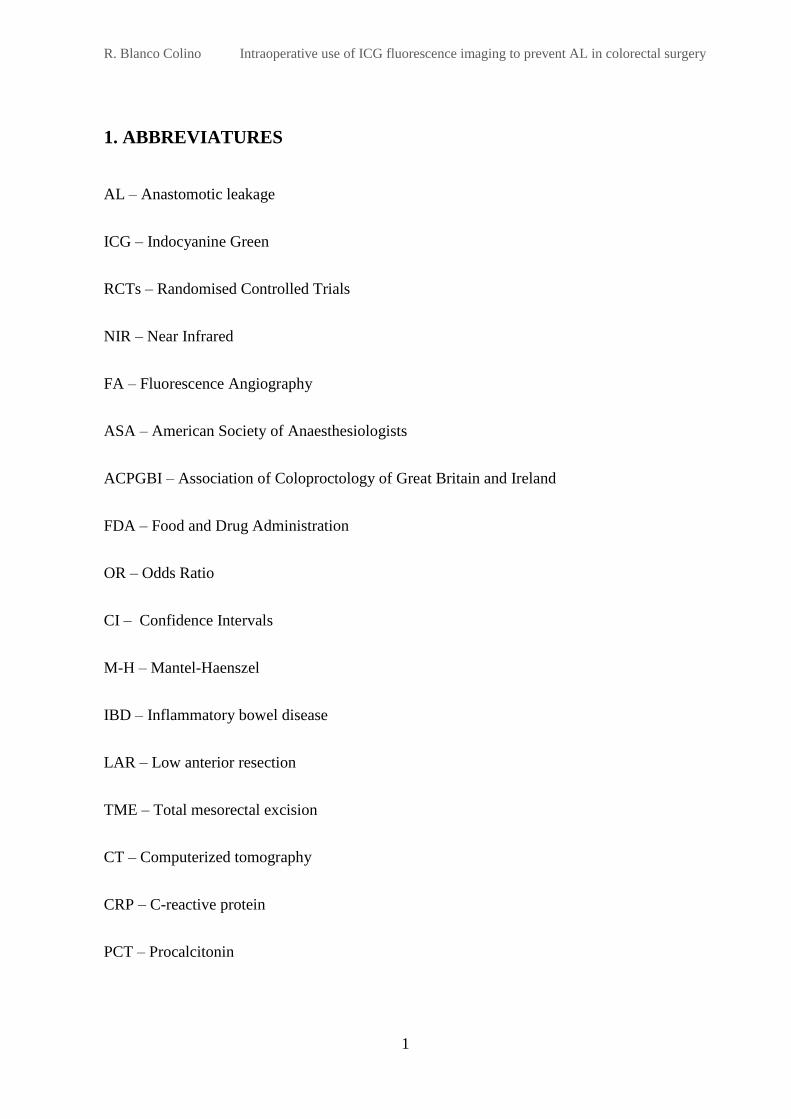
1. ABBREVIATURES
AL – Anastomotic leakage
ICG – Indocyanine Green
RCTs – Randomised Controlled Trials
NIR – Near Infrared
FA – Fluorescence Angiography
ASA – American Society of Anaesthesiologists
ACPGBI – Association of Coloproctology of Great Britain and Ireland
FDA – Food and Drug Administration
OR – Odds Ratio
CI – Confidence Intervals
M-H – Mantel-Haenszel
IBD – Inflammatory bowel disease
LAR – Low anterior resection
TME – Total mesorectal excision
CT – Computerized tomography
CRP – C-reactive protein
PCT – Procalcitonin
R. Blanco Colino Intraoperative use of ICG fluorescence imaging to prevent AL in colorectal surgery
2
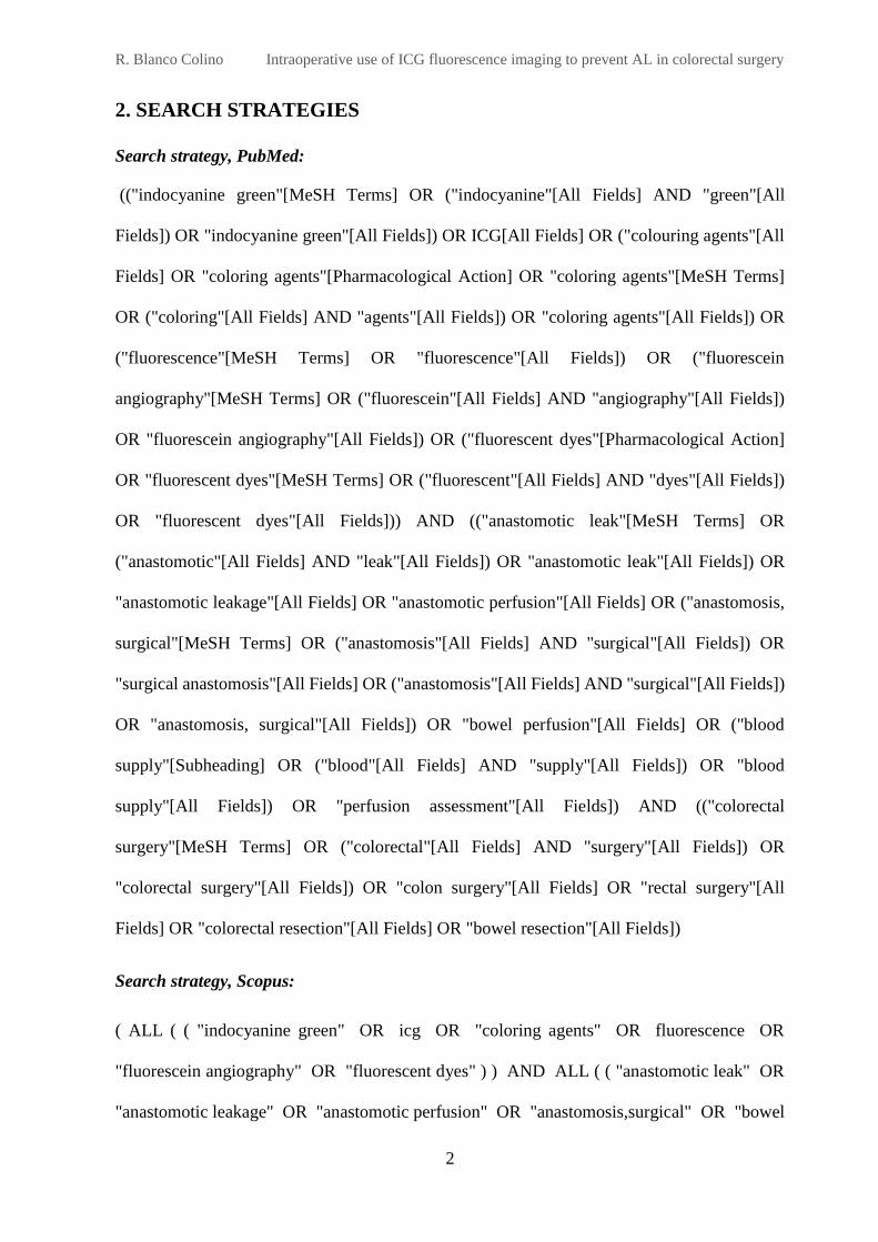
2. SEARCH STRATEGIES
Search strategy, PubMed:
(("indocyanine green"[MeSH Terms] OR ("indocyanine"[All Fields] AND "green"[All
Fields]) OR "indocyanine green"[All Fields]) OR ICG[All Fields] OR ("colouring agents"[All
Fields] OR "coloring agents"[Pharmacological Action] OR "coloring agents"[MeSH Terms]
OR ("coloring"[All Fields] AND "agents"[All Fields]) OR "coloring agents"[All Fields]) OR
("fluorescence"[MeSH Terms] OR "fluorescence"[All Fields]) OR ("fluorescein
angiography"[MeSH Terms] OR ("fluorescein"[All Fields] AND "angiography"[All Fields])
OR "fluorescein angiography"[All Fields]) OR ("fluorescent dyes"[Pharmacological Action]
OR "fluorescent dyes"[MeSH Terms] OR ("fluorescent"[All Fields] AND "dyes"[All Fields])
OR "fluorescent dyes"[All Fields])) AND (("anastomotic leak"[MeSH Terms] OR
("anastomotic"[All Fields] AND "leak"[All Fields]) OR "anastomotic leak"[All Fields]) OR
"anastomotic leakage"[All Fields] OR "anastomotic perfusion"[All Fields] OR ("anastomosis,
surgical"[MeSH Terms] OR ("anastomosis"[All Fields] AND "surgical"[All Fields]) OR
"surgical anastomosis"[All Fields] OR ("anastomosis"[All Fields] AND "surgical"[All Fields])
OR "anastomosis, surgical"[All Fields]) OR "bowel perfusion"[All Fields] OR ("blood
supply"[Subheading] OR ("blood"[All Fields] AND "supply"[All Fields]) OR "blood
supply"[All Fields]) OR "perfusion assessment"[All Fields]) AND (("colorectal
surgery"[MeSH Terms] OR ("colorectal"[All Fields] AND "surgery"[All Fields]) OR
"colorectal surgery"[All Fields]) OR "colon surgery"[All Fields] OR "rectal surgery"[All
Fields] OR "colorectal resection"[All Fields] OR "bowel resection"[All Fields])
Search strategy, Scopus:
( ALL ( ( "indocyanine green" OR icg OR "coloring agents" OR fluorescence OR
"fluorescein angiography" OR "fluorescent dyes" ) ) AND ALL ( ( "anastomotic leak" OR
"anastomotic leakage" OR "anastomotic perfusion" OR "anastomosis,surgical" OR "bowel
R. Blanco Colino Intraoperative use of ICG fluorescence imaging to prevent AL in colorectal surgery
3
perfusion" OR "blood supply" OR "perfusion assessment" ) ) AND ALL ( ( "colorectal
surgery" OR "colon surgery" OR "rectal surgery" OR "colorectal resection" OR "bowel
resection" ) ) – All Fields
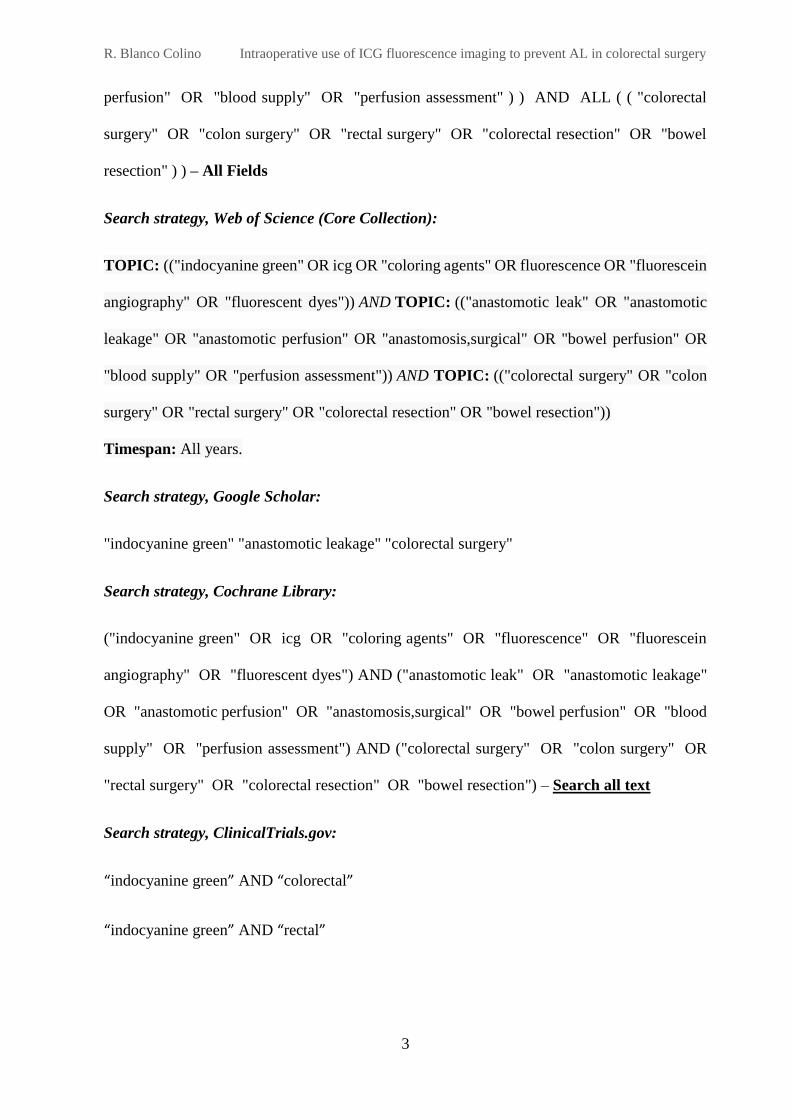
Search strategy, Web of Science (Core Collection):
TOPIC: (("indocyanine green" OR icg OR "coloring agents" OR fluorescence OR "fluorescein
angiography" OR "fluorescent dyes")) AND TOPIC: (("anastomotic leak" OR "anastomotic
leakage" OR "anastomotic perfusion" OR "anastomosis,surgical" OR "bowel perfusion" OR
"blood supply" OR "perfusion assessment")) AND TOPIC: (("colorectal surgery" OR "colon
surgery" OR "rectal surgery" OR "colorectal resection" OR "bowel resection"))
Timespan: All years.
Search strategy, Google Scholar:
"indocyanine green" "anastomotic leakage" "colorectal surgery"
Search strategy, Cochrane Library:
("indocyanine green" OR icg OR "coloring agents" OR "fluorescence" OR "fluorescein
angiography" OR "fluorescent dyes") AND ("anastomotic leak" OR "anastomotic leakage"
OR "anastomotic perfusion" OR "anastomosis,surgical" OR "bowel perfusion" OR "blood
supply" OR "perfusion assessment") AND ("colorectal surgery" OR "colon surgery" OR
"rectal surgery" OR "colorectal resection" OR "bowel resection") – Search all text
Search strategy, ClinicalTrials.gov:
“indocyanine green” AND “colorectal”
“indocyanine green” AND “rectal”
R. Blanco Colino Intraoperative use of ICG fluorescence imaging to prevent AL in colorectal surgery
4
3. BIAS ASSESSMENT WITH ROBINS-I TOOL1
ROBINS-I tool (Stage II): For each study
ROBINS-I tool (Stage III): Overall risk of bias assessment
Kudszus et al. (37) – Overall MODERATE
Kin et al. (38) – Overall MODERATE
Jafari et al. (40) – Overall MODERATE
Kim et al. (41) – Overall MODERATE
Boni et al. (42) – Overall MODERATE
1 Sterne JA, Hernán MA, Reeves BC, Savović J, Berkman ND, Viswanathan M, et al. ROBINS-I: a tool for
assessing risk of bias in non-randomised studies of interventions. Br Med J [Internet]. 2016;355:i4919. Available
from: http://www.bmj.com/lookup/doi/10.1136/bmj.i4919
Co
nfo
und
ing
Sel
ecti
on
of
par
tici
pan
ts
Cla
ssif
icat
ion
of
inte
rven
tio
ns
Dev
iati
on
s
fro
m i
nte
nd
ed
inte
rven
tio
ns
Mis
sin
g d
ata
Mea
sura
men
t
of
ou
tco
mes
Bia
s in
sele
ctio
n o
f
rep
ort
ed r
esu
lt
Kudszus et al. (37) Moderate Low Low Low Low Moderate Low
Kin et al. (38) Moderate Low Low Low Low Low Moderate
Jafari et al. (40) Moderate Low Low Low Low Low Low
Kim et al. (41) Moderate Low Low Low Low Moderate Low
Boni et al. (42) Moderate Low Low Low Low Low Low
R. Blanco Colino Intraoperative use of ICG fluorescence imaging to prevent AL in colorectal surgery
5
4. ICEBERG TRIAL – CASE REPORT FORM
Patient code:
Group assigned: 1 (ICG) - 2 (Control)
PREOPERATIVE DATA
Age (years)*
Gender
BMI (kg/m2)
ASA score* I II III IV
V Unknown
Past Medical History □ Ischaemic Heart Disease*
□ Congestive Heart Failure
□ Cerebrovascular Accident
□ Diabetes □ None □ Diet/tablet □ Insulin*
□ Chronic Kidney Disease
□ Chronic Respiratory Disease
History of previous abdominal surgery Yes No
Current treatment Anticoagulants / Corticosteroids*
Neoadjuvant therapy (If cancer indication) Yes- Chemotherapy Yes-Radiotherapy*
Yes – Chemo + radiotherapy No
Smoking status
For current and ex-smoker record pack year history:
Current* Ex-smoker Never
Unknown
Last pre-operative serum albumin (g/dl)
Last pre-operative serum haemoglobin (g/dl)
INTRAOPERATIVE DATA
Day of surgical intervention (DD/MM/YYYY)
Preoperative bowel preparation Yes No
Surgical procedure
Surgical indication* □ Malignant □ Diverticular disease
□ IBD □ Other
Surgical approach Laparoscopic Robotic
Type of anastomosis* □ Colo-colic
□ Colo-rectal
□ Ileo-colic
□ Other: _______________
Anastomosis - technical details* Hand-sewn - Stapled
Length of operation (minutes)
Clinical Assessment Score
(both segments)
Before anastomosis Proximal: 1 - 2 - 3 Distal: 1 - 2 - 3
After anastomosis Proximal: 1 - 2 - 3 Distal: 1 - 2 - 3
Fluorescence Reflection
Score (both segments)
Before anastomosis Proximal: 1 - 2 - 3 Distal: 1 - 2 - 3
After anastomosis Proximal: 1 - 2 - 3 Distal: 1 - 2 - 3
Change in the surgical plan Yes No
If YES – indicate change:
Performance of protective ileostomy Yes No
R. Blanco Colino Intraoperative use of ICG fluorescence imaging to prevent AL in colorectal surgery
6
POSTOPERATIVE DATA
3rd postoperative day CRP level (mg/L)
If applicable: 4th postop day CRP level (mg/L)
If applicable: 4th postop day PCT level (ng/mL)
CT scan findings None AL Pelvic abscess Other
Anastomotic leak (60 day follow-up)
If AL - Please record postoperative day of diagnosis
No Yes Grade A / Grade B / Grade C
Day:
Length of stay (days)
60-day Clavien-Dindo grade I II III IV V
Readmission (if YES – indicate postoperative day) Yes No
Day:
Reoperation (if YES – indicate postoperative day) Yes No
Day:
Postoperative pain control with NSAIDS Yes No
TNM after surgery (If cancer indication)
(*) Variables associated with AL – for exploratory analysis on primary outcome measure
Clavien Dindo Classification
GRADE DEFINITION
I Any deviation from the normal postoperative course without the need for
pharmacological (other than the “allowed therapeutic regimens”), surgical,
endoscopic or radiological intervention.
Allowed therapeutic regimens are: selected drugs (antiemetics, antipyretics,
analgesics, diuretics and electrolyte replacement), physiotherapy and wound
infections opened at the bedside but not treated with antibiotics.
II Requiring pharmacological treatment with drugs beyond those allowed for
grade I complications. Blood transfusions and total parenteral nutrition are also
included.
IIIa Requiring surgical, endoscopic or radiological intervention, not under general
anaesthetic.
IIIb Requiring surgical, endoscopic or radiological intervention, under general
anaesthetic.
IVa Life-threatening complications requiring critical care management – single
organ dysfunction, or neurological complications including brain haemorrhage
and ischemic stroke (excluding transient ischemic attack).
IVb Life-threatening complications requiring critical care management – multi-
organ dysfunction.
V Death of a patient
R. Blanco Colino Intraoperative use of ICG fluorescence imaging to prevent AL in colorectal surgery
7
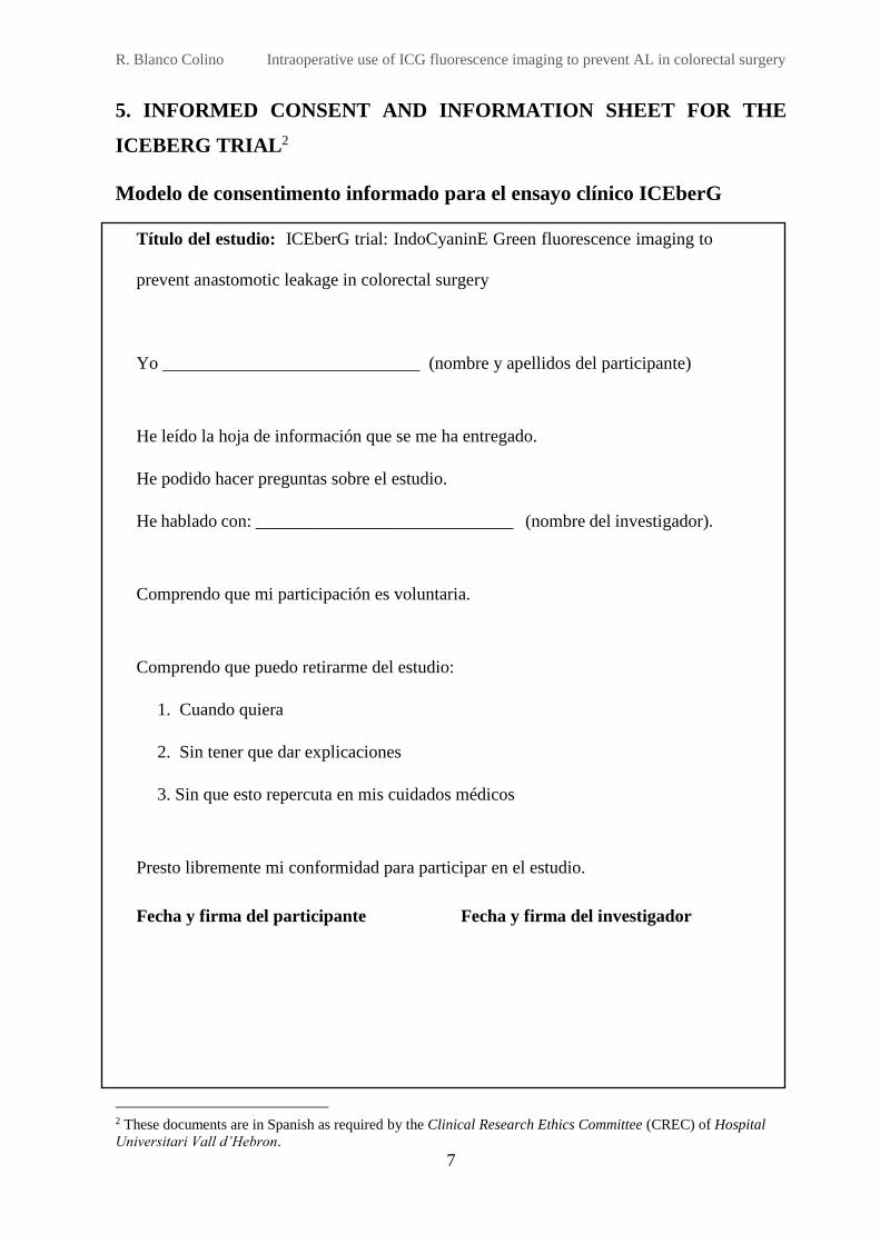
5. INFORMED CONSENT AND INFORMATION SHEET FOR THE
ICEBERG TRIAL2
Modelo de consentimento informado para el ensayo clínico ICEberG
Título del estudio: ICEberG trial: IndoCyaninE Green fluorescence imaging to
prevent anastomotic leakage in colorectal surgery
Yo _____________________________ (nombre y apellidos del participante)
He leído la hoja de información que se me ha entregado.
He podido hacer preguntas sobre el estudio.
He hablado con: _____________________________ (nombre del investigador).
Comprendo que mi participación es voluntaria.
Comprendo que puedo retirarme del estudio:
1. Cuando quiera
2. Sin tener que dar explicaciones
3. Sin que esto repercuta en mis cuidados médicos
Presto libremente mi conformidad para participar en el estudio.
Fecha y firma del participante Fecha y firma del investigador
2 These documents are in Spanish as required by the Clinical Research Ethics Committee (CREC) of Hospital
Universitari Vall d’Hebron.
R. Blanco Colino Intraoperative use of ICG fluorescence imaging to prevent AL in colorectal surgery
8
Hoja de información al paciente para el ensayo clínico ICEberG
Proyecto de investigación titulado ICEberG trial: IndoCyaninE Green fluorescence imaging to
prevent anastomotic leakage in colorectal surgery
Estimado paciente,
Le solicitamos su participación en este proyecto de investigación cuyo objetivo principal es
evaluar si el uso intraoperatorio de verde indocianina para la visualización directa de la
vascularización colorrectal reduce la tasa de fuga de anastomosis.
Beneficios:
Es posible que de su participación en este estudio no obtenga un beneficio directo. Sin embargo,
la evaluación de nuevas estrategias relacionadas con la prevención de la fuga anastomótica
podría contribuir a una futura reducción de esta complicación postoperatoria.
Procedimientos del estudio:
En este estudio se pretenden comparar dos procedimientos. La asignación a uno de ellos vendrá
determinada por el azar. Su médico no intervendrá en este proceso. Usted tendrá una
probabilidad del 50% de recibir cada uno de los procedimientos contemplados en este estudio.
El primer grupo contempla los procedimientos que se hacen usualmente en la práctica habitual
durante la cirugía y en el segundo grupo, además de los procedimientos habituales se empleará
el uso de verde de indocianina. Éste es un colorante que permite visualizar por fluorescencia la
vascularización colorrectal y, por tanto, decidir cambios en el plan quirúrgico según precise
para asegurar una anastomosis en zona viable con buena perfusión.
Molestias y posibles riesgos:
El verde de indocianina es un colorante seguro, de baja toxicidad y buena tolerancia. Se ha
descrito algún caso de reacción anafiláctica en pacientes con alergia a la iodina o a contrastes
iodados (1/300000).
R. Blanco Colino Intraoperative use of ICG fluorescence imaging to prevent AL in colorectal surgery
9
Protección de datos personales:
De acuerdo con la Ley 15/1999 de Protección de Datos de Carácter Personal, los datos
personales que se obtengan serán los necesarios para cubrir los fines del estudio. En ninguno
de los informes del estudio aparecerá su nombre, y su identidad no será revelada a persona
alguna salvo para cumplir con los fines del estudio, y en el caso de urgencia médica o
requerimiento legal. Cualquier información de carácter personal que pueda ser identificable
será conservada por métodos informáticos en condiciones de seguridad. Cualquier información
de carácter personal que pueda ser identificable será conservada por métodos informáticos en
condiciones de seguridad. El acceso a dicha información quedará restringido al personal del
hospital, designado al efecto o a otro personal autorizado que estará obligado a mantener la
confidencialidad de la información.
De acuerdo con la ley vigente, usted tiene derecho al acceso de sus datos personales; asimismo,
y si está justificado, tiene derecho a su rectificación y cancelación. Si así lo desea, deberá
solicitarlo al médico que le atiende en este estudio.
De acuerdo con la legislación vigente, tiene derecho a ser informado de los datos relevantes
para su salud que se obtengan en el curso del estudio. Esta información se le comunicará si lo
desea; en el caso de que prefiera no ser informado, su decisión se respetará.
Su participación en el estudio es totalmente voluntaria, y si decide no participar recibirá todos
los cuidados médicos que necesite y la relación con el equipo médico que le atiende no se verá
afectada.
Agradecimiento:
Le agradecemos el tiempo dedicado a la lectura de esta información. Por favor, tómese el tiempo
necesario para leer toda la información y preguntar las dudas que le pudieran surgir. Si decide
tomar parte en este estudio, deberá firmar el consentimiento informado conforme acepta su
participación voluntaria en el ensayo clínico ICEberG.