Embed Size (px)
Citation preview

CASE REPORTJ Neurosurg Spine 27:312–315, 2017
Hemangioblastomas are rare, slow-growing tumors that are most commonly found in the spinal cord, cerebellum, and brainstem.4 They can occur spo-
radically or as a component of von Hippel-Lindau (VHL) disease; 75% of them are sporadic and typically solitary.15 Solitary sporadic hemangioblastomas have a predilection for the cerebellum, whereas VHL-related tumors are more commonly found in the spinal cord.11
Nearly all reported cases of hemangioblastomas are in the CNS, and extraneural locations are extremely rare. Specifically, intraosseous hemangioblastomas are rarely seen. There have been only 2 case reports in the literature on this condition over the last 20 years.3,9
Due to their location in bone, hemangioblastomas may be a confounding diagnosis to make, particularly in pa-tients with a known primary malignancy. In patients with no known malignancy, the nonspecific imaging findings may prompt a malignancy workup. To our best knowledge, there have been no described cases of intraosseous heman-gioblastomas in the cervical vertebrae. In this report, we describe a case of pathological cervical vertebral fracture secondary to a sporadic, intraosseous hemangioblastoma. We believe that the rarity of its location, unique clinical presentation, nonspecific imaging findings, and subse-
quent treatment improve our understanding of this rare tu-mor, the diversity of its presentation, and its management strategy.
Case ReportHistory
A 69-year-old woman presented to our institution with a 2-week history of worsening bilateral upper-extremity pain after a mechanical fall. She initially presented to an urgent care center, where she was given a neck brace and discharged home on naproxen and prednisone. However, her pain worsened. On presentation, she described sharp, intractable pain radiating down the lateral aspect of her arms in the C-5 dermatomal distribution with minimal neck motion.
Physical ExaminationShe appeared comfortable in a Miami cervical collar.
Her cranial nerve examination revealed left tongue devia-tion, in keeping with her known history of radiation-treat-ed left vagal nerve schwannoma. The remainder of her neurological examination showed intact status, except 4/5 motor strength in bilateral shoulder abduction.
ABBREVIATIONS VHL = von Hippel-Lindau. SUBMITTED January 25, 2016. ACCEPTED March 9, 2017.INCLUDE WHEN CITING Published online June 30, 2017; DOI: 10.3171/2017.3.SPINE1622.
Intraosseous hemangioblastoma of the cervical spine: case reportZhenteng Li, MD,1 Brian Curtis, MD,1 Robert Layser, MD,1 Santosh Kumar Selvarajan, MD,1 James Harrop, MD,2 Lawrence C. Kenyon, MD, PhD,3 Theodore Parsons, MD,3 and Asa Rubin, MD3
Departments of 1Radiology, 2Neurosurgery, and 3Pathology, Thomas Jefferson University Hospital, Philadelphia, Pennsylvania
A 69-year-old woman presented with bilateral upper-extremity radiculopathy and neck pain after a mechanical fall. Ad-mission CT and MRI of the cervical spine demonstrated a pathological C-4 fracture. Subsequent malignancy workup was negative. A CT-guided biopsy of the lesion showed intraosseous hemangioblastoma. Hemangioblastoma is a highly vascular, slow-growing tumor of the CNS; intraosseous location of this tumor is extremely rare. The authors review the diversity of its presentation and the treatment techniques of this rare tumor in an extremely rare location.https://thejns.org/doi/abs/10.3171/2017.3.SPINE1622KEY WORDS intraosseous hemangioblastoma; cervical spine; oncology
©AANS, 2017J Neurosurg Spine Volume 27 • September 2017312
Unauthenticated | Downloaded 12/16/21 03:11 AM UTC

Cervical intravertebral hemangioblastoma
J Neurosurg Spine Volume 27 • September 2017 313
NeuroimagingA cervical spine CT demonstrated an approximately
25% loss of height of the C-4 vertebral body, which was largely replaced by soft tissue with no visible internal tra-beculae (Fig. 1). Subsequent MRI showed abnormal mar-row replacement and enhancement throughout the C-4 vertebral body and posterior elements, sparing the spinous process. Additionally, enhancing soft tissues were pres-ent in the prevertebral ventral epidural space, and in the bilateral neural and transverse foramina at the C-4 level, resulting in mild central stenosis. Expansile tumor was spread within the pedicles and facets, obliterating the neu-ral foramina. At this time, the primary radiological differ-ential considerations were metastasis and plasmacytoma.
However, metastatic workup was negative. A CT-guided biopsy was performed and showed vascular tumor within bone, consistent with intraosseous hemangioblas-toma. Given this diagnosis, selective angiograms were performed for preoperative vascular evaluation and tumor embolization (Fig. 2). Feeding arteries were identified from the muscular branches of V2 segments of the ver-tebral arteries and thyrocervical trunks. Robust feeders were seen from the thyrocervical trunks and were super-selectively embolized with Onyx 18.
OperationThe patient underwent a C-4 corpectomy with C3–5
anterior and posterior instrumented fusion. Intraopera-tively, the longus colli muscles at the C-4 level were in-filtrated and hypervascular. The C-4 vertebral body was comminuted.
Pathological FindingsMacroscopically, a CT-guided core biopsy of the C-4
vertebral body showed 2 fragments of red-pink rubbery soft tissues. Microscopic analysis of the specimens dem-onstrated that the lesion was within bone and was vasofor-mative, with a dual population of cells (Fig. 3). Specimen staining with inhibin was noncontributory. Staining with CD56 and neuron-specific enolase (NSE) was positive in foamy cells. Staining with erythroblast transformation-specific–related gene (ERG), CD31, and CD34 was posi-
tive in endothelial cells. Staining with MelanA, CD10, AE1/AE3, Cam 5.2, epithelial membrane antigen (EMA), and human herpes virus (HHV)–8 was negative. The Ki-67 level was low. Immunohistochemical profile con-firmed the vascular nature of the lesion and did not sup-port metastatic carcinoma or plasmacytoma. These find-ings were consistent with intraosseous hemangioblastoma. Pathological analysis of the surgical specimens confirmed WHO Grade I hemangioblastoma within bone. At this time, immunohistochemistry for inhibin was positive, fur-ther supporting this diagnosis.
Postoperative CourseAt the 1-year follow-up, CT and MR images of the cer-
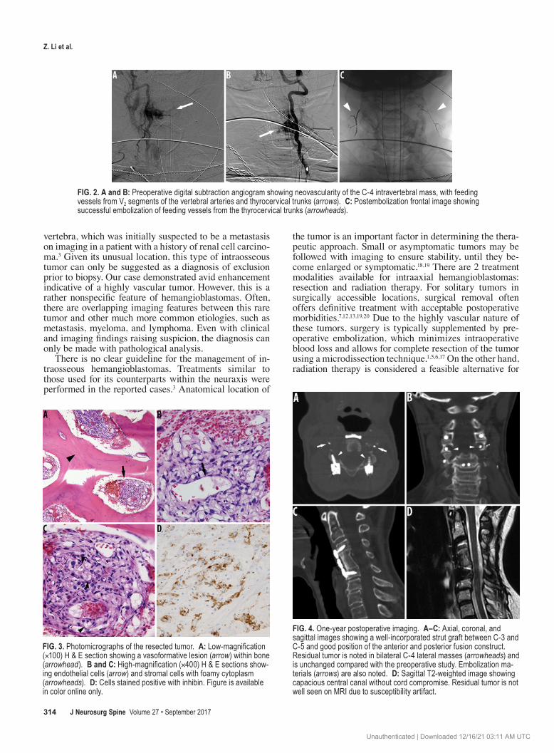
vical spine showed the spinal cord decompressed with re-sidual tumor lateral to the construct, which was unchanged from immediate postoperative imaging (Fig. 4). At the 1-year point, the patient had done exceptionally well. She was without neck pain and her upper-extremity pain had completely resolved.
DiscussionThe differential diagnosis of a lytic lesion with bone
marrow replacement and diffuse enhancement in a cervi-cal vertebra is broad.16 However, given the patient’s age, the most common etiologies are metastasis, myeloma, and—rarely—atypical hemangiomas. Atypical hemangio-mas are rarely completely lucent on CT scans; they typi-cally have vertical internal trabeculae and no diffusion restriction on diffusion-weighted imaging.8
Intracranial hemangioblastomas are described as cys-tic lesions with solid enhancing intramural nodules. Their MR characteristics are hypo- or isointense on T1-weight-ed imaging and hyperintense on T2-weighted imaging.2,11 Due to high vascularity, these tumors typically demon-strate avid, diffuse enhancement and may show vascular flow voids on unenhanced images.
The prevalence of osseous hemangioblastomas is un-known. There have been only 2 reported cases in the last 20 years.3,9 In one example, Cho and colleagues reported a case of sporadic osseous hemangioblastoma in a thoracic
FIG. 1. A C-4 compression fracture secondary to an intravertebral lesion. A and B: Axial and sagittal images showing the C-4 vertebral body largely replaced by irregular, low-density soft tissues with no visible internal trabeculae (arrow). C and D: Axial and sagittal postcontrast fat-suppressed T1-weighted images showing diffuse enhancement throughout the vertebral body, sparing the spinous process. The arrow in panel D designates the C-4 vertebral body, which is intensely enhancing on the postcontrast im-age. E: Sagittal T1-weighted image showing associated marrow replacement. The arrow in panel E designates the C-4 vertebral body, which is homogeneously hypointense, suggesting marrow replacement in the precontrast image.
Unauthenticated | Downloaded 12/16/21 03:11 AM UTC
Z. Li et al.
J Neurosurg Spine Volume 27 • September 2017314
vertebra, which was initially suspected to be a metastasis on imaging in a patient with a history of renal cell carcino-ma.3 Given its unusual location, this type of intraosseous tumor can only be suggested as a diagnosis of exclusion prior to biopsy. Our case demonstrated avid enhancement indicative of a highly vascular tumor. However, this is a rather nonspecific feature of hemangioblastomas. Often, there are overlapping imaging features between this rare tumor and other much more common etiologies, such as metastasis, myeloma, and lymphoma. Even with clinical and imaging findings raising suspicion, the diagnosis can only be made with pathological analysis.
There is no clear guideline for the management of in-traosseous hemangioblastomas. Treatments similar to those used for its counterparts within the neuraxis were performed in the reported cases.3 Anatomical location of
the tumor is an important factor in determining the thera-peutic approach. Small or asymptomatic tumors may be followed with imaging to ensure stability, until they be-come enlarged or symptomatic.18,19 There are 2 treatment modalities available for intraaxial hemangioblastomas: resection and radiation therapy. For solitary tumors in surgically accessible locations, surgical removal often offers definitive treatment with acceptable postoperative morbidities.7,12,13,19,20 Due to the highly vascular nature of these tumors, surgery is typically supplemented by pre-operative embolization, which minimizes intraoperative blood loss and allows for complete resection of the tumor using a microdissection technique.1,5,6,17 On the other hand, radiation therapy is considered a feasible alternative for
FIG. 3. Photomicrographs of the resected tumor. A: Low-magnification (×100) H & E section showing a vasoformative lesion (arrow) within bone (arrowhead). B and C: High-magnification (×400) H & E sections show-ing endothelial cells (arrow) and stromal cells with foamy cytoplasm (arrowheads). D: Cells stained positive with inhibin. Figure is available in color online only.
FIG. 4. One-year postoperative imaging. A–C: Axial, coronal, and sagittal images showing a well-incorporated strut graft between C-3 and C-5 and good position of the anterior and posterior fusion construct. Residual tumor is noted in bilateral C-4 lateral masses (arrowheads) and is unchanged compared with the preoperative study. Embolization ma-terials (arrows) are also noted. D: Sagittal T2-weighted image showing capacious central canal without cord compromise. Residual tumor is not well seen on MRI due to susceptibility artifact.
FIG. 2. A and B: Preoperative digital subtraction angiogram showing neovascularity of the C-4 intravertebral mass, with feeding vessels from V2 segments of the vertebral arteries and thyrocervical trunks (arrows). C: Postembolization frontal image showing successful embolization of feeding vessels from the thyrocervical trunks (arrowheads).
Unauthenticated | Downloaded 12/16/21 03:11 AM UTC

Cervical intravertebral hemangioblastoma
J Neurosurg Spine Volume 27 • September 2017 315
surgically inaccessible lesions or VHL-related, multifocal lesions.3,10,14 Regardless of treatment modality, the prima-ry goal of therapy is to avoid treatment-related morbidi-ties. This patient became symptomatic after pathological fracture of a cervical vertebra secondary to intravertebral hemangioblastoma. Fortunately for her, the hemangioblas-toma was extradural and intravertebral in location, which allowed complete resection with no significant complica-tions.
ConclusionsIntraosseous hemangioblastoma should be considered
in the differential diagnosis of isolated intravertebral low-density lesions with marrow replacement and avid enhancement, after common etiologies are excluded. In reporting this case, we illustrate the diversity of presenta-tion of hemangioblastomas and the effective management techniques of this rare tumor in an extremely rare location.
References 1. Ampie L, Choy W, Khanna R, Smith ZA, Dahdaleh NS, Par-
sa AT, et al: Role of preoperative embolization for intradural spinal hemangioblastomas. J Clin Neurosci 24:83–87, 2016
2. Baker KB, Moran CJ, Wippold FJ II, Smirniotopoulos JG, Rodriguez FJ, Meyers SP, et al: MR imaging of spinal he-mangioblastoma. AJR Am J Roentgenol 174:377–382, 2000
3. Cho H, Lee SH, Kim ES, Eoh W: Intraosseous hemangio-blastoma mimicking spinal metastasis in the patient with renal cell carcinoma. J Korean Neurosurg Soc 49:381–383, 2011
4. Conway JE, Chou D, Clatterbuck RE, Brem H, Long DM, Rigamonti D: Hemangioblastomas of the central nervous system in von Hippel-Lindau syndrome and sporadic disease. Neurosurgery 48:55–63, 2001
5. dos Santos MP, Zhang J, Ghinda D, Glikstein R, Agid R, Rodesch G, et al: Imaging diagnosis and the role of endovas-cular embolization treatment for vascular intraspinal tumors. Neurosurg Focus 39(2):E16, 2015
6. Eskridge JM, McAuliffe W, Harris B, Kim DK, Scott J, Winn HR: Preoperative endovascular embolization of cra-niospinal hemangioblastomas. AJNR Am J Neuroradiol 17:525–531, 1996
7. Fukuda M, Takao T, Hiraishi T, Yoshimura J, Yajima N, Saito A, et al: Clinical factors predicting outcomes after sur-gical resection for sporadic cerebellar hemangioblastomas. World Neurosurg 82:815–821, 2014
8. Hatipoglu HG, Selvi A, Ciliz D, Yuksel E: Quantitative and diffusion MR imaging as a new method to assess osteoporo-sis. AJNR Am J Neuroradiol 28:1934–1937, 2007
9. Higgins JN, Lammie GA, Savy LE, Taylor WJ, Stevens JM: Intraosseous vertebral haemangioblastoma: MRI. Neurora-diology 38 (Suppl 1):S107–S110, 1996
10. Kano H, Niranjan A, Mongia S, Kondziolka D, Flickinger JC, Lunsford LD: The role of stereotactic radiosurgery for intracranial hemangioblastomas. Neurosurgery 63:443–451, 2008
11. Leung RS, Biswas SV, Duncan M, Rankin S: Imaging fea-tures of von Hippel-Lindau disease. Radiographics 28:65–79, 323, 2008
12. Liu A, Jain A, Sankey EW, Jallo GI, Bettegowda C: Sporadic intramedullary hemangioblastoma of the spine: a single insti-tutional review of 21 cases. Neurol Res 38:205–209, 2016
13. Lonser RR, Weil RJ, Wanebo JE, DeVroom HL, Oldfield EH: Surgical management of spinal cord hemangioblastomas in patients with von Hippel-Lindau disease. J Neurosurg 98:106–116, 2003
14. Moss JM, Choi CY, Adler JR Jr, Soltys SG, Gibbs IC, Chang SD: Stereotactic radiosurgical treatment of cranial and spinal hemangioblastomas. Neurosurgery 65:79–85, 2009
15. Neumann HP, Eggert HR, Weigel K, Friedburg H, Wiestler OD, Schollmeyer P: Hemangioblastomas of the central ner-vous system. A 10-year study with special reference to von Hippel-Lindau syndrome. J Neurosurg 70:24–30, 1989
16. Rodallec MH, Feydy A, Larousserie F, Anract P, Campagna R, Babinet A, et al: Diagnostic imaging of solitary tumors of the spine: what to do and say. Radiographics 28:1019–1041, 2008
17. Tampieri D, Leblanc R, TerBrugge K: Preoperative emboli-zation of brain and spinal hemangioblastomas. Neurosur-gery 33:502–505, 1993
18. Wanebo JE, Lonser RR, Glenn GM, Oldfield EH: The natu-ral history of hemangioblastomas of the central nervous system in patients with von Hippel-Lindau disease. J Neuro-surg 98:82–94, 2003
19. Weil RJ, Lonser RR, DeVroom HL, Wanebo JE, Oldfield EH: Surgical management of brainstem hemangioblastomas in patients with von Hippel-Lindau disease. J Neurosurg 98:95–105, 2003
20. Westwick HJ, Giguère JF, Shamji MF: Incidence and prog-nosis of spinal hemangioblastoma: a Surveillance Epidemiol-ogy and End Results study. Neuroepidemiology 46:14–23, 2016
DisclosuresThe authors report no conflict of interest concerning the materi-als or methods used in this study or the findings specified in this paper.
Author ContributionsConception and design: Li. Acquisition of data: Li, Curtis, Layser, Harrop, Kenyon, Parsons. Analysis and interpretation of data: Sel-varajan, Li, Curtis, Layser, Harrop, Kenyon. Drafting the article: Li. Critically revising the article: Selvarajan, Li, Curtis, Layser, Harrop. Reviewed submitted version of manuscript: Selvarajan, Li, Curtis, Layser, Harrop, Kenyon, Parsons. Approved the final version of the manuscript on behalf of all authors: Selvarajan. Administrative/technical/material support: Rubin. Study supervi-sion: Selvarajan, Li.
CorrespondenceSantosh Kumar Selvarajan, TJUH Department of Radiology, 132 South 10th St., Ste. 1087, Main Bldg., Philadelphia, PA 19107. email: [email protected].
Unauthenticated | Downloaded 12/16/21 03:11 AM UTC