Embed Size (px)
Citation preview
INTRASPECIFIC GENE FLOW AND VECTOR COMPETENCE AMONG
Periplaneta americana COCKROACHES (BLATTODEA: BLATTIDAE) IN
CENTRAL TEXAS
A Thesis
by
JENNIFER LYNNE PECHAL
Submitted to the Office of Graduate Studies of Texas A&M University
in partial fulfillment of the requirements for the degree of
MASTER OF SCIENCE
August 2008
Major Subject: Entomology
INTRASPECIFIC GENE FLOW AND VECTOR COMPETENCE AMONG
Periplaneta americana COCKROACHES (BLATTODEA: BLATTIDAE) IN
CENTRAL TEXAS
A Thesis
by
JENNIFER LYNNE PECHAL
Submitted to the Office of Graduate Studies of Texas A&M University
in partial fulfillment of the requirements for the degree of
MASTER OF SCIENCE
Approved by:
Co-Chairs of Committee, Roger E. Gold Jeffery K. Tomberlin Committee Members, James W. Austin Leon H. Russell Head of Department, Kevin M. Heinz
August 2008
Major Subject: Entomology
iii
ABSTRACT
Intraspecific Gene Flow and Vector Competence among Periplaneta americana
Cockroaches (Blattodea: Blattidae) in Central Texas. (August 2008)
Jennifer Lynne Pechal, B.S., Sam Houston State University
Co-Chairs of Advisory Committee: Dr. Roger E. Gold Dr. Jeffery K. Tomberlin
One of the most overlooked areas in forensic entomology is urban, which applies
to insects and their arthropod relatives that have interactions with humans, their
associated structures, and companion animals. American cockroaches, Periplaneta
americana (L.), are common pests of urban environments. Analyzing spatial distribution
of P. americana populations in an artificial, outdoor environment provided insight of
gene flow among populations collected in central Texas. This information provides for a
better understanding of how and if populations were segregated, or if there was a single
unified population. Populations can be genetically differentiated through determining
variation of specific gene regions within populations. This study revealed a ubiquitous
distribution of cockroach populations, and their ability to indiscriminately inhabit areas
within an urban environment. Overall, cockroaches were identified from a large
interbreeding population with no discernable relationship between genetic variation of P.
americana and spatial distribution.
Identifying cockroach populations is relative to understanding the ability of
surrogate species indirectly affecting man by their ability to transfer disease-causing
iv
organisms including bacteria. This may have potentially deleterious health consequences
on animal and/or human populations. There are several pathogens associated with
cockroaches which are overlooked during diagnosis of sudden ailments with symptoms
being similar to food-borne illnesses, including abdominal cramping, diarrhea, nausea,
and fever. Analyzing spatial distributions of Escherichia coli and Campylobacter spp. in
relationship to collected cockroaches allowed for prevalence of bacteria species to be
identified among populations. The prevalence of bacteria isolated from total populations
collected indicated a high prevalence (92.3%) of bacteria carried by the exoskeleton of
P. americana. Gram-negative bacteria acquisition and dissemination of organisms such
as E. coli was prevalent on campus. Screening for E. coli 1057:H7 and Campylobacter
spp. resulted in no positive colony growth. The lack of Campylobacter spp. growth from
cuticular surfaces may have resulted from undesirable conditions required to sustain
colony growth. Data from this study corroborates the potential ability of cockroaches to
mechanically transmit pathogens.
v
ACKNOWLEDGMENTS
I would like to thank the faculty and staff of the Texas A&M University
Department of Entomology, my co-chairs Drs. Roger E. Gold and Jeffery K. Tomberlin.
Thank you to the other members of my committee, Drs. James W. Austin and Leon H.
Russell, who provided invaluable input and information. Thank you all for your support,
advice, and wisdom through the development and completion of this idea. To my fellow
graduate students in the Center for Urban Structural Entomology and F.L.I.E.S. lab, I
thank you for the support, feedback, and technical advice throughout this project.
Without y’all this research may not have come to fruition and my sanity may have gone
off the deep end in the process. Thank you. A special thanks to Dr. Jimmy K. Olson who
through the past two years has become a mentor and friend. I can not truly express how
much I appreciate you pushing me to become a better student and person through my
academic and personal trials and tribulations.
Finally, I would like to thank my family, my dad, mom, sisters, and brother for
being there when needed and knowing the encouragement I needed to finish what they
knew I could accomplish. Also, thank you to the faculty and staff at the Texas
Veterinary Medical Diagnostic Laboratory at Texas A&M University, including my
sister-in-law, for their technical advice, assistance in plating techniques, and bacteria
identification. Last but not least I would like to thank the person who kept it all in
perspective throughout the years. During the writing process you would always humor
me. Thank you for the love, encouragement, and support you have shown.
vi
TABLE OF CONTENTS
Page
ABSTRACT ........................................................................................................ iii
ACKNOWLEDGMENTS ................................................................................... v
TABLE OF CONTENTS .................................................................................... vi
LIST OF TABLES .............................................................................................. viii
LIST OF FIGURES ............................................................................................. x
CHAPTER
I INTRODUCTION AND LITERATURE REVIEW ................... 1
Cockroach Biology .......................................................... 2 Population Molecular Analyses ...................................... 5 Vector Competence of Cockroaches ............................... 7 Bacterial Pathogens Associates with Cockroaches ......... 13 Campylobacter species ........................................ 15 Escherichia coli ................................................... 18
II GENE FLOW AMONG Periplaneta americana (BLATTODEA:BLATTIDAE) IN CENTRAL TEXAS ............ 22
Introduction ..................................................................... 22 Materials and Methods .................................................... 25 Sampling Technique for Cockroaches ................ 25 Molecular Analysis ............................................. 26 Results ............................................................................. 28 Discussion ....................................................................... 37
vii
CHAPTER Page
III EPIDEMIOLOGY AND SPATIAL RELATIONSHIPS OF BACTERIAL SPECIES ASSOCIATED THROUGH MECHANICAL TRANSMISSION BY Periplaneta americana (BLATTODEA:BLATTIDAE) IN
CENTRAL TEXAS ..................................................................... 43
Introduction ..................................................................... 43 Materials and Methods .................................................... 46 Sampling Technique for Cockroaches ................ 46 Screening for Escherichia coli Activity .............. 47 Screening for Campylobacter species Activity ... 49 Koch’s Postulates Experiments ........................... 50 Statistical Analysis .............................................. 50 Results ............................................................................. 51 Discussion ....................................................................... 62
IV DISCUSSION AND CONCLUSIONS ....................................... 78
REFERENCES CITED ....................................................................................... 85
APPENDIX ......................................................................................................... 96
VITA ................................................................................................................... 101
viii
LIST OF TABLES
TABLE Page
1 Naturally occurring pathogens (bacteria, fungi, and parasites) associated with cockroaches ................................................................. 14 2 Sample sites and haplotypes frequencies from each collection site within Texas counties .................................................................... 30 3 Base pair differences between P. americana haplotypes from Texas .................................................................................................... 31 4 Summary of statistics for rDNA genetic variation ............................... 33 5 Mean number of total cockroach populations collected in each quadrant (north, central, south, and west) of the Texas A&M University campus, College Station, Texas ......................................... 53 6 Positive rates of bacterial (E. coli, coliform forming gram-negative, and non-coliform forming gram-negative) prevalence for P. americana populations collected on the Texas A&M campus, College Station, Texas, as categorized by building function ............... 54 7 A linear regression determined the correlation between total cockroach populations compared to the mean temperature of collection dates, for all quadrants on the Texas A&M University campus, College Station, Texas in addition to undisclosed locations from College Station, Texas not found on campus .................................................................................. 56 8 Prevalence of cockroach specimens plated for E. coli, coliform forming gram-negative, and non-coliform forming gram-negative that resulted in too many bacteria colony forming units to count for cockroaches collected on the Texas A&M University campus, College Station, Texas and various undisclosed locations in College Station, Texas .......................................................................... 60 A-1 Collection sites with GPS for College Station, TX including the total number of cockroaches collected at each location ................. 96
ix
TABLE Page A-2 Uncorrected (“P”) distance matrix of 13 haplotypes from populations collected all quadrants on the Texas A&M University campus, College Station, Texas in addition to undisclosed locations from College Station, Texas not found on campus ............... 98
x
LIST OF FIGURES
FIGURE Page
1 Phylogenetic relationship of P. americana rDNA ITS1 region. Neighbor-joining tree with a length = 113, CI = 0.425, and RI = 0.293 resulting from samples collected from quadrants on the Texas A&M University campus College Station, Texas, and from Bryan, Hempstead, and Pleasanton, Texas ...................... 34 2 Phylogenetic trees using a Bayesian analysis with MP branch support are presented above the major branches with posterior bootstrapping probabilities presented behind each node for samples collected from quadrants on the Texas A&M University campus College Station, Texas, and from Bryan, Hempstead, and Pleasanton, Texas ................................................... 35 3 Genealogical relationship among haplotypes of P. americana estimated by TCS. The square is the most bayesian haplotype among the collected populations in Texas. Ovals are haplotypes not observed and each branch represents a single mutation .............. 36 4 The Texas A&M University campus, College Station, Texas, divided into four areas, north (red), central (orange), south (green), and west (white), used for sampling cockroach populations. Images taken from Google™ Earth Plus v. 4.3 ............ 52 5 Prevalence of bacteria (E. coli, coliform forming gram-negative, and non-coliform forming gram-negative) from the total cockroach population collected on the Texas A&M University campus, College Station, Texas ....................................................................... 57 6 Bacterial counts from cockroach populations screened from each quadrant on the Texas A&M University campus, College Station, Texas .................................................................................... 58 7 Comparison of bacteria counts for adults and nymphs in all quadrants collected on the Texas A&M University campus, College Station, Texas ....................................................................... 61
1
CHAPTER I
INTRODUCTION AND LITERATURE REVIEW
Forensic entomology is the study of insects and other arthropods as they pertain
to legal proceedings. First documented in 13th century China, insects were used to
identify a murderer whom committed a crime near a rice field. Ecological succession
studies of forensically important species (i.e. Diptera: Calliphoridae) have been
conducted since the mid 19th century (Benecke 2001). Species specific biology, ecology,
and development data are vital pieces of information used throughout litigations.
Forensic entomology can be categorized into three areas, medical-legal, stored
products, and urban (Smith 1986). One of the most overlooked areas in forensic
entomology is urban, which applies to insects and their arthropod relatives that have
interactions with humans, their associated structures, and companion animals. Formosan
termites (Coptotermes formosanus Shiraki) (Isoptera: Rhinotermitidae) have been
estimated to cost the Southern United States $1 billion/year (Pimentel et al. 2005). Red
imported fire ants (Solenopsis invicta Buren) (Hymenoptera: Formicidae) have an
estimated $300 million/year in damage with an additional $200 million/year allocated
for control in Texas (Pimentel et al. 2005). Insects from Blattodea, Hymenoptera,
_________________
This thesis follows the format of the Journal of Economic Entomology.
2
Coleoptera, and Isoptera are economically important in urban environments.
Damage caused by urban pests is difficult to assess because of additional costs
incurred that are not included with pest control treatment estimates. Controlling
economically damaging urban pests is a multi-billion dollar industry. One of the more
important urban insects is the cockroach (Order: Blattodea) which resides both in and
around homes.
Cockroach Biology
Approximately 4,000 cockroach species have been described world-wide
(Yilmaz et al. 2004, Triplehorn et al. 2005). Cockroaches, as do termites (Order:
Isoptera) date back 350-400 million years (Grimaldi and Engel 2005). The fossil record
places these two groups back to approximately the same era (Thorne et al. 2000).
Molecular work by Grandcolas and D’Haese (2001) determined that the order Isoptera
may be a sister group to the order Blattodea. Inward et al. (2007) supported the previous
study and have proposed termites as a clade within the primitive cockroach family,
Cryptocercidae; thus, identifying Cryptocercus as a sister group to termites. The
relatedness of these two groups could allow genetic information known about termites to
be applied to the study of molecular variation of cockroaches.
Cockroach habitats are typically tropical; however, they can survive in
subtropical and cooler zones so long as they remain indoors or are closely associated
with humans. Cockroaches are gregarious insects that can reside in large numbers in
small spaces within urban environments. Cockroaches have a paurometabolous
3
metamorphosis consisting of three stages, which are the egg, nymph, and adult. Food is
essential for survival. An immature cockroach can survive approximately 10 d without
food, while adults have been documented to last up to six weeks (Baumholtz et al.
1997). Moisture is also instrumental in the longevity of cockroaches, regardless of
developmental stage. Adult cockroaches, depending on species, will die in one to four
weeks without water. In contrast, they can live at least a year when adequate moisture is
present (Baumholtz et al. 1997).
Cockroaches have omnivorous feeding behaviors and are indiscriminate towards
sources of potential nutrients. They have been found to feed on feces, blood, and other
fluids excreted by humans, prior to contacting human food thus raising concerns of
deleterious health consequences for humans (Le Guyader et al. 1989). Cockroaches have
been found to feed directly on human tissue as documented with incidences involving
neglected and abused children (Denic et al. 1997).
Determining areas with high cockroach densities is medically important because
of resulting health problems. Human hypersensitivities to cuticular artifacts and bites
from cockroaches are associated with high infestation rates, as well as being
instrumental in the vectoring of disease-causing pathogens (Brenner 2002). Asthma costs
Americans approximately $12.7 billion annually (Gore and Schal 2007). Cockroach
allergies related to skin and lung irritations are problems in low-income housing areas
(Baumholtz 1997, Rauh et al. 2002). Allergens produced by cockroaches may lead to
broad class allergies to crabs, dust-mites, lobsters, and shrimp (Brenner 2002). Also, in
4
homes with cockroach infestations, allergens are up to fifty times greater in the kitchen
than in any other area of the house (Yin et al. 2001).
Non-physiological ailments may result from the presence of cockroaches.
Psychological effects, including but not limited to phobia(s), social stigmas implying a
lack of sanitation, and general anxiety may result from the presence of cockroaches
(Rivault et al. 1994). Also, these insects are closely associated with animals which may
be infected with medically important pathogens; Blattella germanica (Linnæus)
(Blattodea: Blatteridae) have been found to harbor pathogens in swine production
facilities (Lee et al. 2003, Zurek and Schal 2004).
American cockroaches, Periplaneta americana (L.) (Blattodea: Blattidae), are
considered pests of urban structures (Benson and Zungoli 1997). These cockroaches are
approximately 3.8 cm long with red-brown wings with light markings on their
pronontum and thorax. The female produces an egg case (ootheca) with 6-14 eggs in
parallel rows. A single female has the potential to produce between 210-1440 offspring.
Oothecas are generally hidden in crevices in areas neighboring their foraging and shelter
locations. Development to complete maturity for P. americana can take over a year with
13 molts. American cockroaches can live between two and four years under favorable
conditions (Benson and Zungoli 1997). Periplaneta americana reside in moist climates
and may have population surges after heavy rains (Benson and Zungoli 1997).
Temperature plays a role in their activity level. Previous studies indicate cockroaches are
suited for 28oC, with a minimum threshold of 10–15oC and a maximum threshold of 33–
35oC (Murphy and Heath 1983, Baumholtz et al. 1997).
5
Population Molecular Analyses
Molecular techniques can be used to identify insect species. Polymerase chain
reactions (PCR) use a primer to selectively amplify a targeted sequence of DNA, which
can act as a species-specific marker used for identifications. Amplification length and
rate of success are based on quality and quantity of DNA extracted. Rates of PCR
amplification dropped by 91% when medium-length sequences (300-400 bp) were
amplified, versus short-length sequences (100-200 bp) (Franzten et al. 1998). Genetic
material primed for amplification may undergo damage, degradation, or are completely
unable to replicate during PCR due to small template DNA size, oxidative damage,
and/or enzymatic breakdown of the sample (Taberlet et al. 1996, Franzten et al. 1998).
Eukaryotic ribosomal RNA (rRNA) is arranged with genes being separated by internal
transcribed spacer (ITS) regions, and non-transcribed spacer (NTS) regions. Genes
usually occur in tandem repeating units and have NTS regions between repeating
segments of RNA, while ITS regions separate genes within each unit. Despite looking at
the lesser of the two variable spacer regions, ITS regions still can provide an ample
amount of variation to reveal a relatively moderate level of gene flow amongst the given
cockroach population in central Texas (Mukha et al. 2007).
Defining a population depends on several factors such as spatial distribution,
structures from which collections were made, ecological niches occupied by a
population or the general bias of the collector(s) may contribute to the definition of a
given “population.” Populations can also be distinguished genetically by analyzing
allelic frequencies present in varying populations. Hypothetically, genetic variability
6
decreases in populations secluded from other populations (Cloarec et al. 1999). In
regards to cockroaches, isolated populations may have limited gene fluctuation because
of minimal migration from outside populations contributed minimally to an isolated,
non-diverse gene pool (Mukha et al. 2007).
Only a few cockroaches are needed to establish a new population in a given area.
Mukha et al. (2007) studied B. germanica and identified three cockroach populations
with substantial genetic differentiation, hence, isolated populations, separated between
15 and 115 km. Conversely, Cloarec et al. (1999) demonstrated limited genetic variation
between B. germanica populations in two French cities (Rennes and Sète) approximately
900 km apart by analyzing isoenzymatic genetic markers. Previous studies are
inconclusive as to whether or not populations analyzed over distances are homologous.
Cockroaches can passively and actively disperse to new locales (Jobet et al.
2000). Active movement appears to be confined to temperate climate zones when
alternative ideal habitats are within close proximity (Cloarec et al. 1999). Schoof and
Siverly (1954) indicated a lack of dispersal among P. americana populations through the
sewer system in Phoenix, Arizona, USA. The inability to disperse may have resulted
from sewer systems providing an ideal habitat for cockroaches, including ample amounts
of water, food availability, and shelter. It appears that when the requisites for life are
fulfilled the necessity to actively disperse reduces.
Genetic variation among dispersing populations may result from various genetic
events. Such factors include genetic drift, founder effects, natural selection, migration,
and gene flow (Jobet et al. 2000). Founder effects are thought to occur more frequently
7
in cockroach populations because of their ability to establish new populations with a
limited number of individuals (Cloarec et al. 1999). Gene flow may be caused by long
range passive travel, i.e. people moving location to location with boxes and other storage
materials infested with cockroaches. Cloarec et al. (1999) suggested populations within a
defined geographical area (i.e. a city) were more homologous than populations compared
between greater distances (i.e. city to city). This similarity may result from increased
movement of humans within cities compared to the movement of humans between cities
and consequently the transfer of cockroaches from one site to the next (Cloarec et al.
1999). Populations separated by variable distances retaining similar allelic frequencies
indicate a homologous correlation between populations, hence, gene flow (Cloareac et
al. 1999).
Vector Competence of Cockroaches
Vectors are organisms that are capable of transmitting pathogens (Prescott et al.
2005). Arthropods are known to transmit medically important pathogens which have
resulted in numerous diseases world-wide (Mullens and Durden 2002). There has been
substantial work on the transmission of pathogens by biting arthropods (i.e. Diptera:
Culicidae), but the role of non-biting arthropods has not been as thoroughly investigated
(Healing 1995, Tatfeng et al. 2005). Vector competence is the capability of an organism
(vector) to infect, replicate, and transfer pathogens (Bennett et al. 2002). There are
several pathogens associated with cockroaches which are overlooked during diagnosis of
8
sudden ailments with symptoms being similar to food-borne illnesses including
abdominal cramping, diarrhea, nausea, and fever.
Social insect populations can be distinguished by castes or spatial distribution. A
structured population can impact the virulence of a pathogen (Fries and Camazine 2001).
Small population sizes are more likely to carry pathogens with low virulence when
compared to populations with higher numbers of individuals (Fries and Camazine 2001).
Gregarious behaviors exhibited by cockroaches may also follow the pattern of reduced
virulence due to increased pathogen exposure.
Multiple pathogen transmission routes may occur among populations with
infected individuals. Vertical transmission occurs when an infected mother passes on the
pathogen or disease to her progeny (i.e. generation to generation). Horizontal
transmission occurs within a single generation in which infected individuals pass
organisms to other members within the same population. In bees (Hymenpotera:
Apidae), horizontal transmission of pathogens have been found to stem from drift,
contact between various colonies when foraging, and/or environmental contamination
such as water (Fries and Camazine 2001). Horizontal transmission has the potential to
decrease virulence of transferred pathogens (Fries and Camazine 2001). Kopanic et al.
(1994) determined cockroaches inoculated with a pathogen on their cuticle will transfer
pathogens by walking on surfaces, regurgitation, or defecation. Horizontal transmission
has been proven under laboratory conditions. Cockroaches inoculated with Salmonella
transferred bacteria to uninfected cockroaches confined within the same region (Kopanic
et al. 1994). The resulting amount of colony forming units transferred to uninfected
9
roaches varied throughout the study (Kopanic et al. 1994). German cockroaches have
been shown to horizontally transmit Metarhizium anisopliae from contaminated to non-
contaminated cockroaches under laboratory conditions (Quesada-Moraga et al. 2004).
Vector-borne pathogens appear to be more virulent than directly-transmitted pathogens
(Fries and Camazine 2001).
Cockroaches are important carriers of pathogens due to their unsanitary lifestyle.
Cockroaches breed and forage in sewer systems, septic tank areas, garbage bins, and
latrine pits (Vythilingam et al. 1997, Mpuchane et al. 2006b). They can then enter urban
structures through sewage systems, steam tunnels, and manholes. Specimens collected
near sewer covers had bacteria present on them, thus indicating acquisition through
foraging in filth laden locations (Barcay 2004). Accessibility to human fecal matter
within sewer systems can lead to further distribution of bacterial species via cockroaches
(Schoof and Siverly 1954). Untidy residential areas are prime cockroach habitats
because of the accessibility of food, water, and shelter. Urban environments are not the
only areas susceptible to foraging and harborage of cockroaches. Confined animal
facilities also provide ideal environmental conditions for populations to establish, thus
creating the potential to spread disease-causing organisms (Fischer et al. 2003).
Cockroaches can transmit bacteria, viruses, protozoa, fungi, and helminthes
resulting in food poisoning and a multitude of infections (Rivault et al. 1994). Bacteria
accumulation can occur passively through cuticular contact with environmental objects
in addition to oral ingestion of food sources containing pathogens (Rivault et al. 1994).
Nymphal and adult stages of P. americana lack substantial titers of bacterial pathogens
10
under controlled settings (Barcay 2004). Le Guyader et al. (1989) displayed bacterial
fauna similarities between adults and nymphs, thus indicating shared foraging and
residential locations. Cockroaches can alternatively spread pathogens as their nymphal
cuticle is ecdysed or as they lose body parts (Mpuchane et al. 2006a).
Cockroaches have the ability to carry and transmit pathogens both externally and
internally. There is increased diversity of bacterial fauna in the stomach with declining
fauna in the intestine, and the least amount of diversity occurring on external surfaces
(Elgderi et al. 2006). Mpuchane et al. (2006a) determined B. germanica collected from
kitchens in Botswana had an average bacterial load of log10 5.8-7.4 colony forming
units. Fischer et al. (2003) indicated a high rate of pathogen transfer during nocturnal
periods, when the majority of cockroach species are most active.
There is a positive association between the bacterial fauna of an environment and
the diversity of bacteria carried by cockroaches (Rivault et al. 1994). Additionally,
Rivault et al. (1994) determined through mark and recapture experiments, the ability of
cockroaches to move from floor to floor within an urban structure and from location to
location within the same building. Population movements within a single structure has
unknown contamination rates because the vector competence of cockroaches has yet to
be fully determined. Microorganisms can affect humans in different ways depending on
inoculating dose and health of the infected person. It may require from one hundred to
thousands of cells for an adverse reaction to occur (Healing 1995).
Healing (1995) described pathogen associations of cockroaches studied in
apartment complexes in Rennes, France. He determined comparable bacterial
11
composition within each apartment, but with low levels of species overlap between
facilities (Healing 1995). The lack of species complexities could result from cockroaches
traveling similar routes (i.e. sewers) to different dwellings, resulting in continued
exposure to microbes already established in various residential areas and their cockroach
populations. Also, once sufficient food and water sources have been established,
cockroaches will not seek alternative locations, thus reducing bacterial diversity among
populations.
In the Federal Territory of Kuala Lumpur, it was determined that P. americana
had a higher prevalence of bacterial species than other cockroach species collected
(Vythilingam et al. 1997). The high rate of prevalence may be indicative of P.
americana cuticle being more conducive to carrying organisms. An alternative
explanation might merely be that there were greater P. americana numbers than other
cockroach species, resulting in a greater frequency of pathogens.
The decline in incidence of an illness and removal of potential arthropod vectors
(i.e. cockroaches) from urban establishments indicates the capability of microorganism
transmission through arthropods. Urban buildings can not be completely protected from
cockroaches entering the premises, unless a comprehensive pest control program is
implemented and maintained on a regular basis. Controlling populations and preventing
future population surges is important in reducing the potential for vectoring pathogens.
Mechanical exclusion, biological control, sanitation, and chemical controls such as
pheromones, insect growth regulators, and pesticides can all be used to control
cockroach populations (Benson and Zungoli 1997). Sanitation is an efficient way to
12
eliminate pest populations because it reduces food and water availability, hence forcing
the insects to forage other locations for required nutrients.
A pathogen was once assumed to be viable based on the amount of viable DNA
present in comparison to the known amount dead DNA within a cell (Jamil et al. 1993).
Keer and Birch (2003) explained that mRNA was volatile, with a relatively short half-
life, and that mRNA is a better molecular component to use for viability than DNA
based on mRNA having a shorter half-life. Cells deemed viable had considerably less
degraded DNA than cells known to be dead (Jamil et al. 1993). Determining a
pathogenic organism’s viability also requires analyzing the membrane integrity and
metabolic activity (Keer and Birch 2003). Using the parameters established by Keer and
Birch (2003) suggested the implementation of several molecular techniques to establish
the viability of an organism.
Despite bacteria viability being based upon the amount of RNA present, cell
death can affect the number and state of cellular components present (Keer and Birch
2003). There is not a single physiological characteristic which acts as good indicator of
bacteria viability, and only after several different examination techniques can a proper
estimate of viability be established (Lisle et al. 1999).
Colony growth determined by turbidity and/or colony formation is indicative of
pathogen viability, given proper nutritional conditions. Pathogens unable to be sustained
on media could be interpreted as negative results. However, non-culturable organisms
can be possible health concerns, despite not being detected in clinical tests (Keer and
Birch 2003).
13
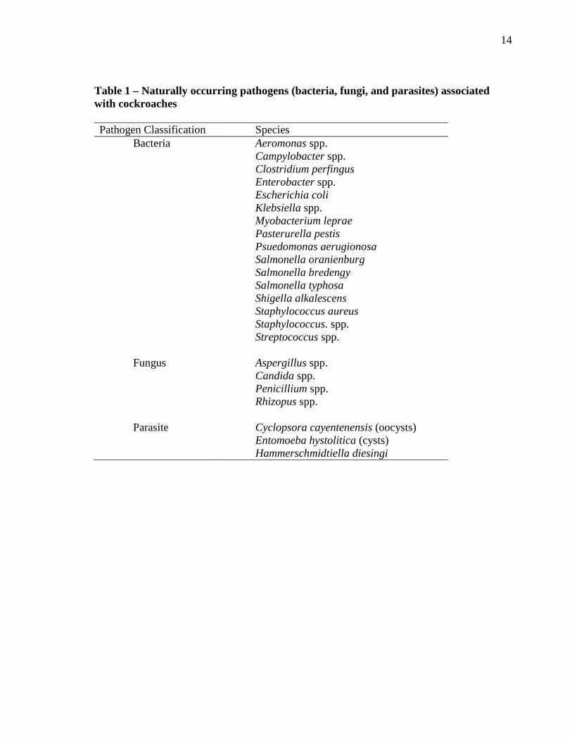
Bacterial Pathogens Associated with Cockroaches
Cockroaches are known to carry pathogens naturally, as seen in Table 1, but are
also known to transmit pathogens such as anthrax, cholera, diphtheria, pneumonia, tetanus,
and tuberculosis (Baumholtz et al. 1997). All of these pathogens can be used as
bioterrorism agents targeting animal or human populations.
Barcay (2004) implied that several disease outbreaks world-wide, such as
dysentery, hepatitis, and gastroenteritis, could be contributed to the spread of pathogens
through the environment by mechanical transmission of cockroaches. A few medically
important pathogens that are carried by P. americana include Campylobacter spp.,
Escherichia coli, Salmonella spp., Shigella spp., Staphylococcus spp., Streptococcus
spp., and a protozoan pathogen, Toxoplasma gondii (Barcay 2004, Graczyk et al. 2005).
Cockroach cuticle can harbor several gram-negative bacteria in the group
Enterobacteriaceae (Mpuchane et al. 2006b). Mpuchane et al. (2006b) suggested the
lack of gram-positive bacteria present on the cuticle could result from cockroach
secretions that inhibit gram-positive survival. Gram-negative bacteria fauna identified
from cockroach cuticle are similar for cockroaches collected from hospitals and
restaurant-type facilities (Elgderi et al. 2006). Fewer bacterial species and lower rates of
positive prevalence were determined for roaches collected in a residential area (Elgderi
et al. 2006). Fungi and yeasts (i.e. Aspergillus spp. and Candida spp.) have been found
on cuticular surfaces of cockroaches collected from intensive care units of a Brazilian
hospital (Lemos et al. 2006).
14
Table 1 – Naturally occurring pathogens (bacteria, fungi, and parasites) associated with cockroaches
Pathogen Classification Species
Bacteria Fungus Parasite
Aeromonas spp. Campylobacter spp. Clostridium perfingus Enterobacter spp. Escherichia coli Klebsiella spp. Myobacterium leprae Pasterurella pestis Psuedomonas aerugionosa Salmonella oranienburg Salmonella bredengy Salmonella typhosa Shigella alkalescens Staphylococcus aureus Staphylococcus. spp. Streptococcus spp. Aspergillus spp. Candida spp. Penicillium spp. Rhizopus spp. Cyclopsora cayentenensis (oocysts) Entomoeba hystolitica (cysts) Hammerschmidtiella diesingi
15
Salmonella has been found on American cockroaches with up to 7–10 d after
initial contact (Schoof and Siverly 1954). Under laboratory conditions, the pronotum of
P. americana inoculated with Salmonella enterica, serotype Oranienburg, produced
viable colonies up to 78 d after inoculation (Schoof and Siverly 1954). Salmonella
oranienburg is also transferred by American cockroach feces to human food sources.
Detection of the bacteria on food sources can last for several years after initial
inoculation (Barcay 2004). Many of these pathogens can result in gastroenteritis along
with other internal and external infections throughout the body, especially in areas with
open wounds or other environments favorable for bacterial growth (Barcay 2004). It is
evident that cockroaches provide a route of transmission for various pathogens.
Specific disease-causing pathogens commonly associated with cockroaches result
in gastro-intestinal related illnesses. Escherichia coli and Campylobacter spp.
transmission has been assumed to occur through mechanical transmission by
cockroaches and result in ailments such as diarrhea, abdominal cramps, and fever
(Altekruse et al. 1999, Zurek and Schal 2004).
Campylobacter species. Campylobacter are microphilic, curved, gram-negative,
non-spore forming motile bacteria (Yan et al. 2005). Campylobacter fetus (formerly
Vibrio fetus) is differentiated into three subspecies: C. fetus, C. interestinales, and C.
jejuni (Blaser et al. 1979). The last two subspecies have been detected in humans since
1947 with increasing annual frequency, but it was not recognized as a human pathogen
until the early 1970’s (Blaser et al. 1979, Butzler 2004). Laboratory tests perfected in
1973 differentiated between the three subspecies (Blaser et al. 1979). Campylobacter
16
spp. was not assumed to be a part of normal human bacterial fauna because it had only
been found in patients displaying symptoms such as diarrhea and fever (Blaser et al.
1979). A small infective dose makes Campylobacter spp. difficult to isolate as the
etiological agent for symptoms as common as fever and diarrhea. Campylobacter
enteritis results from Campylobacter spp. infections and was characterized by diarrhea,
abdominal cramps, malaise, fever, headache, and has a sudden on-set followed by a short
duration period (less than a week) (Blaser et al. 1979). In human patients with symptoms
of diarrhea, C. jejuni has been isolated as the etiological agent more than Shigella spp.,
Salmonella spp, or E. coli 0157:H7 (Blaser et al. 1979, Blaser 1997). Guillain-Barré
syndrome, a demyelinating disease resulting in neuromuscular paralysis, pulmonary
muscle deterioration, and death, has been linked to C. jejuni infections (Blaser 1997,
Sahin et al. 2002).
Diseases associated with this microorganism commonly result from ingesting
undercooked poultry, mishandling raw poultry, and cross-contamination of other
surfaces (i.e. this bacteria has been found to survive in exposed environments containing
oxygen on stainless steel and cotton dishtowel surfaces for over an hour), and survived
in untreated water sources (Yan et at. 2005). Contact with infected children,
consumption of unpasteurized milk and/or contaminated food products can result in the
manifestation of symptoms related to C. jejuni infections. Most U.S. citizens become
infected while traveling to foreign countries (Blaser et. al 1979, Blaser 1997).
Campylobacter jejuni is enteric in livestock such as cattle, swine, poultry,
companion animals (i.e. dogs and cats), and wild animals such as rodents and raccoons
17
(Blaser 1997, Sahin et al. 2002). An earlier study indicated a relationship between the
house fly, Musca domestica (L.) (Diptera: Muscidae), tenebrionid adults and larvae, and
cockroaches as mechanical vectors of C. jejuni in poultry houses (Sahin et al. 2002). An
additional link between the pathogen and humans is through cattle, sheep, and other
livestock which ingest pathogens from contaminated water sources (Blaser et al. 1979).
Consequently, human interactions with livestock increase the potential risk of
contamination.
Similar strains of Campylobacter have also been found to infect humans and
their companion animals, as evidenced by a Danish girl and her dog having the same
strain of quinolone-resitant C. jejuni (Damborg et al. 2004). Transmission from humans
to companion animals is demonstrated by the previous case discussed; however, the
mode of pathogen transmission remains uncertain. Arthropods may play a vital role in
the transfer of bacterial pathogens in such instances. Erythromycin is commonly used to
treat infections with alternatives such as fluoroquinolenes and tetracyclines, but there is
evidence that the usage of antibiotics in humans and animals used for consumption is
increasing, hence pathogens are becoming more resistant to such courses of treatments
(Blaser 1997).
Campylobacter jejuni is susceptible to oxygen in the atmosphere, which may
limit grown in moist locations such as livestock feed and water (Sahin et al. 2002).
Although, once chickens digest Campylobacter spp. and E. coli, the organisms may
develop in the field better than under ideal laboratory conditions (McGee et al. 2004).
18
Campylobacter spp. colonization increased 10,000 times that of laboratory growth
following expulsion from the digestive tract of poultry (McGee et al. 2004).
Escherichia coli. Escherichia coli are gram-negative, rod shaped bacteria with
specific strains considered important pathogens occurring in humans and veterinary
settings. The most common cause of enteric colibacillosis in piglets is E. coli (Zurek
and Schal 2004). In human cases, there are several strains with varying effects ranging
from mild fevers to hospitalizations and death, depending on the strain acquired.
Escherichia coli titers in the environment denote the level of fecal contamination (Le
Guyader et al. 1989, Rivault et al. 1994) Transmission of these organisms can follow an
unsuspected fecal-oral interaction, such as using a contaminated hand towel and then
touching food or the mouth area. One E. coli strain has been cited as one of the primary
causes of Traveler’s diarrhea for individuals visiting foreign countries lacking adequate
sanitation facilities (Nataro and Kaper 1998).
Escherichia coli 0157:H7 is a medically important strain initially reported in
1982. It can cause bloody diarrhea, hemolytic uremic syndrome (HUS), kidney failure,
and death (McGee et al. 2004). This strain of E. coli contains genes comparable to the
Shiga toxin (Tarr 1995). Escherichia coli 0157:H7 has had reported outbreaks in the
United States, Great Britain, and Canada with 20,000 infections and 100 deaths in the
United States (Michino et al. 1999). Mead et al. (1999) estimated 73,480 E. coli 0157:H7
infections with an additional 61 deaths in the United States.
Cattle act as a primary reservoir of E. coli 0157:H7 with 2-24% of their fecal
material contaminated with the pathogen (McGee et al. 2004). Cattle and other livestock
19
(i.e. turkeys) feces is contaminated with Campylobacter spp. and E. coli 0157:H7.
Infected fecal material provided a breeding ground for other insects such as filth flies
(Stomoxys calcitrans (L.) (Diptera: Muscidae), Tabanus spp. (L.) (Diptera: Tanabidae),
and Hydrotaea aenescens (Wiedemann) (Diptera: Muscidae) to acquire pathogens and
therefore becoming mechanical vectors (Szalanski et al. 2004). Outbreaks of E. coli
0157:H7 may result from ingestion of contaminated beef or direct contact with
contaminated cattle and/or their feces (McGee et al. 2004). The hide of cattle appears to
harbor several pathogens, including E. coli 0157:H7, which can contaminate the
carcasses of cattle (McGee et al. 2004). An E. coli 0157:H7 outbreak in Sakai City,
Osaka, Japan in 1996 involved 9,451 cases with 12 deaths (Michino et al. 1999). The
demographic of those infected was as follows: elementary school children; individuals at
child care facilities, nursing homes; an industrial facility; and individuals who ingested a
commercially prepared box lunch with unknown origins (Michino et al. 1999). This
infection was the result of white radishes carrying the pathogen, which correlates with
other studies indicating a presence of E. coli 0157:H7 on vegetables and fruits (Michino
et al. 1999). A more recent outbreak occurred from July–October 2007 in 10 states (IL,
KY, MO, NY, OH, PA, SD, TN, VA, and WI), resulting in 21 reported infections with
eight hospitalizations and four HUS patients from ingestion of contaminated pepperoni
on frozen pizza (CDC 2007).
Cockroaches could be possible mechanical vectors of nosocomial infections,
especially to patients in neonatal units, intensive care, and who are immunocompromised
patients (Fotedar et al. 1991, Gliniewick et al. 2003, Elgderi et al. 2006, Salehzadeh et
20
al. 2007). Nosocomial infections may result from pathogens on contaminated food, a
contaminated water supply, and/or unsanitary facilities, like bathrooms (Lemos et al.
2006). Supella supellectilium (Serville) have been found to carry opportunistic bacteria
species such as Enterobacter agglomerans, Escherichia adecarboxylata, Serratia
marcescens, and Serratia liquefaciens which cause secondary infections in hospitals (Le
Guyader et al. 1989). Salehzadeh et al. (2007) described hospitals infested with
cockroaches contained higher bacterial counts than those found residential areas. This
association of greater rates of bacteria may result from hospital environments being
more conducive to bacterial acquisition from sources of contaminated water, food, and
other objects along with safe harborage through contaminated areas. Multiple drug-
resistant bacterial strains of medical importance have been isolated from cockroaches in
many hospitals (Fotedar et. al 1991, Gliniewick et al. 2003, Elgderi et al. 2006,
Salehzadeh et al. 2007). Understanding the nature of pathogen transmission from urban
insect pests to humans could clarify the epidemiology of many unknown illnesses. The
epidemiology of potentially fatal pathogens needs to be thoroughly examined as they
relate to cockroaches.
Determining gene flow among populations collected in central Texas may allow
for a better understanding of how and if populations are segregated, or if there is a
single, unified population. Currently, a strong link between urban and forensic
entomology does not exist. Cases involving abuse or neglect for young children or
people in full-care facilities would rely on knowledge of both disciplines to successfully
determine biology, development data, and foraging behaviors of alleged species under
21
investigation. Pathogens are important because they cause medically important
infections and diseases within populations. Modes of transmission may be important in
identifying sources and dispersal of pathogens by arthropods. Analyzing the pathogen
fauna among cockroach populations collected in central Texas will help establish
diversity of pathogens carried on their exoskeleton. Also, spatial distribution of bacteria
species may indicate the origins of pathogens, acquisition by cockroaches, and distances
cockroaches are capable of spreading viable organisms.
Therefore, the objectives and hypotheses of this thesis are:
1. Analyze the gene flow among Periplaneta americana cockroach populations in
College Station, Texas (central Texas):
Ho: There is no significant difference in the genetic makeup of field
collected P. americana samples from discrete sites in central Texas.
Ha: There is significant and measurable gene flow among field collected
P. americana samples from discrete sites in central Texas.
2. Determine the epidemiology and/or spatial relationships of Escherichia coli and
Campylobacter spp. associated through mechanical transmission by Periplaneta
americana cockroach specimens in College Station, Texas (central Texas):
Ho: There is no geographic relationship for bacteria recovered among
field collected P. americana samples from discrete sites in central
Texas.
Ha: There are significant differences in the bacteria fauna among field
collected P. americana samples from discrete sites in central Texas.
22
CHAPTER II
GENE FLOW AMONG Periplaneta americana (BLATTODEA: Blattidae) IN
CENTRAL TEXAS
Introduction
Molecular techniques can be used to identify insect species. Polymerase chain
reactions (PCR) use a primer to selectively amplify a targeted sequence of DNA, which
can act as a species-specific marker used for identifications. Amplification length and
rate of success are based on quality and quantity of extracted DNA. PCR amplification
rates dropped by 91% when medium-length sequences (300-400 bp) were amplified,
versus short-length sequences (100-200 bp) (Franzten et al. 1998). Genetic material
primed for amplification may undergo damage, degradation, or are completely unable to
replicate during PCR due to small template DNA size, oxidative damage and/or
enzymatic breakdown of the sample (Taberlet et al. 1996, Franzten et al. 1998).
Eukaryotic rRNA is arranged with genes being separated by internal transcribed spacer
(ITS) regions, and non-transcribed spacer (NTS) regions. Genes usually occur in
repeating, tandem units and have NTS regions between repeating segments of RNA,
while ITS regions separate genes within each strand. Despite looking at the lesser of the
two variable spacer regions, ITS regions still can provide an ample amount of variation
to reveal a relatively moderate level of gene flow amongst the given cockroach
population in central Texas (Mukha et al. 2007).
23
Defining a population depends on several factors such as spatial distribution,
structures from which collections were made, ecological niches occupied by a
population, or the general bias of the collector(s) may contribute to the definition of a
“population.” Differences in allelic frequencies may also be used to distinguish
populations. Hypothetically, genetic variability decreases in populations secluded from
other populations (Cloarec et al. 1999). In regards to cockroaches (Order: Blattodea),
isolated populations may have limited gene fluctuation because of minimal migration
from outside populations contributing to the non-diverse gene pool (Mukha et al. 2007).
Only a few cockroaches are needed to establish a new population in a given area.
Mukha et al. (2007) identified three Blattella germanica (Linnæus) (Blattodea:
Blattidae) populations with substantial genetic differentiation, hence, isolated
populations separated between 15 and 115 km. In contrast, Cloarec et al. (1999)
analyzed isoenzymatic genetic markers from B. germanica populations from two French
cities (Rennes and Sète) approximately 900 km apart and demonstrated limited genetic
variation. Consequently, due to contrasting results in previous studies it is inconclusive
as to whether or not populations analyzed over distances are homologous.
Cockroaches can passively and actively disperse to new locales (Jobet et al.
2000). Gene flow may be caused by long range passive travel, i.e. people moving
location to location with boxes and other storage infested with cockroaches. The
similarity between populations may have resulted from the increased movement of
humans within cities when compared to the movement of humans between cities and
consequently increased transfer of cockroaches from one site to the next (Cloarec et al.
24
1999). Active movement appears to be confined to temperate climate zones when
alternative, ideal habitats are within close proximity (Cloarec et al. 1999). Schoof and
Siverly (1954) indicated a lack of dispersal among American cockroach, Periplaneta
americana (L.) (Blattodea: Blattidae), populations through sewer systems in Phoenix,
Arizona, USA. This inability to disperse may have resulted from the ideal habitat a
sewer system provided, including ample amounts of water, food, and harborage. It
appeared that when requisites for life were fulfilled the necessity to actively disperse
reduced.
Genetic variation among dispersing populations may result from various genetic
events. Genetic drift, founder effects, natural selection, migration, and gene flow are
some factors that might contribute to genetic variation (Jobet et al. 2000). Founder
effects occur more frequently in cockroach populations due to only required a limited
number of individuals to establish new populations (Cloarec et al. 1999). Cloarec et al.
(1999) suggested that populations within a defined geographical area (i.e. a city) were
more homologous than populations compared between greater distances (i.e. city to
city). Populations separated by variable distances retaining similar allelic frequencies
indicated a homologous correlation between populations, hence, gene flow (Cloareac et
al. 1999).
The objective of this study was to determine gene flow among populations
collected in central Texas. This information may allow for a better understanding of how
and if populations were segregated, or if there was a single unified population.
25
Materials and Methods
Sampling Technique for Cockroaches. Periplaneta americana (L.) were
collected within 50 m of neighboring urban structures in College Station, Texas (Table
A-1) and investigated for potential gene flow by phylogenetic analysis among the
collected population(s). Collecting sites on campus were selected from locations with the
highest cockroach populations based on preliminary trapping. Once locations were
established, three collecting containers were placed within a 1.83 m2 square at each
trapping location. Coordinates of each site were determined with a Gormin eTrex® Vista
Cx GPS unit (Garmin Ltd., Olathe, KS, USA). Additional samples from other following
cities in Texas were obtained from the Texas A&M University Insects in Human Society
(ENTO 322) Student Insect Collection including: Pleasanton, Del Rio, Bryan, and
Hempstead, Texas. The cockroaches used from the Texas A&M University Insects in
Human Society Student Insect Collection were preserved by pinning and stored in boxes
turned by the students. Data points for all cockroaches collected were uploaded to
Google Earth.
Containers used for collection were glass mason jars (430 ml) coated with
Elmer’s Acid Free Craft Bond (© Elmer’s Products, Inc., Columbus, Ohio, USA) and
rolled in Quickrete® Playsand (Quickrete® International, Inc., Atlanta, GA, USA),
according to Granovsky (1983). The top 2 cm of the jar opening was lined with H-E-B
brand petroleum jelly (H-E-B, San Antonio, TX, USA), and baited with approximately
51.76 ml beer (Miller Brewing Co., Milwaukee, WI, USA) and 7.04 g of H-E-B brand
white bread (H-E-B, San Antonio, TX, USA) for specimen collections (Barcay 2004).
26
Baited containers were placed in the field immediately after adding the beer/bread
mixture. Jars were set out prior to dusk and collected from the field after 8-12 h. Once
jars were collected from the field, cockroaches were stored in the freezer.
Cockroaches were collected from each jar and stored in individual plastic bags
(16.5 x 14.9 cm) with up to three plastic bags containing cockroaches from each site.
Collected specimens were stored in a freezer at -20oC until further analyses were
conducted. This method should not negatively influence bacterial colony (Szalanski et
al. 2004).
Molecular Analysis. Molecular probes were used to identify different
haplotypes within each cockroach sample. The hind femur from each specimen was used
for genetic analysis. The specific region providing the greatest amount of information
about the genetic flow involved the ITS1 region which is located between the 18S and
5.8S gene. Fragments of both the 18S and 5.8S gene, and the entire IST1 region made-up
the probe in identification of individuals and their genetic composition from the
provided specimens and has been demonstrated in recent studies (Mukta et al. 2007).
A 562-bp section of the nuclear 3’ portion of 18S rDNA, all of ITS1 region, and
the 5’ portion of 5.8S was amplified with the primers rDNA2 (5’-
TTGATTACGTCCCTGCCCTTT-3’) and rDNA 1.58S (5’-
GCCACCTAGTGAGCCGAGCA-3’) with a thermal cycler profile consisting of 40
cycles of 94°C for 45 s, 53°C for 1 min and 72°C for 1 min as described by Szalanski
and Owens (2003) (Vrain et al. 1992, Cherry et al. 1997). Amplified DNA from
individual cockroaches was purified and concentrated with minicolumns according to
27
the manufacturer’s instructions (Wizard PCRpreps, Promega). Samples were sent to The
University of Arkansas Medical School DNA Sequencing Facility (Little Rock, AR,
USA) for direct sequencing in both directions. Consensus sequences were derived from
both of DNA sequences from an individual with Bioedit 5.09 to verify nucleotide
polymorphisms (Hall 1999).
DNA sequences were aligned by CLUSTAL W (Thompson et al. 1994). The
distance matrix option of PAUP* 4.0b10 was used to calculate genetic distances
according to the Kimura 2-parameter model of sequence evolution (Kimura 1980,
Swofford 2001). Maximum likelihood and unweighted parsimony analysis on the
alignments were conducted by PAUP* 4.0b10 (Swofford 2001). Gaps were treated as
missing characters for all analysis. The reliability of trees was tested with a bootstrap test
(Felsenstein 1985). Parsimony bootstrap analysis included 1,000 resamplings with the
Branch and Bound algorithm of PAUP*. For maximum likelihood analysis, the default
likelihood parameters were used (HKY85 six-parameter model of nucleotide
substitution, empirical base frequencies with the exception of the transition/transversion
ratio, will be determined). These parameters were used to carry out a heuristic search by
PAUP* with a neighbor joining tree as the starting tree. Gene flow was evaluated
applying Mitochondrial DNA haplotypes aligned by MacClade v4 (Sinauer Associates,
Sunderland, MA). Haplotype distribution between populations, number of haplotypes,
number of unique haplotypes, haplotype diversity (h), and nucleotide diversity (pi) was
calculated with DNAsp v3.51 and Genealogical relationships among haplotypes were
28
constructed using TCS, with the method described by Templeton et al. (1992) (Rozas
and Rozas 1999, Clement et al. 2000).
Results
DNA sequencing of the ITS1 region from 52 cockroaches samples (Table A-1)
resulted an average size of 560 bp. There were 22 haplotypes observed from four Texas
counties with the 3 haplotype being the most common (Table 2). There were 25 unique
haplotypes. Del Rio, Texas is approximately 462 km from College Station; Pleasanton,
Texas has a distance of approximately 274 km from College Station, Texas; TX;
Hempstead, Texas is approximately 62 km away from College Station, Texas; Bryan,
Texas is a sister city to College Station, Texas separated by approximately 8 km.
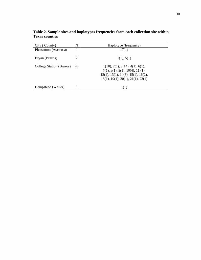
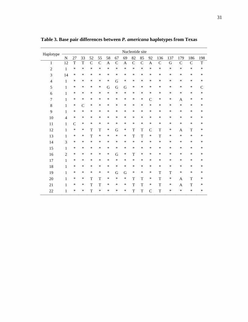
There were 41 polymorphic sites (Table 3). The average number of pairwise
nucleotide differences was 3.992. Out of the 22 haplotypes there were 25 singletons or
unique sequences. Nucleotide diversity, π, was 0.007, and the mean number of pairwise
nucleotide differences between haplotypes, k, was 3.992.Tajima’s D test of neutrality of
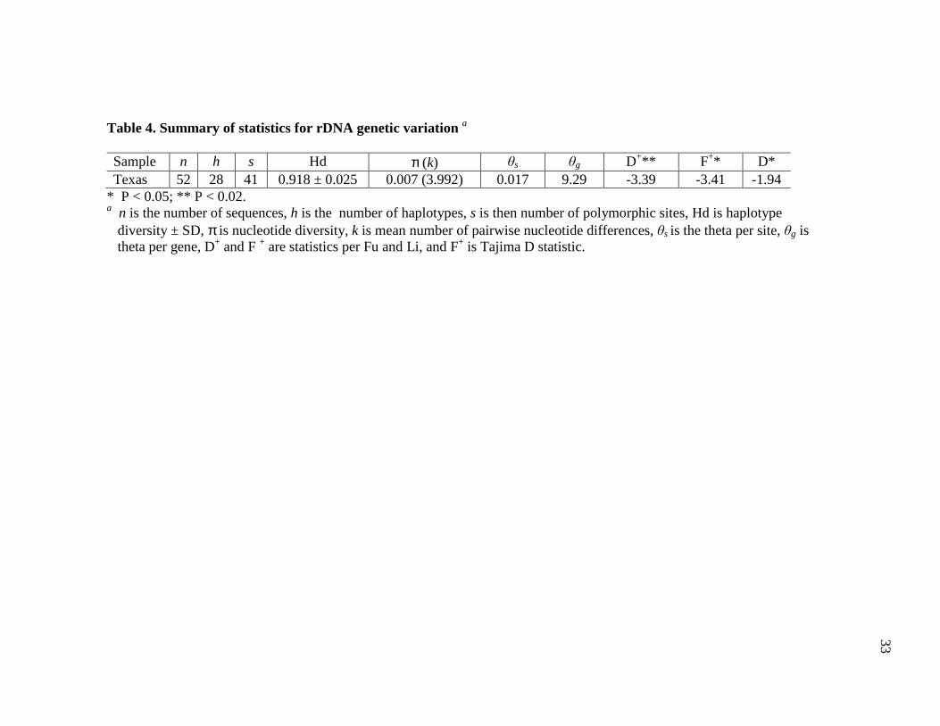
mutations against excess of recent mutations were not significant (Table 4).
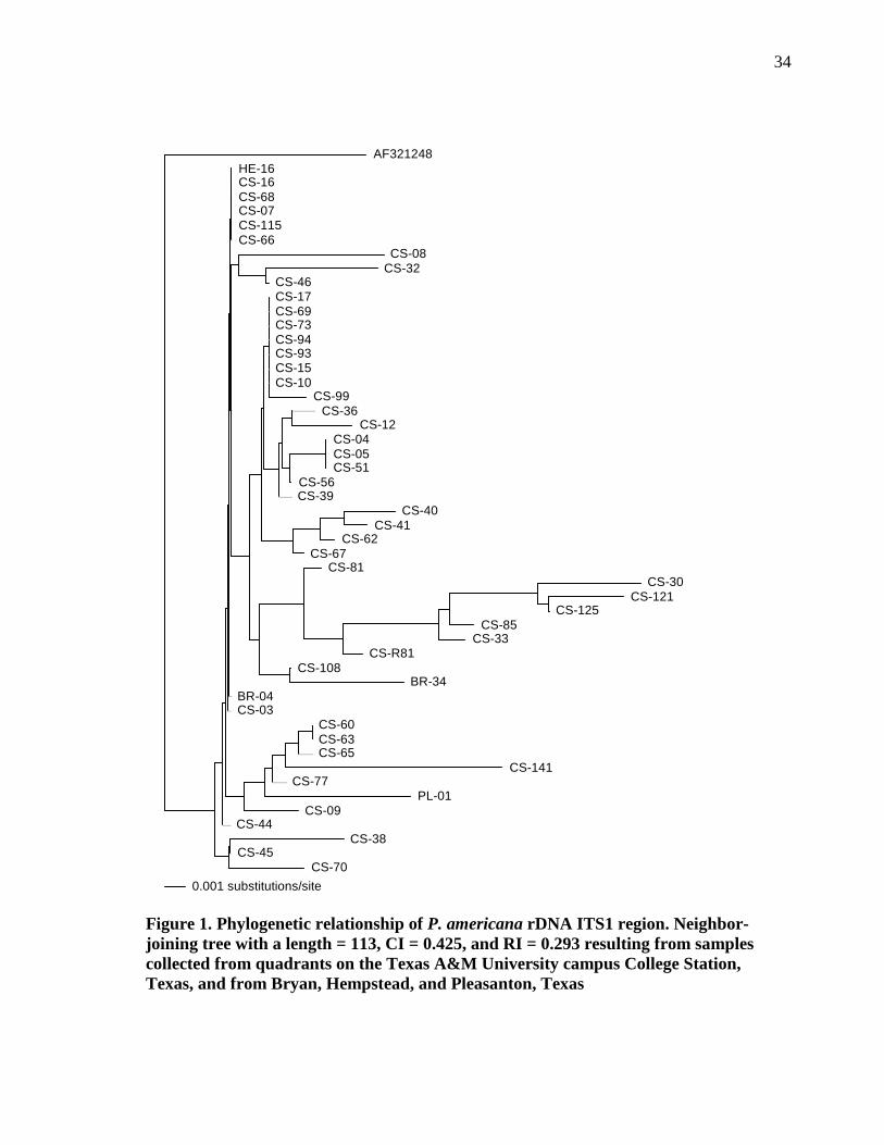
Applying P A U P * version 4.0b10 software, both Neighbor-Joining (NJ) and
Maximum Parsimony (MP) analyses were conducted. Results of the NJ tree using
uncorrected “P” distances is presented as an unrooted cladogram (Figure 1). For MP
analysis, parametric bootstrapping (50% majority-rule) with a full heuristic search was
employed for 1000 pseudoreplicates with a starting seed = 632095753. A total of 560
characters were evaluated with all characters equally weighted; 513 characters remained
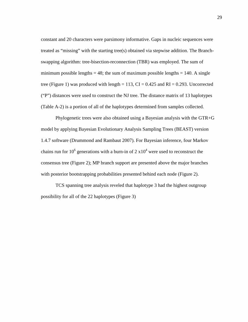
29
constant and 20 characters were parsimony informative. Gaps in nucleic sequences were
treated as “missing” with the starting tree(s) obtained via stepwise addition. The Branch-
swapping algorithm: tree-bisection-reconnection (TBR) was employed. The sum of
minimum possible lengths = 48; the sum of maximum possible lengths = 140. A single
tree (Figure 1) was produced with length = 113, CI = 0.425 and RI = 0.293. Uncorrected
(“P”) distances were used to construct the NJ tree. The distance matrix of 13 haplotypes
(Table A-2) is a portion of all of the haplotypes determined from samples collected.
Phylogenetic trees were also obtained using a Bayesian analysis with the GTR+G
model by applying Bayesian Evolutionary Analysis Sampling Trees (BEAST) version
1.4.7 software (Drummond and Rambaut 2007). For Bayesian inference, four Markov
chains run for 106 generations with a burn-in of 2 x10⁴ were used to reconstruct the
consensus tree (Figure 2); MP branch support are presented above the major branches
with posterior bootstrapping probabilities presented behind each node (Figure 2).
TCS spanning tree analysis reveled that haplotype 3 had the highest outgroup
possibility for all of the 22 haplotypes (Figure 3)
30
Table 2. Sample sites and haplotypes frequencies from each collection site within Texas counties City ( County) N Haplotype (frequency) Pleasanton (Atascosa) 1 17(1) Bryan (Brazos) 2 1(1), 5(1) College Station (Brazos) 48 1(10), 2(1), 3(14), 4(1), 6(1),
7(1), 8(1), 9(1), 10(4), 11 (1), 12(1), 13(1), 14(3), 15(1), 16(2), 18(1), 19(1), 20(1), 21(1), 22(1)
Hempstead (Waller) 1 1(1)
31
Table 3. Base pair differences between P. americana haplotypes from Texas
Haplotype Nucleotide site
N 27 33 52 55 58 67 69 82 85 92 136 137 179 186 198 1 12 T T C C A C A C C A C G C C T
2 1 * * * * * * * * * * * * * * *
3 14 * * * * * * * * * * * * * * *
4 1 * * * * * G * * * * * * * * *
5 1 * * * * G G G * * * * * * * C
6 1 * * * * * * * * * * * * * * *
7 1 * * * * * * * * * C * * A * *
8 1 * C * * * * * * * * * * * * *
9 1 * * * * * * * * * * * * * * *
10 4 * * * * * * * * * * * * * * *
11 1 C * * * * * * * * * * * * * *
12 1 * * T T * G * T T C T * A T *
13 1 * * T * * * * T T * T * * * *
14 3 * * * * * * * * * * * * * * *
15 1 * * * * * * * * * * * * * * *
16 2 * * * * * G * T * * * * * * *
17 1 * * * * * * * * * * * * * * *
18 1 * * * * * * * * * * * * * * *
19 1 * * * * * G G * * * T T * * *
20 1 * * T T * * * T T * T * A T *
21 1 * * T T * * * T T * T * A T *
22 1 * * T * * * * T T C T * * * *
32
Table 3. continued
Haplotype Nucleotide site
N 199 225 239 264 272 303 314 355 366 437 463 488 514 515
1 12 T A G G G G A A G C A C A A
2 1 * * * * * * T T * T * * C *
3 14 * T * * * * * * * * * * * *
4 1 * T * * * * * * * * * * * *
5 1 * T * * * * * * * * * * * *
6 1 * T * * * * * * * * * * * T
7 1 * * G * * * * * * * * * * *
8 1 * * * C * * * * * * * * * *
9 1 A * G * * * * * * * * * * *
10 4 A T * * * * * * * * * * * *
11 1 * * G * * * * * * * * * * *
12 1 A * G * * * * * * * * * * *
13 1 A * G * * * * * * * * * * *
14 3 * * G * * * * * * * * * * *
15 1 A * * * * * * * * * * * * *
16 2 * * * * * * * * * * * * * *
17 1 * * G * G C * * C * C * * *
18 1 * * * * * * * * C * * G * *
19 1 * * G * * * * * * * * * * *
20 1 * * G * * * * * * * * * * T
21 1 * * G * * * * * * * * * * *
22 1 * * G * * * * * * * * * * *
1
Table 4. Summary of statistics for rDNA genetic variation a Sample n h s Hd π (k) θs θg D+** F +* D* Texas 52 28 41 0.918 ± 0.025 0.007 (3.992) 0.017 9.29 -3.39 -3.41 -1.94
* P < 0.05; ** P < 0.02. a n is the number of sequences, h is the number of haplotypes, s is then number of polymorphic sites, Hd is haplotype
diversity ± SD, π is nucleotide diversity, k is mean number of pairwise nucleotide differences, θs is the theta per site, θg is theta per gene, D+ and F + are statistics per Fu and Li, and F+ is Tajima D statistic.
33
34
Figure 1. Phylogenetic relationship of P. americana rDNA ITS1 region. Neighbor-joining tree with a length = 113, CI = 0.425, and RI = 0.293 resulting from samples collected from quadrants on the Texas A&M University campus College Station, Texas, and from Bryan, Hempstead, and Pleasanton, Texas
AF321248HE-16CS-16CS-68CS-07CS-115CS-66
CS-08CS-32
CS-46CS-17CS-69CS-73CS-94CS-93CS-15CS-10
CS-99CS-36
CS-12CS-04CS-05CS-51
CS-56CS-39
CS-40CS-41
CS-62CS-67
CS-81CS-30
CS-121CS-125
CS-85CS-33
CS-R81CS-108
BR-34BR-04CS-03
CS-60CS-63CS-65
CS-141CS-77
PL-01CS-09
CS-44CS-38
CS-45CS-70
0.001 substitutions/site
NJ
34
P_americana_CSR77 P_americana_CSR60 P_americana_CSR65 P_americana_CSR63 P_americana_CS_R141 P_americana_Pleasanton_R1 P_americana_CS_R85 P_americana_CS_R33 P_americana_CS_R30 P_americana_CS_R121 P_americana_CS_R125 P_americana_CSR40 P_americana_CSR32 P_americana_AF321248 P_americana_CSR38 P_americana_CSR70 P_americana_CSR45 P_americana_CSR07 P_americana_CSR44 P_americana_CSR16 P_americana_Hempstead_R16 P_americana_CS_R3 P_americana_Bryan_R4 P_americana_CSR68
P_americana_ CS_R81 P_americana_CSR66
P_americana_CS_R115 P_americana_CSR46 P_americana_CS_R9 P_americana_CSR36 P_americana_CS_R94 P_americana_CS_R15 P_americana_CSR69 P_americana_CSR17 P_americana_CS_R10 P_americana_CS_R99 P_americana_CSR73 P_americana_CS_R12 P_americana_CSR51 P_americana_CSR05 P_americana_CSR04 P_americana_CSR56 P_americana_CS_R93 P_americana_CSR39 P_americana_CS_R7 P_americana_CSR67 P_americana_CSR41 P_americana_CSR62 P_americana_Bryan_R34 P_americana_CS_R108 P_americana_CS_R8 P_brunnea_AF321249 P_fulginosa_AF321250
Figure 2. Phylogenetic trees using a Bayesian analysis with MP branch support are presented above the major branches with posterior bootstrapping probabilities presented behind each node for samples collected from quadrants on the Texas A&M University campus College Station, Texas, and from Bryan, Hempstead, and Pleasanton, Texas
0.98
0.98
35
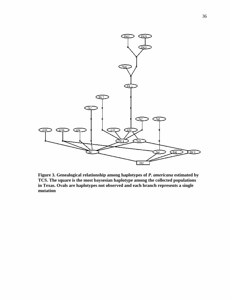
Figure 3. Genealogical relationship among haplotypes of TCS. The square is the most baysesian haplotype among the collected populations in Texas. Ovals are haplotypes not observed and each branch represents a single mutation
Figure 3. Genealogical relationship among haplotypes of P. americanaTCS. The square is the most baysesian haplotype among the collected populations in Texas. Ovals are haplotypes not observed and each branch represents a single
36
P. americana estimated by TCS. The square is the most baysesian haplotype among the collected populations in Texas. Ovals are haplotypes not observed and each branch represents a single
37
Discussion
The purpose of this study was to analyze the spatial distribution of P. americana
populations in an outdoor, urban environment and to determine the extent of gene flow
among the populations. This study attempted to determine genetic variability among P.
americana collected on Texas A&M University in College Station, Texas.
Genetic differentiation occurs between populations in diverse locations for all
organisms (Austin et al. 2004). Inward et al. (2007) suggested both the orders Isoptera
and Blattodea are related, thus their genes would coalesce to a single common ancestor.
It can be assumed that the individual lineages would comprise of similar genetic
material, thus specific gene regions would be applicable for amplification purposes in
both orders. Phylogenetic studies and population genetics performed on termites
commonly used the 16S region of the gene for amplification. The 16S region of the gene
was initially chosen as the amplification site in this study to determine variability among
cockroach populations collected on campus. During this study, the 16S gene region
amplification protocol commonly used in termite studies failed to amplify cockroach
DNA. Differing genetic compositions of the 16S gene region selected may have resulted
from evolution of separate ordinal lineages over time. The universal primers that
annealed for termite DNA simply would not work for cockroach DNA and/or the
annealing temperature may have been to low thus inhibiting annealing or too high which
would damage the primers or DNA. No matter the cause, there was no successful
amplification of the 16S gene region, so the ITS1 region was chosen for amplification
38
because of the availability of comparable sequences available on Genbank (National
Center for Biotechnology Information).
The ITS1 region functions in primary rRNA processing and has a higher rate
differentiation than the 18S gene region of rRNA (James et al. 1996). Mukha et al.
(2007) reported rRNA genes as being the most conserved among populations, while non-
transcribed spacer regions have the most variation, and transcribed spacer regions
between the two extremes. There are conflicting results when analyzing the ITS1 region
for genetic variability in insect populations. Szalanzski et al. (2008) determined a lack of
diversity in the nuclear gene region (ITS1 region) with high levels of differentiation
when examining the mitochondrial DNA region (16S gene) in Cimex lectularius (L.)
(Hemiptera: Cimicidae). The ITS1 region may have indicated low levels of diversity in
this species at this specific loci (Szalanzski et al. 2008). When the ITS1 region was
used, it failed to determine phylogenetic relationships between Reticulitermes termites
(Tripodi et al. 2006). On the other hand, there was sufficient variability in the ITS1
region used to identify diversity among Diabrotica (Coleoptera: Chrysomelidae) species
(Szalanski and Owens 2003). Additionally, Szalanski et al. (2000) demonstrated
differentiation between Nicrophorus americanus (Olivier) (Coleoptera: Silphidae) based
on results from the ITS1 region. The current study may have demonstrated biotic
homogenization within populations of P. americana based on data from the ITS1 region
(McKinney and Lockwood 1999).
Haplotypes are defined by at least a single nucleotide difference within the same
gene region between sequences thus identifying unique genes. Haplotype diversity is the
39
number of haplotypes compared to their relative frequency and determined the
probability of two sequences chosen from a population being different (Austin et al.
2004).) Tajima’s D is a statistical determination of the neutral mutation hypothesis in
natural populations (Tajima 1989). Positive values of D indicate population bottlenecks
while negative values of D suggest expansion of a population (Tajima 1989). Nucleotide
diversity (Pi) in populations assumed neutrality based on the infinite alleles model
(Austin et al. 2004).
Among the 52 sampled there were 22 haplotypes indicating a high amount of
variation in the population. TCS spanning tree analysis defined lineages from nuclear
markers which implied populations moderate levels of gene flow. The lack of isolated
populations was reconfirmed by maximum likelihood and Baysian phylogenetic
analyses.
Periplaneta americana samples from Bryan, College Station, Hempstead, and
Pleasanton, Texas were in a single clade, including P. americana sequence obtained
from Genbank (AF321248). Sequence comparisons reconfirmed speciation and revealed
moderate interbreeding between P. americana. The Smokey Brown cockroach
(Periplaneta fuliginosa) (Serville) and Brown cockroach (Periplaneta brunnea)
(Burmeister) were chosen as outliers because their sequences were available on
Genbank, AF321250 and AF321249, respectively, and are members of the genera as
American cockroaches. Comparing various species allowed a broader analysis of P.
americana to varying genetic sequences as a result of speciation within the same genera.
Comparing the 52 sequences amplified to 22 haplotypes suggested a moderate amount of
40
variation in the population based on nuclear markers. The lack of isolation indicated
interbreeding populations on campus. Differentiation of genetic variation based on
spatial distribution of P. americana populations indicated the success and ability of
breeding with independence among various populations.
Cockroaches might be capable of traveling between collecting sites through
various migration methods such as walking, climbing, dispersal via steam tunnels and
sewer systems throughout campus, and/or depositing their ootheca on materials
transferred by humans. Individuals from one collecting site were able to migrate to other
sites through any of the previously mentioned methods feasibly because the greatest
distance between collecting sites was 1.44 km. Migration of individuals to new locations
provided an opportunities for new genetic material to be introduced into a population
thus increasing some haplotypic diversity. Szalanski and Owens (2003) suggested lack
of variation among southern corn rootworm resulted from motility or population
expansion. Diversity among populations collected on campus most likely resulted from
the ability of cockroaches to travel successfully in urban environments and breed
effectively with cockroaches from other areas. Thus contributing to a constant influx of
genetic material into various populations. It remains unknown what degree of genetic
variability is observed among other cockroach species.
Genetic variability in populations can be achieved through genetic drift, genetic
flow, natural selection, and founder effects (Slatkin 1987). Genetic drift can affect
nuclear genes though the fixation of loci in various locations, but gene flow can impede
the permanent fixation of the alleles (Slatkin 1987). Lenormand (2002) determined gene
41
flow limited adaptation of genes to specific locations because new genes from outside
sources prevent loci from becoming fixed in the environment. Gene flow can prevent
speciation because introduced genetic material can be adapted for survival in a particular
environment differing from the population in which it emigrated (Slatkin 1987). Gene
flow is an indirect method of determining movement within a population. Bossart and
Prowell (1998) indicated the only method that definitively determined gene flow among
a population was through the use of genetic tags used to track movement which has had
successful in marine organisms. Cloarec et al. (1999) defined gene flow as the
movement of cockroaches over long distances by passive transportation, thus increasing
the rate of homogenization among the genetic material between populations. Results
found in the current study correlated with Cloarec et al. (1999) when they determined
German cockroach populations were not isolated in two French cities 900 km apart.
Mukha et al. (2007) determined three B. germanica populations found in farms separated
by 10-100 km and had three populations differentiated by rDNA markers, but they were
still not completely isolated. Species, including highly mobile organisms such as
cockroaches, disperse through an environment until geographical structures such as
oceans, deserts, and mountains impede expansion (Slatkin 1987).
Pesticide use is a common method implemented for supressing cockroach
populations. Although increased and prolonged use of the same pesticides can lead to
resistance. Lenormand (2002) suggested increased gene flow prevented resistance to
pesticides. Introduced genetic material may not have been exposed to similar classes of
pesticides thus specimens would be susceptible to novel pesticides locations. Natural
42
selection differs from genetic drift because not all alleles in different populations are
effected the same and gene flow has no consequence on the outcome of genetic variation
(Slatkin 1987). Founder effects display a portion of variation existing in the entire
population because it comprises of a small number of individuals that colonized a new
area (Cloarec et. al 1999 and Mukha et al. 2007).
To date, this study is the first using rDNA markers to identify spatial
relationships and gene flow among P. americana populations in the United States.
Future studies may analyze a broader range of genes including mitochondrial DNA to
determine if there are lineages formed by maternal genetic material. Also, analyzing
gene flow at several differing sequences within DNA may determine a more
comprehensive evolutionary lineage of divergences in cockroach populations.
43
CHAPTER III
EPIDEMIOLOGY AND SPATIAL RELATIONSHIPS OF BACTERIAL
SPECIES ASSOCIATED THROUGH MECHANICAL TRANSMISSION BY
Periplaneta americana (BLATTODEA: BLATTIDAE) IN CENTRAL TEXAS
Introduction
Arthropods transmit medically important pathogens to humans (Mullens and
Durden 2002). Cockroaches (Order: Blattodea) are important vectors of pathogens due
in part to their unsanitary lifestyle. Cockroach cuticle can harbor several
Enterobacteriaceae species (Mpuchane et al. 2006b). A few medically important
pathogens harbored by the American cockroach, Periplaneta americana (Linnæus)
(Blattodea: Blattidae) include: Campylobacter spp., Escherichia coli, Salmonella spp.,
Shigella spp., Staphylococcus spp., Streptococcus spp., and Toxoplasma gondii (Barcay
2004). Cockroaches also reported to transmit pathogens such as anthrax, cholera,
diphtheria, pneumonia, tetanus, and tuberculosis (Baumholtz et al. 1997). All of which
could be used as bioterrorism agents targeting animal or human populations.
Cockroaches could be competent carriers of nosocomial infection agents,
especially to patients in neonatal units, intensive care, and who are immunocompromised
(Fotedar et al. 1991, Gliniewick et al. 2003, Elgderi et al. 2006, Salehzadeh et al. 2007).
Nosocomial infections may result from pathogens in contaminated food or water, and/or
unsanitary facilities, like bathrooms (Lemos et al. 2006). Salehzadeh et al. (2007)
44
described cockroaches collected in hospitals to have greater bacterial counts than found
in residential areas. The association of higher bacteria rates results from hospital
environments being more conducive to bacterial acquisition from numerous sources such
as water, food, and/or harborage with contaminated objects. Multiple drug-resistant
bacterial strains of medical importance have been isolated from cockroaches in several
hospitals (Fotedar et. al 1991, Gliniewick et al. 2003, Elgderi et al. 2006, Salehzadeh et
al. 2007). Understanding the nature of pathogen transmission from urban insect pests to
humans could clarify the epidemiology of many unknown illnesses. The epidemiology
of potentially fatal pathogens needs to be thoroughly examined as they relate to
cockroaches.
Certain disease causing pathogens commonly associated with cockroaches
resulted in gastro-intestinal related illnesses. Pathogens, such as E. coli and
Campylobacter spp., commonly transmitted by cockroaches may be overlooked during
diagnosis of sudden ailments with symptoms being similar to food-borne illnesses,
including abdominal cramping, diarrhea, nausea, and fever.
Campylobacter are microphilic, curved, gram-negative, non-spore forming
motile bacteria (Yan et al. 2005). Campylobacter spp. are not part of a normal bacterial
fauna in humans but has been found in individuals displaying symptoms such as diarrhea
and fever (Blaser et al. 1979). In human patients with symptoms of diarrhea, C. jejuni
has been isolated to cause diarrhea-like symptoms more than Shigella spp., Salmonella
spp., and E. coli 0157:H7 (Blaser et al. 1979, Blaser 1997).
45
Diseases associated with Campylobacter spp. result from ingesting undercooked
poultry, mishandling raw poultry, and the cross-contaminating of surfaces, for example
this bacteria has been found to survive in exposed environments containing oxygen on
stainless steel and cotton dishtowel surfaces for over an hour, and can survive in
untreated water sources (Yan et at. 2005).
Campylobacter jejuni is enteric in livestock such as cattle, swine, poultry,
companion animals (i.e. dogs and cats), and wild animals such as rodents and raccoons
(Blaser 1997, Sahin et al. 2002). Campylobacter jejuni is atmospheric desiccation and
oxygen which inhibits growth in moist locations such as livestock feed and water (Sahin
et al. 2002). Consequently, human interactions with livestock increase the potential risk
of contamination.
Escherichia coli are gram-negative, rod shaped bacteria with specific strains
considered important pathogens of humans and animals. In human cases, there are
several strains that produce varying effects, ranging from mild fevers to hospitalizations
and death depending on the strain acquired in its host. Escherichia coli titers in the
environment corresponded with levels of fecal contamination (Le Guyader et al. 1989,
Rivault et al. 1994). Transmission of these organisms can follow an unsuspected fecal-
oral interactions, such as using a contaminated hand towel and then touching food or the
mouth area.
Escherichia coli 0157:H7 is a medically important strain initially reported in
1982 (McGee et al. 2004). It can cause bloody diarrhea, hemolytic uremic syndrome
(HUS), and death (McGee et al. 2004). E. coli 0157:H7 had reported outbreaks in the
46
United States, Great Britain, and Canada, with 20,000 infections and 100 deaths in the
United States (Michino et al. 1999).
Pathogens are medically important because of resulting infections and diseases
associated with human and/or animal populations. Modes of transmission are important
in identifying sources and dispersal of pathogens, such as dissemination by arthropods.
Bacterial strains identified were spatially analyzed in this study by determining where
various pathogens were in relationship to different cockroach populations. Analyzing the
pathogen fauna among populations in a given location could help establish the pathogen
diversity cockroaches carry on their exoskeleton based on locations. Also, spatial
distribution of bacterial fauna may indicate acquisition locations and distances
cockroaches are capable of spreading viable organisms.
The objective of this study was to analyze spatial distributions of E. coli and
Campylobacter spp. in relationship to different cockroach populations. This information
may determine the spatial distribution of bacterial fauna and identify locations with high
bacterial titers.
Materials and Methods
Sampling Technique for Cockroaches. Periplaneta americana (L.) were
collected within 50 m of neighboring urban structures in discrete locations in College
Station, Texas (Table A-1). Collecting sites on campus were selected from locations
with the highest cockroach populations during preliminary trapping. Once locations
were established, three collecting containers were placed within a 1.83 m2 square at each
47
trapping location. The north quadrant was approximately 0.29 km2. The central quadrant
was approximately 0.40 km2. The south quadrant was approximately 0.32 km2, and the
west quadrant had an area of approximately 0.58 km2. Coordinates of each site were
determined with a Gormin eTrex® Vista Cx GPS unit (Garmin Ltd., Olathe, KS, USA)
and data points uploaded to Google Earth.
Containers used for collection were glass mason jars (430 ml) coated with
Elmer’s Acid Free Craft Bond (© Elmer’s Products, Inc., Columbus, Ohio, USA) and
rolled in Quickrete® Playsand (Quickrete® International, Inc., Atlanta, GA, USA),
according to Granovsky (1983). The top 2 cm of the jar opening was lined with H-E-B
brand petroleum jelly (H-E-B, San Antonio, TX, USA) and baited with approximately
51.76 ml beer (Miller Brewing Co., Milwaukee, WI, USA), and 7.04 g of H-E-B brand
white bread (H-E-B, San Antonio, TX, USA) for specimen collections (Barcay 2004).
Baited containers were placed in the field immediately after adding the beer/bread
mixture. Jars were set out prior to dusk and collected from the field after 8-12 h. Once
jars were collected from the field, cockroaches were stored in the freezer.
Cockroaches were collected from each jar and stored in individual plastic bags
(16.5 x 14.9 cm), with up to three plastic bags containing roaches from each site.
Collected specimens were stored in a freezer at -20oC until further analyses were
conducted. This method should not negatively influence bacterial colony (Szalanski et
al. 2004).
Screening for Escherichia coli Activity. Media used for screening Escherichia
coli followed the manufacture’s recipe of 32.6 g /L of CHROMagar™ ECC media
48
(CHROMagar, Paris, France). Escherichia coli 0157:H7 specific media was made using
CHROMagar™ 0157 (CHROMagar, Paris, France) at a 29.4 g/L ratio.
Agar was poured into petri dishes (100 x 15 mm, VWR International, West
Chester, PA, USA) making approximately 20 petri dishes/500 ml media. Petri dishes
were divided into thirds and appropriately labeled for the specimen. Working under
sterile conditions, forceps were flame sterilized using 95% ethanol (EtOH) and cooled
prior to touching the cockroach to be plated. Dorsal and ventral sides of each cockroach
were plated within their designated areas. Once the cockroach was plated it was moved
to an isolated area, the forceps were sterilized using the aforementioned flaming
technique. The process was repeated for all P. americana collected.
Escherichia coli samples plated on CHROMagar ECC and CHROMagar 0157
were incubated in a Percival Environmental Chamber Model I36LLVL (Percival
Scientific, Inc., Perry, IA, USA) at 37oC for 24 – 48 h. Escherichia coli colonies were
counted by placing each plate on a white sheet of paper (21.6 x 27.9 cm) after
incubation. Blue colored colonies were identified as E. coli, red colonies were coliform
forming bacteria, and colorless colony forming units were non-coliform forming gram-
negative bacteria and counted. Screening for E. coli 1057:H7 followed the same
technique, but with positives indicated by a mauve coloration
Colonies that were positive for E. coli were stored in sterile 1.5 ml microtubes
with snap caps (VWR International, West Chester, PA, USA) in a 60% Tryptic soy agar
(Fisher Scientific, Pittsburg, PA, USA)/40% glycerin (Fisher Scientific, Fair Lawn, NJ,
USA), and frozen at -80oC, according to Hanahan et al. (1995).
49
Screening for Campylobacter species Activity. Campylobacter specific media
was made using the following recipe: 25 ml of defibrinated sheep blood (Colorado
Serum Co, Denver, CO, USA), 1 tube of antibiotic premix, 21.5 g BBL™ Brucella agar
(BD, Becton, Dickinson and Co., Sparks, MD, USA), and 500 ml distilled water.
Antibiotic premix was made by suspending 159.0 mg Cephalothin (MP Biomedicals,
LLC., Solon, OH, USA), 50.0 mg Trimethoprim Lactate (Research Products
International Corp., Prospect, IL, USA), 100.0 mg Vancomycin hydrochloride (Acros
Organics, Morris Plains, NJ, USA), 3.22 mg Polymyxin B (InvivoGen, San Diego, CA,
USA), and 20.0 mg Amphotericin B (Acros Organics, Morris Plains, NJ, USA) into100
ml distilled, sterile water. The total antibiotic premixture was divided into 20 tubes each
containing 5 ml aliquots, covered with parafilm (American National Can™, Greenwich,
CT, USA), and stored in a -20oC freezer.
Agar was poured into petri dishes (100 x 15 mm, VWR International, West
Chester, PA, USA) making approximately 20 petri dishes/500 ml media. Petri dishes
were divided into thirds and labeled for the appropriate specimen. Working under sterile
conditions, forceps were flame sterilized using 95% ethanol (EtOH), and cooled prior to
touching the roach to be plated. Dorsal and ventral sides of each cockroach were plated
within their designated areas. Once the cockroach was plated it was moved to an isolated
area, the forceps were sterilized using the aforementioned flaming technique. The
process was repeated for all P. americana collected.
Campylobacter spp. specific media was grown in an anaerobic environment for
96 h prior to checking for growth. An anaerobic environment was achieved by placing a
50
BD BBL ™ CampyPak ™ Plus Microaerophilic system envelope with Palladium
catalyst (Becton, Dickinson and Company, Sparks, MD, USA) in an acrylic canister
(17.8 x 12.7 cm, Oggi Co., Anaheim, CA, USA) with a chrome locking clamp with a
silicone gasket that sealed air tight. Campylobacter spp. selective media were removed
from the anaerobic environment after 96 h followed by identification and prevalence of
colonies.
Campylobacter spp. colonies were frozen at -80oC in a Tryptic soy agar and
15% (wt/vol) glycerin solution in sterile 1.5 ml microtubes with snap caps following the
methods of Wasfy et al. (1995).
Koch’s Postulates Experiments. Field collected cockroaches were plated on
CHROMagar ECC and CFU counts were made after 24 h. Cockroaches were sterilized
by shaking individuals in 20 ml of 95% ethanol for 2 min; the cockroaches were plated
on CHROMagar ECC and CFU counts were made after 24 h. Escherichia coli
ATCC25923 cells suspended in sterile saline underwent a 10-fold serial dilution until
log-5. Cockroaches were inoculated with 1 ml aliquots and plated. CFU counts were
made after 24 h.
Statistical Analysis. JMP Statistical Discovery software version 5.1 (SAS
Institute Inc., Cary, North Carolina) was used for the analysis of all results. Oneway
ANOVA, α = 0.05, was performed to analyze the mean total population numbers
collected and quadrant counts. A linear regression, α = 0.05, was performed to determine
the correlation between mean temperatures and mean population totals collected.
Oneway ANOVA, α = 0.05, was performed to analyze the mean number of bacteria
51
colony forming units for E. coli, coliform forming gram-negative, and non-coliform
forming gram-negative, and quadrant counts. Oneway ANOVA, α = 0.05, was
performed to analyze the mean number of bacteria colony forming units for E. coli,
coliform forming gram-negative, and non-coliform forming gram-negative; population
stage of development; and quadrant counts.
Results
Cockroaches (N =687) were collected from four designated areas, north,
central, south and west, from the Texas A&M University campus College Station,
Texas, Figure 4. The mean number of cockroaches collected from Jan–May 2008 was
3.67 ± 4.23 total (3.10 ± 3.31 nymphs and 0.56 ± 1.73 adults). The north quadrant had
the lowest mean of cockroaches collected with 1.86 ± 1.60 total (1.86 ±1.25 nymphs and
0.00 ± 0.65 adults). The central quadrant had a mean of 2.21 ± 1.13 cockroaches with
2.14 ± 0.88 nymphs and 0.07 ± 0.46 adults. The south quadrant had a mean of 4.05 ±
0.94 total (3.25 ± 0.74 nymphs and 0.80 ± 0.39 adults). The mean number of
cockroaches collected in the west quadrant was 7.29 ± 1.60 total (5.86 ± 1.25 nymphs
and 1.43 ± 0.65 adults). There was no significant difference (F = 2.746; df = 4, 160; P =
0.0542) between population means within quadrants (north, central, south, and west).
The mean number of total cockroaches from each location can be seen in Table 5.
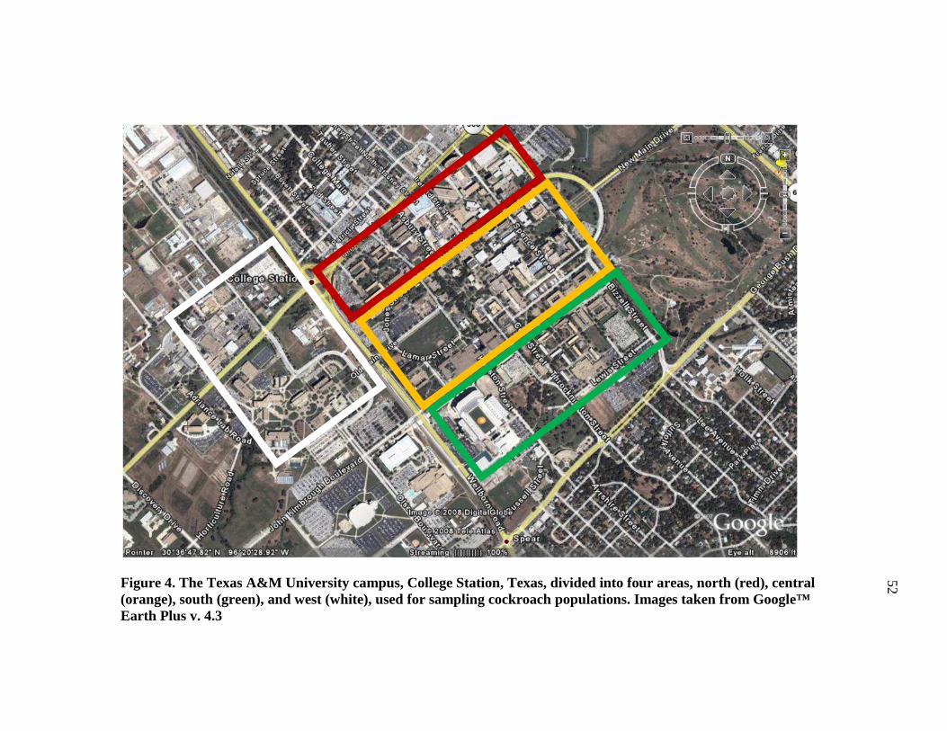
Figure 4. The Texas A&M University campus, College Station, Texas, divided into four areas, north (red), central (orange), south (green), and west (white), used for sampling cockroach populations. Images taken from Google™ Earth Plus v. 4.3
52
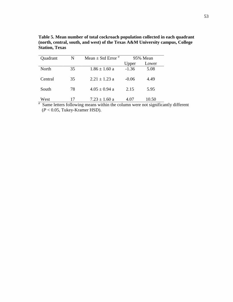
53
Table 5. Mean number of total cockroach population collected in each quadrant (north, central, south, and west) of the Texas A&M University campus, College Station, Texas Quadrant N Mean ± Std Error a 95% Mean
Upper Lower North 35 1.86 ± 1.60 a -1.36 5.08
Central 35 2.21 ± 1.23 a -0.06 4.49
South 78 4.05 ± 0.94 a 2.15 5.95
West 17 7.23 ± 1.60 a 4.07 10.50 a Same letters following means within the column were not significantly different
(P < 0.05, Tukey-Kramer HSD).
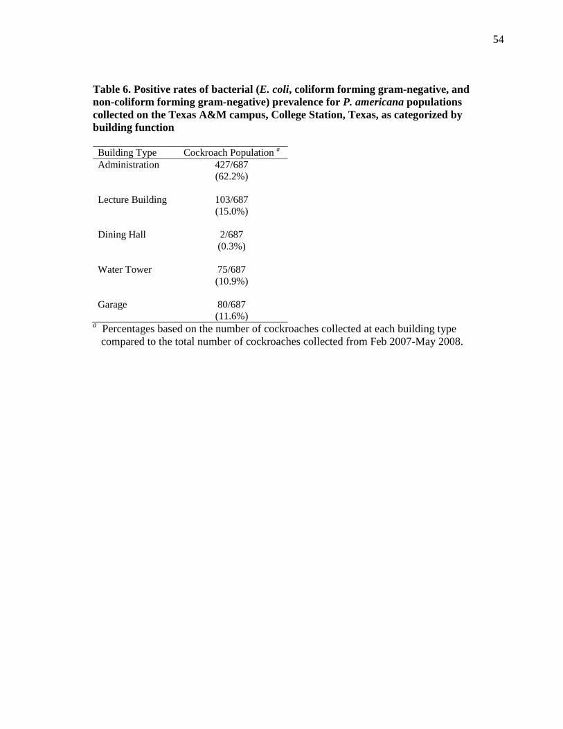
54
Table 6. Positive rates of bacterial (E. coli, coliform forming gram-negative, and non-coliform forming gram-negative) prevalence for P. americana populations collected on the Texas A&M campus, College Station, Texas, as categorized by building function Building Type Cockroach Population a Administration 427/687
(62.2%) Lecture Building 103/687
(15.0%) Dining Hall 2/687
(0.3%) Water Tower 75/687
(10.9%) Garage 80/687
(11.6%) a Percentages based on the number of cockroaches collected at each building type
compared to the total number of cockroaches collected from Feb 2007-May 2008.
55
There were five categories (Table 6) for the building and/or structures from
which cockroach populations were collected adjacent to: administration (primarily
offices, some classrooms, and vending machines); lecture buildings (primarily lecture or
research areas, some offices, and vending machines); dining halls (food establishments
on campus with the primary purpose of food and beverage distribution); water tower;
and garage. The prevalence of bacteria on cockroaches for each building type indicated
that administration buildings had the highest positive rate of cockroaches, while the
dining hall maintained the lowest rate of prevalence on P. americana populations.
Comparing the temperature to number of cockroaches collected indicated no
significant difference (F = 0.383; df = 5, 333; P = 0.5372) between the total population
means and the mean temperature (Table 7). There was no correlation between mean
temperature and total cockroach population means collected for College Station, Texas
locations including the quadrants designated on the Texas A&M University campus.
R2 values for north, central, south, west and College Station, Texas populations were,
0.14, 0.18, 0.07, 0.10, and 0, respectively (Table 7).
Koch’s postulates tested during this experiment resulted in a R2 value of 0.932.
The prevalence of the bacteria isolated from total populations collected indicated a high
prevalence (92.3%) of bacteria on the exoskeleton of P. americana (Figure 5). Bacterial
screening for E. coli resulted in a significant difference (F = 2.468; df = 4, 694;
P = 0.0437) between quadrants (Figure 6). There were also cockroaches that after plated
had too many bacterial colony forming units to count. The north quadrant had 1 E. coli,
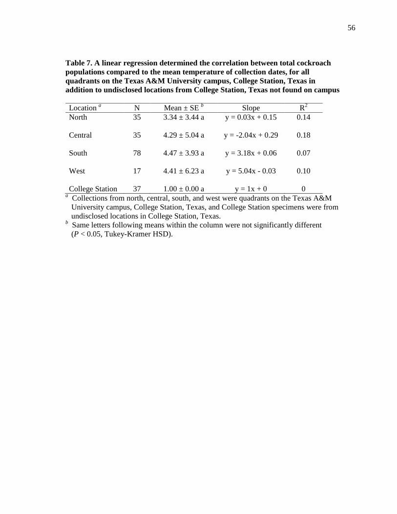
56
Table 7. A linear regression determined the correlation between total cockroach populations compared to the mean temperature of collection dates, for all quadrants on the Texas A&M University campus, College Station, Texas in addition to undisclosed locations from College Station, Texas not found on campus Location a N Mean ± SE b Slope R2 North 35 3.34 ± 3.44 a y = 0.03x + 0.15 0.14 Central 35 4.29 ± 5.04 a y = -2.04x + 0.29 0.18 South 78 4.47 ± 3.93 a y = 3.18x + 0.06 0.07 West 17 4.41 ± 6.23 a y = 5.04x - 0.03 0.10 College Station 37 1.00 ± 0.00 a y = 1x + 0 0
a Collections from north, central, south, and west were quadrants on the Texas A&M University campus, College Station, Texas, and College Station specimens were from undisclosed locations in College Station, Texas.
b Same letters following means within the column were not significantly different (P < 0.05, Tukey-Kramer HSD).
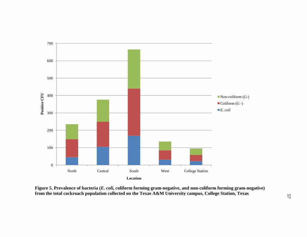
57
Figure 5. Prevalence of bacteria (E. coli, coliform forming gram-negative, and non-coliform forming gram-negative) from the total cockroach population collected on the Texas A&M University campus, College Station, Texas
0
100
200
300
400
500
600
700
North Central South West College Station
Pos
itive
CF
U
Location
Non-coliform (G-)
Coliform (G -)
E. coli
57
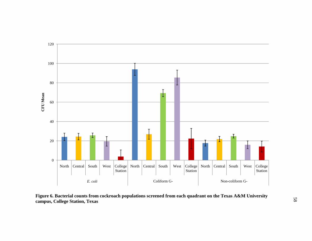
58
Figure 6. Bacterial counts from cockroach populations screened from each quadrant on the Texas A&M University campus, College Station, Texas
0
20
40
60
80
100
120
North Central South West College Station
North Central South West College Station
North Central South West College Station
E. coli Coliform G- Non-coliform G-
CF
U M
ean
E. coli
58
59
7 coliform forming colonies, and 0 non-coliform forming colonies. Central quadrant had
4 E. coli, 2 coliform forming colonies, and 5 non-coliform forming colonies. The south
quadrant had the most with 28 E. coli, 14 coliform forming colonies, and 11 non-
coliform forming colonies. The west quadrant had 0 E. coli, 2 coliform forming colonies,
and 1 non-coliform forming colony. Various locations in College Station resulted in 0
plates with too many to count (Table 8). Coliform forming bacteria were significantly
different (F = 24.728; df = 4, 665; P < 0.001) between quadrants, while non-coliform
forming gram-negative bacteria had no significant difference (F = 2.0573; df = 4, 680;
P = 0.0848) (Figure 6).
There were no significant difference (F = 0.0420; df = 2, 205; P = 0.8379)
between adult and nymph stages of cockroaches collected compared to number of
bacterial colony forming units of E. coli (Table 11). There were no significant difference
(F = 3.0748; df =2, 216; P = 0.0809) between adult and nymph stages of cockroaches
collected compared to number of bacterial colony forming units of coliform forming
bacteria. There were no significant difference (F = 0.0003; df = 2, 216; P = 0.987)
between adult and nymph stages of cockroaches collected compared to number of
bacterial colony forming units of non-coliform forming bacteria plated (Figure 7).
Screening for E. coli 1057:H7 and Campylobacter spp. yielded no positive
colony forming units for all of the samples screened (N = 724).
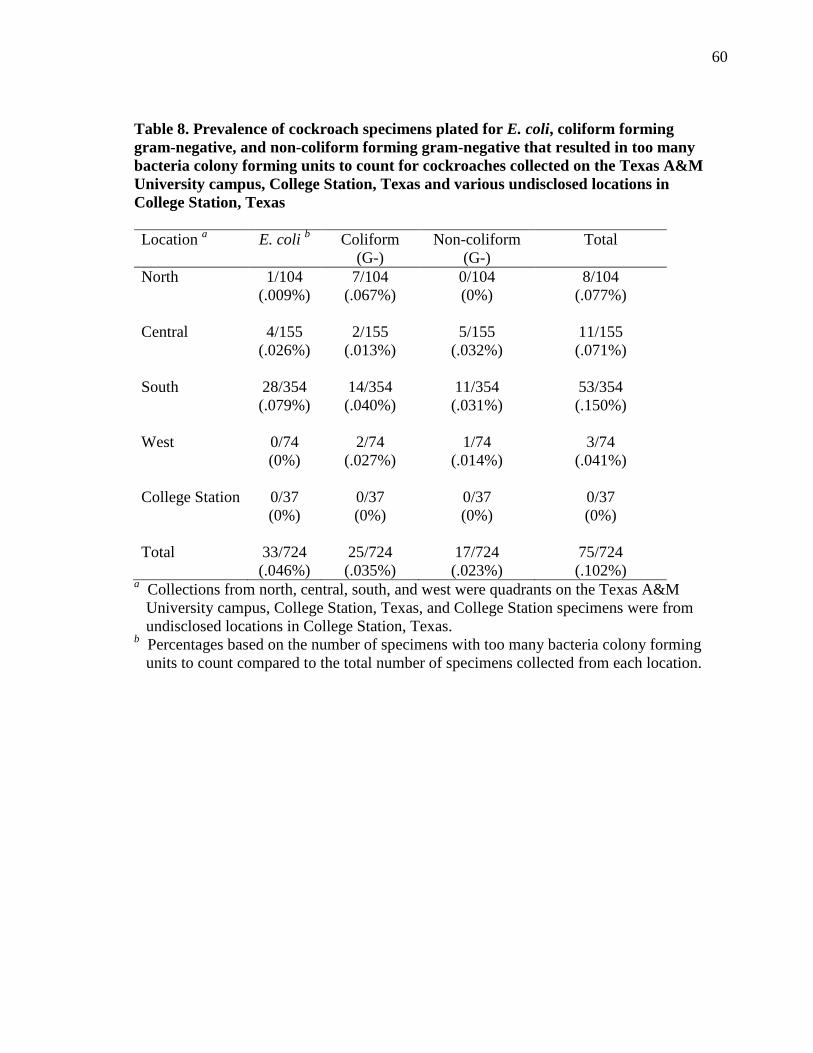
60
Table 8. Prevalence of cockroach specimens plated for E. coli, coliform forming gram-negative, and non-coliform forming gram-negative that resulted in too many bacteria colony forming units to count for cockroaches collected on the Texas A&M University campus, College Station, Texas and various undisclosed locations in College Station, Texas Location a E. coli b Coliform
(G-) Non-coliform
(G-) Total
North 1/104 (.009%)
7/104 (.067%)
0/104 (0%)
8/104 (.077%)
Central 4/155
(.026%) 2/155
(.013%) 5/155
(.032%) 11/155 (.071%)
South 28/354
(.079%) 14/354 (.040%)
11/354 (.031%)
53/354 (.150%)
West 0/74
(0%) 2/74
(.027%) 1/74
(.014%) 3/74
(.041%) College Station 0/37
(0%) 0/37 (0%)
0/37 (0%)
0/37 (0%)
Total 33/724
(.046%) 25/724 (.035%)
17/724 (.023%)
75/724 (.102%)
a Collections from north, central, south, and west were quadrants on the Texas A&M University campus, College Station, Texas, and College Station specimens were from undisclosed locations in College Station, Texas.
b Percentages based on the number of specimens with too many bacteria colony forming units to count compared to the total number of specimens collected from each location.
61
Figure 7. Comparison of bacteria counts for adults and nymphs in all quadrants collected on the Texas A&M University campus, College Station, Texas
0
20
40
60
80
100
120
140
160
Adult Nymph Adult Nymph Adult Nymph
E. coli Coliform Non-coliform
CF
U M
ean
E. coli
61
62
Discussion
The purpose of this study was to determine the amount and viability of bacteria
harbored by P. americana in an outdoor, urban environment by observing commonly
occurring and ubiquitous examples such as E. coli and Campylobacter. The Texas A&M
University campus provided an ideal location to conduct this experiment due to the
familiarity of the structures, buildings, and roadways. Outdoor collecting sites on
campus provided insight into American cockroach population within an artificial
environment. Outdoor locations were chosen because American cockroaches are
considered peridomestic pests and traveled freely between indoor and outdoor locations.
Also, limited building access at times when cockroaches were most active (at night)
made it difficult to maintain a regular collecting schedule of indoor facilities.
Cockroaches have increased activity when most of the buildings on campus were
either unoccupied or closed. This implies that cockroaches can move within a building
with limited restrictions, including foraging areas that are important in food preparation
and handling. The south campus was interesting to note because it was the only quadrant
that had sampling near a dining hall and as seen the density of cockroaches was the
lowest at this location. The low population numbers could result from effective and well-
maintained control strategies, or the facilities indoors provide adequate food, shelter, and
water thus eliminating the need for cockroaches to forage in outdoor locations.
This study is the first to focus on population densities and bacteria associated
with P. americana on a major university campus in the United States. Numerous studies
have collected cockroaches in urban situations, but typically inside schools, hospitals,
63
and homes. Granovsky (1983) suggested trapping rates increased when the external
surfaces of the collecting jar were coated with sand. Collecting containers were placed in
discrete locations and areas that could not be easily seen by the public, because the
disturbance of collecting jars could alter the population numbers. During the course of
this study, only a single site lost a jar by the following morning, because they were too
visible and accessible to the public due to their location next to a busy roadway. Also,
there were times when a single jar was knocked down at a site because of via weather,
wildlife or other unforeseen forces. A single jar being knocked over occurred nine times
over the duration of this study.
Other factors that were not considered prior to collecting was the presence of
feral animals on campus, including but not limited to frogs, snakes, cats, skunks, and
opossums, with the latter three possibly consuming bread from the collecting jars. The
only known incidence of wildlife having a known impact on the number of cockroaches
collected occurred on west campus at the Koldus drain (north) collecting site. A skunk or
skunk surfeit had burrowed under the concrete slab near the collecting location. It is
uncertain how long the animal(s) resided at this location, but on an early collecting trip
on 18 April 2008, their presence was made aware. After that day, the number of
cockroaches from that site declined. Skunks are known carnivores but have been found
to feed on insects when available (Crooks and Van Vuren 1995). It was not known what
caused the decline in the population. It may have been the skunk which had access to a
steady supply of cockroaches as a food sources, or it may have been a normal population
fluctuation due to the season and weather variability.
64
Collecting jars were also placed near areas which maintained requisites of life
such as food, water, and shelter. Jars were typically placed adjacent to a concrete or
brick barrier such as a wall, stairs, or a structure enclosing a flowerbed. When foliage,
typically ivy, was present, the collecting jars were placed below the surface of the
foliage and adjacent to the structures. Observations made throughout the study indicated
a high occurrence of cockroaches in ivy beds. The masonry structures coupled with ivy
or other plant life may have provided adequate coverage for cockroaches to move
without the threat of predation and with increased rates of foraging success. Also, the
masonry and concrete structures may have provided an artificial heat source; thus,
cockroaches could move next to buildings and forage for extended periods of time when
temperatures were less than ideal than if they were foraging in exposed environments.
Lin et al. (2007) demonstrated in Taiwan that heat output by concrete is stronger in the
winter months, with a mean temperature of 14-28oC, but the surface temperature of the
material correlated with ambient temperature. The winter months in Taiwan are
representative of the subtropical climates which are much higher than the temperatures
of winter period in a temperate area such as Texas. Based on observations during this
study, populations collected in areas with a concrete barrier and foliage maintained
higher population than those sites without such structures.
Over the duration of this study, it was interesting to note the lack of cockroach
species diversity being attracted to the collecting traps. There were three Periplaneta
fuliginosa (Serville) (Blattodea: Blattidae) collected amongst other P. americana
species. There were four B. germanica collected during the study near a residential
65
building on campus, but they were not intermingled with American populations. This
was unusual, since German cockroaches are typically indoor pests. It is possible that
there was a large population surge that could no longer be maintained by the area, or that
there were not enough food resources in the building. Either situation could lead
cockroaches to forage greater distances, thus going outside to find adequate food
sources. It is unknown which situation forced German cockroaches outdoors, but it was
unusual for B. germanica to forage outdoors.
There was no significant difference between collecting sites in each quadrant and
population of P. americana collected. Specific areas of campus did appear to yield
higher populations based on observational experience. Categorizing the collection sites
on a university campus into specific types such as food establishments, residential or a
hospital areas is difficult because most buildings include various types of establishments
within a single structure. Buildings on campus usually include food resources like
vending machine, coffee bar, full-scale dining area, lecture hall, and residential areas.
Residential buildings on campus contained food because they serve as the primary living
quarters for many people on campus.
The administration buildings maintained the highest outdoor populations, while
dining halls had the least number of cockroaches collected. The dining halls may have
had adequate control methods in effect to efficiently reduce population numbers.
Alternatively, there may have been sufficient resources inside the buildings which failed
to drive populations outside to forage. Control strategies may be less stringent in areas
where food preparation is not the primary focus, such as in administration and lecture
66
buildings. Haines and Palmer (1955) determined that P. americana was a predominant
species in sewer systems with low population densities indoors and around the home;
although the restrooms of indoor facilities maintained the highest population numbers.
Overall, the building type does not play a significant role in the population densities of
cockroaches. The assumption can be made that the same applies for an area such as a
university where cockroaches were ubiquitous in the environment.
Pai et al. (2003) determined that adult populations of P. americana and B.
germanica were significantly higher than nymph populations collected in hospitals,
which fails to correspond with data found in this study. There were no significant
differences between adult and nymph populations collected around campus. This
difference between studies may result from a difference in collection techniques or that
the Pai et al. (2003) study was conducted indoors, from a single structure (hospitals)
type, while the current study exploited various collecting locations and their outdoor
structures. No significant difference between adult and nymph collections may have
indicated a well-established population on campus, as well as a lack of control methods
to reduce nymphal population numbers, or there may be a naturally higher frequency of
nymphs during the collecting periods.
American cockroaches can take up to one year to reach maturity, and can live up
to three years. Their indoor counterpart, the German cockroach, can reproduce several
times a year, and has a lifespan of 200-300 d (Barcay 2004). It appears that integrated
pest management strategies are not reaching breeding and/or foraging sites on a routine
basis. The lack of a maintained control schedule could indicate why there were various
67
ages of cockroaches (nymphal instars to adults) collected on a consistent basis from all
locations throughout the collection period.
The effect of temperature on population numbers was important to consider
because it was generally assumed between 10-35oC, resulted in a decline of cockroach
activity, thus influencing the numbers acquired during collecting periods (Murphy and
Heath 1983). There was no difference between mean temperatures and the total
population means collected. This implied that overall temperature does not play a
significant factor, population means collected but observation data suggested at lower
temperatures (<10oC) population means were different from populations collected at
higher temperatures. This corresponded with Murphy and Heath (1983) concerning
cockroach activity and temperature.
Prior to collecting, it was hypothesized that population means would increase as
the temperatures rose. The lack of a relationship between temperature and population
indicated another factor may be influencing populations that were not accounted for
during this study. Cockroach populations, based on observational data decreased, when
the minimum temperatures at night were cooler.
Ambient temperatures in Texas fluctuate between December and May because of
seasonal transitions. The collecting period followed an erratic pattern with rising and
falling temperatures, coupled with periods of intense rainfall. For example, during the
last five days in February there was a day with a high of 29oC followed by a low of -1oC
two days later. The rapidly changing temperatures likely influence foraging behaviors
and overall activity. Traps used during this experiment were not designed to collect
68
specimens during rainy conditions, thus population numbers were minimal during these
time periods. Based on observational data, days with rainfall had lower numbers of
cockroaches collected than days without rainfall.
Spatial distribution of natural population is typically patchy. Resource levels
fluctuate over time in individual locations, thus population numbers will also change
over time indication a patchy distribution (Roughgarden 1977). Population fluxes are
normal because collecting cockroaches from outside was coincidental with weather.
Sometimes there is an abundance of cockroaches in a single location, and the next day
there may be none at the same location. Population surges may result from rainfall, food
availability, an overabundance of water in sewer systems, and/ or external weather
conditions. There is limited data correlating the number of cockroaches collected from
outside populations and weather conditions.
Establishing bacteria amounts carried by cockroaches is important because they
can act as potential disease agent carriers. Lipsitch and Moxon (1997) defined virulence
as the capability of organisms to infect or damage the host. Virulence and
transmissibility has been theorized in vertebrate animals, including humans, in which
virulence and transmissions of pathogens may not be directly related (Lipsitch and
Moxon 1997). The pathogen may be in a different part of a host body than where
symptoms are being displayed. Symptoms may result in parts of the body not in the path
of the organisms, and symptoms may result from the immune response of the host to the
threat of pathogens instead of the presence of the actual pathogen (Lipsitch and Moxon
1997). This theory of unrelated virulence and transmissibility may not apply to
69
invertebrates in the same methods as it applies to vertebrates. The exoskeleton of insects
provided protection against invading organisms, but also provided surfaces on which
pathogens may reside. Harboring pathogens on the exterior portion of the body may
increase the potential of viability of pathogens, unlike vertebrates where pathogen entry
into the body occurs with few barriers. It may be difficult to determine which
cockroaches are diseased because pathogens have less opportunity to enter their body
because of the exoskeleton, thus the physiology and behavior of the insect may not be
influenced.
Escherichia coli and Campylobacter spp. are common bacteria that cause gastro-
intestinal illnesses in humans. Escherichia coli was prevalent on surfaces contaminated
by fecal matter and can last anywhere from 1.5 h-16 mo on dry, inanimate surfaces
(Kramer et al. 2006). Scott and Bloomfield (1990) determined E. coli remained viable on
laminate surfaces up to 4 h, and that the bacteria could transfer from contaminated
surface to other objects such as fingertips, stainless steel, or cloth. Bacteria transfer
occurred at the highest rate when a contaminated piece of cloth contacted fingertips,
after contact E. coli was detected up to 48 h after initial contact (Scott and Bloomfield
1990). Kitchens maintained the highest numbers of E. coli resulting in part from poor
sanitary habits after handling contaminated foods such as chicken. Cockroaches, if
indoors, are prone to walk across surfaces that have been wiped down by a potentially
contaminated cloth. In outdoor environments, E. coli has been found to survive up to 20
d the wood shavings of a farm structure (Bale et. al 1993). Indoors, the organisms were
detected up to 21 h on a contaminated piece of paper (Bale et al. 1993). Due to their

70
ubiquitous nature, cockroaches can acquire bacteria from most surfaces in-or-out of
doors, so long as bacteria are present in the environment. Cockroaches can act as
potential carriers of pathogens in the surrounding areas.
Contamination rates of cockroaches compounded with their gregarious behavior
could provide a mode for pathogens to spread to surfaces having direct contact with
food. During this study, 51.7% of all cockroaches trapped were contaminated with E.
coli. This was the lowest percentage of positive bacteria out of all the cockroaches
screened for colony forming units. Despite having the lowest percentage of prevalence,
one out of every two cockroaches on campus was carrying E. coli. A comparison was
made to determine if the life stage (adult or nymph) made an impact on bacteria
associations with the cockroaches and found there to be no significant difference.
Escherichia coli can be found on both internal and external surfaces of
cockroaches (Rivault et al. 1994). The current study concurred with the Le Guyader al.
(1989) study of gram-negative bacteria amounts not having a significant difference
between adults and nymphs. Despite the stigma of cockroaches being filth laden, Bell et
al. (2007) indicated cockroaches spending at least half of their time grooming and
removing foreign objects from their body. The amount of time spent cleaning is
inadequate because of contamination of the habitat and the capability to become re-
inoculated with pathogens present in the environment. The ability to harbor bacteria on
internal and external surfaces provides multiple means of pathogen transmission. In
addition to direct contact with surfaces, cockroaches can disseminate internal organisms
via defecation and/or regurgitation.

71
Compared to previous studies made indoors, the presence of bacteria on
cockroaches appears to correlate with other studies with positives rates of bacteria in
Ghana, France, and Taiwan (Agbodaze and Owusu 1989, Rivault et al. 1994, Pai et al.
2004). Overall, 92.3% of cockroaches collected from outdoor locations on campus
carried gram-negative bacteria on their cuticular surfaces. When compared to another
pest cockroach, the German cockroach, Pai et al. (2005) determined there was no
significant difference between P. americana and B. germanica incident rates of positive
growth of bacterial colonies on the integument and the gut. Although, P. americana had
significantly higher rate of gram-negative colonies than B. germanica (Pai et al. 2005).
Periplaneta americana can harbor more gram-negative bacteria from outside sources
such as sewage, soil, contaminated water, and garbage, than B. germanica acquires from
inside sources.
A previous study indicated cockroaches harbored bacteria present in the
surrounding environment, as opposed to introducing new pathogens into the
environmental fauna (Rivault et al. 1993). During this study, it was assumed
cockroaches were mechanically transmitting pathogens obtained in the environment and
were capable of traveling while harboring these bacteria. Koch’s postulates were tested
and demonstrated the ability of cockroaches to transfer bacterial species. This creates a
public health concern if cockroaches inoculated with bacteria from outside migrated
indoors and transmitted pathogens to sterile surfaces, such as areas in the kitchen.
Chaichanawongsaroj et al. (2004) indicated E. coli levels on cockroaches coincided to E.
coli levels in the environment. Buildings with the most to least amounts of bacteria were
72
as follows: hospitals, food establishments, and residential areas thus cockroaches in
hospitals maintained the highest levels of E. coli (Chaichanawongsaroj et al. 2004).
Rivault et al. (1993) discussed that not all bacteria would be able to survive on surfaces
that a cockroach made contact with. Under proper conditions such as proper humidity,
specific bacteria species can develop at successful rates. Specific food items can provide
bacteria with enough nutritional resources and humidity to grow. This causes concern
because food that is eaten raw such as fruits, vegetables, pastries, and breads may be
cross-contaminated with disease-causing organisms. After retrieving traps, it was
interesting to note the condition of the bread in each jar. Traps containing cockroaches
usually had bread that appeared to be moldy or have black or green spots on it while the
bread in jars without cockroaches lacked these discolorations.
Kopanic et al. (1994) determined a single cockroach contaminated with
Salmonella typhimurioum could infect up to ten other cockroaches within a 24 hour
period in a 1.1 L jar. Theoretically, a single cockroach can contaminate an entire area
given an adequate period of time. In the current study, the time when each cockroach
entered the collecting vessel is unknown, thus making the rate or occurrence of cross-
contamination difficult to determine. Kopanic et al. (1994) previously studied
cockroaches with known inoculated amount placed into a container which resulted in
variable rates of cross-contamination. During this study, the collection jar was 2.3 times
larger than the container used in the previous study, and cockroaches were in traps for
half of the duration of the Kopanic et al. (1994) study. Therefore, cockroaches in this
study may have all been exposed to limited cross-contamination. Data indicated bacteria
73
were present on a single roach while other cockroach(es) were absent of the bacteria
within the same jar.
An alternative may be that under stress the cockroaches defecated or regurgitated
food into the slurry of bread-beer mixture, thus providing a solution for the remaining
cockroaches to become cross-contaminated with bacteria from other cockroaches. A
downside to the methods used for this experiment was that all cockroaches from each
individual jar at each site were placed in a single plastic container prior to freezing. This
may have provided another means of cross-contamination. Potential spreading of
pathogens among the specimens may have been prevented if each cockroach was stored
in individual containers. It would be unrealistic for a single person to collect each
cockroach individually as it was trapped at all of the locations. The methods described in
this paper were sufficient to determine population numbers and bacterial counts with the
least amount of cross-contamination possible.
Data indicated that collection locations were significantly different E. coli,
coliform forming gram-negative bacteria were significantly different while there was no
significant difference between non-coliform forming gram-negative bacterial species and
collecting locations. It was interesting to note differences among collected populations
and prevalence of bacteria, despite collecting sites being up to 1.44 km apart. A
significant difference may indicate the environment of various collecting locations
having differing compositions of bacteria. It is possible that the values for E. coli were
not significantly difference for each quadrant even though the p-value indicated a
significant difference. There were 75 specimens that resulted with too many bacteria
74
colony forming units to count. These numbers should not have affected the overall
significant difference between populations, quadrants, and bacteria species because the
number of too many to count bacteria colony forming units were proportional to initial
rates of prevalence among populations collected on campus. Populations collected in the
south quadrant had significantly different numbers of E. coli colonies when compared to
other locations. It was interesting to note that this was the only location that was
collected near a garage. No differences for non-coliform forming gram-negative bacteria
among collected populations implies that cockroaches may have obtained bacteria from
common means throughout campus, such as soil in the flowerbeds or a common water
source any of which may have been contaminated with bacteria.
A common water source that may have been easily accessible to all specimens is
through the sewer systems. Periplaneta americana may have traveled from one area of
campus to another through various methods of transportation, including but not limited
to steam tunnels, vehicles via infested materials, or physical movement by individual
roaches. Cockroaches are capable of migration by ground movement, climbing vertical
surfaces, swimming, and some limited flight capabilities (Bell et al. 2007). Jackson and
Maier (1955) determined through capture and release experiments that cockroaches
could travel through the sewer up to 107 m. Dispersal can occur rapidly because
cockroaches are capable of traveling at speeds of 0.44-1.5 ms-1 on a horizontal plane and
can become bipedal at their highest speeds (Full and Tu 1991). It is possible cockroaches
remained in locations until resources were depleted and then dispersed in search of food.
75
Nocturnal habits of cockroaches allowed efficient ground movement but not
without the threat of predation by nocturnal wildlife. Safer dispersal methods could be
achieved by traveling through sewer systems. Incidentally, sewers are ideal locations for
acquisition of pathogens by cockroaches. Also, students, faculty, staff, and visitors may
unknowingly transfer cockroaches and/or ootheca to new locations from other residences
or buildings. Increased populations resulted in an increased potential to infest new areas,
hence establish new sites for pathogen acquisition and dispersal.
Several other studies agitated collected cockroaches in saline and used the
solution to plate and determine bacteria numbers. Methods used during the current study
directly plated cockroaches onto media. Humphrey et al. (1995) determined that directly
placing samples on media would increase the rate of prevalence as opposed to placing
the samples into a diluent. Direct plating may replicate what happens outside of
laboratory conditions in a more realistic manner. It does not seem feasible that
cockroaches will be shaken in a solution and then the solution be poured onto a food or
food preparation area. Cockroaches typically walk over surfaces or may stop to feed on a
food resource, thus inoculation periods vary from surface to surface. Direct plating may
replicate P. americana cuticle indiscriminately contacting surfaces and with the possibly
of pathogen transmission.
All specimens collected were negative for E. coli 0157:H7. Presence of this
pathogen usually occurs in livestock area because cattle and sheep act as reservoirs for
the pathogen (McGee et al. 1997). There were no locations on campus that housed
livestock which were regularly sampled for cockroach populations. This may have
76
contributed to why there were no positives for E. coli 0157:H7. Although, E. coli
0157:H7 has been found on vegetables and soft cheese such as feta (Ramsaran et al.
1998, Michino et al. 1999). Escherchia coli 0157:H7 has also been found viable on dry
surfaces such as stainless steel and was detected up to 60 d after inoculation (Maule
2000). Due to pathogenicity of the organism, negative results on all of the specimens
tested were an optimal result.
Campylobacter spp. is normally found in the intestinal gut of animals and
humans. It has microaerophilic properties which make growth of the organism
susceptible to desiccation from oxygen. Altekruse et al. (1999) determined that survival
of the organisms outside of the gut is poor. Unfavorable conditions can result in
Campylobacter spp. to enter a stage where it is viable but nonculturable (Murphy et al.
2006). During this state, the organisms change their morphological characteristics from a
spiral to a coccoid, but it can still result in infections and can colonize a host gut
(Murphy et al. 2006). Growth in substances such as water, litter, and feed are typically
not common in ambient temperatures because of the desiccation associated with being
exposed to atmospheric oxygen (Sahin et al. 2002). Transmission of Campylobacter spp.
to humans occured through consumption of under cooked meats such as beef, pork, and
poultry or consumption of contaminated water sources or non-pasteurized milk (Sahin et
al. 2002). The bacteria are sensitive to drying out and when suspended in a liquid that
has been allowed to dry, Campylobacter spp., will not be detected after four hours
(Humphery et al. 1995). Although, if the organisms is in a solution such as blood, it will
test positive after four hours (Humphrey et al. 1995). Chynoweth et al. (1998)
77
determined C. jejuni could grow in sterile stream water under aerobic conditions for up
to 55 d. Campylobacter spp. have been found on stainless steel, ceramic tile, cotton
dishtowels, and other surfaces commonly associated with food preparation situations for
over an hour (Yan et al. 2005). Campylobacter infections can result from surface water
being contaminated by fecal material or through sewage contamination (Murphy et al.
2006).
The cockroaches in this study failed to have any positive rates of prevalence
when screened for Campylobacter spp. The organism does not grow at temperatures
below 30oC, which could indicate why there were no positives (Park 2002).
Susceptibility to cooler temperatures and exposure to oxygen makes it difficult for
Campylobacter spp. to successfully grow outside of a host body, hence was a possibility
as to why there was no positive colony forming.
Overall, this study displayed the wide distribution of cockroach populations on
campus and their ability to indiscriminately inhabit areas within an urban environment.
Pathogen acquisition and dissemination of gram-negative bacteria, such as E. coli, was
prevalent on campus but without detection of the highly pathogenic strain of E. coli
1057:H7. Also, there was a lack of Campylobacter spp. growth from cuticular plating
which may have resulted from undesirable conditions required to sustain colony growth.
78
CHAPTER IV
DISCUSSION AND CONCLUSIONS
The purpose of the first portion of this study was to analyze the spatial
distribution of P. americana populations in an outdoor, urban environment and
determine gene flow among the population. This study determined genetic variability
among P. americana collected on a major university campus. The 16S region of the gene
was initially chosen as the amplification site in identifying variability among cockroach
populations. 16S gene regions are commonly amplified during termite studies failed to
amplify and thus there was no discernable differentiation between individuals. The ITS1
region was chosen for amplification because there was no successful amplification at the
16S gene region. The current study demonstrated gene flow within populations of P.
americana based on differentiation identified from the ITS1 region.
Among the 52 sequences amplified there were 22 haplotypes indicating a high
amount of variation in the population. Haplotypes isolated during this study will be
made available on Genbank. TCS spanning tree analysis identified discrete lineages
from nuclear markers which demonstrates interbreeding of P. americana populations.
The lack of population structure was reconfirmed by neighbor-joining and Bayesian
phylogenetic analyses. Periplaneta americana samples from Bryan, College Station,
Hempstead, and Pleasanton, Texas were in a single clade including P. americana
sequence obtained from Genbank (AF321248). Samples of P. fuliginosa were in a
79
separate clade along with the sequences from Genbank for a P. fuliginosa (AF321250)
and P. brunnea (AF321249) supports species identification with this marker. Comparing
the 52 sequences amplified to 22 haplotypes suggests a high amount of variation in the
population based on nuclear markers. Genetic variation based on spatial distribution of
P. americana populations indicated the success and ability of breeding with
independence with the individuals collected on campus, hence representing a free-living,
interbreeding population.
Genetic variation of populations occurs through genetic drift, genetic flow,
natural selection, and founder effects (Slatkin 1987). Lenormand (2002) determined
gene flow limited adaptation of genes to specific locations because new genes from
outside sources prevent loci from becoming fixed in the environment. Introduced genetic
material adapted for survival in a particular environment can differ from the population
from which it emigrated, thus preventing speciation via gene flow (Slatkin 1987). Gene
flow acts as an indirect method of determining movement within a population.
This study is the first to date using nuclear markers in identifying spatial
relationships and gene flow among P. americana populations on a university campus in
North America. Future studies may analyze a broader range of genes including
mitochondrial DNA to determine distinct lineages formed by mtDNA. Also, analyzing
gene flow at various sequences may determine a more comprehensive evolutionary
lineage of divergences in cockroach populations. Our study is a step towards the in-
depth analysis of the phylogenetics of cockroaches. The data obtained during this
experiment can contribute a small portion to the overall analysis of a comprehensive
80
phylogenetic study of cockroaches. There was a failure to reject the null hypothesis for
the first objective of this study because there was no significant difference in the genetic
make-up of field collected P. americana samples from locations in central Texas.
The second objective of this study determined the amount and viability of
bacteria harbored by P. americana in an outdoor, urban environment. There was no
significant difference between collecting sites and population means of P. americana
collected. Based on observational experience, specific areas of campus appeared to yield
higher populations. No significant difference between adult and nymph populations may
have indicated a population that is well established on campus, as well as the lack of
control methods implemented to reduce population numbers. Cockroaches are virtually
ubiquitous in urban environments. Overall, the function of buildings adjacent to
collection locations did not play a significant role in population densities of cockroaches.
There was no correlation between temperature and mean populations (total, adults, and
nymphs) when analyzed. Thus affirming populations collected are not correlated to
mean temperatures.
Establishing bacteria amounts carried by cockroaches is important because they
can act as potential disease agent carriers. Escherichia coli and Campylobacter spp. are
common causes of gastro-intestinal illnesses in humans. Despite the stigma of
cockroaches being filth laden, Bell et al. (2007) implicated cockroaches spending at least
half of their time grooming and removing foreign objects from their body. The amount
of time spent cleaning is inadequate because of the pathogen contamination rates of in
the areas cockroaches frequent and their ability to easily become re-inoculated.
81
Therefore, cockroaches can mechanically transfer pathogens present in the environment.
The ability to harbor bacteria on internal and external surfaces provides multiple means
of pathogen transmission. In addition to direct contact with surfaces, cockroaches can
disseminate internal organisms via defecation and/or regurgitation.
Data indicated when collection locations were compared to bacteria amounts
there were significant differences between locations and E. coli; there were significant
differences between locations and coliform forming gram-negative bacteria; and there
were no significant differences between non-coliform forming bacterial species and
collecting locations. It was interesting to note both significant differences and no
significant differences among collected populations and prevalence of bacteria, despite
collecting sites being up to 1.44 km apart. No difference among populations could
demonstrate cockroaches obtained bacteria from a universal substance used throughout
campus, such as soil or a common water source, any of which may have been
contaminated with fecal material. Sewer systems are easily accessible to specimens
throughout campus and may have been a source of contamination.
Cockroaches are capable of migration via ground movement, climbing vertical
surfaces, swimming, and they have some limited flight capabilities (Bell et al. 2007).
Periplaneta americana may have traveled from one area of campus to another through
various methods of transportation. Steam tunnels, automobiles via infested materials, or
physical movement by individual roaches may have been a few methods of dispersal
through the environment.
82
Despite having the lowest percentage of prevalence, one out of every two
cockroaches on campus was carrying E. coli pathogens. All specimens collected were
negative for E. coli 0157:H7. Due to pathogenicity of the organism, negative results on
all of the specimens tested were an optimal result. Cockroaches collected and screened
during this study failed to have any positive rates of prevalence of Campylobacter spp.
Susceptibility to cooler temperatures and exposure to oxygen impedes Campylobacter
spp. growth successfully outside of a host body, hence a possibility as to why there was
no positive colony forming unit. Whole body extractions or fecal remains would be more
likely be used to observe Campylobacter spp. in future studies.
Overall, this study displayed the ubiquitous distribution of cockroach populations
on campus and their ability to indiscriminately inhabit areas within an urban
environment. Gram-negative bacteria acquisition and dissemination of organisms, such
as E. coli, was prevalent on campus but the highly pathogenic strain of E. coli 1057:H7
was not isolated. Also, there was a lack of Campylobacter spp. growth from cuticular
plating which may have resulted from undesirable conditions required to sustain colony
growth.
Data from this study suggested cockroach’s ability to mechanically transfer
pathogens. Insects are known to harbor and transfer pathogens in the environment, thus
having potentially deleterious health consequences on animal and/or human populations.
Dipteran species have been identified as mechanical vectors of pathogens. Houseflies,
Musca domestica (L.) (Diptera: Muscidae) can carry Vibrio chlorerae, E. coli, and
Yersinia pseudotuberculosis (Fotedar 2001, Zurek et al. 2001, De Jesús et al. 2004). In
83
1898, Xenopsylla cheopis (Rothschild) (Siphonaptera: Pulicidae) was reported as the
vector of the etiological agent of plague (Yersinia pestis) (Burroughs 1947, Inglesby et
al. 2000). The consequences of plague outbreaks throughout history are well known, but
more current concerns associated with the disease involves aerosolation of the bacteria
for use as a biological weapon (Inglesby et al. 2000). This technique could apply to other
pathogens with numerous insects acting as mechanical transmitters and having
successful rates of dissemination. Stomoxys calcitrans (L.) (Diptera: Muscidae), Ades
agypeti (L.) (Diptera: Culicidae), and Ae. taeniorhynchus (Wiedemann) (Diptera:
Culicidae) can spread Bacillus anthracis which is also a cause for concern if used as a
biological weapon (Turell and Knudson 1987). The null hypothesis for the second study
was rejected for E. coli and coliform forming bacteria because there was a significant
difference between P. americana samples collected from various locations in central
Texas. Analysis of non-coliform forming bacteria resulted in a failure to reject the null
hypothesis because of there was no geographic relationship for bacteria recovered
among field collected P. americana samples in central Texas.
Zurek and Schal (2004) suggested the capability of German cockroaches to
mechanically transmit the porcine pathogen E. coli F18 through fecal material.
Cockroaches also have been suggested as a vector in sever acute respiratory syndrome
(SARS) which has reached epidemic levels in the past five years (Wu et al. 2004, Lau et
al. 2005). SARS results from contact with individuals infected with a coronavirus, thus
direct contact serving as the primary route of transmission (Wu et al. 2005). Although,
cockroaches have been hypothesized to act as vectors when there was no contact
84
between infected and uninfected individuals (Wu et al. 2005). It is evident that
cockroaches are capable of transmitting various disease-causing organisms by
mechanical transmission. The absolute vectoring capability of cockroaches still remains
unknown because experiments involving etiological agents occurring naturally or those
which can be harmful to large populations, such as anthrax, have yet to be thoroughly
tested.
There were limitations to information and implications resulting from data
collected throughout the study. For example, populations collected for this study were
only collected from selected areas of campus. The genetic data is consistent throughout
the sequences amplified from the various locations on campus. The degree of variation
of the population may be interpreted differently had other markers such as mtDNA or
microsatellite segments been implemented to differentiate individuals. Also, P.
americana were only collected in outdoor environments. It is possible that indoor
populations have different bacterial faunas because of differing bacteria present in the
environment. Future studies could include sampling larger areas for collecting, including
health care facilities, like hospitals and nursing homes, or places associated with
children, such as day cares and schools. It would be interesting to analyze diversity of
bacteria from various locations including gram-positive bacteria, which were not
screened for during this study. Also, identifying specific strains of pathogens through
genetic analysis could allow for better mapping of distances and dispersal of disease-
causing agents throughout the environment.
85
REFERENCES CITED
Agbodaze, D., and S.B. Owusu. 1989. Cockroaches (Periplaneta Americana) as carriers of agents of bacterial diarrhea in Accra, Ghana. Cent. Afr. J. Med. 35: 484-486.
Altekruse, S.F., N.J. Stern, P.I. Fields, and D.L. Swerdlow. 1999. Campylobacter
jejuni-An emerging food borne pathogen. Emerg. Infect. Dis. 5: 28-35. Austin, J.W., A.L. Szalanski, and M.T. Messenger. 2004. Mitochondrial DNA
variation and distribution of the subterranean termite genus Reticulitermes (Isoptera: Rhinotermitidae) in Arkansas and Louisiana. Fla. Entomol. 87: 473-480.
Bale, M.J., P.M. Bennett, J.E. Beringer, and M. Hinton. 1993. The survival of
bacteria exposed to desiccation on surfaces associated with farm buildings. J. Appl. Bacteriol. 75: 519-528.
Barcay, S.J. 2004. Cockroaches, pp. 121-215. In A. Mallis (ed.), Handbook of pest
control, 9th ed. GIE Media, Cleveland, OH. Baumholtz, M.A., L.C. Parish, J.A. Witkowski, and W.B. Nutting. 1997. The
medical importance of cockroaches. Int. J. Dermatol. 36: 90-96. Bell, W.J., L.M. Roth, and C.A. Nalepa. 2007. Cockroaches: ecology, behavior, and
natural history, pp. 17-66. The John Hopkins University Press: Baltimore, MD. Benecke, M. 2001. A brief history of forensic entomology. Forensic Sci. Int.120: 2-14. Benson, E.P., and P.A. Zungoli. 1997. Cockroaches, pg.123-202. In A. Mallis (ed.),
Handbook of pest control, 8th ed. Mallis Handbook and Technical Training Company, Cleveland, OH.
Bennett, K.E., K.E. Olson, M. de L. Muñoz, I. Fernandez-Salas, J.A. Farfan-Ale, S.
Higgs, W.K. Black IV., and B.J. Beaty. 2002. Variation in vector competence for dengue 2 virus among 24 collections of Aedes aegypti from Mexico and the United States. Am. J. Trop. Med. Hyg. 67: 85-95.
Blaser, M.J. 1997. Epidemiologic and clinical features of Campylobacter jejuni
infections. J. Infect. Dis. 176: S103-S105. Blaser, M.J., I.D. Berkowitz, F.M. LaForce, J. Cravens, L.B. Reller, and W-L.L.
Wang. 1979. Campylobater Enteritis: Clinical and epidemiologic features. Ann. Intern. Med. 91: 179-185.
86
Bossart, J.L., and D.P. Prowell. 1998. Genetic estimates of population structure and gene flow: limitations, lessons and new directions. Trends Ecol. Evol. 13: 202-206.
Brenner, R.J. 2002. Cockroaches (Blattaria), pp. 29-44. In G. Mullens and L. Durden
(eds.), Medical and Veterinary Entomology. Elsevier Science, NewYork. Burroughs, A.L. 1947. Sylvatic plague studies: The vector efficiency of nine species of
fleas compared to Xenopsylla cheopis. J. Hyg. 45: 371-396. Butzler, J.-P. 2004. Campylobacter, from obscurity to celebrity. Clin. Microbiol. Infect.
10: 868-876. CDC (Center for Disease Control). 2007. Investigation of outbreak of human
infections caused by E. coli 1057:H7. http://www.cdc.gov/ecoli/2007/october/103107.html.
Chaichanawongsaroj, N., K. Vanichayatanarak, T. Pipatkullachat, M. Polrojpanya,
and S. Somkiatcharoen. 2004. Isolation of gram-negative bacteria from cockroaches trapped from urban environment. Southeast Asian J. Trop. Med. Public Health. 35: 681- 684.
Cherry, T., A.L. Szalanski, T.C. Todd, and T.O. Powers. 1997. The internal
transcribed spacer region of Belonolaimus (Nemata: Belonolaimidae). J. Nematol. 29: 21-29.
Chynoweth, R.W., J.A. Hudson, and K. Thom. 1998. Aerobic growth and survival of
Campylobacter jejuni in food and stream water. Lett. Appl. Micrbiol. 27: 341-344.
Clement, M., D. Posada, and K.A. Crandall. 2000. TCS: A computer program to
estimate gene genealogies. Mol. Ecol. 9: 1657-1659. Cloarec, A., C. Rivault, and M.L. Cariou. 1999. Genetic population structure of the
German cockroach, Blattella germanica: Absence of geographical variation. Entomol. Exp. App. 92: 311-319.
Crooks, K.R., and D. Van Vuren. 1995. Resource utilization by two insular endemic
mammalian carnivores, the island fox and the island spotted skunk. Oecologia 104: 301-307.
Damborg, P., K.E.P. Olsen, E.M. Nielsen, and L. Gaurdabassi. 2004. Occurrence of
Campylobacter jejuni in pets living with human patients infected with C. jejuni. J. Clin. Microbiol. 42: 1363-1364.
87
De Jesús, A.J., A.R. Olsen, J.R. Bryce, and R.C. Whiting. 2004. Quantitative contamination and transfer of Escherichia coli from foods by houseflies, Musca domestica L. (Diptera: Muscidae). Int. J. Food Microbiol. 93: 259-262.
Denic, N., D.W. Huyer, S.H. Sinal, P.E. Lantz, C.R. Smith, and M.M. Silver. 1997.
Cockroach: The omnivorous scavenger. Potential misinterpretations of postmortem injuries. Am. J. Forensic Med. Pathol. 18: 177-180.
Drummond, A.J., and Rambaut, A. 2007. BEAST: Bayesian evolutionary analysis by
sampling trees. BMC Evol. Biol.7:214 Elgderi, R.M., K.S. Ghenghesh, and N. Berbash. 2006. Carriage by the German
cockroach (Blattella germanica) of multiple-antibiotic-resistant bacteria that are potentially pathogenic to humans, in hospitals and households in Tripoli, Libya. Ann. Trop. Med. Parasitol. 200: 55-62.
Felsenstein, J. 1985. Confidence limits on phylogenies: An approach using the
bootstrap. Evolution 39: 783–791. Fischer, O.A., L. Matlova, L. Dvorska, P. Svastova, and I. Pavlik. 2003. Nymphs of
the Oriental cockroach (Blatta orientalis) as passive vectors of causal agents of avian tuberculosis and paratuberculosis. Med. Vet. Entomol. 17: 145-150.
Fotedar, R. 2001. Vector potential of houseflies (Musca domestica) in the transmission
of Vibrio cholera in India. Acta Trop. 78: 31-34. Fotedar, R., U. Banerjee Shriniwas, and A. Verma. 1991. Cockroaches (Blattella
germanica) as carriers of microorganisms of medical importance in hospitals. Epidemiol. Infect. 107: 181-187.
Frantzen, M.A. J., J.B. Silk, J. W.H. Ferguson, R.K. Wayne, and M.H. Kohn. 1998.
Emperical evaluation of preservation methods for faecal DNA. Mol. Ecol. 7: 1423-1428.
Fries, I., and S. Camazine. 2001. Implications of horizontal and vertical pathogen
transmission for honey bee epidemiology. Apidologie 32: 199-214. Full, R.J., and M.S. Tu. 1991. Mechanics of a rapid running insect: two-, four- and six-
legged locomotion. J. Exp. Biol. 156: 215-231. Gliniewick, A., E. Czajka, A.E. Laudy, M. Kochman, K. Grzegorzak, K.
Ziólkowska, B. Sawicka, H. Stypulkowska-Misiurewicz, and K. Pancer. 2003. German cockroaches (Blatella germanica L.) as a potential source of pathogens causing nosocomial infections. Indoor Built Environ. 12: 55-50.
88
Gore, J.C., and C. Schal. 2007. Cockroach allergen biology and mitigation in the indoor environment. Annu. Rev. Entomol. 52: 439-463.
Graczyk, T.K., R. Knight, and L. Tamang. 2005. Mechanical transmission of human
protozoan parasites by insects. Clin. Microbiol. Rev. 18: 128-132. Grandcolas, P., and C. D’Haese. 2001. The phylogeny of cockroach families. Is the
current molecular hypothesis robust? Cladistics 17: 48-55. Granovsky, T.A. 1983. Effect of exterior surface texture on cockroaches jar trap
efficacy. Environ. Entomol. 12: 744-747. Grimaldi, D.A., and M.S. Engel. 2005. Evolution of the Insects. Cambridge University
Press, New York. Haines, T.W., and E.G. Palmer. 1955. Studies of distribution and habitat of
cockroaches in southwest Georgia, 1952-53. Am. J. Trop. Med. Hyg. 4: 1131-1134.
Hall, T.A. 1999. BioEdit: a user-friendly biological sequence alignment editor and
analysis program for Windows 95/98/NT. Nucl. Acids Symp. Ser. 41: 95-98. Hanahan, D., J. Jessee, and F.R. Bloom. 1995. Techniques for transformation of E.
coli, pp. 1-36. In D.M. Glover and B.D. Hames (eds.), DNA Cloning: A practical approach. University Press, New York.
Healing, T.D. 1995. Arthropod pests as disease vectors. Rev. Med. Vet. Entomol. 83:
226-234. Humphrey, T., M. Mason, and K. Martin. 1995. The isolation of Campylobacter
jejuni from contaminated surfaces and its survival in diluents. Int. J. Food Microbiol. 26: 295-303.
Inglesby, T.V., D.T. Dennis, D.A. Henderson, J.G. Bartlett, M.S. Ascher, E. Eitzen,
A.D. Fine, A.M. Friedlander, J. Hauer, J.F. Koerner, M. Layton, J. McDade, M.T. Osterholm, T. O’Toole, G. Parker, T.M. Perl, P.K. Russell, M. Schoch-Spana, and K. Tonat. 2000. Plague as a biological weapon. J. Am. Med. Assoc. 283: 2281-2290.
Inward, D., G. Geccaloni, and P. Eggleton. 2007. Death of an order: A comprehensive
molecular phylogenetic study confirms that termites are eusocial cockroaches. Biol. Lett. 3: 331-335.
89
Jackson, W.B., and P.P. Maier. 1955. Dispersion of marked American cockroaches from sewer manholes in Phoenix, Arizona. Am. J. Trop. Med. Hyg. 4: 141-146.
James, S.A., M.D. Collins, and I.N. Roberts. 1996. Use of an rRNA internal
transcribed spacer region to distinguish phylogenetically closely related species of the genra Zygosacchoromyces and Torulaspora. Int. J. Syst. Bacteriol. 46: 189-194.
Jamil, S., J.T. Keer, S.B. Lucas, H.M. Dockrell, T.J. Chiang, R. Hussain, and N.G.
Stoker. 1993. Use of polymerase chain reaction to assess efficacy of leprosy chemotherapy. Lancet 342: 264-268.
Jobet, E., P. Durand, J. Langand, C.D.M. Müller-Graf, J.-P. Hugot, M.-E.
Bougnoux, C. Rivault, A. Cloarec, and S. Morand. 2000. Comparative genetic diversity of parasites and their hosts: population structure of and urban cockroach and its haplodiploid parasite (oxyuroid nematode). Mol. Ecol. 9: 481-486.
Keer, J.T., and L. Birch. 2003. Molecular methods for the assessment of bacterial
viability. J. Microbiol. Methods. 53: 175-183. Kimura, M. 1980. A simple method for estimating evolutionary rate of base
substitutions through comparative study of nucleotide sequences. J. Mol. Evol. 16: 111–120.
Kopanic Jr., R.J., B.W. Sheldon, and C.G. Wright. 1994. Cockroaches as vectors of
Salmonella: Laboratory and field trials. J. Food Prot. 57: 125-132. Kramer, A., I. Schwebke, and G. Kampf. 2006. How long do nosocomial pathogens
persist on inanimate surfaces? A systematic review. BMC Infect. Dis. 6:130-138. Lau, J.T.F., X. Yang, E. Pang, H.Y. Tsui, E. Wong, and Y.K. Wing. 2005. SARS-
related perceptions in Hong Kong. Emerg. Infect. Dis. 11: 417-424. Lee, D.K., W.J. Lee, and J.K. Sim. 2003. Population densities of cockroaches from
human dwellings in urban areas in the Republic of Korea. J. Vector Ecol. 28: 90-96.
Le Guyader, A., C. Rivault, and J. Chaperon. 1989. Microbial organisms carried by
brown-banded cockroaches in relation to their spatial distribution in a hospital. Epidemiol. Infect. 102: 485-492.
Lemos, A.A., J.A. Lemos, M.A. Prado, F.C. Pimenta, E. Gir, H.M. Silva, and
M.R.R. Silva. 2006. Cockroaches as carriers of fungi of medical importance. Mycoses 49: 23-25.
90
Lenormand, T. 2002. Gene flow and the limits to natural selection. Trends Ecol. Evol. 17: 183-189.
Lin, T.P., Y.F. Ho, and Y.S. Haung. 2007. Seasonal effect of pavement on outdoor
thermal environments in subtropical Taiwan. Build. Environ. 42: 4124-4131. Lipsitch, M., and E.R. Moxon. 1997. Virulence and transmissibility of pathogens:
What is the relationship? Trends Microbiol. 5: 31-37. Lisle, J.T., B.H. Pyle, and G.A. McFeters. 1999. The use of multiple indices of
physiological activity to assess viability in chlorine disinfected Escherichia coli O157:H7. Lett. Appl. Microbiol. 29: 42-47.
Maule, A. 2000. Survival of verocytotoxigenic Escherichia coli 0157 in soil, water and
on surfaces. J. Appl. Microbiol. Symp. Suppl. 88: 71S-78S. McGee, P., L. Scott, J.J. Sheridan, B. Earley, and N. Leonard. 2004. Horizontal
transmission of Escherichia coli 0157:H7 during cattle housing. J. Food Prot. 67: 2651-2656.
McKinney, M.L., and J.L. Lockwood. 1999. Biotic homogenization: A few winners
replacing many losers in the next mass extinction. Trends Ecol. Evol. 14: 450-453.
Mead, P.S., L. Slutsker, V. Dietz, L.F. McCaig, J.S. Bresee, C. Shapiro, P.M.
Griffin, and R.V. Tauxe. 1999. Food-related illness and death in the United States. Emerg. Infect. Dis. 5: 607-625.
Michino, H., K. Araki, S. Minami, S. Takaya, N. Sakai, M. Miyazaki, A. Ono, and
H. Yanagawa. 1999. Massive outbreak of Escherichia coli 0157:H7 infection in schoolchildren in Sakai City, Japan, Associated with consumption of white radish sprouts. Am. J. Epidemiol. 150: 787-796.
Mpuchane, S., J. Allotey, I. Matsheka, M. Simpanya, S. Coetzee, A. Jordaan, N.
Mrema, and B.A. Gashe. 2006a. Carriage of micro-organisms by domestic cockroaches and implications on food safety. Int. J. Trop. Insect Sci. 26: 166-175.
Mpuchane, S., I.M. Matsheka, B.A. Gashe, J. Allotey, G. Murindamombe, and N.
Mrema. 2006b. Microbiological studies of cockroaches from three localities in Gaborone, Botswana. Afr. J. Food Nutr. Sci. 6.
91
Mukha, D.V., A.S. Kagramonova, I.V. Lazebnaya, O.E. Lazebnyi, E.L. Vargo and C. Schal. 2007. Intraspecific variation and population structure of the German cockroach, Blatella germanica, revealed with RFLP analysis of the non-transcribed spacer region of ribosomal DNA. Med. Vet. Entomol. 21: 132-140.
Mullens, G., and L. Durden, eds. 2002. Medical and Veterinary Entomology,
Academic Press, New York. Murphy, C., C. Carroll, and K.N. Jordan. 2006. Environmental survival mechanisms
of the foodborne pathogen Campylobacter jejuni. J. Appl. Microbiol. 100: 623-632.
Murphy Jr., B.F., and J.E. Heath. 1983. Temperature sensitivity in the prothoracic
ganglion of the cockroach, Periplaneta americana, and its relationship to thermoregulation. J. Exp. Biol. 105: 305-315.
Nataro, J.P., and J.B. Kaper. 1998. Diarrheagenic Escherichia coli. Clin. Microbiol.
Rev. 11: 142-201. Pai, H.H., W.C. Chen, and C.F. Peng. 2003. Isolation of non-tuberculous
mycobacteria from hospital cockroaches (Periplaneta americana). J. Hosp. Infect. 53: 224-228.
Pai, H.H., W.C. Chen, and C.F. Peng. 2004. Cockroaches as potential vectors of
nosocomial infections. Infect. Control Hosp. Epidemiol. 35: 979-984. Pai, H.H., W.C. Chen, and C.F. Peng. 2005. Isolation of bacteria with antibiotic
resistance from household cockroaches (Periplaneta americana and Blattella germanica). Acta Trop. 93: 259-265.
Park, S.F. 2002. The physiology of Campylobacter species and its relevance to their
role as foodborne pathogens. Int. J. Food Microbiol. 74: 177-188 Pimentel, D., R. Zuniga, and D. Morrison. 2005. Update on the environmental and
economic costs associated with alien-invasive species in the United States. Ecol. Econ. 52: 273-288.
Prescott, L.M., J.P. Harley, and D.A. Klein. 2005. Microbiology, 6th ed. McGraw-
Hill, Boston, MA. Quesada-Moraga, E., R. Santos-Quirós, P. Valverde-García, and C. Santiago-
Álvarez. 2004. Virulence, horizontal transmission, and sublethal reproductive effects of Metarhizium anisopliae (Anamorphic fungi) on the German cockroach (Blattodea: Blattellidae). J. Invertebr. Pathol. 87: 51-58.
92
Ramsaran, H., J. Chen, B Brunke, A. Hill, and M.W. Griffiths. 1998. Survival of bioluminescent Listeria monocytogenes and Escherichia coli 00157:H7 in soft cheeses. J. Dairy Sci. 81: 1810-1817.
Rauh, V.A., G.L. Chew, and R.S. Garfinkel. 2002. Deteriorated housing contributes to
high cockroach allergen levels inner-city households. Environ. Health Prespect. 110: 323-327.
Rivault, C., A. Cloarec, and A. Le Guyader. 1993. Bacterial load of cockroaches in
relation to urban environment. Epidemiol. Infect. 110: 317-325. Rivault, C., A. Cloarec, and A. Le Guyader. 1994. Cockroaches as vectors pathogenic
bacteria in urban environments. Ecologie 25: 103-109. Roughgarden, J.D. 1977. Patchiness in the spatial distribution of a population caused
by stochastic fluctuations in resources. Oikos 28: 52-59. Rozas, J., and R. Rozas. 1999. DNAsp version 3: an integrated program for molecular
population genetics and molecular evolution analysis. Bioinformatics 15: 174–175.
Sahin, O., T.Y. Morishita, and Q. Zhang. 2002. Campylobacter colonization in
poultry: sources of infection and modes of transmission. Anim. Health Res. Rev. 3: 95-105.
Salehzadeh, A., P. Tavacol, and H. Mahjub. 2007. Bacterial, fungal and parasitic
contamination of cockroaches in public health of Hamadan, Iran. J. Vector Borne Dis. 44: 105-110.
Schoof, H.F., and R.E. Siverly. 1954. The occurrence and movement of Periplaneta
americana (L.) within an urban sewerage system. Am. J. Trop. Med. Hyg. 3: 367-371.
Scott, E., and S.F. Bloomfield. 1990. The survival and transfer of microbial
contamination via cloths, hands and utensils. J. Appl. Bacteriol. 68: 271-278. Slatkin, M. 1987. Gene flow and the geographic structure of natural populations.
Science 236: 787-792. Smith, K.G.V. 1986. A Manual of Forensic Entomology. The Trustees of the British
Museum, Natural History, London and Cornell University Press, Ithaca, NY. Swofford, D.L. 2001. PAUP*: Phylogenetic analysis using parsimony (*and other
methods). ver. 4.0b10. Sinauer, Sunderland, MA.
93
Szalanski, A.L., and C.B. Owens. 2003. Genetic variation of the southern corn rootworm, Diabrotica undecimpunctata howardi (Coleoptera: Chrysomelidae). Fla. Entomol. 86: 329-333.
Szalanski, A.L., D.S. Sikes, R. Bischof, and M. Fritz. 2000. Population genetics and
phylogenetics of the endangerd American burying beetle, Nicophorus americanus (Coleoptera: Silphidae). Ann. Entomol. Soc. Am. 93: 589-594.
Szalanski, A.L., C.B. Owens, T. McKay, and C.D. Steelman. 2004. Detection of
Campylobacter and Escherichia coli O157:H7 from filth flies by polymerase chain reaction. Med. Vet. Entomol. 18: 241-246.
Szalanski, A.L., J.W. Austin, J.A. McKern, C.D. Steelman, and R.E. Gold. 2008.
Mitochondrial and ribosomal internal transcribed spacer 1 diversity of Cimex lectularius (Hemiptera: Cimicidae). J. Med. Entomol. 45: 229-236.
Taberlet, P., S. Griffin, B. Goossens, S. Questiau, V. Manceau, N. Escaravage, L.P.
Waits, and J. Bouvet. 1996. Reliable genotyping of samples with very low DNA quantities using PCR. Nucleic Acids Res. 24: 3189-3194.
Tajima, F. 1989. Statistical method for testing the neutral mutation hypothesis by DNA
polymorphism. Genetics 123: 585-595. Tarr, P.I. 1995. Escherichia coli 0157:H7: Clinical, diagnostic, and epidemiological
aspects of human infection. Clin. Infect. Dis. 20: 1-10. Tatfeng, Y.M., M.U. Usuanlele, A. Orukpe, A.K. Digban, M Okodua, F. Oviasogie,
and A.A. Turay. 2005. Mechanical transmission of pathogenic organisms: the role of cockroaches. J. Vector Borne Dis. 42: 129-134.
Templeton, A.R., K.A. Crandall, and C.F. Sing. 1992. A cladistic analysis of
phenotypic associations with haplotypes inferred from restriction endonuclease mapping and DNA sequence data. III. Cladogram estimation. Genetics. 132: 619-633.
Thompson, J.D., D.G. Higgins, and T.J. Gibson. 1994. CLUSTAL W: improving the
sensitivity of progressive multiples sequence alignments through sequence weighting, position-specific gap penalties and weight matrix choice. Nucleic Acids Res. 22: 4673–4680.
Thorne, B.L., D.A. Grimaldi, and K. Krishna. 2000. Early fossil history of the
termites, pg. 77-93. In T.Abe, D.E. Bingnell, and M. Higashi [eds], Termites: Evolution, Sociality, Symbioses, Ecology. Kluwer Academic Publishers, Dordrechnt, Netherlands.
94
Triplehorn, J.C., and N.F. Johnson. 2005. Borror and DeLong’s Introduction to the Study of Insects, 7th ed. Thomson Brooks/Cole, Belmont, CA.
Tripodi, A.D., J.W. Austin, A.L. Szalanski, J. McKern, M.K. Carroll, R.K. Saran,
and M.T. Messenger. 2006. Phylogeography of Reticulitermes Termites (Isoptera: Rhinotermitidae) in California inferred from mitochondrial DNA sequences. Ann. Entomol. Soc. Am. 99: 697-706.
Turell, M.J., and G.B. Knudson. 1987. Mechanical transmission of Bacillus anthracis
by stable flies (Stomoxys calcitrans) and mosquitoes (Aedes aegypti and Aedes taeniorhynchus). Infect. Immun. 55: 1859-1861.
Vrain, T.C., D.C. Wakarchuk, A.C. Levesque, and R.I. Hamilton. 1992. Intraspecific
rDNA restriction fragment length polymorphism in the Xiphinema americanum group. Fundam. Appl. Nematol. 15: 563-573.
Vythilingam, I., J. Jeffery, P. Oothuman, A.R. Razak, and A. Sulaiman. 1997.
Cockroaches from urban human dwellings: isolation of bacterial pathogens and control. Southeast Asian J. Trop. Med. Public Health 28: 218-222.
Wasfy, M., B. Oyofo, A. Elgindy, and A. Churilla. 1995. Comparison of preservation
media for storage of stool samples. J. Clin.Microbiol. 33: 2176-2178. Wehausen, J.D., R.R. Ramey II, and C.W. Epps. 2004. Experiments in DNA
extraction and PCR amplification from Bighorn sheep feces: the importance of DNA extraction method. J. Hered. 95: 503-509.
Wu, J., F. Xu, W. Zhou, D.R. Feikin, C.Y. Lin, X. He, Z. Zhu, W. Liang, D.P. Chin,
and A. Schuchat. 2004. Risk factors for SARS among persons without known contact with SARS patients, Beijing, China. Emerg. Infect. Dis. 10: 210-216.
Yan, S.S., M.L. Pendrak, S.L. Foley, and J.H. Powers. 2005. Campylobacter infection
and Guillai-Barré syndrome: public health concerns from a microbial food safety perspective. Clin. Appl. Immunol. Rev. 5: 285-305.
Yilmaz, A., A. Tuncer, B. Sekerel, G. Adalioglu, and U. Sarclar. 2004. Cockroach
allergy in a group of Turkish children with respiratory allergies. Turk. J. Pediatr. 46: 344-349.
Yin, Y.Y., S.H. Ko, J.W. Park, I.Y. Lee, H.I. Ree, and C.S. Hong. 2001. Comparison
of allergenic components between German cockroach whole body and fecal extracts. Ann. Allergy, Asthma Immunol. 86: 551-556.
95
Zurek, L., and C. Schal. 2004. Evaluation of the German cockroach (Blattella germanica) as a vector for vertoxigenic Escherichia coli F18 in confined swine production. Vet. Microbiol. 101: 263-267.
Zurek, L., S.S. Denning, C. Schal, and D.W. Watson. 2001. Vector competence of
Musca domestica (Diptera: Muscidae) for Yersinia pseudotuberculosis. J. Med. Entomol. 38: 333-335.
96
APPENDIX
Table A-1. Collection sites with GPS for College Station, TX including the total number of cockroaches collected at each location
Quadrant Location GPS Number Collected
North W Blocker 30o 37'11.16" N / 96o 20' 33.99" W 29
North E Water Tower 30o 36' 4.27" N / 96o 20' 35.75" W 75
North S WERC 30o 37'13.73" N / 96o 20' 17.87" W 0
Central S Arct Bldg B 30o 37' 8.44" N / 96o 20' 17.45" W 0
Central W Arct Bldg C 30o 37' 8.84" N / 96o 20' 16.60" W 0
Central NE Beutel (Dumpster) 30o 36' 52.95" N / 96o 20' 33.93" W 0
Central S Beutel 30o 36' 55.36" N / 96o 20' 33.42" W 0
Central W Bizzell Hall 30o 36' 49.98" N / 96o 20' 28.74" W 0
Central W Board of Regents Annex I 30o 36' 42.39" N / 96o 20' 31.97" W 39
Central W Board of Regents Annex II 30o 36' 42.78" N / 96o 20' 32.45" W 36
Central S Board of Regents Annex 30o 36' 42.21" N / 96o 20' 30.78" W 80
Central N Coke Bldg 30o 36' 52.85" N / 96o 20' 30.79" W 0
Central S Geosci Bldg (Drain) 30o 37' 2.90" N / 96o 20' 11.58" W 0
Central S Geosci Bldg (Dumpster) 30o 37' 3.02" N / 96o 20' 11.31" W 0
Central W J.R. Thompson Hall 30o 37' 3.04" N / 96o 20' 27.38" W 0
Central SE Langford Arct Bldg 30o 37' 7.67" N / 96o 20' 13.63" W 0
Central E Langford Arct Bldg 30o 37' 7.83" N / 96o 20' 13.72" W 0
South Bldg West of Duncan (Door) 30o 36' 42.14" N / 96o 20' 8.23" W 0
South N Commons 30o 36' 57.00" N / 96o 20' 11.96" W 0
South S Duncan (Trash compactor) 30o 36' 41.99" N / 96o 20' 6.97" W 0
South S Duncan (Wall) 30o 36' 42.34" N / 96o 20' 5.93" W 0
South S Duncan (Sewer cover) 30o 36' 42.62" N / 96o 20' 5.64" W 0
South S Duncan (Walk-in) 30o 36' 42.93" N / 96o 20' 5.68" W 1
South W Koldus (N) 30o 36' 41.11" N / 96o 20' 21.97" W 132
South W Koldus (S) 30o 36' 41.86" N / 96o 20' 21.67" W 140
South S Mosher - a 30o 36' 55.54" N / 96o 20' 5.12" W 1
South S South Campus Garage (Wall) 30o 36' 47.33" N / 96o 19' 59.10" W 2
South S South Campus Garage (Drain) 30o 36' 47.50" N / 96o 19' 58.81" W 78
West E Borlog Center (Sewer cover) 30o 36' 30.19" N / 96o 20' 56.30" W 0
West N HFS Bldg 30o 36' 33.85" N / 96o 21' 0.48" W 0
West W Kleberg (Sewage cover) 30o 36' 36.11" N / 96o 20' 50.99" W 14
West W Kleberg Drain (S) 30o 36' 36.57" N / 96o 20' 50.32" W 0
West W Kleberg Drain (N) 30o 36' 36.70" N / 96o 20' 50.54" W 59
West N Sat Utilities 1 30o 36' 30.36" N / 96o 20' 50.20" W 0
97
Table A-1. continued
Quadrant Location GPS Number Collected
West N Vet Med Sci (Glass enclave) 30o 36' 47.88" N / 96o 21' 8.62" W 0
West N Vet Med Sci (Loading dock) 30o 36' 48.47" N / 96o 21' 7.48" W 0
West N Vet Med Sci (Door-N) 30o 36' 49.09" N / 96o 21' 7.12" W 1
West E Vivarium III (Corner) 30o 36' 51.28" N / 96o 21' 9.54" W 0
West NE Vivarium III (Door-E) 30o 36' 51.6" N / 96o 21' 9.70" W 0
College Station, Texas (various locations) 37
Total 724
98
Table A-2. Uncorrected (“P”) distance matrix of 13 haplotypes from populations collected all quadrants on the Texas A&M University campus, College Station, Texas in addition to undisclosed locations from College Station, Texas not found on campus
Haplotype 1 2 3 4 5 6 7 8 9 10 11 12 8 0.01794 0.07034 0.06697 0.02882 0.00716 0.01269 0.00716 0.00896 0.01787 0.00897 - 10 0.01622 0.06672 0.06524 0.02691 0.00536 0.01093 0.00536 0.00357 0.01607 0.00181 0.01073 0.00181 14 0.01441 0.06535 0.06346 0.02531 0.00181 0.00905 0.00181 0.00363 0.00896 0.00361 0.00721 0.00362 4 0.01622 0.06310 0.06160 0.02150 0.00357 0.01087 0.00357 0.00179 0.01429 0.00179 0.01075 0.00180 3 0.01619 0.06318 0.06163 0.02867 0.00714 0.01087 0.00714 0.00536 0.01786 0.00180 0.01250 0.00180 5 0.02162 0.06160 0.05976 0.01973 0.00898 0.01626 0.00898 0.00718 0.01971 0.00718 0.01619 0.00722 17 0.02170 0.07284 0.07078 0.03257 0.00908 0.01627 0.00908 0.01090 0.01624 0.01088 0.01447 0.01088 1 0.01260 0.06308 0.06158 0.02510 0.00000 0.00725 0.00000 0.00179 0.01071 0.00180 0.00716 0.00180 12 0.03259 0.05411 0.05445 0.03419 0.01967 0.02717 0.01967 0.02145 0.01964 0.02156 0.02693 0.02158 19 0.02533 0.06011 0.06173 0.03239 0.01445 0.01994 0.01445 0.01625 0.01980 0.01446 0.01986 0.01453 21 0.02717 0.05591 0.05625 0.03237 0.01430 0.02177 0.01430 0.01608 0.01786 0.01619 0.02156 0.01621 22 0.02350 0.05590 0.05618 0.02875 0.01074 0.01810 0.01074 0.01252 0.01430 0.01259 0.01797 0.01260 16 0.01813 0.05617 0.05458 0.01988 0.00543 0.01276 0.00543 0.00723 0.01618 0.00724 0.01266 0.00725
98
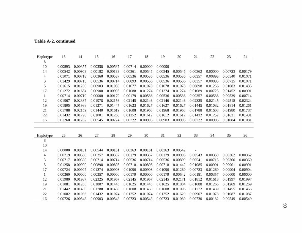
99
Table A-2. continued
Haplotype 13 14 15 16 17 18 19 20 21 22 23 24 8 10 0.00893 0.00357 0.00358 0.00537 0.00714 0.00000 0.00000 - 14 0.00542 0.00903 0.00182 0.00183 0.00361 0.00545 0.00545 0.00545 0.00362 0.00000 0.00723 0.00179 4 0.01071 0.00718 0.00360 0.00537 0.00536 0.00536 0.00536 0.00536 0.00357 0.00893 0.00540 0.01071 3 0.01429 0.00715 0.00536 0.00714 0.00893 0.00536 0.00536 0.00536 0.00357 0.00893 0.00715 0.01071 5 0.01615 0.01260 0.00903 0.01080 0.01077 0.01078 0.01078 0.01078 0.00898 0.01256 0.01083 0.01435 17 0.01272 0.01634 0.00908 0.00908 0.01088 0.01274 0.01274 0.01274 0.01089 0.00723 0.01452 0.00901 1 0.00714 0.00719 0.00000 0.00179 0.00179 0.00536 0.00536 0.00536 0.00357 0.00536 0.00539 0.00714 12 0.01967 0.02337 0.01978 0.02156 0.02145 0.02146 0.02146 0.02146 0.02325 0.02145 0.02518 0.02324 19 0.01805 0.01988 0.01271 0.01447 0.01623 0.01627 0.01627 0.01627 0.01445 0.01082 0.01814 0.01261 21 0.01788 0.02159 0.01440 0.01619 0.01608 0.01968 0.01968 0.01968 0.01788 0.01608 0.01980 0.01787 22 0.01432 0.01798 0.01081 0.01260 0.01252 0.01612 0.01612 0.01612 0.01432 0.01252 0.01621 0.01431 16 0.01260 0.01262 0.00545 0.00724 0.00722 0.00903 0.00903 0.00903 0.00722 0.00903 0.01084 0.01081
Haplotype 25 26 27 28 29 30 31 32 33 34 35 36 8 10 14 0.00000 0.00181 0.00544 0.00181 0.00363 0.00181 0.00363 0.00542 - 4 0.00719 0.00360 0.00357 0.00357 0.00179 0.00357 0.00179 0.00903 0.00543 0.00359 0.00362 0.00362 3 0.00717 0.00360 0.00714 0.00714 0.00536 0.00714 0.00536 0.00899 0.00541 0.00718 0.00360 0.00360 5 0.01258 0.00900 0.00898 0.00898 0.00718 0.00898 0.00718 0.01442 0.01085 0.00901 0.00901 0.00901 17 0.00724 0.00907 0.01274 0.00908 0.01090 0.00908 0.01090 0.01269 0.00723 0.01269 0.00904 0.00904 1 0.00360 0.00000 0.00357 0.00000 0.00179 0.00000 0.00179 0.00542 0.00181 0.00357 0.00000 0.00000 12 0.01980 0.01987 0.02325 0.01967 0.02145 0.01967 0.02145 0.02171 0.01812 0.01618 0.01997 0.01997 19 0.01081 0.01263 0.01807 0.01445 0.01625 0.01445 0.01625 0.01804 0.01088 0.01265 0.01269 0.01269 21 0.01442 0.01450 0.01788 0.01430 0.01608 0.01430 0.01608 0.01996 0.01272 0.01439 0.01455 0.01455 22 0.01082 0.01086 0.01432 0.01074 0.01252 0.01074 0.01252 0.01629 0.00907 0.01078 0.01087 0.01087 16 0.00726 0.00548 0.00903 0.00543 0.00723 0.00543 0.00723 0.01089 0.00730 0.00182 0.00549 0.00549
99
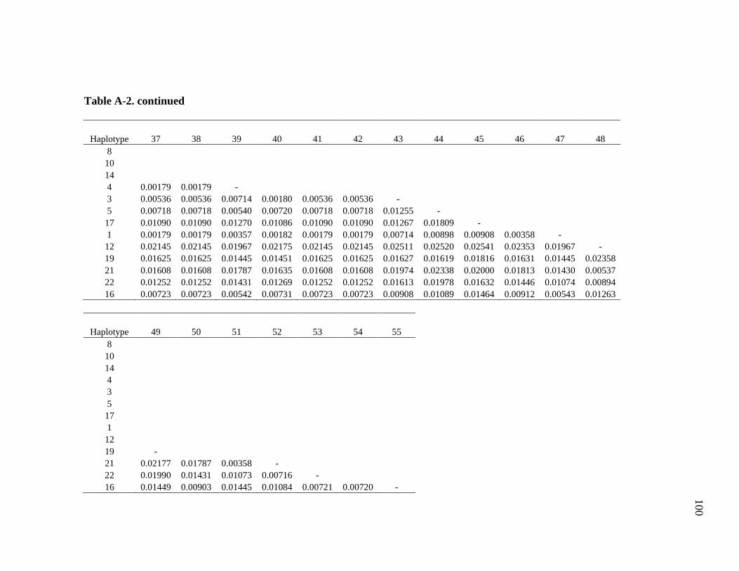
100
Table A-2. continued
Haplotype 37 38 39 40 41 42 43 44 45 46 47 48 8 10 14 4 0.00179 0.00179 - 3 0.00536 0.00536 0.00714 0.00180 0.00536 0.00536 - 5 0.00718 0.00718 0.00540 0.00720 0.00718 0.00718 0.01255 - 17 0.01090 0.01090 0.01270 0.01086 0.01090 0.01090 0.01267 0.01809 - 1 0.00179 0.00179 0.00357 0.00182 0.00179 0.00179 0.00714 0.00898 0.00908 0.00358 - 12 0.02145 0.02145 0.01967 0.02175 0.02145 0.02145 0.02511 0.02520 0.02541 0.02353 0.01967 - 19 0.01625 0.01625 0.01445 0.01451 0.01625 0.01625 0.01627 0.01619 0.01816 0.01631 0.01445 0.02358 21 0.01608 0.01608 0.01787 0.01635 0.01608 0.01608 0.01974 0.02338 0.02000 0.01813 0.01430 0.00537 22 0.01252 0.01252 0.01431 0.01269 0.01252 0.01252 0.01613 0.01978 0.01632 0.01446 0.01074 0.00894 16 0.00723 0.00723 0.00542 0.00731 0.00723 0.00723 0.00908 0.01089 0.01464 0.00912 0.00543 0.01263
Haplotype 49 50 51 52 53 54 55 8 10 14 4 3 5 17 1 12 19 - 21 0.02177 0.01787 0.00358 - 22 0.01990 0.01431 0.01073 0.00716 - 16 0.01449 0.00903 0.01445 0.01084 0.00721 0.00720 -
100
101
VITA
Name: Jennifer Lynne Pechal
Address: Department of Entomology Texas A&M University College Station, Texas 77845-2475 E-mail Address: [email protected]
Education: B.S. in Biology, minor in Chemistry with a Forensic Emphasis, Sam Houston State University, 2006
M.S. in Entomology, Texas A&M University, 2008