Embed Size (px)
Citation preview
The Egyptian Journal of Hospital Medicine (2008) Vol., 33: 512– 503
Intrathecal Ketorolac Injection in Albino Rats;
Pharmacological and Histological study
*Tarek A. Atia, **Mostafa I. Shalaby, ***Nemat M. Al-Baz
*Histology, ** Anesthesiology & ICU, ***Pharmacology Departments; Faculty
of Medicine, Al-Azhar University, Cairo, Egypt
Abstract Introduction: Ketorolac tromethamine is a potent injectable non-steroidal anti-inflammatory drug (NSAID). Ketorolac provides successful analgesia after intrathecal or
epidural injection. It is frequently used to manage post-operative pain, cancer pain, and
arthritis either intrathecally, or intramuscular. However, its long term administration could
induce renal toxicity and/or gastro-intestinal ulceration. Aim of the study: The aim of this study was to assess the analgesic potency of ketorolac
after intrathecal injection. Also, we aimed to study the histological effect of ketorolac on the
spinal cord and the duodenum after treatment in an animal model. Methods: 40 adult male albino rats, weighing 250-350 gm, were used and divided into 4
groups, 10 rats each. Group S (control) received 10μl normal saline intrathecally, group K50
received 50μg ketorolac intrathecally, group K50 + omeprazole (proton pump inhibitor) received 50μg ketorolac intrathecally plus 0.2 mg omeprazole orally, and finally, group K100
received 100μg ketorolac intrathecally. All animals were treated for four successive days.
Result: The rat tail flick latency was longer in K50, K50 + omeprazole, and K100 groups
when compared to normal control (P = 0.002). Also, the hind-paw withdrawal latency was longer in treated groups when compared to those of the control group (P = 0.0001). Moreover,
K50 group showed decreased phase II response by 61%, K50 + omeprazole group showed
decreased phase II by 62%, while K100 group showed decreased it by 76%. Histological examination revealed no changes in the spinal cord of all treated animals. Also,
examination of the duodenum showed normal duodenal mucosa in group K50 and those of
group K50 + omeprazole. On the other hand, cellular infiltration as well as destruction of the mucous acini have been noticed in the duodenum of K100 group.
Conclusion: Ketorolac could be a good alternative drug used intrathecally to manage pain.
Key word; Ketorolac, analgesics, intrathecal, rats
Introduction
The study of intrathecal application of
drugs to manage pain is important for two reasons. First, it is directly relevant to
anesthesia practice in that the intrathecal
space is often instrumented as part of peri-operative, or chronic pain care. Second, it
provides important information regarding
mechanisms of analgesic action and of pain
transmission, which could guide pharmaceutical development of both
intrathecal and systemic drug development.
A good example of these rationales is examination of cyclooxygenase (COX)
enzyme expression and inhibition in the
spinal cord as it relates to pain treatment. COX is expressed in the normal spinal cord
in small amounts, both isoforms COX-1
and COX-2. Brocks and, Jamali (1992).
Indeed, the constitutive presence of COX-2
in the spinal cord has been suggested to underlie the early analgesic effect of COX
inhibitors after surgery or other peripheral
injury and at times before peripheral COX-2 expression is increased. After peripheral
injury, spinal COX-2 expression is greatly
enhanced, leading to increased spinal
release of prostaglandins with resultant increased substance-P release and central
sensitization. Gillis and Brogden. (1997)
For this reason, spinally administered COX inhibitors produce analgesia after injury
(Conklin and Eisenach, 2003).
Ketorolac tromethamine is an injectable non-steroidal anti-inflammatory
drug (NSAID) approved in 1990 for
treating post-operative pain. Ketorolac is
112
Tarek A. Atia et al
522
frequently used to manage postoperative
pain, renal colic, arthritis, and cancer pain either intrathecally or intramuscular.
Ketorolac has also been reported to provide
successful analgesia when injected through
epidural way (Gillis and Brogden 1997) .Ketorolac, a peripherally acting drug, has
become a popular alternative to opoids for
postoperative analgesia, because of its minimal central nervous system side effects
specifically respiratory depression,
sedation, or nausea and vomiting (Miranda et al., 1993).As a NSAID drug, ketorolac
inhibits platelet aggregation, and its long
term administration could induce renal
toxicity and/or gastro-intestinal ulceration. Ketorolac has also been reported to
provide successful analgesia when injected
by intrathecal and epidural way in animal models. To consider the possible reaction of
intrathecal ketorolac in man, it is necessary
to establish the pharmacokinetic and the effects upon spinal cord after intrathecal
delivery in well defined experiment.
Analgesic effect of intrathecal
administration of ketorolac has been investigated in mouse, rat, and dog models
before its recent used in man. (Eisenach et
al., 2002).
Material and Methods
1- Pharmacological study:
Forty adult male albino rats weighing
250 – 350 g were subjected to the present study. Animals were housed with free
access to food and water, and maintained
on a 12 hour light/dark cycle. Rats were anesthetized with 2% halothane in
oxygen/air, and then polyethylene catheters
(Gauge 27) were inserted through a small incision in the atlanto-occipital membrane,
and then passed 8cm caudally to the level
of the lumber enlargement. To confirm
correct placement of the catheter we inject 10μl of 2% lidocaine followed by 10μl
0.9% saline to flush the catheter
(Yamamoto and, Yaksh. 1992). All animals were developed bilateral motor block of the
hind limbs within 30 seconds that lasted
within two days. Animals were divided into 4 groups,
10 rats each. First, group S (control),
injected with 10μl sterile saline 0.9%
intrathecally. Second, group K50, where
animals were injected intrathecally with 50μg ketorolac dissolved in 10ul normal
saline. Third, group K50 + Omeperazole,
where animals received 0.2mg omeperazole
(proton pump inhibitor) orally one hour before intrathecal injected with 50μg
ketorolac dissolved in 10μl normal saline.
Lastly, group K100, where animals were injected intrathecally with 100μg ketorolac
dissolved in 10μl normal saline. All doses
were given daily for four successive days. At the fourth day, 15 minutes after
intrathecal injection rat flick test, and hot
plate test were assessed.
A- Rat flick test:
The nociceptive threshold was
measured by latency of the tail flick responses elicited by radiant heat applied to
the lower third of the tail. The mean tail
flick latency (TFL) of three measurements was taken as the basal threshold. Adjust the
amplitude of radiant heat, so that the basal
TFL was within 4-6 seconds (Sec.). The
TFL taken at 15 minutes intervals after intrathecal injection was expressed as the
percentage change from basal tail flick
latency, with cut-off limit of 150% above baseline to avoid unnecessary skin damage.
In the present study the cut-off time was 14
Sec.
B- Hot plate test:
The hind-paw withdrawal latency
(HWL) was measured by the hot plate test. The HWL to noxious heat stimulation was
tested by the hot plate maintained at
temperature of 52°C. The time of the hind-paw withdrawal was measured in seconds
to be referred as HWL to thermal
stimulation. The HWL was measured
before intrathecal injection of ketorolac as the basal threshold 4-6 Sec. A cut-off limit
of 15 Sec. was set up to avoid tissue
damage (Sun et al., 2003).
C- Formalin test:
The formalin modified test (
Malmberg, and Yaksh 1993). was performed 15 minutes after the last
intrathecal injection. Rats were anesthetized
with 2% halothane in oxygen/air, and then
50ul of 5% formalin was injected subcutaneously into the dorsal surface of
the right hind-paw with 26-gauge needle.
Intrathecal Ketorolac Injection in Albino Rats;………….
523
After formalin injection, flinches were
counted for 1 minute interval at 1 minute, 5 minutes and 10 minutes, and then every 10
minutes for 1 hour. Two phases of
spontaneous flinching behavior observed.
Phase I; begins immediately after formalin injection, and lasts to the second
observation interval (5 minutes). Phase II;
begins at the 10th minutes and lasts through
60 minutes. Thus, the mean of the first 2
measurements (at one and five minutes)
was the phase I value, and the mean of the remaining measurements was phase II
value.
2- Histological study:
At the fifth day, rats were sacrificed, bilateral laminecomty was performed, and
spinal cord with the companying catheter
tips located at the lumber enlargements were removed from the vertebral canals.
Laparotomy was performed; part of the
duodenum was removed. Samples were fixed in 10% formalin buffered saline,
embedded in paraffin, and cut out into 6μm
thick sections. Duodenal Sections were
stained with hematoxylin and eosin stain, and spinal cord sections were stained with
Toluidin blue to demonstrate nerve cells,
with Nissl granules (Drury and Wallington 1980).
Statistical analysis:
Data from nociceptive tests were presented as mean ± SD. Differences
between groups were determined by
analysis of variance (ANOVA). P<0.05 was considered as significant difference.
Result
Pharmacological study:
A- As regard to the effect of intrathecal
administration of ketorolac on rat tail flick responses; the mean TFL was
longer in groups K50, K50+ omeprazol,
and K100 than control group (S group),
as shown in table-1. B- As regard to the hot plate test, the mean
value of HWL was longer in groups
K50, K50+ omeprazol, and K100 than control group (S group), as shown in
table-2.
C- The effect of intrathecal administration of ketorolac on formalin test is
represented in table-3. As regard to
phase I, there were non significant
difference between ketorolac injected groups and control (S) group, whereas
there were significant reduction in the
number of flinching in ketorolac injected groups than in S group.
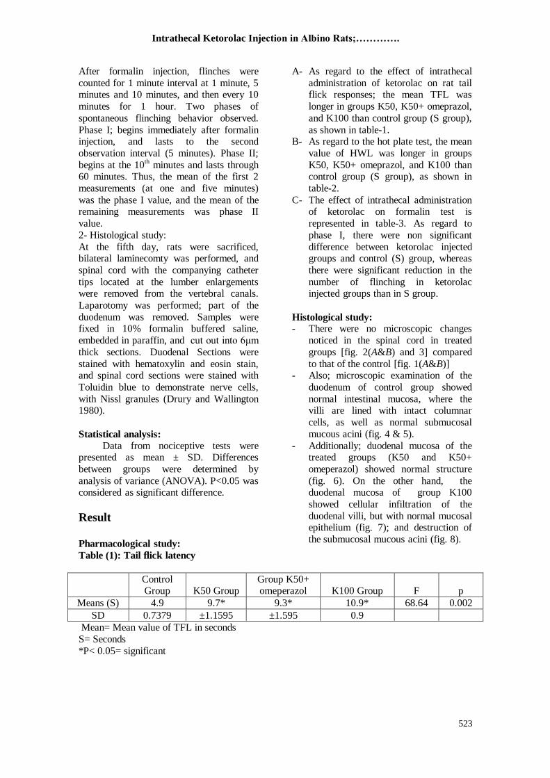
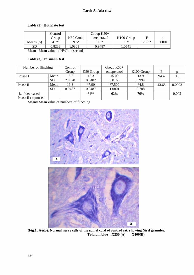
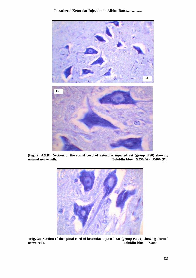
Histological study: - There were no microscopic changes
noticed in the spinal cord in treated
groups [fig. 2(A&B) and 3] compared
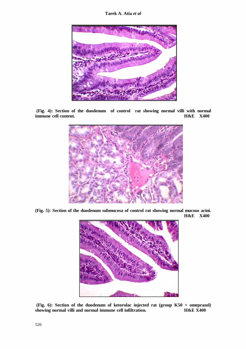
to that of the control [fig. 1(A&B)] - Also; microscopic examination of the
duodenum of control group showed
normal intestinal mucosa, where the villi are lined with intact columnar
cells, as well as normal submucosal
mucous acini (fig. 4 & 5).
- Additionally; duodenal mucosa of the treated groups (K50 and K50+ omeperazol) showed normal structure
(fig. 6). On the other hand, the duodenal mucosa of group K100
showed cellular infiltration of the
duodenal villi, but with normal mucosal epithelium (fig. 7); and destruction of
the submucosal mucous acini (fig. 8).
Table (1): Tail flick latency
Control Group K50 Group
Group K50+ omeperazol K100 Group F p
Means (S) 4.9 9.7* 9.3* 10.9* 68.64 0.002
SD 0.7379 ±1.1595 ±1.595 0.9
Mean= Mean value of TFL in seconds
S= Seconds
*P< 0.05= significant
1
Tarek A. Atia et al
524
Table (2): Hot Plate test
Control
Group K50 Group
Group K50+
omeperazol K100 Group F p
Means (S) 4.7* 9.5* 9.3* 11* 76.32 0.0001
SD 0.8233 1.0801 0.9487 1.0541
Mean =Mean value of HWL in seconds
Table (3): Formalin test
Number of flinching
Control
Group K50 Group
Group K50+
omeperazol K100 Group F p
Phase I
Mean 16.7 15.3 15.00 13.9 94.4
0.8 SD 2.9078 0.9487 0.8165 0.994
Phase II
Mean 19.3 *7.90 *7.500 *4.8 43.68
0.0002
SD 0.9487 0.9487 1.0801 0.788
%of decreased Phase II responses
61%
62%
76%
0.002
Mean= Mean value of numbers of flinching
(Fig.1; A&B): Normal nerve cells of the spinal cord of control rat, showing Nissl granules.
Toluidin blue X250 (A) X400(B)
Intrathecal Ketorolac Injection in Albino Rats;………….
525
(Fig. 2; A&B): Section of the spinal cord of ketorolac injected rat (group K50) showing
normal nerve cells. Toluidin blue X250 (A) X400 (B)
(Fig. 3): Section of the spinal cord of ketorolac injected rat (group K100) showing normal
nerve cells. Toluidin blue X400
Tarek A. Atia et al
526
(Fig. 4): Section of the duodenum of control rat showing normal villi with normal
immune cell content. H&E X400
(Fig. 5): Section of the duodenum submucosa of control rat showing normal mucous acini.
H&E X400
(Fig. 6): Section of the duodenum of ketorolac injected rat (group K50 + omeprazol)
showing normal villi and normal immune cell infiltration. H&E X400
Intrathecal Ketorolac Injection in Albino Rats;………….
527
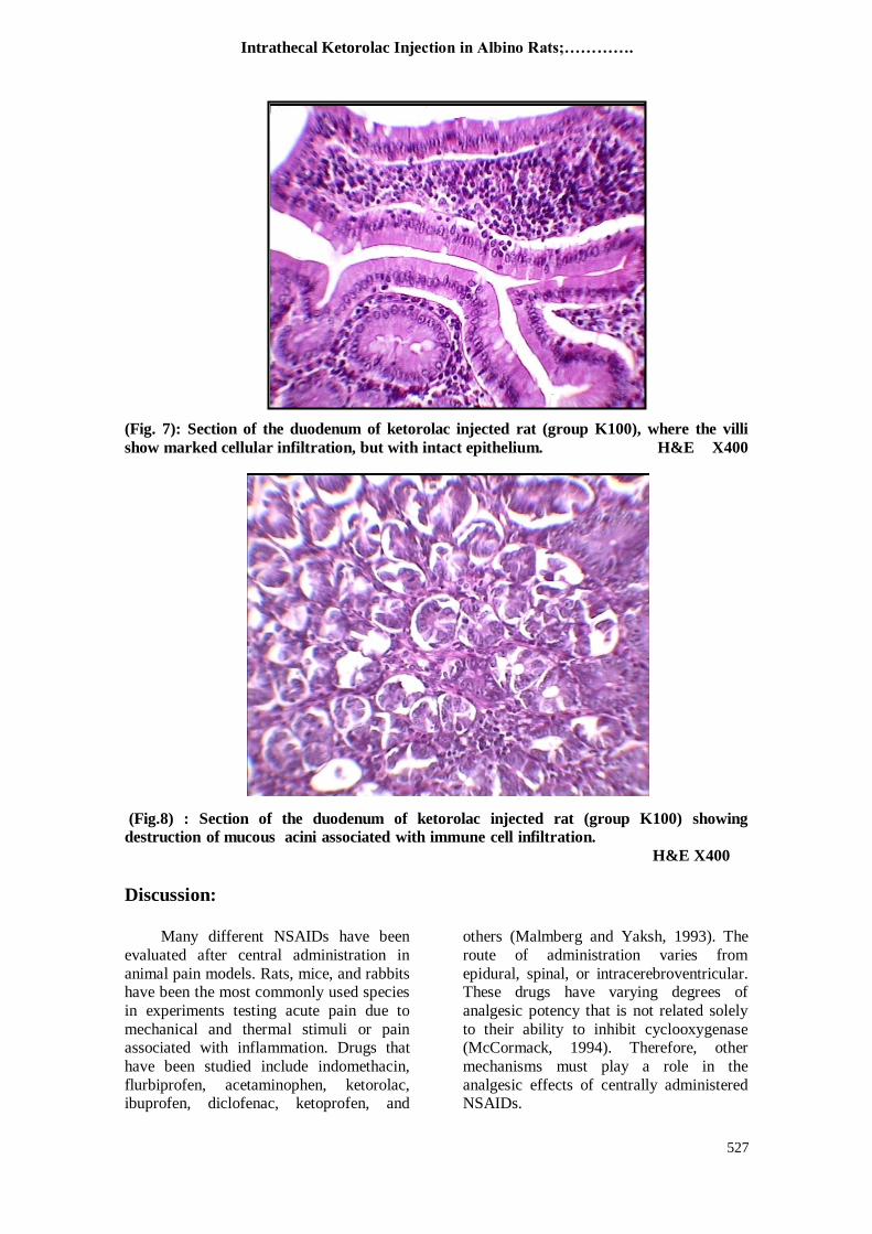
(Fig. 7): Section of the duodenum of ketorolac injected rat (group K100), where the villi
show marked cellular infiltration, but with intact epithelium. H&E X400
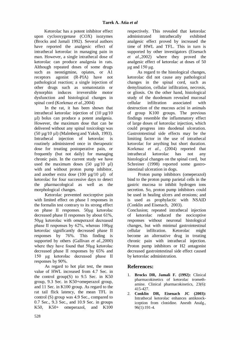
(Fig.8) : Section of the duodenum of ketorolac injected rat (group K100) showing
destruction of mucous acini associated with immune cell infiltration.
H&E X400
Discussion:
Many different NSAIDs have been
evaluated after central administration in
animal pain models. Rats, mice, and rabbits have been the most commonly used species
in experiments testing acute pain due to
mechanical and thermal stimuli or pain associated with inflammation. Drugs that
have been studied include indomethacin,
flurbiprofen, acetaminophen, ketorolac, ibuprofen, diclofenac, ketoprofen, and
others (Malmberg and Yaksh, 1993). The
route of administration varies from
epidural, spinal, or intracerebroventricular. These drugs have varying degrees of
analgesic potency that is not related solely
to their ability to inhibit cyclooxygenase (McCormack, 1994). Therefore, other
mechanisms must play a role in the
analgesic effects of centrally administered NSAIDs.
Tarek A. Atia et al
528
Ketorolac has a potent inhibitor effect
upon cyclooxygenase (COX) isozymes (Brocks and Jamali 1992). Several authors
have reported the analgesic effect of
intrathecal ketorolac in managing pain in
man. However, a single intrathecal dose of ketorolac can produce analgesia in rats.
Although repeated doses of some drugs
such as neostigmine, opiates, or A1 receptors agonist (R-PIA) have not
pathological reaction; a single injection of
other drugs such as somatostatin or dynorphin induces irreversible motor
dysfunction and histological changes in
spinal cord (Korkmaz et al.,2004)
In the rat, it has been shown that intrathecal ketorolac injection of (10
µg/10
µl) bolus can produce a potent analgesia.
However, the maximum dose that can be delivered without any spinal toxicology
was
(50 µg/10 µl) (Malmberg and Yaksh, 1993).
Intrathecal injection of ketorolac is routinely administered once in therapeutic
dose for treating postoperative pain, or
frequently (but not daily) for managing
chronic pain. In the current study we have used the maximum doses (50 µg/10 µl)
with and without proton pump inhibitor,
and another extra dose (100 µg/10 µl) of ketorolac for four successive days to detect
the pharmacological as well as the
morphological changes.
Ketorolac prevented nociceptive pain with limited effect on phase I responses in
the formalin test contrary to its strong effect
on phase II responses. 50μg ketorolac decreased phase II responses by about 61%,
50μg ketorolac with omeperazol decreased
phase II responses by 62%, whereas 100μg ketorolac significantly decreased phase II
responses by 76%. This finding is
supported by others (Gallivan et al.,2000)
where they have found that 50μg ketorolac decreased phase II responses by 65% and
150 μg ketorolac decreased phase II
responses by 90%. As regard to hot plat test, the mean
value of HWL increased from 4.7 Sec. in
the control group(S) to 9.5 Sec. in K50 group, 9.3 Sec. in K50+omeperazol group,
and 11 Sec. in K100 group. As regard to the
rat tail flick latency, the mean TFL in
control (S) group was 4.9 Sec., compared to 0.7 Sec., 9.3 Sec., and 10.9 Sec. in groups
K50, K50+ omeperazol, and K100
respectively. This revealed that ketorolac
administrated intrathecally exhibited analgesic effect proved by increased the
time of HWL and TFL. This in turn is
supported by other investigators (Eisenach
et al.,2002) where they proved the analgesic effect of ketorolac at doses of 50
μg and 150 μg.
As regard to the histological changes, ketorolac did not cause any pathological
changes in the spinal cord, such as
demylination, cellular infiltration, necrosis, or gliosis. On the other hand, histological
study of the duodenum revealed mucosal
cellular infiltration associated with
destruction of the mucous acini in animals of group K100 groups. The previous
findings resemble the inflammatory effect
of large doses of ketorolac injection, which could progress into duodenal ulceration.
Gastrointestinal side effects may be the
limiting factor in the use of intrathecal ketorolac for anything but short duration.
Korkmaz et al., (2004) reported that
intrathecal ketorolac has not any
histological changes on the spinal cord, but Schreiner (1998) reported some gastro-
intestinal ulceration in dogs.
Proton pump inhibitors (omeperazol) bind to the proton pump parietal cells in the
gastric mucosa to inhibit hydrogen ions
secretion. So, proton pump inhibitors could
be used in healing ulcers and erosions; and is used as prophylactic with NSAID
(Conklin and Eisenach, 2003).
Conclusion; repeated intrathecal injection of ketorolac reduced the nociceptive
responses without neuronal histological
changes, but with minimal gastrointestinal cellular infiltration. Ketorolac might
become an alternative drug in treating
chronic pain with intrathecal injection.
Proton pump inhibitors or H2 antagonist decreased gastrointestinal side effect caused
by ketorolac administration.
References:
1. Brocks DR, Jamali F. (1992): Clinical
pharmacokinetics of ketorolac trometh-
amine. Clinical pharmacokinetics, 23(6):
415-427.
2. Conklin DR, Eisenach JC (2003): Intrathecal ketorolac enhances antiknock-
iception from clonidine. Anesth Analg.,
96(1):191-4.
Intrathecal Ketorolac Injection in Albino Rats;………….
529
3. Drury RAB, Wallington FA. (1980): Histological techniques. Oxford University
Press, New York, Toronto.
4. Eisenach JC, Curry R, Yaksh TL.
(2002): Phase I safety assessment of
intrathecal ketorolac. Pain, 99(3): 599-604. 5. Gillis JC, Brogden RN. (1997): Ketorolac:
a reappraisal of its pharmacodynamic and
pharmacokinetic properties and therapeutic
use in pain management. Drug, 53: 139-
188.
6. Gallivan ST, Johnston SA, Broadstone
RV, Jortner BS, Reimer M. (2000). The
clinical, cerebrospinal fluid, and
histopathologic effects of epidural ketorolac
in dogs. Vet Surg. 29(5):436-41.
7. Korkmaz HA, Maltepe F, Erbayraktar S,
Yilmaz O, Guray M, Canda MS, Gunerli A, Gokmen N. (2004).Antinociceptive and
neurotoxicologic screening of chronic
intrathecal administration of ketorolac
tromethamine in the rat. Anesth Analg.,
98(1):148-52.
8. Malmberg AB, Yaksh TL. (1993): Pharmacology of the spinal action of
ketorolac, morphine, ST-91, U50488H, and
L-PIA on the formalin test and an
isobolographic analysis of the NSAID
interaction. Anesthesiology, 79(2):270-81.
9. McCormack K (1994): The Spinal Actions
of Nonsteroidal Anti-Inflammatory Drugs
and the Dissociation Between Their Anti-
Inflammatory and Analgesic Effects. Drugs., 47:28-45.
10. Miranda HF, Sierralta F, Sierralta F,
Pinardi G. (1993): Previous administration
of indomethacin or naloxone did not
influence ketorolac antinociception in mice.
Anesth. Analg., 77: 750-753.
11. Schreiner MS. (1998). Gastric fluid
volume: is it really a risk factor for
pulmonary aspiration? Anesth Analg.,
87(4):754-756.
12. Sun YG, Gu XL, Lundeberg T, Yu LC.
(2003): An antinociceptive role of galanin in the arcuate nucleus of hypothalamus in
intact rats and rats with inflammation. Pain,
106: 143-150.
13. Yamamoto T, Yaksh TL. (1992): Comparison of the antinociceptive effects
of pre- and post-treatment with intrathecal
morphine and MK801 and NMDA
antagonist on formalin test in the rat.
Anesthesiology, 77: 757- 763.
حقه مبدة الكتروالك داخل الىخبع الشىكى للفئران البيضبء
Tarek A. Atia et al
530
دراست هستىلىجيت وفبرمبكىلىجيت
وعمت الببز –مصطفى شلبى –طبرق عطيت
القبهرة -مه اقسبم الهستىلىجيب والتخذير واالدويت بكليت الطب جبمعت االزهر
ب قيب ػذ دق بدة اىنخشالك عبد ىالىخبة غيش اسخيشيذ حؼط حبثيشا سن
قذ . حسخخذ بنثشة ىخخفيف االى بؼذ اىؼييبث اىجشاديت, داخو اىذبو اىشم
ىدظج بؼط اآلثبس اىجببيت ىيؼقبس ف صسة اػشاض حس ببىني حقشدبث ببىقبة قذ ذفج اىذساست ف زا اىبذث اى حقيي اسخخذا دق اىؼقبس داخو اىخبع . اىعيت
قذ . بثيش رىل ػي سيج اىذبو اىشم االث ػشش ف اىفئشا اىبيعبء مسن ح
013-113اسخخذ ىيذساست اسبؼ رمس اىفئشا اىبيعبء يخشاح ص بي دقج اىجػت , جشا قسج إى أسبغ جػبث ف مو جػت ػششة فئشا
اسخخذج مجػت األى بجشػت ػششة ينشىخش ذيه اىيخ اىطبيؼ
ظببطت دقج اىجػت اىثبيت بخسي ينشجشا بدة اىنخشالك دقج ييجشا 3, 1اىجػت اىثبىثت بخسي ينشجشا بدة اىنخشالك ببإلظبفت إى
بدة االيبشاصه اىاقيت ىيؼذة دقج اىجػت اىشابؼت ببئت ينشجشا
قذ اظشث اىخبئج حبثش سيج االث ػشش غ .ىل ىذة اسبؼت ايب خخبىيت اىنخشالك ردق اىنخشالك دذ بخشميض اىبئت ينشجشا بيب ى حظش ا حغيشاث سخىجيت
ف سيج اىذبو اىشم قذ خيصج ز اىذساست اى سالت اسخخذا ػقبس اىنخشالك
ت اىبسبت اسخؼو ؼ بدة اقيت ىجذاس مسن ىألى خبصت إرا اسخؼو ببىجشػ . اىؼذة