Embed Size (px)
Citation preview
antibodies
Review
Intravenous Immune Globulin Uses in the Fetus andNeonate: A Review
Mahdi Alsaleem 1,2
1 Pediatrics Department, Neonatology, Children’s Mercy Hospital, Kansas City, MO 64108, USA;[email protected]
2 Pediatrics Department, University of Kansas, Wichita, KS 67208, USA
Received: 31 August 2020; Accepted: 2 November 2020; Published: 4 November 2020�����������������
Abstract: Intravenous immune globulin (IVIG) is made after processing plasma from healthy donors.It is composed mainly of pooled immunoglobulin and has clinical evidence-based applicationsin adult and pediatric populations. Recently, several clinical applications have been proposedfor managing conditions in the neonatal population, such as hemolytic disease of the newborn,treatment, and prophylaxis for sepsis in high-risk neonates, enterovirus parvovirus and COVID-19related neonatal infections, fetal and neonatal immune-induced thrombocytopenia, neonatalhemochromatosis, neonatal Kawasaki disease, and some types of immunodeficiency. The dosing,mechanism of action, effectiveness, side effects, and adverse reactions of IVIG have been relativelywell studied in adults but are not well described in the neonatal population. This review aimsto provide the most recent evidence and consensus guidelines about the use of IVIG in the fetusand neonate.
Keywords: immunoglobulins; fetus; neonates; sepsis; hemolysis; hyperbilirubinemia; necrotizingenterocolitis; coronavirus; coronavirus disease 19 (COVID-19)
1. Introduction
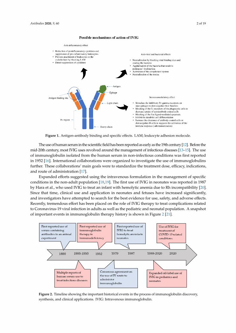
Immunoglobulin therapy is defined as the use of a combination of antibodies obtained fromhealthy human donors to treat different conditions [1,2]. The principal components of intravenousimmunoglobulin (IVIG) are IgG antibodies, which compromise about 90% of the IVIG. Antibodiesare glycoproteins synthesized and secreted by plasma cells (activated B cells) to respond to antigenicstimulation with the primary purpose of a specific immune response to result in different physiologicaland/or pathological processes [2,3]. The basic structural unit is primarily formed by two heavy andtwo light chains [4,5]. The difference between the heavy chains results in different kinds of antibodies:IgG, IgA, IgM, IgE, and IgD. After synthesis, formed antibodies functions by binding with a specificantigen epitope. This binding subsequently results in specific actions that ultimately help neutralizeand inactivate the pathogenic organisms or trigger a specific immune response (Figure 1).
IVIG clinical applications and indications have expanded rapidly in recent years [6,7]. Several ofthese clinical applications have extended to include children, neonates, and fetuses [8,9]. Although theFood and Drug Administration (FDA) has not yet approved IVIG therapy for use in the neonate, it hasbeen used off-label in the management of challenging and progressing conditions in many fetusesand neonates [9]. The roles that IVIG may play in immunomodulation (inhibition or activation ofthe immune response, modulation of FcgR expression on B cells, inducing phagocytosis or directcytotoxicity, regulating apoptosis, modulation of antigen-presenting cells) were the driving factors tostudy the use of IVIG in this population subset [10,11].
Antibodies 2020, 9, 60; doi:10.3390/antib9040060 www.mdpi.com/journal/antibodies
Antibodies 2020, 9, 60 2 of 19Antibodies 2020, 9, x FOR PEER REVIEW 2 of 19
Figure 1. Antigen-antibody binding and specific effects. LAM; leukocyte adhesion molecule.
IVIG clinical applications and indications have expanded rapidly in recent years [6,7]. Several of these clinical applications have extended to include children, neonates, and fetuses [8,9]. Although the Food and Drug Administration (FDA) has not yet approved IVIG therapy for use in the neonate, it has been used off-label in the management of challenging and progressing conditions in many fetuses and neonates [9]. The roles that IVIG may play in immunomodulation (inhibition or activation of the immune response, modulation of FcgR expression on B cells, inducing phagocytosis or direct cytotoxicity, regulating apoptosis, modulation of antigen-presenting cells) were the driving factors to study the use of IVIG in this population subset [10,11].
The use of human serum in the scientific field has been reported as early as the 19th century [12]. Before the mid-20th century, most IVIG uses revolved around the management of infectious diseases [13–15]. The use of immunoglobulin isolated from the human serum in non-infectious conditions was first reported in 1952. [16]. International collaborations were organized to investigate the use of immunoglobulins further. These collaborations’ main goals were to standardize the treatment dose, efficacy, indications, and route of administration [17].
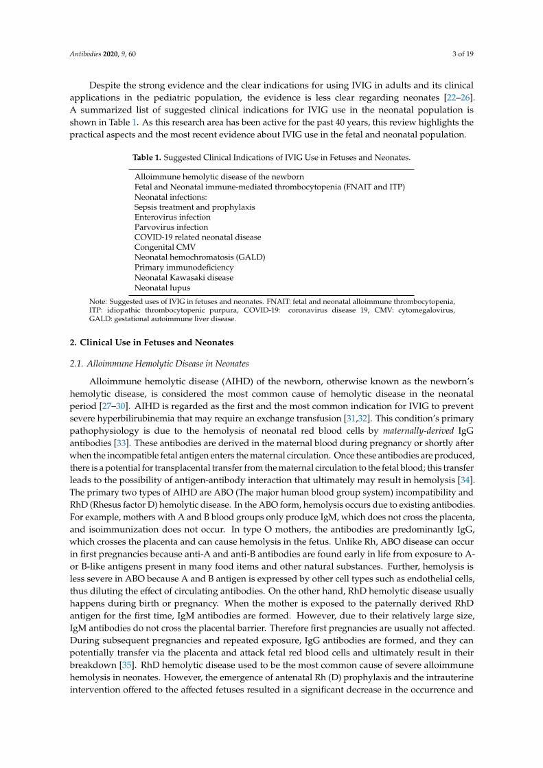
Expanded efforts suggested using the intravenous formulation in the management of specific conditions in the non-adult population [18,19]. The first use of IVIG in neonates was reported in 1987 by Hara et al., who used IVIG to treat an infant with hemolytic anemia due to Rh incompatibility [20]. Since that time, clinical use and application in neonates and fetuses have increased significantly, and investigators have attempted to search for the best evidence for use, safety, and adverse effects. Recently, tremendous effort has been placed on the role of IVIG therapy to treat complications related to Coronavirus-19 viral infection in adults as well as the pediatric and neonatal population. A snapshot of important events in immunoglobulin therapy history is shown in Figure 2 [21].
Figure 1. Antigen-antibody binding and specific effects. LAM; leukocyte adhesion molecule.
The use of human serum in the scientific field has been reported as early as the 19th century [12]. Before themid-20th century, most IVIG uses revolved around the management of infectious diseases [13–15]. The useof immunoglobulin isolated from the human serum in non-infectious conditions was first reportedin 1952 [16]. International collaborations were organized to investigate the use of immunoglobulinsfurther. These collaborations’ main goals were to standardize the treatment dose, efficacy, indications,and route of administration [17].
Expanded efforts suggested using the intravenous formulation in the management of specificconditions in the non-adult population [18,19]. The first use of IVIG in neonates was reported in 1987by Hara et al., who used IVIG to treat an infant with hemolytic anemia due to Rh incompatibility [20].Since that time, clinical use and application in neonates and fetuses have increased significantly,and investigators have attempted to search for the best evidence for use, safety, and adverse effects.Recently, tremendous effort has been placed on the role of IVIG therapy to treat complications relatedto Coronavirus-19 viral infection in adults as well as the pediatric and neonatal population. A snapshotof important events in immunoglobulin therapy history is shown in Figure 2 [21].Antibodies 2020, 9, x FOR PEER REVIEW 3 of 19
Figure 2. Timeline showing the important historical events in the process of immunoglobulin discovery, synthesis, and clinical applications. IVIG: Intravenous immunoglobulin.
Despite the strong evidence and the clear indications for using IVIG in adults and its clinical applications in the pediatric population, the evidence is less clear regarding neonates [22–26]. A summarized list of suggested clinical indications for IVIG use in the neonatal population is shown in Table 1. As this research area has been active for the past 40 years, this review highlights the practical aspects and the most recent evidence about IVIG use in the fetal and neonatal population.
Table 1. Suggested Clinical Indications of IVIG Use in Fetuses and Neonates.
Alloimmune hemolytic disease of the newborn
Fetal and Neonatal immune-mediated thrombocytopenia (FNAIT and ITP)
Neonatal infections: Sepsis treatment and prophylaxis Enterovirus infection Parvovirus infection COVID-19 related neonatal disease Congenital CMV
Neonatal hemochromatosis (GALD)
Primary immunodeficiency
Neonatal Kawasaki disease
Neonatal lupus
Note: Suggested uses of IVIG in fetuses and neonates. FNAIT: fetal and neonatal alloimmune thrombocytopenia, ITP: idiopathic thrombocytopenic purpura, COVID-19: coronavirus disease 19, CMV: cytomegalovirus, GALD: gestational autoimmune liver disease.
2. Clinical Use in Fetuses and Neonates
2.1. Alloimmune Hemolytic Disease in Neonates
Alloimmune hemolytic disease (AIHD) of the newborn, otherwise known as the newborn’s hemolytic disease, is considered the most common cause of hemolytic disease in the neonatal period [27–30]. AIHD is regarded as the first and the most common indication for IVIG to prevent severe hyperbilirubinemia that may require an exchange transfusion [31,32]. This condition’s primary pathophysiology is due to the hemolysis of neonatal red blood cells by maternally-derived IgG antibodies [33]. These antibodies are derived in the maternal blood during pregnancy or shortly after when the incompatible fetal antigen enters the maternal circulation. Once these antibodies are
Figure 2. Timeline showing the important historical events in the process of immunoglobulin discovery,synthesis, and clinical applications. IVIG: Intravenous immunoglobulin.
Antibodies 2020, 9, 60 3 of 19
Despite the strong evidence and the clear indications for using IVIG in adults and its clinicalapplications in the pediatric population, the evidence is less clear regarding neonates [22–26].A summarized list of suggested clinical indications for IVIG use in the neonatal population isshown in Table 1. As this research area has been active for the past 40 years, this review highlights thepractical aspects and the most recent evidence about IVIG use in the fetal and neonatal population.
Table 1. Suggested Clinical Indications of IVIG Use in Fetuses and Neonates.
Alloimmune hemolytic disease of the newbornFetal and Neonatal immune-mediated thrombocytopenia (FNAIT and ITP)Neonatal infections:Sepsis treatment and prophylaxisEnterovirus infectionParvovirus infectionCOVID-19 related neonatal diseaseCongenital CMVNeonatal hemochromatosis (GALD)Primary immunodeficiencyNeonatal Kawasaki diseaseNeonatal lupus
Note: Suggested uses of IVIG in fetuses and neonates. FNAIT: fetal and neonatal alloimmune thrombocytopenia,ITP: idiopathic thrombocytopenic purpura, COVID-19: coronavirus disease 19, CMV: cytomegalovirus,GALD: gestational autoimmune liver disease.
2. Clinical Use in Fetuses and Neonates
2.1. Alloimmune Hemolytic Disease in Neonates
Alloimmune hemolytic disease (AIHD) of the newborn, otherwise known as the newborn’shemolytic disease, is considered the most common cause of hemolytic disease in the neonatalperiod [27–30]. AIHD is regarded as the first and the most common indication for IVIG to preventsevere hyperbilirubinemia that may require an exchange transfusion [31,32]. This condition’s primarypathophysiology is due to the hemolysis of neonatal red blood cells by maternally-derived IgGantibodies [33]. These antibodies are derived in the maternal blood during pregnancy or shortly afterwhen the incompatible fetal antigen enters the maternal circulation. Once these antibodies are produced,there is a potential for transplacental transfer from the maternal circulation to the fetal blood; this transferleads to the possibility of antigen-antibody interaction that ultimately may result in hemolysis [34].The primary two types of AIHD are ABO (The major human blood group system) incompatibility andRhD (Rhesus factor D) hemolytic disease. In the ABO form, hemolysis occurs due to existing antibodies.For example, mothers with A and B blood groups only produce IgM, which does not cross the placenta,and isoimmunization does not occur. In type O mothers, the antibodies are predominantly IgG,which crosses the placenta and can cause hemolysis in the fetus. Unlike Rh, ABO disease can occurin first pregnancies because anti-A and anti-B antibodies are found early in life from exposure to A-or B-like antigens present in many food items and other natural substances. Further, hemolysis isless severe in ABO because A and B antigen is expressed by other cell types such as endothelial cells,thus diluting the effect of circulating antibodies. On the other hand, RhD hemolytic disease usuallyhappens during birth or pregnancy. When the mother is exposed to the paternally derived RhDantigen for the first time, IgM antibodies are formed. However, due to their relatively large size,IgM antibodies do not cross the placental barrier. Therefore first pregnancies are usually not affected.During subsequent pregnancies and repeated exposure, IgG antibodies are formed, and they canpotentially transfer via the placenta and attack fetal red blood cells and ultimately result in theirbreakdown [35]. RhD hemolytic disease used to be the most common cause of severe alloimmunehemolysis in neonates. However, the emergence of antenatal Rh (D) prophylaxis and the intrauterineintervention offered to the affected fetuses resulted in a significant decrease in the occurrence and
Antibodies 2020, 9, 60 4 of 19
the severity of hemolysis associated with Rh (D) hemolytic disease [36,37]. Due to this decreasein incidence, hemolysis due to ABO incompatibility became more common; however, only about15% of the affected pregnancies with ABO incompatibility will develop hemolysis. Only a smallerpercentage will develop severe hyperbilirubinemia [38,39]. In the presence of hemolysis unexplainedby either RhD or ABO incompatibility, investigation for other minor blood groups (Duffy, Kell, P, andothers) or different Rh antigens (E, C, and c) incompatibility is recommended.
The clinical presentation of AIHD can affect the fetus and/or the newborn at various severitiesbased on the hemolysis degree. Severe hemolysis during pregnancy can result in hydrops fetalis(severe anemia, resulting in heart failure and fluid accumulation in different bodily cavities(pleural effusion, skin edema, pericardial effusion, or ascites)) [40,41]. Rates of morbidities andmortality in fetal hydrops are high and may warrant intrauterine intervention to perform a fetalblood transfusion [42]. In other cases of mild or moderate hemolysis, anemia and associatedhyperbilirubinemia in the neonate are the most common clinical manifestations.
Significant efforts have been made to understand and to manage the hyperbilirubinemiaassociated with AIHD in recent years [43,44]. The main aim of most of the studies andclinical trials has been to provide efficient, timely, and aggressive interventions to preventthe devastating complications of hyperbilirubinemia (acutely known as acute bilirubin-inducedencephalopathy and long-term disabilities, and permanent neurodevelopmental deficits also knownas kernicterus) [30,43–45]. The primary etiology of the brain damage in these two conditions is thepenetration of bilirubin through the blood-brain barrier and eventually deposition in the centralnervous system [46].
IVIG has been proposed as a potential intervention that can decrease hemolysis severity and,therefore, the associated hyperbilirubinemia [47,48]. The exact mechanism of the action of IVIG to reducehemolysis is unclear. Scientists suggest IVIG works most likely by blocking the antibodies’ receptorslocated on the red blood cells’ surface. Blocking these receptors will prevent the antigen/antibodyinteractions between the antigens found on the red blood cells and the maternal antibodies, decreasingrecognition of the targeted red blood cells by the circulating macrophage and subsequently decreasingthe degree of hemolysis [32,49]. The first reported use of IVIG in AIHD of the newborn was publishedin 1987 [20]. This report was followed by other case reports and case series that suggested usingIVIG as a useful intervention to halt severe hyperbilirubinemia [50–52]. This intervention’s primarybeneficial effect is to decrease the need for exchange transfusion (a high-risk procedure performed inadvanced intensive care units to prevent the risk of bilirubin-induced brain damage).
The American Academy of Pediatrics (AAP) [53], in their report in 2004, recommended the use ofIVIG for alloimmune hemolytic disease of the newborn if the serum bilirubin level continues to risedespite intensive phototherapy or approaches the levels for which exchange transfusion is required [54].The dose suggested is 500 mg−1 g for each kg of body weight given via the intravenous route to beinfused over two hours. The AAP used the evidence obtained from the systemic review performed byGottstein et al., and other previous observations that showed the beneficial effects and the favorableoutcomes after using IVIG to manage severe hyperbilirubinemia [20,32]. The AAP also advised usingIVIG in the rare types of Rh disease (Anti-C and Anti-E) but acknowledged the limited evidence behindthis recommendation [54].
Supported by these recommendations by the AAP, there was a significant increase in IVIG use insevere hyperbilirubinemia due to AIHD. A recent Cochrane review by Zwiers et al. was done in 2018,to further evaluate this practice’s evidence-based aspects [55]. In their meta-analysis, 27 full-text articleswere screened for eligibility. Nine studies were eligible, and a total of 658 participants were includedfor the final analysis. The results did not support the AAP’s recommendations. They concludedthat there was not enough evidence that IVIG use in AIHD prevents exchange transfusion. In theirconclusion, the authors recommended using IVIG if performing exchanging transfusion is not possibleat the admitting facility until a transport arrangement can be made to a higher-level center.
Antibodies 2020, 9, 60 5 of 19
More recently, two studies were performed to evaluate the efficacy of IVIG. El Fekey et al. foundin their randomized controlled trial that the use of IVIG in addition to phototherapy resulted in adecrease in bilirubin levels and the number of exchange transfusions performed [56]. In contrast tothese findings, Al-lawama et al. found in their retrospective observation that infants who receivedIVIG in addition to phototherapy were noted to be at higher risk for rebound hyperbilirubinemiaand the need for exchange transfusion [57]. However, both of these studies were limited by the smallsample size and confounding variables’ effects.
Louis et al. did a systematic review and meta-analysis that included 12 studies about the safetyand efficacy of IVIG in neonates with RhD hemolytic disease. They found after analyzing the databased on high vs. low risk of bias that IVIG is beneficial in RhD hemolytic disease of the newborn instudies with a high risk of bias; however, this benefit was not clear in the studies that carried a lowrisk of bias (evaluated by risk assessment including reviewing; appropriate randomization, allocation,completing the outcome data, selective reporting, and others) [58].
Such conflicting outcomes could be explained by the different response to IVIG therapy based onthe primary etiology. De Haas et al. and Armstrong et al. suggested in their report that IVIG may bemore effective if the hemolysis is due to ABO groups incompatibility vs. RhD incompatibility [59,60].Another possible explanation may be related to the origin and the characteristics of the specific IVIGformulations used in the different studies.
One randomized double-blinded placebo-controlled trial was conducted to address the use ofIVIG in hemolytic disease of newborns as a prophylaxis measure rather than treatment. The subjectswere infants affected by hemolysis due to Rh disease. A total of 41 infants out of the 80 includedin the study received IVIG as a prophylactic measure to prevent the need for exchange transfusion.Seven infants in the intervention group required an exchanged transfusion compared to 6 from thecontrol group. Therefore, the authors concluded the prophylactic IVIG did not significantly decreasethe need for exchange transfusion in infants with alloimmunization due to Rh hemolytic disease [61].
2.2. Neonatal and Fetal Alloimmune Thrombocytopenia
Fetal and neonatal alloimmune thrombocytopenia (FNAIT) is thrombocytopenia caused bymaternal-fetal antiplatelet antibodies, resulting in platelet destruction [62,63]. The most commonlyaccepted theory for pathophysiology is maternal IgG antibody formation against fetal paternallyderived antigens. These IgG antibodies can pass through the placenta and subsequently form anantigen-antibody complex that ultimately results in platelet destruction.
Many platelet antigens have been identified as potential triggers for this immune process. Humanplatelet antigen (HPA)-1a is considered the most common trigger for maternal antibody formation andhence fetal and neonatal thrombocytopenia [64]. FNAIT usually affects the first-born child more thansubsequent children, in contrast to AIHD, which affects subsequent pregnancies with a more severedegree of hemolysis [63]. Postnatal clinical manifestations vary significantly. More than half of thecases are asymptomatic and identified mainly by screening complete blood count laboratory evaluationfor other reasons. Severe thrombocytopenia can lead to rapid progressive bleeding. The most fearedcomplication is spontaneous intracranial hemorrhage (ICH) [65,66].
The main principles of treatment of FNAIT are anticipation, antenatal IVIG therapy, postnatalrecognition, and timely interventions. Risk anticipation is based on the maternal history of a previouschild who developed thrombocytopenia during the second or the last third of gestation or shortly afterdelivery due to FNAIT in previous pregnancies. The mothers identified have a risk for subsequentdeliveries with potential risk for ICH,
Multiple studies have addressed the benefits of antenatal management of FNAIT. Bussel et al.found in their prospective study that IVIG administration in mothers who had a history of infantsaffected by FNAIT resulted in a significant increase in fetal platelet counts. None of the neonateshad ICH [67]. A recent meta-analysis was done by Winkelhorst et al. to evaluate the effect of IVIGin the management of FNAIT. Four randomized trials and 22 nonrandomized observations were
Antibodies 2020, 9, 60 6 of 19
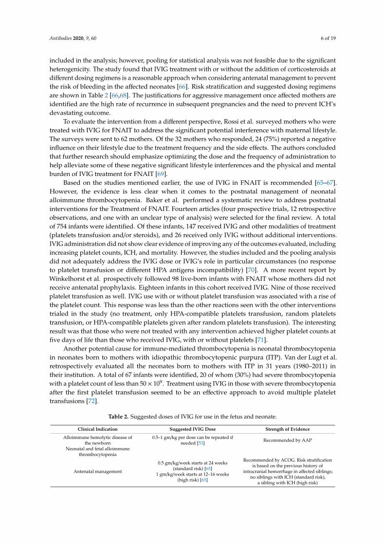
included in the analysis; however, pooling for statistical analysis was not feasible due to the significantheterogenicity. The study found that IVIG treatment with or without the addition of corticosteroids atdifferent dosing regimens is a reasonable approach when considering antenatal management to preventthe risk of bleeding in the affected neonates [66]. Risk stratification and suggested dosing regimensare shown in Table 2 [66,68]. The justifications for aggressive management once affected mothers areidentified are the high rate of recurrence in subsequent pregnancies and the need to prevent ICH’sdevastating outcome.
To evaluate the intervention from a different perspective, Rossi et al. surveyed mothers who weretreated with IVIG for FNAIT to address the significant potential interference with maternal lifestyle.The surveys were sent to 62 mothers. Of the 32 mothers who responded, 24 (75%) reported a negativeinfluence on their lifestyle due to the treatment frequency and the side effects. The authors concludedthat further research should emphasize optimizing the dose and the frequency of administration tohelp alleviate some of these negative significant lifestyle interferences and the physical and mentalburden of IVIG treatment for FNAIT [69].
Based on the studies mentioned earlier, the use of IVIG in FNAIT is recommended [65–67].However, the evidence is less clear when it comes to the postnatal management of neonatalalloimmune thrombocytopenia. Baker et al. performed a systematic review to address postnatalinterventions for the Treatment of FNAIT. Fourteen articles (four prospective trials, 12 retrospectiveobservations, and one with an unclear type of analysis) were selected for the final review. A totalof 754 infants were identified. Of these infants, 147 received IVIG and other modalities of treatment(platelets transfusion and/or steroids), and 26 received only IVIG without additional interventions.IVIG administration did not show clear evidence of improving any of the outcomes evaluated, includingincreasing platelet counts, ICH, and mortality. However, the studies included and the pooling analysisdid not adequately address the IVIG dose or IVIG’s role in particular circumstances (no responseto platelet transfusion or different HPA antigens incompatibility) [70]. A more recent report byWinkelhorst et al. prospectively followed 98 live-born infants with FNAIT whose mothers did notreceive antenatal prophylaxis. Eighteen infants in this cohort received IVIG. Nine of those receivedplatelet transfusion as well. IVIG use with or without platelet transfusion was associated with a rise ofthe platelet count. This response was less than the other reactions seen with the other interventionstrialed in the study (no treatment, only HPA-compatible platelets transfusion, random plateletstransfusion, or HPA-compatible platelets given after random platelets transfusion). The interestingresult was that those who were not treated with any intervention achieved higher platelet counts atfive days of life than those who received IVIG, with or without platelets [71].
Another potential cause for immune-mediated thrombocytopenia is neonatal thrombocytopeniain neonates born to mothers with idiopathic thrombocytopenic purpura (ITP). Van der Lugt et al.retrospectively evaluated all the neonates born to mothers with ITP in 31 years (1980–2011) intheir institution. A total of 67 infants were identified, 20 of whom (30%) had severe thrombocytopeniawith a platelet count of less than 50 × 109. Treatment using IVIG in those with severe thrombocytopeniaafter the first platelet transfusion seemed to be an effective approach to avoid multiple platelettransfusions [72].
Table 2. Suggested doses of IVIG for use in the fetus and neonate.
Clinical Indication Suggested IVIG Dose Strength of Evidence
Alloimmune hemolytic disease ofthe newborn
0.5–1 gm/kg per dose can be repeated ifneeded [53] Recommended by AAP
Neonatal and fetal alloimmunethrombocytopenia
Antenatal management
0.5 gm/kg/week starts at 24 weeks(standard risk) [65]
1 gm/kg/week starts at 12–16 weeks(high risk) [65]
Recommended by ACOG. Risk stratificationis based on the previous history of
intracranial hemorrhage in affected siblings;no siblings with ICH (standard risk),
a sibling with ICH (high risk)
Antibodies 2020, 9, 60 7 of 19
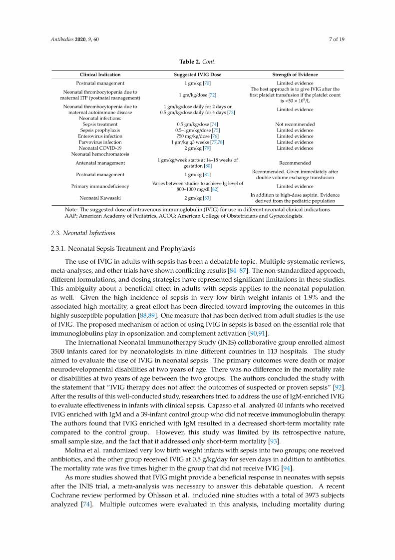
Table 2. Cont.
Clinical Indication Suggested IVIG Dose Strength of Evidence
Postnatal management 1 gm/kg [70] Limited evidence
Neonatal thrombocytopenia due tomaternal ITP (postnatal management) 1 gm/kg/dose [72]
The best approach is to give IVIG after thefirst platelet transfusion if the platelet count
is <50 × 109/LNeonatal thrombocytopenia due to
maternal autoimmune disease1 gm/kg/dose daily for 2 days or
0.5 gm/kg/dose daily for 4 days [73] Limited evidence
Neonatal infections:Sepsis treatment 0.5 gm/kg/dose [74] Not recommended
Sepsis prophylaxis 0.5–1gm/kg/dose [75] Limited evidenceEnterovirus infection 750 mg/kg/dose [76] Limited evidenceParvovirus infection 1 gm/kg q3 weeks [77,78] Limited evidenceNeonatal COVID-19 2 gm/kg [79] Limited evidence
Neonatal hemochromatosis
Antenatal management 1 gm/kg/week starts at 14–18 weeks ofgestation [80] Recommended
Postnatal management 1 gm/kg [81] Recommended. Given immediately afterdouble volume exchange transfusion
Primary immunodeficiency Varies between studies to achieve Ig level of800–1000 mg/dl [82] Limited evidence
Neonatal Kawasaki 2 gm/kg [83] In addition to high-dose aspirin. Evidencederived from the pediatric population
Note: The suggested dose of intravenous immunoglobulin (IVIG) for use in different neonatal clinical indications.AAP; American Academy of Pediatrics, ACOG; American College of Obstetricians and Gynecologists.
2.3. Neonatal Infections
2.3.1. Neonatal Sepsis Treatment and Prophylaxis
The use of IVIG in adults with sepsis has been a debatable topic. Multiple systematic reviews,meta-analyses, and other trials have shown conflicting results [84–87]. The non-standardized approach,different formulations, and dosing strategies have represented significant limitations in these studies.This ambiguity about a beneficial effect in adults with sepsis applies to the neonatal populationas well. Given the high incidence of sepsis in very low birth weight infants of 1.9% and theassociated high mortality, a great effort has been directed toward improving the outcomes in thishighly susceptible population [88,89]. One measure that has been derived from adult studies is the useof IVIG. The proposed mechanism of action of using IVIG in sepsis is based on the essential role thatimmunoglobulins play in opsonization and complement activation [90,91].
The International Neonatal Immunotherapy Study (INIS) collaborative group enrolled almost3500 infants cared for by neonatologists in nine different countries in 113 hospitals. The studyaimed to evaluate the use of IVIG in neonatal sepsis. The primary outcomes were death or majorneurodevelopmental disabilities at two years of age. There was no difference in the mortality rateor disabilities at two years of age between the two groups. The authors concluded the study withthe statement that “IVIG therapy does not affect the outcomes of suspected or proven sepsis” [92].After the results of this well-conducted study, researchers tried to address the use of IgM-enriched IVIGto evaluate effectiveness in infants with clinical sepsis. Capasso et al. analyzed 40 infants who receivedIVIG enriched with IgM and a 39-infant control group who did not receive immunoglobulin therapy.The authors found that IVIG enriched with IgM resulted in a decreased short-term mortality ratecompared to the control group. However, this study was limited by its retrospective nature,small sample size, and the fact that it addressed only short-term mortality [93].
Molina et al. randomized very low birth weight infants with sepsis into two groups; one receivedantibiotics, and the other group received IVIG at 0.5 g/kg/day for seven days in addition to antibiotics.The mortality rate was five times higher in the group that did not receive IVIG [94].
As more studies showed that IVIG might provide a beneficial response in neonates with sepsisafter the INIS trial, a meta-analysis was necessary to answer this debatable question. A recentCochrane review performed by Ohlsson et al. included nine studies with a total of 3973 subjectsanalyzed [74]. Multiple outcomes were evaluated in this analysis, including mortality during
Antibodies 2020, 9, 60 8 of 19
hospitalization, the combined outcome of death or major disability at two years of age, and lengthof hospital stay. After thorough analysis, the authors found that IVIG administration, including IgMenriched formulation, did not result in any beneficial effect. The authors concluded that the evidenceis strong against the use of IVIG or IgM-enriched IVIG in proven or suspected neonatal sepsis and thatno further research is recommended to address this topic [74].
Another meta-analysis was performed by Ohlsson and Lacy to compare the outcomes between thepreterm or low birth weight infants who received IVIG prophylaxis for sepsis and those who did notreceive sepsis IVIG prophylaxis. There was no difference between the two groups regarding mortality orthe incidence of necrotizing enterocolitis, bronchopulmonary dysplasia, and intraventricular bleeding.However, there was a decrease in the rate of sepsis (3%) associated with those who received prophylacticIVIG (R.R.: 0.85, 95% CI (0.74–0.98) with a number need to treat of 33) [75].
Multiple explanations have been suggested for the limited effect of IVIG for prevention or treatmentof neonatal sepsis, including insufficient dosing, different sepsis causative agents, the definitionfor sepsis, the immature immune function of the premature infants, and lower functional complementlevels [95].
2.3.2. Neonatal Enterovirus Infection
Enteroviruses are RNA viruses that belong to the picornaviridae family. In contrast tothe mild clinical presentation in adults, neonatal infection with enteroviruses can have severesystematic involvement.
IVIG has been suggested as a possible beneficial intervention when administered to neonateswith enterovirus infection. However, the literature’s evidence is not clear about this effect and isbased on only a few studies. Abzug et al. evaluated the neonatal response to IVIG in 16 neonateswho developed enterovirus infection. Nine infants were randomized to receive IVIG (750 mg/kg).A mild increase in serum neutralizing titers was noted in the infants who received IVIG but didnot significantly reduce the viral load in blood or urine [76]. In one case report, IVIG resulted in agood outcome in a neonate who developed disseminated infection caused by echovirus (a memberof the enterovirus family) [96]. Another case series showed a promising beneficial response in twoout of six infants who developed myocarditis and central nervous system (CNS) infections caused byechovirus 11 infection [97]. The most extensive study to date is the one done by Yen et al. in which datafrom 65 neonates with severe enteroviral infection were retrospectively analyzed. A total of 41 (63%)received IVIG, 29 of whom received it within the first 72 h of illness. The outcomes were significantlymore favorable in the group of infants who received the therapy early in the disease course. However,the dose and frequency of IVIG administration were not stated in this study [98].
2.3.3. Neonatal Parvovirus Infection
Parvovirus is a small single-stranded DNA virus. Perinatal maternal infection can result in severeconsequences for the fetus, including hydrops and fetal anemia. These effects’ primary etiology areresults of the viral targeting of the packed red blood cell precursors located in the bone marrow.
The evidence of using IVIG to treat neonates with parvovirus infection is minimal and onlyreported in the literature in two infants who were treated successfully with IVIG. One infant wastreated at 15 days of life; information about dosing and frequency was not mentioned in that report [77].The second infant was treated with IVIG 1 g/kg administered every three weeks for eight months [78].
2.3.4. Neonatal Coronavirus Disease 2019 (COVID-19)
Coronavirus disease is an infectious disease caused by severe acute respiratory distresssyndrome-related coronavirus 2 (SARS-CoV-2) [99,100]. At the time of this article, COVID-19 is still anongoing pandemic that has resulted in an overwhelming number of cases worldwide. The diseasemainly affects adults and tends to have severe consequences in the elderly. However, there areincreasing reports of cases in the pediatric population [101,102]. COVID-19 can result in a unique
Antibodies 2020, 9, 60 9 of 19
vasculitis-Kawasaki-like illness in the pediatric age group. This poorly understood association usuallyresults in multisystem inflammatory effects [103]. Neonatal cases of COVID-19 have been describedrecently in the literature [104]. The exact mode of transmission, the disease’s progression, and theassociated morbidity and mortality remain unclear due to the low number of reported cases.
The use of IVIG has been shown by some studies to have a possible beneficial impact onadults with COVID-19 respiratory illness [105–107]. This effect has not been well investigated inthe neonatal population [108]. Huaping et al. reported a case series of 10 neonates born to motherswith COVID-19–related pneumonia [109]. One female infant born at 34 weeks and six days ofgestation developed severe symptoms most likely related to maternal COVID-19 infection. The infant’smanifestations were shortness of breath, fever, gastrointestinal bleeding, and disseminated intravascularcoagulation. She responded successfully to IVIG 2 g/kg. Based on the results from this case series andanother case report [79], Yuanqiang et al. suggested in their review that the use of immunoglobulins inneonates may be beneficial. However, further exploration is needed [110].
2.3.5. Neonatal Congenital cytomegalovirus (CMV)
Intrauterine infection with cytomegalovirus is one of the most common infections during pregnancy.Only about 10–15% of the infected fetus with CMV will show symptoms after birth; however, thereis significant morbidity and mortality seen in these symptomatic infected infants. Due to this highmorbidity, scientific basic and clinical research has been directed toward evaluating and managingcongenital CMV [111]. One of the possible treatment options that have been suggested is the use ofIVIG. In a relatively large, randomized placebo-controlled double-blinded trial, 124 pregnant womenwith primary CMV infection were randomized to either received IVIG or placebo every four weeks until36 weeks of gestation. The authors found the use of IVIG didn’t improve the newborns’ outcomes [112].Tanimura et al. did another small clinical trial to evaluate IVIG efficacy in mothers diagnosed withprimary CMV infection. IVIG with a high titer of anti-CMV antibodies failed to decrease the risk ofmaternal-fetal transmission [113].
2.4. Neonatal Hemochromatosis
Neonatal hemochromatosis (N.H.), also known as gestational alloimmune liver disease (GALD),is a rare condition that affects neonates. The pathophysiology is best explained as an immune processcaused by the maternal transfer of IgG antibodies directed toward antigens located on the fetalhepatocytes [114]. The prognosis was poor for this condition, as most infants affected developedsevere liver failure [115]. Traditional treatment for N.H. depended mostly on the use of chelatingagents to decrease iron disposition in the hepatocytes. However, this treatment showed only mildimprovement. Most of the treated infants required liver transplantation to improve survival. With agreater in-depth understanding of this condition’s pathophysiology, newer treatment modalities havefocused on controlling the immune-mediated process [116].
Treatment protocols were developed to prevent the severe consequences of anticipated N.H.by intervening during pregnancy. Whittington et al. showed in a relatively large sample that IVIGadministered during pregnancy to mothers who had previous infants who developed N.H. resulted insignificantly better outcomes. A total of 188 pregnant women who received treatment were comparedto other women with high-risk pregnancies who did not receive treatment. The final analysis showed asignificant difference in outcomes, as only 30% of the untreated pregnancies resulted in healthy infantscompared to 94% in the treated group [80].
Another recent report, by Okada, et al., noted a significantly favorable outcome for infants withneonatal hemochromatosis by administering IVIG to high-risk N.H. pregnancies. In their small trial,they treated a total of eight pregnancies in six women with IVIG (1 g/kg) administered at the beginningof the second trimester at weekly or biweekly dosing frequency. This regimen was continued until18 weeks of gestation, then weekly until the time of delivery. Only three out of the eight infants born in
Antibodies 2020, 9, 60 10 of 19
this study developed liver dysfunction. In these three infants, the abnormalities were transient andresolved without treatment [117].
Another successful approach used a double exchange transfusion followed by IVIG (1g/kg)immediate administration to clear the attacking maternal antibodies. Rand et al. reported promisingoutcomes and significantly improved prognosis defined as survival without the need for a livertransplant. In total, 12 out of 16 (75%) infants with N.H. survived without the need for livertransplantation compared to only 23 (17%) out of 131 infants in the historical control group [81]. Furtherreports confirmed favorable outcomes with a similar postnatal management approach [118,119].
2.5. Primary Immunodeficiency
Making the diagnosis of primary immunodeficiency in neonates may be challenging in the first fewmonths of life due to the presence of maternal antibodies in the fetal circulation [120]. However, certainconditions may manifest early in life, mainly those associated with a severe deficiency in the humoralimmune response (X-linked hyper-IgM syndrome, severe combined immunodeficiency (SCID), X-linkedagammaglobulinemia, and others). Treatment with IVIG in this age group remains controversial.IVIG replacement therapy is primarily indicated for those with recurrent severe or unusual infectionsassociated with different types of immune deficiencies like hypogammaglobulinemia, common variableimmune deficiency, and others.
Agammaglobulinemia, due to the complete absence of B cells, requires IVIG replacement therapyto protect against different pathogens in this critical period of life [22]. The dosing regimen andadministration schedule can vary, but generally, achieving an IgG level goal of 500 mg/dl to 800 mg/dlis recommended to prevent serious complications. Usually, these levels are achieved with lifelongadministration of IVIG of 400–500 mg/kg at monthly intervals [82,121].
2.6. Kawasaki Disease
Kawasaki disease (K.D.) is a form of vasculitis that affects the pediatric population.The pathophysiology for this condition is not clearly known. Multiple hypotheses and theorieshave been proposed to explain the disease process, with infectious or autoimmune triggering or bothbeing the most widely accepted explanation [122]. K.D. disease rarely affects neonates. Only a fewcases have been reported in the literature [123,124]. The low incidence in the neonatal group is possiblyrelated to the immature immune system and the relatively high concentration of maternal antibodiesin the neonatal circulation.
The treatment algorithm in neonates affected with K.D. follows the same steps involved in thepediatric population [83]. Although high-dose aspirin and IVIG (2 g/kg) usually result in completerecovery in the majority of pediatric cases, the response is less predictable in the neonatal cases [124,125].
2.7. Neonatal Lupus
Neonatal lupus is the description used to describe infants who are affected by maternally derivedantibodies (anti-Ro/SSA and anti-La/SSB). These antibodies can be found in the maternal serum withmultiple autoimmune conditions, not limited to systemic lupus erythematosus.
Congenital atrioventricular heart block (AVHB) associated with neonatal lupus carries a significantrisk for morbidity and mortality. The rule of IVIG use in this condition has been studied at adifferent dosing regimen with and without other interventions (dexamethasone and plasmapheresis).Friedman et al. showed in their prospective open-label trial that the use of low dose IVIG at 400 mg/kgin pregnant mothers who had a previous newborn with AVHB did not prevent the recurrence insubsequent pregnancies [126]. Other small-scale trials using a combination management approachconsisting of weekly plasmapheresis, IVIG every two weeks and continued soon after birth, in additionto betamethasone, have shown to be safe and effective in second-degree heart block but not in completeheart block [127].
Antibodies 2020, 9, 60 11 of 19
In thrombocytopenia due to maternally derived antibodies seen in infants born to mothers withan autoimmune condition, IVIG use at 1 g/kg/day for two days or 0.5 g/kg/day for four days mayimprove affected neonates platelets counts [73].
A summary of suggested IVIG doses to use in the fetus and newborn is shown in Table 2.
3. Safety of IVIG Use in Neonates
As mentioned in the previous sections, although the evidence is not clear, IVIG does provide apotential treatment option for several conditions that can present in the fetal and neonatal periods.However, the use of IVIG is not-FDA approved and most often is based on off-label use after assessingthe risk and benefits of IVIG administration. IVIG administration in neonates is generally safe [128].However, rare side effects from IVIG administration have been reported, including:
3.1. Necrotizing Enterocolitis
(NEC) associated with IVIG has been observed more specifically in term neonates, generatinga significant debate about IVIG safety in this age group [129–131]. Yang et al. performed ameta-analysis to evaluate this association [132]. Five original studies were included. After completingthe statistical analysis, they found that infants who received IVIG for severe hyperbilirubinemiatreatment were at higher risk for NEC than those who did not receive IVIG (OR: 4.54; 95% CI 2.34–8.79).Further analysis did not show a difference in the mortality rates between the case and the controlgroups (95% CI, 0.15–5.13). As it is well known that NEC in neonates can be multifactorial, the authorsassessed the risk of bias in these studies. They found no difference in the baseline characteristicsbetween the cases and the control groups regarding gestational age by weeks, highest total bilirubinlevels, gender, small for gestational age (SGA) status, or formula feeding.
Clinicians and scientists have attempted to understand the relationship between IVIGadministration and NEC. Few proposed mechanisms for this associated have been suggested; one studyhypothesized that IVIG affects the intestinal blood flow; however, the authors did not show in theirexperiment any change in the blood flow measured immediately and at 12–18 h in the superiormesenteric or the celiac arteries after IVIG administration [133]. Another hypothesis focuses on thepossible effect of IVIG on the cytokines and inflammatory response on the intestinal endothelial cellsthat can result in a disturbance in the immune hemostasis. This disturbance, in turn, can lead to bowelischemia and, subsequently, bacterial transmural translocation [134]. Mesenteric vessel thrombosisand high viscosity due to IVIG administration have also been suggested as a potential risk factor thatcan increase the risk of NEC [135]. Another possible explanation is that the high level of bilirubinindicates a more severe hemolysis degree, which can lead to more hemodynamic disruption to theintestinal blood flow and, therefore, higher risk for NEC. However, the number and the severity ofNEC episodes were not correlated with the level of bilirubin.
3.2. Thrombosis
Blood vessels Thrombosis after IVIG administration has been reported in the literature withan incidence of about 1–18%. Data are limited about this complication in the neonatal population.Hinson et al. reported a preterm neonate who developed inferior vena cava thrombosis after his motherreceived IVIG and steroids for FNAIT [136].
3.3. Anaphylaxis
As IVIG contains immunoglobulins pooled from thousands of individual donors, a theoreticalrisk exists for possible anaphylaxis, especially in newborns with IgA deficiency. However, multiplestudies reported safe use of IVIG and no cases of anaphylaxis.
Antibodies 2020, 9, 60 12 of 19
3.4. Apnea and Cardiac Arrhythmia
Cardiac and respiratory continuous monitoring during IVIG infusion in neonates is recommendedto monitor vital signs and basic cardiac rhythm. Sinan, et al. reported two newborns who developedsupraventricular tachycardia during IVIG transfusion. Both babies responded well to medical therapyand were converted successfully to sinus rhythm [137]. Another rare complication that can occurduring IVIG is apnea [138].
4. Conclusions
Despite lacking FDA approval, intravenous immunoglobulin (IVIG) has been used more recentlyto manage different clinical conditions in fetuses and neonates. The rationale behind its use is based onthe immunomodulatory, anti-inflammatory, and immune-protective effects. Continuous monitoringduring and after the infusion is recommended to observe for rare adverse effects associated with IVIGuse in this population subset. Different clinical practice guidelines supported the use of IVIG in neonatalautoimmune hemolytic anemia, neonatal hemochromatosis, and antenatal management of neonatalalloimmune thrombocytopenia, neonatal hemochromatosis, and neonatal Kawasaki. The evidenceis limited for other conditions (postnatal management of neonatal alloimmune thrombocytopenia,neonatal thrombocytopenia due to maternal autoimmune disease, neonatal infections, primaryimmunodeficiency, and others. Because of the unclear risk-benefit ratio of using IVIG to treat infectiousand immune-mediated diseases, further studies are needed to evaluate IVIG’s efficacy and safety infetuses and neonates.
Funding: This research received no specific funding.
Acknowledgments: The author thanks the Medical Writing Center at Children’s Mercy Hospital for editingthis manuscript.
Conflicts of Interest: The author declare no conflict of interest.
References
1. Wong, P.H.; White, K.M. Impact of immunoglobulin therapy in pediatric disease: A review of immunemechanisms. Clin. Rev. Allergy Immunol. 2016, 51, 303–314. [CrossRef]
2. Barahona Afonso, A.F.; João, C.M.P. The production processes and biological effects of intravenousimmunoglobulin. Biomolecules 2016, 6, 15. [CrossRef] [PubMed]
3. Chaigne, B.; Mouthon, L. Mechanisms of action of intravenous immunoglobulin. Transfus. Apher. Sci. 2017,56, 45–49. [CrossRef]
4. Davies, D.R.; Padlan, E.A.; Sheriff, S. Antibody-antigen complexes. Annu. Rev. Biochem. 1990, 59, 439–473.[CrossRef]
5. Teplyakov, A.; Obmolova, G.; Malia, T.J.; Luo, J.; Muzammil, S.; Sweet, R.; Almagro, J.C.; Gilliland, G.L.Structural diversity in a human antibody germline library. MAbs Taylor Fr. 2016, 8, 1045–1063. [CrossRef]
6. João, C.; Negi, V.S.; Kazatchkine, M.D.; Bayry, J.; Kaveri, S.V. Passive serum therapy to immunomodulationby IVIG: A fascinating journey of antibodies. J. Immunol. 2018, 200, 1957–1963. [CrossRef]
7. De Ranieri, D. Intravenous Immunoglobulin and Its Clinical Applications. Pediatr. Ann. 2017, 46, e6–e7.[CrossRef]
8. Prasad, A.; Chaudhary, S. Intravenous immunoglobulin in pediatrics: A review. Med. J. Armed Forces India2014, 70, 277–280. [CrossRef]
9. Navarro, M.; Negre, S.; Golombek, S.; Matoses, M.L.; Vento, M. Intravenous immune globulin: Clinicalapplications in the newborn. NeoReviews 2010, 11, e370–e378. [CrossRef]
10. Roberts, S.C.; Jain, S.; Tremoulet, A.H.; Kim, K.K.; Burns, J.C.; Anand, V.; Anderson, M.; Ang, J.; Ansusinha, E.;Arditi, M.; et al. The Kawasaki Disease Comparative Effectiveness (KIDCARE) trial: A phase III,randomized trial of second intravenous immunoglobulin versus infliximab for resistant Kawasaki disease.Contemp. Clin. Trials 2019, 79, 98–103. [CrossRef]
Antibodies 2020, 9, 60 13 of 19
11. Mack, C.L.; Spino, C.; Alonso, E.M.; Bezerra, J.A.; Moore, J.; Goodhue, C.; Ng, V.L.; Karpen, S.J.; Venkat, V.;Loomes, K.M.; et al. A phase I/IIa trial of intravenous immunoglobulin following portoenterostomy in biliaryatresia. J. Pediatr. Gastroenterol. Nutr. 2019, 68, 495. [CrossRef]
12. Kantha, S.S. A centennial review; the 1890 tetanus antitoxin paper of von Behring and Kitasato and therelated developments. Keio J. Med. 1991, 40, 35–39. [CrossRef]
13. Flexner, S. Experimental cerebro-spinal meningitis in monkeys. J. Exp. Med. 1907, 9, 142. [CrossRef]14. Flexner, S. Concerning a serum-therapy for experimental infection with diplococcus intracellularis.
J. Exp. Med. 1907, 9, 168. [CrossRef]15. McKhann, C.F.; Chu, F.T. Use of placental extract in prevention and modification of measles. Am. J. Dis. Child.
1933, 45, 475–479. [CrossRef]16. Bruton, O.C. Agammaglobulinemia. Pediatrics 1952, 9, 722–728. [PubMed]17. HYPOGAMMAGLOBULINAEMIA MWPO. Hypogammaglobulinaemia in the United Kingdom; Her Majesty’s
Stationery Office: London, UK, 1971.18. Eibl, M. Treatment of defects of humoral immunity. Birth Defects Orig. Artic. Ser. 1983, 19, 193.19. Finlayson, J.; Alving, B. Overview of potential uses for imunoglobulin preparations, possible etiologies of
adverse reactions, and ideal characteristics of intravenous preparations. In Immunoglobulins: Characteristicsand Uses of Intravenous Preparations Department of Health and Human Services; Bethesda: Rockville, MD, USA,1979; pp. 133–229.
20. Sato, K.; Hara, T.; Kondo, T.; Iwao, H.; Honda, S.; Ueda, K. High-dose intravenous gammaglobulin therapyfor neonatal immune haemolytic jaundice due to blood group incompatibility. Acta Paediatr. 1991, 80,163–166. [CrossRef]
21. Eibl, M.M. History of immunoglobulin replacement. Immunol. Allergy Clin. N. Am. 2008, 28, 737–764.[CrossRef]
22. Perez, E.E.; Orange, J.S.; Bonilla, F.; Chinen, J.; Chinn, I.K.; Dorsey, M.; El-Gamal, Y.; Harville, T.O.; Hossny, E.;Mazer, B.; et al. Update on the use of immunoglobulin in human disease: A review of evidence. J. AllergyClin. Immunol. 2017, 139, S1–S46. [CrossRef]
23. Shemer, A.; Kivity, S.; Shoenfeld, Y. Clinical indications for intravenous immunoglobulin utilization in atertiary medical center: A 9-year retrospective study. Transfusion 2018, 58, 430–438. [CrossRef]
24. Balch, A.; Wilkes, J.; Thorell, E.; Pavia, A.; Sherwin, C.M.; Enioutina, E.Y. Changing trends in IVIG usein pediatric patients: A retrospective review of practices in a network of major USA pediatric hospitals.Int. Immunopharmacol. 2019, 76, 105868. [CrossRef]
25. Diep, C.; Shih, A.W.; Jamula, E.; Heddle, N.M.; Parvizian, M.; Hillis, C.M. Impact of organizationalinterventions on reducing inappropriate intravenous immunoglobulin (IVIG) usage: A systematic reviewand meta-analysis. Transfus. Apher. Sci. 2018, 57, 215–221. [CrossRef]
26. Forbat, E.; Ali, F.; Al-Niaimi, F. Intravenous immunoglobulins in dermatology. Part 2: Clinical indicationsand outcomes. Clin. Exp. Dermatol. 2018, 43, 659–666. [CrossRef]
27. Firouzi, M.; Yazdanmehr, R.; Eliasy, H.; Birjandi, M.; Goudarzi, A.; Almasian, M. The prevalence of the ABOhemolytic disease of the newborn and its complications in an Iranian population. Iran. J. Pediatr. Hematol.Oncol. 2018, 8, 37–47.
28. Abbas, S.H.; Nafea, L.T.; Abbas, R.S. Prevalence of ABO Incompatibility and its effect on NeonatesHyperbilirubinemia. Res. J. Pharm. Technol. 2020, 13, 141–146. [CrossRef]
29. Metcalf, R.A.; Khan, J.; Andrews, J.; Mayock, D.; Billimoria, Z.; Pagano, M.B. Severe ABO Hemolytic Diseaseof the Newborn Requiring Exchange Transfusion. J. Pediatr. Hematol. Oncol. 2019, 41, 632–634. [CrossRef][PubMed]
30. Watchko, J.F. Neonatal indirect hyperbilirubinemia and kernicterus. In Avery’s Diseases of the Newborn;Elsevier: Amsterdam, The Netherlands, 2018; pp. 1198.e5–1218.e5.
31. Alcock, G.S.; Liley, H. Immunoglobulin infusion for isoimmune haemolytic jaundice in neonates.Cochrane Database Syst. Rev. 2002. [CrossRef]
32. Gottstein, R.; Cooke, R.W.I. Systematic review of intravenous immunoglobulin in haemolytic disease of thenewborn. Arch. Dis. Child. Fetal Neonatal Ed. 2003, 88, F6–F10. [CrossRef]
33. Arneth, B. Neonatal Immune Incompatibilities between Newborn and Mother. J. Clin. Med. 2020, 9, 1470.[CrossRef]
Antibodies 2020, 9, 60 14 of 19
34. Ramasethu, J.; Luban, N.L. Alloimmune hemolytic disease of the newborn. In Williams Hematology, 7th ed.;McGraw-Hill: New York, NY, USA, 2006; pp. 751–765.
35. Ross, M.B.; de Alarcon, P. Hemolytic disease of the fetus and newborn. NeoReviews 2013, 14, e83–e88.[CrossRef]
36. Lyons, P.; McLaughlin, N. Rh Isoimmunization. In Obstetrics in Family Medicine: A Practical Guide; Springer:Heidelberg, Germany, 2020; pp. 107–112.
37. Sperling, J.D.; Dahlke, J.D.; Sutton, D.; Gonzalez, J.M.; Chauhan, S.P. Prevention of RhD alloimmunization:A comparison of four national guidelines. Am. J. Perinatol. 2018, 35, 110–119. [CrossRef] [PubMed]
38. Bhat, Y.; Pavan Kumar, C. Morbidity of ABO haemolytic disease in the newborn. Paediatr. Int. Child Health2012, 32, 93–96. [CrossRef]
39. Atsumi, Y.; Sakakibara, H.; Suzuki, S.; Yuza, Y. Anemia Due to ABO Incompatibility in a Neonate.Indian J. Pediatr. 2018, 85, 810–811. [CrossRef]
40. Hall, V.; darshini Avulakunta, I. Hemolytic Diseases of the Newborn; StatPearls Publishing: St. Petersburg, FL,USA, 2020.
41. Hendrickson, J.E.; Delaney, M. Hemolytic disease of the fetus and newborn: Modern practice and futureinvestigations. Transfus. Med. Rev. 2016, 30, 159–164. [CrossRef]
42. Zwiers, C.; van Kamp, I.; Oepkes, D.; Lopriore, E. Intrauterine transfusion and non-invasive treatmentoptions for hemolytic disease of the fetus and newborn–review on current management and outcome.Expert Rev. Hematol. 2017, 10, 337–344. [CrossRef] [PubMed]
43. Alkén, J.; Håkansson, S.; Ekéus, C.; Gustafson, P.; Norman, M. Rates of extreme neonatal hyperbilirubinemiaand kernicterus in children and adherence to national guidelines for screening, diagnosis, and treatment inSweden. JAMA Netw. Open 2019, 2, e190858. [CrossRef]
44. Bhutani, V.; Johnson, L. A proposal to prevent severe neonatal hyperbilirubinemia and kernicterus. J. Perinatol.2009, 29, S61–S67. [CrossRef]
45. Bhutani, V.K.; Johnson, L.H.; Maisels, M.J.; Newman, T.B.; Phibbs, C.; Stark, A.R.; Yeargin-Allsopp, M.Kernicterus: Epidemiological strategies for its prevention through systems-based approaches. J. Perinatol.2004, 24, 650–662. [CrossRef]
46. Pichon, J.-B.L.; Riordan, S.M.; Watchko, J.; Shapiro, S.M. The neurological sequelae of neonatalhyperbilirubinemia: Definitions, diagnosis and treatment of the kernicterus spectrum disorders (KSDs).Curr. Pediatr. Rev. 2017, 13, 199–209.
47. Bhutani, V.K.; Maisels, M.J.; Stark, A.R.; Buonocore, G. Management of jaundice and prevention of severeneonatal hyperbilirubinemia in infants≥ 35 weeks gestation. Neonatology 2008, 94, 63–67. [CrossRef]
48. Ullah, S.; Rahman, K.; Hedayati, M. Hyperbilirubinemia in neonates: Types, causes, clinical examinations,preventive measures and treatments: A narrative review article. Iran. J. Public Health 2016, 45, 558.
49. Ballow, M. The IgG molecule as a biological immune response modifier: Mechanisms of action of intravenousimmune serum globulin in autoimmune and inflammatory disorders. J. Allergy Clin. Immunol. 2011, 127,315–323. [CrossRef]
50. Ergaz, Z.; Arad, I. Intravenous immunoglobulin therapy in neonatal immune hemolytic jaundice. J. Perinat.Med. Off. J. WAPM 1993, 21, 183–187. [CrossRef]
51. Voto, L.S.; Sexer, H.; Ferreiro, G.; Tavosnanska, J.; Orti, J.; Mathet, E.R.; Margulies, M.; Margulies, M. Neonataladministration of high-dose intravenous immunoglobulin in rhesus hemolytic disease. J. Perinat. Med. Off.J. WAPM 1995, 23, 443–451. [CrossRef]
52. Tanyer, G.; Siklar, Z.; Dallar, Y.; Yildirmak, Y.; Trias, Ü. Brief report. Multiple dose IVIG treatment in neonatalimmune hemolytic jaundice. J. Trop. Pediatr. 2001, 47, 50–53. [CrossRef]
53. Soukka, H.; Halkola, L.; Aho, H.; Rautanen, M.; Kero, P.; Kääpä, P. Methylprednisolone attenuates thepulmonary hypertensive response in porcine meconium aspiration. Pediatr. Res. 1997, 42, 145. [CrossRef]
54. Hyperbilirubinemia AAoPSo. Management of hyperbilirubinemia in the newborn infant 35 or more weeksof gestation. Pediatrics 2004, 114, 297. [CrossRef]
55. Zwiers, C.; Scheffer-Rath, M.E.A.; Lopriore, E.; de Haas, M.; Liley, H.G. Immunoglobulin for alloimmunehemolytic disease in neonates. Cochrane Database Syst. Rev. 2018. [CrossRef] [PubMed]
56. El Fekey, S.W.I.; El-Sharkawy, H.M.; Ahmed, A.A.-E.E.; Nassar, M.A.-E.; Elgendy, M.M. Effect of IntravenousImmunoglobulin in Reducing Bilirubin Levels in Hemolytic Disease of Newborn. Egypt. J. Hosp. Med. 2019,74, 957–968.
Antibodies 2020, 9, 60 15 of 19
57. Al-lawama, M.; Badran, E.; Ala’Elrimawi, A.B.M.; Alkhatib, H. Intravenous Immunoglobulins as AdjunctTreatment to Phototherapy in Isoimmune Hemolytic Disease of the Newborn: A Retrospective Case-ControlStudy. J. Clin. Med. Res. 2019, 11, 760. [CrossRef]
58. Louis, D.; More, K.; Oberoi, S.; Shah, P.S. Intravenous immunoglobulin in isoimmune haemolytic diseaseof newborn: An updated systematic review and meta-analysis. Arch. Dis. Child. Fetal Neonatal Ed. 2014,99, F325–F331. [CrossRef] [PubMed]
59. de Haas, M.; Thurik, F.F.; Koelewijn, J.M.; van der Schoot, C.E. Haemolytic disease of the fetus and newborn.Vox Sang. 2015, 109, 99–113. [CrossRef]
60. Armstrong, B.; Smart, E. Haemolytic diseases. ISBT Sci. Ser. 2008, 3, 93–109. [CrossRef]61. Smits-Wintjens, V.E.H.J.; Walther, F.J.; Rath, M.E.A.; Lindenburg, I.T.M.; Pas, A.B.T.; Kramer, C.M.;
Oepkes, D.; Brand, A.; Lopriore, E. Intravenous immunoglobulin in neonates with rhesus hemolyticdisease: A randomized controlled trial. Pediatrics 2011, 127, 680–686. [CrossRef]
62. Zdravic, D.; Yougbare, I.; Vadasz, B.; Li, C.; Marshall, A.H.; Chen, P.; Kjeldsen-Kragh, J.; Ni, H. Fetal andneonatal alloimmune thrombocytopenia. In Seminars in Fetal and Neonatal Medicine; Elsevier: Amsterdam,The Netherlands, 2016.
63. Brojer, E.; Husebekk, A.; Debska, M.; Uhrynowska, M.; Guz, K.; Orzinska, A.; Debski, R.; Maslanka, K.Fetal/neonatal alloimmune thrombocytopenia: Pathogenesis, diagnostics and prevention. Arch. Immunol.Ther. Exp. 2016, 64, 279–290. [CrossRef] [PubMed]
64. Kjær, M.; Bertrand, G.; Bakchoul, T.; Massey, E.; Baker, J.M.; Lieberman, L.; Tanael, S.; Greinacher, A.;Murphy, M.F.; Arnold, D.M.; et al. Maternal HPA-1a antibody level and its role in predicting the severity ofFetal/Neonatal Alloimmune Thrombocytopenia: A systematic review. Vox Sang. 2019, 114, 79–94. [CrossRef][PubMed]
65. Winkelhorst, D.; Oepkes, D. Foetal and neonatal alloimmune thrombocytopenia. Best Pract. Res. Clin. Obstet.Gynaecol. 2019, 58, 15–27. [CrossRef]
66. Winkelhorst, D.; Murphy, M.; Greinacher, A.; Shehata, N.; Bakchoul, T.; Massey, E.; Baker, J.; Lieberman, L.;Tanael, S.; Hume, H.; et al. Antenatal management in fetal and neonatal alloimmune thrombocytopenia:A systematic review. Blood 2017, 129, 1538–1547. [CrossRef]
67. Bussel, J.B.; Zabusky, M.R.; Berkowitz, R.L.; McFarland, J.G. Fetal Alloimmune Thrombocytopenia. N. Engl.J. Med. 1997, 337, 22–26. [CrossRef]
68. Pacheco, L.D.; Berkowitz, R.L.; Moise, K.J., Jr.; Bussel, J.B.; McFarland, J.G.; Saade, G.R. Fetal and neonatalalloimmune thrombocytopenia: A management algorithm based on risk stratification. Obstet. Gynecol. 2011,118, 1157–1163. [CrossRef]
69. Rossi, K.Q.; Lehman, K.J.; O’Shaughnessy, R.W. Effects of antepartum therapy for fetal alloimmunethrombocytopenia on maternal lifestyle. J. Matern. Fetal Neonatal Med. 2016, 29, 1783–1788. [CrossRef][PubMed]
70. Baker, J.M.; on behalf of the International Collaboration for Transfusion Medicine Guidelines (ICTMG);Shehata, N.; Bussel, J.; Murphy, M.F.; Greinacher, A.; Bakchoul, T.; Massey, E.; Lieberman, L.; Landry, D.; et al.Postnatal intervention for the treatment of FNAIT: A systematic review. J. Perinatol. Off. J. Calif. Perinat. Assoc.2019, 39, 1329–1339. [CrossRef] [PubMed]
71. Winkelhorst, D.; Oostweegel, M.; Porcelijn, L.; Middelburg, R.A.; Zwaginga, J.J.; Oepkes, D.; Van DerBom, J.G.; De Haas, M.; Lopriore, E. Treatment and outcomes of fetal/neonatal alloimmune thrombocytopenia:A nationwide cohort study in newly detected cases. Br. J. Haematol. 2019, 184, 1026–1029. [CrossRef]
72. van der Lugt, N.M.; van Kampen, A.; Walther, F.J.; Brand, A.; Lopriore, E. Outcome and management inneonatal thrombocytopenia due to maternal idiopathic thrombocytopenic purpura. Vox Sang. 2013, 105,236–243.
73. Roberts, I.; Murray, N. Neonatal thrombocytopenia: Causes and management. Arch. Dis. Child. FetalNeonatal Ed. 2003, 88, F359–F364. [CrossRef]
74. Ohlsson, A.; Lacy, J.B. Intravenous immunoglobulin for suspected or proven infection in neonates.Cochrane Database Syst. Rev. 2015, CD001239. [CrossRef]
75. Ohlsson, A.; Lacy, J.B. Intravenous immunoglobulin for preventing infection in preterm and/or low birthweight infants. Cochrane Database Syst. Rev. 2020, 1, CD000361. [CrossRef]
76. Abzug, M.J.; Keyserling, H.L.; Lee, M.L.; Levin, M.J.; Rotbart, H.A. Neonatal enterovirus infection: Virology,serology, and effects of intravenous immune globulin. Clin. Infect. Dis. 1995, 20, 1201–1206. [CrossRef]
Antibodies 2020, 9, 60 16 of 19
77. Rugolotto, S.; Padovani, E.M.; Sanna, A.; Chiaffoni, G.P.; Marradi, P.L.; Borgna-Pignatti, C. Intrauterineanemia due to parvovirus B19: Successful treatment with intravenous immunoglobulins. Haematologica 1999,84, 668–669.
78. Manchanda, A.; Datta, V.; Jhunjhunwala, K.; Saili, A.; Kumar, A.; Agarwal, N. Parvovirus B19 nonimmunehydrops in a neonate. Indian J. Pediatr. 2007, 74, 585–586. [CrossRef]
79. Chen, F.; Liu, Z.S.; Zhang, F.R.; Xiong, R.H.; Chen, Y.; Cheng, X.F.; Wang, W.Y.; Ren, J. Frist case of severechildhood novel coronavirus pneumonia in China. Zhonghua er ke za zhi Chin. J. Pediatr. 2020, 58, E005.
80. Whitington, P.F.; Kelly, S.; Taylor, S.A.; Nóbrega, S.; Schreiber, R.A.; Sokal, E.M.; Hibbard, J.U. AntenatalTreatment with Intravenous Immunoglobulin to Prevent Gestational Alloimmune Liver Disease: ComparativeEffectiveness of 14-Week versus 18-Week Initiation. Fetal Diagn. Ther. 2018, 43, 218–225. [CrossRef] [PubMed]
81. Rand, E.B.; Karpen, S.J.; Kelly, S.; Mack, C.L.; Malatack, J.J.; Sokol, R.J.; Whitington, P.F. treatment of neonatalhemochromatosis with exchange transfusion and intravenous immunoglobulin. J. Pediatr. 2009, 155, 566–571.[CrossRef]
82. Quartier, P.; Debré, M.; De Blic, J.; De Sauverzac, R.; Sayegh, N.; Jabado, N.; Haddad, E.; Blanche, S.;Casanova, J.-L.; Smith, C.E.; et al. Early and prolonged intravenous immunoglobulin replacement therapyin childhood agammaglobulinemia: A retrospective survey of 31 patients. J. Pediatr. 1999, 134, 589–596.[CrossRef]
83. McCrindle, B.W.; Rowley, A.H.; Newburger, J.W.; Burns, J.C.; Bolger, A.F.; Gewitz, M.; Baker, A.L.;Jackson, M.A.; Takahashi, M.; Shah, P.B.; et al. Diagnosis, Treatment, and Long-Term Management ofKawasaki Disease: A Scientific Statement for Health Professionals From the American Heart Association.Circulation 2017, 135, e927–e999. [CrossRef]
84. Werdan, K.; Pilz, G.; Bujdoso, O.; Fraunberger, P.; Neeser, G.; Schmieder, R.E.; Viell, B.; Marget, W.;Seewald, M.; Walger, P.; et al. Score-based immunoglobulin G therapy of patients with sepsis: The SBITSstudy. Crit. Care Med. 2007, 35, 2693–2701.
85. Alejandria, M.M.; Lansang, M.A.; Dans, L.F.; Mantaring, J.B., III. Intravenous immunoglobulin for treatingsepsis, severe sepsis and septic shock. Cochrane Database Syst. Rev. 2013, 2013, Cd001090. [CrossRef]
86. Aubron, C.; Berteau, F.; Sparrow, R.L. Intravenous immunoglobulin for adjunctive treatment of severeinfections in ICUs. Curr. Opin. Crit. Care 2019, 25, 417–422. [CrossRef]
87. Yang, Y.; Yu, X.; Zhang, F.; Xia, Y. Evaluation of the Effect of Intravenous Immunoglobulin Dosing onMortality in Patients with Sepsis: A Network Meta-analysis. Clin. Ther. 2019, 41, 1823.e4–1838.e4. [CrossRef]
88. Stoll, B.J.; Gordon, T.; Korones, S.B.; Shankaran, S.; Tyson, J.E.; Bauer, C.R.; Fanaroff, A.A.; Lemons, J.A.;Donovan, E.F.; Oh, W.; et al. Late-onset sepsis in very low birth weight neonates: A report from the NationalInstitute of Child Health and Human Development Neonatal Research Network. J. Pediatr. 1996, 129, 63–71.[CrossRef]
89. Ohlsson, A.; Lacy, J.B. Intravenous immunoglobulin for preventing infection in preterm and/orlow-birth-weight infants. Cochrane Database Syst. Rev. 2004, CD000361. [CrossRef]
90. Baley, J.E. Neonatal sepsis: The potential for immunotherapy. Clin Perinatol. 1988, 15, 755–771. [CrossRef]91. Cohen-Wolkowiez, M.; Benjamin, D.K., Jr.; Capparelli, E. Immunotherapy in neonatal sepsis: Advances in
treatment and prophylaxis. Curr. Opin. Pediatr. 2009, 21, 177–181. [CrossRef]92. Brocklehurst, P.; Farrell, B.; King, A.; Juszczak, E.; Darlow, B.; Haque, K.; Salt, A.; Stenson, B.; Tarnow-Mordi, W.
Treatment of neonatal sepsis with intravenous immune globulin. N. Engl. J. Med. 2011, 365, 1201–1211.93. Capasso, L.; Borrrelli, A.C.; Parrella, C.; Lama, S.; Ferrara, T.; Coppola, C.; Catania, M.; Iula, V.D.; Raimondi, F.
Are IgM-enriched immunoglobulins an effective adjuvant in septic VLBW infants? Ital. J. Pediatr. 2013, 39,1–4. [CrossRef] [PubMed]
94. Bancalari Molina, A.; Muñoz Pérez, T.; Martínez Bengoechea, P. Prolonged intravenous immunoglobulintreatment in very low birth weight infants with late onset sepsis. J. Neonatal-Perinat. Med. 2019, 13, 1–6.[CrossRef]
95. Wynn, J.; Seed, P.; Cotten, C. Does IVIg administration yield improved immune function in very prematureneonates? J. Perinatol. 2010, 30, 635–642. [CrossRef]
96. Watanabe, Y.; Sugiura, T.; Sugimoto, M.; Togawa, Y.; Kouwaki, M.; Koyama, N.; Saitoh, S. Echovirustype 7 virus-associated hemophagocytic syndrome in a neonate successfully treated with intravenousimmunoglobulin therapy: A case report. Front. Pediatr. 2019, 7, 469. [CrossRef]
Antibodies 2020, 9, 60 17 of 19
97. Johnston, J.M.; Overall, J.C., Jr. Intravenous immunoglobulin in disseminated neonatal echovirus 11 infection.Pediatr. Infect. Dis. J. 1989, 8, 254–256. [PubMed]
98. Yen, M.-H.; Huang, Y.-C.; Cheng-Hsun, C.; Liu, C.-C.; Chiu, N.-C.; Lien, R.; Chang, L.-Y.; Chiu, C.-H.;Tsao, K.-C.; Lin, T.-Y. Effect of intravenous immunoglobulin for neonates with severe enteroviral infectionswith emphasis on the timing of administration. J. Clin. Virol. 2015, 64, 92–96. [CrossRef]
99. Jones, D.S. History in a Crisis—Lessons for Covid-19. N. Engl. J. Med. 2020, 382, 1681–1683. [CrossRef]100. Fauci, A.S.; Lane, H.C.; Redfield, R.R. Covid-19—Navigating the Uncharted. N. Engl. J. 2020, 382, 1268–1269.
[CrossRef]101. McMichael, T.M.; Currie, D.W.; Clark, S.; Pogosjans, S.; Kay, M.; Schwartz, N.G.; Lewis, J.; Baer, A.;
Kawakami, V.; Lukoff, M.D.; et al. Epidemiology of Covid-19 in a Long-Term Care Facility in King County,Washington. N. Engl. J. 2020, 382, 2005–2011. [CrossRef]
102. Toubiana, J.; Poirault, C.; Corsia, A.; Bajolle, F.; Fourgeaud, J.; Angoulvant, F.; Debray, A.; Basmaci, R.;Salvador, E.; Biscardi, S.; et al. Kawasaki-like multisystem inflammatory syndrome in children during thecovid-19 pandemic in Paris, France: Prospective observational study. BMJ (Clin. Res.) 2020, 369, m2094.
103. Viner, R.M.; Whittaker, E. Kawasaki-like disease: Emerging complication during the COVID-19 pandemic.Lancet 2020, 395, 1741–1743. [CrossRef]
104. Zeng, L.; Xia, S.; Yuan, W.; Yan, K.; Xiao, F.; Shao, J.; Zhou, W. Neonatal Early-Onset Infection WithSARS-CoV-2 in 33 Neonates Born to Mothers With COVID-19 in Wuhan, China. JAMA Pediatr. 2020, 174,722–725. [CrossRef]
105. Sakoulas, G.; Geriak, M.; Kullar, R.; Greenwood, K.; Habib, M.; Vyas, A.; Ghafourian, M.; Dintyala, V.N.K.;Haddad, F. Intravenous Immunoglobulin (IVIG) Significantly Reduces Respiratory Morbidity in COVID-19Pneumonia: A Prospective Randomized Trial. A Prospect. Randomized Trial 2020. [CrossRef]
106. Saghazadeh, A.; Rezaei, N. Towards treatment planning of COVID-19: Rationale and hypothesis forthe use of multiple immunosuppressive agents: Anti-antibodies, immunoglobulins, and corticosteroids.Int. Immunopharmacol. 2020, 84, 106560. [CrossRef]
107. Xie, Y.; Cao, S.; Dong, H.; Li, Q.; Chen, E.; Zhang, W.; Yang, L.; Fu, S.; Wang, R. Effect of regular intravenousimmunoglobulin therapy on prognosis of severe pneumonia in patients with COVID-19. J. Infect. 2020, 81,318–356. [CrossRef]
108. De Rose, D.U.; The Study Group of Neonatal Infectious Diseases of The Italian Society of Neonatology (SIN);Piersigilli, F.; Ronchetti, M.P.; Santisi, A.; Bersani, I.; Dotta, A.; Danhaive, O.; Auriti, C. Novel Coronavirusdisease (COVID-19) in newborns and infants: What we know so far. Ital. J. Pediatr. 2020, 46, 1–8. [CrossRef]
109. Zhu, H.; Wang, L.; Fang, C.; Peng, S.; Zhang, L.; Chang, G.; Xia, S.; Zhou, W. Clinical analysis of 10 neonatesborn to mothers with 2019-nCoV pneumonia. Transl. Pediatr. 2020, 9, 51. [CrossRef]
110. Yu, Y.; Chen, P. Coronavirus Disease 2019 (COVID-19) in Neonates and Children From China: A Review.Front. Pediatr. 2020, 8, 287. [CrossRef] [PubMed]
111. Nagano, N.; Morioka, I. Congenital cytomegalovirus infection: Epidemiology, prediction, diagnosis, andemerging treatment options for symptomatic infants. Expert Opin. Orphan Drugs 2020, 8, 1–9. [CrossRef]
112. Revello, M.G.; Lazzarotto, T.; Guerra, B.; Spinillo, A.; Ferrazzi, E.; Kustermann, A.; Guaschino, S.;Vergani, P.; Todros, T.; Frusca, T.; et al. A randomized trial of hyperimmune globulin to prevent congenitalcytomegalovirus. N. Engl. J. Med. 2014, 370, 1316–1326. [CrossRef]
113. Tanimura, K.; Tairaku, S.; Deguchi, M.; Sonoyama, A.; Morizane, M.; Ebina, Y.; Morioka, I.; Yamada, H.Prophylactic intravenous immunoglobulin injections to mothers with primary cytomegalovirus infection.Kobe J. Med. Sci. 2014, 60, E25–E29.
114. Kasko, O.; Klose, E.; Rama, G.; Newberry, D.; Jnah, A. Gestational Alloimmune Liver Disease: A Case Study.Neonatal Netw. 2018, 37, 271–280. [CrossRef]
115. Taylor, S.A.; Kelly, S.; Alonso, E.M.; Whitington, P.F. The Effects of Gestational Alloimmune Liver Disease onFetal and Infant Morbidity and Mortality. J. Pediatr. 2018, 196, 123.e1–128.e1. [CrossRef] [PubMed]
116. Whitington, P.F.; Pan, X.; Kelly, S.; Melin-Aldana, H.; Malladi, P. Gestational alloimmune liver disease incases of fetal death. J. Pediatr. 2011, 159, 612–616. [CrossRef]
117. Okada, N.; Sasaki, A.; Saito, J.; Mitani, Y.; Yachie, A.; Takahashi, H.; Matsubara, S.; Tenkumo, C.;Tanaka, H.; Hata, T.; et al. The Japanese experience and pharmacokinetics of antenatal maternal high-doseimmunoglobulin treatment as a prophylaxis for neonatal hemochromatosis in siblings. J. Matern. FetalNeonatal Med. 2020, 33, 142–148. [CrossRef]
Antibodies 2020, 9, 60 18 of 19
118. Nair, J.; Kumar, V.H.S. Liver Failure and Conjugated Hyperbilirubinemia in a Preterm Neonate: Role of EarlyIVIG and Exchange Transfusion. AJP Rep. 2018, 8, e95–e98. [CrossRef]
119. Roos Mariano da Rocha, C.; Rostirola Guedes, R.; Kieling, C.O.; Rossato Adami, M.; Cerski, C.T.; GonçalvesVieira, S.M. Neonatal Liver Failure and Congenital Cirrhosis due to Gestational Alloimmune Liver Disease:A Case Report and Literature Review. Case Rep. Pediatr. 2017, 2017, 7432859. [CrossRef] [PubMed]
120. Palmeira, P.; Quinello, C.; Silveira-Lessa, A.L.; Zago, C.A.; Carneiro-Sampaio, M. IgG placental transfer inhealthy and pathological pregnancies. Clin. Dev. Immunol. 2012, 2012, 985646. [CrossRef]
121. Orange, J.S.; Grossman, W.J.; Navickis, R.J.; Wilkes, M.M. Impact of trough IgG on pneumonia incidence inprimary immunodeficiency: A meta-analysis of clinical studies. Clin. Immunol. 2010, 137, 21–30. [CrossRef]
122. Dietz, S.M.; Van Stijn, D.; Burgner, D.; Levin, M.; Kuipers, I.M.; Hutten, B.A.; Kuijpers, T.W. DissectingKawasaki disease: A state-of-the-art review. Eur. J. Pediatr. 2017, 176, 995–1009. [CrossRef] [PubMed]
123. Stanley, T.V.; Grimwood, K. Classical Kawasaki disease in a neonate. Arch. Dis. Child. Fetal Neonatal Ed.2002, 86, F135–F136. [CrossRef]
124. Tsuchida, S.; Yamanaka, T.; Tsuchida, R.; Nakamura, Y.; Yashiro, M.; Yanagawa, H. Epidemiology of infantKawasaki disease with a report of the youngest neonatal case ever reported in Japan. Acta Paediatr. 1996,85, 995–997. [CrossRef]
125. Burns, J.C.; Wiggins, J.W.; Toews, W.H.; Newburger, J.W.; Leung, D.; Wilson, H.; Glodé, M.P. Clinical spectrumof Kawasaki disease in infants younger than 6 months of age. J. Pediatr. 1986, 109, 759–763. [CrossRef]
126. Friedman, D.M.; Llanos, C.; Izmirly, P.M.; Brock, B.; Byron, J.; A Copel, J.; Cummiskey, K.; Dooley, M.A.;Foley, J.; Graves, C.; et al. evaluation of fetuses in a study of intravenous immunoglobulin as preventivetherapy for congenital heart block: Results of a multicenter, prospective, open-label clinical trial. Arthr.Rheum. 2010, 62, 1138–1146. [CrossRef] [PubMed]
127. Ruffatti, A.; Cerutti, A.; Favaro, M.; Del Ross, T.; Calligaro, A.; Hoxha, A.; Marson, P.; Leoni, L.; Milanesi, O.Plasmapheresis, intravenous immunoglobulins and bethametasone-a combined protocol to treat autoimmunecongenital heart block: A prospective cohort study. Clin. Exp. Rheumatol. 2016, 34, 706–713.
128. Christensen, R.D.; Hardman, T.; Thornton, J.; Hill, H.R. A randomized, double-blind, placebo-controlledinvestigation of the safety of intravenous immune globulin administration to preterm neonates. J. Perinatol.Off. J. Calif. Perinat. Assoc. 1989, 9, 126–130.
129. Navarro, M.; Negre, S.; Matoses, M.L.; Golombek, S.G.; Vento, M. Necrotizing enterocolitis following the useof intravenous immunoglobulin for haemolytic disease of the newborn. Acta Paediatr. 2009, 98, 1214–1217.[CrossRef]
130. Marshall, L.R.; Barr, A.L.; French, N.P.; Lown, J.A.; Knowles, S. A fatal case of necrotizing enterocolitis in aneonate with polyagglutination of red blood cells. J. Paediatr. Child Health 1993, 29, 63–65. [CrossRef]
131. Lieberman, L.; Spradbrow, J.; Keir, A.; Dunn, M.; Lin, Y.; Callum, J. Use of intravenous immunoglobulinin neonates at a tertiary academic hospital: A retrospective 11-year study. Transfusion 2016, 56, 2704–2711.[CrossRef]
132. Yang, Y.; Pan, J.J.; Zhou, X.G.; Zhou, X.Y.; Cheng, R.; Hu, Y.H. The effect of immunoglobulin treatment forhemolysis on the incidence of necrotizing enterocolitis—A meta-analysis. Eur. Rev. Med. Pharmacol. Sci.2016, 20, 3902–3910.
133. Louis, D.; Patil, S.; Saini, S.S.; Kumar, P. A Doppler velocimetry evaluation of intestinal blood flowcharacteristics in neonates receiving intravenous immunoglobulin therapy: A prospective observationalstudy. Indian J. Pediatr. 2015, 82, 553–557. [CrossRef]
134. Sanges, M.; Spadaro, G.; Miniero, M.; Mattera, D.; Sollazzo, R.; D’Armiento, F.P.; De Palma, G.D.; Pecoraro, A.;Borrelli, F.; Genovese, A.; et al. Efficacy of subcutaneous immunoglobulins in primary immunodeficiencywith Crohn’s-like phenotype: Report of a case. Eur. Rev. Med. Pharmacol. Sci. 2015, 19, 2641–2645.
135. Wittstock, M.; Benecke, R.; Zettl, U.K. Therapy with intravenous immunoglobulins: Complications andside-effects. Eur. Neurol. 2003, 50, 172–175. [CrossRef]
136. Hinson, A.R.; Saxonhouse, M. Neonatal thrombosis after antenatal treatment of neonatal alloimmunethrombocytopenia with intravenous immunoglobulin. J. Clin. Neonatol. 2019, 8, 186. [CrossRef]
Antibodies 2020, 9, 60 19 of 19
137. Tufekci, S.; Coban, A.; Bor, M.; Yasa, B.; Nisli, K.; Ince, Z. Cardiac rhythm abnormalities during intravenousimmunoglobulin G (IVIG) infusion in two newborn infants: Coincidence or association? Clin. Case Rep. 2015,3, 731. [CrossRef]
138. Kumar, A.; Kapoor, R.; Basu, S. Apnea as a complication of intravenous immunoglobulin therapy in aneonate. Indian J. Pediatr. 2014, 81, 1415. [CrossRef]
Publisher’s Note: MDPI stays neutral with regard to jurisdictional claims in published maps and institutionalaffiliations.
© 2020 by the author. Licensee MDPI, Basel, Switzerland. This article is an open accessarticle distributed under the terms and conditions of the Creative Commons Attribution(CC BY) license (http://creativecommons.org/licenses/by/4.0/).