Embed Size (px)
Citation preview
Intravenous Therapy
Marjorie Wiltshire, RN
:OBJECTIVES
• Define key terms related to intravenous therapy.
• Demonstrate the procedure for IV insertion,
conversion to a saline lock,
• administration of IV fluids, discontinuation of the
IV
• Identify possible complications of intravenous
therapy and nursing interventions to treat each.
• Describe the nursing care of a patient that has a
saline lock, a continuous IV infusion, and
intermittent IV medication administration.
Purpose of IV therapy
– Provide fluid and electrolyte maintenance,
restoration, and replacement
– Administer medication and nutritional feedings
– Administer blood and blood products
– Administer chemotherapy to cancer patients
– Administer patient-controlled analgesics
– Keep a vein open for quick access

Intravenous access devices
1. Peripheral catheter.
2. Peripherally inserted central catheter
PICC.
3. Central line.
4. Subcutaneous injection port.
Peripheral catheter
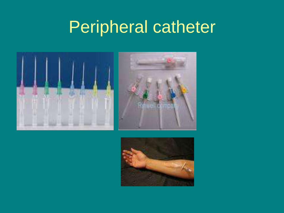
• This is the most common intravenous access method in both hospitals and pre-hospital services. A peripheral IV line consists of a short catheter (a few centimeters long) inserted through the skin into a peripheral vein (any vein that is not inside the chest or abdomen). This is usually in the form of a cannula-over-needle device, in which a flexible plastic cannula comes mounted on a needle. Any accessible vein can be used although arm and hand veins are used most commonly, with leg and foot veins used to a much lesser extent. On infants the scalp veins are sometimes used.
Peripheral catheter
Peripheral catheter
• The caliber of cannula is commonly indicated in gauge, with 14 being a very large cannula (used in resuscitation settings) and 24-26 the smallest. The most common sizes are 16-gauge (midsize line used for blood donation and transfusion), 18- and 20-gauge (all-purpose line for infusions and blood draws), and 22-gauge (all-purpose pediatric line). 12- and 14-gauge peripheral lines actually deliver equivalent volumes of fluid faster than central lines, accounting for their popularity in emergency medicine. These lines are frequently called "large bores" or "trauma lines".

IV site assessment
Note the location (hand, wrist, forearm, antecubital fossa?).
Site should be visually inspected and palpated every 2hr.
The IV site should be free of redness, swelling, tenderness.
The IV dressing should be clean and secure.
For adults, change catheter and rotate site
every 48 - 72 hours. Replace catheters
inserted under emergency conditions
within 24 hours.
Complications
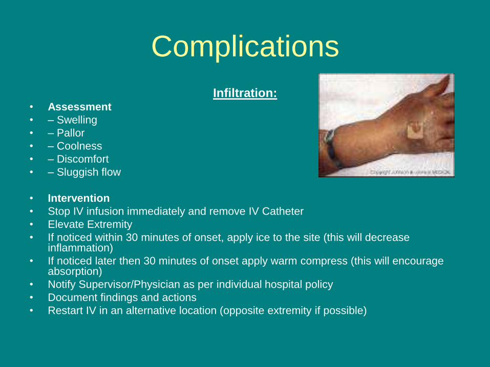
Infiltration: • Assessment
• – Swelling
• – Pallor
• – Coolness
• – Discomfort
• – Sluggish flow
• Intervention
• Stop IV infusion immediately and remove IV Catheter
• Elevate Extremity
• If noticed within 30 minutes of onset, apply ice to the site (this will decrease inflammation)
• If noticed later then 30 minutes of onset apply warm compress (this will encourage absorption)
• Notify Supervisor/Physician as per individual hospital policy
• Document findings and actions
• Restart IV in an alternative location (opposite extremity if possible)
Complications
Plebitis • • Assessment
• – Redness
• – Swelling
• – Warmth
• – Pain along vein route
• – Vein is hard “cordlike”
• – IV may be sluggish
• Interventions
• Stop IV infusion immediately and remove IV Catheter
• Elevate Extremity
• If noticed within 30 minutes of onset, apply ice to the site (this will decrease inflammation)
• If noticed later then 30 minutes of onset apply warm compress (this will encourage absorption)
• Notify Supervisor/Physician as per individual hospital policy
• Document findings and actions
• Restart IV in an alternative location (opposite extremity if possible)
Complications
Infection - Local, Systemic • Assessment
• – Redness, swelling, pain at site
• – Pus at site
• – Fever, chills
• Interventions
• – Prevention!! Adhere to policy for site change and site care.
• – Use appropriate technique for IV starts and site care.
• – Once it occurs, involve physician, discontinue IV and Rx infection per physician order
Complications
(systemic) Air embolus:
• Signs and Symptoms of Air Embolism include:
• Abrupt drop in blood pressure
• Weak, rapid pulse
• Cyanosis
• Chest Pain
• Immediate corrective action for suspected Air Embolism includes:
• Notify Supervisor and Physician immediately
• Immediately place patient on left side with feet elevated (this allows pulmonary artery to absorb small air bubbles)
• Administer O2 if necessary
• Preventive Measures to avoid Air Embolism includes:
• Clear all air from tubing before attaching it to the patient
• Monitor solution levels carefully and change bag before it becomes empty
• Frequently check to assure that all connections are secure
Complications
(systemic)
speed shock
• a sudden adverse physiologic reaction to IV medications or drugs
that are administered too quickly.
• Some signs of speed shock are a flushed face, headache, a tight
feeling in the chest, irregular pulse, loss of consciousness, and
cardiac arrest. • Nursing interventions: notify physician immediately, patent IV for
fluids, reversal, emergency equipment and monitoring.
Six Rights of IV Fluid
Administration
• Right Patient: treat as any drug, use MAR for
accuracy in administration
• Right Drug: solution for IVs
• Right Dose: consider w/time, Amount of IV
solution to hang
• Right Time: rate of solution administration
• Right Route: specific order for IV admin
• Right Documentation: Always!!!!
IV FLUIDS
• The Three Types of Intravenous Fluids are:
• Hypertonic solutions - Any solution that has a higher osmotic pressure than another solution (that is, has a higher concentration of solutes than another solution), which means it draws fluid out of the cell and into the extra-cellular space.
• Hypotonic solutions - Any solution that has a lower osmotic pressure than another solution (that is, has a lower concentration of solutes than another solution), which means it pushes fluid into the cell.
• Isotonic solutions - Any solution that has the same osmotic pressure than another solution (that is, has the same concentration of solutes than another solution), which means it does not draw or push fluid into the cell.
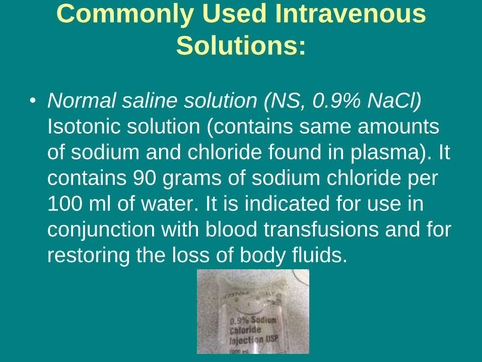
Commonly Used Intravenous
Solutions:
• Normal saline solution (NS, 0.9% NaCl)
Isotonic solution (contains same amounts
of sodium and chloride found in plasma). It
contains 90 grams of sodium chloride per
100 ml of water. It is indicated for use in
conjunction with blood transfusions and for
restoring the loss of body fluids.
Commonly Used Intravenous
Solutions:
• Ringer's Solution or Lactated Ringer's (LR)
Isotonic solution (replaces electrolytes in
amounts similarly found in plasma). It
contains sodium chloride, potassium
chloride, calcium chloride, and sodium
lactate. It is indicated for use as the choice
for burn patients, and in most cases of
dehydration. It is also recommended for
supportive treatment of trauma.

Commonly Used Intravenous
Solutions:
• Five percent dextrose and water (D5W) -
Isotonic solution (after administration and
metabolism of the glucose; D5W becomes
a hypotonic solution). It contains 5 grams
of dextrose per 100 ml of water. It is
indicated for use as a calorie replacement
solution and in cases where glucose is
needed for metabolism purposes.
Commonly Used Intravenous
Solutions:
• ½ Normal Saline Solution – Hypotonic
solution that pushes fluid from the
extracellular space into the cell. Watch if
given to patients with increased ICP i.e.
stroke, head trauma or neurosurgery.
Methods of Intravenous
Administration
– IV push
– Intermittent venous access device
– Intermittent infusion (or piggyback)
– Continuous infusion
– Electronic pumps and controllers
– Patient-controlled analgesia
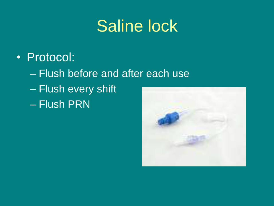
Saline lock
• Protocol:
– Flush before and after each use
– Flush every shift
– Flush PRN
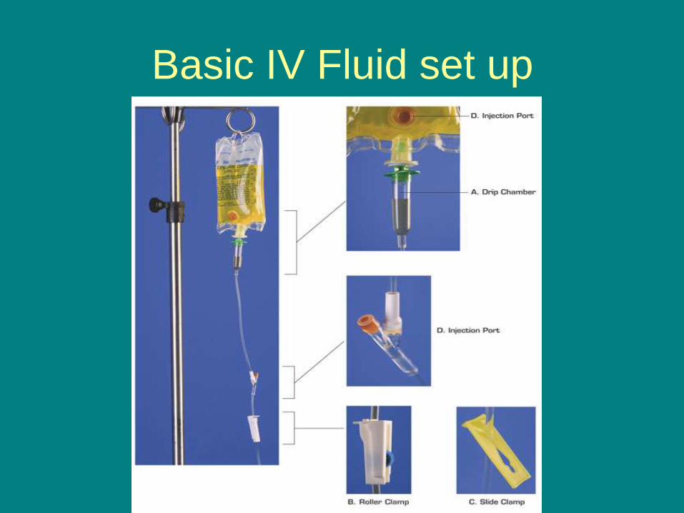
Basic IV Fluid set up
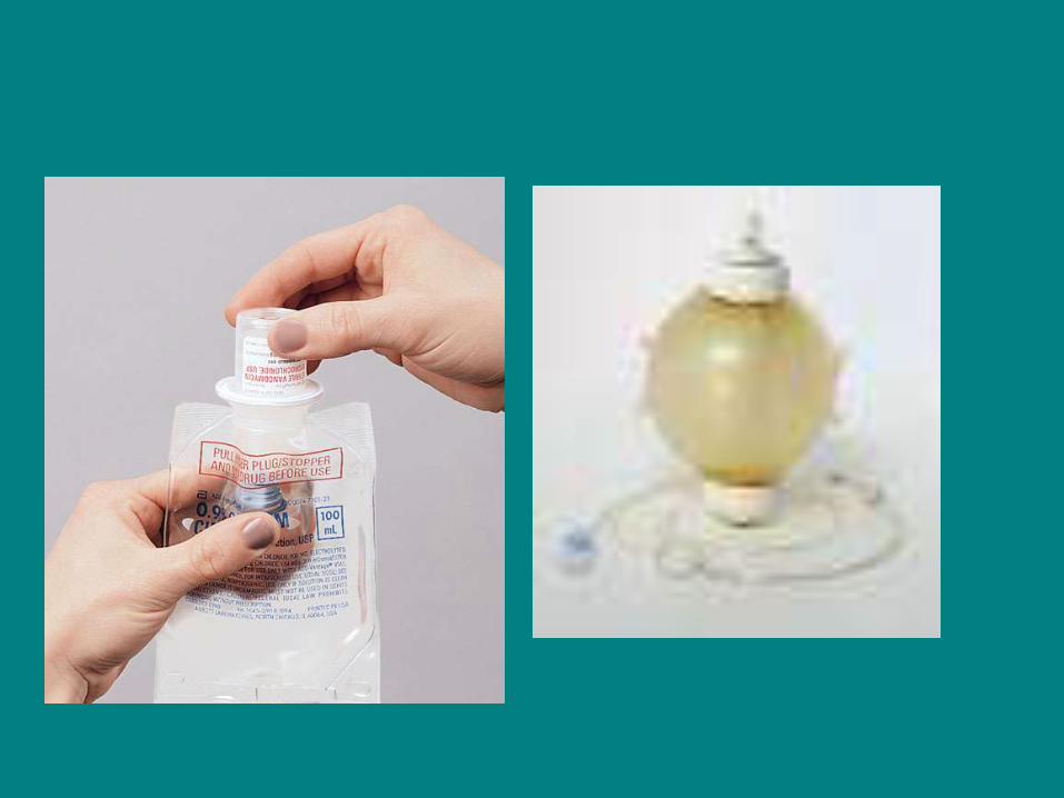

IV tubing
• Microdrip:an apparatus for delivering relatively small measured amounts of IV solutions at specific flow rates over time, as when it is necessary to keep a vein open. The size of the drops is controlled by the fixed diameter of the plastic delivery tube. With a microdrip, 60 drops delivers 1 mL of solution.
• Macrodrip: an apparatus that is used to deliver measured amounts of IV solutions at specific flow rates based on the size of drops of the solution. The size of the drops is controlled by the fixed diameter of a plastic delivery tube. Different macrodrips deliver 10, 15, or 20 drops per milliliter of solution.

General guidelines for tubing
• Change IV tubing, including piggyback
tubing, no more frequently than at 72 hour
intervals.
• Change tubing used for blood or blood
products within 24 hours of completing
infusion.
• Do not leave TPN fluids hanging more
than 24 hours
***Morningside Ministries policy is to change the tubing every 24 hours***
Nursing assessment for fluid volume deficit and fluid
volume overload during IV therapy include:
FVD (Fluid Volume Deficit)
• Dry Skin (Capillary refill > 3 seconds)
• Elevated or Subnormal Temperature
• Thirst
• Dry Mucus Membranes
• Decreased Urine Output
• Soft Sunken Eyeballs ( > then 10% loss of total body fluid volume decreases intraocular pressure and cause eyes to appear to be sunken in)
• Decrease Tearing and Salivating
• Hypotension
FVO (Fluid Volume Overload)
• Pitting Edema (1+ - 4+)
• Puffy Eyelids
• Acute weight gain
• Elevated blood pressure
• Bounding pulse
• Dyspnea and shortness of breath (Usually first sign)
• Ascites or third spacing
Other nursing assessment observations that are
important during IV therapy include:
• Close monitoring of weight gain/loss
• Accurate I and O (normal urine output is approximately 1 Ml / Kg of body wt. per hour)
• Assessing for signs of edema (skin that is tight and shiny)
• Assessing for skin turgor that when pinched takes longer then 3 seconds to return to normal.
• Assessing lung sounds (crackles will be heard with FVO)
• Notification to physician if urine output is < 30cc for two consecutive hours
• Monitor sodium and hematocrit levels
Critical Elements
• Assess IV site every two hours and prn.
• Verify patency.
• Observe the Six Rights of IV fluid administration.
• Prime tubing, clearing air.
• Identify tubing parts that are to remain sterile and maintain sterility.
• Apply principles of sterile technique throughout procedure.
• Document tubing change and site care.
• Secures IV catheter with sterile dressing per agency policy.

IV drip rate calculation
• Calculate an IV drip rate based on the volume of fluid to be
infused over time:
Gtt/min=volume to be infused(mL) x drop factor(gtt/mL)
time (minutes)
• Calculate an IV drip rate for a medication that is administered
based on a specific dosage to be infused per minute:
Gtt/min=dosage per minute to be administered x drop factor
concentration of medication per mL
Review
• IV site changed every 72 hours routinely
• IV tubing changed every 24 hours routinely
• Nurse may attempt IV only twice before seeking
another nurse to attempt starting IV
• IV push medications are not routinely performed
in MSM skilled facilities
• All nurses upon employment must complete IV
competency training and be skill checked with
1st IV start with a resident by a nurse who is
authorized to observe.