Embed Size (px)
Citation preview

University of Liège!GBI001.1 JGV 1/05/10
Introduction au génie biomédical!GBI0 001!
Imagerie médicale!
Prof. Jacques Verly!Departement dʼElectricité, Electronique et Informatique!
(Institut Montefiore)!
University of Liège!GBI001.2 JGV 1/05/10
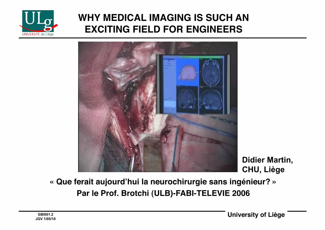
WHY MEDICAL IMAGING IS SUCH AN EXCITING FIELD FOR ENGINEERS!
« Que ferait aujourdʼhui la neurochirurgie sans ingénieur? »!Par le Prof. Brotchi (ULB)-FABI-TELEVIE 2006!
Didier Martin, CHU, Liège
University of Liège!GBI001.3 JGV 1/05/10
OVERVIEW OF IMAGING MODALITIES!
University of Liège!GBI001.4 JGV 1/05/10
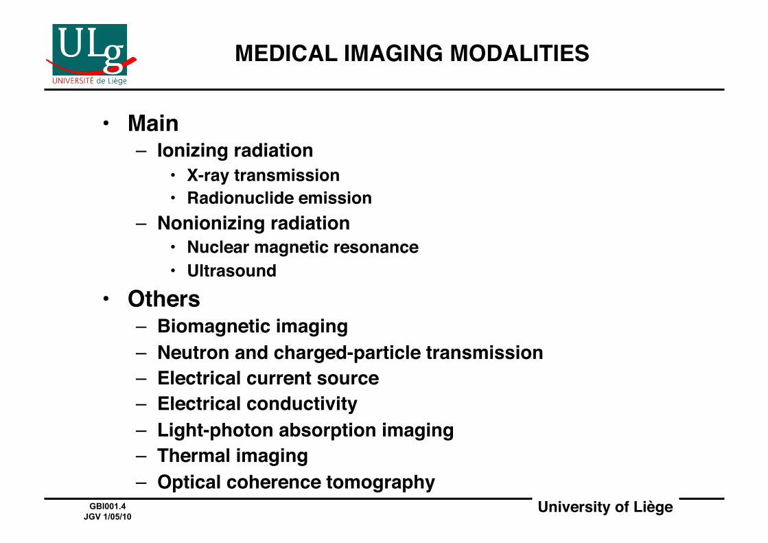
MEDICAL IMAGING MODALITIES!
• Main!– Ionizing radiation!
• X-ray transmission!• Radionuclide emission!
– Nonionizing radiation!• Nuclear magnetic resonance!• Ultrasound!
• Others!– Biomagnetic imaging!– Neutron and charged-particle transmission!– Electrical current source!– Electrical conductivity!– Light-photon absorption imaging!– Thermal imaging!– Optical coherence tomography!
University of Liège!GBI001.5 JGV 1/05/10
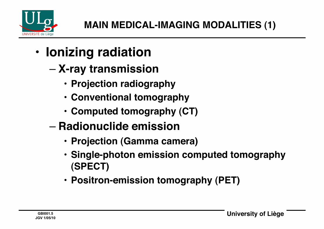
• Ionizing radiation!– X-ray transmission!
• Projection radiography!• Conventional tomography!• Computed tomography (CT)!
– Radionuclide emission!• Projection (Gamma camera)!• Single-photon emission computed tomography
(SPECT)!• Positron-emission tomography (PET)!
MAIN MEDICAL-IMAGING MODALITIES (1)!
University of Liège!GBI001.6 JGV 1/05/10
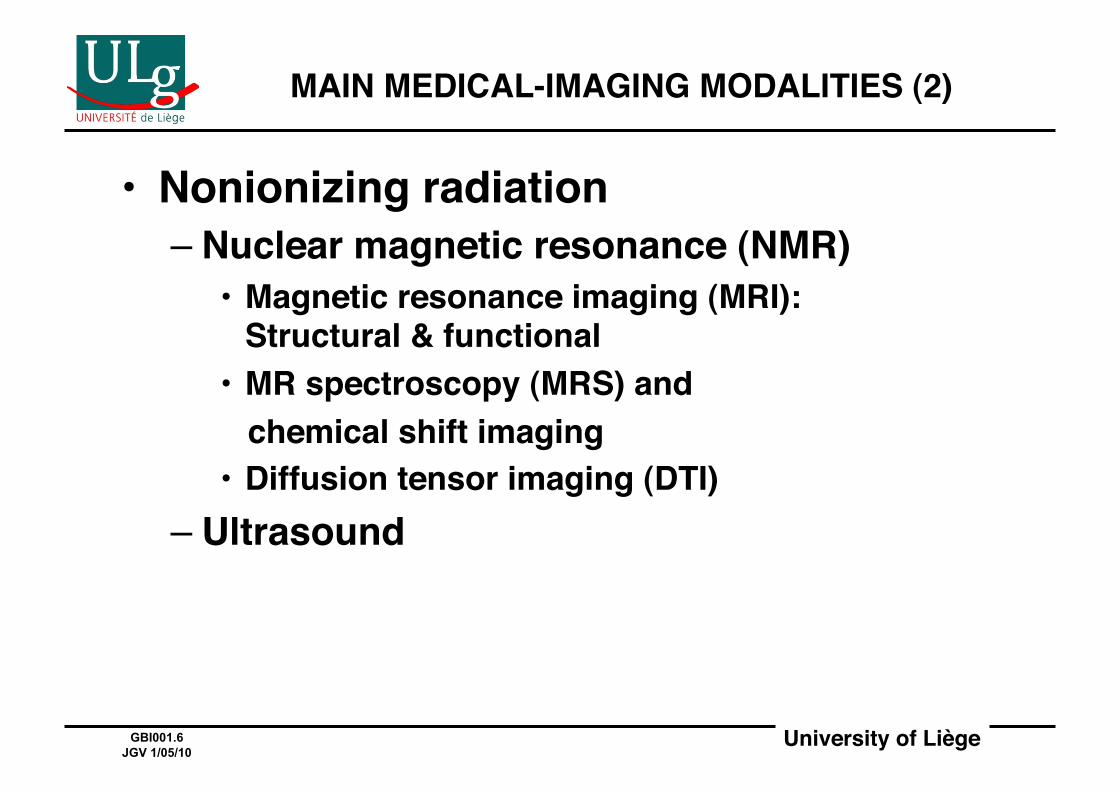
• Nonionizing radiation!– Nuclear magnetic resonance (NMR)!
• Magnetic resonance imaging (MRI):Structural & functional!
• MR spectroscopy (MRS) and ! chemical shift imaging!• Diffusion tensor imaging (DTI)!
– Ultrasound!
MAIN MEDICAL-IMAGING MODALITIES (2)!
University of Liège!GBI001.7 JGV 1/05/10
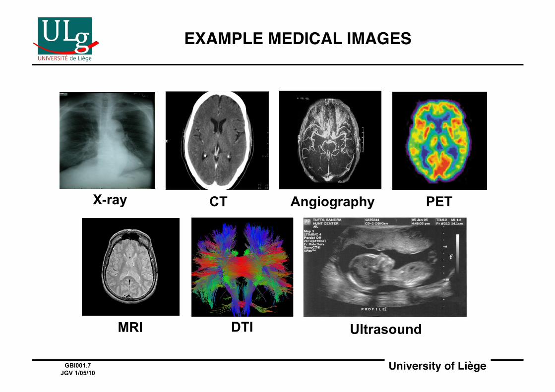
EXAMPLE MEDICAL IMAGES!
X-ray CT Angiography PET
MRI DTI Ultrasound

University of Liège!GBI001.8 JGV 1/05/10
EXAMPLE MEDICAL IMAGING EQUIPMENT!
University of Liège!GBI001.9 JGV 1/05/10
PHYSICS OF IONIZING RADIATION!

University of Liège!GBI001.10 JGV 1/05/10
STRUCTURE OF ATOMS (1)!
University of Liège!GBI001.11 JGV 1/05/10
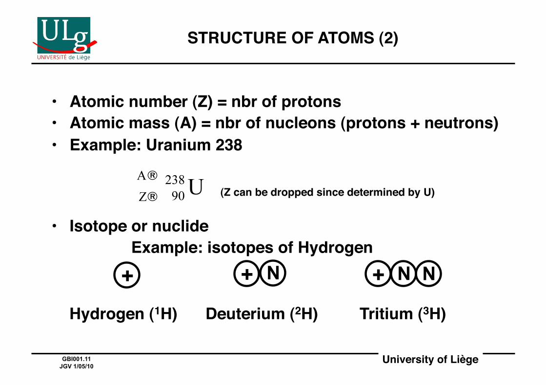
• Atomic number (Z) = nbr of protons!• Atomic mass (A) = nbr of nucleons (protons + neutrons)!• Example: Uranium 238!
! ! ! !
! ! (Z can be dropped since determined by U)!
• Isotope or nuclide!! ! Example: isotopes of Hydrogen!
Hydrogen (1H) ! Deuterium (2H)! Tritium (3H)!
STRUCTURE OF ATOMS (2)!
+ + N + N N
University of Liège!GBI001.12 JGV 1/05/10
RADIOACTIVE DECAY, RADIONUCLIDES, AND RADIOACTIVE MATERIALS!
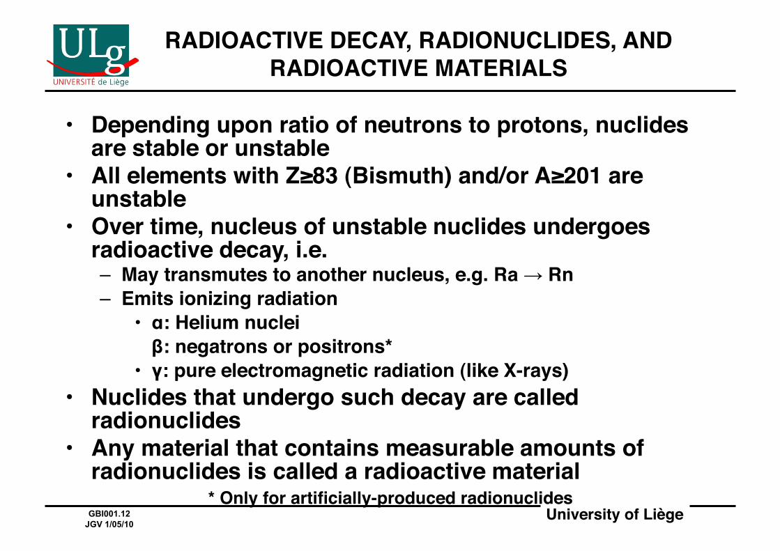
• Depending upon ratio of neutrons to protons, nuclides are stable or unstable!
• All elements with Z≥83 (Bismuth) and/or A≥201 are unstable!
• Over time, nucleus of unstable nuclides undergoes radioactive decay, i.e.!– May transmutes to another nucleus, e.g. Ra → Rn!– Emits ionizing radiation!
• α: Helium nuclei!! β: negatrons or positrons* !• γ: pure electromagnetic radiation (like X-rays)!
• Nuclides that undergo such decay are called radionuclides!
• Any material that contains measurable amounts of radionuclides is called a radioactive material!
* Only for artificially-produced radionuclides!
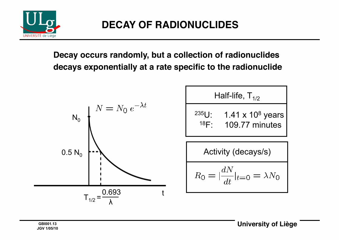
University of Liège!GBI001.13 JGV 1/05/10
DECAY OF RADIONUCLIDES!
Decay occurs randomly, but a collection of radionuclides !decays exponentially at a rate specific to the radionuclide!
Half-life, T1/2
235U: 1.41 x 10⁸ years 18F: 109.77 minutes
Activity (decays/s)
N0
0.5 N0
T1/2 = 0.693 λ
t

University of Liège!GBI001.14 JGV 1/05/10
WHAT IS IONIZING (OR PENETRATING) RADIATION ?!
• Ionizing radiation consists of charged and/or neutral "particles" that create ion-pairs in target material!
• An ion-pair is a pair of oppositely-charged ions held together by Coulomb attraction without formation of a covalent bond!
• An ion-pair results from the ejection of one or more orbital electrons!
• An ion-pair behaves as one unit!
• Examples: α-particles, X-rays, γ-rays!
University of Liège!GBI001.15 JGV 1/05/10
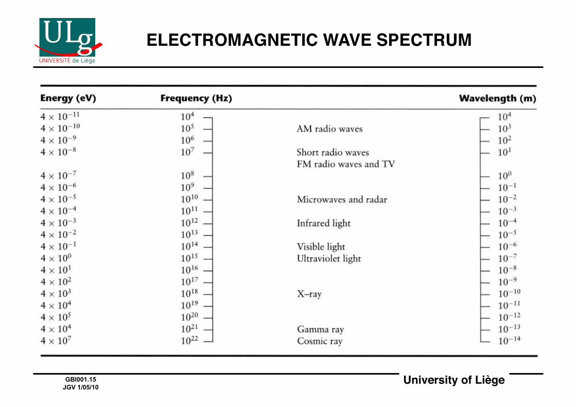
ELECTROMAGNETIC WAVE SPECTRUM!
University of Liège!GBI001.16 JGV 1/05/10
SOURCES OF IONIZING RADIATION!
• Natural!– Terrestrial: natural radioactivity (mostly 232Th, 235U, 238U )!– Extraterrestrial: cosmic rays (principally protons)!
• Man-made!– X-rays (since 1895)!– Artificial radioactivity!
• Unavoidable by-product of nuclear reactions!• Deliberately-produced by particle accelerators!
– Industrial applications!– Medical applications (e.g. 18F)!
University of Liège!GBI001.17 JGV 1/05/10
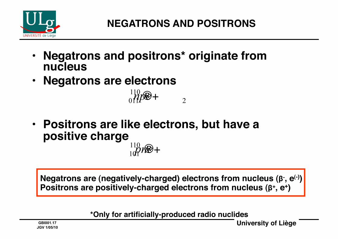
NEGATRONS AND POSITRONS!
• Negatrons and positrons* originate from nucleus!
• Negatrons are electrons!
• Positrons are like electrons, but have a positive charge!
Negatrons are (negatively-charged) electrons from nucleus (β-, e(-))Positrons are positively-charged electrons from nucleus (β+, e+)!
*Only for artificially-produced radio nuclides!
University of Liège!GBI001.18 JGV 1/05/10
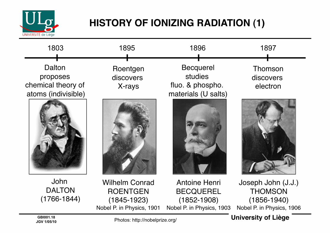
HISTORY OF IONIZING RADIATION (1)!
1803 1895 1896 1897
Dalton proposes
chemical theory of atoms (indivisible)#
Roentgen discovers
X-rays#
Becquerel studies
fluo. & phospho. materials (U salts)#
Thomson discovers electron#
John DALTON
(1766-1844)#
Wilhelm Conrad ROENTGEN (1845-1923)
Nobel P. in Physics, 1901#
Antoine Henri BECQUEREL (1852-1908)
Nobel P. in Physics, 1903#
Joseph John (J.J.)THOMSON (1856-1940)
Nobel P. in Physics, 1906#
Photos: http://nobelprize.org/
University of Liège!GBI001.19 JGV 1/05/10
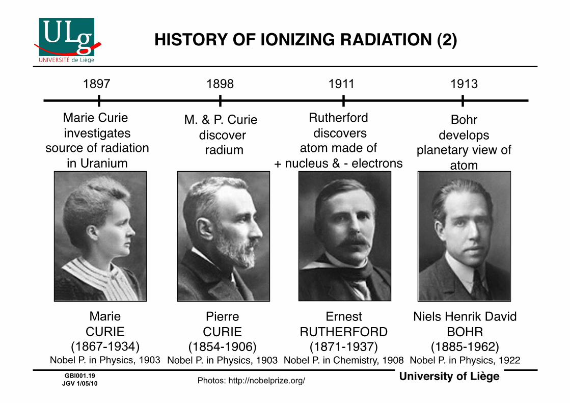
HISTORY OF IONIZING RADIATION (2)!
1897 1898 1911 1913
Marie Curie investigates
source of radiation in Uranium#
M. & P. Curie discover radium#
Rutherford discovers
atom made of + nucleus & - electrons#
Bohrdevelops
planetary view of atom#
Marie CURIE
(1867-1934) Nobel P. in Physics, 1903
Pierre CURIE
(1854-1906)Nobel P. in Physics, 1903#
ErnestRUTHERFORD
(1871-1937)Nobel P. in Chemistry, 1908#
Niels Henrik David BOHR
(1885-1962)Nobel P. in Physics, 1922#
Photos: http://nobelprize.org/
University of Liège!GBI001.20 JGV 1/05/10
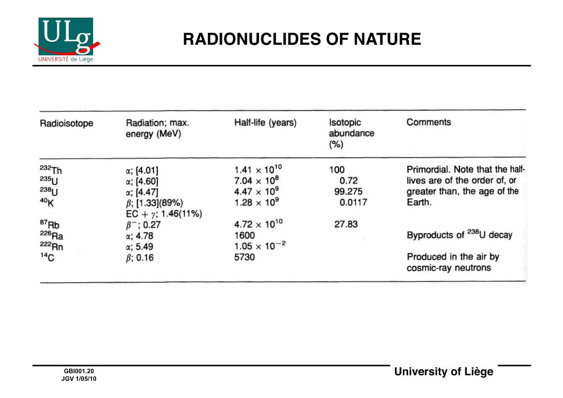
RADIONUCLIDES OF NATURE!
University of Liège!GBI001.21 JGV 1/05/10
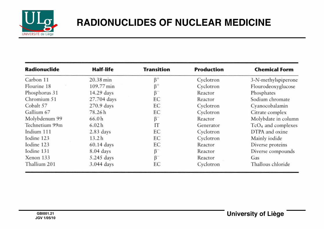
RADIONUCLIDES OF NUCLEAR MEDICINE!
University of Liège!GBI001.22 JGV 1/05/10
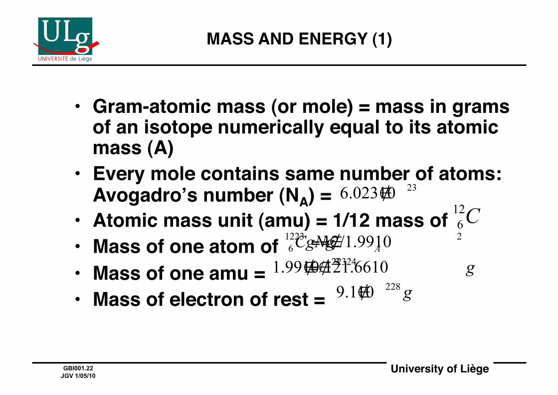
MASS AND ENERGY (1)!
• Gram-atomic mass (or mole) = mass in grams of an isotope numerically equal to its atomic mass (A)!
• Every mole contains same number of atoms: Avogadroʼs number (NA) =!
• Atomic mass unit (amu) = 1/12 mass of !• Mass of one atom of !• Mass of one amu = !• Mass of electron of rest = !
University of Liège!GBI001.23 JGV 1/05/10
MASS AND ENERGY (2)!
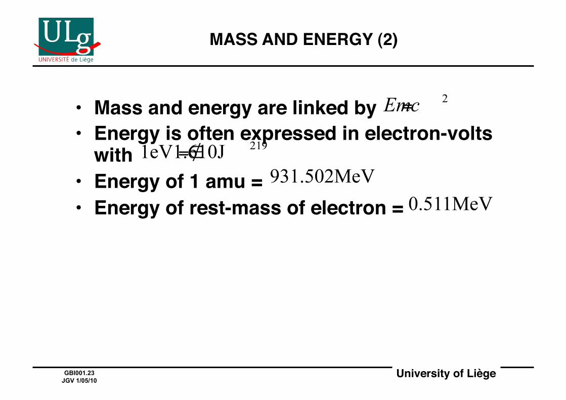
• Mass and energy are linked by !• Energy is often expressed in electron-volts
with !• Energy of 1 amu = !• Energy of rest-mass of electron = !

University of Liège!GBI001.24 JGV 1/05/10
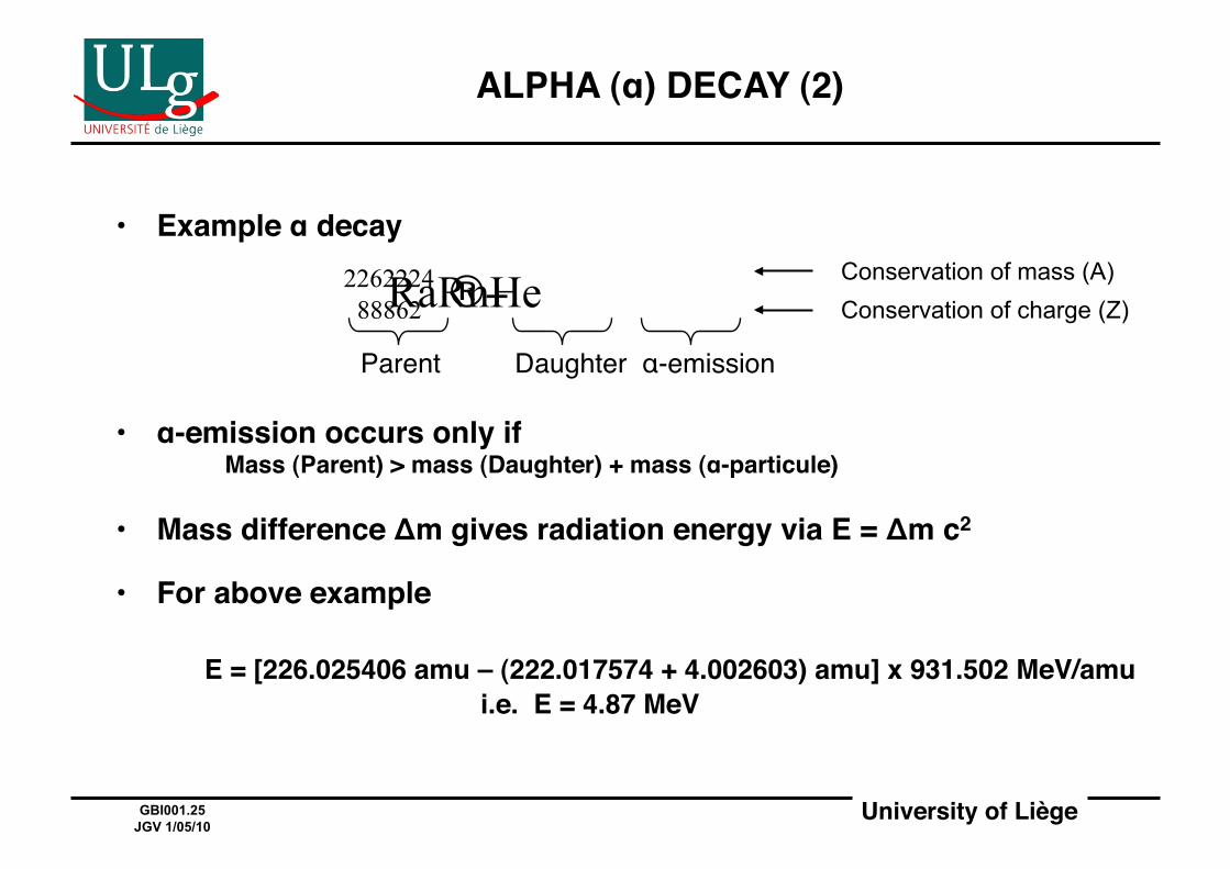
ALPHA (α) DECAY (1)!
Too many protons causing excessive repulsive force → a Helium nucleus is emitted!
http://library.thinkquest.org/3471/radiation_types_body.html
University of Liège!GBI001.25 JGV 1/05/10
ALPHA (α) DECAY (2)!
• Example α decay!
• α-emission occurs only if!! ! Mass (Parent) > mass (Daughter) + mass (α-particule)!
• Mass difference Δm gives radiation energy via E = Δm c2!
• For above example!
! E = [226.025406 amu – (222.017574 + 4.002603) amu] x 931.502 MeV/amu!! ! ! ! i.e. E = 4.87 MeV!
Parent# Daughter α-emission
Conservation of mass (A) Conservation of charge (Z)
University of Liège!GBI001.26 JGV 1/05/10
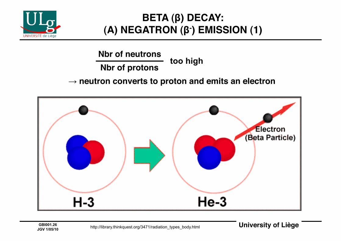
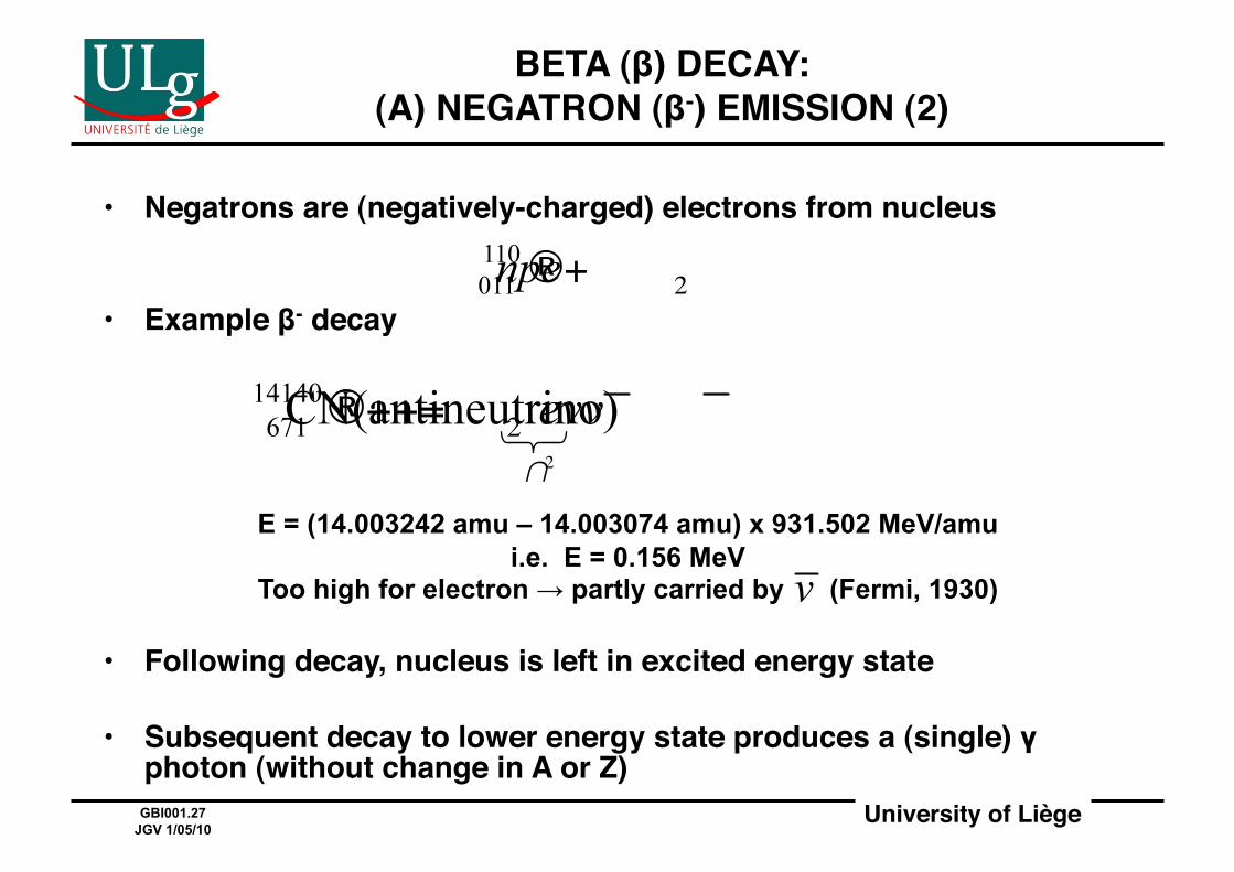
BETA (β) DECAY: (A) NEGATRON (β-) EMISSION (1)!
too high
→ neutron converts to proton and emits an electron!
http://library.thinkquest.org/3471/radiation_types_body.html
Nbr of neutrons!Nbr of protons!
University of Liège!GBI001.27 JGV 1/05/10
• Negatrons are (negatively-charged) electrons from nucleus!
• Example β- decay!
• Following decay, nucleus is left in excited energy state!
• Subsequent decay to lower energy state produces a (single) γ photon (without change in A or Z) !
BETA (β) DECAY: (A) NEGATRON (β-) EMISSION (2)!
E = (14.003242 amu – 14.003074 amu) x 931.502 MeV/amu i.e. E = 0.156 MeV
Too high for electron → partly carried by (Fermi, 1930)
University of Liège!GBI001.28 JGV 1/05/10
NEUTRINO ( )!
• Neutrino = "little neutral one"!• Existence postulated by Fermi in 1930!• Zero electrical charge!• Resting mass smaller than that of electron!• Interacts very weakly with matter → difficult to
detect!• Only detected experimentally in 1950!• Antineutrino ( ) in antiparticule to neutrino!• When a particle collides with its antiparticle,
they annihilate and release electromagnetic radiation, e.g. γ-rays!
University of Liège!GBI001.29 JGV 1/05/10
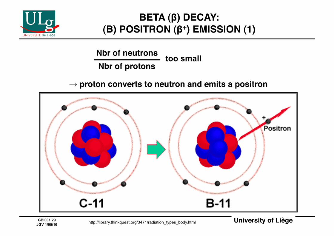
BETA (β) DECAY: (B) POSITRON (β+) EMISSION (1)!
too small
→ proton converts to neutron and emits a positron!
http://library.thinkquest.org/3471/radiation_types_body.html
Nbr of neutrons!Nbr of protons!
University of Liège!GBI001.30 JGV 1/05/10
BETA (β) DECAY: (B) POSITRON (β+) EMISSION (2)!
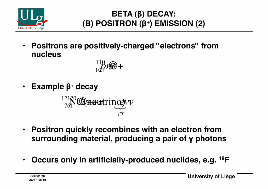
• Positrons are positively-charged "electrons" from nucleus!
• Example β+ decay!
• Positron quickly recombines with an electron from surrounding material, producing a pair of γ photons!
• Occurs only in artificially-produced nuclides, e.g. 18F!
University of Liège!GBI001.31 JGV 1/05/10
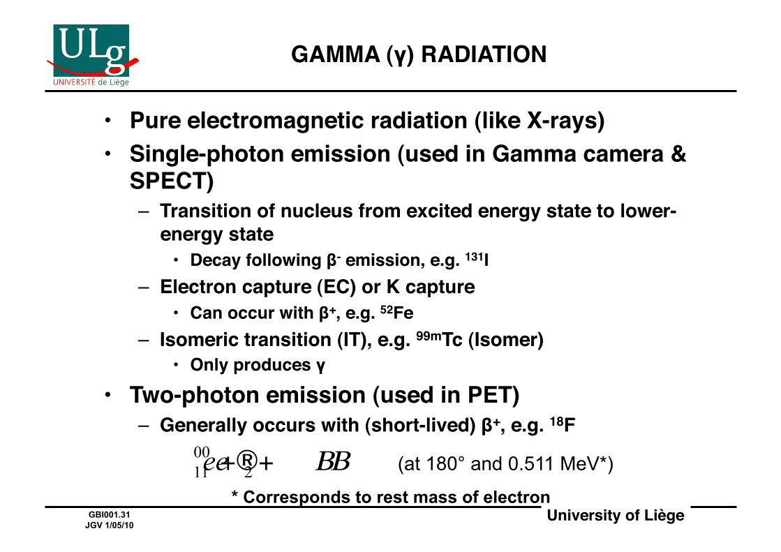
GAMMA (γ) RADIATION!
• Pure electromagnetic radiation (like X-rays)!• Single-photon emission (used in Gamma camera &
SPECT)!– Transition of nucleus from excited energy state to lower-
energy state!• Decay following β- emission, e.g. 131I!
– Electron capture (EC) or K capture!• Can occur with β+, e.g. 52Fe!
– Isomeric transition (IT), e.g. 99mTc (Isomer)!• Only produces γ!
• Two-photon emission (used in PET)!– Generally occurs with (short-lived) β+, e.g. 18F!
(at 180° and 0.511 MeV*)
* Corresponds to rest mass of electron

University of Liège!GBI001.32 JGV 1/05/10
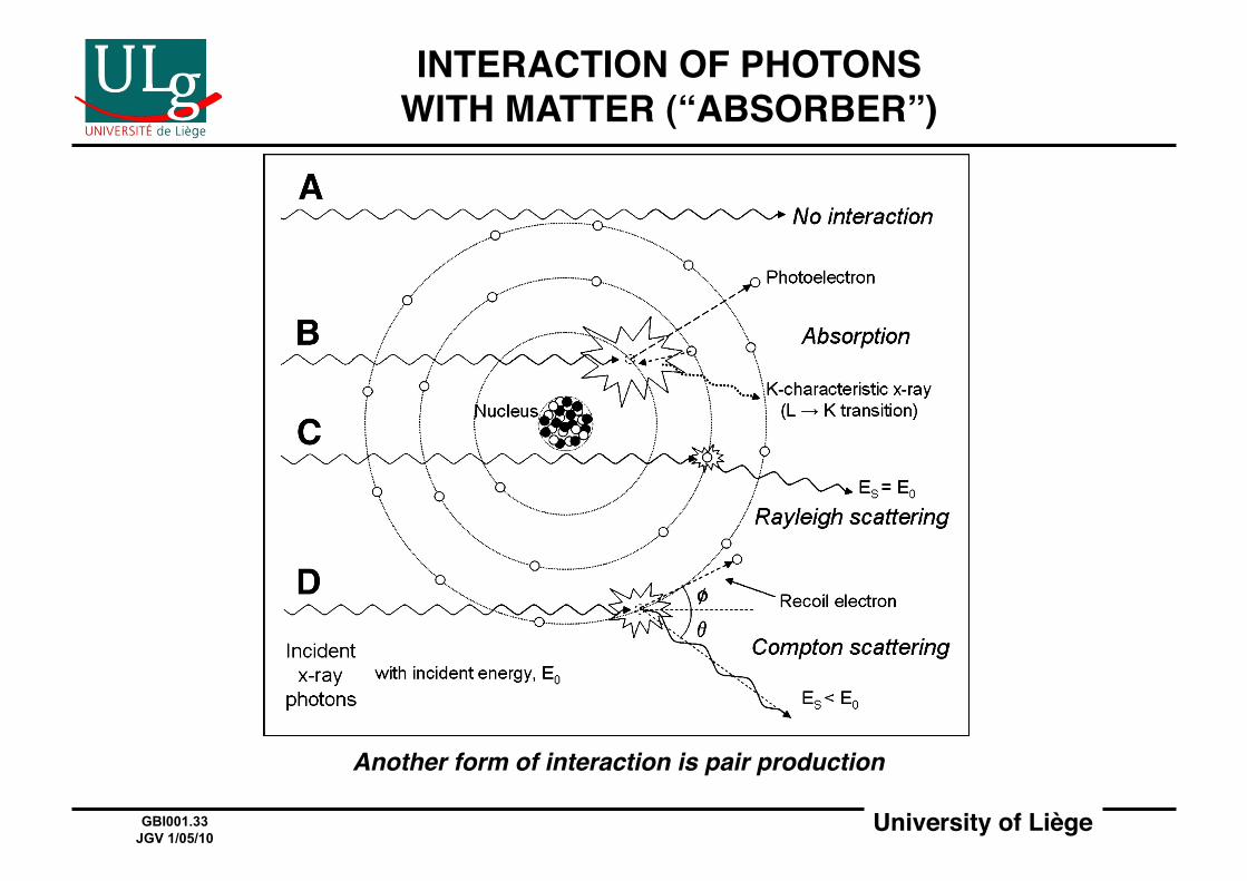
INTERACTION OF IONIZING RADIATION WITH MATTER (“ABSORBER”)!
• Charged particles (p, α, β-, β+, e, …) – In general: many small energy transfers resulting in
ionization – β+ is short-lived and quickly recombines with e
• Neutral "particles" (n, X, γ, …) – In general: largely-unimpeded travel, then one-time
large energy transfer – Neutrons (n) – Photons (X, γ)
• Photoelectric effect (E < 0.2 MeV) • Rayleigh scattering • Compton scattering (0.2 ≤ E < 5 MeV) • Pair production* (E ≥ 1.2 MeV)
(opposite of annihilation)#
University of Liège!GBI001.33 JGV 1/05/10
INTERACTION OF PHOTONS WITH MATTER (“ABSORBER”)!
Another form of interaction is pair production!
University of Liège!GBI001.34 JGV 1/05/10
"ATTENUATION" OF IONIZING RADIATION BY MATTER!
• Charged particles!– Rate of loss of energy (dE/dx) given by Bethe-Block (or
stopping-power) equation!– Uniform loss rate along particle track!
• β emission in water: 2 MeV cm-1!– Stopping distance ("range″)!
• 10 MeV α particle in water: 1 cm!• Neutral “particles”!
– Microscopically: cross-section σ!– Macroscopically: linear attenuation coefficient, μ
(probability of one interaction per unit length)!– Mean free path (1/μ):!
• 1 MeV γ photon in Pb: 1.3 cm!
University of Liège!GBI001.35 JGV 1/05/10
IMAGING BY X-RAY TRANSMISSION!
University of Liège!GBI001.36 JGV 1/05/10
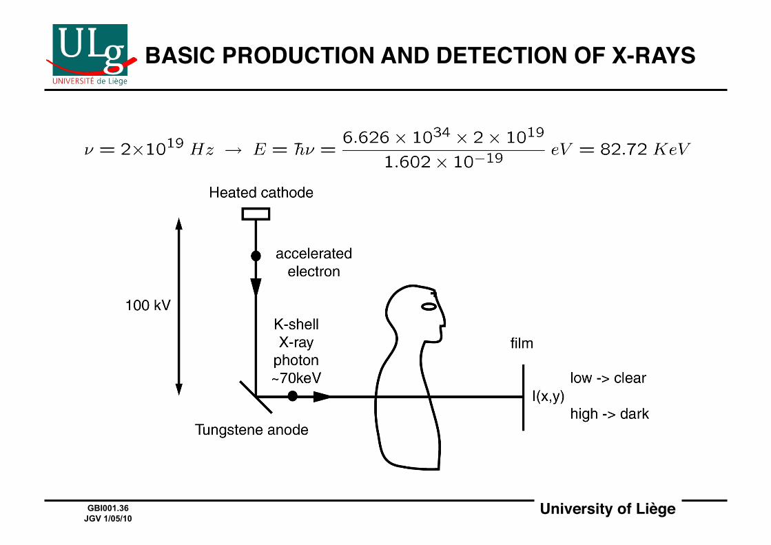
BASIC PRODUCTION AND DETECTION OF X-RAYS!
University of Liège!GBI001.37 JGV 1/05/10
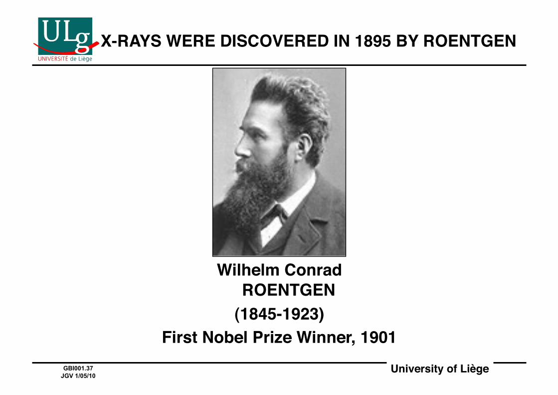
X-RAYS WERE DISCOVERED IN 1895 BY ROENTGEN!
Wilhelm Conrad ROENTGEN!
(1845-1923)!First Nobel Prize Winner, 1901!
University of Liège!GBI001.38 JGV 1/05/10
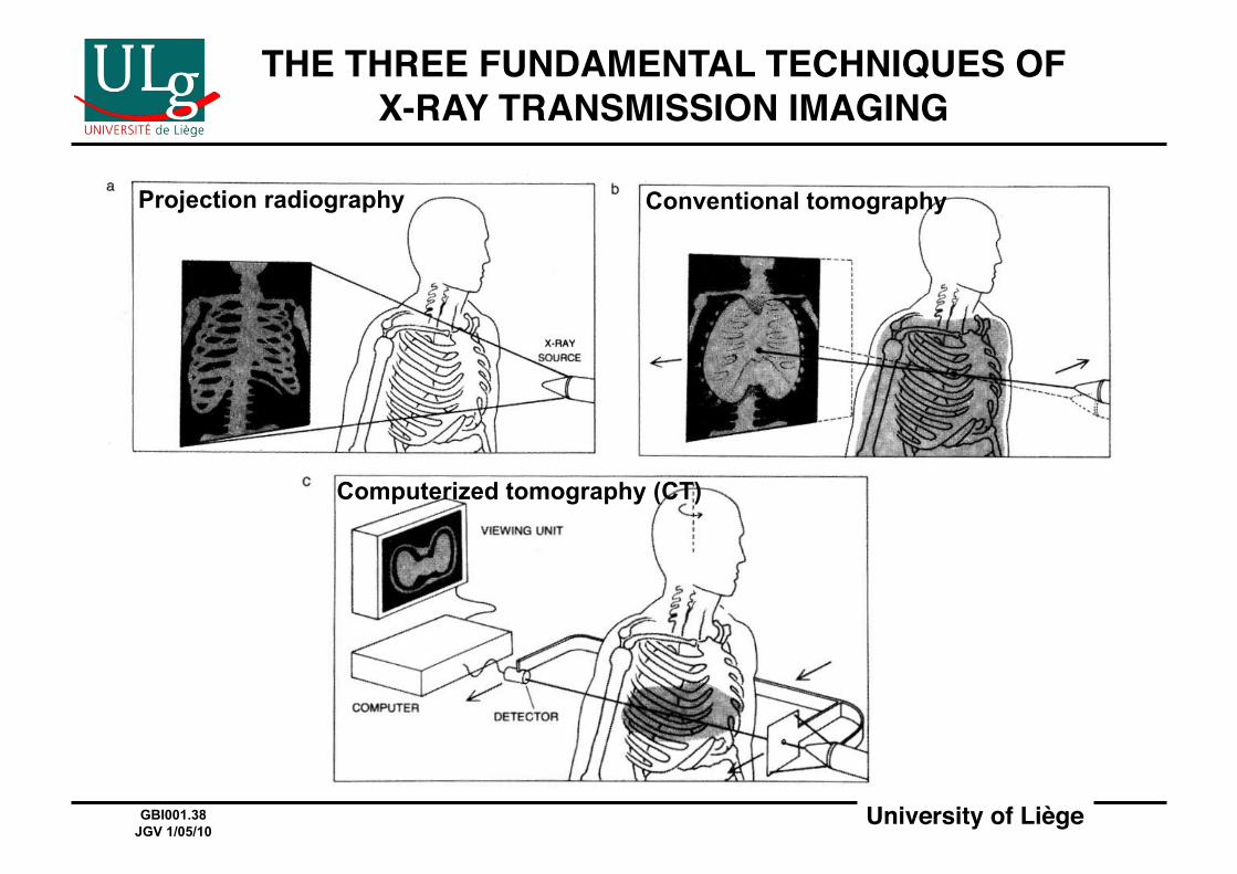
THE THREE FUNDAMENTAL TECHNIQUES OF X-RAY TRANSMISSION IMAGING!
Projection radiography Conventional tomography
Computerized tomography (CT)

University of Liège!GBI001.39 JGV 1/05/10
MAIN INTERACTION OF X-RAYS WITH MATTER:IONIZATION!
• Ionization: formation of ion pairs!• Binding energy: 13.6 Z2 for K-shell electron!• Photoelectric effect: process by which photon knocks off
electron!• Coulomb interaction: process by which e knocks off e!• A 50 KeV X-ray photon will ionize ~2000 atoms in air!• Photoelectric effect and Coulomb interactions are mostly
responsible for attenuation, stopping, and damage!
Visible (500 nm) 2.48 eV X-ray ~70 KeV γ-ray ~1 MeV
University of Liège!GBI001.40 JGV 1/05/10
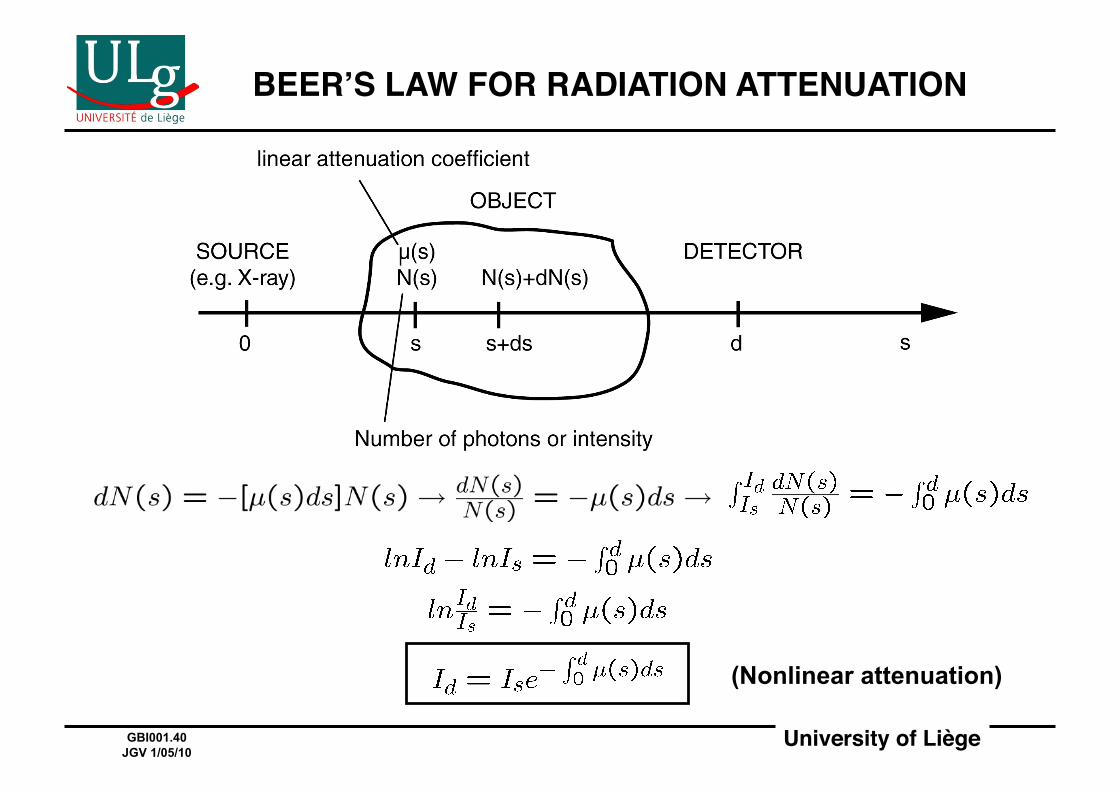
BEERʼS LAW FOR RADIATION ATTENUATION!
(Nonlinear attenuation)

University of Liège!
EXAMPLE OF ATTENUATION AND CONTRAST!CALCULATION!
" PATIENT "
LUNG (~AIR)
LUNG LUNG
LUNG
LUNG LUNG
CHEST CHEST
CHEST CHEST
chest
chest chest
chest RIB RIB
RIB RIB
tumor
tumor
35cm
3 3
3 3 3
3
29
29
13 13
13 13
1.5 1.5 1.5 1.5
1.5 1.5 1.5 1.5
0.14
0.14
0.14 0.05
0.4 0.4
I0
X-ray I
µ (cm-1) 24%
24%
55%
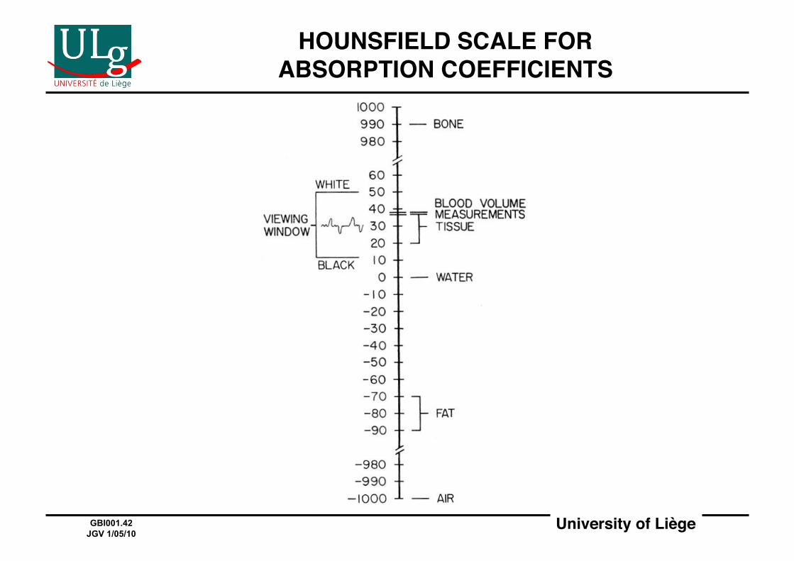
University of Liège!GBI001.42 JGV 1/05/10
HOUNSFIELD SCALE FOR ABSORPTION COEFFICIENTS!
University of Liège!GBI001.43 JGV 1/05/10
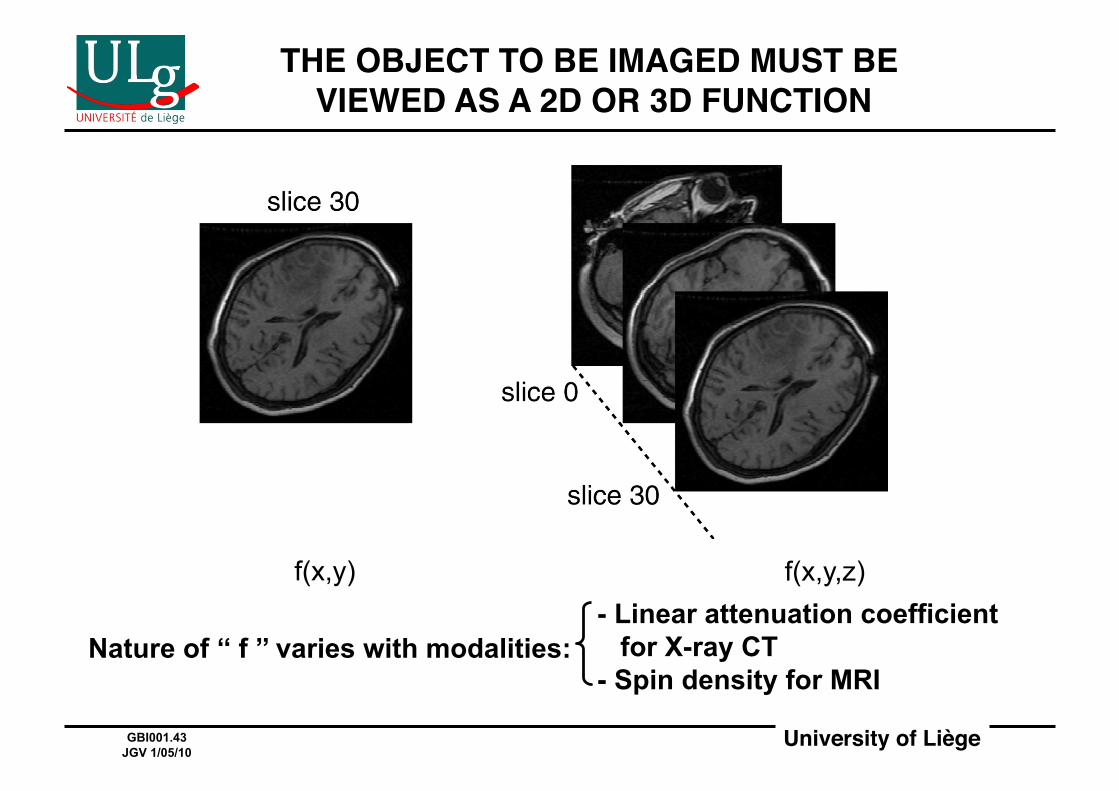
THE OBJECT TO BE IMAGED MUST BE VIEWED AS A 2D OR 3D FUNCTION!
f(x,y) f(x,y,z)
Nature of ‘‘ f ’’ varies with modalities: - Linear attenuation coefficient for X-ray CT - Spin density for MRI

University of Liège!GBI001.44 JGV 1/05/10
LINE INTEGRALS AND PROJECTIONS!
Shown in 2D but generalizable to 3D
and even ND
Line integral along l

University of Liège!GBI001.45 JGV 1/05/10
FUNDAMENTAL PROBLEM:2D IMAGE RECONSTRUCTION FROM PROJECTIONS!
p Є(-∞,∞)!
y
x
f(x,y)
p l Φ
2D function p(p,Φ)
is the Radon transform of f(x,y)
(Rf)(p,Φ)=RADON{f(x,y)}
(Radon, 1917)
Amazingly, it is possible to recover f(x,y) from all p(p,Φ)’s!
University of Liège!GBI001.46 JGV 1/05/10
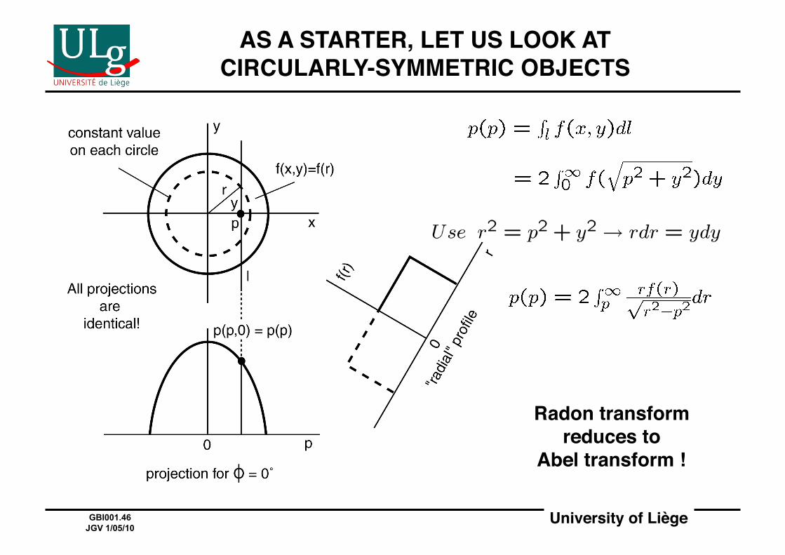
AS A STARTER, LET US LOOK ATCIRCULARLY-SYMMETRIC OBJECTS!
Radon transform!reduces to!
Abel transform !!
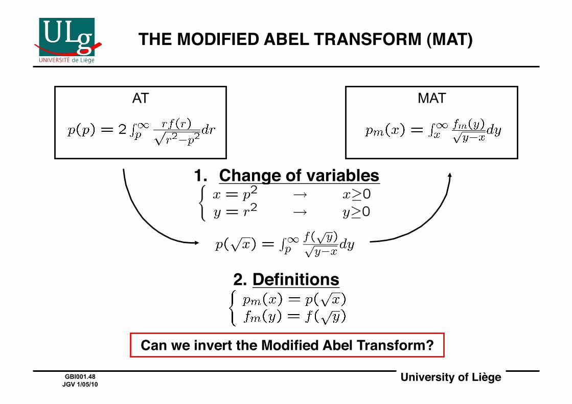
University of Liège!GBI001.47 JGV 1/05/10
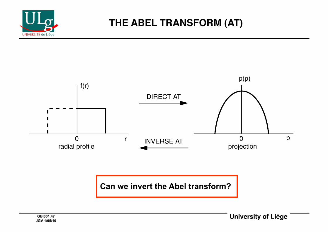
THE ABEL TRANSFORM (AT)!
Can we invert the Abel transform?
University of Liège!GBI001.48 JGV 1/05/10
THE MODIFIED ABEL TRANSFORM (MAT)!
1. Change of variables!
AT
2. Definitions!
Can we invert the Modified Abel Transform?!
MAT
University of Liège!GBI001.49 JGV 1/05/10
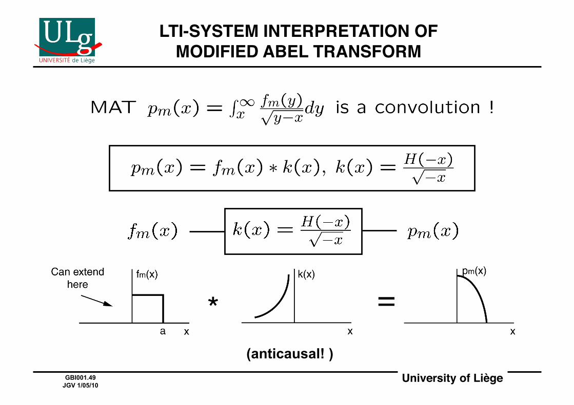
LTI-SYSTEM INTERPRETATION OFMODIFIED ABEL TRANSFORM!
(anticausal! )
University of Liège!GBI001.50 JGV 1/05/10
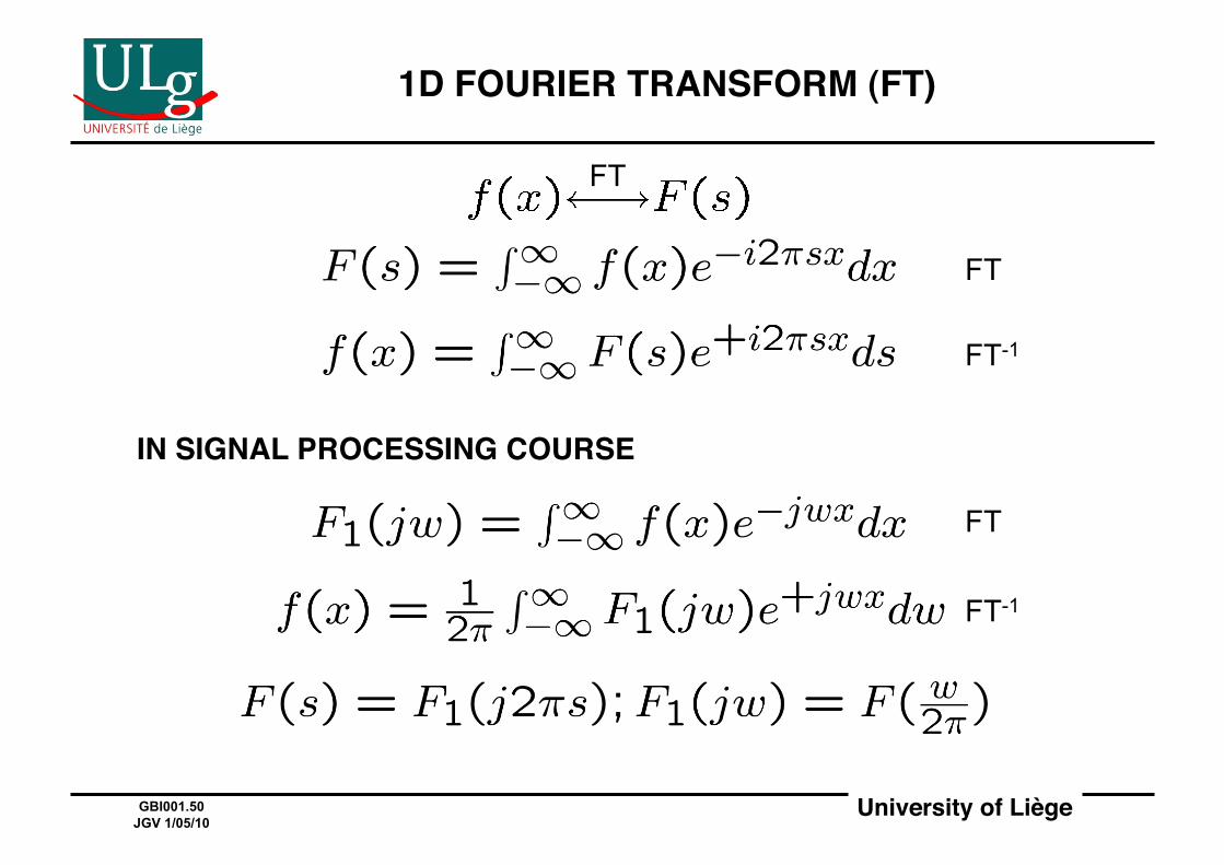
1D FOURIER TRANSFORM (FT)!
IN SIGNAL PROCESSING COURSE!
FT
FT-1
FT
FT-1
FT
University of Liège!GBI001.51 JGV 1/05/10
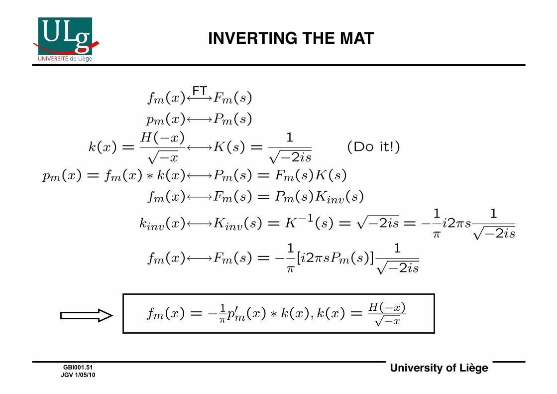
INVERTING THE MAT!
FT

University of Liège!GBI001.52 JGV 1/05/10
LTI-SYSTEM INTERPRETATION OF MAT AND INVERSE MAT!
MAT INVERSE MAT
University of Liège!GBI001.53 JGV 1/05/10
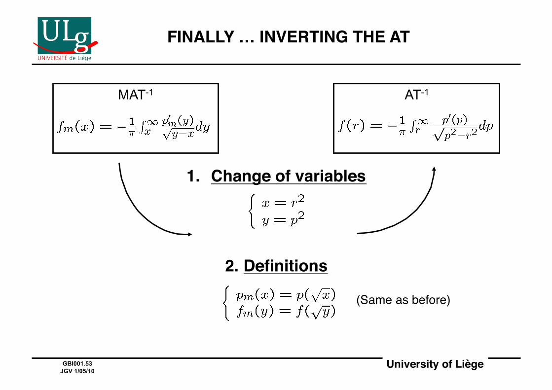
FINALLY … INVERTING THE AT!
1. Change of variables!
2. Definitions!
(Same as before)#
MAT-1 AT-1

University of Liège!GBI001.54 JGV 1/05/10
THE ABEL TRANSFORM AND ITS INVERSE!
There are tables of AT pairs (just as for FTʼs)!
Example:!
AT
AT
University of Liège!GBI001.55 JGV 1/05/10
FROM ABEL TO RADON!
• The AT and its inverse are the key to understanding image reconstruction from projections for circularly-symmetric objects!
• The existence of AT-1 is reassuring and gives hopes that one can reconstruct arbitrary objects from their projections!
• The RT generalizes the AT to arbitrary objects!
• Amazingly, the RT also has an inverse !!
• The fundamental result for understanding most of image reconstruction from projections is the projection-slice theorem!

University of Liège!GBI001.56 JGV 1/05/10
KEY INITIAL CONTRIBUTORS TO IMAGE RECONSTRUCTION FROM PROJECTIONS!
1917 1956 1963 1968
Radon proposes
a backprojection method#
Bracewell proposes
projection-slice theorem#
Cormackpublishes
a first paper#
Hounsfield files
patent for CT scanner#
Johann RADON
(1887-1956)#
Ronald Newbold BRACEWELL (1921-2007 )
Allan M.CORMACK(1924-1998)
Nobel P. in Physiology or Medicine, 1979#
Godfrey N.HOUNSFIELD (1919-2004)
Nobel P. in Physiology or Medicine, 1979#
Photos: http://nobelprize.org/

University of Liège!GBI001.57 JGV 1/05/10
2D FOURIER TRANSFORM (FT)!
Most theorems and properties of 1D FT!extend to 2D FT!
2D FT
2D FT-1
FT

University of Liège!GBI001.58 JGV 1/05/10
2D FOURIER TRANSFORMIN POLAR COORDINATES!
x
y
r θ
v
u Φ
ρ (x,y) (u,v)
University of Liège!GBI001.59 JGV 1/05/10
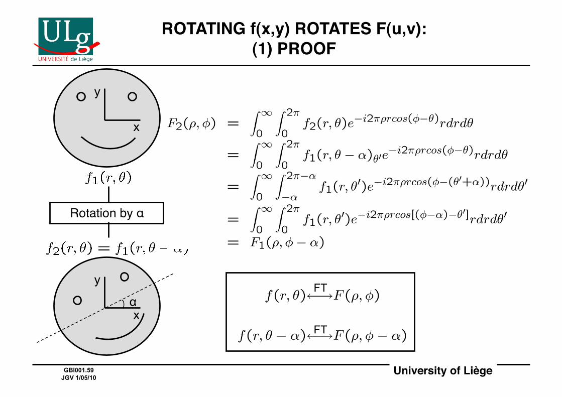
ROTATING f(x,y) ROTATES F(u,v):(1) PROOF!
Rotation by α#
x
y
x
y
α FT
FT
University of Liège!GBI001.60 JGV 1/05/10
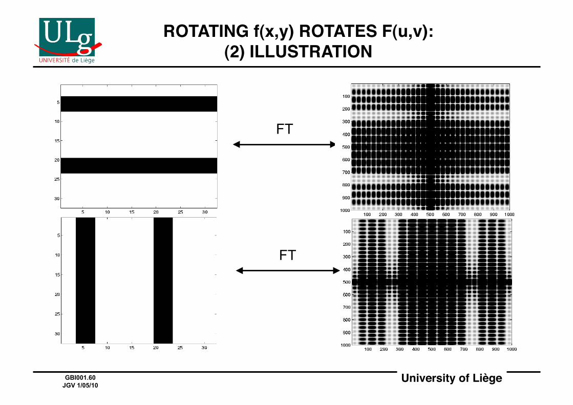
ROTATING f(x,y) ROTATES F(u,v):(2) ILLUSTRATION!
FT
FT
University of Liège!GBI001.61 JGV 1/05/10
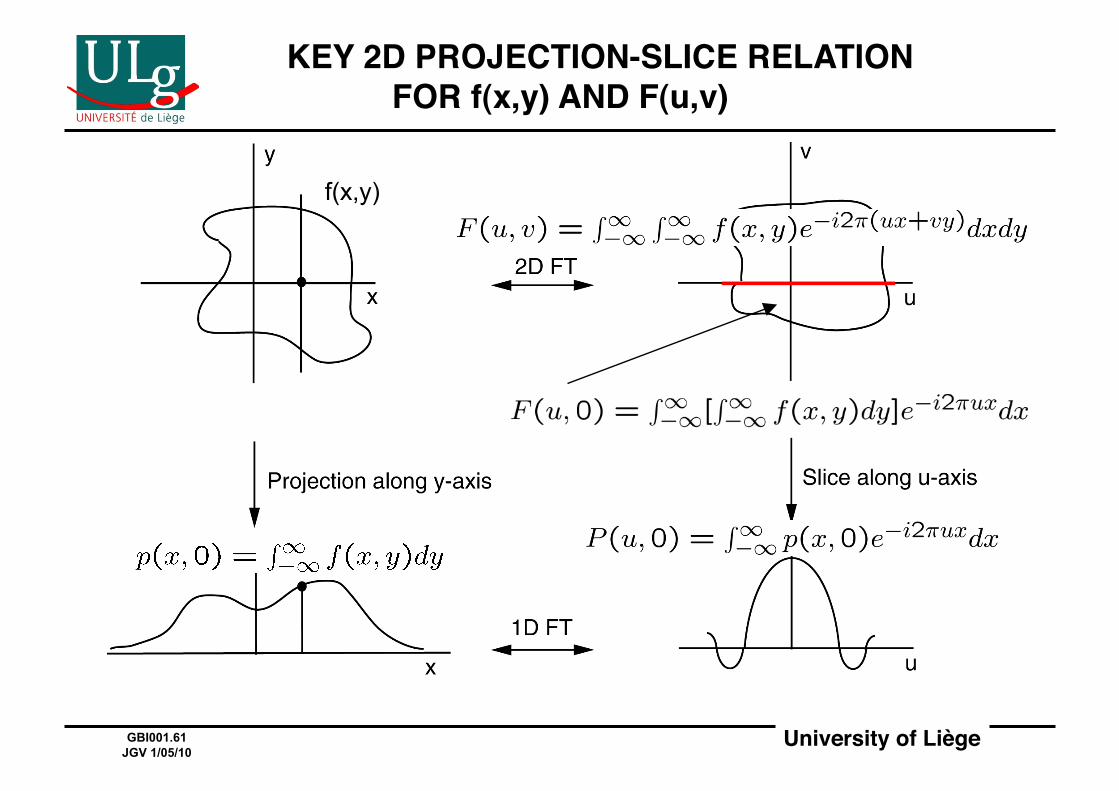
! KEY 2D PROJECTION-SLICE RELATION FOR f(x,y) AND F(u,v)!
f(x,y)
University of Liège!GBI001.62 JGV 1/05/10
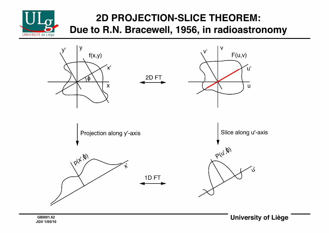
2D PROJECTION-SLICE THEOREM:Due to R.N. Bracewell, 1956, in radioastronomy!

University of Liège!GBI001.63 JGV 1/05/10
2D PROJECTION-SLICE THEOREM(FOR ARBITRARY OBJECTS)!
2D FT f(x,y) F(u,v)
p(x’,Φ) P(u’,Φ) 1D FT
PROJECT (RADON TRANSFORM)
SLICE (SET v’=0)

University of Liège!GBI001.64 JGV 1/05/10
2D PROJECTION-SLICE THEOREMFOR CIRCULARLY-SYMMETRIC OBJECTS!
HANKEL TRANSFORM f(r) F(ρ)
p(x) P(u) 1D FT
PROJECT (ABEL TRANSFORM) same !
f(r)
p(x) F(ρ)
AT
FT
HT
University of Liège!GBI001.65 JGV 1/05/10
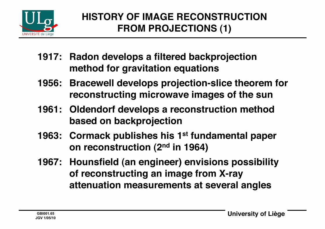
HISTORY OF IMAGE RECONSTRUCTION FROM PROJECTIONS (1)!
1917:! Radon develops a filtered backprojection method for gravitation equations!
1956:! Bracewell develops projection-slice theorem for reconstructing microwave images of the sun
1961:! Oldendorf develops a reconstruction method based on backprojection!
1963:! Cormack publishes his 1st fundamental paper on reconstruction (2nd in 1964)
1967:! Hounsfield (an engineer) envisions possibility of reconstructing an image from X-ray attenuation measurements at several angles!
University of Liège!GBI001.66 JGV 1/05/10
HISTORY OF IMAGE RECONSTRUCTION FROM PROJECTIONS (2)!
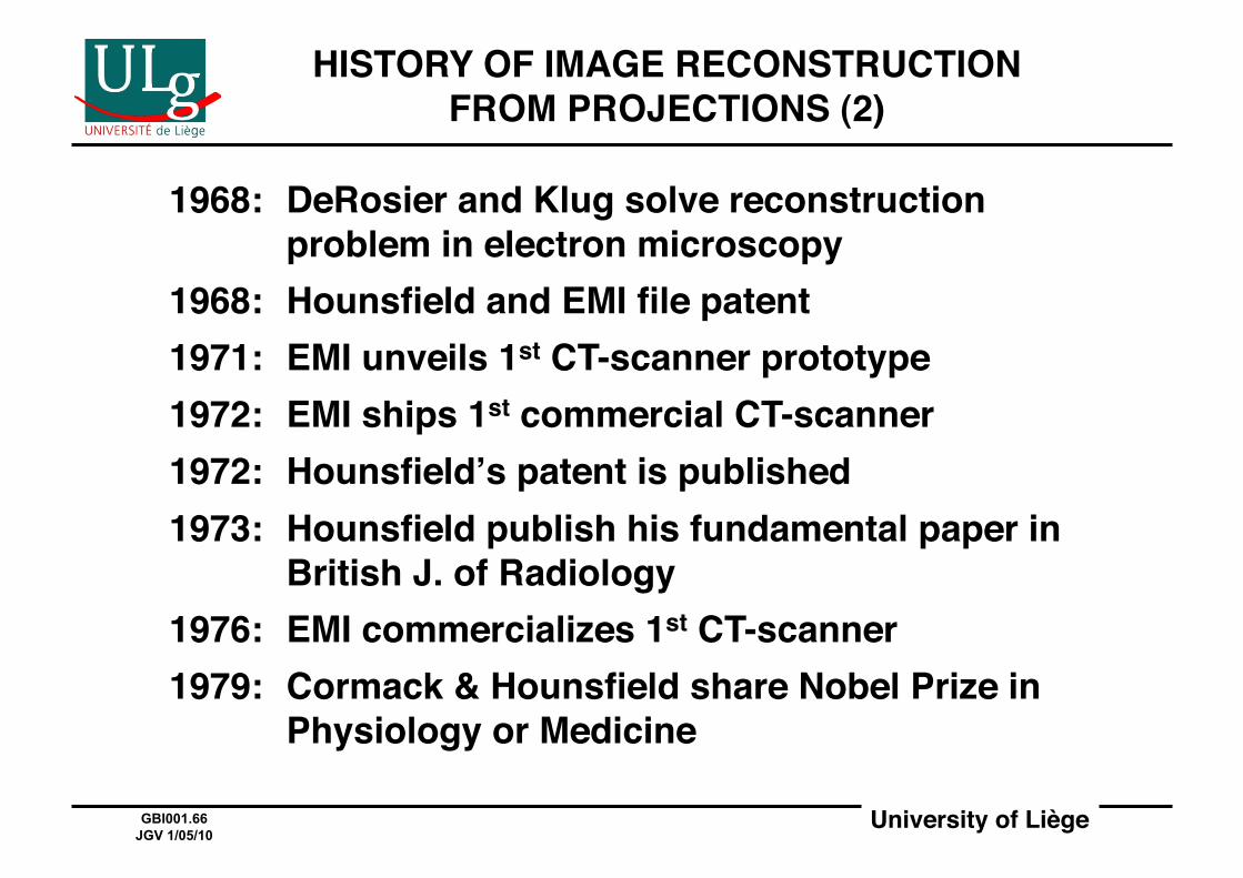
1968:! DeRosier and Klug solve reconstruction problem in electron microscopy!
1968:! Hounsfield and EMI file patent!1971:! EMI unveils 1st CT-scanner prototype!1972:! EMI ships 1st commercial CT-scanner!1972:! Hounsfieldʼs patent is published!1973:! Hounsfield publish his fundamental paper in
British J. of Radiology!1976:! EMI commercializes 1st CT-scanner!1979:! Cormack & Hounsfield share Nobel Prize in
Physiology or Medicine!
University of Liège!GBI001.67 JGV 1/05/10
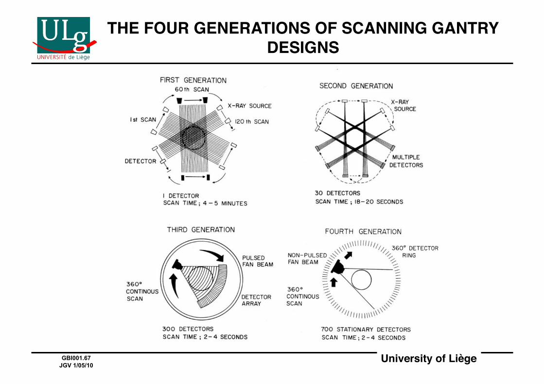
THE FOUR GENERATIONS OF SCANNING GANTRY DESIGNS!
University of Liège!GBI001.68 JGV 1/05/10
IMAGING BY RADIONUCLIDE EMISSION!
University of Liège!GBI001.69 JGV 1/05/10
IMAGING BY RADIONUCLIDE EMISSION INVOLVES THREE DISTINCT TASKS!
• Design, production, and administration of clinically-useful radionuclides (aka radioactive tracers or radiopharmaceutical), e.g. 18F.!
• Design and use of instruments for locating (i.e. "imaging") these tracers and following their temporal evolution in body!
• Determination of relations between tracers and physiology!
University of Liège!GBI001.70 JGV 1/05/10
THREE MAIN TECHNIQUES FOR MEASURING RADIOACTIVITY!
• Photography (X-ray film)!• Ionization (effective for α, but not for γ)!• Luminescence (effective for α, β, γ)!
– Basis for widely-used scintillation detectors!– Used in Gamma camera, SPECT, and PET!
University of Liège!GBI001.71 JGV 1/05/10
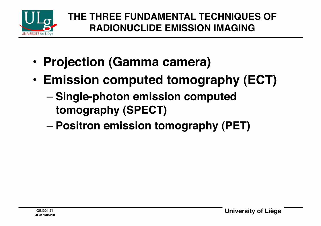
THE THREE FUNDAMENTAL TECHNIQUES OF RADIONUCLIDE EMISSION IMAGING!
• Projection (Gamma camera)!• Emission computed tomography (ECT)!
– Single-photon emission computed tomography (SPECT)!
– Positron emission tomography (PET)!
University of Liège!GBI001.72 JGV 1/05/10
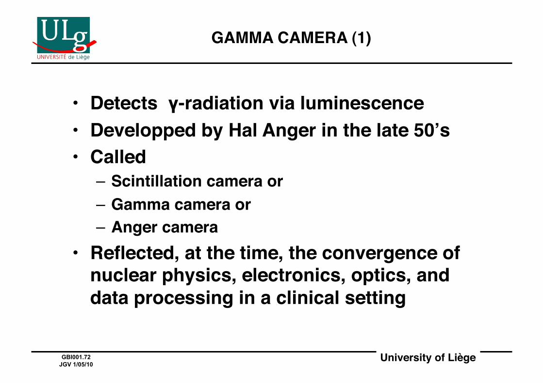
GAMMA CAMERA (1)!
• Detects γ-radiation via luminescence!• Developped by Hal Anger in the late 50ʼs!• Called!
– Scintillation camera or!– Gamma camera or!– Anger camera!
• Reflected, at the time, the convergence of nuclear physics, electronics, optics, and data processing in a clinical setting!
University of Liège!GBI001.73 JGV 1/05/10
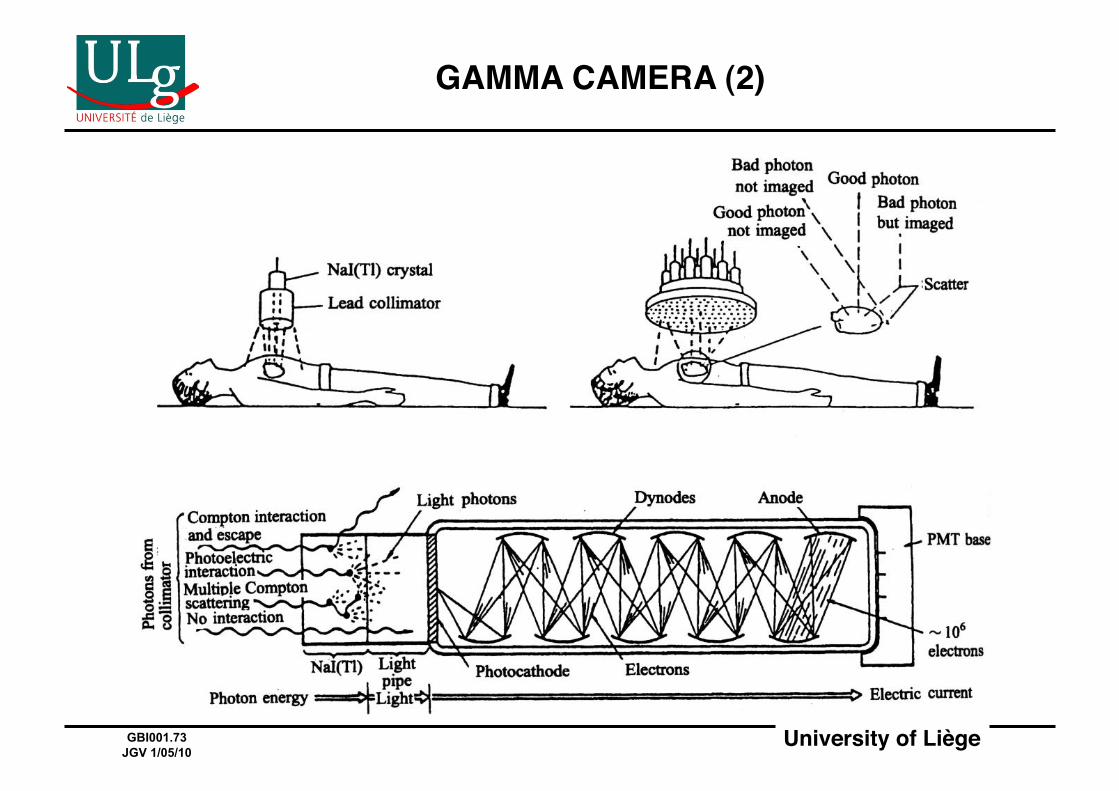
GAMMA CAMERA (2)!
University of Liège!GBI001.74 JGV 1/05/10
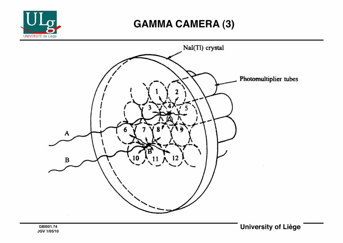
GAMMA CAMERA (3)!
University of Liège!GBI001.75 JGV 1/05/10
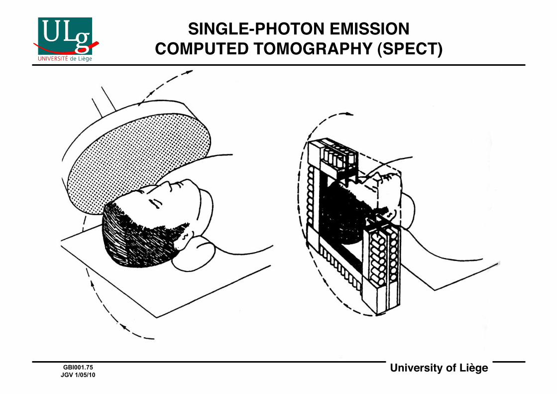
SINGLE-PHOTON EMISSION COMPUTED TOMOGRAPHY (SPECT)!
University of Liège!GBI001.76 JGV 1/05/10
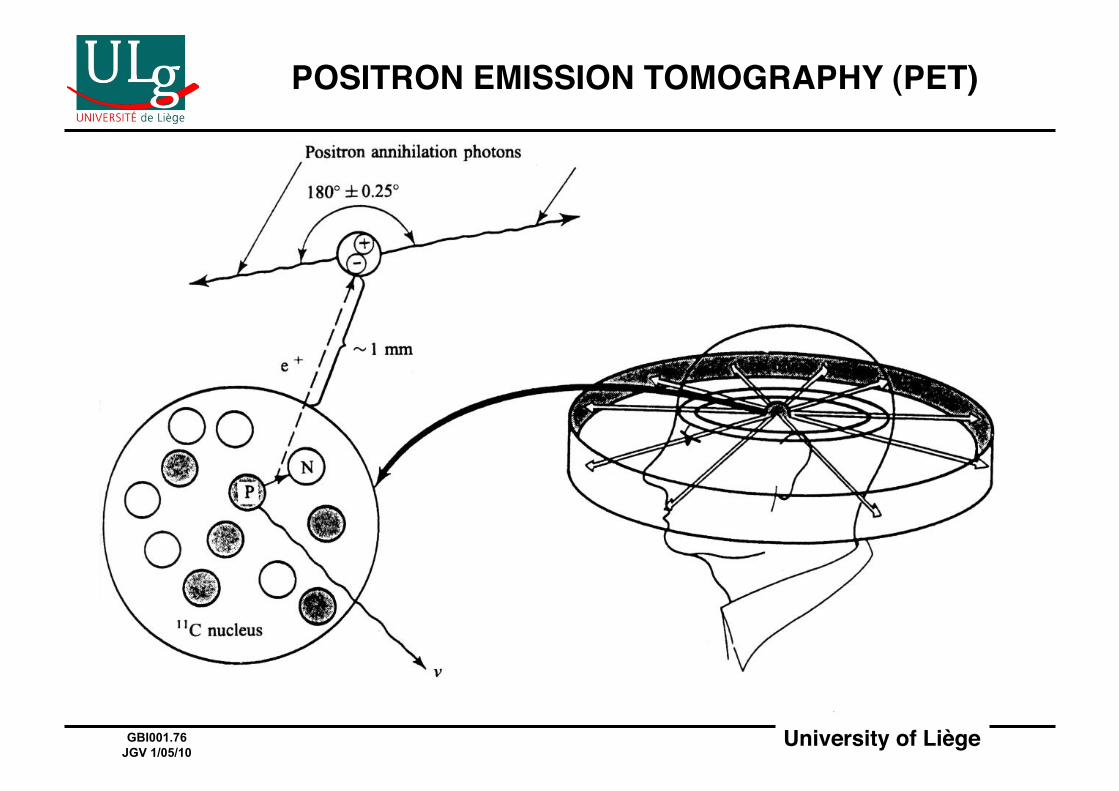
POSITRON EMISSION TOMOGRAPHY (PET)!
University of Liège!GBI001.77 JGV 1/05/10
NEXT TIME …!
• Magnetic resonance imaging (MRI)!• Image-guided surgery!• Image segmentation!• Image registration!
University of Liège!GBI001.78 JGV 1/05/10
ACKNOWLEDGMENTS!
Many thanks to the following people for helping putting this presentation together:!
Lara VigneronBenoît JaspartSophie le Maire Philippe Ries!


![Partie I : Anatomiestaff.univ-batna2.dz/sites/default/files/kebabla-mebarek/files/anato... · Anatomie et physiologie Génie Biomédical - ème2 année [Univ. Batna2 : 2020/2021]](https://img.pdfslide.net/doc/110x75/6130723e1ecc515869441a43/partie-i-anatomie-et-physiologie-gnie-biomdical-me2-anne-univ-batna2.jpg)


![Master [120] : ingénieur civil biomédical€¦ · scientifiques et techniques liés au génie biomédical, et ce dans un contexte européen et mondial en pleine évolution. Intrinsèquement](https://img.pdfslide.net/doc/110x75/605d99a6426a38780b7cf996/master-120-ingnieur-civil-biomdical-scientifiques-et-techniques-lis-au.jpg)