Embed Size (px)
Citation preview

Introduction of Cestodes Introduction of Cestodes (Tapeworms)(Tapeworms)
Phylum PlatyhelminthesPhylum PlatyhelminthesClass CestodaClass Cestoda
Order PseudophyllideaOrder PseudophyllideaOrder CyclophyllideaOrder Cyclophyllidea

Morphology
• Flat, segmented body with various length (several mm ~ several meters)
• 3 regions of worm body:– Scolex: suckers, hooklets, grooves – Neck: germinal portion– Strobila: immature, mature, gravid proglottids
(segments)

• Monoecious (each segment): reproductive system highly developed
• Digestive system degenerated
• All species are parasitic
TapewormsTapeworms

Taenia soliumTaenia solium链状带绦虫链状带绦虫
Pork tapeworm / Hook tapewormPork tapeworm / Hook tapeworm猪肉绦虫猪肉绦虫 //有钩绦虫有钩绦虫
Taenia saginataTaenia saginata肥胖带绦虫肥胖带绦虫
Beef tapeworm / Hookless tapewormBeef tapeworm / Hookless tapeworm牛肉绦虫牛肉绦虫 //无钩绦虫无钩绦虫
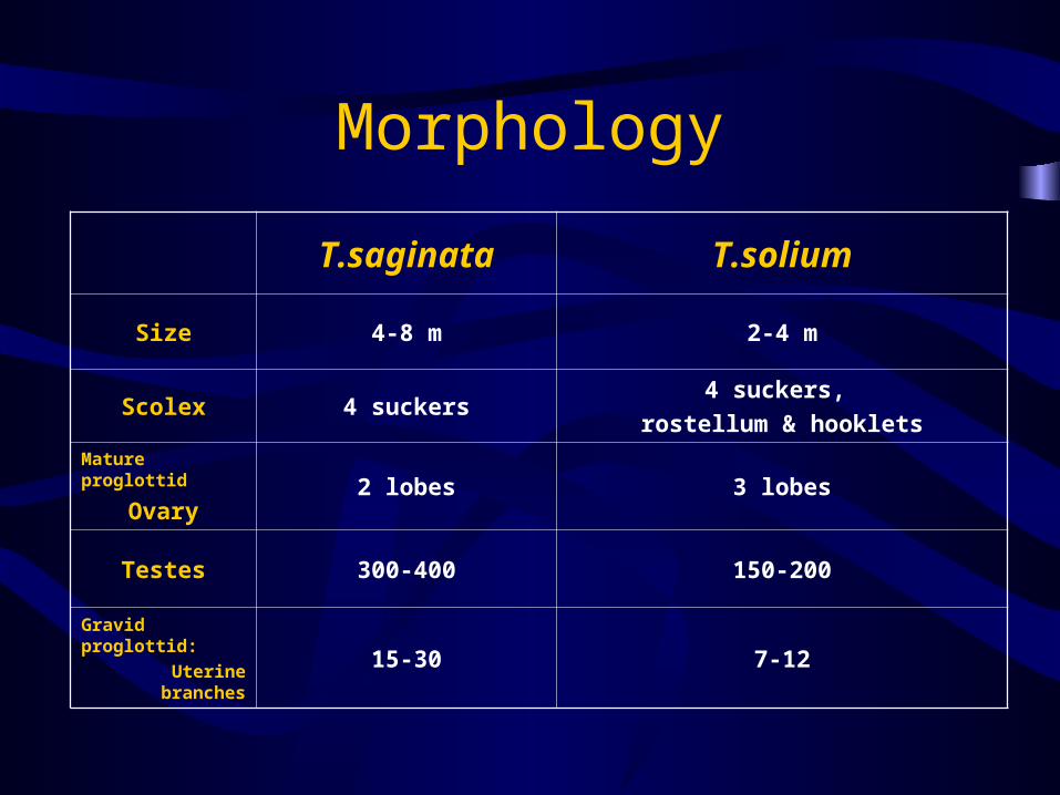
Morphology
T.saginata T.solium
Size 4-8 m 2-4 m
Scolex 4 suckers4 suckers,
rostellum & hooklets
Mature proglottid
Ovary2 lobes 3 lobes
Testes 300-400 150-200
Gravid proglottid:
Uterine branches15-30 7-12
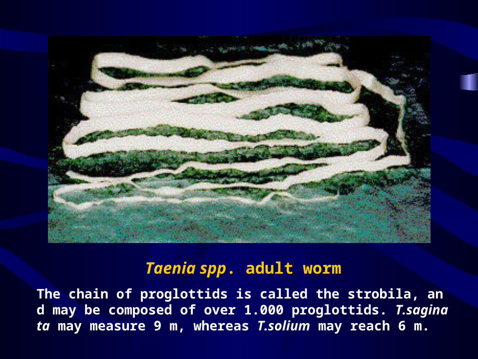
Taenia spp. adult worm
The chain of proglottids is called the strobila, and may be composed of over 1.000 proglottids. T.saginata may measure 9 m, whereas T.solium may reach 6 m.

The beef tapeworm (Living specimen)

The scolex of T. solium
hooklets
rostullum
suckers

The scolex of T. saginata

Taenia saginata, fresh specimen

Gravid proglottid of T. solium
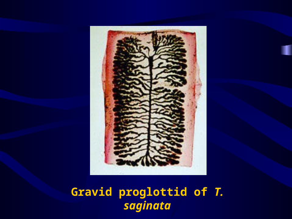
Gravid proglottid of T. saginata

• Larva
Cysticercus bovis
Cysticercus cellulosae
– Ovoid, cystic, size = a bean
– Invaginated scolex and neck

T.solium: cysticercus cellulosae with invaginated scolex
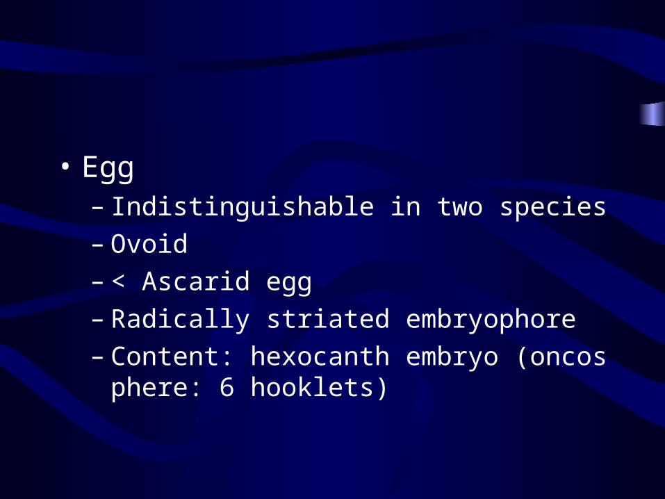
• Egg – Indistinguishable in two species– Ovoid– < Ascarid egg– Radically striated embryophore– Content: hexocanth embryo (oncosphere: 6 hoo
klets)

Taenia spp. egg Can not differentiate T. saginata from T. solium

Taenia spp. egg

Life Cycle
• Definitive host– Human being; No reservoir host
• Discharged stage– Eggs or gravid proglottids in feces

Life cycle of Taenia saginata

Life cycle of Taenia solium

T.saginata T.solium
D.H Human Human Human
I.H Cattle Swine Human
Habitation Small intestine Small intestineTissue(brain, eye,
skin etc.)
Infective stage Cysticercus bovisCysticercus
CellulosaeEgg
Disease Taeniasis Taeniasis Cysticercosis

Pathogenesis
• Taeniasis ( Infected by eating cysticerus; Pathogenic factor: adult worm)
– Deprivation of nutrition– Disfunction of the intestine: vomiting or diarrhea– Allergic reactions– Appendicitis– Obstructions of the intestine

• Cysticercosis (Intrinsic or extrinsic auto-infection; Cr
oss infection due to T.solium egg only; Pathogenic factor:
cysticercus cellulosae)
– Symptoms vary with site & intensity of infectio
n
– Clinical aspects: headache, dizziness, epilepsy,
blurred vision, subcutaneous nodule etc

Diagnosis
• Taeniasis– Anal swab: to find egg at perianal region– Fecal exam: to find segment (species identificat
ion)
• Cysticercosis– Biopsy (subcutaneous nodule)– X-ray/CT/MRI: cerebral cysticercosis– Ophthalmoscopy: ophthalmic cysticercosis

Epidemiology
• Distribution– Cosmopolitan– In china: mainly in minority regions

• Epidemic factors– Egg or gravid proglottid contamination of grass
and soil– Method of raising domestic animals– Unhygienic dinning habit of eating raw or
undercooked meat

Control
• Treatment– Paziquantel – Areca nut + pumpkin seed+ purge
• Scientific cattle and pig raising
• Avoid to consume raw meat
• Meat inspection

Echinococcus granulosus
细粒棘球绦虫 /包生绦虫

Morphology
• Adult worm– 3-6 mm long with 4 segments– Scolex & cervical portion (2 rows of 28-40 hoo
klets and 4 suckers)– Immature, mature, gravid segments
• Egg– Similar to the Taenia egg
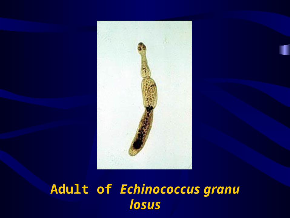
Adult of Echinococcus granulosus

• Hydatid cyst
– Round & cystic
– Cyst wall: laminated layer, germinal layer
– Contents: cystic fluid, brood capsules, protoscol
ex, daughter & grand daughter cyst, (hydatic sa
nd)

• Hydatid sand – The protoscoleces generally settle down at the b
ottom of the cyst and are known as hydatid sand.

Protoscoleces with double row hooklets and calcareous corpuscles

Protoscoleces

Hydatid sand

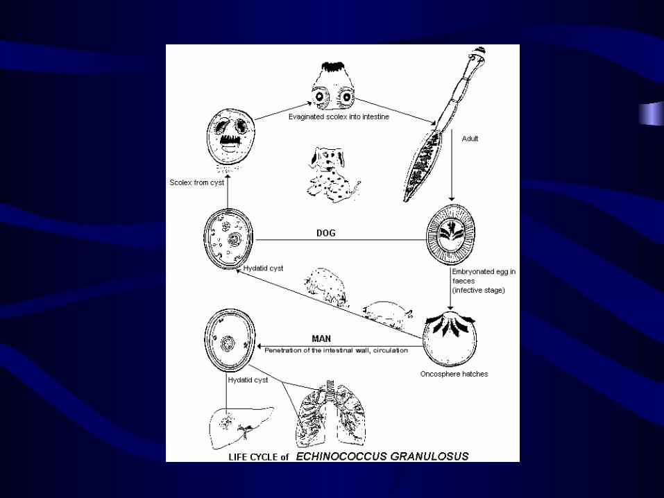
Life Cycle
• Adult worm – In the small intestine of the dog and other
carnivores
• Larva (hydatid cyst)– In the tissue of human being, sheep, horse, pig,
etc.


• Infective stage: egg
• Infective route: mouth
• Location: liver, lung, brain, eye, kidney, mu
scles, bone and heart
• Zoonotic parasite

Pathogenesis
• ‘Echinicoccosis’, ‘Hydatidosis’– Depend on the location and the number of hyda
tid cysts
• Pressure: liver, pulmonary, etc
• Allergy: anaphylactic shock
• Regeneration: secondary infection

Epidemiology
• Distribution
– Forest type (human are seldom involved)
• Wolf-moose/reindeer
• Dingo-wallaby
– Animal raising type (human are involved)
• Dog-sheep/cattle/pig
• Endemic factors
– High resistant egg
– Intimate contact between dog, animals and man
in local district
– Contamination of the feces by infected dogs
– Improper the viscera disposition

Diagnosis
• Physical (hepatic hypertrophy)
• History of residence in endemic area
• X-ray/Ultrasonography
• Immunological means
• Biopsy and puncture are forbidden unless during operation

Treatment and Control
• Surgical removal of the cyst
• Long-term Mebendazole therapy– 40 mg/kg/day × 1-6 months
• Personal protection
• Reasonable disposition of the viscera from infected animals
• Treatment of sheep dogs periodically