Embed Size (px)
Citation preview

1050
C H A P T E R 76Emergencies
■ INTRODUCTIONEmergencies include serious injuries
from accidents, burns, stings, bites, and possible poisoning. Sudden illness, or an ongoing illness that suddenly becomes worse, can also be an emergency. These conditions all require immediate veteri-nary attention.
You can reduce the likelihood of many of these situations, for example, by keep-ing harmful substances away from your pet. However, it is impossible to ensure that your pet will never have a medical emergency. By their very nature, emer-gencies are typically sudden and unex-pected. Regardless, you can be prepared to respond if an emergency occurs.
Keep information about your pet’s medical history and your veterinarian’s phone number easily accessible. Make sure you know where the closest 24-hour veterinary hospital is located. It is also a good idea to keep a first aid kit on hand to treat minor emergencies.
Emergency patients present special challenges because underlying problems may not be evident for 24 to 48 hours. Many variables contribute to the overall success of emergency treatment, includ-ing the severity of the illness or injury, the amount of blood or fluid lost, age of the animal, previous health problems, and time delay in beginning therapy.
Emergencies resulting from poisonings are discussed in the chapter on poisoning (see page 1145) later in this section.
Know Your PetKnowing your pet’s habits will help you
recognize when something is wrong. Sud-den changes in your pet’s normal physical condition, gait, activity level, eating habits, elimination habits, or grooming habits can indicate a medical problem. Being able to recognize an emergency and get your pet to a veterinarian quickly is
one of the most important things you can do to ensure successful treatment.
First Aid KitYou can purchase a ready-made pet first
aid kit or make one yourself. A pet first aid kit generally includes basic items sim-ilar to those of a human first aid kit. The first aid kit should have a secure lid and be kept where you can find it quickly.
Be sure you know how to properly use the first aid kit. You may be able to enroll in animal first aid and CPR classes through your veterinarian’s office, local
Stocking a Pet First Aid KitYou should keep a stocked first aid kit
available and know how to use it. Check the expiration dates of all medications at least once a year, and replace them when necessary. Include the following items:■ Muzzle■ Bandaging materials (including gauze,
sterile pads, stretch bandage, bandaging tape)
■ Duct or packaging tape■ Small scissors■ Hydrogen peroxide■ Cotton balls or swabs■ Chlorhexidine wash (0.5%)■ Saline solution■ Antibiotic ointment■ Splinting materials■ Tweezers or forceps■ Bulb syringe■ Thermometer (for rectal use)■ Lubricating jelly■ Disposable gloves■ Kaolin-pectin (for mild diarrhea)*■ Activated charcoal (to deactivate
poisons)**Always check with your veterinarian before using any over-the-counter medication.
5608_09_076-085.fm Page 1050 Thursday, August 23, 2007 9:48 AM

Chapter 76 EMERGENCIES 1051
community college, or groups such as the Red Cross.
Of course, a first aid kit is not a substi-tute for veterinary care. Take your pet to your veterinarian as soon as possible to determine the extent of the injury or ill-ness and for followup care.
■ EMERGENCY CARE FOR DOGS AND CATS
Emergency care begins with your call to the veterinarian. Be prepared to describe the emergency situation. Your veterinar-ian may instruct you on how to adminis-ter first aid and how to safely transport your pet. You may be able to identify life-threatening airway, breathing, and circulation problems with the help of a veterinary professional on the telephone. Follow instructions regarding immediate treatment and transport. Calling ahead also gives the veterinary staff some time to prepare for your arrival.
What to Do at the Scene and Transport
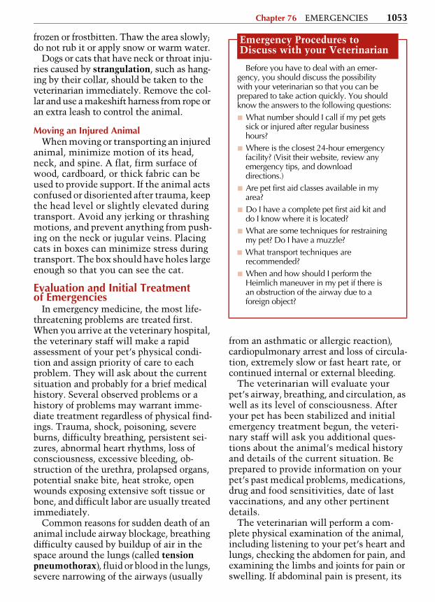
You can provide basic medical care at the scene of the injury. Remember that any animal that is injured or in pain may bite or scratch. Injured animals must be approached carefully, and you should first take precautions for your own safety. Using a muzzle is often a prudent safety measure; one can be easily made from a piece of cloth or a ready-made muzzle can be included in the first aid kit. Never muzzle a dog with chest injuries or a dog with a short nose (brachycephalic breeds like Pugs), and never leave a muz-zled dog alone. Placing a light towel or cloth over the animal’s head can decrease the pet’s awareness of nearby activity or noises that may cause fearful and aggres-sive reactions.
If your pet is not breathing, you may need to perform mouth-to-nose resuscita-tion and chest compressions. Request instructions from your veterinarian or pet emergency hotline. To perform mouth-to-nose resuscitation, close the animal’s mouth, place your lips over the animal’s
nostrils, and initially give 3 to 4 strong breaths (see CARDIOPULMONARY-CEREBRAL RESUSCITATION, page 1057). If the animal does not start breathing on its own, breathe for the animal 10 to 12 times per minute. If you cannot detect a heartbeat, perform 5 chest compressions to 1 quick breath. Continue this pattern until the animal starts breathing on its own, or you get to veterinary assistance. Of course, in this situation, someone else will have to drive during transport.
Emergencies Requiring Immediate Veterinary Care
■ Severe trauma■ Heat exhaustion or stroke■ Frostbite or exposure to cold■ Electric shock■ Profuse bleeding from the nose, mouth,
ears, or rectum■ Painful eyes with squinting, pupils that
appear larger or smaller than usual, pro-truding eyeball
■ Frequent vomiting and/or diarrhea, with or without blood
■ Retching or unproductive vomiting, parti-cularly if the stomach or abdomen looks bloated
■ Difficulty breathing or other respiratory distress
■ Collapse or coma■ Paralysis or severe neck or back pain
(arching, twisted)■ Painful or bloated abdomen■ Clusters of seizures within a 24-hour
period or a seizure that does not stop after several minutes
■ Prolonged labor or difficulty giving birth■ Suspected poisonings, insect bite reac-
tions, snake bites, scorpion bites, toad poisoning
■ Extreme lethargy■ Prolapse of the rectum or uterus
(From The Pill Book Guide to Medications for Your Dog and Cat by Current Medical Directions Inc., copyright © 1998 by Bantam Books, a division of Random House Inc. Used by permission of Ban-tam Books, a division of Random House, Inc.)
5608_09_076-085.fm Page 1051 Thursday, August 23, 2007 9:48 AM

1052 Section 9 SPECIAL SUBJECTS
Bleeding requires immediate first aid. Press down firmly on the bleeding area with your fingers or the palm of your hand, and then apply a firm, but not tight, bandage. Any long pieces of fabric or gauze can be used. Often washcloths and hand towels are enough when applied with mild pressure. If the original band-age becomes soaked with blood, do not remove it; simply place additional mate-rial on top and continue to apply pressure. These bandages can be secured in place using duct or packaging tape.
Burns can be difficult to evaluate because the fur makes it hard to examine the injury. Large deep burns, chemical burns, and electrical burns need imme-diate attention, as do burns involving the airway or face. Use cold water on the affected area, and cover the burn with a nonstick dressing.
Dogs or cats that are choking may cough forcefully, drool, gag, or paw at their mouth. They may also hold their mouth open and show signs of agitation. If you think your pet is choking, do not stick your fingers in its mouth because you can easily be bitten or push the object further in. Instead, you can try to dislodge the object by thumping the animal be-tween the shoulder blades or by applying several quick, squeezing compressions on both sides of the ribcage.
Do not remove foreign objects that have penetrated the skull, chest, or abdomen. Prevent the object from moving or pene-trating further. If an arrow has penetrated the abdomen, do not let the shaft of the arrow move during transport. It may be necessary to stabilize the shaft of the arrow just outside the body and, holding it firmly, cut the shaft off.
Heat stroke is another emergency. Nor-mal rectal temperature for cats and dogs is about 101.5°F to 102°F (38.6°C to 38.9°C). Signs of heat stroke include skin that is hot to the touch, vomiting, drooling, rapid panting, distress, loss of coordination, col-lapse, and unconsciousness. Remove the animal from the heat. Use cold water, ice packs, or wet towels to cool the head and body. Offer small amounts of water after the pet has begun to cool down. Do not immerse the animal in cold water.
Hypothermia (overexposure to cold) is rare in pets. It usually results when an ani-mal has been lost or outside in very cold weather for a long time after another accident or injury, such as a car accident. Signs of hypothermia include slow pulse, shallow breathing, disorientation, collapse, and unconsciousness. Shivering is not a usual sign of hypothermia in pets. If the ani-mal is wet, first dry it thoroughly, and then place wrapped warm (not hot) water bottles around the body. White, numb skin may be
A temporary muzzle can be created from a strip of cloth. Find a cloth bandage, rope, or other long strip of fabric. Tie a knot in the center of the bandage. Make another loose knot several inches above the first knot. Slip the loop over the dog’s muzzle and gently pull the knot tight (Step 1). Cross the ends of the bandage under the dog’s jaw (Step 2). Firmly tie the crossed ends behind the dog’s neck (Step 3). Adapted, with permission, from www.petfocused.com, © morefocus group inc. 2007.
Step 3Step 2Step 1
5608_09_076-085.fm Page 1052 Thursday, August 23, 2007 9:48 AM
Chapter 76 EMERGENCIES 1053
frozen or frostbitten. Thaw the area slowly; do not rub it or apply snow or warm water.
Dogs or cats that have neck or throat inju-ries caused by strangulation, such as hang-ing by their collar, should be taken to the veterinarian immediately. Remove the col-lar and use a makeshift harness from rope or an extra leash to control the animal.
Moving an Injured AnimalWhen moving or transporting an injured
animal, minimize motion of its head, neck, and spine. A flat, firm surface of wood, cardboard, or thick fabric can be used to provide support. If the animal acts confused or disoriented after trauma, keep the head level or slightly elevated during transport. Avoid any jerking or thrashing motions, and prevent anything from push-ing on the neck or jugular veins. Placing cats in boxes can minimize stress during transport. The box should have holes large enough so that you can see the cat.
Evaluation and Initial Treatment of Emergencies
In emergency medicine, the most life-threatening problems are treated first. When you arrive at the veterinary hospital, the veterinary staff will make a rapid assessment of your pet’s physical condi-tion and assign priority of care to each problem. They will ask about the current situation and probably for a brief medical history. Several observed problems or a history of problems may warrant imme-diate treatment regardless of physical find-ings. Trauma, shock, poisoning, severe burns, difficulty breathing, persistent sei-zures, abnormal heart rhythms, loss of consciousness, excessive bleeding, ob-struction of the urethra, prolapsed organs, potential snake bite, heat stroke, open wounds exposing extensive soft tissue or bone, and difficult labor are usually treated immediately.
Common reasons for sudden death of an animal include airway blockage, breathing difficulty caused by buildup of air in the space around the lungs (called tension pneumothorax), fluid or blood in the lungs, severe narrowing of the airways (usually
from an asthmatic or allergic reaction), cardiopulmonary arrest and loss of circula-tion, extremely slow or fast heart rate, or continued internal or external bleeding.
The veterinarian will evaluate your pet’s airway, breathing, and circulation, as well as its level of consciousness. After your pet has been stabilized and initial emergency treatment begun, the veteri-nary staff will ask you additional ques-tions about the animal’s medical history and details of the current situation. Be prepared to provide information on your pet’s past medical problems, medications, drug and food sensitivities, date of last vaccinations, and any other pertinent details.
The veterinarian will perform a com-plete physical examination of the animal, including listening to your pet’s heart and lungs, checking the abdomen for pain, and examining the limbs and joints for pain or swelling. If abdominal pain is present, its
Emergency Procedures to Discuss with your Veterinarian
Before you have to deal with an emer-gency, you should discuss the possibility with your veterinarian so that you can be prepared to take action quickly. You should know the answers to the following questions:■ What number should I call if my pet gets
sick or injured after regular business hours?
■ Where is the closest 24-hour emergency facility? (Visit their website, review any emergency tips, and download directions.)
■ Are pet first aid classes available in my area?
■ Do I have a complete pet first aid kit and do I know where it is located?
■ What are some techniques for restraining my pet? Do I have a muzzle?
■ What transport techniques are recommended?
■ When and how should I perform the Heimlich maneuver in my pet if there is an obstruction of the airway due to a foreign object?
5608_09_076-085.fm Page 1053 Thursday, August 23, 2007 9:48 AM

1054 Section 9 SPECIAL SUBJECTS
source must be determined. Some signs, such as vomiting or diarrhea, can indicate which body systems may be involved. Samples of blood and urine may be col-lected for laboratory testing. All this infor-mation helps the veterinarian to identify specific problems and to determine a diag-nosis, treatment, and monitoring plan.
AirwayAny obstruction of the airways can be
life-threatening. If the large airways (the trachea, commonly called the windpipe, and its 2 main branches, called the bron-chi) are completely obstructed, the animal is unable to breathe and will be uncon-scious. If the large airways are only par-tially obstructed, the animal’s breathing will be noisy. The animal may be fearful, and its skin may appear to have a bluish tinge due to lack of oxygen. Possible causes of large airway problems include foreign objects, swelling (perhaps because of an allergic reaction), paralysis of the larynx, collapse of the trachea, and a longer soft palate than is usual. A poten-tially very serious situation called aspi-ration refers to breathing in stomach contents that have been vomited. This can lead to respiratory problems such as pneumonia and is one of the reasons why animals must be fasted before anesthesia and surgery.
Animals with severe small airway ob-struction have difficulty breathing. They may wheeze and push out their dia-phragm when exhaling. They may be fearful or anxious, and their skin, eyes, nailbeds, and other tissues may appear bluish. The animal may be sedated and given medication to expand the airways and make breathing easier. Common causes include allergic reactions; asthma (cats); and fluid, blood, mucus, or foreign material in the lungs.
If an animal is unconscious and not breathing, a tube will be placed through the larynx into the trachea to help it breathe. This is called tracheal intubation. If the upper airway is obstructed, oxygen can be given through a tube inserted below
the obstruction to open the airway. The veterinarian will listen to ensure that the air is reaching the animal’s lungs. Heart sounds and pulses are checked and when absent, cardiopulmonary-cerebral resusci-tation is done.
If the large airways are only partially obstructed by a foreign object, the veteri-narian will remove the object if possible. A tracheal tube may be placed to deliver oxygen. The animal may need to be sedated to relieve anxiety. When tracheal intubation is necessary, sedation, likely general anesthesia, will be needed.
BreathingWhen animals have difficulty breath-
ing, they breathe faster and breathing takes more effort. The breathing pattern often changes, followed by changes in pos-ture. For example, dogs may stand with the elbows spread out and the back arched. Cats may crouch on all 4 limbs and raise their chest slightly, or sit high on the rear haunches and extend their head and neck. Obvious labored, open-mouth breathing and a blue tinge to the skin due to lack of oxygen develop last. These signs indicate significant loss of respiratory function.
The veterinarian will carefully observe the breathing pattern and listen to the animal’s chest. X-rays or other tests may be necessary after the animal has been stabilized. Oxygen is administered by a tracheal tube, a mask, a hood, or other method. The animal may be sedated to relieve anxiety. After the animal has been stabilized, additional tests may be done.
One cause of breathing problems is pleural space disease. The pleural space is the area between the membrane covering the lungs and the membrane lining the chest cavity. In this condition, air, fluid, or abdominal contents are in the pleural space. The veterinarian will hear muffled lung sounds over the affected regions. Treatment of pleural space disease begins with the veterinarian using a needle or catheter to remove the air or fluid that is free in the chest cavity. This procedure
5608_09_076-085.fm Page 1054 Thursday, August 23, 2007 9:48 AM

Chapter 76 EMERGENCIES 1055
relieves the tension within the chest, allowing the heart to beat and the lungs to expand. A chest tube is then placed to continue to relieve the pressure.
CirculationSeveral indicators are used to determine
how well the animal’s circulatory system is functioning. These include heart rate, mucous membrane color, capillary refill time, and pulse intensity. Listening to the animal’s heart and breath sounds will help the veterinarian determine what circulatory problems may exist.
Shock is the medical term for changes that develop when the body attempts to compensate for limited heart function, blood volume, or circulation. Shock can develop in many emergency situations, including head trauma, excessive blood or fluid loss, and severe infection. Signs of shock include a rapid heart rate, pale mucous membrane color, very low blood pressure, very little urinary output, and weak pulses. As shock progresses, deliv-ery of oxygen and other nutrients to the tissues drops, eventually leading to organ failure and ultimately death.
Recognizing the type and stage of shock early is vital to treatment. Shock is typi-cally classified into 3 categories: hypovo-lemic, cardiogenic, and distributive. Hypovolemic shock develops when blood volume is low, usually from loss of blood due to traumatic injury or loss of fluid through metabolic processes. Cardiogenic shock results when the heart fails, no longer pumping blood to the lungs or the rest of the body. Distributive shock is caused by blood flow being directed away from the central circulation because of a problem with peripheral blood vessels and is usually associated with serious wide-spread infections.
The goal of treatment for shock is to deliver blood to the tissues, bringing oxy-gen and other nutrients. Oxygen can be administered by a mask, bag, nasal or tra-cheal tube, or another method. Bleeding must be controlled. However, internal bleeding may not be evident until blood
pressure and circulation are restored. Flu-ids and blood products will be given intra-venously as needed to replace what has been lost. Abdominal counterpressure may be used to reduce abdominal bleed-ing, when present.
Medication to relieve pain is usually provided quickly. Narcotics or local anes-thetics can be used. Your veterinarian may use other medications as well.
Specific Diagnostics and TherapyIn an emergency, after your pet has been
stabilized the veterinarian will ask you for a more complete history, conduct a more systematic physical examination, and begin specific diagnostic and treatment procedures.
TraumaAccidents, falls, and fights with other
animals may result in different types of trauma. In blunt trauma, the animal has been struck by an object (such as a car) but the skin was not penetrated. Blunt trauma is commonly associated with internal bleeding, organ rupture, fractures, and head injuries. In penetrating trauma, a sharp object, such as an arrow or bullet, pierces the skin, and injuries are related to the path of the penetrating object. Falling from a height can cause multiple bone fractures, as well as injuries to the chest and organs. A dog bitten by another larger dog can have deep penetrating bite wounds and will frequently have spinal injuries and tracheal rupture from the thrashing motions experienced during the attack.
For all types of trauma, airway, breath-ing, and circulation are evaluated and sta-bilized as described above. Control of bleeding, oxygen if needed, and pain relief are also given immediate attention. After stabilization, the nervous system, chest, abdomen, and bones are carefully eval-uated. Blood tests, urine tests, and x-rays may be performed as needed. Trauma to the eye is also a common emergency (see EYE EMERGENCIES, page 1063).
5608_09_076-085.fm Page 1055 Thursday, August 23, 2007 9:48 AM

1056 Section 9 SPECIAL SUBJECTS
Animals that have suffered a trauma often have multiple injuries, some of which may not be immediately obvious. Whenever the animal is moved or being examined, the neck and spine should be kept still in case there are spinal fractures or other problems that cannot be readily seen. Broken legs may be wrapped with bandages or splinted. Because many prob-lems are not apparent for 12 to 24 hours after trauma occurs, your pet needs care-ful monitoring in the veterinary hospital.
Thoracic (Chest) TraumaSome traumatic injuries to the chest are
potentially life-threatening. Bruising of the lungs, air in the space around the lungs (called pneumothorax), heart rhythm problems, internal bleeding, and rib fractures are emergencies that may result from blunt trauma.
Tests to help determine the extent and severity of the problems include chest x-rays, blood tests, and an electrocardio-gram (EKG). A procedure called centesis, in which a needle is inserted into the chest or abdomen of the animal to remove fluid or air, may also be performed. Labo-ratory examination of any fluid removed can help with diagnosis.
Severe bruising of the lungs causes labored breathing and lack of oxygen to the tissues. Oxygen treatment may require sedation and tracheal intubation, in which a tube is inserted into the tra-chea to help the animal breathe. When bruising is severe, ventilation (in which air is forced mechanically in and out of the lungs) may be required to provide the best chances for survival.
If air or fluid is trapped inside the ani-mal’s chest cavity, a tube may be inserted through an incision in the chest to drain the air or fluid. Surgery may be recom-mended either immediately or after sev-eral days if the animal does not improve.
A condition called flail chest may be caused by blunt trauma that breaks 3 or more ribs. The portions of ribs that have broken off, called flail segments, may be stabilized by using an external frame of
metal rods or cast material that is formed to the shape of the chest. The animal must generally be anesthetized, and sur-gery is usually necessary.
Bite wounds over the chest must be cleaned and drained. The puncture holes seen on the skin from the bite are only a small portion of the significant damage that may have occurred to the muscles and tissues underneath the skin. Surgery may be required if the wound is penetrat-ing or if there is significant bruising or swelling.
Heart rhythm abnormalities, particu-larly fast heart rhythms, are often seen after trauma. A sedative may be used to relieve anxiety and slow the animal’s heart rate. The animal should be treated aggressively for shock, and an electrocar-diogram should be done to look for pri-mary heart rhythm problems.
Abdominal TraumaThe extent and severity of abdominal
injuries are not always obvious imme-diately. If the abdominal wall is torn open, the injury is clearly serious. Bruising, road burns, cuts, scrapes, swelling, a change in the shape of the abdomen, or pain may indicate internal injuries. If your pet appears to have abdominal pain and is in shock, internal bleeding may be present. The spleen, liver, or kidneys may be injured. All abdominal organs have blood vessels that can be injured by blunt trauma.
It may take several hours for specific signs of an injury to an abdominal organ to appear. Your veterinarian will watch closely for sharp abdominal pain. Abdom-inal x-rays or ultrasonography may show the location of injury. If fluid is present in the abdomen, a needle may be inserted into various locations to remove it. The fluid is then examined for any evidence of infection, rupture of an organ, cancer, or other problems.
Sometimes, a catheter is placed through an incision in the animal’s abdomen, and warm saline solution is flowed into the abdomen. The fluid is left in the abdomen
5608_09_076-085.fm Page 1056 Thursday, August 23, 2007 9:48 AM

Chapter 76 EMERGENCIES 1057
for several minutes and then drained. This process is called peritoneal lavage. If the fluid is clear, significant internal abdomi-nal bleeding is unlikely. Some blood in the fluid indicates mild bleeding. More blood in the fluid indicates significant abdominal bleeding that warrants careful monitoring and may require surgery. However, bleeding may be present even if the peritoneal lavage shows little or no blood in the fluid. Pelvic fractures in par-ticular may cause complications, includ-ing bleeding, that often are not found by peritoneal lavage.
If internal bleeding or other serious internal problem is found, emergency surgery may be needed.
Cardiopulmonary-Cerebral Resuscitation
Cardiac arrest occurs when there is no heartbeat and breathing stops. The pur-pose of cardiopulmonary-cerebral resusci-tation (CPCR) is to replace the work that the lungs and heart are not performing on their own. The success of CPCR efforts depends on the underlying cause of the cardiac arrest as well as on the speed and effectiveness of the treatment. The initial steps of CPCR are meant to get the needed oxygen and blood to the animal’s tissues. CPCR progresses with advanced life sup-port measures. Heart rhythms are moni-tored, drugs are administered, and the heart is defibrillated when necessary.
Defibrillation is a process of shocking the heart in a specific way to restore a coordinated heart beat and a pulse. Often, immediate defibrillation offers the best chance of recovery after cardiac arrest. Defibrillation is considered the best treat-ment for one of the most common arrest arrhythmias, ventricular fibrillation. Other heart rhythm problems are gener-ally thought not to be made any worse by the procedure.
Basic Life SupportMouth-to-nose resuscitation is the first
step of CPCR. This continues until the veterinary staff can insert a tube to deliver oxygen and help the animal breathe with mechanical ventilation. Chest compres-sions are used to promote circulation when no pulse is present. The animal is positioned on its back or its side (depend-ing on size) and supported as needed while compressions are performed over the area of the heart. The goal is to improve the return of the blood to the heart between beats.
Constant monitoring is important during basic life support procedures. If circulation continues to fail, additional procedures or drugs may be necessary.
Advanced Life SupportIn advanced life support, heart rhythms
are monitored by electrocardiogram, and drugs or defibrillation may be used. The purpose is to reestablish the heartbeat.
Fluids are administered, usually intra-venously (IV), to promote circulation. Sometimes, blood products are also given.
If basic life support has not been success-ful, meaning that the animal does not begin to breathe on its own or have a nor-mal heartbeat after 5 to 10 minutes, open-chest cardiopulmonary resuscitation (CPR, see page 1058) may be started. Sometimes open-chest CPR is begun ear-lier. In severe trauma with blood loss, the chest may be opened to assess the injury as well as to perform CPR. In a large dog, exter-nal compressions may not generate enough blood flow. You should make your wishes
Mouth-to-nose resuscitation is the first step of cardiopulmonary-cerebral resuscitation.
5608_09_076-085.fm Page 1057 Thursday, August 23, 2007 9:48 AM

1058 Section 9 SPECIAL SUBJECTS
known to the veterinary team as quickly as possible regarding your permission for open-chest CPR. This technique can be life-saving, but it carries additional expense and requires surgery to close the chest.
Open-chest Cardiopulmonary Resuscitation (Emergency Thoracotomy)
When chest compressions are not suc-cessful in restoring the heartbeat, the chest may need to be opened to allow access to the heart. The visual examina-tion, along with the EKG, allows the type of heart arrhythmia to be determined. If blood has pooled around the heart in the tissue sac called the pericardium, the pressure from this fluid can be relieved by opening the sac. If the heart is not beating, the veterinarian grasps the heart with one or both hands and compresses it. The compression is then released to allow the heart chambers to refill with blood. If blood loss is severe or poor circulation has been prolonged, certain blood vessels may be clamped to direct blood flow to the brain. This is a temporary measure, usually up to 10 minutes.
If the heart begins to beat again, the open chest is rinsed with sterile, warm salt water, and a chest tube is placed before the chest is closed. Treatment continues while the underlying cause of the arrest is determined.
Fluid TherapyMaintaining volume in the blood ves-
sels and the tissues of the emergency patient is vital. Signs of dehydration (fluid loss in the tissues) include dry skin that does not “smooth” back into its normal position after being pinched, dry mucous membranes, and sunken eyes. When fluid is given, it is distributed evenly through-out the body. This rehydrates a dehy-drated animal. However, excessive water volume can cause swelling, called edema. Specific types of fluids are given to injured animals to meet their specific bodily needs.
Monitoring the Critically Ill AnimalCritically ill animals must be moni-
tored closely in the veterinary hospital for a period of time depending on the severity of the illness or injury. Close attention helps the veterinarian determine whether treatment is effective, or whether other treatments may be needed. Often, second-ary problems become evident or develop during treatment.
Several parameters are generally evalu-ated daily or more often while the animal is hospitalized. These include the balance of fluids in the body, glucose (blood sugar) levels, electrolytes, oxygen levels, level of consciousness, blood pressure, heart rate and rhythm, coagulation of blood, red blood cell count and hemoglobin (a pro-tein that carries oxygen) level, gastroin-testinal function, kidney function, and effects of medications.
NutritionIt is always preferable if an animal will
eat on its own or at least take food orally. Small amounts of a liquid diet can gener-ally be given orally. When the pet refuses oral feeding, a tube can be placed to pro-vide nutrition through the gastrointesti-nal tract. In the rare event that nutrition cannot be given through the gastrointesti-nal tract, then nutrition is usually pro-vided through specific intravenous fluids.
Pain ControlSigns of pain include fast heart rate and
pale mucous membranes. These can mimic signs of shock. An animal in pain also has higher levels of stress hormones. However, animals may be in pain without showing obvious signs. In these cases, if the animal has a known painful condi-tion, pain medication is generally admin-istered. Pain medication makes the animal more comfortable but can also reduce signs of other conditions, possibly making diagnosis more difficult.
Nursing CareCritically ill animals need skilled,
knowledgeable, attentive nursing care.
5608_09_076-085.fm Page 1058 Thursday, August 23, 2007 9:48 AM

Chapter 76 EMERGENCIES 1059
Animals that are unable to walk should be turned from one side to the other, every 4 hours if possible, to prevent ulcers and other problems. Physical therapy to main-tain range of motion, muscle tone, and blood flow may be a part of nursing care. Catheter sites should be inspected fre-quently for signs of infection or displace-ment. Bandages that become soiled or wet should be changed. If fluid collects in limbs, light compression wraps may be used. These should be changed every day. Any animal with a critical illness should have 24-hour nursing care.
After the EmergencyIf your pet needs to stay in the veteri-
nary hospital, ask about the visitation pol-icy. Many veterinary hospitals encourage owner visits and have daily visiting hours. Talking to your pet and maintaining con-tact when possible can help reduce your pet’s stress and anxiety (as well as your own).
When it is time to bring your pet home, make sure that you completely under-stand all care instructions. Often, you will be required to restrict an animal’s activity for a certain period of time, particularly after surgery or if your pet has fractured a bone. Dogs can be crated or restrained in a small room; when taken outside, they should remain on a leash to prevent run-ning or jumping. Cats can be kept in a small room where they will not be tempted to jump onto furniture or counters.
Medication should be given according to instructions, and all label directions followed closely. Tips on administering medications are given in the Dog Basics and Cat Basics chapters (see page 12 and page 338). Bandages should be changed as directed, and wounds or sutures (stitches) checked for any swelling, redness, or dis-charge that might indicate infection. You will probably be advised to avoid bathing your pet until any sutures are removed.
Your veterinarian will usually schedule a followup appointment to check on your
pet’s recovery. In the meantime, call if any problems arise or if you have any questions about care.
■ EMERGENCY CARE FOR HORSESEquine emergencies can be challenging
for veterinarians and emotionally charged for owners. Preparation before an emer-gency occurs is key. Discuss the best facil-ities for treatment with your veterinarian ahead of time. Have phone numbers and other information on hand. Know how to get to the facility you have chosen, plan how you are going to transport the horse, and keep driving directions handy. Assem-ble and keep a first aid kit on hand to deal with immediate needs and transport in case your horse requires emergency treatment.
The most common types of equine emergencies are abdominal pain (colic), trauma and lacerations, and ill foals.
Common Emergency Procedures in Horses
Emergency procedures for horses follow the same general principles as those for small animals. However, there are special considerations for horses. Because horses cannot lie down for extended periods of time, some conditions that are considered less serious in other animals are emergen-cies in horses. Also, treatments often vary between smaller animals and horses.
Monitoring a horse in an emergency situation is crucial. In a horse that is in severe shock, pulse and breathing must be closely monitored.
Fluid TherapyPreventing and treating dehydration is a
primary concern. Horses require about 60 milliliters per kilogram of fluids per day normally. For an adult horse, this is about 1 liter (about 1 quart) per hour. Athletic horses often require more fluids. Heart rate, pulse, urine production, and other tests help determine whether a horse is dehydrated. Dehydration often develops along with other injuries and illnesses. Diarrhea is a major cause of dehydration,
5608_09_076-085.fm Page 1059 Thursday, August 23, 2007 9:48 AM
1060 Section 9 SPECIAL SUBJECTS
particularly in foals. If the horse is losing fluids, such as with diarrhea, very large quantities of fluids may be needed to correct the fluid loss. For example, a 1,000-pound horse that has become 5% dehydrated will need 25 liters (about 6¼ gallons) of fluids to correct that loss.
If a lot of blood has been lost, or the horse is exhausted or overheated, emer-gency fluid replacement is needed.
Nasogastric IntubationNasogastric intubation is an essential
and possibly life-saving procedure com-monly used in cases of equine colic. A tube is placed through the nostril down into the stomach to remove fluid that has accumulated in the stomach due to a blockage in the small intestine. Removing the fluid not only relieves the pain of the distended stomach, it may also prevent rupture of the stomach.
AbdominocentesisIn abdominocentesis, a needle or
syringe is inserted through the abdominal wall to obtain a fluid sample. The fluid is used in the evaluation of abdominal dis-eases such as colic and weight loss. If the horse has suffered an intestinal rupture, this test will generally detect the prob-lem. For the first 2 to 4 hours after a rup-ture of the intestine, the horse may not show any signs. Internal bleeding can also be detected by abdominocentesis.
TrocarizationTrocarization is a technique used to
relieve the pressure in the abdomen when it becomes distended with gas due to an intes-tinal blockage. Such blockage can result in severe bloating (distention), pain, and rapid or uneven breathing. The veterinarian will identify the segment of intestine that is involved by rectal examination in adult horses. In foals or small horses, x-rays or ultrasonography can be used. If the problem is in the large intestine, trocarization may be used. After the pressure is relieved, the trocar is removed, and an antibiotic is infused. Because infection and other prob-lems are possible after trocarization, the
horse is usually monitored carefully for 24 hours for signs of any complications. Trocarization may not solve the blockage problem, and the horse may still need addi-tional treatment or even abdominal surgery.
TracheostomyIf the trachea is blocked due to swelling,
a foreign object, or excessive bleeding, the airway needs to be opened. An emergency procedure for inserting a tube through the neck to allow breathing is called a trache-ostomy. A local anesthetic may be given. The incision is made in the neck, and a tube is inserted into the trachea to help the horse breathe.
Once the horse can breathe without using the tube, it is removed. The incision site generally closes in 10 to 14 days and heals in 3 weeks. During that time, anti-biotics may be used, and the site is cleaned and monitored.
Transporting an Injured HorseIn many equine emergencies, your vet-
erinarian will travel to your location to assess and treat your horse. However, in some cases it may be necessary to trans-port your ill or injured horse for treatment.
Before loading an injured horse, be sure that the horse is stabilized and the injury immobilized as much as possible. A low ramp facilitates loading and unloading of an injured horse. While in the trailer, the horse may lean on the wall or partitions to help reduce the weight load on the injured leg. It will be easier for the horse to travel with partitions in place rather than loose in a makeshift stall. A sling can be placed under the abdomen to help the horse take weight off the injured limb. Many trailers have standing stalls at 45° angles (slant load trailers), which help horses balance during transport. If a regular straight-load trailer is used, the horse should face back-ward for a foreleg injury, and forward for a hindleg injury, to help cushion sudden stops. Providing hay helps relieve anxiety. Frequent stops should be made to check on the status of the horse and provide drinking water.
5608_09_076-085.fm Page 1060 Thursday, August 23, 2007 9:48 AM

Chapter 76 EMERGENCIES 1061
If the horse is severely injured and can-not stand, it can be pulled onto the trailer using a large tarp or blanket. The horse should be kept sedated during transport, to avoid injuries. A head protector or bandage can be used to protect the eyes and head from self-induced trauma. Bandages should also be applied to the lower legs to avoid trauma caused by paddling or thrashing.
Trauma and First AidCommon traumatic injuries of horses
include fractures, cuts, puncture wounds, infections, and a muscle weakness syn-drome called exertional rhabdomyolysis. These conditions require immediate vet-erinary care. In cases of trauma, keeping the horse calm is a primary concern. This can help prevent further injury. Emer-gency first aid may also be required.
Eye InjuriesEye injuries are usually caused by
trauma. They include cuts, scratches, and penetrating injuries from foreign bodies. Direct blows to the eye can cause retinal detachment. The eyes should be protected
from direct sunlight as much as possible (see EYE EMERGENCIES, page 1063).
Fractures and DislocationsBone fractures, particularly in the legs,
are one of the more common musculo-skeletal injuries in horses. Luxations, or dislocations, are also common. Initially, the goals are to relieve anxiety, prevent further injury, and allow safe transporta-tion to the veterinary facility. Emergency splinting or other stabilization of injured legs should be performed.
Horses usually cannot bear weight on a leg with a traumatic fracture. Certain stress fractures and other skeletal and ten-don injuries may take some weight. Gen-erally, the first indication of a fracture is the sound of a loud crack (if you are present at the time of the injury) or sud-den lameness. Other indications of a break are a misaligned or visibly unstable leg. A horse may lie down, or be unable to get up after a fall, if the injury is severe. If the horse is lying down, it should be examined before attempts are made to get it to stand. If the horse is standing, it
A horse with a severe injury may be loaded into a trailer using a blanket or tarp.
5608_09_076-085.fm Page 1061 Thursday, August 23, 2007 9:48 AM

1062 Section 9 SPECIAL SUBJECTS
should be examined before attempts are made to move it.
For examination, the horse should be restrained and sedated, if necessary, to relieve pain and anxiety. Fractures are often accompanied by significant skin and other tissue injuries. The veterinarian will begin by locating and assessing the injury. Bleeding must be controlled. Wounds are cleaned and debrided (foreign matter and dead tissue are removed), then bandaged. The fracture is then stabilized. A splint is usually used to stabilize the leg to prevent further injury during transport. Many splints are commercially available and are part of equine first aid kits. The splints should be well padded to avoid the development of sores.
Head InjuriesHead injuries can result in severe
damage to the central nervous system. In many head injuries, swelling and bleeding continue after the initial injury, and quick veterinary care is needed to minimize the damage. Causes of head injury in horses include direct trauma from a fall, blows to the head, and falling over backward.
Horses with head injuries should be han-dled and moved with extreme caution. If the horse is down, it may need short-term general anesthesia.
Heat StrokeHeat stroke is an emergency. A rectal
temperature of more than 104.9°F (40.5°C) in a horse indicates overheating. Foals are especially susceptible to heat stroke. The first sign of heat stroke is that a horse stops sweating. Horses may also breathe heavily and begin to breathe through the mouth instead of the nose. Horses with heat stroke should be contin-uously hosed with cold water, stood in the shade and, if possible, placed in a cooling breeze. Seek veterinary attention immediately.
Wounds and LacerationsWounds and lacerations are common in
horses. The steps involved in the manage-ment of these injuries are similar to those
for small animals (see WOUND MANAGE-MENT, page 1065). Control of bleeding is an immediate concern. In addition to wound management, a tetanus shot may be required.
Other Common Conditions Requiring Emergency Treatment
Two other common conditions that require emergency treatment in horses are esophageal obstruction (commonly called choke) and postcastration evisceration.
Esophageal Obstruction (Choke)Esophageal obstruction, or choke, is
common in horses (see also ESOPHAGEAL OBSTRUCTION, page 613). It is generally caused by feed blocking the esophagus. A horse is more likely to develop choke if it bolts its food or does not chew it com-pletely, has been recently sedated, is dehydrated, or is fed poor-quality feed.
Coughing, drooling, and frequent attempts to swallow are obvious signs of choke. There may also be a discharge from the nose containing saliva and feed mate-rial. A veterinarian should be called imme-diately if choke is suspected.
If esophageal obstruction is confirmed, the horse is muzzled to prevent any further feed intake. The horse is then sedated to relax the muscles of the esophagus. This clears many obstructions without surgery. However, if the obstruction has not cleared after about 1 hour, a tube is usually inserted through the nose and into the esophagus. Water or saline solution is passed through the tube to flush the esoph-agus. Mineral oil should never be used because of the risks associated with aspira-tion. If repeated attempts to clear the obstruction are unsuccessful, further tests may be required to determine the cause of blockage (such as a foreign object).
Horses that have choked are at risk of recurrence for the next 2 to 4 weeks. In addition, the damaged esophagus may take 4 weeks or longer to heal. Feeding a slurried, pelleted diet or grass can help prevent recurrence. Permanent damage, resulting in a narrowed esophagus, some-times develops as a result of choke.
5608_09_076-085.fm Page 1062 Thursday, August 23, 2007 9:48 AM

Chapter 76 EMERGENCIES 1063
Postcastration EviscerationEvisceration, or internal tissue such as
intestine protruding through the incision, is a risk after open castrations. The risk is increased in Standardbred and Belgian horses or after castration of an adult stallion.
Evisceration is first identified by a structure hanging out of the surgical incision. It is important to keep the horse quiet and to support the structure with a clean, moistened towel to avoid further stretching or damage. The horse is gener-ally anesthetized for treatment, which often requires surgery.
Emergencies in FoalsCritically ill foals are common. Imme-
diately after birth, the foal must begin breathing on its own and adapt to its new environment. These critical events are particularly difficult if the lungs are unde-veloped, viral or bacterial infection is present, or the birth is abnormal.
The foal should begin breathing within 1 minute of birth. During the first hour of life, the breathing rate of a healthy foal can be high, but the rate should decrease to 30 to 40 breaths per minute within a few hours. It is not unusual for a newborn foal to appear slightly blue initially. This should resolve within minutes of birth.
The heart rate of a healthy newborn foal has a regular rhythm and should be at least 60 beats per minute. Heart murmurs are normal during the first few days. Mur-murs that persist beyond the first week of life in an otherwise healthy foal should be investigated.
Slow or difficult labor or delivery (called dystocia) is associated with many emer-gency medical issues for both mares and foals. The goal in a normal birth of a healthy foal is to minimally disturb the bonding process. This also applies to high-risk births, although some disruption of normal bonding is inevitable.
A slow heart rate (less than 40 beats per minute) is expected during forceful contrac-tions. The pulse rate should increase rapidly once the foal’s chest clears the birth canal. If
the heart rate does not increase, immediate intervention is required. Chest compres-sions may be used if no rib fractures are present in the foal.
Foals that are not spontaneously breath-ing are generally resuscitated using mouth-to-nose or artificial ventilation such as a squeeze bag attached to a face mask. The airway is checked to ensure that it is clear. The airway may be suc-tioned if necessary. If the foal does not breathe or move spontaneously within seconds of birth, it should be rubbed vigorously over its body. If vigorous rubbing does not result in spontaneous breathing, intubation is sometimes necessary to help the foal breathe.
Because approximately 5% of foals are born with fractured ribs, the ribs should be examined. Fractures are usually multi-ple and consecutive on one side of the chest. These fractures normally heal without intervention. However, chest compressions may not be possible on a foal with rib fractures.
■ EYE EMERGENCIESOphthalmic (eye) emergencies require
fast diagnosis. Therapy must be appro-priate and often aggressive to save the animal’s vision.
Traumatic ProptosisTraumatic proptosis is a bulging of the
eye caused by injury. It may follow blunt trauma (such as being hit by a car or a fight with another animal), during which the eyeball is dislodged from the orbit. Eyelid spasms prevent the eyeball from returning to its proper position. Then, bleeding and swelling push the eye further from the orbit. The eye becomes dry, and vision may worsen or fail.
Treatment involves surgically replacing the eye into the socket. Stitches and stents placed during the surgery are removed when a brisk blink reflex returns (usually 7 to 21 days). Potential complications include corneal tears, optic nerve damage, vision loss, muscle injury, inflammation,
5608_09_076-085.fm Page 1063 Thursday, August 23, 2007 9:48 AM

1064 Section 9 SPECIAL SUBJECTS
and infection. About 40 to 60% of dogs, but very few cats, recover vision.
Bleeding in the EyeIf trauma damages the blood vessels in
the eye, bleeding occurs. Proptosis may also be present. Parts of the eye may swell, or the animal may not be able to close the eye.
Therapy usually includes topical and systemic (whole body) antibiotics, cortico-steroids, or other drugs. The upper and lower eyelids may need to be sewn together to protect the cornea until a brisk blink reflex returns. Glaucoma is a potential complication.
Foreign Objects in the EyeForeign objects in the eye are common in
dogs, cats, and horses. These can be organic material, sand, metal fragments, glass shards, or other small objects. Redness, tearing, and swelling of the eye are com-mon. The eyelids may spasm, and the ani-mal may scratch at the affected eye. The animal may need to be sedated for exami-nation and treatment. The foreign object can usually be removed by flushing the eye with saline solution or using small forceps that look like tweezers. Sutures may be used to close the wound. Topical and sys-temic (whole body) antibiotics, pain medi-cation, and other drugs may be prescribed. Vision is usually restored after the foreign body is removed.
Penetrating InjuriesInjuries due to objects penetrating the
eye are most common in dogs and cats. Lead pellets, bullets, splinters, and plant spines (such as cactus) can cause this type of injury. The eye should be examined for evidence of lens injury and other damage. Lens rupture is common with cat claw injuries. If the lens has been penetrated, it should be removed as soon as possible because perforation of the lens leads to rapid cataract formation. Bleeding, swell-ing, and glaucoma may develop also. Retinal detachments are common and often cause at least partial loss of vision.
Therapy aims to control swelling and maintain normal levels of pressure within the eye. A variety of drugs, including anti-biotics and corticosteroids, may be used.
Corneal InjuriesCorneal ulcers may develop when the
eye is not lubricated enough (for example, when there are not enough tears). They also may be caused by scratches or other injuries. Sometimes, the cornea erodes and weakens, resulting in a bulge. Iris pro-lapse is an abnormal shift of the iris. Dogs, cats, and horses are more likely to develop these problems, but they can be seen in other small animals as well. These condi-tions usually require immediate surgery, followed by appropriate medications. Potential complications include scarring or pigmentation of the cornea, cataract formation, and rarely, infection.
Corneal lacerations or tears are most common in dogs and horses and rare in cats. Bites, self-inflicted trauma, and other accidents can partially or totally penetrate the cornea. Partial-thickness corneal lacerations are usually very pain-ful. The wound may need to be closed with sutures. To provide additional pro-tection and support, the sutured lacera-tion may be covered. This may include stitching the eyelids closed temporarily. Antibiotics and other medications may be prescribed. Potential complications include corneal scarring, cataracts, glau-coma, and other serious eye disease.
GlaucomaGlaucoma is increased pressure in the
eye. High intraocular pressure results in fluid buildup in the eye, bulging eyes, and signs of pain. Dog breeds most often affected with primary glaucoma include the American Cocker Spaniel, Basset Hound, Chow Chow, Akita, Chinese Shar-Pei, Norwegian Elkhound, and Samoyed. Glaucoma is diagnosed by mea-suring the pressure in the animal’s eyes. This procedure is called tonometry. The goals of therapy are to lower pressure rapidly and to preserve as much vision as
5608_09_076-085.fm Page 1064 Thursday, August 23, 2007 9:48 AM

Chapter 76 EMERGENCIES 1065
possible. Medication is usually tried first, but if the glaucoma does not improve, sur-gery may be needed. Referral to a veteri-nary ophthalmologist (eye specialist) may be required.
Dislocation of the LensThe lens of the eye can become dislo-
cated and push forward. Lens dislocation usually affects middle-aged dogs of the terrier breeds. It is most common in Smooth and Wire Fox Terriers and Jack Russell Terriers. The eye may bulge from excess fluid and high intraocular pressure and appear red. Spasm of the eyelid and tearing may be evident. Treatment con-sists of lowering the pressure within the eye to normal levels. The lens is removed as soon as possible. Topical and systemic (whole body) antibiotics and corticoster-oids are usually prescribed.
Acute Vision LossAcute loss of vision may occur with
many eye disorders, brain or nerve diseases, or generalized illnesses. Blind-ness may be sudden. Usually, vision is lost in both eyes at the same time, but loss of vision in one eye can occur. For acute vision loss, large amounts of the retina must be involved. The optic nerve may be damaged. Evaluation of vision requires a thorough examination. However, because visual field evaluation cannot be per-formed in animals, subjective tests of vision are necessary. The veterinarian may perform tests of your pet’s response to flashing lights, moving objects, and other images. Referral to a veterinary ophthalmologist may be required.
Inflammation of the Optic NerveThere are 2 main types of inflammation
that affect the optic nerve. One, papillitis, is an inflamed optic nerve head that can be seen with an ophthalmoscope. In the other, retrobulbar optic neuritis, the ani-mal’s pupils are dilated, and blindness is common. Examination, blood tests, and other diagnostic procedures are done to confirm optic neuritis and to determine
the type of problem. Because the inflam-mation is usually due to an underlying generalized disease, therapy is directed at the particular disease. Your veterinarian will watch for the pupils to return to nor-mal size and respond to light. It may take several days for vision to be restored. Unfortunately, in some cases, optic neur-itis can cause permanent blindness.
Degeneration or Detachment of the Retina
In sudden acquired retinal degeneration, vision is lost over several days. The pupils are generally much larger than normal and do not respond well to light. Middle-aged dogs are affected most often. Dogs with liver disease and certain hormone disorders may be more susceptible. There is no effective treatment.
Retinal detachment can cause vision loss in one or both eyes. Once retinal detachment is detected, immediate treat-ment can help restore vision. Some breeds, such as the Shih Tzu, are more prone to retinal detachment. Collie eye anomaly (see page 151) may also be related, but this form of retinal detachment is usually treatable. Trauma can cause retinal detachment. Certain diseases, including high blood pressure in cats and dogs, also contribute. Some retinal detachments involve holes and tears in the retina. Referral to a veterinary ophthalmologist may be required.
■ WOUND MANAGEMENTWounds are cuts, tears, burns, breaks, or
other damage to living tissue. Wounds are often classified as clean, contaminated, or infected. Clean wounds are those created under sterile conditions, such as surgical incisions. The number of bacteria present determines the difference between con-taminated and infected wounds.
Initial Wound ManagementGeneral wound care begins after the
animal has been stabilized if it has under-gone a trauma or is in shock. First aid,
5608_09_076-085.fm Page 1065 Thursday, August 23, 2007 9:48 AM

1066 Section 9 SPECIAL SUBJECTS
such as pressure to stop bleeding and basic bandaging, is generally done quickly.
Cleaning, or debridement, removes dead tissue and foreign material from the wound, reduces bacterial contamination, and helps prevent infection. If the wound is already infected, a tissue sample may be collected for culture. Irrigation of the wound, called lavage, washes away both visible and microscopic debris. This reduces the risk of infection. A syringe is used to spray a solution onto or into the wound to clean it. Antibiotics are used to treat bacterial infections. Medications for pain relief are also usually given.
After initial inspection, irrigation, and cleaning, the veterinarian will decide whether to close the wound or to manage it as an open wound. Each wound must be assessed individually. If there is too little skin to close the wound, or the risk of infection is high, the wound may not be closed.
Sutures, staples, or surgical glue can be used to close the wound. Sometimes, layers of closure are required. A wound may be closed after it has been treated for some time. This is common if an infection is present, but is successfully treated with antibiotics. Such wounds may be closed after 24 to 72 hours or longer.
Wounds that are left open are usually managed with repeated bandaging and debridement. Wet-to-dry dressings are often used. These dressings help clean the wound at every bandage change. In the early stages of healing, the bandage may need to be changed as often as twice daily. Dry, nonstick dressings are used after healing has progressed.
DrainsDrains are used to help remove fluid
from a wound or body cavity. This pre-vents the body from “walling off” the fluid, which can lead to infection. Drains can be passive or active. In passive drain-age techniques, gravity draws the fluid out. In active drainage techniques, some type of suction is required to pull fluid
from the wound. Laboratory tests may be run on the extracted fluid.
BandagesBandages help stop bleeding, keep the
wound clean, protect the wound from fur-ther injury, and prevent the wound from excessive drying. Bandages have 3 layers.
The first layer of the bandage is directly on the wound and is sometimes called the dressing. It may be made of gauze or a mesh material that promotes early heal-ing. This layer allows fluid to pass through to the secondary layer of the bandage, and also prevents tissue from drying out. Removing the bandages can cause some pain, but it helps debride and clean the tis-sue. Wet-to-dry bandages are made with moistened gauze that is placed directly on the wound. They can also be painful to remove but result in less tissue drying than dry bandages.
The second layer of a bandage absorbs fluid, pads the wound, and supports or immobilizes the limb. This layer is fre-quently cast padding or roll cotton.
The third layer provides some pressure on the wound, and holds the inner layers in place and protects them from the envi-ronment. This layer is usually adhesive tape or elastic wraps.
Before you bring your pet home, make sure you understand how to change your pet’s bandages and clean the wound, if necessary.
SurgerySometimes, a wound requires surgical
treatment. Different types of wounds need different surgical procedures. For example, flaps of skin may be stretched over the wound to close it. Muscle flaps are also used for deep wounds. Some-times, skin (or muscle) from other areas, or grafts, are taken and surgically attached to cover a wound.
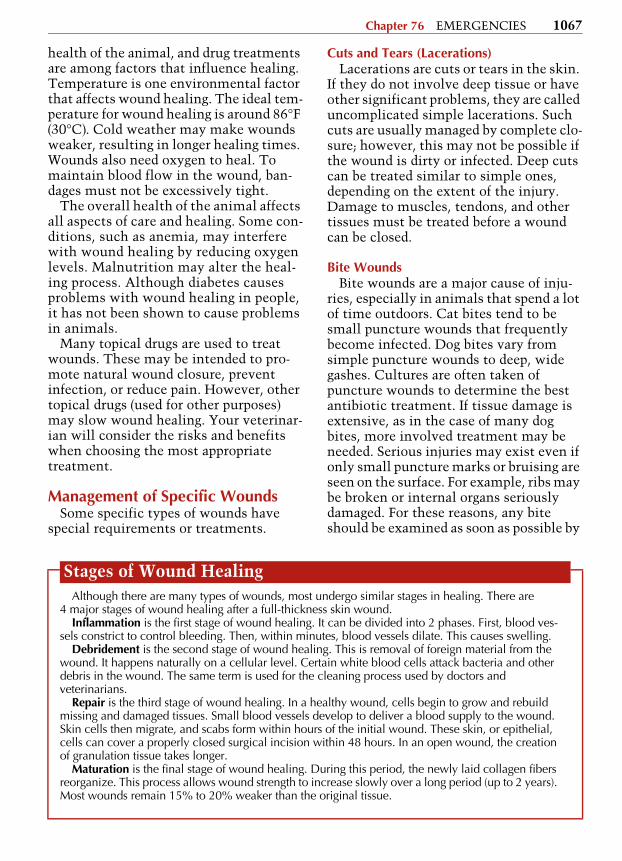
Wound HealingWounds heal in 4 stages (see BOX). Many
factors affect how well and how quickly the wounds heal. Environment, the overall
5608_09_076-085.fm Page 1066 Thursday, August 23, 2007 9:48 AM
Chapter 76 EMERGENCIES 1067
health of the animal, and drug treatments are among factors that influence healing. Temperature is one environmental factor that affects wound healing. The ideal tem-perature for wound healing is around 86°F (30°C). Cold weather may make wounds weaker, resulting in longer healing times. Wounds also need oxygen to heal. To maintain blood flow in the wound, ban-dages must not be excessively tight.
The overall health of the animal affects all aspects of care and healing. Some con-ditions, such as anemia, may interfere with wound healing by reducing oxygen levels. Malnutrition may alter the heal-ing process. Although diabetes causes problems with wound healing in people, it has not been shown to cause problems in animals.
Many topical drugs are used to treat wounds. These may be intended to pro-mote natural wound closure, prevent infection, or reduce pain. However, other topical drugs (used for other purposes) may slow wound healing. Your veterinar-ian will consider the risks and benefits when choosing the most appropriate treatment.
Management of Specific WoundsSome specific types of wounds have
special requirements or treatments.
Cuts and Tears (Lacerations)Lacerations are cuts or tears in the skin.
If they do not involve deep tissue or have other significant problems, they are called uncomplicated simple lacerations. Such cuts are usually managed by complete clo-sure; however, this may not be possible if the wound is dirty or infected. Deep cuts can be treated similar to simple ones, depending on the extent of the injury. Damage to muscles, tendons, and other tissues must be treated before a wound can be closed.
Bite WoundsBite wounds are a major cause of inju-
ries, especially in animals that spend a lot of time outdoors. Cat bites tend to be small puncture wounds that frequently become infected. Dog bites vary from simple puncture wounds to deep, wide gashes. Cultures are often taken of puncture wounds to determine the best antibiotic treatment. If tissue damage is extensive, as in the case of many dog bites, more involved treatment may be needed. Serious injuries may exist even if only small puncture marks or bruising are seen on the surface. For example, ribs may be broken or internal organs seriously damaged. For these reasons, any bite should be examined as soon as possible by
Stages of Wound HealingAlthough there are many types of wounds, most undergo similar stages in healing. There are
4 major stages of wound healing after a full-thickness skin wound.Inflammation is the first stage of wound healing. It can be divided into 2 phases. First, blood ves-
sels constrict to control bleeding. Then, within minutes, blood vessels dilate. This causes swelling.Debridement is the second stage of wound healing. This is removal of foreign material from the
wound. It happens naturally on a cellular level. Certain white blood cells attack bacteria and other debris in the wound. The same term is used for the cleaning process used by doctors and veterinarians.
Repair is the third stage of wound healing. In a healthy wound, cells begin to grow and rebuild missing and damaged tissues. Small blood vessels develop to deliver a blood supply to the wound. Skin cells then migrate, and scabs form within hours of the initial wound. These skin, or epithelial, cells can cover a properly closed surgical incision within 48 hours. In an open wound, the creation of granulation tissue takes longer.
Maturation is the final stage of wound healing. During this period, the newly laid collagen fibers reorganize. This process allows wound strength to increase slowly over a long period (up to 2 years). Most wounds remain 15% to 20% weaker than the original tissue.
5608_09_076-085.fm Page 1067 Thursday, August 23, 2007 9:48 AM

1068 Section 9 SPECIAL SUBJECTS
a veterinarian. After examination, the wound is generally cleaned thoroughly. Antibiotics and pain medication are com-monly administered.
Degloving InjuriesIn degloving injuries, the skin is sheared
or torn off. Degloving injuries can occur on the limbs or torso as well as the paws. Loss of skin is often extensive, and deeper tissues are often involved. Animals hit by cars or caught underneath the hood often have degloving injuries. Sometimes the skin is not completely removed. It remains attached to surrounding skin but not to the tissues beneath the skin. The skin is loose, usually bruised, and fragile. It may die later because it lacks blood supply. Any dead skin or other tissue must be removed. Tis-sue that can heal is usually saved. Infection is a major complication, and preventing infection is a main goal of initial treatment.
Gunshot InjuriesIn gunshot injuries, most of the damage
is not visible. The wounds are typically very deep. Gunshot wounds are also con-taminated because the bullet or pellet drags skin, hair, and dirt through the wound. If the bullet exits the body, the exit wound will be larger than the entrance wound. High-speed bullets create shock waves that affect surround-ing tissue and organs. There may be blunt force trauma as a result.
Often, surgery is needed to determine the amount of damage done by a gunshot wound. Organ and deep tissue injuries can be life-threatening. Fractures are common and may require additional treatment or surgery.
Pressure WoundsPressure wounds, also called decubital
ulcers, develop as a result of prolonged pressure on an area of skin. They are most common in paralyzed or immobile ani-mals. When tissue does not get enough blood or oxygen, it begins to die. Pressure wounds can be extremely difficult to treat and are best prevented. Preventive mea-
sures include changing the position of the animal frequently, maintaining adequate nutrition and cleanliness, and providing a sufficiently padded bed. If pressure wounds are mild or caught early, cleaning and bandaging may be enough to prevent further damage. More severe wounds require surgery. Grafts may be needed.
■ LIGHTNING STRIKE AND ELECTROCUTION
Injury or death of a pet due to high-voltage electrical currents may be the result of lightning, fallen transmission wires, faulty electrical circuits, or chew-ing on an electrical cord. Lightning strike is seasonal and tends to be geographically restricted. Investigation of possible elec-trocution should always proceed with caution because the electrification resulting from broken transmission wires, for example, may still be present.
Certain types of trees, especially hard-woods such as oaks and those that are tall and have spreading root systems just beneath the ground surface, tend to be struck by lightning more often than others. Electrification of such roots charges a wide surface area, particularly when the ground is already damp; passage of charged roots beneath a shallow pool of water causes it to become electrified. Fallen or sagging trans-mission wires also may electrify a pool of water, fence, or building, and an animal may also directly contact such wires.
Varying degrees of electric shock may occur. In most instances of lightning strike, death is instantaneous and the animal falls without a struggle. Occasionally, the ani-mal becomes unconscious but may recover in a few minutes to several hours; residual nervous signs (including depression, para-plegia, and increased skin sensitivity to nor-mally painless stimulus) may persist for days or weeks or be permanent. Death from electric shock usually results from cardiac or respiratory arrest.
Lightning strike and electrocution should be considered true emergencies in which your veterinarian should be con-
5608_09_076-085.fm Page 1068 Thursday, August 23, 2007 9:48 AM

Chapter 76 EMERGENCIES 1069
tacted right away. Even if the effect of electrocution appears mild (as in a pet chewing on an electrical cord), signs of shock or other complications may occur later. Animals that survive may require supportive and symptomatic therapy.
■ MINOR INJURIES AND ACCIDENTS
A number of minor injuries are gener-ally not life-threatening but also demand immediate attention.
Broken NailsControl any bleeding by applying pres-
sure or using styptic power, cornstarch, or even white flour. Because broken nails can be very painful, you may need to place a muzzle on your dog (see FIGURE, page 1052). Try to gently remove the broken piece with clippers. Seek veterinary care if the broken nail cannot be removed easily.
Bumps, Bruises, Twists, and SprainsMild to moderate pain, tenderness,
swelling, and limping can indicate a bruise, strain, or sprain. Keep your pet quiet and restrict exercise. Contact your veterinarian if signs continue for more than a few days.
Fish HooksFish hooks often become lodged in the
mouth, lips, nose, or paws. Do not try to remove a fish hook that is embedded in the eyes, mouth, or ears—this must be done under anesthesia by your veteri-narian. If a hook embedded in the mouth is attached to a line, prevent your pet from swallowing the hook by tying the line to the animal’s collar. Otherwise, cut the hook free from the line, but leave several inches of line attached so that the hook is easier to find (especially in thick fur). If the barb has entered the skin, push it through in the same direc-tion so that you can snip off the barb with wire cutters or pliers. Then remove the hook by pulling it back out in the direction it entered the skin. Clean and
cover the wound, and take your pet to the veterinarian immediately.
Insect Bites and StingsBites and stings often occur on the face
or elsewhere on the head. They are often so small that they cannot be seen through the fur. Apply a cold pack to the affected area to reduce swelling and itch-ing. Look for a stinger. If you find one in the skin, you can use a credit card or other flat, rigid object to scrape it out. Do not squeeze a stinger with tweezers or other tool because this can release more venom into the wound. If your pet has an allergic reaction with a great deal of swelling in the head or neck area that may affect breathing, or if you find a stinger in the tongue or the roof of the mouth, take your pet to your veterinar-ian immediately.
Porcupine QuillsPorcupine quills have small barbs on
their ends, which makes them very pain-ful to remove. They should be removed by your veterinarian because of the need for anesthesia and the risk of more bleeding and tissue damage.
SkunkingSkunk spray is an oil that is best
removed while still wet. If you can iden-tify the specific area that was sprayed, clean it first before wetting the entire dog to avoid spreading the oil to other areas. There are numerous commercial products and recipes for homemade deskunking rinses. Be aware that many homemade concoctions are too harsh to use in the face and eye area, and many will bleach the hair coat. In addition to the objection-able odor, skunk spray can cause eye irri-tation and temporary blindness. Skunk bites can transmit rabies, but skunk spray has not been shown to transmit the virus.
Swallowed ObjectsDogs tend to swallow numerous inap-
propriate objects including toys, trash,
5608_09_076-085.fm Page 1069 Thursday, August 23, 2007 9:48 AM

1070 Section 9 SPECIAL SUBJECTS
or chicken bones. In many cases, swal-lowed objects can be treated with a wait-and-see approach; most of the time, the object will pass through without any trouble. However, swallowing sharp objects, such as needles or pieces of glass, or any type of long item, such as string, pantyhose, or fishing line, is very dangerous because a serious bowel con-
dition can result. Although cats are more selective than dogs, they often swallow string, fishing line, or tinsel, or get the material wrapped and knotted around their tongue. In these situations, or if your pet has persistent vomiting, a ten-der abdomen, or is not having bowel movements, you should contact your veterinarian immediately.
C H A P T E R 77Diagnostic Tests and Imaging
■ INTRODUCTIONWhen you have a health problem, your
doctor will order blood tests, x-rays, or other tests to help pinpoint the cause of the problem. When your pet has a health problem, your veterinarian will often order similar tests to determine the cause and seriousness of your pet’s con-dition. Depending on the tests needed and the facilities available at your veter-inarian’s clinic, the tests may be per-formed in-house at the clinic or at a specialized laboratory or test facility in another location.
If the tests are to be carried out by the veterinarian or a technician within their clinic, then the staff has access to the medical history leading up to the prob-lem. If the tests are to be performed else-where, it is important that a detailed history of the problem be included with the samples or be available when the pet arrives. The staff at the external labora-tory will need this information to cor-rectly perform the tests and interpret the results. Usually, the referring veterinary clinic will provide this information to the external laboratory. However, if you are asked to bring the information to the clinic with your pet, it is very important that you do so to help the laboratory do their work correctly.
■ TYPES OF VETERINARY MEDICAL TESTS
There are several categories of diagnos-tic tests that may be performed to help your veterinarian determine the cause of your pet’s disorder.
HistologyHistology is the study of the structure,
function, and chemical composition of cells within the body. Experts in histology (called pathologists) examine small tissue samples to determine if they are normal or diseased. These experts can often point to a cause for the abnormal cells.
Small tissue samples will often be sent to a pathologist if your veterinarian sus-pects conditions such as cancer or other diseases that cause tissue changes.
MicrobiologyMicrobiology is the study of small or-
ganisms such as bacteria, viruses, fungi, and other single-celled life forms. In a vet-erinary laboratory, specialists in microbi-ology can perform any of hundreds of tests looking for signs of infection. Common tests include growing and identifying bac-teria, viruses, and fungi. Bacteria can also be tested to see which antibiotics should be effective for eliminating them from the body. Other tests use antibodies to detect
5608_09_076-085.fm Page 1070 Thursday, August 23, 2007 9:48 AM