Embed Size (px)
Citation preview
Introduction to CardiologyIntroduction to Cardiology

Introduction to CardiologyIntroduction to Cardiology
Topics to be Discussed:Topics to be Discussed:Cardiovascular DiseaseCardiovascular DiseaseEMS System RoleEMS System RoleCardiovascular Anatomy and PhysiologyCardiovascular Anatomy and PhysiologyCardiovascular ElectrophysiologyCardiovascular Electrophysiology
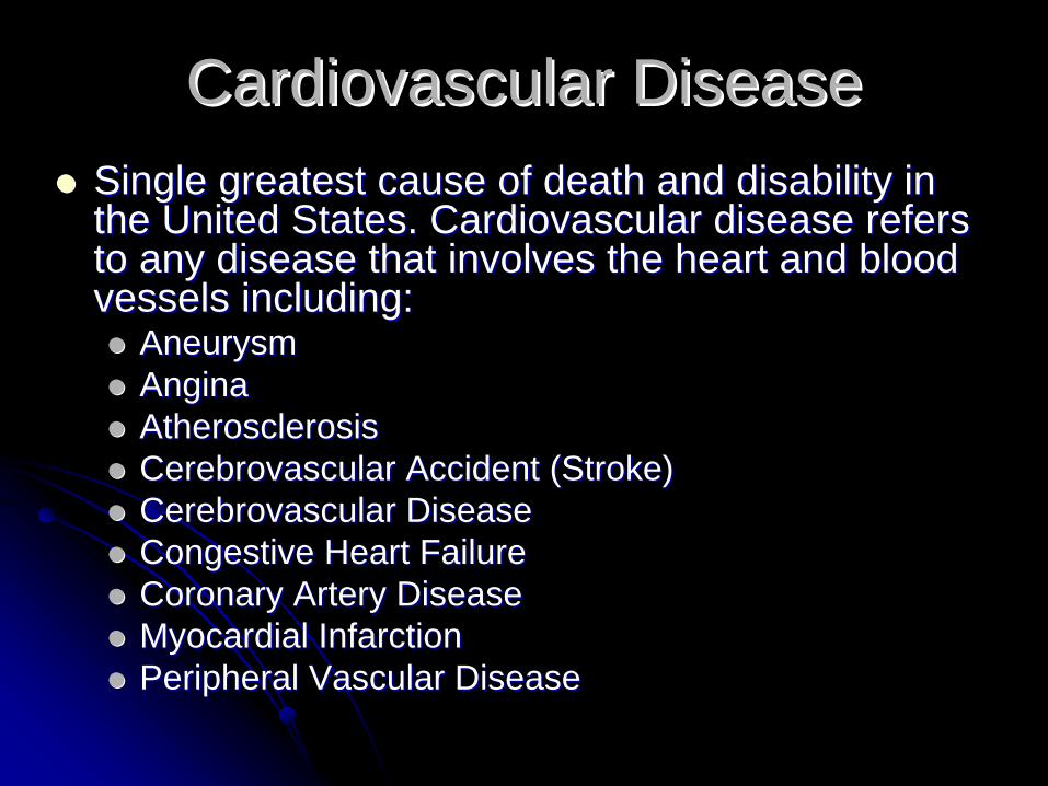
Cardiovascular DiseaseCardiovascular DiseaseSingle greatest cause of death and disability in Single greatest cause of death and disability in the United States. Cardiovascular disease refers the United States. Cardiovascular disease refers to any disease that involves the heart and blood to any disease that involves the heart and blood vessels including:vessels including:
AneurysmAneurysmAngina Angina Atherosclerosis Atherosclerosis Cerebrovascular Accident (Stroke) Cerebrovascular Accident (Stroke) Cerebrovascular Disease Cerebrovascular Disease Congestive Heart Failure Congestive Heart Failure Coronary Artery Disease Coronary Artery Disease Myocardial Infarction Myocardial Infarction Peripheral Vascular DiseasePeripheral Vascular Disease
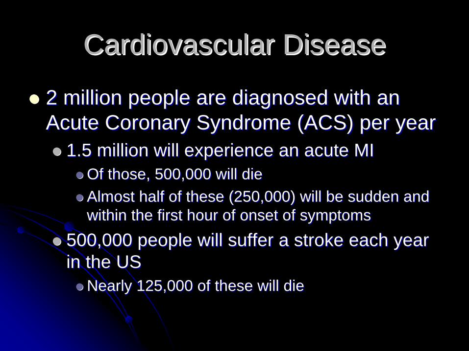
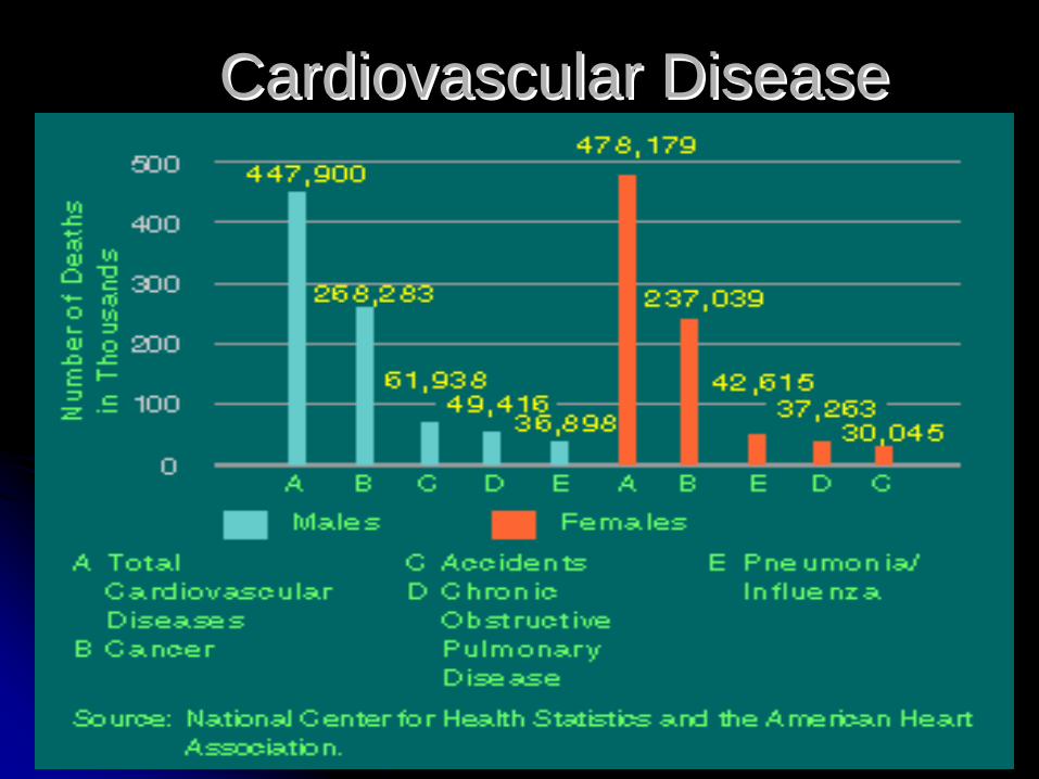
2 million people are diagnosed with an 2 million people are diagnosed with an Acute Coronary Syndrome (ACS) per yearAcute Coronary Syndrome (ACS) per year
1.5 million will experience an acute MI1.5 million will experience an acute MIOf those, 500,000 will dieOf those, 500,000 will dieAlmost half of these (250,000) will be sudden and Almost half of these (250,000) will be sudden and within the first hour of onset of symptomswithin the first hour of onset of symptoms
500,000 people will suffer a stroke each year 500,000 people will suffer a stroke each year in the USin the US
Nearly 125,000 of these will dieNearly 125,000 of these will die
Cardiovascular DiseaseCardiovascular Disease
Cardiovascular MorbidityCardiovascular Morbidity
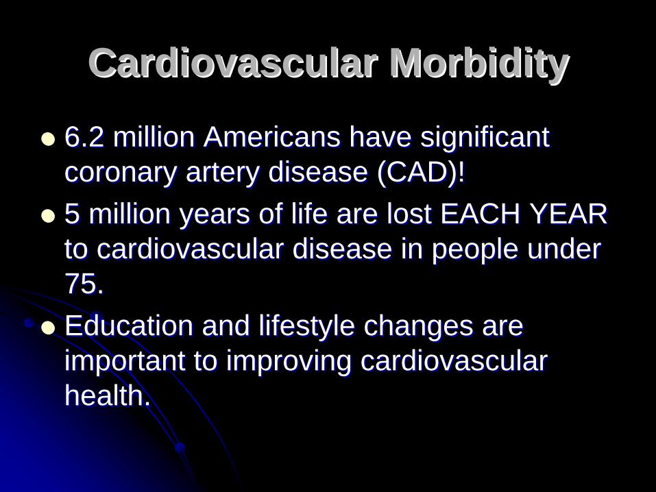
6.2 million Americans have significant 6.2 million Americans have significant coronary artery disease (CAD)!coronary artery disease (CAD)!5 million years of life are lost EACH YEAR 5 million years of life are lost EACH YEAR to cardiovascular disease in people under to cardiovascular disease in people under 75.75.Education and lifestyle changes are Education and lifestyle changes are important to improving cardiovascular important to improving cardiovascular health.health.
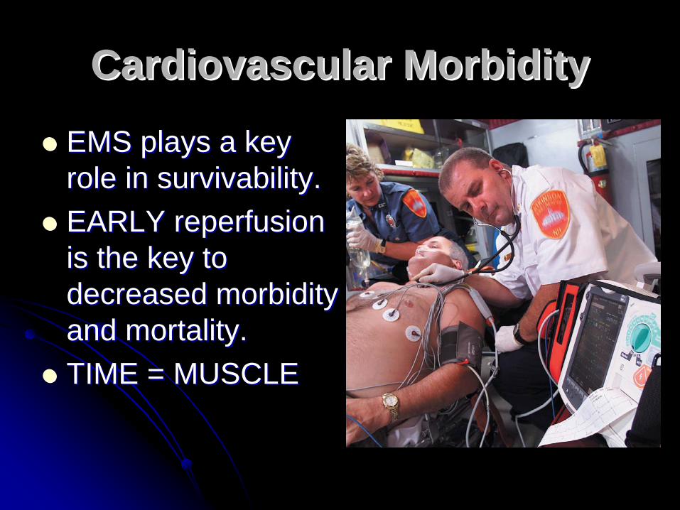
EMS plays a key EMS plays a key role in survivability.role in survivability.EARLY reperfusion EARLY reperfusion is the key to is the key to decreased morbidity decreased morbidity and mortality.and mortality.TIME = MUSCLETIME = MUSCLE
Cardiovascular MorbidityCardiovascular Morbidity
Cardiovascular Disease Cardiovascular Disease
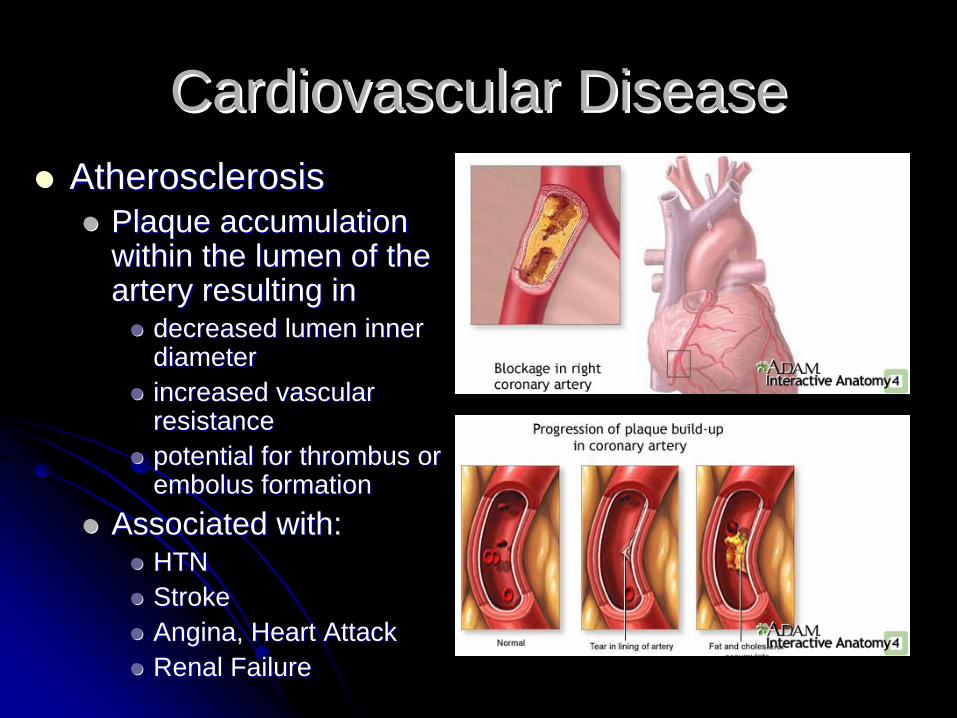
Cardiovascular DiseaseCardiovascular DiseaseAtherosclerosisAtherosclerosis
Plaque accumulation Plaque accumulation within the lumen of the within the lumen of the artery resulting inartery resulting in
decreased lumen inner decreased lumen inner diameterdiameterincreased vascular increased vascular resistanceresistancepotential for thrombus or potential for thrombus or embolus formationembolus formation
Associated with: Associated with: HTNHTNStrokeStrokeAngina, Heart AttackAngina, Heart AttackRenal FailureRenal Failure
Cardiovascular DiseaseCardiovascular Disease
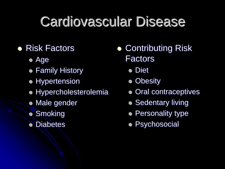
Risk FactorsRisk FactorsAgeAgeFamily HistoryFamily HistoryHypertensionHypertensionHypercholesterolemiaHypercholesterolemiaMale genderMale genderSmokingSmokingDiabetesDiabetes
Contributing Risk Contributing Risk FactorsFactors
DietDietObesityObesityOral contraceptivesOral contraceptivesSedentary livingSedentary livingPersonality typePersonality typePsychosocialPsychosocial
EMS System RoleEMS System Role
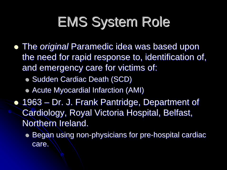
The The originaloriginal Paramedic idea was based upon Paramedic idea was based upon the need for rapid response to, identification of, the need for rapid response to, identification of, and emergency care for victims of:and emergency care for victims of:
Sudden Cardiac Death (SCD)Sudden Cardiac Death (SCD)Acute Myocardial Infarction (AMI)Acute Myocardial Infarction (AMI)
1963 1963 –– Dr. J. Frank Pantridge, Department of Dr. J. Frank Pantridge, Department of Cardiology, Royal Victoria Hospital, Belfast, Cardiology, Royal Victoria Hospital, Belfast, Northern Ireland.Northern Ireland.
Began using nonBegan using non--physicians for prephysicians for pre--hospital cardiac hospital cardiac care.care.
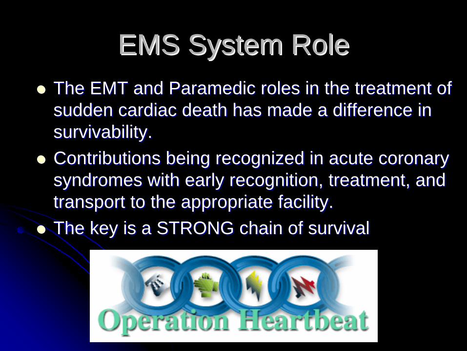
EMS System RoleEMS System RoleThe EMT and Paramedic roles in the treatment of The EMT and Paramedic roles in the treatment of sudden cardiac death has made a difference in sudden cardiac death has made a difference in survivability.survivability.Contributions being recognized in acute coronary Contributions being recognized in acute coronary syndromes with early recognition, treatment, and syndromes with early recognition, treatment, and transport to the appropriate facility.transport to the appropriate facility.The key is a STRONG chain of survivalThe key is a STRONG chain of survival
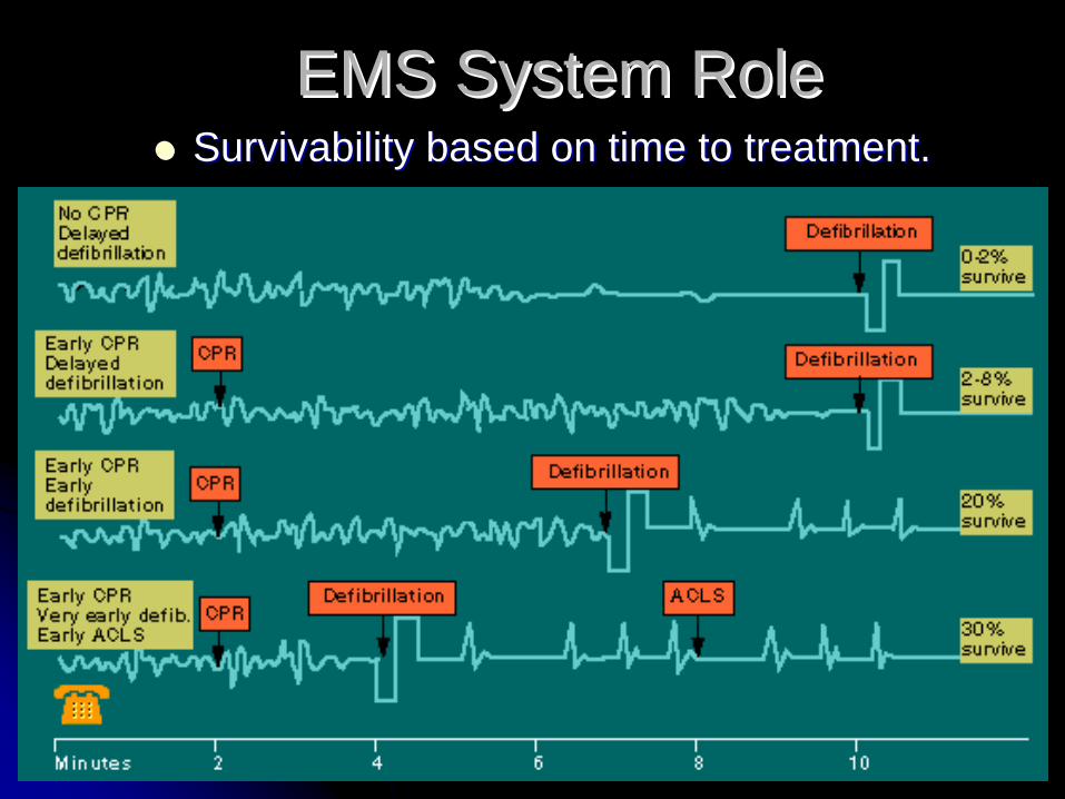
EMS System RoleEMS System RoleSurvivability based on time to treatment.Survivability based on time to treatment.
Cardiac Anatomy Cardiac Anatomy and Physiologyand Physiology

Anatomy ReviewAnatomy Review

Anatomy ReviewAnatomy Review
Anatomy ReviewAnatomy Review

Anatomy ReviewAnatomy Review
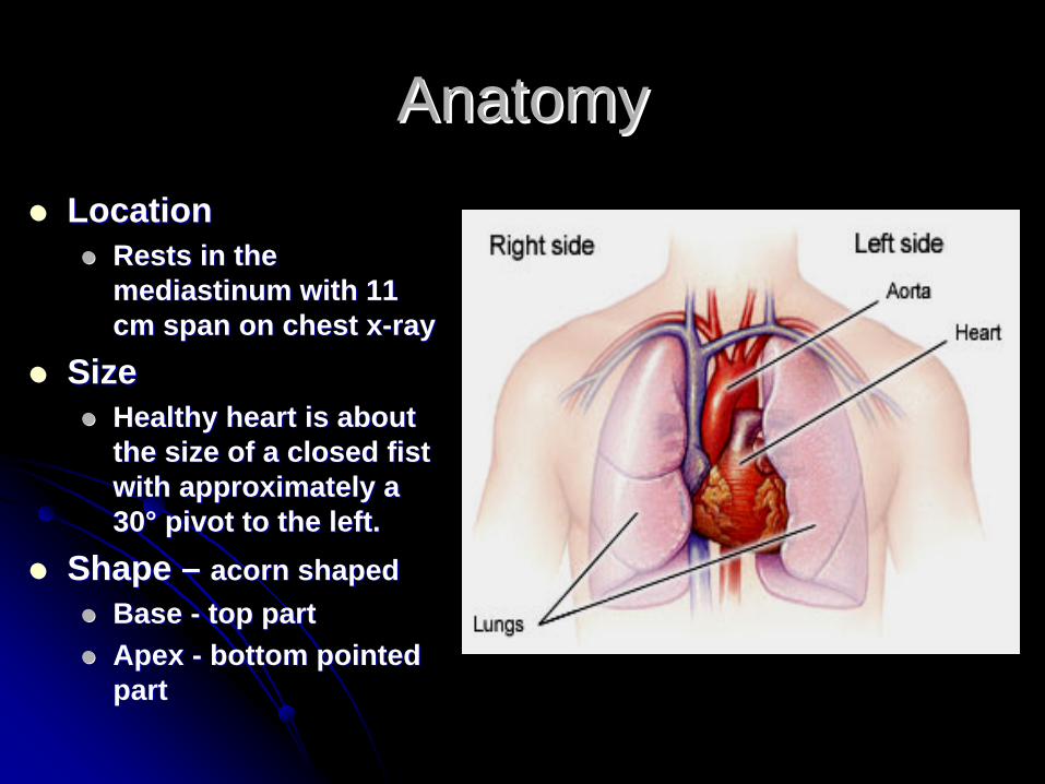
AnatomyAnatomyLocationLocation
Rests in the Rests in the mediastinum with 11 mediastinum with 11 cm span on chest xcm span on chest x--rayray
SizeSizeHealthy heart is about Healthy heart is about the size of a closed fist the size of a closed fist with approximately a with approximately a 3030°° pivot to the left.pivot to the left.
Shape Shape –– acorn shapedacorn shapedBase Base -- top parttop partApex Apex -- bottom pointed bottom pointed part part
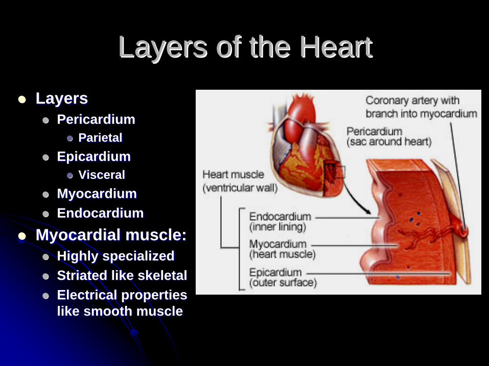
Layers of the HeartLayers of the HeartLayersLayers
PericardiumPericardiumParietalParietal
EpicardiumEpicardiumVisceralVisceral
MyocardiumMyocardiumEndocardiumEndocardium
Myocardial muscle: Myocardial muscle: Highly specializedHighly specializedStriated like skeletalStriated like skeletalElectrical properties Electrical properties like smooth musclelike smooth muscle

Layers of the HeartLayers of the Heart

Pericardial SpacePericardial Space
Pericardial space contains about 25 mL of serous fluid for lubrication. (By definition: >90 mL constitutes a tamponade.)

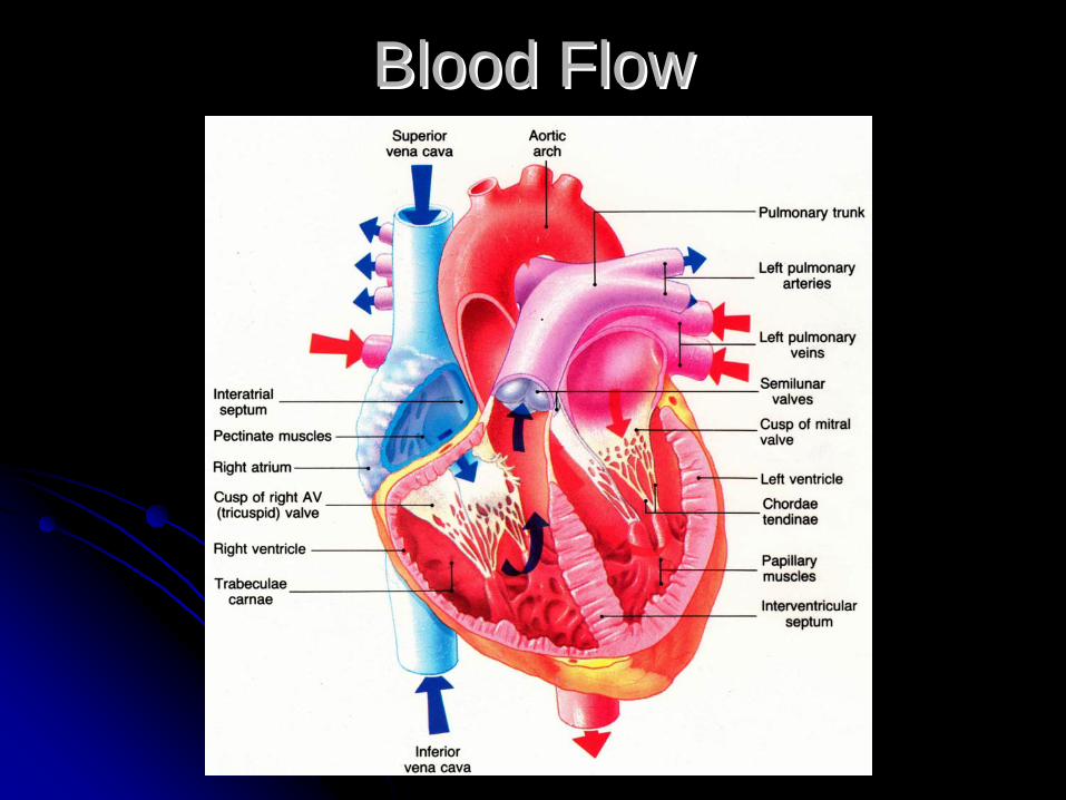
ChambersChambersAtria Atria -- less muscularless muscular
80% of blood flow 80% of blood flow from right atrium is from right atrium is passive, remaining passive, remaining 20% provides atrial 20% provides atrial kick. kick. Loss of atrial kick can Loss of atrial kick can significantly reduce significantly reduce cardiac output (CO).cardiac output (CO).
What cardiac rhythms What cardiac rhythms result in a loss of result in a loss of atrialatrialkick?kick?
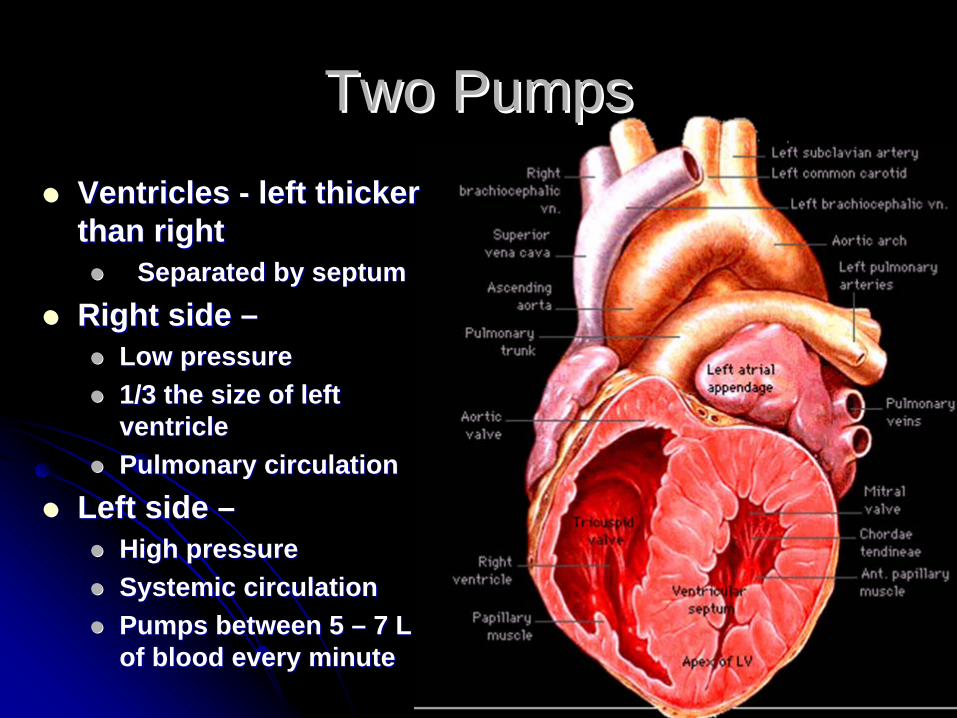
Two PumpsTwo PumpsVentricles Ventricles -- left thicker left thicker than rightthan right
Separated by septumSeparated by septumRight side Right side ––
Low pressureLow pressure1/3 the size of left 1/3 the size of left ventricle ventricle Pulmonary circulationPulmonary circulation
Left side Left side ––High pressureHigh pressureSystemic circulationSystemic circulationPumps between 5 Pumps between 5 –– 7 L 7 L of blood every minuteof blood every minute
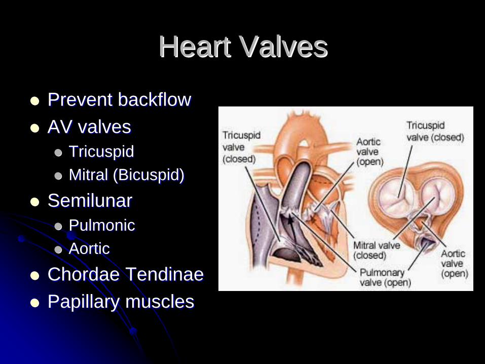
Heart ValvesHeart Valves
Prevent backflowPrevent backflowAV valves AV valves
TricuspidTricuspidMitral (Bicuspid)Mitral (Bicuspid)
SemilunarSemilunarPulmonicPulmonicAorticAortic
Chordae TendinaeChordae TendinaePapillary musclesPapillary muscles

Heart ValvesHeart Valves
S1 (“lub”) or the first heart sound is caused by the closure of the AV valvesS2 (“dub”) or the second heart sound is caused by the closure of the semilunar valves.

Heart SoundsHeart Sounds
Extra heart sounds include:
S3-ventricular gallop (Ken-tuc-ky). Commonly associated with failure of the left ventricle in CHF.S4-atrial gallop (Ten-nes-see). Can also be associated with LVF or restrictive cardiomyopathy.
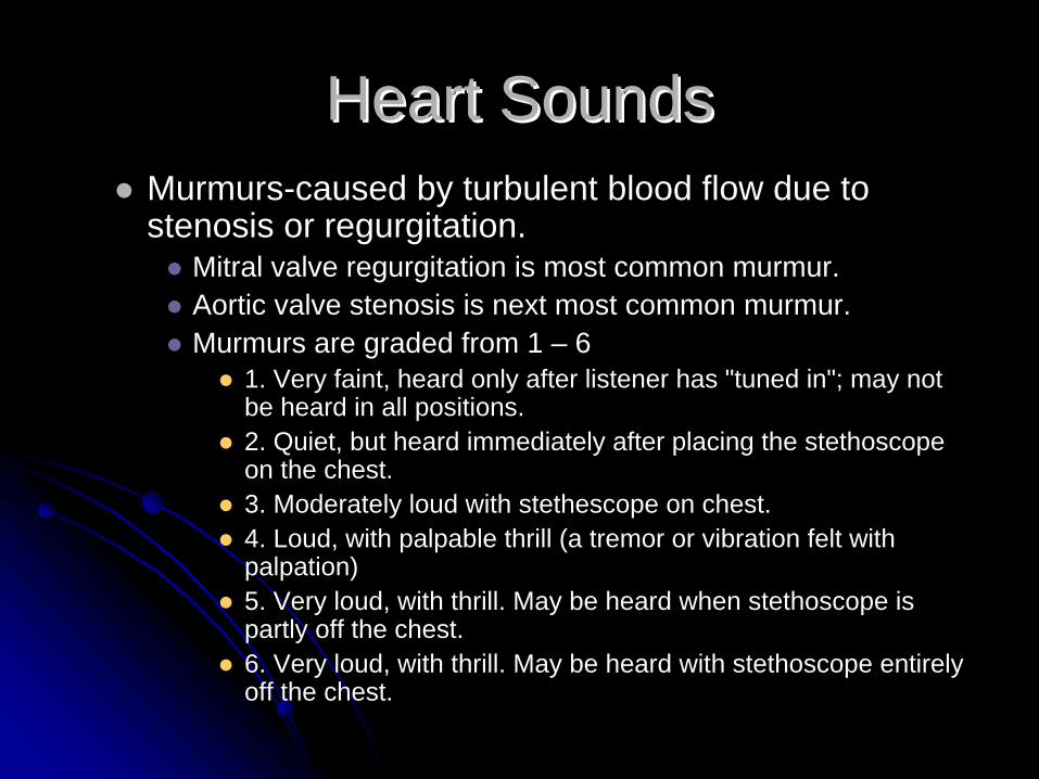
Murmurs-caused by turbulent blood flow due to stenosis or regurgitation.
Mitral valve regurgitation is most common murmur.Aortic valve stenosis is next most common murmur.Murmurs are graded from 1 – 6
1. Very faint, heard only after listener has "tuned in"; may notbe heard in all positions.2. Quiet, but heard immediately after placing the stethoscope on the chest. 3. Moderately loud with stethescope on chest.4. Loud, with palpable thrill (a tremor or vibration felt with palpation)5. Very loud, with thrill. May be heard when stethoscope is partly off the chest. 6. Very loud, with thrill. May be heard with stethoscope entirely off the chest.
Heart SoundsHeart Sounds

AnatomyAnatomy
Great Vessels:Great Vessels:Vena CavaVena CavaPulmonary Arteries Pulmonary Arteries (2)(2)Pulmonary Veins (4)Pulmonary Veins (4)AortaAortaThe Great Vessels The Great Vessels are attached to the are attached to the base of the heart.base of the heart.
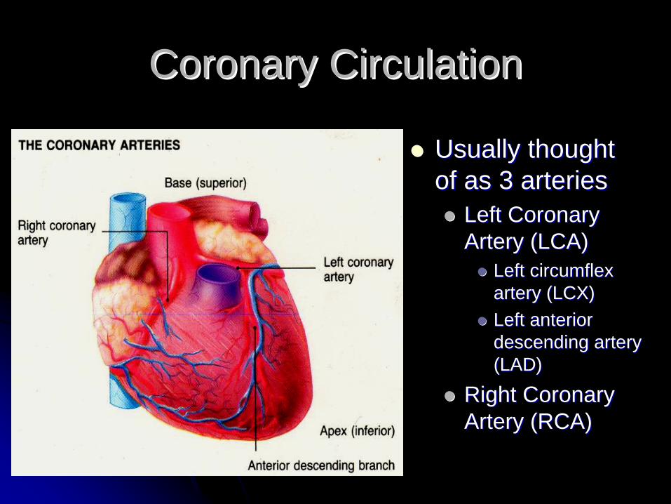
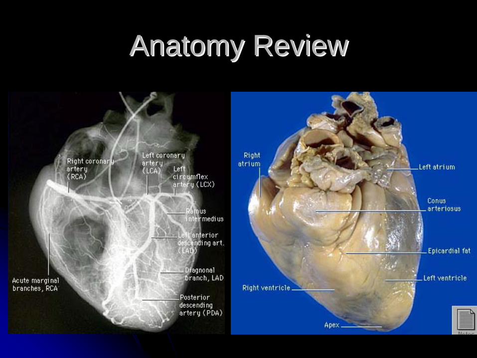
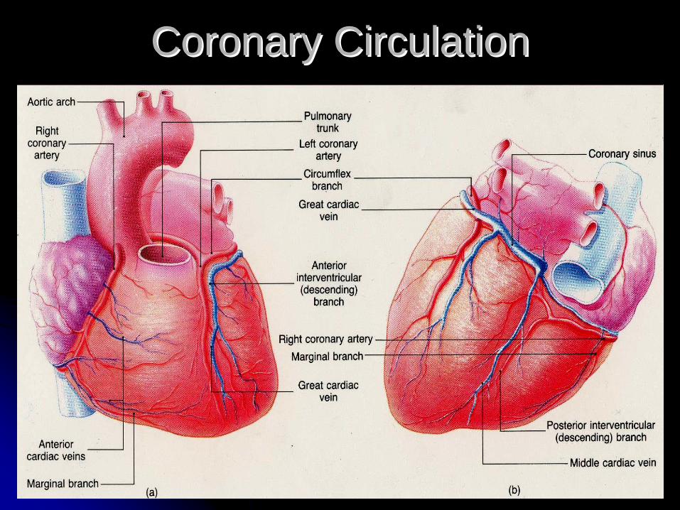
Coronary CirculationCoronary Circulation
Usually thought Usually thought of as 3 arteriesof as 3 arteries
Left Coronary Left Coronary Artery (LCA) Artery (LCA)
Left circumflex Left circumflex artery (LCX)artery (LCX)Left anterior Left anterior descending artery descending artery (LAD)(LAD)
Right Coronary Right Coronary Artery (RCA)Artery (RCA)
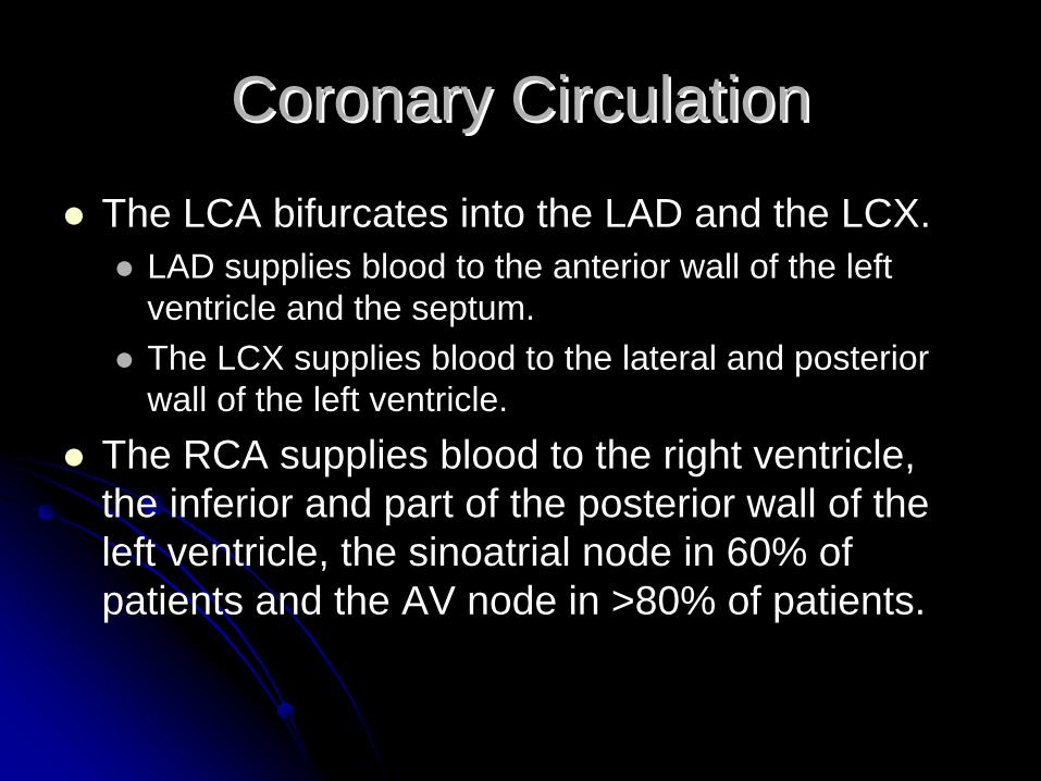
The LCA bifurcates into the LAD and the LCX.LAD supplies blood to the anterior wall of the left ventricle and the septum.The LCX supplies blood to the lateral and posterior wall of the left ventricle.
The RCA supplies blood to the right ventricle, the inferior and part of the posterior wall of the left ventricle, the sinoatrial node in 60% of patients and the AV node in >80% of patients.
Coronary CirculationCoronary Circulation
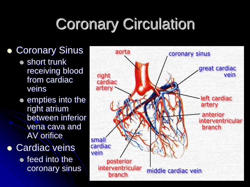
Coronary CirculationCoronary CirculationCoronary SinusCoronary Sinus
short trunk short trunk receiving blood receiving blood from cardiac from cardiac veinsveinsempties into the empties into the right atrium right atrium between inferior between inferior vena cava and vena cava and AV orificeAV orifice
Cardiac veinsCardiac veinsfeed into the feed into the coronary sinuscoronary sinus
Coronary CirculationCoronary Circulation
Blood FlowBlood Flow
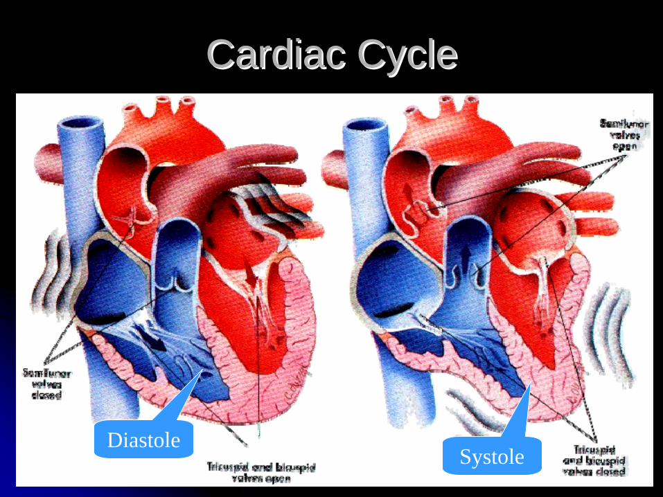
Cardiac CycleCardiac Cycle
DiastoleSystole

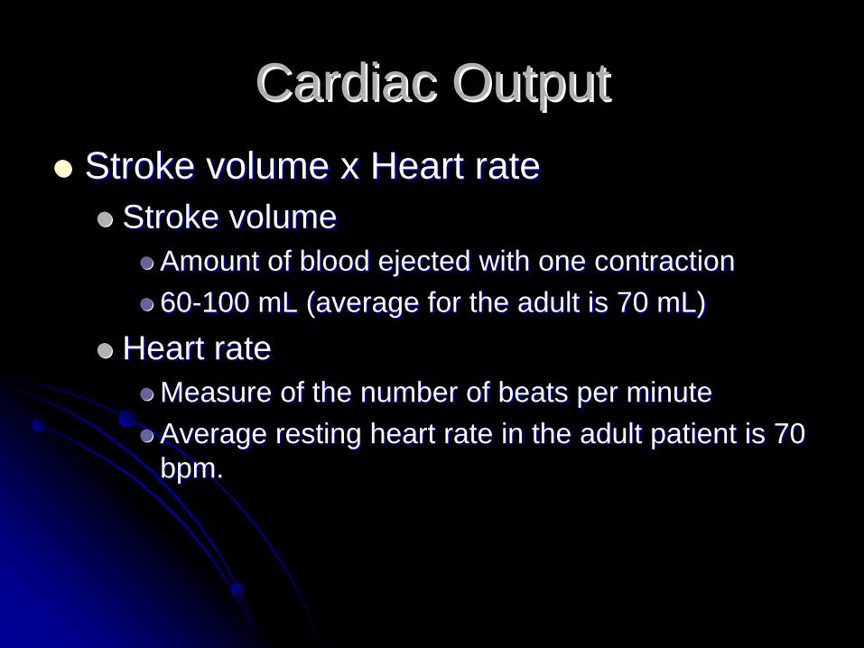
Cardiac OutputCardiac OutputStroke volume x Heart rateStroke volume x Heart rate
Stroke volumeStroke volumeAmount of blood ejected with one contractionAmount of blood ejected with one contraction6060--100 mL (average for the adult is 70 mL)100 mL (average for the adult is 70 mL)
Heart rateHeart rateMeasure of the number of beats per minuteMeasure of the number of beats per minuteAverage resting heart rate in the adult patient is 70 Average resting heart rate in the adult patient is 70 bpm.bpm.
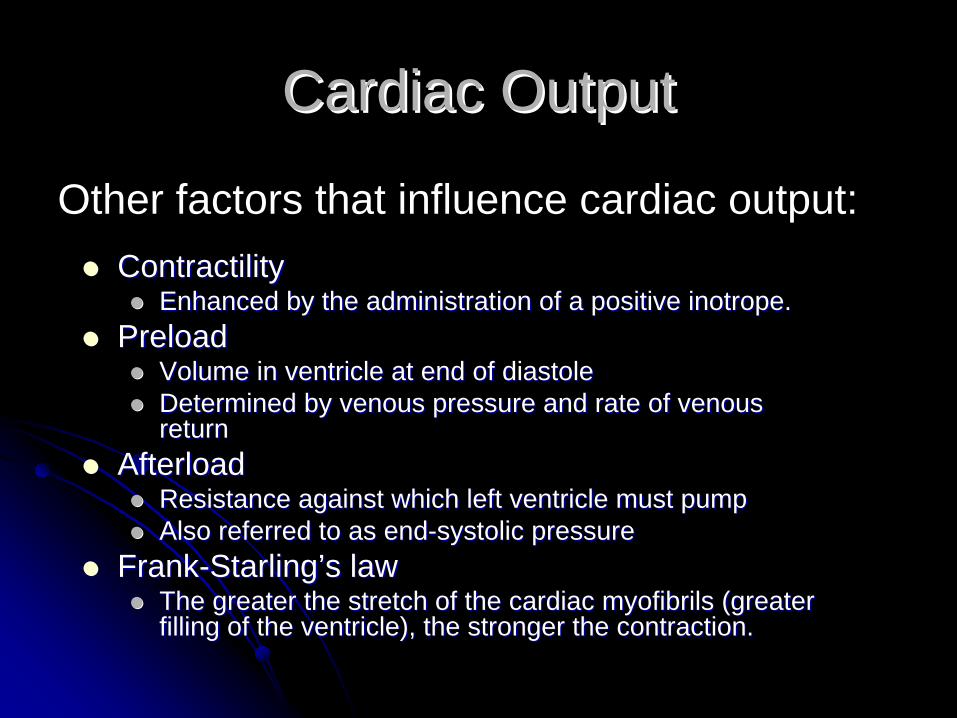
ContractilityContractilityEnhanced by the administration of a positive inotrope.Enhanced by the administration of a positive inotrope.
PreloadPreloadVolume in ventricle at end of diastoleVolume in ventricle at end of diastoleDetermined by venous pressure and rate of venous Determined by venous pressure and rate of venous returnreturn
AfterloadAfterloadResistance against which left ventricle must pumpResistance against which left ventricle must pumpAlso referred to as endAlso referred to as end--systolic pressuresystolic pressure
FrankFrank--StarlingStarling’’s laws lawThe greater the stretch of the cardiac myofibrils (greater The greater the stretch of the cardiac myofibrils (greater filling of the ventricle), the stronger the contraction.filling of the ventricle), the stronger the contraction.
Cardiac OutputCardiac Output
Other factors that influence cardiac output:

Vascular SystemVascular System
Consists of Arteries, Consists of Arteries, Veins, and Capillaries.Veins, and Capillaries.
Largest artery is the Largest artery is the AortaAorta
ascending thoracicascending thoracicdescending thoracicdescending thoracicabdominalabdominal
Largest veins are the Largest veins are the Vena cavaVena cava
superior superior InferiorInferior
Capillary beds is where Capillary beds is where gas exchange occurs.gas exchange occurs.
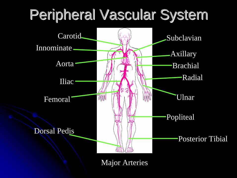
Peripheral Vascular SystemPeripheral Vascular SystemSubclavian
AxillaryBrachial
Radial
Ulnar
Aorta
Femoral
Iliac
Carotid
Popliteal
Dorsal PedisPosterior Tibial
Innominate
Major Arteries
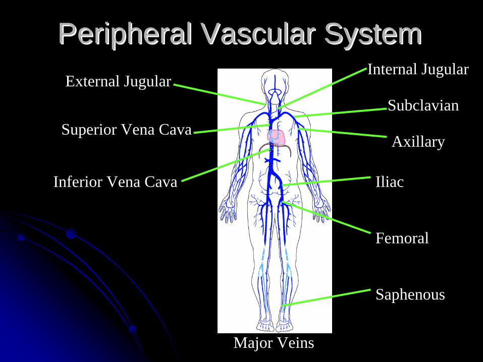
Peripheral Vascular SystemPeripheral Vascular SystemInternal Jugular
Subclavian
Axillary
Iliac
Femoral
Saphenous
External Jugular
Superior Vena Cava
Inferior Vena Cava
Major Veins
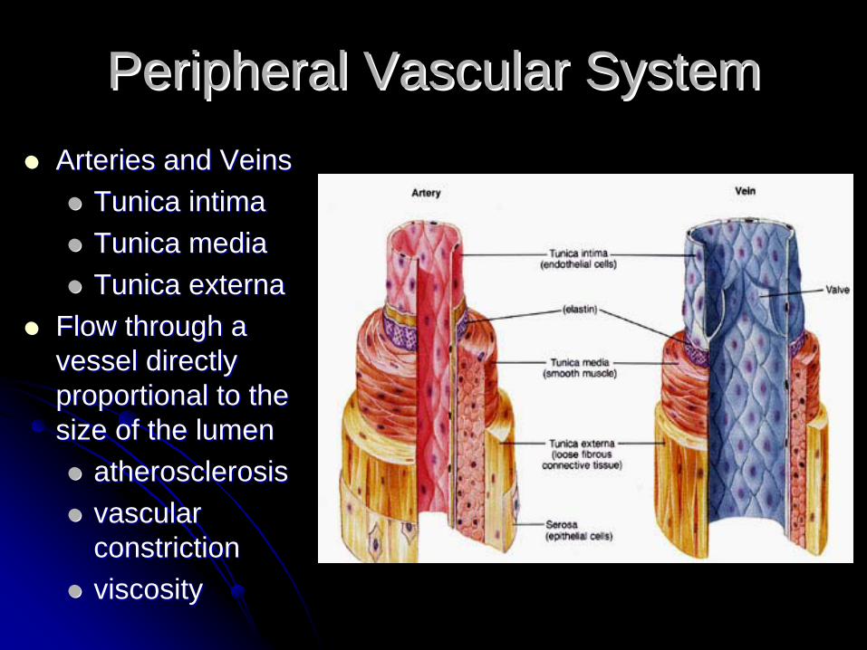
Peripheral Vascular SystemPeripheral Vascular SystemArteries and Veins Arteries and Veins
Tunica intimaTunica intimaTunica media Tunica media Tunica externaTunica externa
Flow through a Flow through a vessel directly vessel directly proportional to the proportional to the size of the lumensize of the lumen
atherosclerosisatherosclerosisvascular vascular constrictionconstrictionviscosityviscosity

CapillariesCapillaries

Peripheral Vascular SystemPeripheral Vascular SystemBlood return to the heart Blood return to the heart is dependent on:is dependent on:Skeletal muscle pumpSkeletal muscle pump
Muscular contraction Muscular contraction squeezes adjacent veins squeezes adjacent veins causing a milking actioncausing a milking actionValves prevent back flowValves prevent back flow
Respiratory MovementsRespiratory MovementsDiaphragm contraction Diaphragm contraction exerts pressure in abdomen exerts pressure in abdomen and decreases pressure in and decreases pressure in thoracic cavitythoracic cavityBlood moves to an area of Blood moves to an area of lowerlower (negative)(negative) pressure in pressure in thoraxthorax

Peripheral Vascular SystemPeripheral Vascular System
Factors affecting Venous ReturnFactors affecting Venous ReturnConstriction of veinsConstriction of veinsSympathetic stimulation causes contraction of Sympathetic stimulation causes contraction of the smooth muscle walls of veinsthe smooth muscle walls of veins
Venous reservoir. The vVenous reservoir. The venous system contains approximately 65% of blood volume at any given timeBlood vessel constriction returns 20% or 1 liter of blood to active circulation
GravityGravity
Peripheral Vascular SystemPeripheral Vascular System
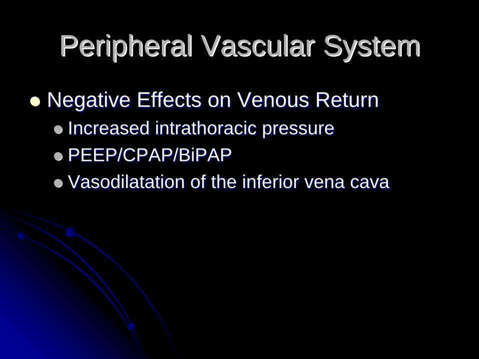
Negative Effects on Venous ReturnNegative Effects on Venous ReturnIncreased intrathoracic pressureIncreased intrathoracic pressurePEEP/CPAP/BiPAPPEEP/CPAP/BiPAPVasodilatation of the inferior vena cavaVasodilatation of the inferior vena cava
Peripheral Vascular SystemPeripheral Vascular System
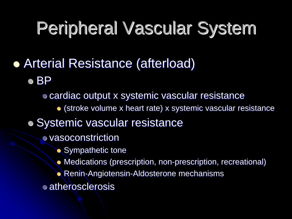
Arterial Resistance (afterload)Arterial Resistance (afterload)BPBP
cardiac output x systemic vascular resistancecardiac output x systemic vascular resistance(stroke volume x heart rate) x systemic vascular resistance(stroke volume x heart rate) x systemic vascular resistance
Systemic vascular resistanceSystemic vascular resistancevasoconstrictionvasoconstriction
Sympathetic toneSympathetic toneMedications (prescription, nonMedications (prescription, non--prescription, recreational)prescription, recreational)ReninRenin--AngiotensinAngiotensin--Aldosterone mechanismsAldosterone mechanisms
atherosclerosisatherosclerosis

ElectrophysiologyElectrophysiology
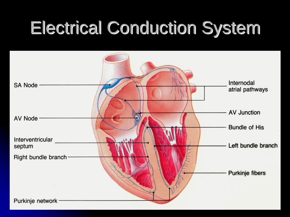
Electrical Conduction SystemElectrical Conduction System
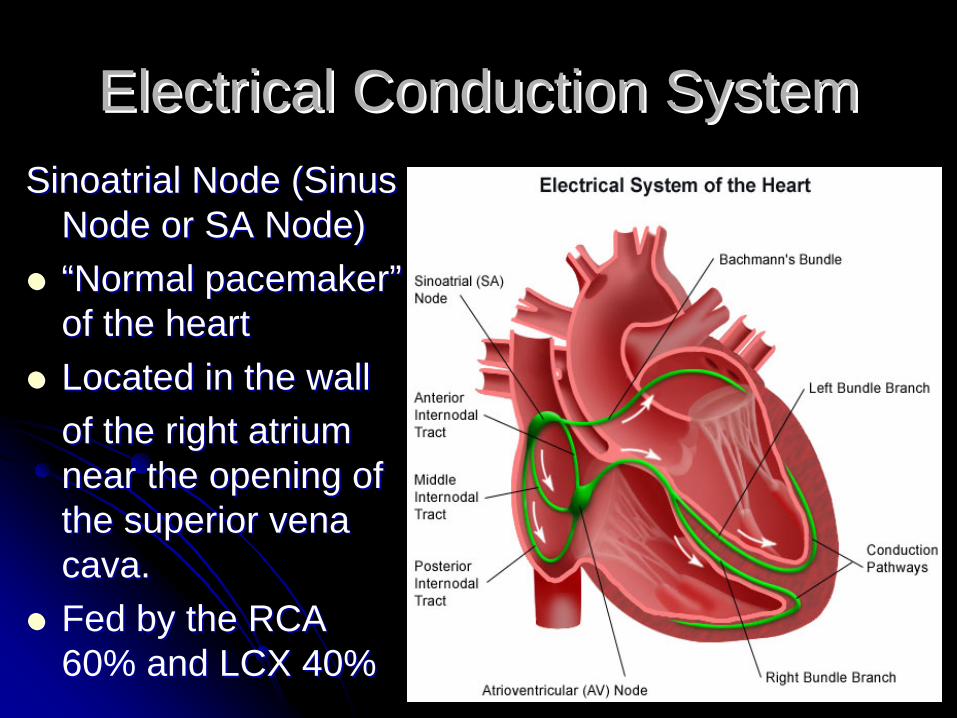
Electrical Conduction SystemElectrical Conduction SystemSinoatrial Node (Sinus Sinoatrial Node (Sinus
Node or SA Node)Node or SA Node)““Normal pacemakerNormal pacemaker””of the heartof the heartLocated in the wall Located in the wall of the right atrium of the right atrium near the opening of near the opening of the superior vena the superior vena cava.cava.Fed by the RCA Fed by the RCA 60% and LCX 40%60% and LCX 40%

Internodal Atrial PathwaysInternodal Atrial PathwaysBachmanBachman’’s Bundles BundleAnterior, middle, and Anterior, middle, and posterior pathwaysposterior pathwaysAccessory pathwaysAccessory pathways
Electrical Conduction SystemElectrical Conduction System

Atrioventricular Junction Atrioventricular Junction (AV junction)(AV junction)AV nodeAV node
““GatekeeperGatekeeper””Slows conduction to the Slows conduction to the ventricles allowing timeventricles allowing timefor ventricles to fillfor ventricles to fillMay receive impulses as May receive impulses as high as 600 bpm, high as 600 bpm, generally will not allow generally will not allow impulses more than 180 impulses more than 180 bpm to reach ventriclesbpm to reach ventriclesBundle of HisBundle of His
Electrical Conduction SystemElectrical Conduction System
Electrical Conduction SystemElectrical Conduction System
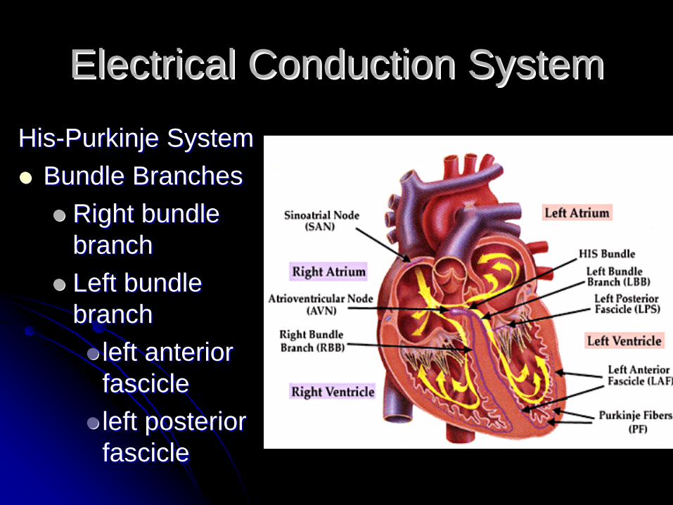
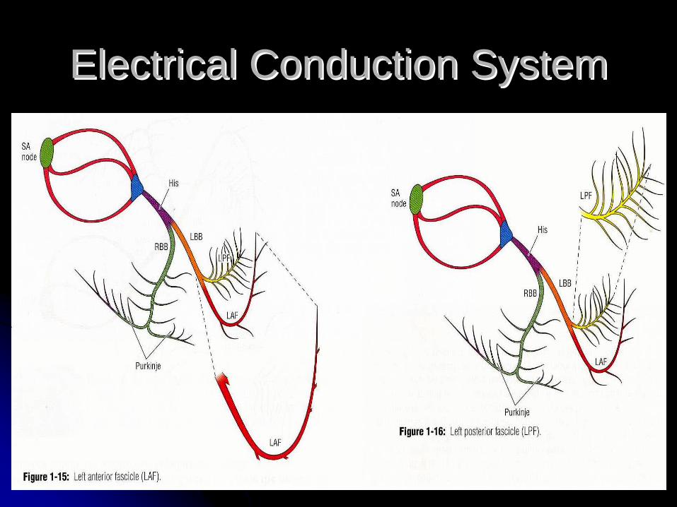
HisHis--Purkinje SystemPurkinje SystemBundle BranchesBundle Branches
Right bundle Right bundle branchbranchLeft bundle Left bundle branchbranch
left anterior left anterior fasciclefascicleleft posterior left posterior fasciclefascicle
Electrical Conduction SystemElectrical Conduction System
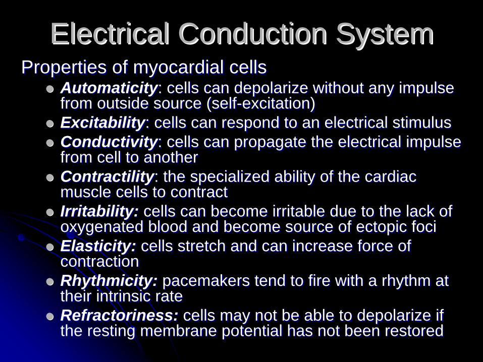
Electrical Conduction SystemElectrical Conduction SystemProperties of myocardial cellsProperties of myocardial cells
AutomaticityAutomaticity: cells can depolarize without any impulse : cells can depolarize without any impulse from outside source (selffrom outside source (self--excitation)excitation)ExcitabilityExcitability: cells can respond to an electrical stimulus: cells can respond to an electrical stimulusConductivityConductivity: cells can propagate the electrical impulse : cells can propagate the electrical impulse from cell to anotherfrom cell to anotherContractilityContractility: the specialized ability of the cardiac : the specialized ability of the cardiac muscle cells to contractmuscle cells to contractIrritability: Irritability: cells can become irritable due to the lack of cells can become irritable due to the lack of oxygenated blood and become source of ectopic focioxygenated blood and become source of ectopic fociElasticity: Elasticity: cells stretch and can increase force of cells stretch and can increase force of contractioncontractionRhythmicity: Rhythmicity: pacemakers tend to fire with a rhythm at pacemakers tend to fire with a rhythm at their intrinsic ratetheir intrinsic rateRefractoriness: Refractoriness: cells may not be able to depolarize if cells may not be able to depolarize if the resting membrane potential has not been restoredthe resting membrane potential has not been restored

Electrical Conduction SystemElectrical Conduction SystemMyocardial CellsMyocardial Cells
Three groups of cardiac muscleThree groups of cardiac muscleAtrialAtrialVentricularVentricularExcitatory/Conductive FibersExcitatory/Conductive Fibers
Atria contract from superior to inferiorAtria contract from superior to inferiorVentricles contract from inferior to superiorVentricles contract from inferior to superiorAtria and ventricles are separatedAtria and ventricles are separatedConduction from atria to the ventricles only Conduction from atria to the ventricles only through AV bundlethrough AV bundle““All or NoneAll or None”” principle of muscle contractionprinciple of muscle contraction
ElectrophysiologyElectrophysiology
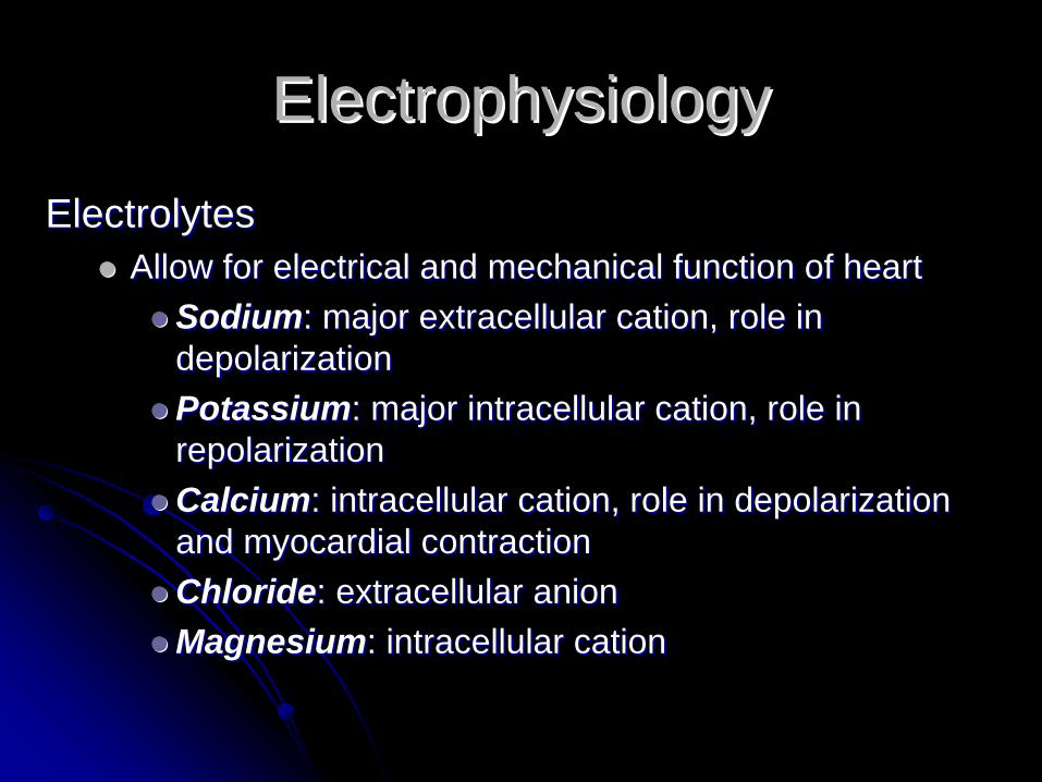
ElectrolytesElectrolytesAllow for electrical and mechanical function of heartAllow for electrical and mechanical function of heart
SodiumSodium: major extracellular cation, role in : major extracellular cation, role in depolarizationdepolarizationPotassiumPotassium: major intracellular cation, role in : major intracellular cation, role in repolarizationrepolarizationCalciumCalcium: intracellular cation, role in depolarization : intracellular cation, role in depolarization and myocardial contractionand myocardial contractionChlorideChloride: extracellular anion: extracellular anionMagnesiumMagnesium: intracellular cation: intracellular cation
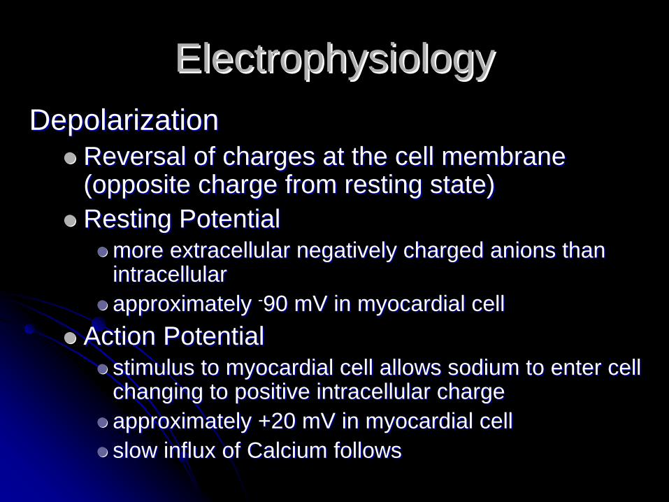
ElectrophysiologyElectrophysiologyDepolarizationDepolarization
Reversal of charges at the cell membrane Reversal of charges at the cell membrane (opposite charge from resting state)(opposite charge from resting state)Resting PotentialResting Potential
more extracellular negatively charged anions than more extracellular negatively charged anions than intracellularintracellularapproximately approximately --90 mV in myocardial cell90 mV in myocardial cell
Action PotentialAction Potentialstimulus to myocardial cell allows sodium to enter cell stimulus to myocardial cell allows sodium to enter cell changing to positive intracellular chargechanging to positive intracellular chargeapproximately +20 mV in myocardial cellapproximately +20 mV in myocardial cellslow influx of Calcium followsslow influx of Calcium follows

ElectrophysiologyElectrophysiology
ElectrophysiologyElectrophysiology
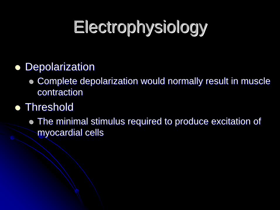
DepolarizationDepolarizationComplete depolarization would normally result in muscle Complete depolarization would normally result in muscle contractioncontraction
ThresholdThresholdThe minimal stimulus required to produce excitation of The minimal stimulus required to produce excitation of myocardial cellsmyocardial cells

ElectrophysiologyElectrophysiology
RepolarizationRepolarizationProcess of returning to resting potential stateProcess of returning to resting potential state
Sodium influx stops and potassium leaves cellSodium influx stops and potassium leaves cellSodium pumped to outside the cellSodium pumped to outside the cell
Relative refractory periodRelative refractory periodcell will respond to a second action potential but cell will respond to a second action potential but the action potential must be stronger than usualthe action potential must be stronger than usual
Absolute refractory periodAbsolute refractory periodcell will not respond to a repeated action potential cell will not respond to a repeated action potential regardless of how strong it isregardless of how strong it is
ElectrophysiologyElectrophysiology
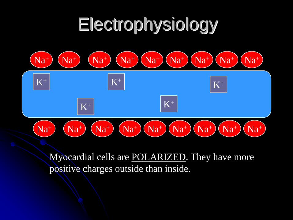
Na+
Na+ Na+ Na+ Na+ Na+ Na+ Na+
Na+ Na+ Na+ Na+ Na+ Na+ Na+Na+
Na+ Na+
K+
K+
K+
K+
K+
Myocardial cells are POLARIZED. They have more positive charges outside than inside.

ElectrophysiologyElectrophysiology
Na+
Na+ Na+ Na+ Na+ Na+ Na+ Na+
Na+ Na+ Na+ Na+ Na+ Na+ Na+Na+
Na+ Na+
K+ K+ K+ K+ K+
Stimulation of cell opens “fast” channels in cell membrane. Na+
rapidly enters cell. Now there are more positive charges inside than outside. The cell is DEPOLARIZED.

ElectrophysiologyElectrophysiology
Depolarization causes CaDepolarization causes Ca++++ to be released to be released from the cisternae of the sarcoplasmic from the cisternae of the sarcoplasmic reticulum in the cell.reticulum in the cell.CaCa++++ release catalyzes a chemical reaction release catalyzes a chemical reaction that promotes the sliding of the actin and that promotes the sliding of the actin and myosin filaments or muscle contraction.myosin filaments or muscle contraction.
Calcium couples the electrical Calcium couples the electrical event of depolarization to the event of depolarization to the
mechanical event of contraction.mechanical event of contraction.
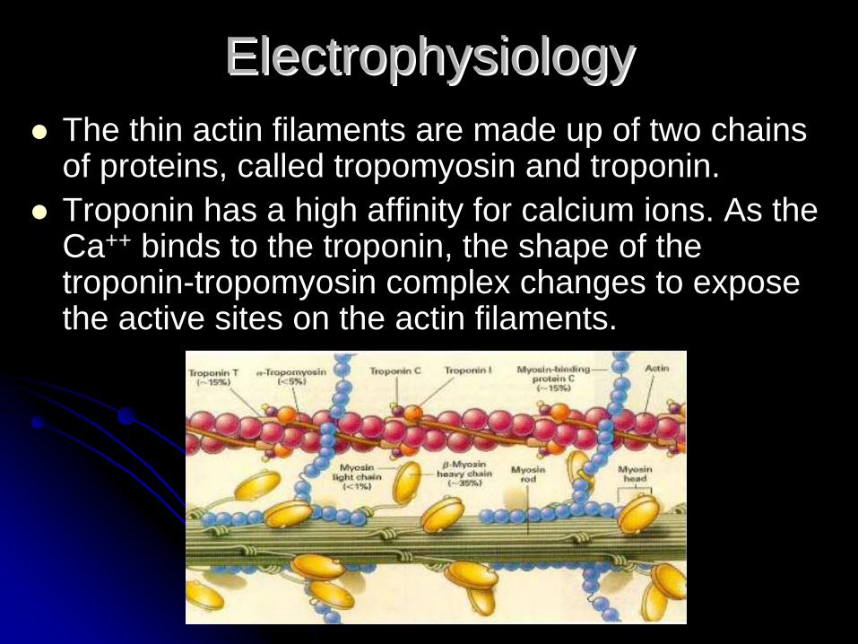
The thin actin filaments are made up of two chains of proteins, called tropomyosin and troponin. Troponin has a high affinity for calcium ions. As the Ca++ binds to the troponin, the shape of the troponin-tropomyosin complex changes to expose the active sites on the actin filaments.
ElectrophysiologyElectrophysiology
ElectrophysiologyElectrophysiology
Na+
Na+
Na+
K+
Na+ Na+ Na+ Na+ Na+ Na+ Na+Na+ K+
Na+ Na+ Na+ Na+ Na+ Na+Na+K+ K+
K+
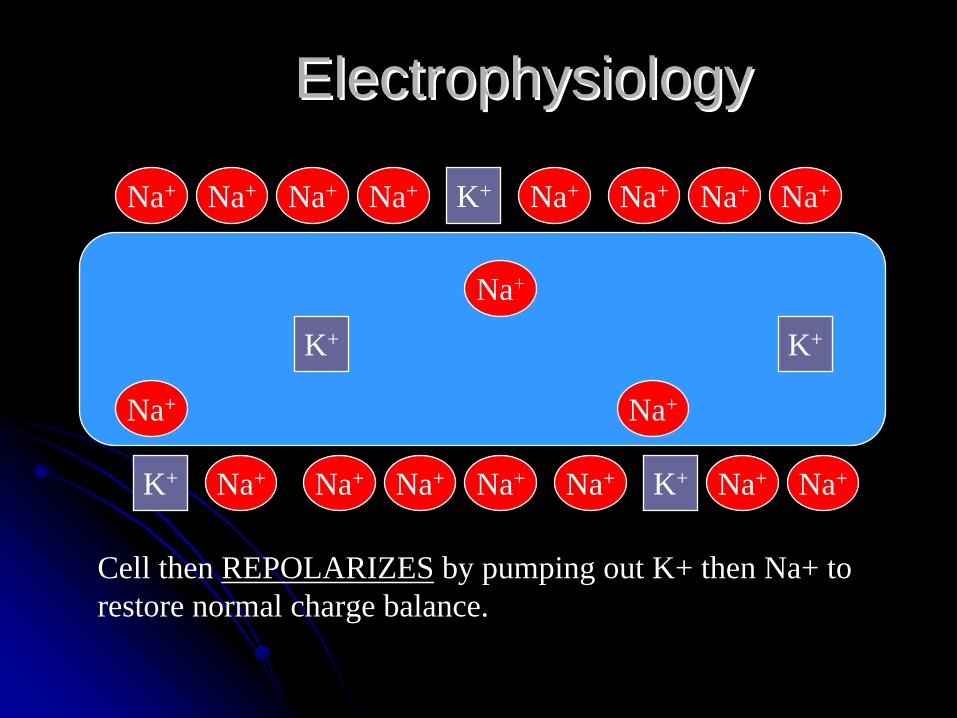
Cell then REPOLARIZES by pumping out K+ then Na+ to restore normal charge balance.

ElectrophysiologyElectrophysiology
Finally, the Na+-K+ pump in the cell membrane restores the proper balance of sodium and potassiuim.
Na+
Na+
Na+K+
Na+ Na+ Na+ Na+ Na+ Na+ Na+Na+ K+
Na+ Na+ Na+ Na+ Na+ Na+Na+K+ K+
K+
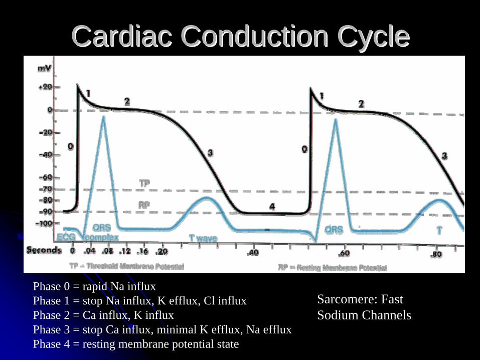
Cardiac Conduction CycleCardiac Conduction Cycle
Phase 0 = rapid Na influxPhase 1 = stop Na influx, K efflux, Cl influxPhase 2 = Ca influx, K influxPhase 3 = stop Ca influx, minimal K efflux, Na efflux Phase 4 = resting membrane potential state
Sarcomere: Fast Sodium Channels
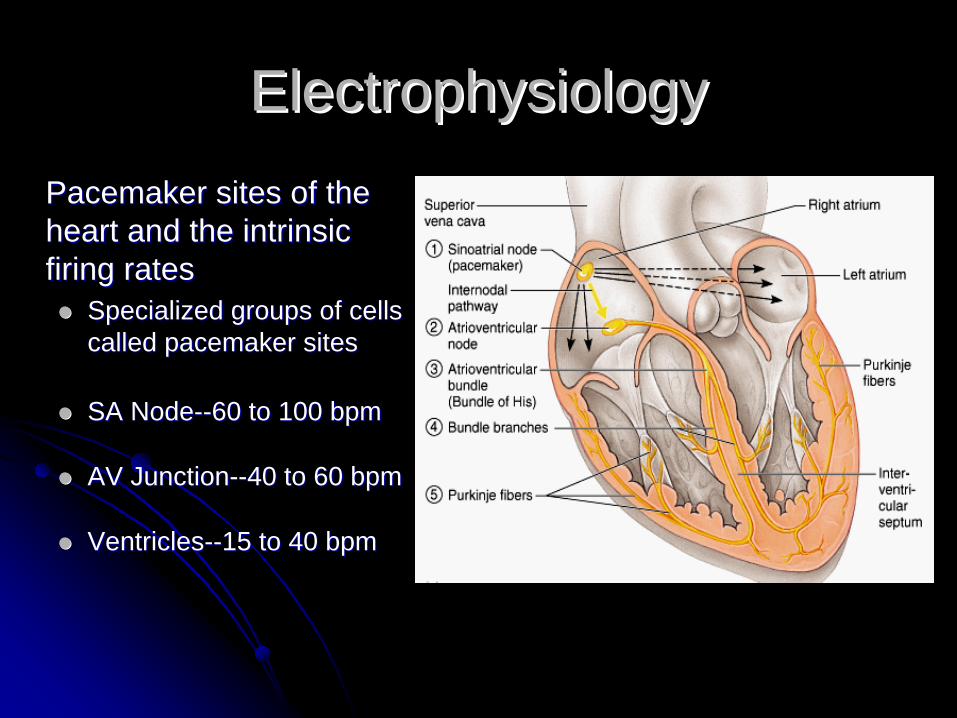
ElectrophysiologyElectrophysiologyPacemaker sites of the Pacemaker sites of the heart and the intrinsic heart and the intrinsic firing ratesfiring rates
Specialized groups of cells Specialized groups of cells called pacemaker sitescalled pacemaker sites
SA NodeSA Node----60 to 100 bpm60 to 100 bpm
AV JunctionAV Junction----40 to 60 bpm40 to 60 bpm
VentriclesVentricles----15 to 40 bpm15 to 40 bpm

ElectrophysiologyElectrophysiology
SA Node
Internodal Pathways
AV Node
Bundle of His
Bundle Branches
Purkinje Fibers
ElectrophysiologyElectrophysiology
Na+
Na+ Na+ Na+ Na+ Na+ Na+ Na+
Na+ Na+ Na+ Na+ Na+ Na+ Na+Na+
Na+ Na+
K+
K+
K+
K+
K+
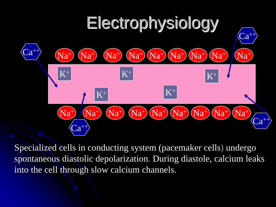
Specialized cells in conducting system (pacemaker cells) undergo spontaneous diastolic depolarization. During diastole, calcium leaks into the cell through slow calcium channels.
Ca++
Ca++Ca++
Ca++

ElectrophysiologyElectrophysiology
Na+
Na+ Na+ Na+ Na+ Na+ Na+ Na+
Na+ Na+ Na+ Na+ Na+ Na+ Na+Na+
Na+ Na+
K+ K+ K+ K+ K+
When a critical amount of calcium has entered the cell, fast channels open, sodium enters, and rapid DEPOLARIZATION begins.

ElectrophysiologyElectrophysiology
•• Electrical impulse Electrical impulse from depolarizing from depolarizing pacemaker cell pacemaker cell spreads to working spreads to working myocardial cells myocardial cells and stimulates and stimulates them.them.
• Depolarization and contraction result.

Communication between myocardial cells allows for contraction to occur.Syncytium
ElectrophysiologyElectrophysiology

ElectrophysiologyElectrophysiology
Ectopic Impulse Ectopic Impulse FormationFormationEnhanced AutomaticityEnhanced Automaticity
Pacemaker cellsPacemaker cellslost function of contractilitylost function of contractilityacquired function of impulse acquired function of impulse formationformation
May lead to ectopic beatsMay lead to ectopic beats
Picture to the right shows Picture to the right shows the Bundle of Kent, an the Bundle of Kent, an accessory pathway found accessory pathway found in WPW.in WPW.
ElectrophysiologyElectrophysiology
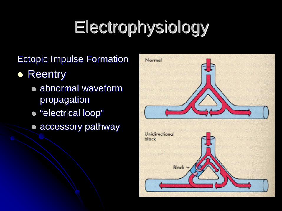
Ectopic Impulse FormationEctopic Impulse Formation
ReentryReentryabnormal waveform abnormal waveform propagationpropagation““electrical loopelectrical loop””accessory pathwayaccessory pathway

Effects of ANS on ElectrophysiologyEffects of ANS on Electrophysiology
The autonomic nervous The autonomic nervous system is mainly system is mainly activated by centers activated by centers located in the spinal cord, located in the spinal cord, brain stem, and brain stem, and hypothalamushypothalamusMedulla contains the Medulla contains the cardioacceleratory and cardioacceleratory and cardioinhibitory centerscardioinhibitory centersBaroreceptor reflex Baroreceptor reflex controls arterial pressurecontrols arterial pressure

Parasympathetic Nervous Parasympathetic Nervous SystemSystem
AcetylcholineAcetylcholineActivates muscarinic and Activates muscarinic and nicotinic receptorsnicotinic receptors
CholinesteraseCholinesteraseSympathetic Nervous Sympathetic Nervous SystemSystem
Adrenergic receptors Adrenergic receptors stimulated by epinephrine stimulated by epinephrine and norepinphrineand norepinphrine
AlphaAlpha11 and alphaand alpha2 2 receptorsreceptorsBetaBeta11 and betaand beta22 receptorsreceptors
Inotropic effectInotropic effectDromotropic effectDromotropic effectChronotropic effectChronotropic effect
Effects of ANS on ElectrophysiologyEffects of ANS on Electrophysiology
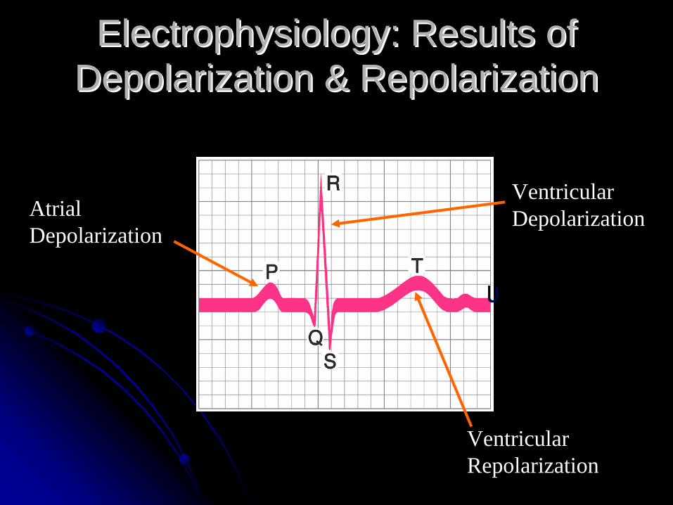
Electrophysiology: Results of Electrophysiology: Results of Depolarization & RepolarizationDepolarization & Repolarization
Atrial Depolarization
Ventricular Depolarization
Ventricular Repolarization
Questions?Questions?