Embed Size (px)
Citation preview
Page 1 of 15
Introduction to Cellular Biochemistry
Student Learning Outcomes
Upon completion of this Introductory monograph on The Cell, the properly prepared student will be able
to successfully explain, define, list and apply the introductory Cellular Biochemistry concepts in this
document as demonstrated by examination at an average score of 75%.
Specifically, the properly prepared student will be able to:
1) Give one difference between prokaryotic an eukaryotic cells;
2) Discuss the two primary determiners on cell size;
3) Illustrate, label and discuss the purpose of the lipid bilayer in the cell membrane, as well as extending
that to peripheral and integral proteins;
4) Discuss and explain the role of cholesterol in cell membranes;
5) Superficially explain why PC, PS and PE are found in different layers of the lipid bilayer membrane;
6) Explain the general function of glycoproteins and glycolipids;
7) illustrate and explain the theoretical generation of a liposome;
8) Illustrate, explain and provide an example of the three intercellular connections;
9) List the conditions necessary for regulating cell membrane permeability and provide a brief
explanation for each;
10) Compare, contrast and illustrate the differences between diffusion and osmosis;
11) Compare and contrast the differences between passive and active transport;
12) Illustrate and explain the process of phagocytosis;
13) Illustrate and explain the process of pinocytosis;
14) List, define and give an example of the three transporters;
15) List, define and give one function of the sub-cellular organelles;
16) List and explain how gold therapy is used to treat rheumatoid arthritis;
17) Explain what N and 2N mean from a nuclear perspective;
18) Illustrate, label and explain the 4 phases of mitosis;
19) Illustrate, label and explain the sequences of meiosis;
20) Compare, contrast and illustrate the elementary difference between mitosis and meiosis;
21) Explain the difference between “haploid cells” and “diploid cells”;

Page 2 of 15
22) Explain selective passage of materials across a cell membrane;
23) Explain what semi-permeable membranes do;
24) List, define and compare and contrast between hydrophilic and hydrophobic;
25) List, define and compare and contrast between lipophilic and lipophobic;
at an average score of 75% on the assessment tool.

Page 3 of 15
Statement of Purpose
Why would anyone want to bring in an introductory, fundamental, elementary, chapter on The Cell in a
General Chemistry I course? While most wouldn’t, WNC and UNR-Orvis School of Nursing recently
completed a transfer agreement from WNC to UNR-Orvis that could potentially have pre-BSN students
taking CHEM 121 at WNC and bypassing BIOL 190/L prior to enrolling in BIOL 223 (Human Anatomy and
Physiology I; A&P I) at WNC. A lack of background on the cell prior to entering A&P I can be academically
hurtful and impact a student’s success negatively. The goal of this chapter is to provide enough content
to give the student the tools to build upon in courses like A&P I.
The Cell
The emphasis on this chapter is that of the eukaryotic cell. Prokaryotic cells will be discussed in BIOL 251
(General Microbiology). The simplest and biggest difference between the two is that prokaryotic cells do
not have a nuclear membrane to separate nuclear content from the rest of the cell; the region that
contains the nuclear material in a prokaryotic cell is called the nucleoid. Eukaryotic cells have a clearly
demarcated nuclear membrane to keep the nuclear contents isolated (compartmentalized) from the rest
of the cell.
The size of a cell varies from as small as 200 nanometers (one billionth of a meter) up to the size of an
ostrich egg. The minimal size of any cell is dependent on the content of the cells. The primary contributors
to the size of a cell are macromolecules like DNA (the "brains" of the cell) and protein (the ultimate signal
coded for in the DNA). Both macromolecules (large molecules) are required for control of cellular activity
and to sustain cellular activity. The size of the average cell in the human body is between 0.5 micrometers
and 20 micrometers (one millionth of a meter; the old unit was micron). The diameter of the average red
blood cell is between 8 and 10 micrometers.
The Potential Size of The Cell is Dependent upon Two Characteristics
1) The relationship between the nucleus (NOOK lee uss) and the cytoplasm (SIGH toe plazum), e.g., a
lymphocyte (LIMM foe sight; a type of white blood cell) has a large nucleus and little cytosol (SICH toe
soll). This cell synthesizes many useful proteins, e.g., antibodies, but does not have the cytosol to retain
them for very long, hence, the synthesized compounds are released into the general circulation.
On the other hand, monocytes (another type of white blood cell [WBC]) have a large nucleus and a large
volume of cytosol. These cells function, among many, as macrophages (MACK row fa juz). These cells
synthesize lytic substances that work intracellularly on phagocytized (fa GOE si tized) micro-organisms,
cellular debris, excretions or anything else the body does not want or need.
2) The amount of surface for nutrient and/or waste transport. The more surface area on a cell, the more
nutrients the cell may take up and the more waste that may be efficiently excreted. The more a cell takes
up and utilizes, the larger the cell.
Any Body Cell Has Three Parts
Part I - Membrane Systems
Page 4 of 15
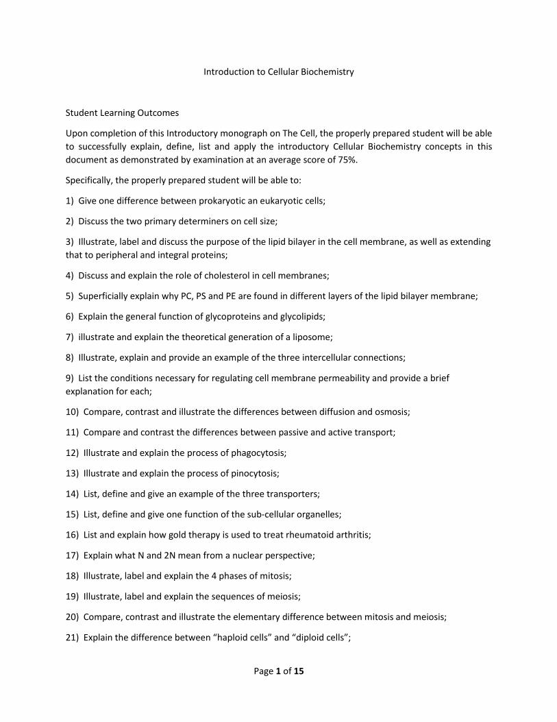
This membrane has three-dimensional structure (above), although we'll examine it from only 2
dimensions. Note that it is a lipid (fat) bilayer (two layers) membrane, i.e., there are two layers of lipid
that surround the cell.
The outer-most and inner-most of the two layers are polar (hydrophilic [high droe FILL ick] - water loving
- or lipophobic [lye poe FOE bick] - fat fearing) lipids. This is important for they must interact with the
aqueous solvent of which bodies and cells consist: water. Water is a polar molecule.
The two middle-most regions of this lipid bilayer are apolar (hydrophobic - water fearing -- or lipophilic -
fat loving). It is this middle region that gives the membranes a powerful way of separating the cell
"innards" from the outside and from other cells, allowing different cells to "bunch together" to form
different kinds of tissues and, hence, organs and organ systems and organisms.
The outer layer of a typical cell is primarily phosphatidylcholine (PC or lecithin) and sphingomyelin; the
inner layer is primarily phosphatidylethanolamine and phosphatidylserine. The value of this is that the
outer layer is a bit more rigid and the inner layer is more flexible, much like taking two sheets of corrugated
aluminum roofing and layering them, then bending them. With the aluminum roofing, the inner layer
seems to extend beyond the edges of the outer layer when it's bent. By making the inner layer of a
membrane more flexible, it bends, as it were, to retain its alignment with the outer layer without
extending beyond the outer layer.
There is also protein associated with the cell membrane: peripheral proteins that are attached to either
the inner layer or the outer layer of the membrane that act as receptors for molecules that are unable to
get through the membrane and integral proteins that are completely inserted through the membrane.
The latter proteins are often-times ion channels, as their outer layer is hydrophobic and more interactive
with the cell membrane, while the center portion is hydrophilic. This hydrophilic region allows ions to
traverse the cell membrane to regulate ion balance inside and outside the cell.
Glycoproteins (GLY koe PRO teens; a combination of carbohydrate and protein) are also found on the
surface of the membrane. As a general rule, these compounds are the compounds used for cell
recognition or cell identification. Glycolipids (glye-coe-LIP-ids; a combination of carbohydrates and lipids)
are also found in the membrane. They tend to stabilize the structure of the membrane.
Page 5 of 15
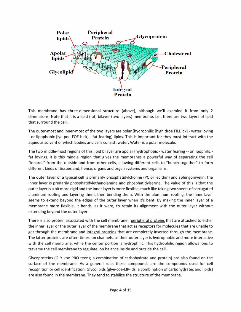
Cholesterol is found in cell membranes, as well. Cholesterol (ko LESS turr all) works with the rigidity of the
membrane. As a general rule, the more cholesterol in the membrane, the more rigid the membrane; the
converse is equally true.
Inside the cell, underneath the cell membrane is a system of microtubules that acts as the cytoskeleton.
These microtubules provide a framework to give a shape to the cell. They also act as the cell's irrigation
system - more on this, later.
Pharmacologists, biochemists and physiologists have been studying the chemical properties of the various
cell membranes in the human body in hopes of understanding how to use the information to make a
carrier (called a liposome - fat sac or fat pocket) that will take a specific drug from a syringe, through the
blood without enzymatic modification to a specific target cell or tissue. The figure, above, shows
graphically how this, theoretically, occurs.
A polar lipid that has the same characteristics as the membrane to which it is to migrate and traverse is
mixed with the drug it is to carry. The polar regions of the lipid rearrange around the drug in such a manner
that the apolar regions bind with the drug so that a three-dimensional cage is formed around the drug.
This cage is called a micelle (MYE cell; liposome). The liposomes are then injected into the blood, travel to
the target organ and keep the drug safe from blood enzymes that might inactivate it prior to getting to
the target organ, tissue or tumor. There has been some success with this in the lab.
Cells are connected to each other by one of three Intercellular Connections: Tight Junctions, Gap Junctions
or Desmosomes.
The Figure, below, illustrates each type of connection.

Page 6 of 15
Tight junctions occur by fusing membranes. These are commonly found in the intestine and blood-brain
barrier. It is this kind of connection in the central nervous system that makes it so difficult to get drugs to
cross the blood-brain barrier and, hence, to treat disorders of the nervous system.
Gap junctions occur between cells to connect them by narrow channels and are separated by small spaces
(synapses). These are common in the nervous system.
Desmosomes consist of filaments that surround the cell and cement or suture cells together to form solid,
"crumbly" tissues like the liver and kidney.
Cell Membrane Functions
Cell membranes control the passage of substances across themselves selectively (allow some materials to
cross without difficulty, e.g., water) and semipermeably (SE mye PER mee abb lee; restricts the passage
of other compounds, e.g., glucose and proteins).
The permeability of the cell membrane depends on a number of conditions:
1) Membrane thickness: the thicker it is, the longer it takes the compound to cross the
membrane;
2) The size of the materials: tiny molecules like urea easily pass through the membrane,
while slightly larger molecules, like glucose will not and very large molecules like proteins
simply won't cross the membrane;
3) Lipid solubility: like dissolves like, i.e., if the compound is soluble in lipid, it will cross
the membrane easily; conversely, if the molecule is polar, it will not cross;
4) Electrical charge: the time of crossing increases or decreases based upon the charge of
the material AND the membrane;
5) Active transport systems: more on this coming up;
6) Binding sites: more on this coming up.
Passive Transmembrane Movement
Passive movements are caused by pressure or concentration changes WITHOUT the use of energy.
The first passive movement to be examined is DIFFUSION (dye FEW zhun). By definition, diffusion is the
net movement of solute from a region of higher solute concentration to a region of lower solute
concentration. The difference between the two regions is called the Concentration Gradient. The Figure
at top right, next page, illustrates this concept of solute movement. One of the best analogies is that of
placing a dye tablet in a beaker of water. At first, there is no movement of the dye into the water and
there is no concentration gradient, i.e., there is only dye tablet and water. As time goes by, though, the
dye begins release some of itself into the surrounding water - now there are 3 regions: the dye tablet, the
clear water and the region in the water that has varying concentrations of dye (the concentration
gradient). As more time goes by, the dye is uniformly distributed throughout the water, making a
homogeneous solution.
Page 7 of 15
Facilitated diffusion is defined as
diffusion assisted by integral proteins in
the membrane which act as carriers, e.g.,
glucose. Facilitated diffusion has a rate
that is faster than diffusion, proper. The
rate of facilitated diffusion is
proportional to the concentration
gradient, i.e., if there is a lot more of a
substance outside a cell than inside the
cell, then the rate is rapid. It is
proportional to the amount of carrier
available, i.e., if there are only 5 carriers
and 500 molecules to be carried, then the
rate of uptake will be very slow.
Conversely if there are 5000 carriers and
500 molecules, then the rate of uptake
will be very rapid. The rate of facilitated
diffusion depends on how quickly the
carrier and substance combine, e.g.,
insulin. Insulin catalyzes the rapid binding
of glucose to the glucose transporter to
drive glucose inside the cell. This is called
enhancement and greatly increases the
efficiency of glucose uptake into our cells.
Osmosis is DIFFERENT from diffusion.
Osmosis is defined as the movement of water from a region of higher water concentration to a region of
lower water concentration across a semi-permeable membrane until there are equal water
concentrations on either side of the membrane.
The Figure, bottom left, illustrates
the movement of water across a
semi-permeable membrane to
equalize, if you will, the water
concentrations on either side of the
membrane. Note in the expansion
of the semi-permeable membrane
that the reason that the sugar does
not cross the membrane is because
it is too large to pass through the
pores in the membrane, while the
water is not. Osmotic pressure is
defined as the pressure needed to
stop the flow of water across the

Page 8 of 15
membrane. THE KEY: the greater the concentration of solute, the
greater the osmotic pressure. The converse is equally as true.
One useful application of hypertonic solutions is to infuse these
solutions into a patient who has received head trauma to reduce
the swelling (edema) of the brain and reduce the damage to the
brain. Hypotonic solutions may be used to rapidly rehydrate a
dehydrated patient.
Active Transmembrane Movement
Active movements are defined as those movements that are
caused by the release of energy to move material across the membrane from low concentration to high
concentration, i.e., AGAINST or ACROSS a concentration gradient. These movements require energy in the
form of ATP (Adenosine TriPhosphate; uh DENN o sin tri PHOS phate). We use up to 40% of the ATP we
synthesize daily for active transport. Considering that we synthesize about 4 pounds of ATP per day, that
comes to 1.6 pounds of ATP a day we use in these movements. Active movements require integral
proteins, e.g., Glucose/Sodium (Na+) transporter.
The first active movement to be discussed in this chapter is
endocytosis. There are 2 sub-categories under this heading:
phagocytosis (FAGG oh sigh TOE siss; cell eating), Figure, top
right, and pinocytosis (PEE noe sigh TOE siss; cell drinking),
Figure, middle right.
Phagocytosis is initiated by the recognition that something is
where it's not supposed to be, e.g., a micro-organism. The cell
responding (a white blood cell called a PMN) extends
pseudopodia (sue doe POE dee uh; false feet) around the
organisms or particle and then encloses the "object" with the
pseudopodia to form a phagosome (FAGG oh some; an eating sac or eating pocket). The phagosome is
internalized and it differentiates and fuses with a lysosome (LYE so some) and, presto!, the "object" is
hydrolyzed.
Pinocytosis is similar to phagocytosis. Cell drinking begins with a water droplet "foraging" around on the
surface of a cell. Once the water finds a small invagination (inn VAJ i NA shun), it falls into it and causes
the membrane to change shape so that the pinosome (drinking sac or drinking pocket) is internalized and
the water is sent to the appropriate compartment in
the cell.
The last active transport mechanism to be discussed
in this monograph is exocytosis (getting something
out of the cell; Figure, bottom right). Exocytosis
occurs when a cell has an excretion or a secretion it
wishes to release. The exocytic vesicle containing the
secretion or excretion migrates to the cell membrane
where it fuses with the membrane, exvaginates and
Page 9 of 15
"dumps" out the particles, e.g. hormone, enzyme, waste. This is how pancreatic enzymes are dumped into
the GI tract. Remember, this transport requires ATP, too.
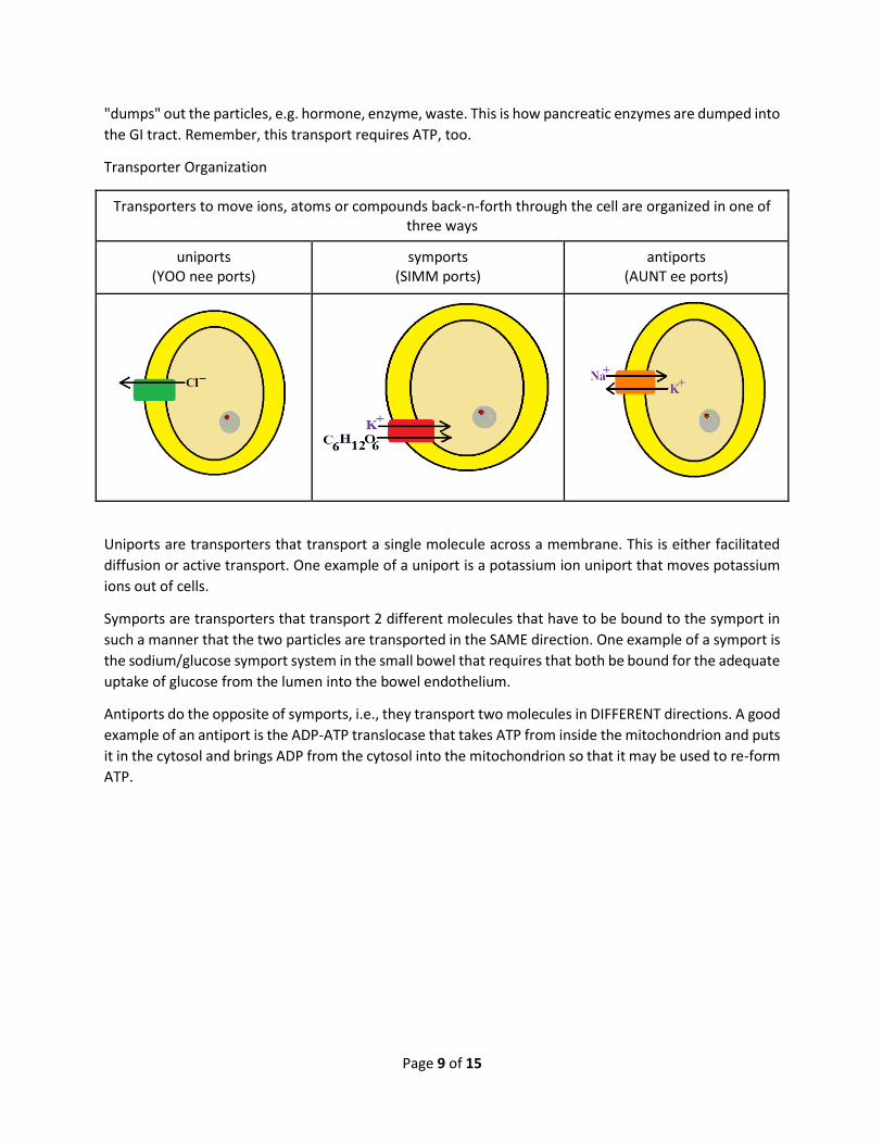
Transporter Organization
Transporters to move ions, atoms or compounds back-n-forth through the cell are organized in one of three ways
uniports (YOO nee ports)
symports (SIMM ports)
antiports (AUNT ee ports)
Uniports are transporters that transport a single molecule across a membrane. This is either facilitated
diffusion or active transport. One example of a uniport is a potassium ion uniport that moves potassium
ions out of cells.
Symports are transporters that transport 2 different molecules that have to be bound to the symport in
such a manner that the two particles are transported in the SAME direction. One example of a symport is
the sodium/glucose symport system in the small bowel that requires that both be bound for the adequate
uptake of glucose from the lumen into the bowel endothelium.
Antiports do the opposite of symports, i.e., they transport two molecules in DIFFERENT directions. A good
example of an antiport is the ADP-ATP translocase that takes ATP from inside the mitochondrion and puts
it in the cytosol and brings ADP from the cytosol into the mitochondrion so that it may be used to re-form
ATP.
Page 10 of 15
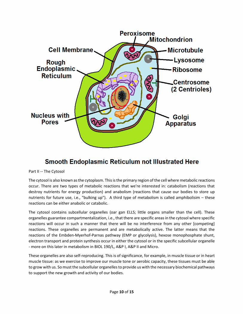
Part II -- The Cytosol
The cytosol is also known as the cytoplasm. This is the primary region of the cell where metabolic reactions
occur. There are two types of metabolic reactions that we're interested in: catabolism (reactions that
destroy nutrients for energy production) and anabolism (reactions that cause our bodies to store up
nutrients for future use, i.e., "bulking up"). A third type of metabolism is called amphibolisim – these
reactions can be either anabolic or catabolic.
The cytosol contains subcellular organelles (oar gan ELLS; little organs smaller than the cell). These
organelles guarantee compartmentalization, i.e., that there are specific areas in the cytosol where specific
reactions will occur in such a manner that there will be no interference from any other [competing]
reactions. These organelles are permanent and are metabolically active. The latter means that the
reactions of the Embden-Myerhof-Parnas pathway (EMP or glycolysis), hexose monophosphate shunt,
electron transport and protein synthesis occur in either the cytosol or in the specific subcellular organelle
- more on this later in metabolism in BIOL 190/L, A&P I, A&P II and Micro.
These organelles are also self-reproducing. This is of significance, for example, in muscle tissue or in heart
muscle tissue: as we exercise to improve our muscle tone or aerobic capacity, these tissues must be able
to grow with us. So must the subcellular organelles to provide us with the necessary biochemical pathways
to support the new growth and activity of our bodies.
Page 11 of 15
The cytosol also contains inclusions: for storage (e.g., glycogen), for waste (e.g., urates) and/or for raw
materials for cellular activity (e.g., fat, pigment granules).
In addition, the cytosol contains salts that help maintain the ionic environment of the cell and maintain
the pH of the cell.
The cytosol also contains enzymes (biological catalysts), which may be bound (which allows for enzyme
orientation in the case of sequential reactions; bound to internal membranes and/or to filaments) and/or
free (dissolved in the cytosol), as well.
The Subcellular Organelles
Rough Endoplasmic Reticulum: This structure is for protein synthesis. There are high numbers of these in
antibody-producing cells, liver cells and pancreatic cells.
Smooth endoplasmic reticulum: For steroid synthesis and complex carbohydrate synthesis; in liver
(detoxification center).
Ribosomes: the primary function of ribosomes is protein synthesis; bound ribosomes are found on the
rough endoplasmic reticulum (and is why7 it’s called “rough”; free ribosomes are found in cytosol
(remember, too, that the consistency of the cytosol is like jelly - NOT water); proteins are synthesized on
ribosomes, then transported in the tubular endoplasmic reticulum to the Golgi apparatus.
Golgi Apparatus: this structure is located near nucleus; it’s used in protein packaging; the synthesis of
glycoproteins, glycolipids, mucus. Proteins come in one side of Golgi and go out the other. There are high
levels of the Golgi Apparatus in liver and pancreas.
Lysosomes: Are called the suicide sac or the garbage disposal of the cell. They have powerful hydrolytic
enzymes. They are implicated in cell death and digestion due to increased intracellular release.
Lysosomes contain very acidic contents. They are also implicated in rheumatoid arthritis.
Gold therapy for rheumatoid arthritis
1) inhibits lysosomal enzymes directly by stabilizing lysosomal membranes;
2) lymphocyte responses to mitogens/antigens are inhibited by gold in culture;
3) monocyte activity decreases after gold therapy
all of which lead to reduced joint erosion.
Peroxisomes: Many cells produce hydrogen peroxide, which is toxic to the cell. Catalase is synthesized in
peroxisomes which hydrolyzes (detoxifies, if you will) hydrogen peroxide to oxygen and water. The
peroxisomes protect the rest of the cell from the toxicity of the hydrogen peroxide. Peroxisomes are found
primarily in the liver and kidney.
Mitochondrion: the mito are called the "powerhouse of the cell". Mitochondria have a double membrane
system and are responsible for ATP synthesis, urea synthesis, lipid oxidation, among others. It's the ONLY
sub-cellular organelle to contain its own DNA, BUT still requires nuclear DNA to function. There is a theory
that mito were originally bacteria that got trapped in the primordial goo from which cells arose.
Mitochondria contain 5 complexes necessary for ATP synthesis.
Page 12 of 15
Microtubules are used to make the cytoskeleton. They are aka microfilaments. Microtubules are used
for irrigation system of cell and used in cell division.
Centrosomes consists of 2 centrioles. Centrosomes are used in cell division; they are used to form cilia
and flagella which move materials across cell surfaces (lungs) or propel cells in fluid (sperm).
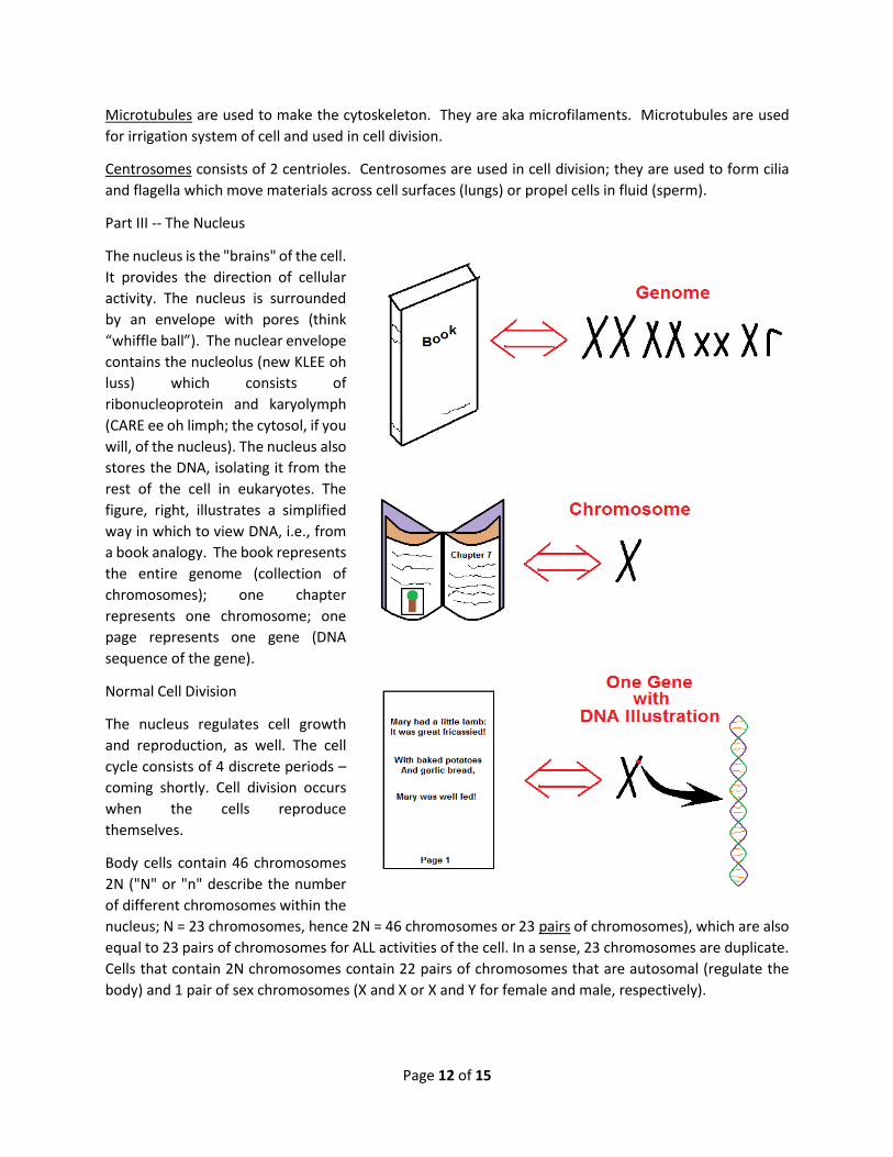
Part III -- The Nucleus
The nucleus is the "brains" of the cell.
It provides the direction of cellular
activity. The nucleus is surrounded
by an envelope with pores (think
“whiffle ball”). The nuclear envelope
contains the nucleolus (new KLEE oh
luss) which consists of
ribonucleoprotein and karyolymph
(CARE ee oh limph; the cytosol, if you
will, of the nucleus). The nucleus also
stores the DNA, isolating it from the
rest of the cell in eukaryotes. The
figure, right, illustrates a simplified
way in which to view DNA, i.e., from
a book analogy. The book represents
the entire genome (collection of
chromosomes); one chapter
represents one chromosome; one
page represents one gene (DNA
sequence of the gene).
Normal Cell Division
The nucleus regulates cell growth
and reproduction, as well. The cell
cycle consists of 4 discrete periods –
coming shortly. Cell division occurs
when the cells reproduce
themselves.
Body cells contain 46 chromosomes
2N ("N" or "n" describe the number
of different chromosomes within the
nucleus; N = 23 chromosomes, hence 2N = 46 chromosomes or 23 pairs of chromosomes), which are also
equal to 23 pairs of chromosomes for ALL activities of the cell. In a sense, 23 chromosomes are duplicate.
Cells that contain 2N chromosomes contain 22 pairs of chromosomes that are autosomal (regulate the
body) and 1 pair of sex chromosomes (X and X or X and Y for female and male, respectively).

Page 13 of 15
Somatic cells contain 2 sets of each
chromosome. These cells are called
diploid (DYE ployd) cells and are
identified, as well, by 2N or 2n. In
diploid cells, 2 chromosomes in a pair
are called homologous chromosomes.
Somatic cell division (body cell
division) occurs when a parent cell
produces 2 identical daughter cells.
The division of the nuclear material is
called mitosis; the cytoplasmic
division is called cytokinesis.
The daughter cells have the same
number and the same kind of
chromosomes (KROME uh somes) as
does their parent cell.
Reproductive cell division also has
division of its/their nuclear material.
This is called meiosis (my OH siss);
cytokinesis (sigh to kunn EE siss)also occurs. When a parent cell divides by meiosis, haploid cells (2N N
chromosome-containing cells) are formed. It is by this mechanism that spermatogenesis (spur ma toe GEN
uh siss) occurs in the testes and oogenesis (oh oh GENN uh siss) in the ovaries.
There are two (2) successive nuclear divisions in meiosis: reduction division (meiosis I) and equatorial (or
equational) division (meiosis II). The chromosome number does NOT double in meiosis, rather, it halves
producing haploid cells: N, n or 23 chromosomes.
To Restate and Expand
Chromosomes contain genes. Each identical gene site is called a locus. Plural is loci. Alternate forms of the
same gene are called alleles. HAPLOIDY is N number of chromosomes, i.e., 1/2 the number of
chromosomes and come from successful meiosis. DIPLOIDY is 2N number of chromosomes, i.e., all the
chromosomes that are supposed to be there and come from successful mitosis. In the human, haploidy is
23 chromosomes and diploidy is 46 chromosomes or 23 pairs of chromosomes.
In terms of the reproductive cell divisions, the sex cells are called gametes (GAMM eets). In the female
they are also called ova; in the male, sperm. Union/fusion of gametes is called fertilization and forms a
zygote.
Page 14 of 15
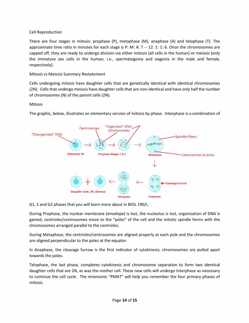
Cell Reproduction
There are four stages in mitosis: prophase (P), metaphase (M), anaphase (A) and telophase (T). The
approximate time ratio in minutes for each stage is P: M: A: T -- 12: 1: 1: 6. Once the chromosomes are
capped off, they are ready to undergo division via either mitosis (all cells in the human) or meiosis (only
the immature sex cells in the human, i.e., spermatogonia and oogonia in the male and female,
respectively).
Mitosis vs Meiosis Summary Restatement
Cells undergoing mitosis have daughter cells that are genetically identical with identical chromosomes
(2N). Cells that undergo meiosis have daughter cells that are non-identical and have only half the number
of chromosomes (N) of the parent cells (2N).
Mitosis
The graphic, below, illustrates an elementary version of mitosis by phase. Interphase is a combination of
G1, S and G2 phases that you will learn more about in BIOL 190/L.
During Prophase, the nuclear membrane (envelope) is lost, the nucleolus is lost, organization of DNA is
gained, centrioles/centrosomes move to the “poles” of the cell and the mitotic spindle forms with the
chromosomes arranged parallel to the centrioles.
During Metaphase, the centrioles/centrosomes are aligned properly at each pole and the chromosomes
are aligned perpendicular to the poles at the equator.
In Anaphase, the cleavage furrow is the first indicator of cytokinesis; chromosomes are pulled apart
towards the poles.
Telophase, the last phase, completes cytokinesis and chromosome separation to form two identical
daughter cells that are 2N, as was the mother cell. These new cells will undergo Interphase as necessary
to continue the cell cycle. The mnemonic “PMAT” will help you remember the four primary phases of
mitosis.
Page 15 of 15
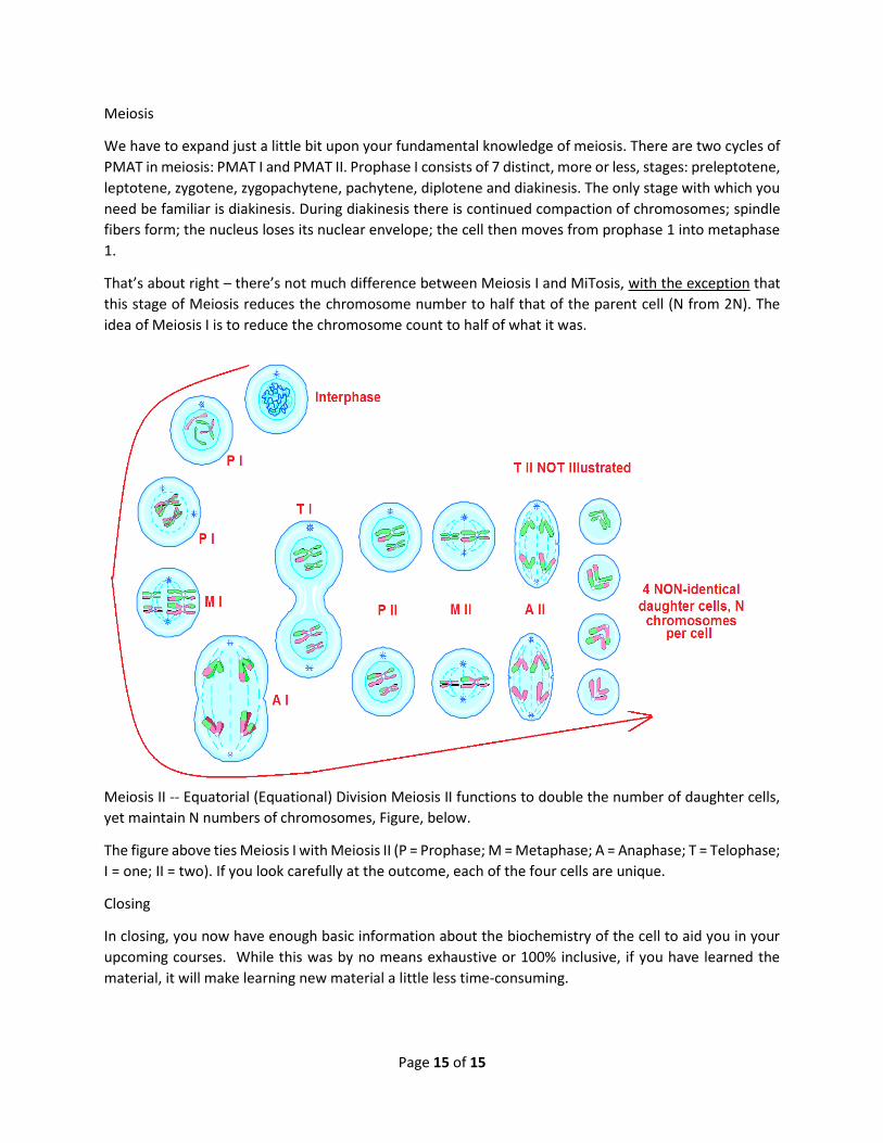
Meiosis
We have to expand just a little bit upon your fundamental knowledge of meiosis. There are two cycles of
PMAT in meiosis: PMAT I and PMAT II. Prophase I consists of 7 distinct, more or less, stages: preleptotene,
leptotene, zygotene, zygopachytene, pachytene, diplotene and diakinesis. The only stage with which you
need be familiar is diakinesis. During diakinesis there is continued compaction of chromosomes; spindle
fibers form; the nucleus loses its nuclear envelope; the cell then moves from prophase 1 into metaphase
1.
That’s about right – there’s not much difference between Meiosis I and MiTosis, with the exception that
this stage of Meiosis reduces the chromosome number to half that of the parent cell (N from 2N). The
idea of Meiosis I is to reduce the chromosome count to half of what it was.
Meiosis II -- Equatorial (Equational) Division Meiosis II functions to double the number of daughter cells,
yet maintain N numbers of chromosomes, Figure, below.
The figure above ties Meiosis I with Meiosis II (P = Prophase; M = Metaphase; A = Anaphase; T = Telophase;
I = one; II = two). If you look carefully at the outcome, each of the four cells are unique.
Closing
In closing, you now have enough basic information about the biochemistry of the cell to aid you in your
upcoming courses. While this was by no means exhaustive or 100% inclusive, if you have learned the
material, it will make learning new material a little less time-consuming.


![Cellular Biochemistry of the Stepwise Development of ... › content › canres › ... · [CANCER RESEARCH 44, 5463-5474, December 1984] Cellular Biochemistry of the Stepwise Development](https://img.pdfslide.net/doc/110x75/5f138bb3dafc99707f18b76b/cellular-biochemistry-of-the-stepwise-development-of-a-content-a-canres.jpg)