Embed Size (px)
Citation preview

DTU Compute
Introduction to Medical Image AnalysisRasmus R. PaulsenDTU Compute
http://www.compute.dtu.dk/courses/02511http://www.compute.dtu.dk/courses/02512
DTU Compute
31/1/2018Introduction to Medical Image Analysis2 DTU Compute, Technical University of Denmark
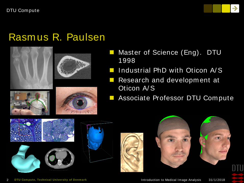
Rasmus R. Paulsen Master of Science (Eng). DTU
1998 Industrial PhD with Oticon A/S Research and development at
Oticon A/S Associate Professor DTU Compute

DTU Compute
31/1/2018Introduction to Medical Image Analysis3 DTU Compute, Technical University of Denmark
Teaching Assistants Mariam Andersson
Ph.D. student at DTU Compute
DTU Compute
31/1/2018Introduction to Medical Image Analysis4 DTU Compute, Technical University of Denmark
A statement from me and the TA’s The lectures and exercises are offers
– We do not notice if you are here or not We want to lift you as far as possible
– Make you understand our topic and the learning objectives We are grateful for critical and constructive feedback
We do expect that you are here to learn We do expect that you are responsible for your own learning The TA WILL help the ones that have a hard time understanding
a problem The TA WILL NOT help the ones that do not even try to
understand the problem
DTU Compute
31/1/2018Introduction to Medical Image Analysis5 DTU Compute, Technical University of Denmark
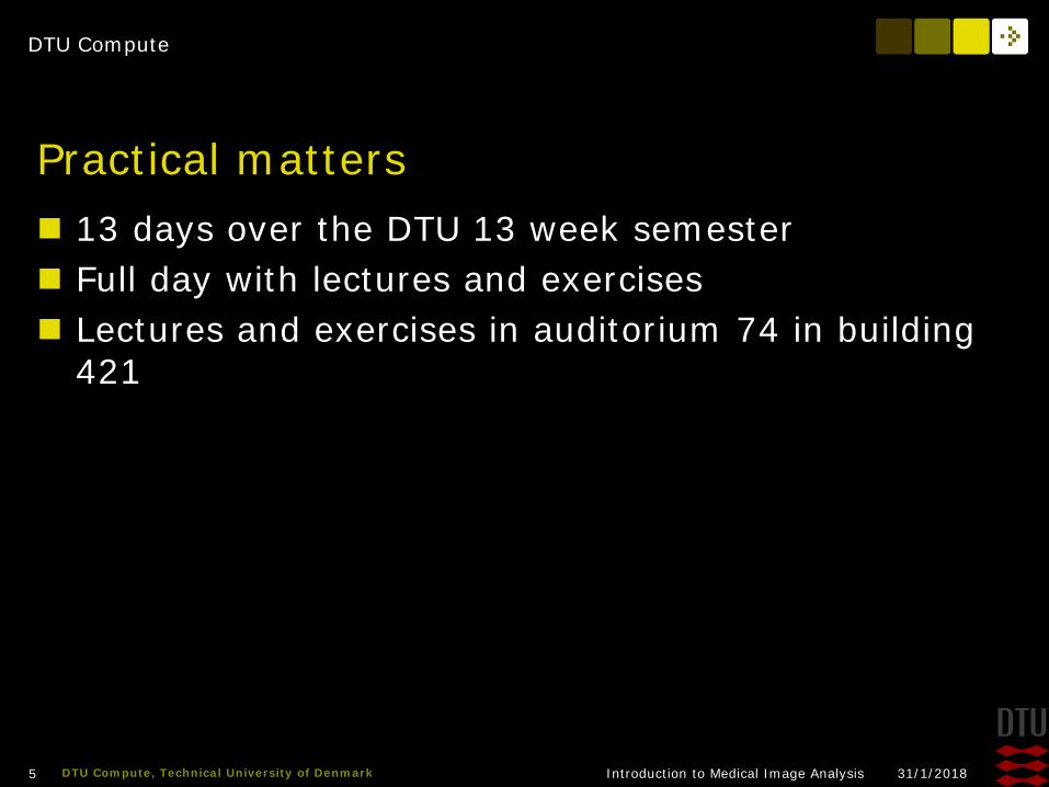
Practical matters 13 days over the DTU 13 week semester Full day with lectures and exercises Lectures and exercises in auditorium 74 in building
421

DTU Compute
31/1/2018Introduction to Medical Image Analysis6 DTU Compute, Technical University of Denmark
Week 1 - today
9.00 Introduction and practical mattersLecture – Digital ImagesExercises
12.00 – 13.00 Lunch break13.00 - 16.00 Exercises

DTU Compute
31/1/2018Introduction to Medical Image Analysis7 DTU Compute, Technical University of Denmark
Next weeks
8:30-9:00 Exercises with TA9:00-11ish Lecture11ish-12 Exercises12.00 – 13.00 Lunch break13.00 - 16.00 Exercises with TA
DTU Compute
31/1/2018Introduction to Medical Image Analysis8 DTU Compute, Technical University of Denmark
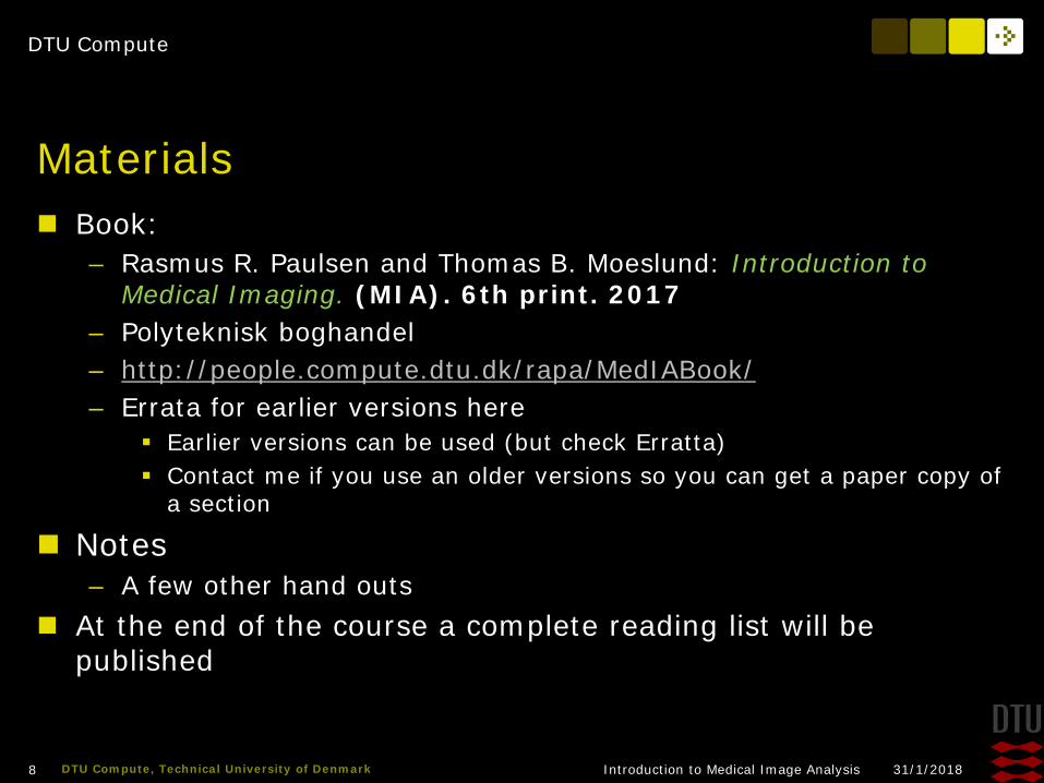
Materials Book:
– Rasmus R. Paulsen and Thomas B. Moeslund: Introduction to Medical Imaging. (MIA). 6th print. 2017
– Polyteknisk boghandel– http://people.compute.dtu.dk/rapa/MedIABook/– Errata for earlier versions here
Earlier versions can be used (but check Erratta) Contact me if you use an older versions so you can get a paper copy of
a section
Notes– A few other hand outs
At the end of the course a complete reading list will be published
DTU Compute
31/1/2018Introduction to Medical Image Analysis9 DTU Compute, Technical University of Denmark
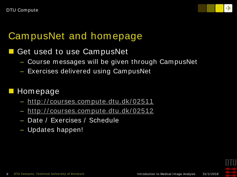
CampusNet and homepage Get used to use CampusNet
– Course messages will be given through CampusNet– Exercises delivered using CampusNet
Homepage– http://courses.compute.dtu.dk/02511– http://courses.compute.dtu.dk/02512– Date / Exercises / Schedule– Updates happen!
DTU Compute
31/1/2018Introduction to Medical Image Analysis10 DTU Compute, Technical University of Denmark
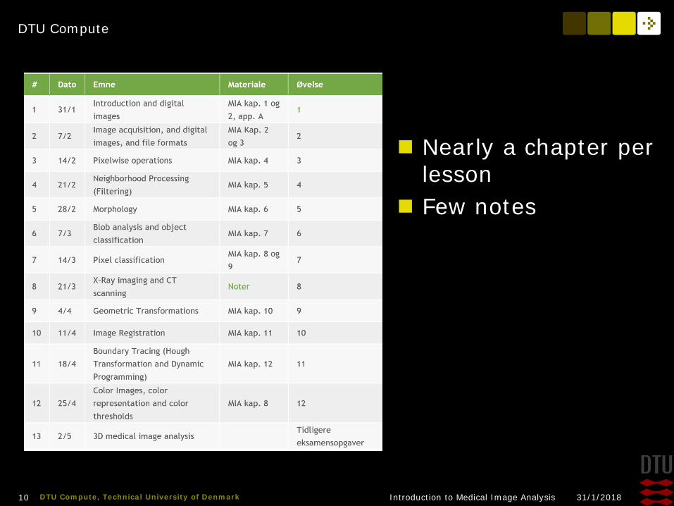
Nearly a chapter per lesson
Few notes
DTU Compute
31/1/2018Introduction to Medical Image Analysis12 DTU Compute, Technical University of Denmark
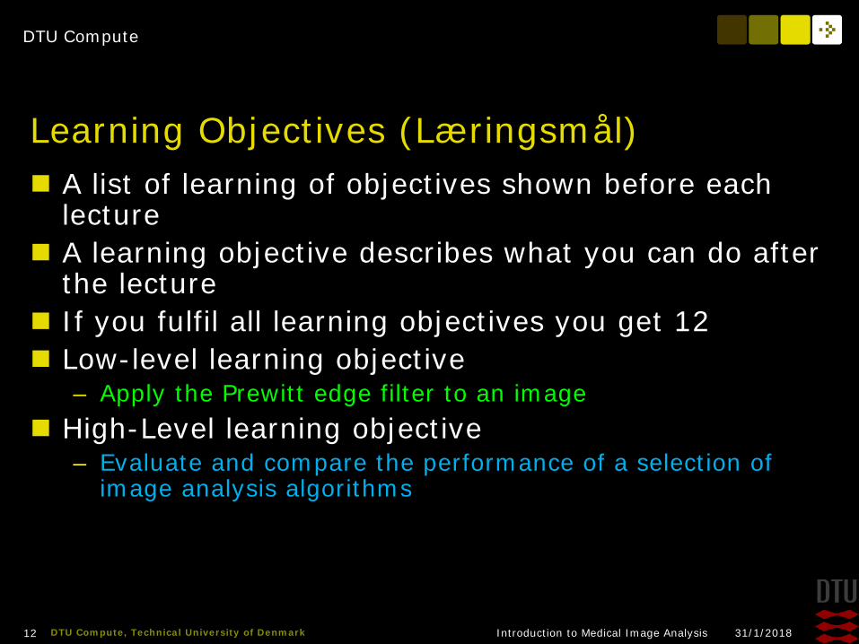
Learning Objectives (Læringsmål) A list of learning of objectives shown before each
lecture A learning objective describes what you can do after
the lecture If you fulfil all learning objectives you get 12 Low-level learning objective
– Apply the Prewitt edge filter to an image High-Level learning objective
– Evaluate and compare the performance of a selection of image analysis algorithms
DTU Compute
31/1/2018Introduction to Medical Image Analysis13 DTU Compute, Technical University of Denmark
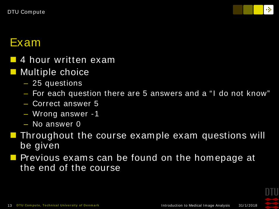
Exam 4 hour written exam Multiple choice
– 25 questions– For each question there are 5 answers and a “I do not know”– Correct answer 5 – Wrong answer -1– No answer 0
Throughout the course example exam questions will be given
Previous exams can be found on the homepage at the end of the course
DTU Compute
31/1/2018Introduction to Medical Image Analysis14 DTU Compute, Technical University of Denmark
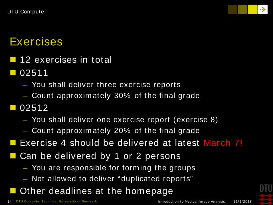
Exercises 12 exercises in total 02511
– You shall deliver three exercise reports– Count approximately 30% of the final grade
02512– You shall deliver one exercise report (exercise 8)– Count approximately 20% of the final grade
Exercise 4 should be delivered at latest March 7! Can be delivered by 1 or 2 persons
– You are responsible for forming the groups– Not allowed to deliver “duplicated reports”
Other deadlines at the homepage
DTU Compute
31/1/2018Introduction to Medical Image Analysis15 DTU Compute, Technical University of Denmark
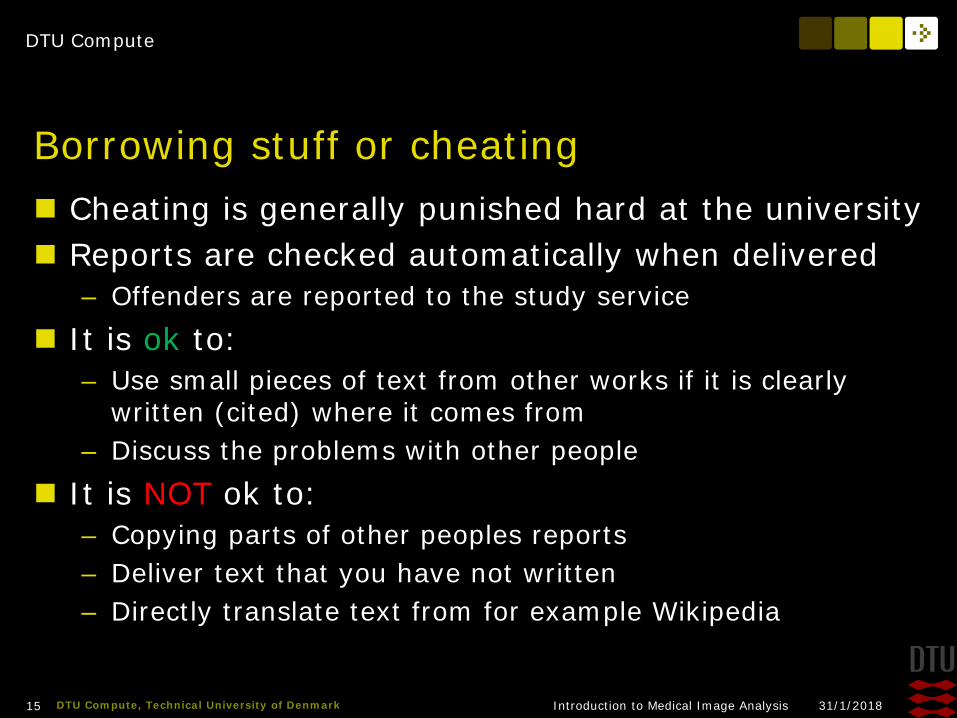
Borrowing stuff or cheating Cheating is generally punished hard at the university Reports are checked automatically when delivered
– Offenders are reported to the study service It is ok to:
– Use small pieces of text from other works if it is clearly written (cited) where it comes from
– Discuss the problems with other people It is NOT ok to:
– Copying parts of other peoples reports– Deliver text that you have not written– Directly translate text from for example Wikipedia

DTU Compute
31/1/2018Introduction to Medical Image Analysis16 DTU Compute, Technical University of Denmark
Matlab and computers No databar We assume that you can use your own portable
computer with Matlab Try to arrange yourself into groups with at least one
working Matlab installation

DTU Compute
31/1/2018Introduction to Medical Image Analysis17 DTU Compute, Technical University of Denmark
Exercise report When doing the exercises:
– Create a Matlab file and keep all your Matlab code for the exercise in this one
– Create a text document (word, Latex, OpenOffice etc) and paste the results here
Writing the report– Evaluate the results
What do you see? Why does the results look like they do? Can it be improved? How?

DTU Compute
31/1/2018Introduction to Medical Image Analysis19 DTU Compute, Technical University of Denmark
About Math We assume that you are comfortable with
”Gymnasiematematik”– Vectors– Pythagoras– Differentation– and so on…
Use the appendix in the book Formelsamling:
– http://www.polyteknisk.dk/home/Detaljer/9788750210092

DTU Compute
31/1/2018Introduction to Medical Image Analysis20 DTU Compute, Technical University of Denmark
Questions! How many of you:
– use image manipulation software Photoshop Paintshop GIMP Something else (what?)
– Adjust images before putting them on Facebook or somewhere else?
DTU Compute
31/1/2018Introduction to Medical Image Analysis21 DTU Compute, Technical University of Denmark
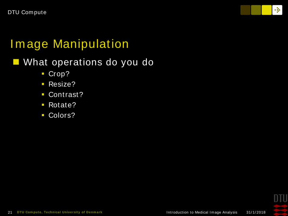
Image Manipulation What operations do you do
Crop? Resize? Contrast? Rotate? Colors?

DTU Compute
31/1/2018Introduction to Medical Image Analysis22 DTU Compute, Technical University of Denmark
Camera / smartphone Bring your own camera/smartphone to the exercises Learn to transfer photos from your camera/phone so
you can use them on your computer
DTU Compute
31/1/2018Introduction to Medical Image Analysis23 DTU Compute, Technical University of Denmark
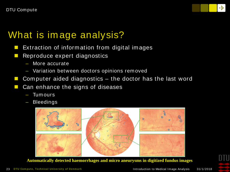
What is image analysis? Extraction of information from digital images Reproduce expert diagnostics
– More accurate– Variation between doctors opinions removed
Computer aided diagnostics – the doctor has the last word Can enhance the signs of diseases
– Tumours– Bleedings
Automatically detected haemorrhages and micro aneurysms in digitized fundus images

DTU Compute
31/1/2018Introduction to Medical Image Analysis24 DTU Compute, Technical University of Denmark
Examples
Shape changes in brain structures
Recognise and track the heart Face recognition and tracking
Cochlear implant planning
DTU Compute
31/1/2018Introduction to Medical Image Analysis25 DTU Compute, Technical University of Denmark
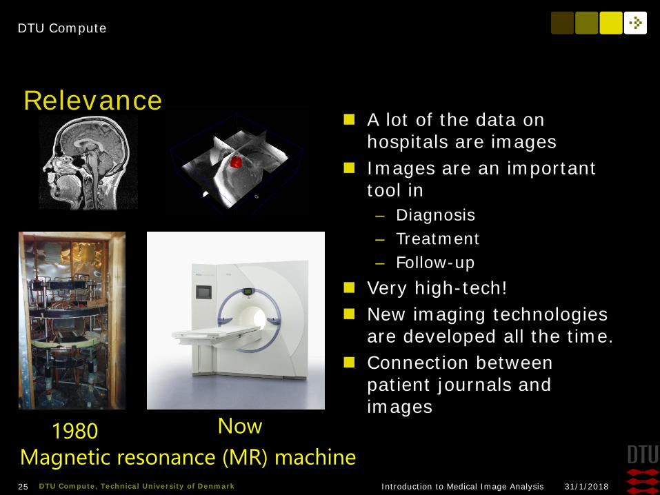
A lot of the data on hospitals are images
Images are an important tool in– Diagnosis– Treatment– Follow-up
Very high-tech! New imaging technologies
are developed all the time. Connection between
patient journals and images
1980
Relevance
Magnetic resonance (MR) machineNow1980

DTU Compute
31/1/2018Introduction to Medical Image Analysis26 DTU Compute, Technical University of Denmark
Relevance Siemens PET/MR
machine Installed at
Rigshospitalet december 2011
Extremely advanced New types of images
and information

DTU Compute
31/1/2018Introduction to Medical Image Analysis27 DTU Compute, Technical University of Denmark
Relevance 7 tesla MR scanner installed at Hvidovre hospital
– New anatomical details visible– More information in brain images
Fast and accurate 3D face scanner installed at the Bloodbank at Glostrup Hospital– Creating a database of normal human faces– Can be used to identify facial features connected to non-
normal growth
DTU Compute
31/1/2018Introduction to Medical Image Analysis28 DTU Compute, Technical University of Denmark
Digital Images – Learning Objectives Describe the fundamental properties of a digital image Read and show an image in Matlab Describe the commonly used image coordinate systems Describe the binary, the label, the multispectral, and the
16-bit image Show and manipulate your own images in Matlab
DTU Compute
31/1/2018Introduction to Medical Image Analysis29 DTU Compute, Technical University of Denmark
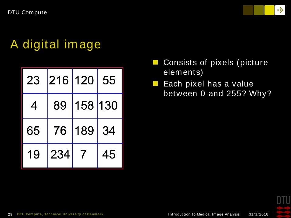
A digital image Consists of pixels (picture
elements) Each pixel has a value
between 0 and 255? Why?

DTU Compute
31/1/2018Introduction to Medical Image Analysis30 DTU Compute, Technical University of Denmark
Bits and Bytes! A bit is a tiny tiny little switch that can be either 0 or 1 – the
“memory of a computer” consists of insanely many bits One byte is 8 bits together. It is the “basic” unit in a computer. With 8 bits how many possible values can be made?
– (2^8 = 256)
00000001 = 1 00000010 = 2 00000100 = 4 00001010 = 10 00001111 = 15
128 64 32 16 8 4 2 1

DTU Compute
31/1/2018Introduction to Medical Image Analysis31 DTU Compute, Technical University of Denmark
Bit the Byte!
MSB LSB
LSB = Least significant bitMSB = Most significant bit

DTU Compute
31/1/2018Introduction to Medical Image Analysis32 DTU Compute, Technical University of Denmark
Binary numbers Decimal 7
– 0000 0111 Decimal 69
– 0100 0101 Decimal 125
– 0111 1101 Decimal 227
– 1110 0011
DTU Compute
31/1/2018Introduction to Medical Image Analysis33 DTU Compute, Technical University of Denmark
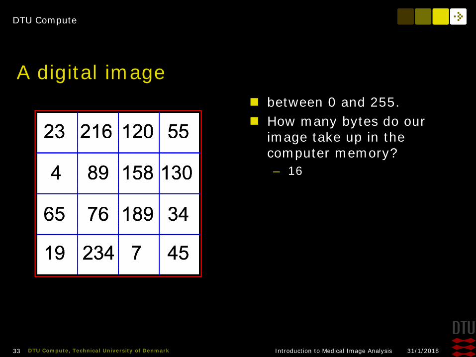
A digital image between 0 and 255. How many bytes do our
image take up in the computer memory? – 16

DTU Compute
31/1/2018Introduction to Medical Image Analysis34 DTU Compute, Technical University of Denmark
Grayscale digital images 0 is black and 255 is white! The values in between are
shown as shades of gray

DTU Compute
31/1/2018Introduction to Medical Image Analysis35 DTU Compute, Technical University of Denmark
Typical Grayscale image Traditional film X-ray Scanned on a flatbed scanner Do you know what an X-ray is? Bone is white and air is black
– The more radiation the darker What are they used for?
– Fractures– Arthrisis– Osteoporosis
DTU Compute
31/1/2018Introduction to Medical Image Analysis36 DTU Compute, Technical University of Denmark
Image Resolution Determines how much the image fills in the memory
and on the hard disk Spatial resolution Gray level resolution
DTU Compute
31/1/2018Introduction to Medical Image Analysis37 DTU Compute, Technical University of Denmark
Spatial? Spatial
– relating to the position, area and size of things Example:
– This task is designed to test the child's spatial awareness
Danish– Rumlig – barnet har en god rumlig forståelse
DTU Compute
31/1/2018Introduction to Medical Image Analysis38 DTU Compute, Technical University of Denmark
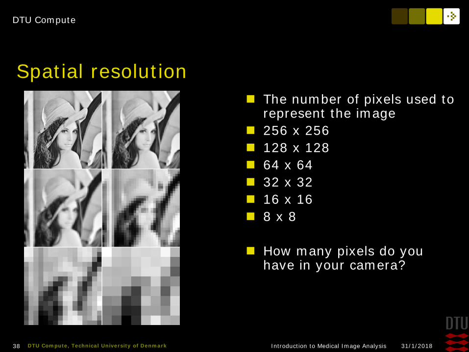
Spatial resolution The number of pixels used to
represent the image 256 x 256 128 x 128 64 x 64 32 x 32 16 x 16 8 x 8
How many pixels do you have in your camera?
DTU Compute
31/1/2018Introduction to Medical Image Analysis39 DTU Compute, Technical University of Denmark
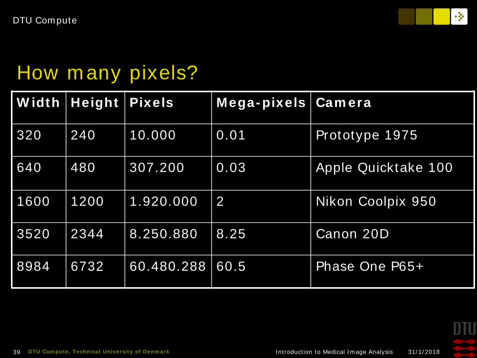
How many pixels?Width Height Pixels Mega-pixels Camera
320 240 10.000 0.01 Prototype 1975
640 480 307.200 0.03 Apple Quicktake 100
1600 1200 1.920.000 2 Nikon Coolpix 950
3520 2344 8.250.880 8.25 Canon 20D
8984 6732 60.480.288 60.5 Phase One P65+
DTU Compute
31/1/2018Introduction to Medical Image Analysis40 DTU Compute, Technical University of Denmark
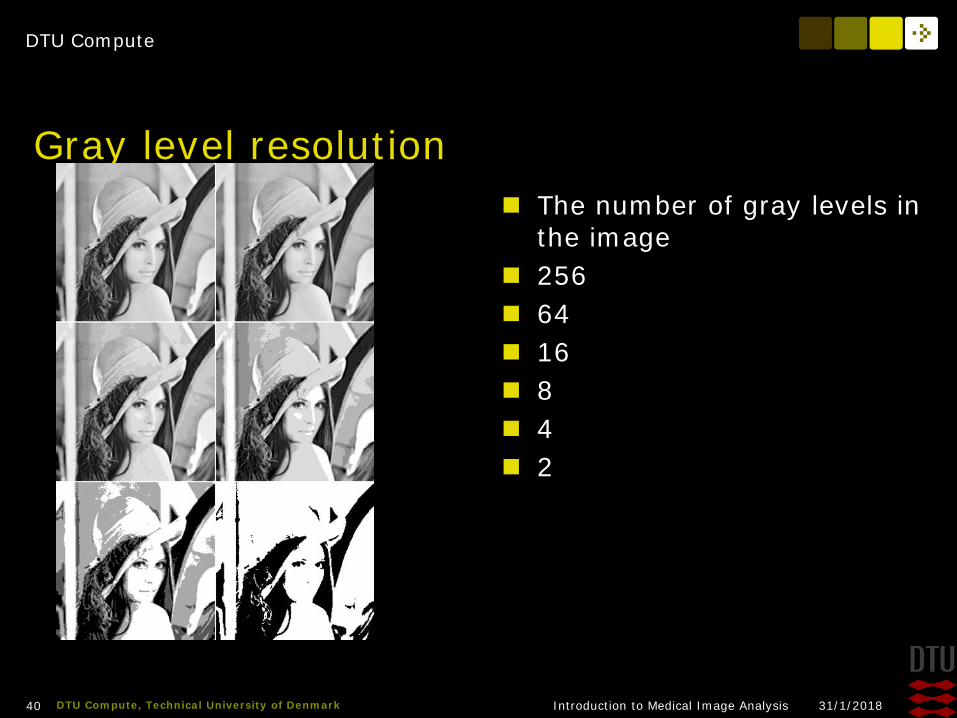
Gray level resolution The number of gray levels in
the image 256 64 16 8 4 2
DTU Compute
31/1/2018Introduction to Medical Image Analysis41 DTU Compute, Technical University of Denmark
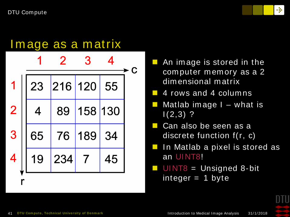
Image as a matrix An image is stored in the
computer memory as a 2 dimensional matrix
4 rows and 4 columns Matlab image I – what is
I(2,3) ? Can also be seen as a
discrete function f(r, c) In Matlab a pixel is stored as
an UINT8! UINT8 = Unsigned 8-bit
integer = 1 byte

DTU Compute
31/1/2018Introduction to Medical Image Analysis42 DTU Compute, Technical University of Denmark
Pixel coordinates – Matlab matrix Used in Matlab Origin is in upper left corner 1-based (row, column) system M rows and N columns
What is the coordinates of the pixel with value 34?
DTU Compute
31/1/2018Introduction to Medical Image Analysis43 DTU Compute, Technical University of Denmark
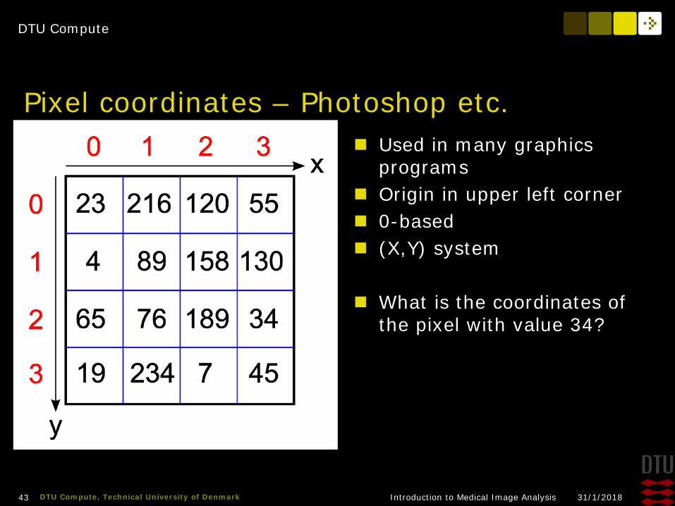
Pixel coordinates – Photoshop etc. Used in many graphics
programs Origin in upper left corner 0-based (X,Y) system
What is the coordinates of the pixel with value 34?
DTU Compute
31/1/2018Introduction to Medical Image Analysis44 DTU Compute, Technical University of Denmark
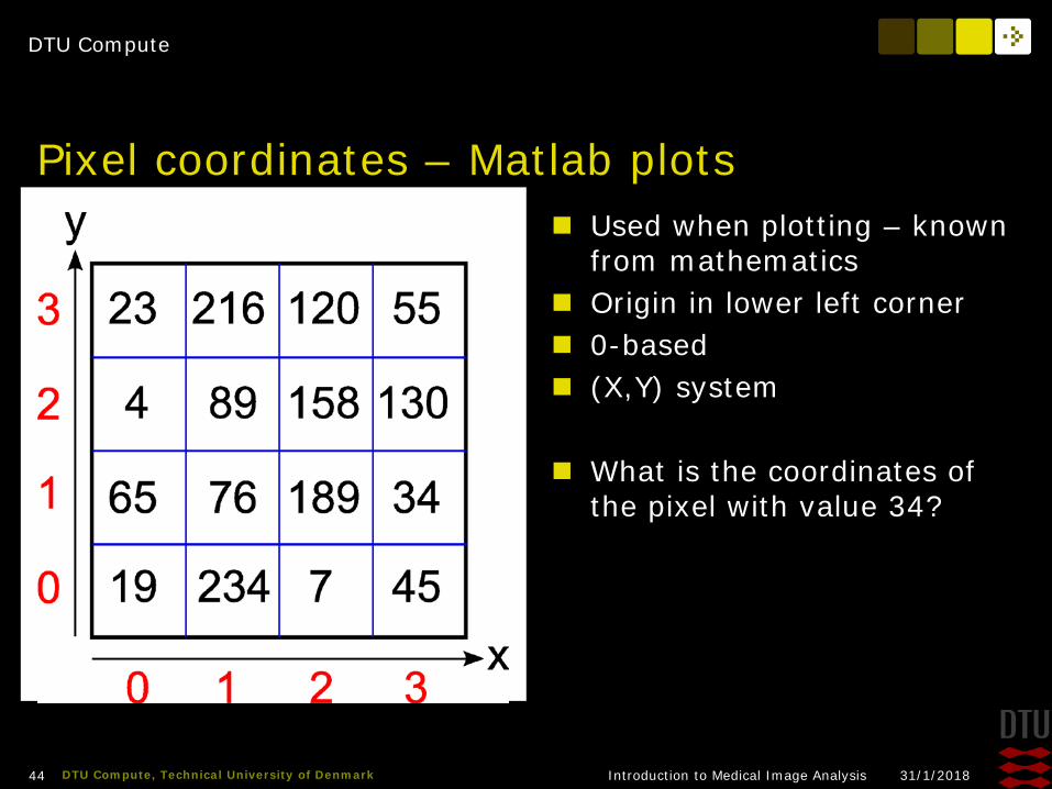
Pixel coordinates – Matlab plots Used when plotting – known
from mathematics Origin in lower left corner 0-based (X,Y) system
What is the coordinates of the pixel with value 34?
DTU Compute
31/1/2018Introduction to Medical Image Analysis45 DTU Compute, Technical University of Denmark
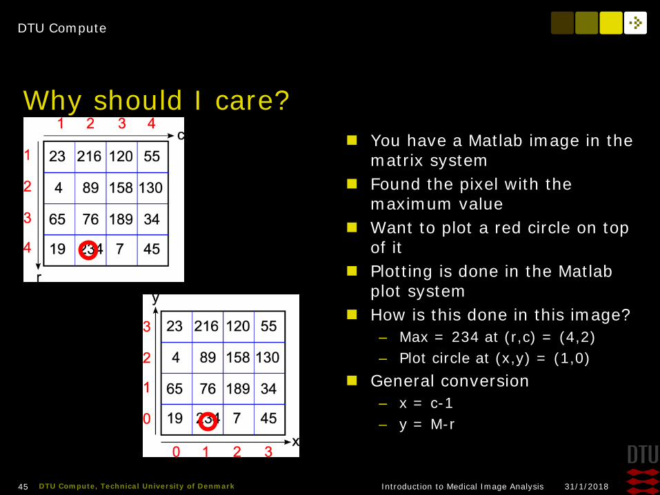
Why should I care? You have a Matlab image in the
matrix system Found the pixel with the
maximum value Want to plot a red circle on top
of it Plotting is done in the Matlab
plot system How is this done in this image?
– Max = 234 at (r,c) = (4,2)– Plot circle at (x,y) = (1,0)
General conversion– x = c-1– y = M-r
DTU Compute
31/1/2018Introduction to Medical Image Analysis46 DTU Compute, Technical University of Denmark
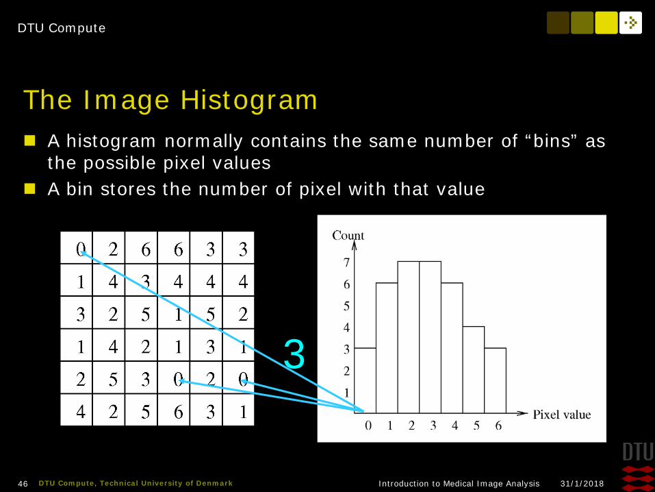
The Image Histogram A histogram normally contains the same number of “bins” as
the possible pixel values A bin stores the number of pixel with that value
3
DTU Compute
31/1/2018Introduction to Medical Image Analysis47 DTU Compute, Technical University of Denmark
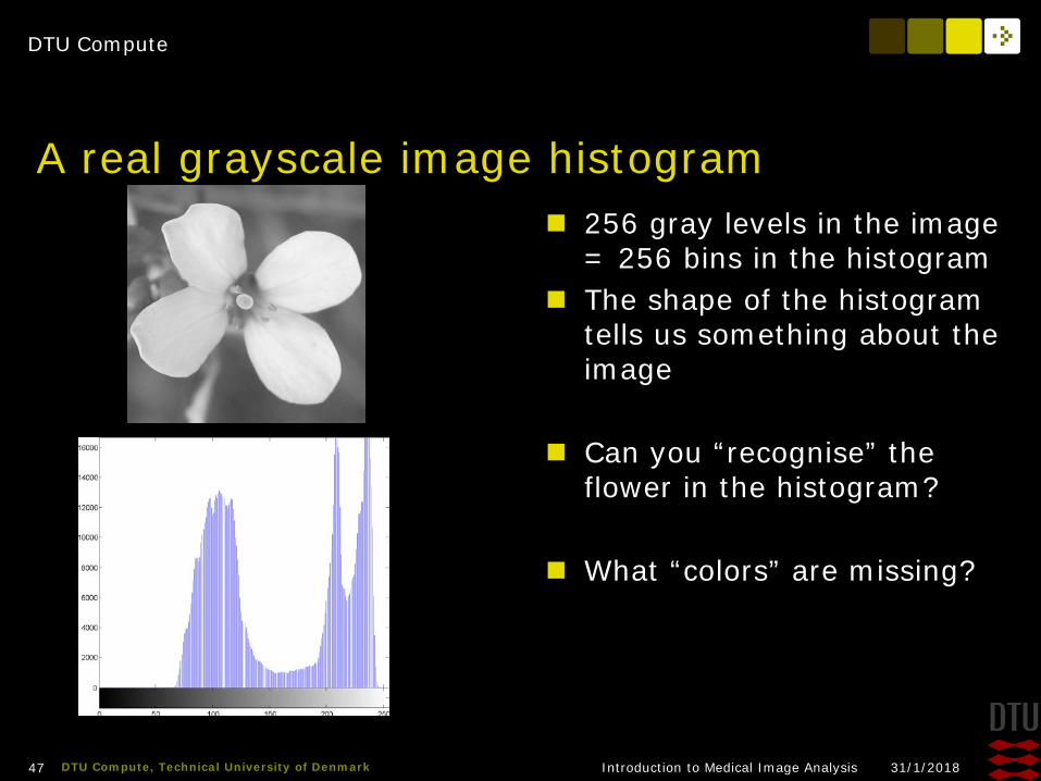
A real grayscale image histogram 256 gray levels in the image
= 256 bins in the histogram The shape of the histogram
tells us something about the image
Can you “recognise” the flower in the histogram?
What “colors” are missing?

DTU Compute
31/1/2018Introduction to Medical Image Analysis48 DTU Compute, Technical University of Denmark
The histogram function Can be seen as a function
h(v) v is the pixel value h(2) = 7 h(5) = 4
Total number of pixels is the sum of all h
DTU Compute
31/1/2018Introduction to Medical Image Analysis49 DTU Compute, Technical University of Denmark
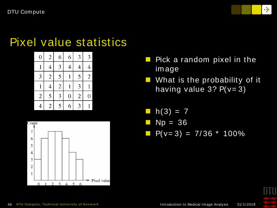
Pixel value statistics Pick a random pixel in the
image What is the probability of it
having value 3? P(v=3)
h(3) = 7 Np = 36 P(v=3) = 7/36 * 100%

DTU Compute
31/1/2018Introduction to Medical Image Analysis50 DTU Compute, Technical University of Denmark
DTU Compute
31/1/2018Introduction to Medical Image Analysis51 DTU Compute, Technical University of Denmark
Normalised histogram A normalised histogram is made by dividing each bin count with
the total number of pixels H(v) is the normalised histogram function H(v) is the probability that a random pixel has value v
Equal to a probability density function

DTU Compute
31/1/2018Introduction to Medical Image Analysis52 DTU Compute, Technical University of Denmark
Other Image Types Colour images Binary Images Label Images 16-bit images

DTU Compute
31/1/2018Introduction to Medical Image Analysis53 DTU Compute, Technical University of Denmark
Colour images Anyone heard of RGB? RGB = Red, Green, and Blue Television, computers, digital
cameras use the “RGB color space”
Additive colours: Final colour is made by mixing red, green, and blue
Typically the values of R, G, and B lie between 0 and 255 (total 3 bytes)!
DTU Compute
31/1/2018Introduction to Medical Image Analysis54 DTU Compute, Technical University of Denmark
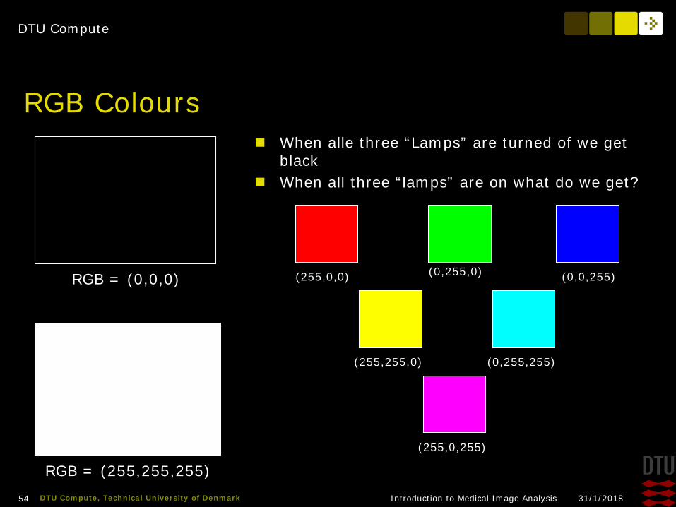
RGB Colours When alle three “Lamps” are turned of we get
black When all three “lamps” are on what do we get?
RGB = (0,0,0)
RGB = (255,255,255)
(255,0,0) (0,255,0) (0,0,255)
(255,255,0) (0,255,255)
(255,0,255)

DTU Compute
31/1/2018Introduction to Medical Image Analysis55 DTU Compute, Technical University of Denmark
Processing RGB images Each pixel in a colour image contains 3 values Equal to a “vector function” in mathematics Much more complicated to analyse Medical images are typically grayscale Therefore we convert from colours to grayscale
before the analysis

DTU Compute
31/1/2018Introduction to Medical Image Analysis56 DTU Compute, Technical University of Denmark
Converting colour to grayscale
v = 0.2989 * R + 0.5870 * G + 0.1140 * B
Is it possible to convert a grayscale image back to a color image?

DTU Compute
31/1/2018Introduction to Medical Image Analysis57 DTU Compute, Technical University of Denmark
DTU Compute
31/1/2018Introduction to Medical Image Analysis58 DTU Compute, Technical University of Denmark
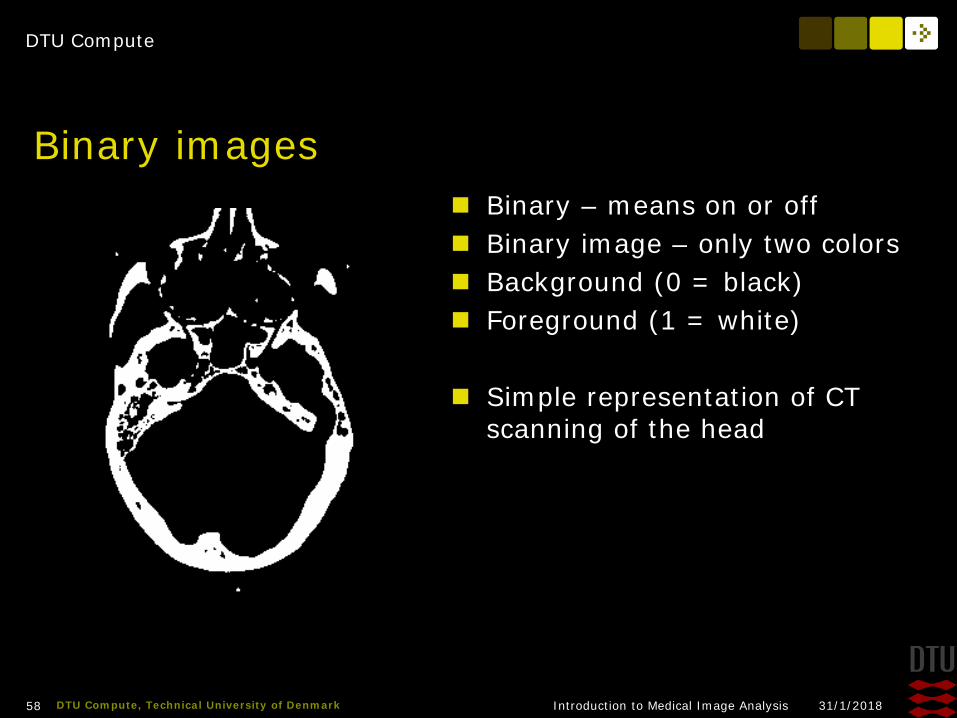
Binary images Binary – means on or off Binary image – only two colors Background (0 = black) Foreground (1 = white)
Simple representation of CT scanning of the head
DTU Compute
31/1/2018Introduction to Medical Image Analysis59 DTU Compute, Technical University of Denmark
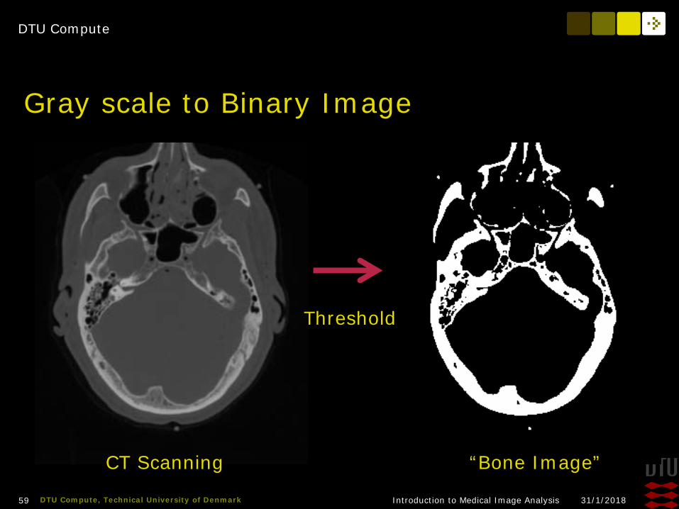
Gray scale to Binary Image
Threshold
CT Scanning “Bone Image”
DTU Compute
31/1/2018Introduction to Medical Image Analysis60 DTU Compute, Technical University of Denmark
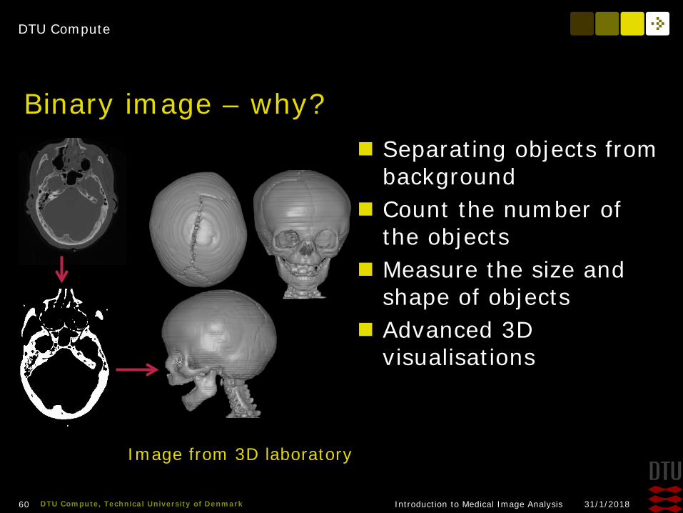
Binary image – why? Separating objects from
background Count the number of
the objects Measure the size and
shape of objects Advanced 3D
visualisations
Image from 3D laboratory
DTU Compute
31/1/2018Introduction to Medical Image Analysis61 DTU Compute, Technical University of Denmark
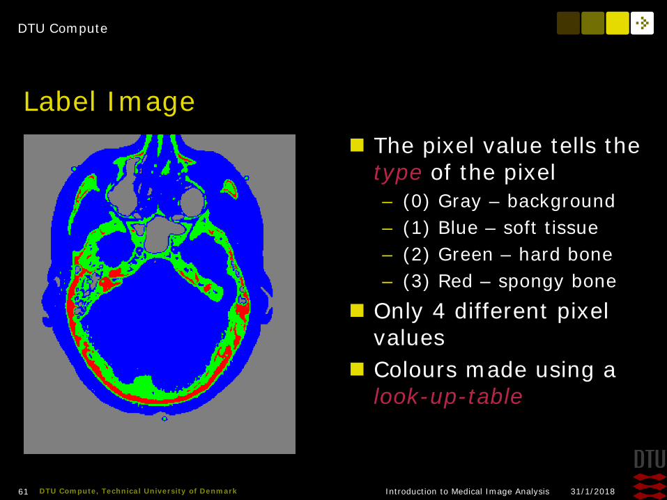
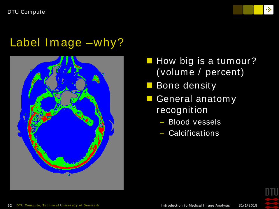
Label Image The pixel value tells the
type of the pixel– (0) Gray – background– (1) Blue – soft tissue– (2) Green – hard bone– (3) Red – spongy bone
Only 4 different pixel values
Colours made using a look-up-table
DTU Compute
31/1/2018Introduction to Medical Image Analysis62 DTU Compute, Technical University of Denmark
Label Image –why? How big is a tumour?
(volume / percent) Bone density General anatomy
recognition– Blood vessels– Calcifications

DTU Compute
31/1/2018Introduction to Medical Image Analysis63 DTU Compute, Technical University of Denmark
Label Image – how?
Classification

DTU Compute
31/1/2018Introduction to Medical Image Analysis64 DTU Compute, Technical University of Denmark
Multispectral images There are more visual
information than what can be seen with the human eye
Standard cameras captures the red, green, blue colours
Capture systems that capture more bands and other frequencies exist
Creates multispectral images– Each pixel contains perhaps 20
values from different spectral bands
Infrared

DTU Compute
31/1/2018Introduction to Medical Image Analysis65 DTU Compute, Technical University of Denmark
Multispectral System - VideometerLab Integrating sphere Light emitting diodes with
different wavelengths– From near infrared to
ultraviolet High resolution B/W camera Water in bread Classification of fungi Skin diseases

DTU Compute
31/1/2018Introduction to Medical Image Analysis66 DTU Compute, Technical University of Denmark
16-bit images 256 values fine for the
human eye Pixel values not only for
display– Physical meaning
Computed Tomography– X-ray attenuation
Hounsfield unit– 0 water– -1000 air– -120 fat– 400+ bone
DTU Compute
31/1/2018Introduction to Medical Image Analysis67 DTU Compute, Technical University of Denmark
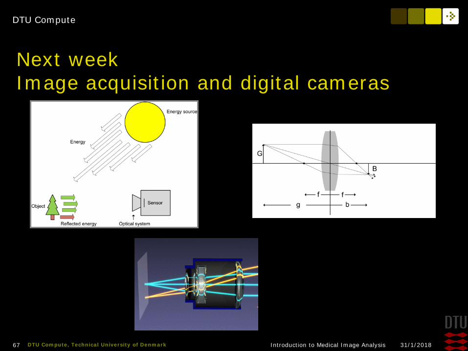
Next weekImage acquisition and digital cameras

DTU Compute
31/1/2018Introduction to Medical Image Analysis68 DTU Compute, Technical University of Denmark
Exercises