Embed Size (px)
Citation preview

Importance of Developing a Decision Support System for Diagnosis of Glaucoma
Murat Durucu
Industrial Engineering Department, Management Faculty, Istanbul Technical University, Istanbul, Turkey
Abstract Glaucoma is a condition of irreversible blindness, early diagnosis and appropriate interventions to make the patients able to see longer time. In this study, it addressed that the importance of developing a decision support system for glaucoma diagnosis. Glaucoma occurs when pressure happens around the eyes it causes some damage to the optic nerves and deterioration of vision. There are dif-ferent levels ranging blindness of glaucoma disease. The diagnosis at an early stage allows a chance for therapies that slows the progression of the disease.
By using Optical Coherence Tomography (OCT) images and pattern recognition systems, it is possible to develop a support system for doctors to make their deci-sions on glaucoma. Thus, in this recent study we develop an evaluation and sup-port system to the usage of doctors. Pattern recognition system based computer software would help the doctors to make an objective evaluation for their patients. It is intended that after development and evaluation processes of the software, the system is planning to be serve for the usage of doctors in different hospitals.
Keywords Decision Support System, Glaucoma, Image Processing, Pattern Recognition.
Introduction
This study focused on necessity for developing an objective decisions support sys-tem for evaluating level and occurrence of glaucoma disease. Glaucoma is diag-nosed with considering patients’ family history and by using clinical techniques, such as tonometry, ophthalmoscopy, perimetry, gonioscopy and pachymetry. Glaucoma is an irreversible blindness for the patients in late stages. The diagnosis at an early stage allows for therapies that slow the progression of the disease. Also diagnosis at an early stage can decrease the socio-economic wages for the patients and country they live in (Mazhar 2013). However, problems have been experi-
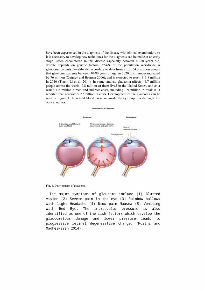
enced in the diagnosis of the disease with clinical examination, so it is necessary to develop new techniques for the diagnosis can be made at an early stage. Often encountered in this disease especially between 40-80 years old, despite depends on genetic factors. 3:54% of the population worldwide is glaucoma patients. World-wide, according to data from 2013, 64.3 million people that glaucoma patients be-tween 40-80 years of age, in 2020 this number increased by 76 million (Quigley and Broman 2006), and is expected to reach 111.8 million in 2040 (Tham, Li et al. 2014). In some studies, glaucoma affects 44.7 million people across the world, 2.8 million of them lived in the United States, and as a result, 1.6 million direct, and indirect costs, including 0.9 million in total; It is reported that generate $ 2.5 bil-lion in costs. Development of the glaucoma can be seen in Figure 1. Increased blood pressure inside the eye pupil, is damages the optical nerves.
Fig. 1. Development of glaucoma
The major symptoms of glaucoma include (1) Blurred vision (2) Severe pain in the eye (3) Rainbow hallows with light Headache (4) Brow pain Nausea (5) Vom-iting with Red Eye. The intraocular pressure is also identified as one of the risk factors which develop the glaucomatous damage and lower pressure leads to pro-gressive retinal degenerative change. (Murthi and Madheswaran 2014).
There are different levels of the glaucoma in the literature. The comparison be-tween normal vision and the glaucoma eye can be seen in Figure 2. Description of the levels of the glaucoma for ophthalmologist can be found in Table 1.

3
Fig. 2. Level samples for patients’ visions
Table 1. Glaucoma levels
Light or early level glaucoma Glaucoma is defined as the optic nerve abnormalities that are similar, but there is no abnormality in the visual field white upon white field test.
Mid-level glaucoma Glaucoma is an optic nerve abnormalities compatible with. En-countered a hemisphere and 5 is very hard glaucoma anomaly.
Extreme, last term level glau-coma
Glaucoma is consistent with glaucoma optic nerve and visual field abnormalities anomalies are found in both hemispheres, are seen at least 5 degrees loss of vision in one hemisphere fixed.
Uncertain glaucoma Visual fields were not yet occurred, or is not suitable for patients with visual field testing or visual field tests applicable / unreli-able state.
In recent years, imaging technology from Heidelberg retinal tomography (HRT), Stereoscopic disc Photo (SDP) and Optical Coherence Tomography (OCT) imaging technology such as is used for the diagnosis of glaucoma (Mwanza and Budenz 2016). This better accuracy and faster imaging techniques in response technique of OCT has become the most common method used by ex-perts. Retinal Nerve Fiber Layer (RNFL), optic nerve head (ONH) and reasonable analysis are applied to detect any glaucoma damage OCT (Bai, Niwas et al. 2016). Due to all economic facts that, early diagnosis of glaucoma is very important as patients’ life quality as economical costs. Clinically, the diagnosis of Glaucoma can be done through measurement of CDR, defined as the ratio of the vertical height of the optic cup to the vertical height of the optic disc. An increment in the cupping of Optic Nerve Head (ONH) corresponds to the increased ganglion cell death and hence CDR can be used to measure the probability of developing the disease. A CDR value that is greater than 0.65 indicates the high glaucoma risk (Li and Chutatape 2003).
In the Figure 2 you can find the medical imaging for normal eye and affected eye (Murthi and Madheswaran 2014). With the defect of optical nerves irre-versible blindness will be begun.

4
Fig. 3. Medical imaging of normal eye and affected eye (Murthi and Madheswaran 2014)
Methodology
To date, procedures that have been employed for detection of glaucomatous visual field progression may be broadly grouped into four categories: subjective clinical judgment, defect classification systems, trend analyses, and event analyses.
Clinical Judgment
Clinical judgment consists of simple subjective observation of sequential visual field test results and represents the oldest method for identification of progressive visual field defects. This approach is advantageous for a number of reasons: 1) it demands no additional computation; 2) it is highly flexible as observers with any degree experience may apply it to the results of any instrumentation; and 3) it is easy to perform. However, the subjectivity of this approach means that it is also poorly controlled, and criteria can vary considerably from one evaluator to an-other. A comparison of visual field series evaluated for deterioration, stability, or improvement by six expert observers illustrates clinical judgment’s disadvantages (Werner, Bishop et al. 1988). When using clinical judgment to assess data, care should be taken when a patient has been examined with a variety of threshold esti-mation algorithms.

5
Defect Classification Systems
Visual field defect classification systems use predetermined criteria to grade single test results, providing a discrete score for each visual field test result. The advantages of this approach are that test results are immediately stratified into broadly similar defect magnitudes, interpretation is relatively simple, and progres-sion can be easily defined as worsening of the score over time. There are, how-ever, a number of drawbacks to use of classification systems. They do not provide information on the spatial configuration of defects and may not be scaled linearly, for example, a change from 0 to 3 may not be equal to a change from 10 to 13.
Trend Analyses
Trend analyses evaluate test parameters sequentially to determine temporal pat-terns that may exist within the data (Holmin and Krakau 1980, Holmin and Krakau 1982). Such analyses are of value because they are capable of determining long-term characteristics with use of information from all visual field examina-tions performed on a patient, and therefore have the potential to discriminate sub-tle progressive loss from considerable degrees of test variability (Fitzke, Hitchings et al. 1996).
Event Analyses
Event analyses are valuable because they attempt to identify single events of significant change relative to a reference examination (Hitchings 1994). Event analyses can be relatively simple, and can look for statistically significant differ-ences between one examination and another, such as used within the DELTA pro-gram of the Octopus perimeter. This particular method employs a paired t test to determine whether significant differences are present between one test result and another.
Conclusion
Although OCT images or HRT precision and quickness, especially in the early stages, difficulty and mistakes are experienced in diagnosis of glaucoma. To be in the discretion of the doctor's diagnosis and placement process, it is difficult to ob-

6
tain objective results. It is very important to develop an objective decision support system for diagnosis and level the glaucoma disease for patients.
In recent years computer aided diagnosis (CAD) is playing a major role in screening the glaucoma. The CAD system is simple, repetitive, not prone to inter or intra observer variability and fast in diagnosis. Also CAD can screen many pa-tients in a small time. There is a scarcity of ophthalmologists in many developing countries, where CAD can be very useful. The proposed decision support system for glaucoma can differentiate normal and glaucoma classes accurately
By using OCT images and pattern recognition systems, it is possible to develop a support system for doctors to make their decisions on glaucoma. For this pur-pose, an evaluation and support system will be developed, and will be offered to usage of doctors. Pattern recognition system based computer software will be evaluating the level of glaucoma between none to severe, end-stage glaucoma. Af-ter evaluation processes of the software, the system is planning to be serve for the usage of medical personnel in different hospitals.
References
Bai, X. L., S. I. Niwas, W. S. Lin, B. F. Ju, C. K. Kwoh, L. P. Wang, C. C. Sng, M. C. Aquino and P. T. K. Chew (2016). "Learning ECOC Code Matrix for Multiclass Classification with Application to Glaucoma Diagnosis." Journal of Medical Systems 40(4): 10.
Fitzke, F. W., R. A. Hitchings, D. Poinoosawmy, A. I. McNaught and D. P. Crabb (1996). "Anal-ysis of visual field progression in glaucoma." The British Journal of Ophthalmology 80(1): 40-48.
Hitchings, R. A. (1994). "Perimetry--back to the future?" The British Journal of Ophthalmology 78(11): 805-806.
Holmin, C. and C. E. Krakau (1980). "Visual field decay in normal subjects and in cases of chronic glaucoma." Albrecht Von Graefes Arch Klin Exp Ophthalmol 213(4): 291-298.
Holmin, C. and C. E. Krakau (1982). "Regression analysis of the central visual field in chronic glaucoma cases. A follow-up study using automatic perimetry." Acta Ophthalmol (Copenh) 60(2): 267-274.
Li, H. and O. Chutatape (2003). A Model-Based Approach For Automated Feature Extraction In Fundus Images. Proc. of the 9th IEEE International Conference on Computer Vision.
Mazhar, S. (2013). "Nuggets in clinical approach to diagnosis of glaucoma." Journal of Clinical Ophthalmology and Research 1(3): 175-181.
Murthi, A. and M. Madheswaran (2014). "Medical Decision Support System to Identify Glau-coma using Cup to Disc Ratio." Journal of Theoretical and Applied Information Technology 68(2): 406-413.
Mwanza, J. C. and D. L. Budenz (2016). "Optical coherence tomography platforms and parame-ters for glaucoma diagnosis and progression." Current Opinion in Ophthalmology 27(2): 102-110.
Quigley, H. A. and A. T. Broman (2006). "The Number of People With Glaucoma Worldwide In 2010 and 2020." Br. J. Ophthalmol 90(3): 262-267.
Tham, Y.-C., X. Li, T. Y. Wong, H. A. Quigley, T. Aung and C.-Y. Cheng (2014). "Global Prevalence of Glaucoma and Projections of Glaucoma Burden through 2040: A Systematic Review and Meta-Analysis." Ophthalmology 121(11): 2081-2090.

7
Werner, E. B., K. I. Bishop, J. Koelle and et al. (1988). "A comparison of experienced clinical observers and statistical tests in detection of progressive visual field loss in glaucoma using automated perimetry." Archives of Ophthalmology 106(5): 619-623.