Embed Size (px)
Citation preview
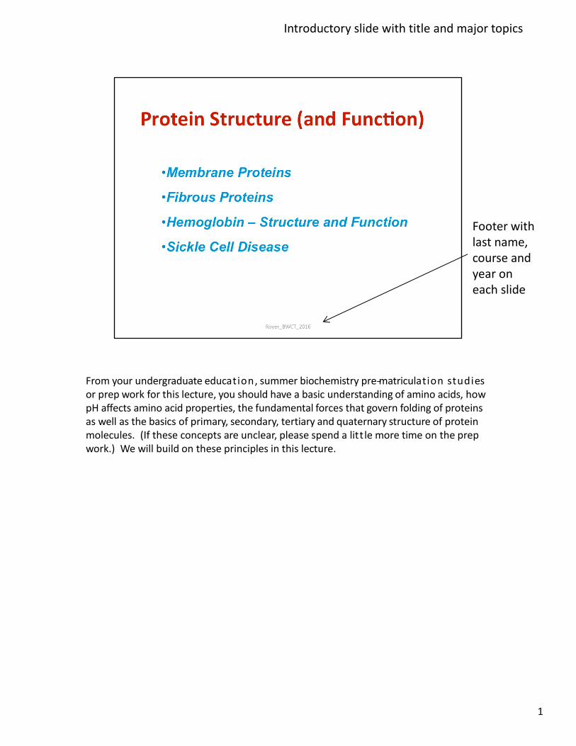
From your undergraduate education, summer biochemistry pre-‐matriculation studies or prep work for this lecture, you should have a basic understanding of amino acids, how pH affects amino acid properties, the fundamental forces that govern folding of proteins as well as the basics of primary, secondary, tertiary and quaternary structure of protein molecules. (If these concepts are unclear, please spend a litt le more time on the prep work.) We will build on these principles in this lecture.
1
Introductory slide with title and major topics
Footer with last name, course and year on each slide
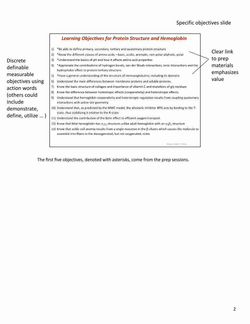
The first five objectives, denoted with asterisks, come from the prep sessions.
2
Clear link to prep materials emphasizes value
Specific objectives slide
Discrete definable measurable objectives using action words (others could include demonstrate, define, utilize … )
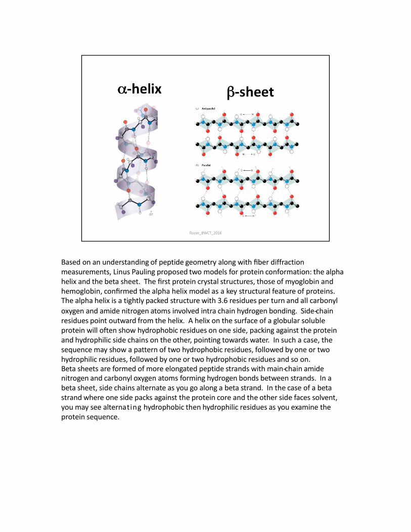
Based on an understanding of peptide geometry along with fiber diffraction measurements, Linus Pauling proposed two models for protein conformation: the alpha helix and the beta sheet. The first protein crystal structures, those of myoglobin and hemoglobin, confirmed the alpha helix model as a key structural feature of proteins. The alpha helix is a tightly packed structure with 3.6 residues per turn and all carbonyl
oxygen and amide nitrogen atoms involved intra chain hydrogen bonding. Side-‐chain residues point outward from the helix. A helix on the surface of a globular soluble protein will often show hydrophobic residues on one side, packing against the protein and hydrophilic side chains on the other, pointing towards water. In such a case, the sequence may show a pattern of two hydrophobic residues, followed by one or two hydrophilic residues, followed by one or two hydrophobic residues and so on. Beta sheets are formed of more elongated peptide strands with main-‐chain amide nitrogen and carbonyl oxygen atoms forming hydrogen bonds between strands. In a beta sheet, side chains alternate as you go along a beta strand. In the case of a beta strand where one side packs against the protein core and the other side faces solvent, you may see alternating hydrophobic then hydrophilic residues as you examine the protein sequence.
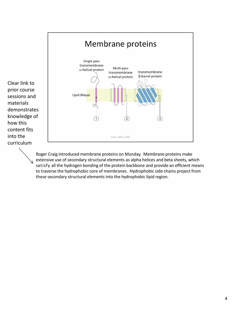
Roger Craig introduced membrane proteins on Monday. Membrane proteins make extensive use of secondary structural elements as alpha helices and beta sheets, which satisfy all the hydrogen bonding of the protein backbone and provide an efficient means to traverse the hydrophobic core of membranes. Hydrophobic side chains project from these secondary structural elements into the hydrophobic lipid region.
4
Clear link to prior course sessions and materials demonstrates knowledge of how this content fits into the curriculum
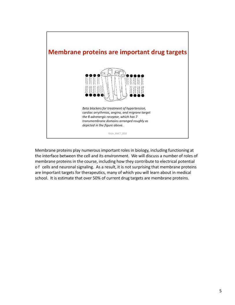
Membrane proteins play numerous important roles in biology, including functioning at the interface between the cell and its environment. We will discuss a number of roles of membrane proteins in the course, including how they contribute to electrical potential o f cells and neuronal signaling. As a result, it is not surprising that membrane proteins are important targets for therapeutics, many of which you will learn about in medical school. It is estimate that over 50% of current drug targets are membrane proteins.
5
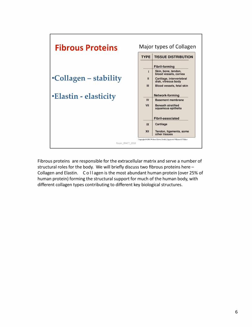
Fibrous proteins are responsible for the extracellular matrix and serve a number of structural roles for the body. We will briefly discuss two fibrous proteins here – Collagen and Elastin. C o l l agen is the most abundant human protein (over 25% of human protein) forming the structural support for much of the human body, with different collagen types contributing to different key biological structures.
6
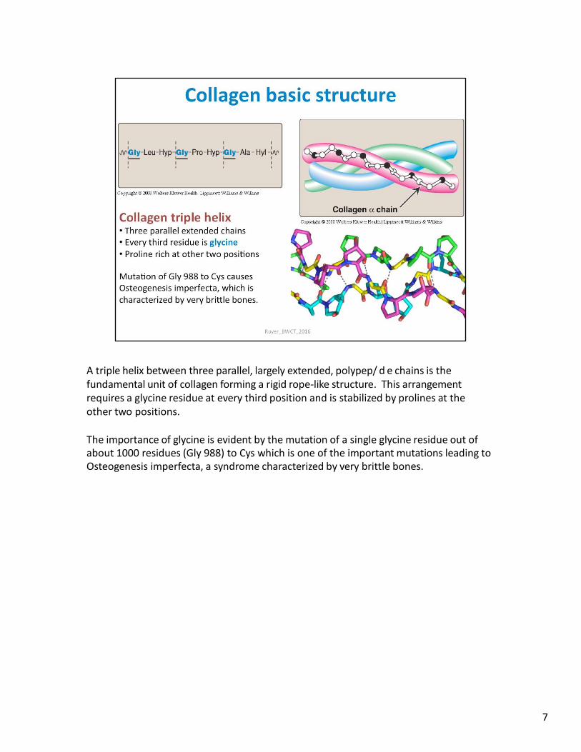
A triple helix between three parallel, largely extended, polypep/ d e chains is the fundamental unit of collagen forming a rigid rope-like structure. This arrangement requires a glycine residue at every third position and is stabilized by prolines at the other two positions.
The importance of glycine is evident by the mutation of a single glycine residue out of about 1000 residues (Gly 988) to Cys which is one of the important mutations leading to Osteogenesis imperfecta, a syndrome characterized by very brittle bones.
7
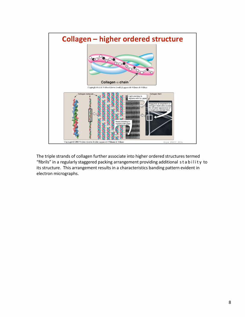
The triple strands of collagen further associate into higher ordered structures termed “fibrils” in a regularly staggered packing arrangement providing additional s t a b i l i t y to its structure. This arrangement results in a characteristics banding pattern evident in electron micrographs.
8
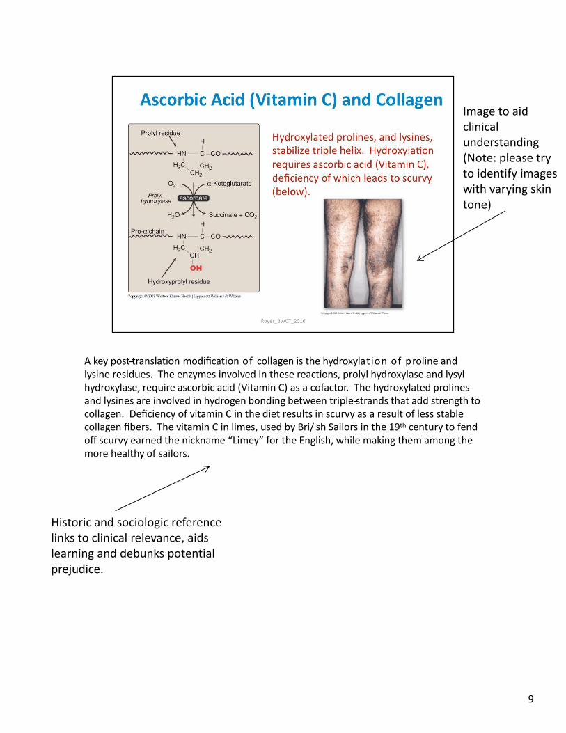
A key post-‐translation modification of collagen is the hydroxylation of proline and lysine residues. The enzymes involved in these reactions, prolyl hydroxylase and lysyl hydroxylase, require ascorbic acid (Vitamin C) as a cofactor. The hydroxylated prolines and lysines are involved in hydrogen bonding between triple-‐strands that add strength to collagen. Deficiency of vitamin C in the diet results in scurvy as a result of less stable collagen fibers. The vitamin C in limes, used by Bri/ sh Sailors in the 19th century to fend off scurvy earned the nickname “Limey” for the English, while making them among the more healthy of sailors.
9
Image to aid clinical understanding (Note: please try to identify images with varying skin tone)
Historic and sociologic reference links to clinical relevance, aids learning and debunks potential prejudice.
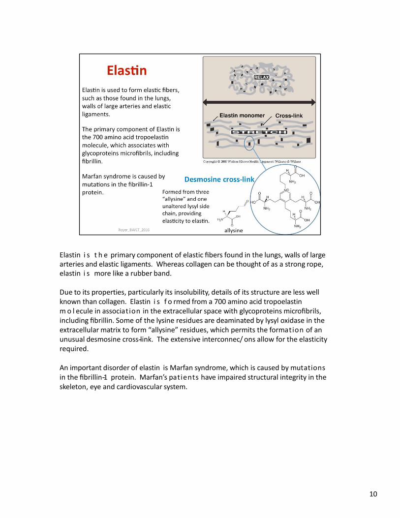
Elastin i s t h e primary component of elastic fibers found in the lungs, walls of large arteries and elastic ligaments. Whereas collagen can be thought of as a strong rope, elastin i s more like a rubber band.
Due to its properties, particularly its insolubility, details of its structure are less well known than collagen. Elastin i s f o rmed from a 700 amino acid tropoelastin m o l ecule in association in the extracellular space with glycoproteins microfibrils, including fibrillin. Some of the lysine residues are deaminated by lysyl oxidase in the extracellular matrix to form “allysine” residues, which permits the formation of an unusual desmosine cross-‐link. The extensive interconnec/ ons allow for the elasticity required.
An important disorder of elastin is Marfan syndrome, which is caused by mutations in the fibrillin-‐1 protein. Marfan’s patients have impaired structural integrity in the skeleton, eye and cardiovascular system.
10
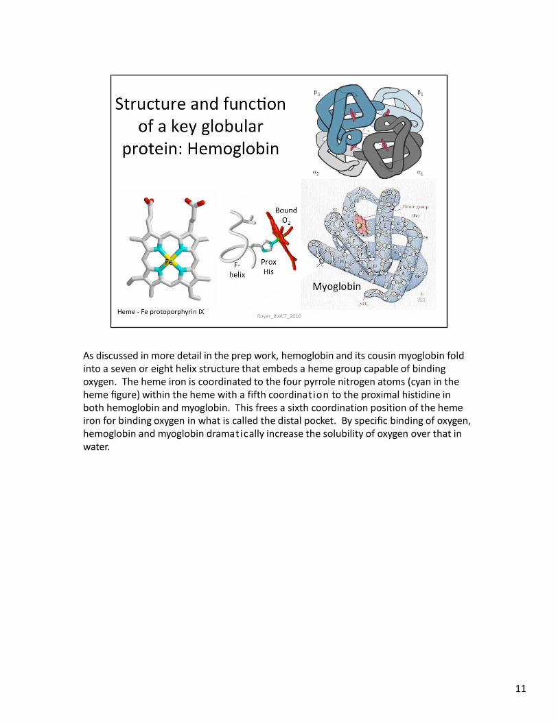
As discussed in more detail in the prep work, hemoglobin and its cousin myoglobin fold into a seven or eight helix structure that embeds a heme group capable of binding oxygen. The heme iron is coordinated to the four pyrrole nitrogen atoms (cyan in the heme figure) within the heme with a fifth coordination to the proximal histidine in both hemoglobin and myoglobin. This frees a sixth coordination position of the heme iron for binding oxygen in what is called the distal pocket. By specific binding of oxygen, hemoglobin and myoglobin dramatically increase the solubility of oxygen over that in water.
11
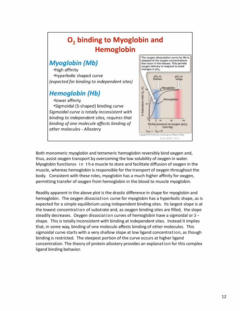
Both monomeric myoglobin and tetrameric hemoglobin reversibly bind oxygen and, thus, assist oxygen transport by overcoming the low solubility of oxygen in water. Myoglobin functionss i n t h e muscle to store and facilitate diffusion of oxygen in the muscle, whereas hemoglobin is responsible for the transport of oxygen throughout the body. Consistent with these roles, myoglobin has a much higher affinity for oxygen, permitting transfer of oxygen from hemoglobin in the blood to muscle myoglobin.
Readily apparent in the above plot is the drastic difference in shape for myoglobin and hemoglobin. The oxygen dissociation curve for myoglobin has a hyperbolic shape, as is expected for a simple equilibrium using independent binding sites. Its largest slope is at the lowest concentration of substrate and, as oxygen binding sites are filled, the slope steadily decreases. Oxygen dissociation curves of hemoglobin have a sigmoidal or S -‐ shape. This is totally inconsistent with binding at independent sites. Instead it implies that, in some way, binding of one molecule affects binding of other molecules. This sigmoidal curve starts with a very shallow slope at low ligand concentration, as though binding is restricted. The steepest portion of the curve occurs at higher ligand concentration. The theory of protein allostery provides an explanation for this complex ligand binding behavior.
12
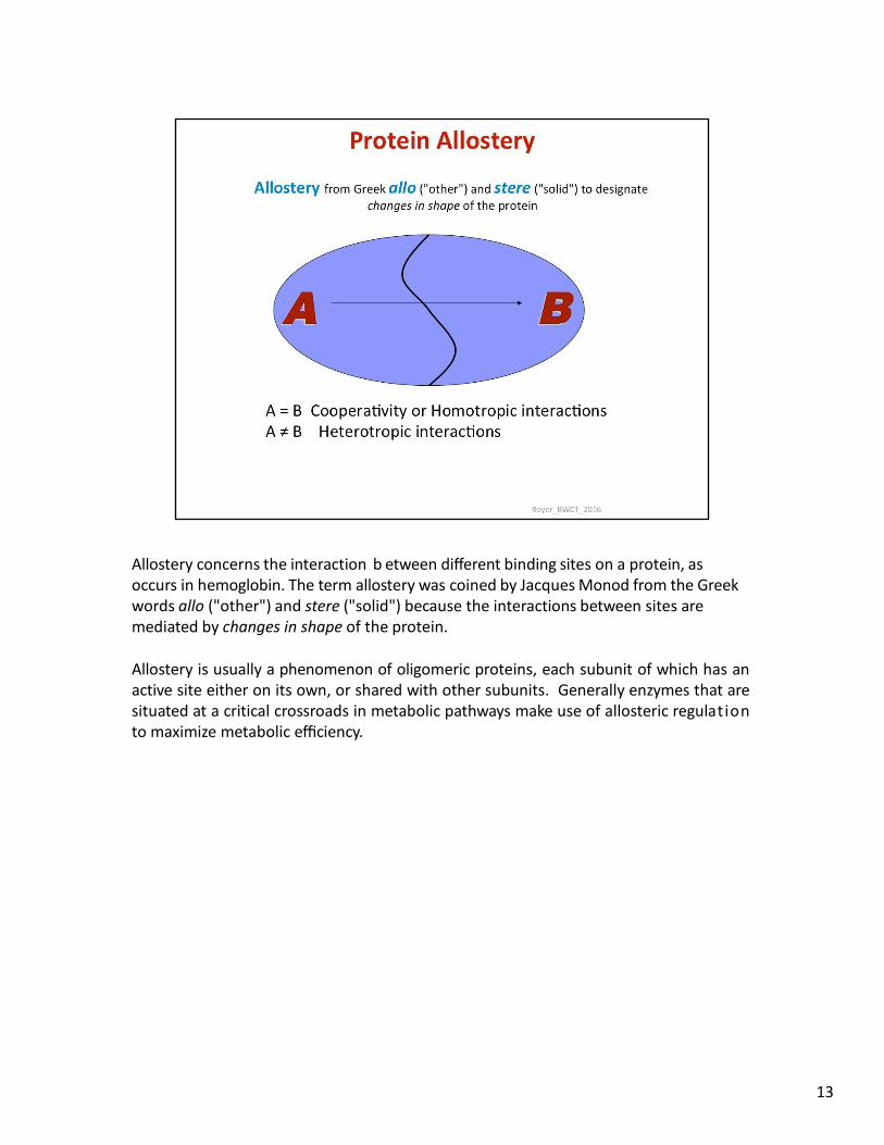
Allostery concerns the interaction b etween different binding sites on a protein, as occurs in hemoglobin. The term allostery was coined by Jacques Monod from the Greek words allo ("other") and stere ("solid") because the interactions between sites are mediated by changes in shape of the protein.
Allostery is usually a phenomenon of oligomeric proteins, each subunit of which has an active site either on its own, or shared with other subunits. Generally enzymes that are situated at a critical crossroads in metabolic pathways make use of allosteric regulation to maximize metabolic efficiency.
13
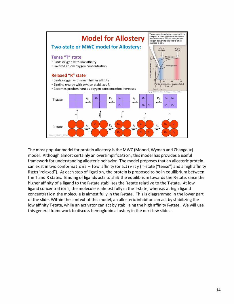
The most popular model for protein allostery is the MWC (Monod, Wyman and Changeux) model. Although almost certainly an oversimplification, this model has provides a useful framework for understanding allosteric behavior. The model proposes that an allosteric protein can exist in two conformations – low affinity (or act i v i t y ) T-state (“tense”) and a high affinity R-state (“relaxed”). At each step of ligation, the protein is proposed to be in equilibrium between the T and R states. Binding of ligands acts to shiS the equilibrium towards the R-‐state, since the higher affinity of a ligand to the R-‐state stabilizes the R-‐state relative to the T-‐state. At low ligand concentrations, the molecule is almost fully in the T-‐state, whereas at high ligand concentration the molecule is almost fully in the R-‐state. This is diagrammed in the lower part of the slide. Within the context of this model, an allosteric inhibitor can act by stabilizing the low affinity T-‐state, while an activator can act by stabilizing the high affinity R-‐state. We will use this general framework to discuss hemoglobin allostery in the next few slides.
14
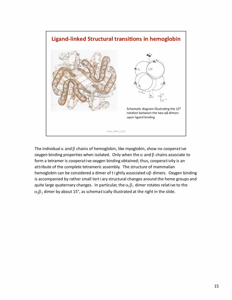
The individual and chains of hemoglobin, like myoglobin, show no cooperative
oxygen binding properties when isolated. Only when the and chains associate to
form a tetramer is cooperative oxygen binding obtained; thus, cooperativity is an
attribute of the complete tetrameric assembly. The structure of mammalian
hemoglobin can be considered a dimer of t i ghtly associated dimers. Oxygen binding
is accompanied by rather small tert i ary structural changes around the heme groups and
quite large quaternary changes. In particular, the dimer rotates relative to the
dimer by about 15°, as schematically illustrated at the right in the slide.
15
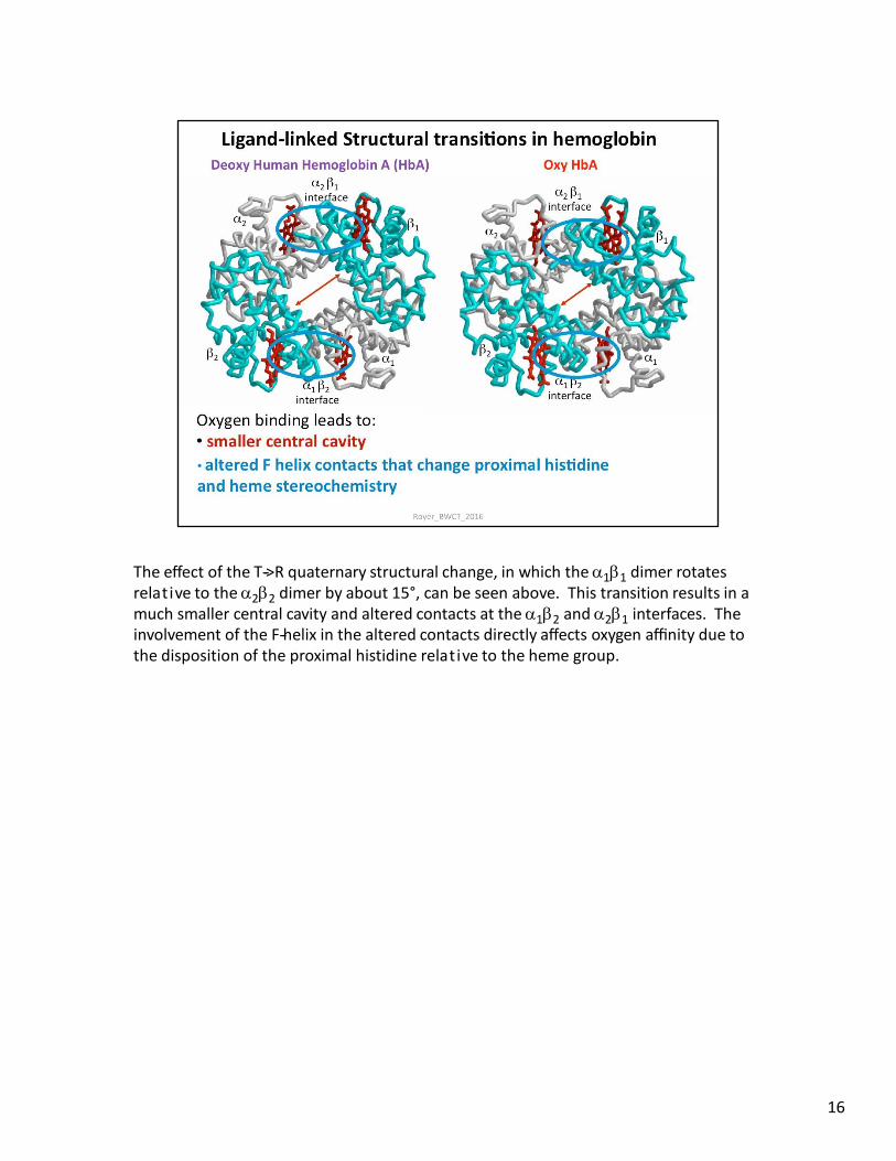
The effect of the T-‐>R quaternary structural change, in which the 11 dimer rotates relative to the 22 dimer by about 15°, can be seen above. This transition results in a much smaller central cavity and altered contacts at the 12 and 21 interfaces. The involvement of the F-‐helix in the altered contacts directly affects oxygen affinity due to the disposition of the proximal histidine relative to the heme group.
16
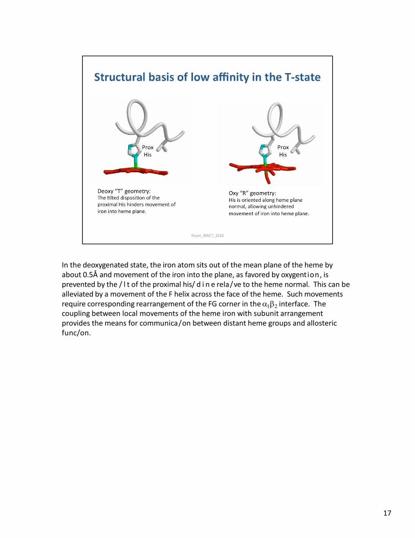
In the deoxygenated state, the iron atom sits out of the mean plane of the heme by about 0.5Å and movement of the iron into the plane, as favored by oxygention, is prevented by the / l t of the proximal his/ d i n e rela/ve to the heme normal. This can be alleviated by a movement of the F helix across the face of the heme. Such movements require corresponding rearrangement of the FG corner in the interface. The coupling between local movements of the heme iron with subunit arrangement provides the means for communica/on between distant heme groups and allosteric func/on.
17
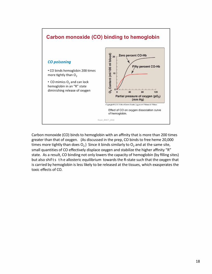
Carbon monoxide (CO) binds to hemoglobin with an affinity that is more than 200 times greater than that of oxygen. (As discussed in the prep, CO binds to free heme 20,000 times more tightly than does O2.) Since it binds similarly to O2 and at the same site,
small quantities of CO effectively displace oxygen and stabilize the higher affinity “R” state. As a result, CO binding not only lowers the capacity of hemoglobin (by filling sites) but also shif t s t h e allosteric equilibrium towards the R-state such that the oxygen that is carried by hemoglobin is less likely to be released at the tissues, which exasperates the toxic effects of CO.
18
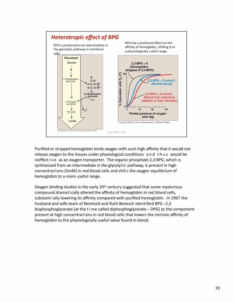
Purified or stripped hemoglobin binds oxygen with such high affinity that it would not release oxygen to the tissues under physiological conditions a n d t h u s would be ineffect i v e as an oxygen transporter. The organic phosphate 2,3 BPG, which is synthesized from an intermediate in the glycolytic pathway, is present in high concentrations (5mM) in red blood cells and shiS s the oxygen equilibrium of hemoglobin to a more useful range.
Oxygen binding studies in the early 20th century suggested that some mysterious compound dramatically altered the affinity of hemoglobin in red blood cells, substant i ally lowering its affinity compared with purified hemoglobin. In 1967 the husband and wife team of Reinhold and Ruth Benesch identified BPG -‐ 2,3 bisphosphoglycerate (at the t i me called diphosphoglycerate – DPG) as the component present at high concentrations in red blood cells that lowers the intrinsic affinity of hemoglobin to the physiologically useful value found in blood.
19
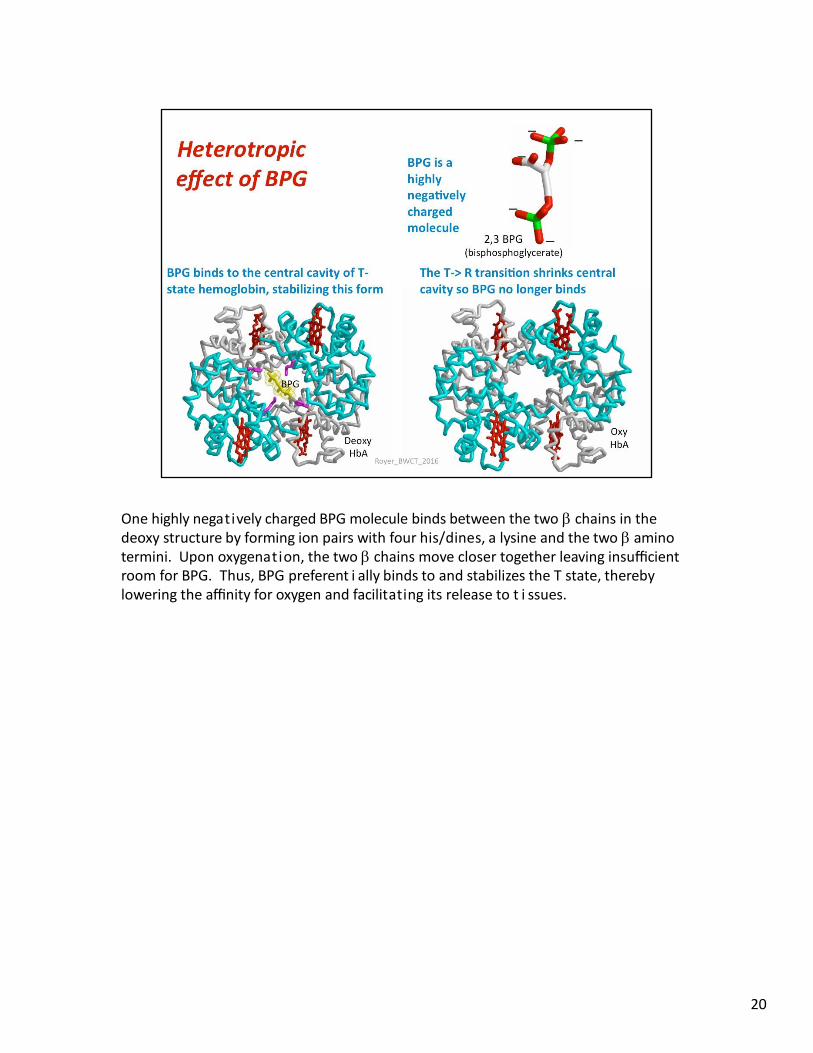
One highly negatively charged BPG molecule binds between the two chains in the deoxy structure by forming ion pairs with four his/dines, a lysine and the two amino termini. Upon oxygenation, the two chains move closer together leaving insufficient room for BPG. Thus, BPG preferent i ally binds to and stabilizes the T state, thereby lowering the affinity for oxygen and facilitating its release to t i ssues.
20
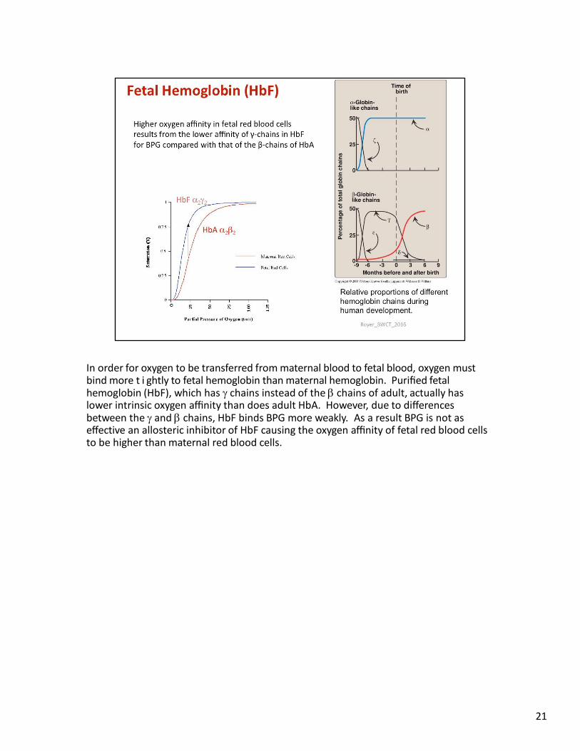
In order for oxygen to be transferred from maternal blood to fetal blood, oxygen must bind more t i ghtly to fetal hemoglobin than maternal hemoglobin. Purified fetal hemoglobin (HbF), which has chains instead of the chains of adult, actually has lower intrinsic oxygen affinity than does adult HbA. However, due to differences between the and chains, HbF binds BPG more weakly. As a result BPG is not as effective an allosteric inhibitor of HbF causing the oxygen affinity of fetal red blood cells to be higher than maternal red blood cells.
21
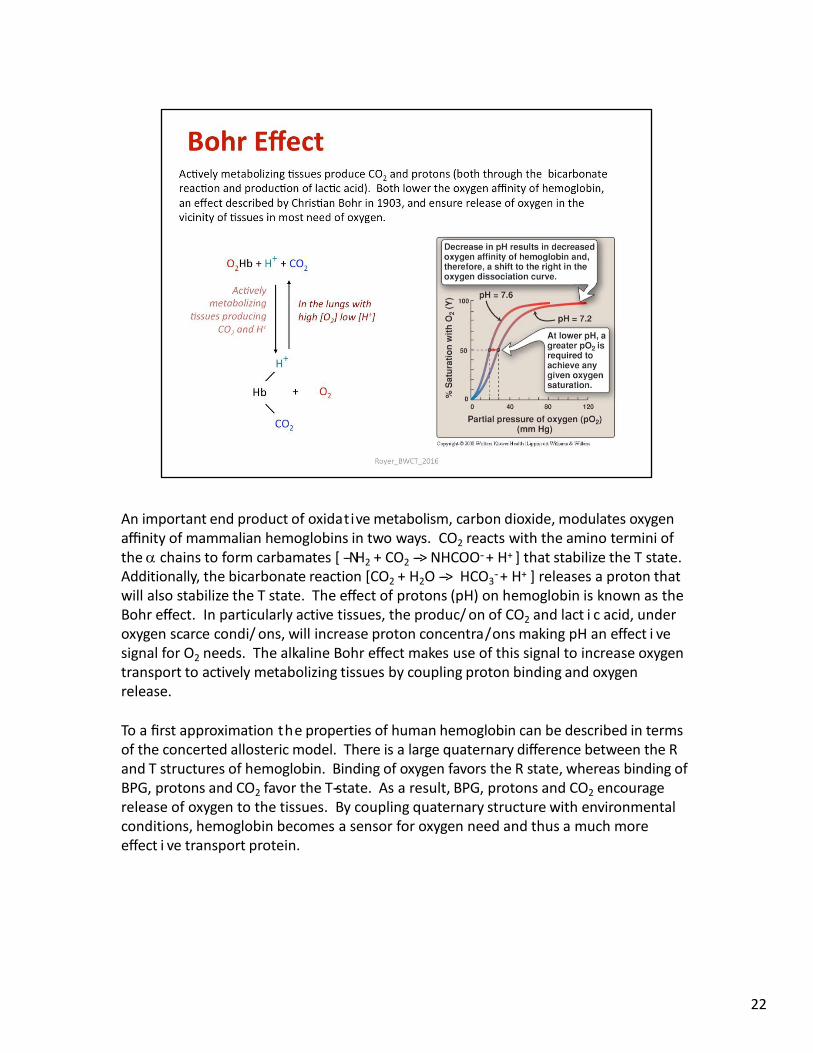
An important end product of oxidative metabolism, carbon dioxide, modulates oxygen affinity of mammalian hemoglobins in two ways. CO2 reacts with the amino termini of the chains to form carbamates [ -‐NH2 + CO2 -‐-‐> NHCOO-‐ + H+ ] that stabilize the T state. Additionally, the bicarbonate reaction [CO2 + H2O -‐-‐> HCO3
-‐ + H+ ] releases a proton that will also stabilize the T state. The effect of protons (pH) on hemoglobin is known as the Bohr effect. In particularly active tissues, the produc/ on of CO2 and lact i c acid, under oxygen scarce condi/ ons, will increase proton concentra/ons making pH an effect i ve signal for O2 needs. The alkaline Bohr effect makes use of this signal to increase oxygen transport to actively metabolizing tissues by coupling proton binding and oxygen release.
To a first approximation the properties of human hemoglobin can be described in terms of the concerted allosteric model. There is a large quaternary difference between the R and T structures of hemoglobin. Binding of oxygen favors the R state, whereas binding of BPG, protons and CO2 favor the T-‐state. As a result, BPG, protons and CO2 encourage release of oxygen to the tissues. By coupling quaternary structure with environmental conditions, hemoglobin becomes a sensor for oxygen need and thus a much more effect i ve transport protein.
22
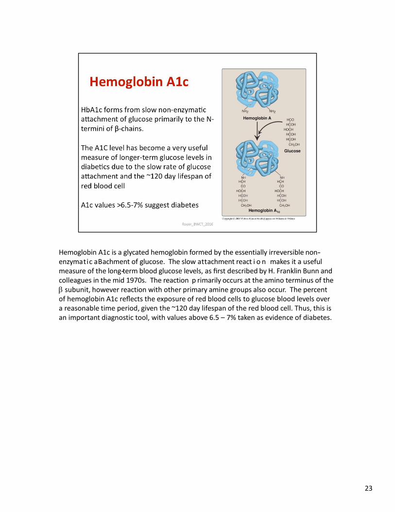
Hemoglobin A1c is a glycated hemoglobin formed by the essentially irreversible non-‐ enzymatic aBachment of glucose. The slow attachment react i o n makes it a useful measure of the long-‐term blood glucose levels, as first described by H. Franklin Bunn and colleagues in the mid 1970s. The reaction p rimarily occurs at the amino terminus of the subunit, however reaction with other primary amine groups also occur. The percent of hemoglobin A1c reflects the exposure of red blood cells to glucose blood levels over a reasonable time period, given the ~120 day lifespan of the red blood cell. Thus, this is an important diagnostic tool, with values above 6.5 – 7% taken as evidence of diabetes.
23
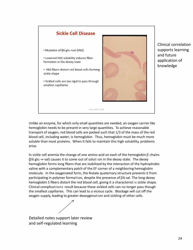
Unlike an enzyme, for which only small quantities are needed, an oxygen carrier like hemoglobin needs to be present in very large quantities. To achieve reasonable transport of oxygen, red blood cells are packed such that 1/3 of the mass of the red blood cell, including water, is hemoglobin. Thus, hemoglobin must be much more soluble than most proteins. When it fails to maintain this high solubility, problems arise.
In sickle cell anemia the change of one amino acid on each of the hemoglobin chains (6 glu -‐-‐> val) causes it to come out of solut i on in the deoxy state. The deoxy hemoglobin forms long fibers that are stabilized by the interaction of the hydrophobic valine with a complementary patch of the EF corner of a neighboring hemoglobin molecule. In the oxygenated form, the R-‐state quaternary structure prevents it from participating in polymer formation, despite the presence of 6 val. The long deoxy hemoglobin S fibers distort the red blood cell, giving it a characterist i c sickle shape. Clinical complications result because these sickled cells can no longer pass though the smallest capillaries. This can lead to a vicious cycle. Blockage will cut off the oxygen supply, leading to greater deoxygenation and sickling of other cells.
24
Clinical correlation supports learning and future application of knowledge
Detailed notes support later review and self-regulated learning
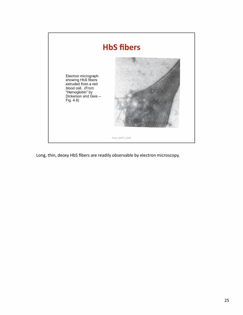
Long, thin, deoxy HbS fibers are readily observable by electron microscopy.
25
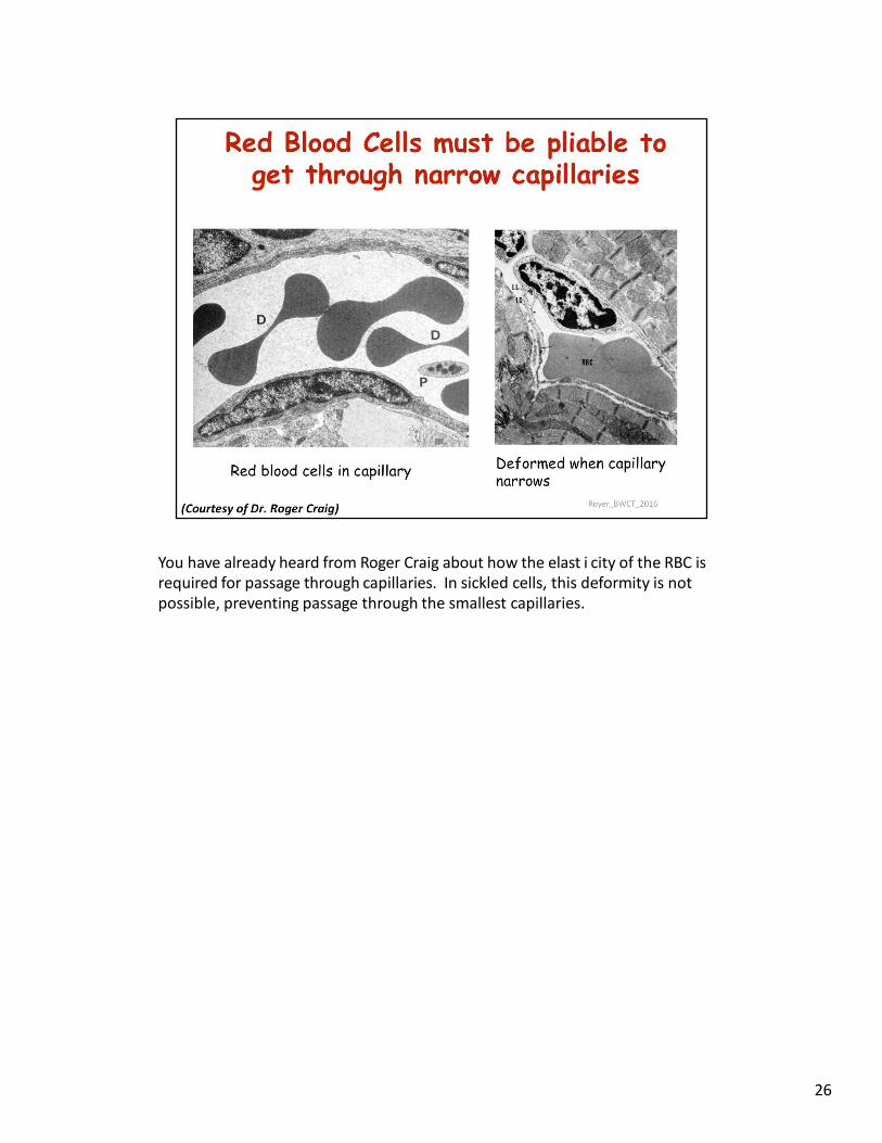
You have already heard from Roger Craig about how the elast i city of the RBC is required for passage through capillaries. In sickled cells, this deformity is not possible, preventing passage through the smallest capillaries.
26
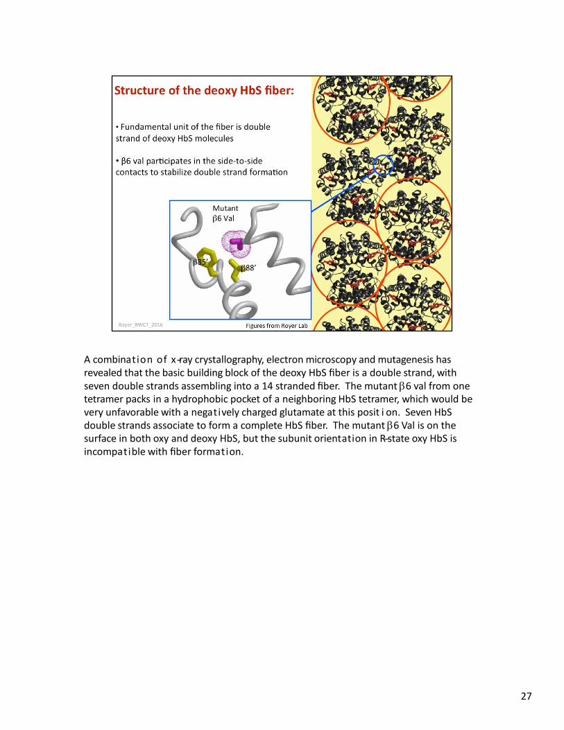
A combination of x-‐ray crystallography, electron microscopy and mutagenesis has revealed that the basic building block of the deoxy HbS fiber is a double strand, with seven double strands assembling into a 14 stranded fiber. The mutant 6 val from one tetramer packs in a hydrophobic pocket of a neighboring HbS tetramer, which would be very unfavorable with a negatively charged glutamate at this posit i on. Seven HbS double strands associate to form a complete HbS fiber. The mutant 6 Val is on the surface in both oxy and deoxy HbS, but the subunit orientation in R-‐state oxy HbS is incompatible with fiber formation.
27
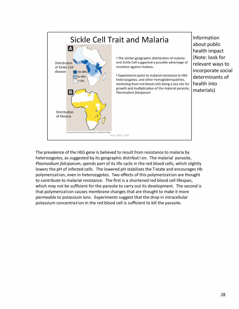
The prevalence of the HbS gene is believed to result from resistance to malaria by heterozygotes, as suggested by its geographic distribut i on. The malarial parasite, Plasmodium falciparum, spends part of its life cycle in the red blood cells, which slightly lowers the pH of infected cells. The lowered pH stabilizes the T-‐state and encourages Hb polymerization, even in heterozygotes. Two effects of this polymerization are thought to contribute to malarial resistance. The first is a shortened red blood cell lifespan, which may not be sufficient for the parasite to carry out its development. The second is that polymerization causes membrane changes that are thought to make it more permeable to potassium ions. Experiments suggest that the drop in intracellular potassium concentration in the red blood cell is sufficient to kill the parasite.
28
Information about public health impact (Note: look for relevant ways to incorporate social determinants of health into materials)
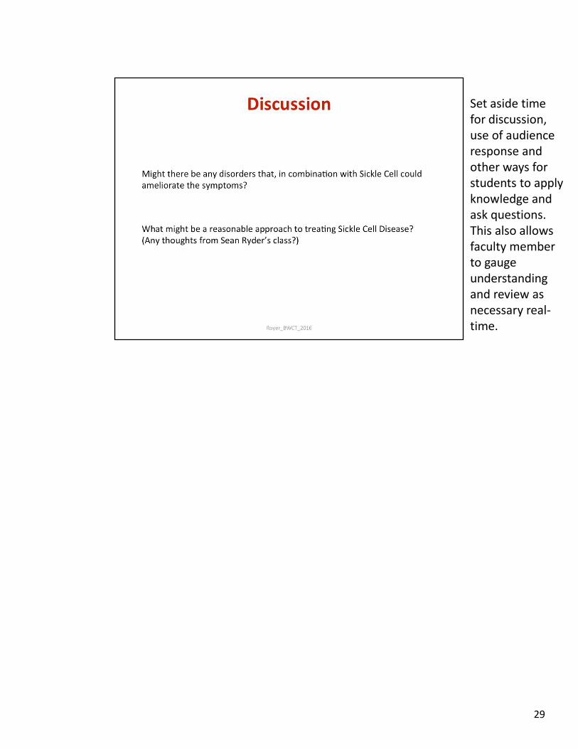
29
Set aside time for discussion, use of audience response and other ways for students to apply knowledge and ask questions. This also allows faculty member to gauge understanding and review as necessary real-time.
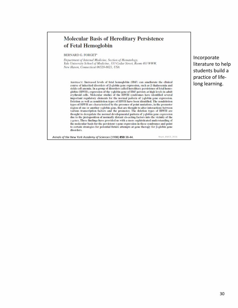
30
Incorporate literature to help students build a practice of life-long learning.
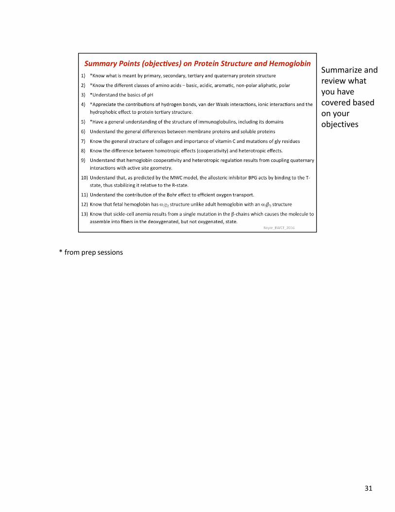
* from prep sessions
31
Summarize and review what you have covered based on your objectives


![PART ONE : HYDROSPHERE [Section 6] Topics Covered Slide Numbers 6 Hydrosphere [2-4]](https://img.pdfslide.net/doc/110x75/5681426b550346895dae9105/part-one-hydrosphere-section-6-topics-covered-slide-numbers-6-hydrosphere.jpg)