Embed Size (px)
Citation preview

Intronic Deletion Affecting a Negative RegulatoryRegion of TP73 is Related to Breast andColorectal Carcinomas
Cristina Pena,1 Jose M. Garcia,1 Gemma Dominguez,1 Javier Silva,1 Vanesa Garcia,1 Enric Carcereny,1
Javier Vargas,2 Mariano Provencio,1 Pilar Espana,1 and Felix Bonilla 1*1Department of Medical Oncology, Hospital Universitario Puerta de Hierro, Madrid, Spain2Department of Pathology, Hospital Universitario Santa Cristina, Madrid, Spain
The TP73 gene encodes a nuclear protein that has high homology with TP53. TP73 is rarely mutated in human cancer. Thepresence of a 1-kb regulatory fragment within the first intron of TP73 was recently reported. This fragment exerts silenceractivity on TP73 mediated by ZEB. We searched for possible mutations in this negative regulatory region in 45 colorectal and43 breast cancer patients and in 34 healthy donors. The study was carried out using the SSCP method, and the allelic variantsdetected were sequenced. The expression of TP73 was analyzed by quantitative RT-PCR, and loss of heterozygosity (LOH)was assessed by microsatellite study. In several samples, we identified an allele variant that corresponds to a deletion of 73bp in tumor tissues and normal counterparts, localized between �489 and �417 from the ATG start site of exon 2. Amongthe 88 tumor samples, 35 (40%) showed at least 1 allele with the cited deletion, versus 7 of the 34 (21%) healthy donors (P �0.045). When we classified the patients according to the number of variations into homozygous or heterozygous groups, thesignificance was clearer (P � 0.03). No LOH was detected in the heterozygous cases. There was a positive quantitativecorrelation between the expression of TP73 and the presence of the allelic variant (P � 0.029). These data suggest that thisallelic variant is common in breast and colorectal cancers and that it could alter the expression of the TP73 gene with anadditive effect. © 2004 Wiley-Liss, Inc.
Gene TP73, which codes for a protein with asignificant homology to TP53, has been identifiedin 1p36.3 (Kaghad et al., 1997). Both proteins aresimilar in a specific DNA-binding region in boththe tetramerization and the amino terminal trans-activation domains (Yang et al., 1998). Specificcharacteristics of TP73 are monoallelic or biallelicexpression and marked idiosyncrasy, with varia-tions from person to person and even from tissue totissue (Kaghad et al., 1997; Nomoto et al., 1998;Tsao et al., 1999)
In addition to its full-length form, TP73 has var-ious isoforms that have different functions. Theseisoforms are generated by alternative splicing ofthe carboxy-terminal region or by truncation of theNH2 terminal (Zaika et al., 1999; Ng et al., 2000;Ishimoto et al., 2002; Stiewe et al., 2002).
When TP73 is overexpressed, it shows some ofthe functional characteristics of TP53, such as theability to promote apoptosis (Kadhad et al., 1997) orthe capacity to activate the transcription of TP53-responsive genes involved in the cell cycle (Jost etal., 1997; Zhu et al., 1998). These data, togetherwith the localization of TP73 on 1p36, a regionfrequently deleted in a variety of human tumors(Dominguez et al., 2001), suggest that TP73 may
have tumor-suppressor activity like TP53. How-ever, TP73 does not appear to be induced by DNAdamage (Kadhad et al., 1997), and the three classicviral oncogenes that target and inactivate TP53 donot target the TP73 protein physically or function-ally (Higashino et al., 1998; Marin et al., 1998).Furthermore, the lack of inactivating mutations inall human tumors (Stiewe et al., 2002), the relation-ship between overexpression and tumorigenesis inmany studies (Stiewe et al., 2002), and the fact that,unlike Tp53 (�/�) mice, Tp73 knockout mice donot develop tumors (Yang et al., 2000) do not sup-port a role for the TP73 gene as a classic tumorsuppressor. Moreover, it has been reported that thedifferent truncated isoforms show oncogeneticfunctions (Ng et al., 2000; Fillippovich et al., 2001;
Supported by: Fundacion Banco Santander Central Hispano;Grant number: CAM 08.1/0069/2000.2; Aventis Pharma, S.A.
*Correspondence to: Felix Bonilla, Department of Medical On-cology, Hospital Universitario Puerta de Hierro, C/ San Martin dePorres 4, E-28035 Madrid, Spain. E-mail: [email protected]
Received 20 May 2003; Accepted 11 November 2003DOI 10.1002/gcc.10322
GENES, CHROMOSOMES & CANCER 39:257–262 (2004)
BRIEF COMMUNICATION
© 2004 Wiley-Liss, Inc.
Sayan et al., 2001; Ishimoto et al., 2002; Stiewe etal., 2002).
The most common alterations reported in TP73are loss of heterozygosity (LOH) (Dominguez etal., 2001) and increase of protein expression(Stiewe et al., 2002); promoter hypermethylationhas occasionally been described (Martinez-Del-gado et al., 2002). Several studies of various tumorsfailed to find mutations in the gene, suggesting thatmutated TP73 plays little, if any, role in the devel-opment of those tumors (Stiewe et al., 2002).
There is growing interest in understanding theregulation of TP73. For example, a 1-kb negativeregulatory fragment has recently been identified inthe first intron immediately upstream of exon 2(Fontemaggi et al., 2001). This fragment containssix E-boxes, which directly bind ZEB (Fontemaggiet al., 2001), a negative regulator and the vertebratehomologue of the Drosophila melanogaster Zfh-1protein.
To explore structural alterations in this domainin human primary tumors, we searched for possiblemutations in this negative regulatory region andcorrelated them with the expression of TP73, theLOH at 1p36, and the clinicopathological charac-teristics of the patients.
DNA was extracted from 88 tumor and normaltissues of breast (43) and colorectal (45) cancers andfrom 34 peripheral blood samples of healthy donorsusing a nonorganic method (S-4520 Kit, Oncor,Inc., Gaithersburg, MD).
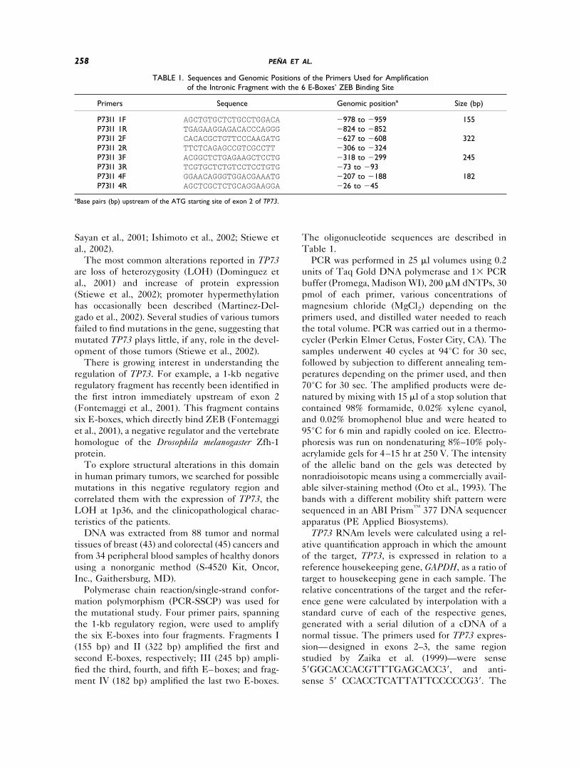
Polymerase chain reaction/single-strand confor-mation polymorphism (PCR-SSCP) was used forthe mutational study. Four primer pairs, spanningthe 1-kb regulatory region, were used to amplifythe six E-boxes into four fragments. Fragments I(155 bp) and II (322 bp) amplified the first andsecond E-boxes, respectively; III (245 bp) ampli-fied the third, fourth, and fifth E–boxes; and frag-ment IV (182 bp) amplified the last two E-boxes.
The oligonucleotide sequences are described inTable 1.
PCR was performed in 25 �l volumes using 0.2units of Taq Gold DNA polymerase and 1� PCRbuffer (Promega, Madison WI), 200 �M dNTPs, 30pmol of each primer, various concentrations ofmagnesium chloride (MgCl2) depending on theprimers used, and distilled water needed to reachthe total volume. PCR was carried out in a thermo-cycler (Perkin Elmer Cetus, Foster City, CA). Thesamples underwent 40 cycles at 94°C for 30 sec,followed by subjection to different annealing tem-peratures depending on the primer used, and then70°C for 30 sec. The amplified products were de-natured by mixing with 15 �l of a stop solution thatcontained 98% formamide, 0.02% xylene cyanol,and 0.02% bromophenol blue and were heated to95°C for 6 min and rapidly cooled on ice. Electro-phoresis was run on nondenaturing 8%–10% poly-acrylamide gels for 4–15 hr at 250 V. The intensityof the allelic band on the gels was detected bynonradioisotopic means using a commercially avail-able silver-staining method (Oto et al., 1993). Thebands with a different mobility shift pattern weresequenced in an ABI Prism™ 377 DNA sequencerapparatus (PE Applied Biosystems).
TP73 RNAm levels were calculated using a rel-ative quantification approach in which the amountof the target, TP73, is expressed in relation to areference housekeeping gene, GAPDH, as a ratio oftarget to housekeeping gene in each sample. Therelative concentrations of the target and the refer-ence gene were calculated by interpolation with astandard curve of each of the respective genes,generated with a serial dilution of a cDNA of anormal tissue. The primers used for TP73 expres-sion—designed in exons 2–3, the same regionstudied by Zaika et al. (1999)—were sense5�GGCACCACGTTTGAGCACC3�, and anti-sense 5� CCACCTCATTATTCCCCCG3�. The
TABLE 1. Sequences and Genomic Positions of the Primers Used for Amplificationof the Intronic Fragment with the 6 E-Boxes’ ZEB Binding Site
Primers Sequence Genomic positiona Size (bp)
P73I1 1F AGCTGTGCTCTGCCTGGACA �978 to �959 155P73I1 1R TGAGAAGGAGACACCCAGGG �824 to �852P73I1 2F CACACGCTGTTCCCAAGATG �627 to �608 322P73I1 2R TTCTCAGAGCCGTCGCCTT �306 to �324P73I1 3F ACGGCTCTGAGAAGCTCCTG �318 to �299 245P73I1 3R TCGTGCTCTGTCCTCCTGTG �73 to �93P73I1 4F GGAACAGGGTGGACGAAATG �207 to �188 182P73I1 4R AGCTCGCTCTGCAGGAAGGA �26 to �45
aBase pairs (bp) upstream of the ATG starting site of exon 2 of TP73.
258 PENA ET AL.

primers for GAPDH were sense 5�CATCTT-CTTTTGCGTCGCC3� and antisense 5�AAAAG-CAGCCCTGGTGAC3�. For the synthesis of first-strand cDNA, 400 ng of total RNA wasretrotranscribed using the Gold RNA PCR CoreKit (PE Biosystems, Foster City, CA) according tothe manufacturer’s protocol.
Real-time PCR was performed in a light-cyclerapparatus (Roche Diagnostics, Mannheim, Ger-many) using the LightCycler-FastStart DNA Mas-ter SYBR Green I Kit (Roche Diagnostic, Mann-heim, Germany). Each reaction was performed in afinal volume of 20 �l that contained 2 �l of thecDNA product, 3 mM MgCl2, and 0.5 �M of eachprimer, as well as 1 � reaction mix including Fast-Star DNA polymerase, reaction buffer, dNTPs,and SYBR green. Thermal cycling for both geneswas initiated with a denaturation step of 95°C for10 min and subjected to 30 cycles (denaturation at94°C for 0 sec, annealing at 56°C for 5 sec, andelongation at 72°C for 5 sec, in which fluorescencewas acquired).
Two microsatellite markers, D1S214 andD1S450, were used to determine LOH at 1p36 intumor tissue (Source: J. Weissenbach, Genethon,Whitehead Institute Center for Genome Research,Cambridge, MA).
The following parameters were obtained fromthe medical records of the 88 patients: age, tumorsize, lymph node metastases, presence of steroidreceptors (estrogen and progesterone), pathologicalstage, histological grade, proliferation index,ERBB2 expression, vascular invasion, and TP53immunostaining status. Pathological stage was as-sessed using the tumor-node-metastases (TNM)classification. The steroid receptor content was de-termined by an immunohistochemical procedure.The proliferative index was calculated by Ki-67antigen (Immunotech, Westbrook, ME) in immu-nohistochemical analyses. ERBB2 expression wasevaluated by a monoclonal mouse antibody (CB11;Novocastra Lab., Ltd., Newcastle, UK). The im-munohistochemistry of TP53 was analyzed withthe cl 1801 mouse monoclonal antibody (OncogeneScience, Manhasset, NY).
The chi-square test, along with the Mantel-Haenszel test, was used to compare the analyzedvariables . Statistical analysis was performed withEPI-INFO version 6.04.
No aberrant band at SSCP was found in ampli-fied fragments I, III, and IV, but in fragment II weobserved two electrophoretic mobility patterns inboth the tumor tissue samples and their normalcounterparts of several patients (Fig. 1). The se-
quence assay of this fragment revealed a 73-bpdeletion between �489 and �417 from the ATGstarting site of exon 2. In this amplicon, there aretwo contiguous repeat regions of 73 bp each, with89% homology. In the allelic variant, the end of thefirst repeated unit and most of the second are lost.Despite this, the sequence of the repetition unit ismaintained because the deletion is equal in size, 73bp (Figs. 2 and 3).
Among the 45 studied colorectal carcinomas, 18(40%) showed at least one allele with the deletionversus 7 of 34 (21%) among the healthy donors(P � 0.06). Similarly, 17 of 43 breast cancer pa-tients had one or two alleles with the deletion,compared with 7 of 34 (21%) found among healthydonors (P � 0.07). This distribution of allele fre-quency is close to statistical significance, probablyaffected by the small number of samples, becausewhen we compared the distribution of the alleles in
Figure 1. Photograph showing the three patterns found after SSCPof the fragment II amplification: homozygous for the wild-type allele,patient 1; homozygous for the variant allele, patient 2; heterozygous,patient 3 (N, normal tissue; T, tumor tissue).
259DELETED TP73 IN BREAST AND COLON CANCER

the global tumor series we found 35 of 88 (40%)tumors with at least one allele with the deletionversus the 21% among healthy donors (P � 0.045).
The statistical significance is more evident whenthe patients are classified according to the numberof variations into homozygous or heterozygousgroups (P � 0.044 for colon cancer and P � 0.038for breast cancer; P � 0.03 in the global series).This greater statistical significance is essentially aresult of the much greater frequency of homozy-gosity for the allelic variant in the patients than inhealthy donors: 10 of the 43 breast cancer patients(23%) and 10 of the 45 colorectal cancer patients(22%) versus only 1 of the 34 healthy donors (3%).
The frequency of the rest of the haplotypes wasmore homogeneous (Table 2).
We analyzed the expression of TP73 in 12 colo-rectal carcinomas in order to identify a possiblecorrelation with the 73-bp deletion. Of the 12cases, 5 did not harbor the allelic variant, 4 had onlyone allele with the deletion, and 3 were homozy-gous for the deletion. The samples were dividedinto 3 groups according to their expression pat-terns. In the first group were samples that showedno differences between normal tissue and the tu-mor counterpart; in the second were samples thathad between 4- and 10-fold overexpression of TP73in the tumor tissue; and in the third were those
Figure 2. Sequence of intron 1 harbored among primers P73I1 2 (in lowercase and bold). The twocontiguous repeated regions of 73 bp with 89% homology are shown in brackets (the distinctive basesappear in lowercase). The shaded region represents the fragment deleted in the sequence assay, formed byfragments of the two repeated regions. The amplified second E-box is framed.
Figure 3. Fragment of the sequence assay result for WT (wild-type allele) and DEL (fragment with thedeletion). The arrows and the underlined bases show the beginning and the end of the deletion.
260 PENA ET AL.

with more than 10-fold overexpression of TP73(P � 0.029; Table 3).
LOH was not found in any tumors heterozygousfor the deletion. This was expected because weobserved the wild-type and variant alleles in bothnormal and tumor tissue in the SSCP study (Fig. 1;patient 3).
There was no correlation between the presenceof the TP73 allelic variant and the clinicopatholog-ical characteristics of the patients. Surprisingly, atendency to significance was observed in relation totumor differentiation in colon carcinoma, in which80% of the individuals who were homozygous forthe deletion had well-differentiated carcinomas,compared to 33% of all individuals who were ho-mozygous for the wild type (P � 0.062), suggestingthat the small number of tumors with the TP73variant, when distributed among the differentstages, could be an important limitation of thisanalysis.
In this study, we analyzed the negative regula-tory fragment in the first intron of the TP73 gene,in which we found a deletion of 73 bp. The greaterpresence of this deletion in colorectal and breastcancer patients compared to healthy controls wasstatistically significant. On the other hand, we alsofound a significant correlation between the dele-tion and the expression of TP73.
In this negative regulatory fragment, there aresix specific E-boxes that bind to the zinc finger/homeodomain of ZEB, suggesting that TP73 is aspecific target for transcription repression by ZEB
(Fontemaggi et al., 2001). ZEB was originally iso-lated as a DNA-binding protein that exerts its re-pressor activity by directly binding to the consen-sus boxes present on target genes (Postigo et al.,1997, 1999). It has previously been reported thatMyo D and other basic helix-loop-helix proteinssuch as the MEF-2-family genes might displaceZEB from its target genes in order to inhibit itsrepressor function and activate their transcription(Postigo et al., 1997). This implies that competitionwith ZEB for the binding to E-boxes of the TP73gene intronic fragment differentially regulates theexpression of TP73 (Fontemaggi et al., 2001).
It has become increasingly clear that regulationof gene expression is driven by a fine balance in thedifferential activities of transcriptional regulators.Hence, it is important to identify the region inwhich these proteins exert their activity. Further-more, a mechanism has been reported in which asingle nucleoide polymorphism in noncoding DNAcan differentially affect the gene’s regulation(Knight et al., 2003). Therefore, a genetic changein the TP73 intron 1 regulator region could alter thecorrect expression of that gene; thus, the allelicvariation observed in colorectal and breast carcino-mas may affect gene expression. We identified acorrelation between colorectal patients both withand without the deletion and the expression of theTP73 gene. Despite the statistical significance ofthis finding, further studies of larger series areneeded to verify these results. Furthermore, stud-ies that include the differential expression of eachof the TP73 isoforms should be performed.
In the present series, the percentages of het-erozygotes and homozygotes for the wild-type al-lele were similar between healthy donors and tu-mor patients. However, among the healthycontrols, only 1 (3%) was homozygous for the de-letion, compared to 20 (23%) of the tumor patients.This deletion may have an additive effect in dis-ease development similar to that observed for tu-mor-suppressor genes, in which the inactivation oftwo allelic copies is needed for the development ofthe disease.
Both suppressor and oncogenic activities havevariously been assigned to the TP73 gene product,depending on the isoform expressed (Ng et al.,2000; Fillippovich et al., 2001; Sayan et al., 2001;Ishimoto et al., 2002; Stiewe et al., 2002). More-over, a marked idiosyncrasy in the expression(mono- or biallelic) of this gene has been describedin distinct samples (Kaghad et al., 1997; Nomoto etal., 1998; Tsao et al., 1999). The deletion reportedhere could determine the efficiency with which
TABLE 2. Distribution of Patients and Healthy ControlsAccording to Allelic Variants Found
Alleledistribution
Healthydonors %
Breastcancerpatients %
Colorectalcancerpatients %
Totaltumorspatients %
Wt/wt 27 79 26 60 27 60 53 60Wt/del 6 18 7 17 8 18 15 17Del/del 1 3 10 23 10 22 20 23p* 0.038 0.044 0.030
*The p value is calculated in each one of the group, breast, colorectal,or total tumor patients with respect to the healthy donors.
TABLE 3. Distributions of the 12 Colorectal CarcinomasAccording to Their Allelic Variants and Their Level of
Expression (P � 0.029)
Ratio N/T Wt/wt Wt/del Del/del Total
R � 0.25 4 1 0 50.25 � R � 0.1 1 2 0 30.1 � R 0 1 3 4Total 5 4 3 12
261DELETED TP73 IN BREAST AND COLON CANCER

each copy is expressed, which in turn could affectthe expression of each isoform. If so, this wouldexplain the germinal presence of the deletion andits apparent additive effect in the development ofthe disease.
ACKNOWLEDGMENTS
We thank C. Zalallos and M. M. Garcia for theirhelp with the collection of tissue samples andRobin Rycroft for his assistance with the Englishmanuscript.
REFERENCES
Dominguez G, Silva JM, Silva J, Garcia JM, Sanchez A, Navarro A,Gallego I, Provencio M, Espana P, Bonilla F. 2001. Wild type p73overexpression and high-grade malignancy in breast cancer.Breast Cancer Res Tr 66:183–190.
Fillippovich I, Sorokina N, Gatei M, Haupt Y, Hobson K, MoallemE, Spring K, Mould M, McGuckin MA, Lavin MF, Khanna KK.2001. Transactivation-deficient p73alpha (p73Deltaexon2) inhib-its apoptosis and competes with p53. Oncogene 20:514–522.
Fontemaggi G, Gurtner A, Strano S, Higashi Y, Sacchi A, Piaggio G,Blandino G. 2001. The transcriptional repressor ZEB regulatesp73 expression at the crossroad between proliferation and differ-entiation. Mol Cell Biol 21:8461–8470.
Higashino F, Pipas JM, Shenk T. 1998. Adenovirus E4orf6 oncop-rotein modulates the function of the p53-related protein, p73.Proc Natl Acad Sci USA 95:15683–15687.
Ishimoto O, Kawahara C, Enjo K, Obinata M, Nukiwa T, Ikawa S.2002. Possible oncogenic potential of Anp73: a newly identifiedisoform of human p73. Cancer Res 62:636–641.
Jost CA, Marin, MC, Kaelin WG. 1997. P73 is a human p53-relatedprotein that can induce apoptosis. Nature 389:191–194.
Kaghad M, Bonnet H, Yang A, Creancier L, Biscan JC, Valent A,Minty A, Chalon P, Lelias JM, Dumont X, Ferrara P, McKeon F,Caput D. 1997. Monoallelically expressed gene related to p53 at1p36, a region frequently deleted in neuroblastoma and otherhuman cancers. Cell 90:809–819.
Knight JC, Keating BJ, Rocett KA, Kwiatkowski DP. 2003. In vivocharacterization of regulatory polymorphisms by allele-specificquantification of RNA polymerase loading. Nat Genet 33:469–475.
Marin MC, Jost CA, Irwin MS, DeCaprio JA, Caput D, Kaellin WC
Jr. 1998. Viral Oncoproteins discriminate between p53 and p53homolog p73. Mol Cell Biol 18:6316–6324.
Martinez-Delgado B, Melendez B, Cuadros M, Jose Garcia M, Nom-dedeu J, Rivas C, Fernandez-Piqueras J, Benitez J. 2002. Fre-quent inactivation of the p73 gene by abnormal methylation orLOH in non-Hodgkin’s lymphomas. Int J Cancer 102:15–19.
Ng SW, Yiu GK, Liu Y, Huang LW, Palnati M, Jun SH, BerkowitzRS, Mok SC. 2000. Analysis of p73 in human borderline andinvasive ovarian tumor. Oncogene 19:1885–1890.
Nomoto S, Haruki N, Kondo M, Konishi H, Takahashi T, Taka-hashi T, Takahashi T. 1998. Search for mutations and examina-tion of allelic expression imbalance of the p73 gene at 1p36.3 inhuman lung cancers. Cancer Res 58:1380–1383.
Oto M, Miyake S, Yuasa Y. 1993. Optimization of nonradioisotopicsingle strand conformation polymorphism analysis with a conven-tional minislab gel electrophoresis apparatus. Ann Biochem 213:19–22.
Postigo AA, Dean DC. 1997. ZEB, a vertebrate homolog of Drosoph-ila Zfh-1, is a negative regulator of muscle differentiation. EMBOJ 16:3935–3943.
Postigo AA, Ward E, Skeath JB, Dean DC. 1999. Zfh-1, the Dro-sophila homolog of ZEB, is a transcriptional repressor that regu-lates somatic myogenesis. Mol Cell Biol 19:7255–7263.
Sayan AE, Sayan BS, Findikli N, Ozturk M. 2001. Acquired expres-sion of transcriptionally active p73 in hepatocellular carcinomacells. Oncogene 20:5111–5117.
Stiewe T, Putzer BM. 2002. Role of p73 in malignancy: tumorsuppressor or oncogene? Cell Death Differ 9:237–245.
Stiewe T, Zimmermann, Frilling A, Esche H, Putzer BM. 2002.Transactivation-deficient ATA-p73 acts as an oncogene. CancerRes 62:3598–3602.
Tsao H, Zhang X, Majewski P, Haluska FG. 1999. Mutational andexpression analysis of the p73 gene in melanoma cell lines. CancerRes 59:172–174.
Yang A, Kaghad M, Wang Y, Gillet E, Fleming MD, Dotsch V,Andrews NC, Caput D, McKeon F. 1998. P63, a p53 homolog at3q27–29, encodes multiple products with transactivating, death-inducing, and dominant-negative activities. Mol Cell 2:305–316.
Yang A, Walker N, Bronson R, Kaghad M, Oosterwegel M, BonninJ, Vagner C, Bonnet H, Dikkes P, Sharpe A, McKeon F, Caput D.2000. P73-deficient mice have neurological, pheromonal and in-flammatory defects but lack spontaneous tumours. Nature 2:99–103.
Zaika AI, Kovalev S, Marchenko ND, Moll UM. 1999. Overexpres-sion of the wild type p73 gene in breast cancer tissues and celllines. Cancer Res 59:3257–3263.
Zhu J, Jiang J, Zhou W, Chen X. 1998. The potential tumor sup-pressor p73 differentially regulates cellular p53 target genes. Can-cer Res 58:5061–5065.
262 PENA ET AL.