Embed Size (px)
Citation preview
Biomedical Signal and ImageComputing Laboratory
Invariant SPHARM Shape Descriptors for Complex Geometry in MR Region of Interest Analysis
Ashish Uthama1
Rafeef Abugharbieh1
Anthony Traboulsee2
Martin J. McKeown1,2
Presented by Bernard Ng1
1 Biomedical Signal and Image Computing Laboratory, Department of ECE, University of British Columbia2 Department of Medicine, University of British Columbia
Biomedical Signal and ImageComputing Laboratory
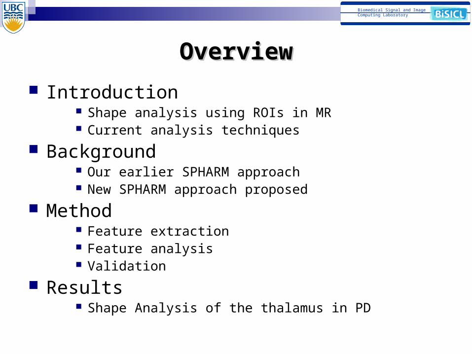
OverviewOverview
Introduction Shape analysis using ROIs in MR Current analysis techniques
Background Our earlier SPHARM approach New SPHARM approach proposed
Method Feature extraction Feature analysis Validation
Results Shape Analysis of the thalamus in PD
Biomedical Signal and ImageComputing Laboratory
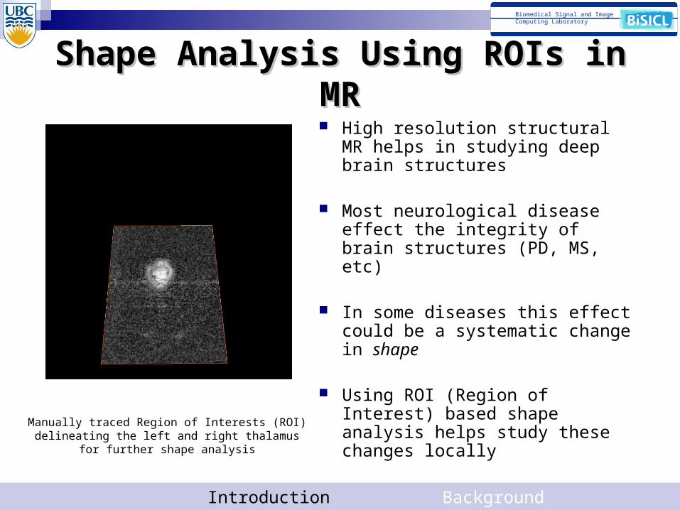
Shape Analysis Using ROIs in MRShape Analysis Using ROIs in MR High resolution structural MR helps
in studying deep brain structures
Most neurological disease effect the integrity of brain structures (PD, MS, etc)
In some diseases this effect could be a systematic change in shape
Using ROI (Region of Interest) based shape analysis helps study these changes locally
Introduction Background Method Results 3
Manually traced Region of Interests (ROI) delineating the left and right thalamus for further shape analysis
Biomedical Signal and ImageComputing Laboratory
Current Analysis TechniquesCurrent Analysis Techniques
Voxel count to represent volume Very simplistic measure Does not capture shape
Template based representation (medial, atlas, etc)
Most require manual selection of Land Marks Requires mutual registration
Automated feature extraction Limited to spherical topology
Introduction Background Method Results 4
Biomedical Signal and ImageComputing Laboratory
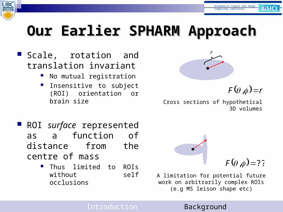
Our Earlier SPHARM ApproachOur Earlier SPHARM Approach
Scale, rotation and translation invariant
No mutual registration Insensitive to subject (ROI)
orientation or brain size
ROI surface represented as a function of distance from the centre of mass
Thus limited to ROIs without self occlusions
Introduction Background Method Results 5
A limitation for potential future work on arbitrarily complex ROIs (e.g MS leison shape etc)
rF ,
??, F
Cross sections of hypothetical 3D volumes
r
Biomedical Signal and ImageComputing Laboratory
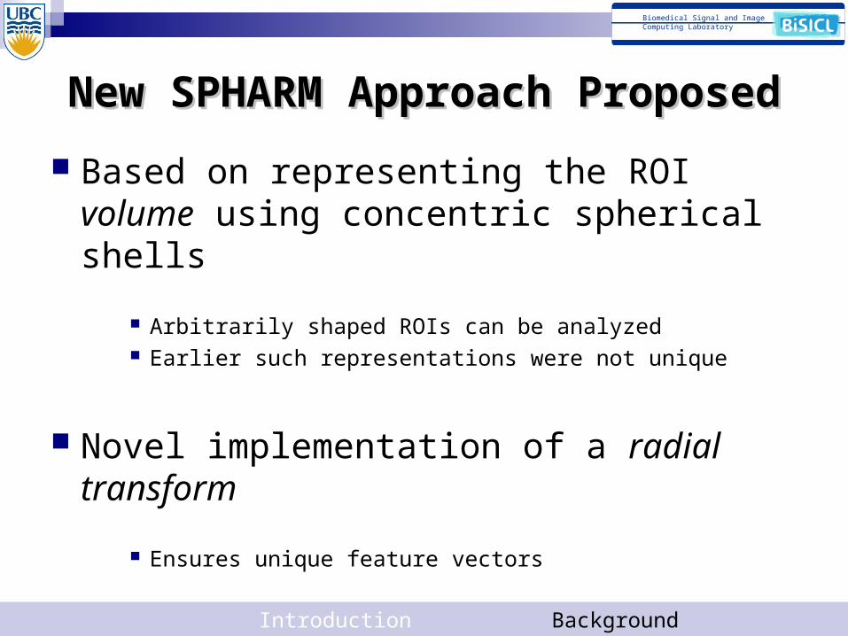
New SPHARM Approach ProposedNew SPHARM Approach Proposed
Based on representing the ROI volume using concentric spherical shells
Arbitrarily shaped ROIs can be analyzed Earlier such representations were not unique
Novel implementation of a radial transform
Ensures unique feature vectors
Introduction Background Method Results 6
Biomedical Signal and ImageComputing Laboratory
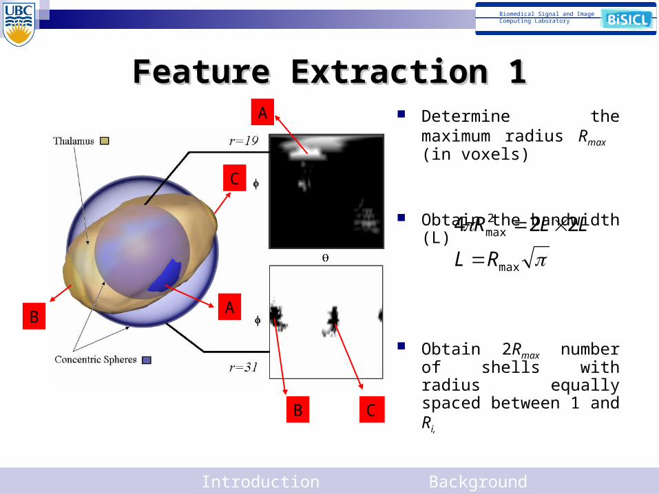
Feature Extraction 1Feature Extraction 1
BA
C
A
B C
Determine the maximum radius Rmax (in voxels)
Obtain the bandwidth (L)
Obtain 2Rmax number of shells with radius equally spaced between 1 and Ri,
Intersect these shells with the binary ROI mask, interpolating as required
Introduction Background Method Results 7
max
2max 224
RL
LLR
Biomedical Signal and ImageComputing Laboratory
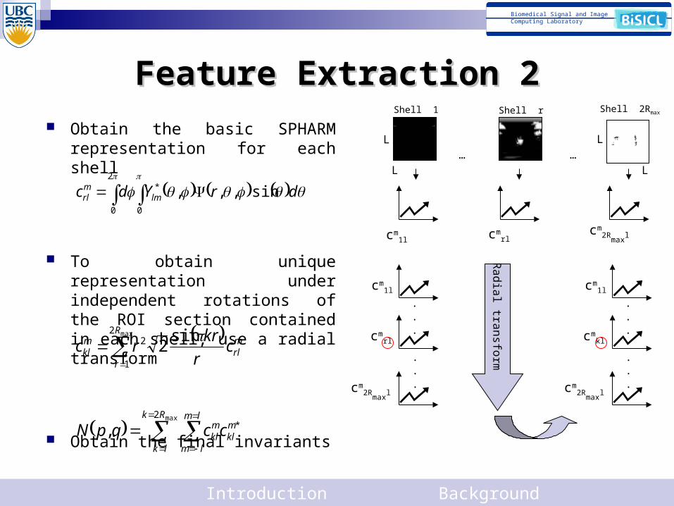
Feature Extraction 2Feature Extraction 2 Obtain the basic SPHARM
representation for each shell
To obtain unique representation under independent rotations of the ROI section contained in each shell, use a radial transform
Obtain the final invariants
Introduction Background Method Results 8
L
L
L
L
Shell 1 Shell r Shell 2Rmax
… …
cm1l cm
rlcm
2Rmax
l
cm1l
cmrl
cm2R
maxl
.
.
.
.
.
.
Ra
dia
l tran
sform
cm1l
cmkl
cm2R
maxl
.
.
.
.
.
.
drYdc lmmrl sin,,,
2
0 0
*
mrl
R
r
mkl c
r
krrc
sin2
max2
1
2
max2
*,Rk
lk
lm
lm
mkl
mklccqpN
Biomedical Signal and ImageComputing Laboratory
Feature Analysis
Obtain features for both groups (e.g. PD vs. Healthy controls)
Reshape each feature into a vector
Use a permutation test to determine if the two groups have a significant difference
Does not need a generating probability distribution Best suited for long feature vectors seen in biomedical
applications
Introduction Background Method Results 9
Biomedical Signal and ImageComputing Laboratory
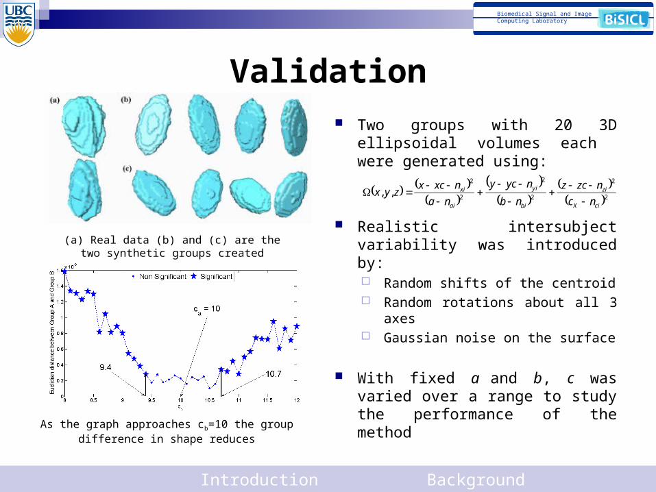
Validation Two groups with 20 3D ellipsoidal
volumes each were generated using:
Realistic intersubject variability was introduced by: Random shifts of the centroid Random rotations about all 3 axes Gaussian noise on the surface
With fixed a and b, c was varied over a range to study the performance of the method
Introduction Background Method Results 10
(a) Real data (b) and (c) are the two synthetic groups created
2
2
2
2
2
2
,,ciX
zi
bi
yi
ai
xi
nc
nzcz
nb
nycy
na
nxcxzyx
As the graph approaches cb=10 the group difference in shape reduces
Biomedical Signal and ImageComputing Laboratory
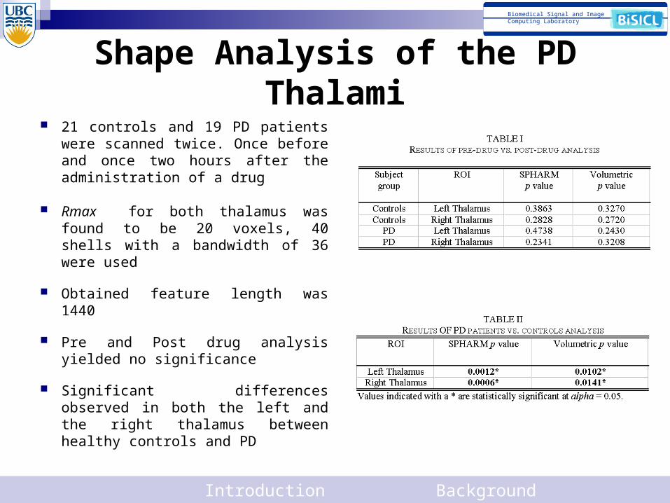
Shape Analysis of the PD Thalami 21 controls and 19 PD patients
were scanned twice. Once before and once two hours after the administration of a drug
Rmax for both thalamus was found to be 20 voxels, 40 shells with a bandwidth of 36 were used
Obtained feature length was 1440
Pre and Post drug analysis yielded no significance
Significant differences observed in both the left and the right thalamus between healthy controls and PD
Introduction Background Method Results 11
Biomedical Signal and ImageComputing Laboratory
Conclusion
SPHARM based invariant feature vectors for a 3D volume
Unique radial transform to obtain unique vectors
Validated with synthetic data
Application to real data Significant shape changes were observed in addition to
volumetric changes indicating that atrophy is not isotropic
Introduction Background Method Results 12