Embed Size (px)
Citation preview

Investigation of the Role of Probiotics on Toxicity of Aflatoxin B1
Muhammad Khalid Tipu
Thesis submitted in the partial fulfilment for the requirement
of the degree of Doctor of Philosophy
May, 2015.
University College of Pharmacy,
University of the Punjab, Lahore, Pakistan.

Investigation of the Role of Probiotics on Toxicity of Aflatoxin B1
Muhammad Khalid Tipu
Thesis submitted in the partial fulfilment for the requirement
of the degree of Doctor of Philosophy
May, 2015.
University College of Pharmacy,
University of the Punjab, Lahore, Pakistan.


ii
DEDICATED TO MY ALL STUDENTS

iii
Acknowledgments
The first and foremost thanks, prayers and gratitude are for Almighty Allah,
most benevolent and merciful who enables me to accomplish the huge task
with flying color. I pay homage and respects to Holy Prophet Hazart
Muhammad (Peace be upon him) who is source of eternal guidance for whole
mankind in this world and hereafter
I have been lucky enough to have the research supervision of an eminent
researcher and experienced academician, Prof. Dr. Bashir Ahmad(R),
University College of Pharmacy, University of Punjab, Lahore, Pakistan. His
support and guidance in achieving this goal are highly appreciable. He was
always there to be a source of inspiration and encouragement, which guide me
to complete my doctorate thesis.
I am very much obliged to Prof. Dr. Khushi Muhammad, Dean, Faculty of
Veterinary Sciences, University of Veterinary and Animal Science, for his
supervision and personal efforts in my research work and thesis write-up. I am
very thankful to Prof. (R) Dr. Syed Saeed-ul-Hassan, who encouraged me a lot
in completion of my research work.
I am personally grateful to Prof. Dr. Khalid Hussain, Department of
Pharmaceutical Chemistry, University College of Pharmacy, University of the
Punjab, Lahore, Pakistan, for his efforts and contributions in the preparation
of research proposal and the present research work. I also express my
thanks to Prof. Dr. Nadeem Irfan Bokhari, Department of Pharmaceutics,
University College of Pharmacy, University of the Punjab,Lahore,

iv
Pakistan, for his contribution in statistical analysis of the data. I am in very much
debt to Prof. Dr. Mobasher Ahmad, Dean and Principal, University College of
Pharmacy, University of Punjab, Lahore, Pakistan, for facilitating the serum
biochemical analysis.
I am highly grateful to Prof. Dr. Gul Majeed Khan, who allowed me to use the
premises and laboratory facilities of the Department of Pharmacy, Quaid-i-
Azam University, Islamabad, Pakistan, to conduct my research work. I feel
lucky enough to have the company of Dr. Ihsan-ul-Haq (Lecturer) and Dr.
Tofeeq-ur- Rehma (Assistant Professor) at the Department of Pharmacy,
Quaid-i-Azam University, Islamabad, Pakistan. Their encouragement and
fruitful discussion were a source of energy for me to achieve this goal.
I am also grateful to Dr. Zahid Hussain, Livestock Officer, Chiniot,
Pakistan, who facilitated me in immunological analysis of samples. I am
thankful to M r . Abdul Muqeet Khan, Analyst at Quality Operation Lab at
University of Veterinary and Animal Sciences, Lahore, Pakistan, for
assisting me in analysis of the aflatoxin B1 in isolated l iver tissues. I am
also grateful to Dr. Imtiaz Khan, Associate Professor of Pathobiology at Pir
Mahar University of Arid Agriculture, Rawalpindi for histopathological
examination of the tissues. I am highly obliged to Dr. Muhammad Imran,
Assistant Professor, Department of Microbiology, Quaid-i-Azam University, and
Islamabad-Pakistan for his contribution a n d guidance in microbial
characterization of lactobacilli in yogurt (Dhai).
I am personally thankful to Mr. Zahid Hussain (Lab Assistant) and Mr. Faisal
(Lab Attendant) at the Department of Pharmacy, Quaid-i-Azam University,

v
Islamabad, Pakistan, and Mr. Ejaz (Lab Assistant), Noor Hussain and Javed in
Pharmacology Section at University College of Pharmacy, University of Punjab,
Lahore, Pakistan, for their assistance in my research work.
I am in personal debt to Mr. Daud Butt, Mr. Fahad Khan and Mr. Syed
Naqeebullah students of mine at the Department of Pharmacy, Quaid-i-Azam
University, Islamabad, Pakistan for their selfless efforts in fermentation of rice
for AFB production. I thank Mr. Abdul Hadi, owner of Pakeeza Chicks,
Islamabad, Pakistan a n d his team for providing the housing facilities for
broiler birds during my research work.
Last but not least I am also thankful to Ms. Zainab, Mr. Ali Hassan and Mr.
Shaheer who because of t h e i r innocent queries/question, kept me charged
and determined to complete the task in time.
Muhammad Khalid Tipu

vi
TABLE OF CONTENTS
Page
CERTIFICATE OF APPROVAL i
DEDICATION ii
ACKNOWLEDGMENTS iii
LIST OF TABLES ix
LIST OF FIGURES xi
LIST OF PLATES xiii
LIST OF ABBREVIATIONS xiv
ABSTRACT xvi
CHAPTER 1 Introduction 1
CHAPTER 2 Review of Literature 5
2.1 Aflatoxins 5
2.1.1 Historical Review 5
2.1.2 Toxicokinetics 6
2.1.3 Toxicodynamics 7
2.1.4 Aflatoxicosis 8
2.1.4.1 Acute aflatoxicosis 8
2.1.4.2 Chronic aflatoxicosis 9
2.1.4.3 Occurrence AFB in Pakistan 16
2.2 Control of Aflatoxins 17
2.2..1 Agricultural Control 18
2.2.1.1 Pre-harvest control 18
2.2.1.2 Post-Harvest control 19
2.2..2. Dietary control 19
2.2..2.1 Chemopreventive agents 20

vii
2.2.2.2 Adsorbents 24
2.2.2.2.1 Mycosorb (MYC) 25
2.2.2.2.2 Probiotics (Lactobacilli) 29
2.3 Justification 37
2.4 Aims and Objectives 39
CHAPTER 3 Material and Methods 41
3.1 Plan of Study 41
3.2 Preparation of feed containing different treatment 41
3.2.1 Production of AFB 41
3.2.2 Preparation of PBT 44
3.2.3 Preparation of SLM containing feed 44
3.2.4 Preparation of MYC containing feed 45
3.3 Sampling 45
3.3.1 Collection of blood and serum 45
3.3.2 Collection of internal organ 45
3.4 Analysis of samples 45
3.4.1 Determination of NDV antibody response 45
3.4.1.1 Preparation of RBCs 46
3.4.1.2 Preparation of 4HA unit of virus 46
3.4.1.3 Determination of BDV antibody titre 47
3.4.2. Determination of AFB residue in liver 51
3.4.3 Clinical biochemistry 55
3.4.3.1 Total Serum protein 55
3.4.3.2 Serum Albumin 55
3.4.3.3 Serum bilirubin 56
3.4.3.4 Serum Glutamate pyruvate transferase 56

viii
3.4.3.5 Serum creatinine 57
3.4.3.6 Serum blood urea nitrogen 57
3.4.4 Histopathological examination 58
3.5 Statistical analysis 58
Chapter 4 Results 60
4.1 NDV antibody titre 60
4.2 Total leukocytes count 64
4.3 Relative weight of bursa of Fabricius 67
4.4 Relative weight of spleen 69
4.5 Relative weight of liver 72
4.6 Total body weight 73
4.7 Total serum protein 77
4.8 Serum albumin 80
4.9 Serum glutamate pyruvate transferase 82
4.10 Serum bilirubin 85
4.11 Serum blood urea nitrogen 88
4.12 Serum Creatinine 90
4.13 AFB-residue in liver 92
4.14 Histopathological examination 94
Chapter 5 Discussion and Conclusion 99
Chapter 6 References 111

ix
LIST OF TABLES
Table No. Title Page
No.
2.1 Dietary Control of Aflatoxins 21
3.1 General plan for Hemagglutination (HA) test 48
3.2 General plan for Hemagglutination inhibition (HI) test 49
3.3 Table of Brugh 50
4.1 Mean of LOG of antibody titer of birds taking different
therapies for two week 60
4.2 The Distribution of serum of birds given various
treatments according to Well Number in HI Test 61
4.3 Mean of total leukocytes counts (cells×1000/µl) in birds
taking different therapies for two week 66
4.4 Mean relative weight (g/Kg) of bursa of Fabricius of birds
taking different therapies for two week 67
4.5 Mean relative weight (g/Kg)of spleen of birds taking
different therapies for two week 69
4.6 Mean relative weight of liver (g/Kg) of birds taking
different therapies for two week 73
4.7 Mean total body weight (Kg) of birds taking different
therapies for two week 75
4.8 Mean total serum protein level (g/dl) of birds taking
different therapies for two week 77
4.9 Mean total serum albumin level (g/dl) of birds taking 80

x
different therapies for two week
4.10 Mean SGPT (U/L) of birds taking different therapies for
two week 83
4.11 Mean serum bilirubin level (mg/dl) of birds taking different
therapies for two week 85
4.12 Mean serum BUN level (mg/dl) of birds taking different
therapies for two week 88
4.13 Mean serum creatinine level (mg/dl) of birds taking
different therapies for two week 90
4.14 Mean of AFB residue in liver of birds taking different
therapies for two week 92

xi
LIST OF FIGURES
Figure
No. Title
Page
No.
2.1 Chemical Structure of Aflatoxins 5
2.2 Metabolism of Aflatoxins 7
2.3 Mechanism of AFB toxicity 8
2.4 Mechanism of action of adsorbents 24
2.5 Mechanism of Lactobacilli in aflatoxicosis 32
3.1 Standard of AFB 52
3.2 AFB in bird of group III at 4th week of age 53
3.3 AFB in bird of group VI at 4th week of age 53
3.4 AFB in bird of group VI at 6th week of age 54
3.5 AFB in bird of group VII at 6th week of age 54
3.6 AFB in bird of group IV at 4th week of age 55
4.1 Effect of different treatments on NDV antibody titre 62
4.2 Effect of different treatments on total leukocytes counts 65
4.3 Effect of different treatments on relative weight of bursa of
Fabricius
68
4.4 Effect of different treatments on relative weight of spleen 70
4.5 Effect of different treatment on relative weight of liver 74
4.6 Effect of different treatment on total body weight 76
4.7 Effect of different treatment on total serum protein 78
4.8 Effect of different treatment on serum albumin level 81
4.9 Effect of treatment on serum GPT level 84

xii
4.10 Effect of treatment on serum bilirubin level 87
4.11 Effect of treatment on serum BUN level 89
4.12 Effect of treatment on serum Creatinine level 91
4.13 Effect of treatment on liver AFB residue level 93

xiii
LIST OF PLATES
Page No.
a) Bursal follicle (10X) in PBT treated Birds 96
b) Bursal follicle (40X) in PBT treated Birds 96
c) Depleted Bursal follicle (10X) in AFB treated Birds 96
d) Depleted Bursal follicle (40X) in AFB treated Birds 96
e) Kidney (40X)of MYC for two weeks treated 97
f) Kidney (40X)of AFB for two weeks treated 97
g) Kidney (40X)of SLM for two weeks treated 97
h) Kidney (40X)of PBT for two weeks treated 97
i) Liver (10X) of AFB treated birds for two weeks 98
j) Liver (40X)of AFB treated birds for two weeks 98
k) Liver(10X) of PBT treated birds for two weeks 98
l) Liver (40X)of PBT treated birds for two weeks 98

xiv
LIST OF ABBREVIATIONS
AFB Aflatoxin B1
AFM Aflatoxin M1
ALT Alanine aminotransferase
AOAC Association of analytical chemists
AST Aspartate transaminase
BF Bursa of Fabricius
BUN Blood urea nitrogen
CYP Cytochrome
EG Esterified glucomannan
FAO Food and Agricultural organization
FDA Food and Drug Administration
GIT Gastrointestinal tract
GMT Geometric mean titre
HA Heamagglutination
HCS Hydro pericardium syndrome
HI Heamagglutination inhibition
HPLC High performance liquid chromatography
HSCAS Hydrated sodium calcium aluminosilicate
IARC International Agency for Cancer
research
IB Infectious bronchitis
IBD Infectious bursal disease

xv
MYC Mycosorb
NDV Newcastle disease virus
PBT Probiotics
ppb Part per billion
ROS Reactive oxygen species
SGOT Serum glutamic oxaloacetate transferase
SGPT Serum glutamic pyruvic transaminase
SLM Silymarin
SOD Super oxide dismutase
TLC Total leukocytes counts
TBW Total body weight
TSP Total serum Protein
UV/VIS Ultraviolet and visible spectroscopy
WHO World Health Organization

xvi
Investigation of the Role of Probiotics on Toxicity of Aflatoxin B1
Abstract
The aflatoxins are unavoidable contaminant of feed and food commodities.
Among these, aflatoxin B1 (AFB) has established hepatotoxic, immuno-toxic and
carcinogenic properties. AFB is grouped as class I carcinogenic by International
Agency for Cancer Research (IARC). Various methods to prevent dietary exposure
of AFB are: use of chemopreventive agents like Silymarin (SLM) and adsorbents
e.g. mycosorb (MYC). Among adsorbents, Probiotics (Live Lactobacilli) have
exhibited good AFB binding properties in vitro as well as in vivo. Traditional
Pakistani yogurt (Dhai) has been reported to be good source of such Lactobacilli.
The current study was designed to evaluate protective effect of these Lactobacilli
(PBT) species against AFB toxicity on liver, immunity and kidneys.
One day old broilers (n=240) were reared under standard environmental
conditions. On 3rd week of age, the broilers were segregated into different treatment
groups: I (Basal diet), II (300 g of yogurt [PBT 1X] ), III (400 ppb of AFB), IV ( 600 g
of yogurt [PBT 2X]+400 ppb of AFB), V (300 g of yogurt [PBT 1X]+400 ppb of AFB),
VI (SLM 600mg/Kg body weight+400 ppb of AFB), VII (MYC 1g/Kg of feed+400 ppb
of AFB). The above treatments were continued for two weeks i.e. 4th & 5th week of
age. All birds were vaccinated against regional prevailing diseases such as
Newcastle disease, Infectious bursal disease, etc.
Two ml blood was collected on 4th, 5th, 6th and 7th weeks of age for
measuring leukocytes count, serum antibody titre against Newcastle Disease Virus
(NDV) and clinical chemistry. Birds after weighing were euthanized and internal

xvii
organs such as liver, spleen, bursa of Fabricius and kidney were collected for
histopathological examination and determination of AFB residue in the liver. Data
thus obtained were analyzed by two way ANOVA with LSD test (α=0.05) for multiple
comparison. Ingestion of AFB resulted in significant decline in total body weight and
relative weight of spleen and bursa of Fabricius, serum NDV antibody titer, total
serum proteins and serum albumin. Moreover, significant rise in relative weight of
liver, serum GPT, bilirubin and AFB residue in the liver were recorded in AFB-treated
birds (p<0.05). Histopathological examination revealed vacuolar degeneration, bile
duct hyperplasia and loss of hepatic chord in the AFB-treated birds.
MYC intake significantly restored the negative effects of AFB in birds by its
adsorptive action during exposure (p<0.05). The SLM intake caused substantial
protection against harmful effects of AFB but the effect appeared on second week of
exposure (p<0.05). However, protective effects of both SLM and MYC were lost
when intake was stopped. Birds receiving PBT showed better NDV-antibody titer,
normalized organs weight, serum total proteins, albumin, GPT and bilirubin (p<0.05)
Histopathological findings also reflected shielding effects of PBT. The protective
action of PBT was observed during the exposure as well as post exposure. It is
concluded that Lactobacilli (PBT) in the Pakistani food significantly ameliorate the
negative effect of AFB on immune system and liver presumably via its adsorptive
properties which result in declined AFB bioavailability. Furthermore, investigations
are needed to elucidate mechanism of protective action of such PBT persisting even
after stopping of intake of PBT.

1
1. INTRODUCTION
Aflatoxin B1, B2, G1 and G2, are fungal secondary metabolites, which
are produced by some strains of Aspergillus flavus and Aspergillus
parasiticus (Wei and Jong, 1986; Karimi Torshizi et al., 2010). These are natural
contaminants of raw materials used for preparation of feed and food
commodities. Among these aflatoxin B1 (AFB) is classified as group I
carcinogen in humans (IARC, 2002). Additionally, it has ability to produce
hepatotoxic, carcinogenic and immunotoxic effects. These toxins enter into
the body through contaminated feeds/foods, and are either metabolized or
deposited in liver to cause deleterious effects. In lactating animals AFB and
aflatoxin B2 are excreted in milk such as aflatoxin M1 (AFM) and aflatoxin M2
(El-Nezami et al., 1995; Prandini et al., 2009). First record about the toxicity of
aflatoxins appeared in literature in 1960 when increased rate of death in turkey
poultry and ducklings was found to be due to contaminated peanut oil used in
the production of poultry feed, because the aflatoxins were isolated from the
feed (Hartley et al., 1963). The toxins were found to be involved in outbreak of
liver disorders and other symptoms of aflatoxicosis resulting in human deaths
in various part of the world such as India (Krishnamachari et al., 1975a; Tandon
et al., 1978) and Kenya (Ngindu et al., 1982; Azziz-Baumgartner et al., 2005).
The source was found to be the contaminated g ra in used for the
preparation o f food.
Aflatoxicosis is of two types: acute poisoning resulting in hemorrhagic
necrosis of liver, proliferation of bile duct, edema and lethargy and death
due to liver cirrhosis (Tandon et al., 1978; Azziz-Baumgartner et al., 2005)

2
and chronic sub-clinical exposure causing nutritional disorder (Gong et al.,
2002), immunosuppression (Meissonnier et al., 2008b), mutagenicity (Wild and
Turner, 2002) and carcinogenic consequences (Williams et al., 2004). These
toxins can cause toxicity in respiratory tract by exposure through inhalation
(Kelly et al., 1997). The immunosuppressant effects are the major health and
economic problems for both animals as well as humans which results in failure
of immune-prophylaxis (Dimitri and Gabal, 1996; Gabal and Dimitri, 1998).
Because of their profound toxicity, amount of these toxins in food is
essential to be regulated. The permissible limit by FDA is 20 ppb of AFB for
human food with exception of milk which is 0.5 ppb for AFM (JECFA, 2001). The
maximum level set by the European Union is 2 µg/Kg of AFB in food stuff
intended for direct human consumption whereas 0.10 µg/Kg of the AFB in food
for infants and special medical purposes. According to OJL, the level of AFM
is 0.05 µg/Kg and 0.025 µg/Kg for milk products for infant and special medical
purposes, respectively (Regulation, 2006).
Several strategies have been established to remove these toxins from
food commodities such as proper maturing of crops, minimizing post-harvest
damage and improving storage conditions. Sequestering a gen t s (adsorbents)
are also used in poultry feed and animal ration to reduce the aflatoxins
exposure. However, s ome poor quality adsorbents also adsorb essential
nutrients like vitamins resulting in stunted growth of animals, and
also show irreproducible results in experiments (Chung et al., 1990). On
account of these limitations these adsorbents are not used in human food.
Therefore, many researchers have started using live cultured microbes
(Probiotics) l i ke bacteria, yeast and molds to reduce the level of the toxins in
foods and feeds.

3
The term probiotics (PBT) was first used by Lilly and Stillwell in
1965 (Lilly and Stillwell, 1965). The concept of PBT arouse from study of Nobel
laureate Eli Metchnikoff, who linked the longer longevity of Bulgarian peasants
with consumption of fermented milk products (Gupta and Garg, 2009).
According to the Food and Agriculture Organization (FAO) and the World
Health Organization (WHO) PBT are live micro-organisms which, when
administered in adequate amounts confer health benefits on the host
(Quigley, 2010). The most commonly used bacterial genera as PBT are
Lactobacillus, Bifidobacterium, Enterococcus, and Streptococcus. Some fungal
strains belonging to Saccharomyces have also been used as PBT (Jin et al.,
2000; Alvarez-Olmos and Oberhelman, 2001). PBT are beneficial in
different pathological conditions such as antibiotic associated diarrhea
(D'Souza, 2002), Helicobacter pylori infection (Lesbros-Pantoflickova et al.,
2007) and cancer (Aso et al., 1995). Apart from these effects, Lactobacillus
and Bifidobacteria are reported to reduces level of free mycotoxin, cyanotoxin
and heavy metals in aqueous solution (Salminen et al., 2010a). The same
has been reported by various scientists that different strains of
Lactobacilli and Bifidobacteria on incubation with aflatoxins containing culture
media reduce the level of the free aflatoxins, either by binding or metabolizing
the toxin (Peltonen et al., 2001b; Fazeli et al., 2009b; Hernandez-Mendoza et al.,
2009a).
Due to humid and warm climate in Pakistan, grains/foods are more
susceptible to aflatoxins contamination (Mobeen, 2011). In Pakistan, all four
aflatoxins have identified at level higher than permissible limit in both various
unprocessed and processed food (Mushtaq et al., 2012). The wheat, the staple
food of Pakistan, is harvested and stored during the summer. The stores houses

4
in the country lack proper system to control temperature and humidity which
encourage the fungal growth. The Dhai is traditional dairy food product of
Pakistan and subcontinent which is prepared by fermentation like kefir and
yogurt in Europe. The traditional fermented milk product is prepared by mixing
raw and heat treated milk with starter culture (Jhaag: local term) containing
Lactobacillales as dominating microbial group while it also have other undefined
complex microbial consortia for fermentation. Dhai serves as very good and
economical source of probiotic Lactobacillales (Aslam and Qazi, 2010).
In Ayurveda, Dahi is advised in various liver and kidney aliments;
moreover it is also suggested in various type of intoxication as folklore remedy.
The probiotics from Dahi (Yogurt) for example Lactobacilli, have been used in
alcoholic liver disease, non-alcoholic liver disease, minimal hepatic
encephalopathy and overt hepatic encephalopathy (Hitchins and McDonough,
1989; Lewis and Freedman, 1998; Bajaj et al., 2008; Nabavi et al., 2015).
We hypothesized that low priced freely available fermented milk product
containing probiotics will be beneficial in combating aflatoxicosis related
economical and health issues of a developing country like Pakistan. The current
study was designed to evaluate whether the traditional product offers a
comparable benefit to established strategies (Silymarin and mycosorb) for
prevention /treatment of aflatoxicosis.

5
2. Review of Literature
2.1. Aflatoxins
Mycotoxins are the secondary metabolites produced by certain fungal
species. These highly toxic compounds are produced as natural protectant. But
these become a serious health and economical problem when fungus
contaminates the crops/grains used for human and animal food. Aflatoxins are
produced by mainly Aspergillus flavus and Asergillus parasiticus (Wei and Jong,
1986). The word ‘Afla’ is combination of ‘A’ from Aspergillus and ‘fla’ from flavus
(Schoental, 1967). There are about 18 different mycotoxins identified, but AFB,
aflatoxin B2, G1 and G2 are more common. AFM and aflatoxin M2 are metabolites
of AFB and aflatoxin B2 (Wu et al., 2009). These compounds are coumarone
derivative showed intense blue and green fluorescence in UV light. Hence named
B (blue) and G (green) are used. The chemical structures of these are shown in
Fig 2.1.
Fig. 2.1: Chemical Structures of Aflatoxins (Williams et al., 2004)
2.1.1. Historical Review

6
The first record about aflatoxins appeared in 1960 when unexpected high
death rate observed in turkey poultry, was traced to consumption of highly
contaminated feed (Nesbitt et al., 1962). These compounds were discovered by
Hartley in 1963. Hartley reported the isolation and chemical characteristics of the
toxic metabolites of Aspergillus flavus. The four toxic compounds were isolated
from sterilized groundnuts which were inoculated by the fungus. The mixture of
AFB, aflatoxins-B2, -G1 and -G2 were separated and recrystallized using various
solvents by column chromatography. Then AFB was re-crystallized using
trichloroethylene and chloroform (Hartley et al., 1963). In 1966, Shotwell and his
co-workers reported a method of production of the aflatoxins by culturing the
fungus (NRRL 2999) on fermented rice. AFB was separated from almost all
impurities and from the other aflatoxins by chromatography on silica gel with 1%
ethyl alcohol in chloroform which was further purified by recrystallization
(Shotwell et al., 1966).
2.1.2. Toxicokinetics
Because of lipophilicity and low molecular weight, AFB is rapidly absorbed
in gastrointestinal tract. It reversibly bound mainly to albumin in plasma. After
distribution, AFB is mainly accumulated in liver, muscle, and Kidney (Smith and
Henderson, 1991). In another study higher concentration (3 µg/Kg) of aflatoxins
were found in kidney, gizzard and liver of broilers given mixture of AFB and
aflatoxin B2 for four weeks (Wolzak et al., 1986). In a study, Mico and his
colleagues found detectable amount of AFB, AFM, aflatoxin B2 and aflatoxicol in
liver, kidney and thigh muscle of broiler fed diet containing 50 ppb of aflatoxins
(Micco et al., 1988). AFB is oxidized by cytochrome P450 enzyme especially
CYP2A6 and CYP1A1 to various metabolites: AFB-8,9-exo-epoxide, AFB-8,9
endo-epoxide, AFM, aflatoxin P1 and aflatoxin Q1. Reduction of AFB produces

7
aflatoxicol which can be detected in liver (Fernández et al., 1994; Diaz et al.,
2010; Yunus et al., 2011a).The AFB-8,9-epoxide is detoxified by glutathione-S-
transferase to AFB-mercapturic acid which is excreted in urine. Metabolites of
aflatoxin P1, aflatoxin Q1 and aflatoxicol are inactive and excreted by renal and
biliary route after conjugation with glucuronic acid or sulfate (Yunus et al., 2011a;
Gross-Steinmeyer and Eaton, 2012). The metabolism of AFB is illustrated in Fig
.2.2.
Fig.2.2 Metabolism of AFB, GST: Glutathione-S-transferase, UDPGT: Uridine-diphosphate glucuronosyltransferase, ST: Sulphotransferase, CYP450(s): Cytochromes P450.
2.1.3. Toxicodynamics
The toxic effects of AFB are exerted by its epoxide metabolite. The highly
reactive metabolite covalently binds with macromolecules. It forms AFB-Lysine
adduct with serum albumin which is one of biomarker for aflatoxin exposure. The
epoxide metabolite is converted to dihydrodiol by epo-hydrolase. Binding of both

8
epoxide and dihydrodiol with cellular proteins result in cell death and toxicity
(Gross-Steinmeyer and Eaton, 2012). Secondly the epoxide metabolite form
adduct with N-7 in guanine in DNA. The adduct strand either undergoes
depurination or the AFB-N(7)-guanine adduct is converted to open ring
formamidopyrimidine. Both forms cause mutation in p-53 gene or activate the ras
oncogene (Eaton and Gallagher, 1994; Smela et al., 2002; Bedard and Massey,
2006). The mechanism of toxicity is shown in Fig. 2.3
Fig.2.3 Mechanism of AFB toxicity
2.1.4. Aflatoxicosis
The exposure to the aflatoxins causes deleterious manifestation in
humans, animals and birds. These effects are collectively termed as aflatoxicosis.
The aflatoxicosis is of two types:
2.1.4.1. Acute Aflatoxicosis

9
Acute aflatoxicosis occurs due to excessive intake of aflatoxins particularly
AFB in contaminated diet. Symptoms include vomiting, abdominal pain, hepatitis
and death. The lethal dose of AFB is about 10-20 mg for adult (Etzel, 2002).
Different outbreaks of aflatoxicosis have been reported by various scientists in
different part of world. Krishnamachari and his colleagues described an outbreak
of hepatitis in 1975. The outbreak affected the human and even street dogs. The
symptoms observed were jaundice, ascites and portal hypertension with high
death rate. The autopsy revealed the proliferation of bile duct and giant cells. The
cause was traced to consumption of 2-6 mg of the aflatoxins for a month due to
use of highly contaminated maize as staple food (Krishnamachari et al., 1975b).
Another outbreak of liver disease with jaundice and ascites was investigated by
Tandon and his coworkers in some rural area of India. The corn sample had
shown high level of AFB (Tandon et al., 1978). In 1982 Similarly Ngindu with his
colleagues observed hepatitis in 20 people with 60 % mortality. The liver of
deceased contained about 89 ppb of AFB (Ngindu et al., 1982).
In 1991 an outbreak of aflatoxicosis was described in Malaysia. There was
death of 13 children associated with consumption of local food contaminated with
the aflatoxins. The cause of death was acute hepatic and renal failure (Cheng,
1992). An outbreak of aflatoxicosis was recorded in 2004 which claimed 317
sufferings with 125 deaths. The symptoms followed somewhat same pattern as
described by Etzel, 2000. The causative agent was AFB, confirmed by analysis of
corn consumed, AFB-lysine adduct and positive Hepatitis B surface antigen in
blood of affected ones (Azziz-Baumgartner et al., 2005; Probst et al., 2007).
2.1.4.2. Chronic Aflatoxicosis
The chronic aflatoxicosis results from low but continuous ingestion of AFB
from environment. The low level contamination does not cause sudden necrosis

10
or other sign of acute toxicity. Rather it results in different effects with prolonged
consequences. These are:
I. Immunosuppressive Effects
The laboratory and farm animals (chronic exposure) are main source of
immunological effects. The variability of the aflatoxins level in food commodities
and biomarkers of the toxins are main hindrance for generalization of the results.
However these studies are linked to human in broader term (Williams et al.,
2004). The deleterious effects of AFB on various cells of immune system are as
follow:
a. Effects on Dendritic Cells
Dendritic cells play a central role in immune response to pathogens. They
are produced by distinct hematopoietic lineage (Merad et al., 2013). The classical
type is present in most of the tissue and triggered immune response by
presenting the antigen to T-lymphocytes, phagocytic property and production of
cytokines (Geissmann et al., 2010). AFB ingestion resulted in impairment of their
phagocytic activity and T-cell stimulating ability in porcine (Mehrzad et al., 2014)
b. Effect on Lymphocytes
. AFB had detrimental effects on lymphocytes. AFB produced oxidative
stress indicated by reduced glutathione peroxidase, glutathione reductase and
catalase activity in broiler chickens (Chen et al., 2013a). AFB caused decrease in
cells proliferation in Jurkat T-cell line and increased expression of IL-8 (Luongo et
al., 2013). Similarly in human lymphocytes, AFB induced cytotoxic effects. It
caused decrease in cellular oxygen consumption and mediated apoptosis as
indicated by intracellular activity of caspase and annexin-V positive cells. These
effects were produced within two hours at 100 µM concentration and 24 hours at
50 µM concentration (Al-Hammadi et al., 2014).

11
c. Effects on Macrophages:
Macrophages have imperative role in both innate and acquired immunity.
They not only have role in phagocytosis but also act as antigen presenting cells
(Rossi et al., 1986). In swine, AFB exposure at concentration of 1.5 µg/ml for 24
hours reduced viability of alveolar macrophages to 41% and expression of
apoptosis-related heat shock protein 72 (HSP 72). The reduction in phagocytic
activity (36% of control) was observed in same at level of 100 ng/ml for 24 hours
(Liu et al., 2002). The secretions of various cytokines was also affected by AFB.
It reduced anti-inflammatory cytokine IL-10 while enhanced the pro-inflammatory
cytokine IL-6. The expression of toll like receptor 2 (TLR2) and differentiation
CD14 was reduced significantly by AFB in murine macrophages (Bruneau et al.,
2012).
d. Cell Mediated Immunity:
Dimitri and Gabal had described the immunosuppressant activity of the
aflatoxins in rabbits where AFB showed reduction in lymphocyte stimulation and
abolished the tuberculin test in the treated rabbits. The lymphocyte stimulation
indices and diameter of skin reactions were significantly reduced in the AFB-
treated groups. In addition, rabbits (25%) from the AFB-treated groups failed to
produce any detectable response to the tuberculin test. It was concluded that
AFB inhibited the lymphocyte proliferation and negatively influenced the
tuberculin skin test (Gabal and Dimitri, 1998).
Similarly Meissonnier and colleagues studied the impact of dietary AFB on
cell mediated immunity in pigs. The results of experiment indicated an increase in
the level of pro inflammatory and regulatory cytokines. These findings suggested
that AFB causes reduction in cell mediated immunity coupled with inflammatory
response (Meissonnier et al., 2008b).

12
e. Humoral Immunity:
AFB exposure decreased the antibody titer (enzyme-linked
immunosorbent assay) and antibody level (protein electrophoresis) in a study
conducted by Gabal and Dimitri (1998). It was found that antibody titer and the
immunoglobulin levels were significantly decreased in the aflatoxin-treated
groups. Chen and co-workers reported decrease in serum immunoglobulin,
relative weight of bursa of Fabricius. Moreover, increase in percentage of
apoptotic bursal cells was observed (Chen et al., 2014a)
II. Hepatotoxic Effects
a. Liver Functions:
Chronic exposure to the aflatoxins especially AFB produced a direct injury
to hepatocytes causing macroscopic, microscopic lesions and impairing their
functions and consequently the serum profile. Exposure to 100 ppb of AFB in
broilers resulted in elevated level of gamma glutamyl transferase (GGT),
glutamate-oxaloacetate transferase (GOT) and glutamate-pyruvate transferase
(GPT). There was significant accumulation of AFB residue in liver than muscle
(Bintvihok and Kositcharoenkul, 2006). Similarly, Chen and his colleagues
observed the negative effect of AFB in duckling. They reported that AFB (110 to
210 ppb) given for 14 days, reduced serum glucose, protein and albumin level in
duckling. While the serum aminotransferases, alkaline phosphatase (ALP) and
serum urea nitrogen were elevated by increasing the AFB level (Chen et al.,
2014e).
In a study conducted in broilers by Denli and colleagues, AFB (1mg/Kg of
feed) significantly increased the liver weight and serum alkaline phosphatase.
Perilobular inflammation along with vacuolar degeneration was also observed in
histopathological examination of liver (Denli et al., 2009). Similarly, Weaver and

13
co-workers reported the hyperplasia of liver bile ductule and karyomegaly in pig
upon exposure to combination of 150 ppb of AFB and 1100 ppb of
deoxynivalenol (Weaver et al., 2013). Yang and colleagues studied effect of
contaminated corn in broilers. They reported that an increment in contamination
level adversely affected the liver histology with rise in apoptosis. Reduction in
total hepatic proteins and glutathione peroxidase were also observed at more
than 50% of contamination level. Whereas, rise in activity of glutathione-S-
transferase (GST), glutathione reductase and superoxide dismutase (SOD) along
with high hepatic malanodialdehyde level were also observed (Yang et al.,
2012).
b. Carcinogenesis:
Liu and Wu reported that long term exposure to AFB was linked to
increased risk of hepatic cell carcinoma (HCC), (Liu and Wu, 2010). William and
co-workers described the higher HCC prevalence (16 to 32 times) in developing
countries especially in sub-Saharan Africa and Asia-Pacific region was related to
higher exposure of AFB (Williams et al., 2004). Wild and Turner described the
liver as major site for activation of AFB (Wild and Turner, 2002). The metabolic
activation of AFB into AFB-8, 9-exo-epooxide metabolite mainly by CYP3A4 was
studied by Kamdem and colleagues. They also studied relative contribution of
CYPs in activation and detoxification of AFB. They suggested that inhibition of
CYP3A4 might be alternative chemoprevention (Kamdem et al., 2006). Mulder
and his colleagues studied role of AFB metabolite-DNA adduct in mutation and
consequently carcinogenesis by using the p53 gene knockout mice (Mulder et al.,
2014). Kirk and his team described that mutation by the AFB metabolite-adduct
at codon 249 (AGG to AGT: Arginine to Serine) in tumour suppressor p53 gene
and infection with hepatitis B virus had synergistic effect in HCC (Kirk et al.,

14
2005). William and his colleagues also reported an increment in odd ratio with
both AFB exposure and hepatitis B virus (HBV) infection than AFB alone
(Williams et al., 2004). Kew studied the impact of HBV X gene and protein in
induction of HCC. He studied the anti-apoptotic role of HBV X protein and its
inhibitory effect on the p-53 gene. The protein also interfered with other
nucleotide excision repair mechanism either p-53 gene dependent or
independent (Kew, 2011).
III. Nutritional Toxicity
Consumption of the aflatoxins resulted in reduced weight gain and
negative effect on nutritional status. Marin with his colleagues reported dose
dependent reduction in weight gain in weaning piglet given 140 and 280 ppb of
AFB (Marin et al., 2002). Similarly significant weigh reduction, feed consumption
and feed conversion ratio (FCR) were reported by Verma and co-workers in
broiler chickens (Verma et al., 2004). In a cross sectional study, Gong and her
colleagues observed stunted growth and low weight in children less than 5 year
in Benin and Togo. They determined the AFB exposure by measuring the AFB-
albumin adducts which ranged from 5 to 1064 Pico g/mg of albumin in 99% of
children (Gong et al., 2002).
Obuseh and co-workers studied the relation between AFB-albumin adduct,
vitamin A and E levels in HBV infected patient having either Human
Immunodeficiency virus (HIV) or none. They measured the level of AFB-albumin
adduct, vitamin A and E in HIV positive and negative patients. Low serum vitamin
A and E were correlated with the higher AFB-albumin adduct level (Obuseh et al.,
2011). Tang with his colleagues reported the negative co-relation between
vitamin A and E level with AFB exposure. They determined the AFB-albumin
adduct, vitamin A and vitamin E level in 507 Ghanaian peoples. High exposures

15
of AFB caused decrease in serum vitamin A and E level which influenced the
aflatoxin deleterious effects (Tang et al., 2009). Hendrickse reported that AFB
exposure modulated the recovery from Kwashiorkor by affecting the protein
synthesis (Hendrickse, 1997) .
Costanzo and co-workers studied the impact of AFB on vitamin D receptor
in osteosarcoma cell line SAOA-2. They reported that exposure to 5 to 50 ng of
AFB down regulated vitamin D receptor (58% and 86% respectively) in the cell
line. They suggested that early exposure with AFB might increase the risk of
rickets in African children (Costanzo et al., 2014). Hussein and his colleagues
studied the relation between AFB-adduct and oxidative stress in wheat milling
workers. They measured AFB-adduct, GOT, GPT, GGT, ALP, GST, SOD,
malaonodialdehyde, zinc and vitamin C. Significant correlation of zinc with GST
and SOD activities showed the role of Zn in oxidative stress induced by AFB.
Similarly positive correlation of vitamin C with GST while negative with the
malaonodialdehyde, indicated the role of the antioxidant vitamins in AFB toxicity
(Saad-Hussein et al., 2014).
IV. Inhalation Toxicity
Apart from all these deleterious effects, inhalation of the AFB in worker of
poultry and concerned industries, is becoming alarming. Kelly and co-workers
studied the impact of inhalation exposure to AFB on human lung because
inhalation of AFB in certain occupations was considerable. The data indicated
that human lung microsomes activated AFB to form the exo-AFB-8, 9-epooxide
and that cytochrome P450 (3A subfamily) might be responsible for this activity.
The relatively low amount of AFB activation in human lung compared to that
in human liver could be explained by the scarcity of cytochrome P450 containing
cells in the lung. In situ AFB activation and resultant carcinogenic risk were

16
distinctly possible in occupational settings where inhalation of AFB contaminated
dusts occurred (Kelly et al., 1997). Shen and his team studied toxicity of aflatoxin
G1 in rats. He administered the toxin through intra-tracheal route. After
determination of surfactant protein, they conferred that alveolar type II cells were
the target for acute toxicity (Shen et al., 2012). Study conducted on poultry
workers in Portugal necessitated the assessment and prevention of inhaled AFB
exposure to worker in such work places (Viegas et al., 2012).
2.1.4.3. Occurrence of AFB/Aflatoxicosis in Pakistan
Due to humid and warm climate in Pakistan, grains/foods are more
susceptible to aflatoxins contamination (Mobeen, 2011). In Pakistan, all four
aflatoxins have been identified at level higher than permissible limit in both
various unprocessed and processed food (Mushtaq et al., 2012). A study was
conducted to evaluate contamination of aflatoxins in animal feed and raw feed
from 2006 to 2009. It was revealed that 61% of samples collected were
contaminated with AFB and other aflatoxins. Moreover, the contamination level
was more than the permissible limit i.e. 20 ppb (Khan et al., 2011).
Another study revealed that more than 80% of wheat samples were
contained AFB more than the permissible limit. Impact of weather was significant
on AFB level in wheat grains (Rashid et al., 2012). Anjum and colleagues
analyzed the 100 sample of starter feed for broiler and layer for aflatoxin and
ochratoxin. It was found that 40 % of samples contained higher level of both
mycotoxins (Anjum et al., 2011). A study was conducted to determine occurrence
of aflatoxicosis in broilers. Sick and dead birds (n=1105) were examined from
June 2009 to May 2010. It indicated that 8.78 % of birds were aflatoxicosis
positive. Necropsy findings showed pale liver, atrophied bursa and thymus.

17
Occurrence was more in autumn season as compared to winter (Rashid et al.,
2013).
Aslam and his colleagues studied the level of AFM in milk supplied
through various supply chains. It was found that AFM concentration was higher
than permissible limit. AFM concentration was 2.60, 2.59 and 1.93 ppb in
autumn, rainy (monsoon) and summer season respectively (Aslam et al., 2016).
An incident of aflatoxicosis was reported bovine farm. It affected 45 animals with
30% mortality. Postmortem investigation revealed visceral hemorrhage, prolapse
and damaged hepatic portal system (Sohooa et al., 2015).
Despite of high level of aflatoxin and aflatoxicosis in broiler and cattle,
Aflatoxicosis either acute or chronic in Pakistani population has not been
reported. However, Qureshi and colleagues suggested aflatoxins contamination
(10-17%) was positively correlated with higher incidence of hepatocellular
carcinoma (Qureshi et al., 1990). Qazi and Fayyaz described a link between the
long term ingestion of aflatoxin in food with liver cancer in Karachi (Qazi and
Fayyaz, 2006).
2.2. Control of Aflatoxins
Because of the profound toxicity, the AFB level in foods is
required to be regulated. Its level in food and food ingredients may not
exceed the permissible limit, which is 20 ppb for human food with exception of
milk which is 0.5 ppb for AFM (JECFA, 2001). The maximum level of AFB as per
European Union is 2 µg/Kg in food stuff intended for direct human consumption
and is 0.10 µg/Kg in food for infants and special medical purposes. According
to OJEUL (2006) the level of AFM is 0.05 µg/Kg and 0.025 µg/Kg for milk
products for infant and special medical purposes, respectively (Regulation,

18
2006). Several strategies have been established to remove such toxins from
food commodities at different levels. These include:
2.2.1. Agricultural Control
At this level emphasis is laid down on agricultural practices to reduce
contamination/infection by toxigenic fungus. Such methods include: proper
maturing of crops, minimizing post-harvest damage and improving storage
conditions (Hell et al., 2000).
2.2.1.1 Pre-harvest Control
In this method, genetically resistant varieties against Aspergillus infection,
e.g. Mp420 and Mp13E were developed by Brown and his team (Brown et al.,
1999). After identifying the source of resistance to such pathogens, the
incorporated African maize varieties were used in Nigeria (Menkir et al., 2006).
Similar transgenic approach was implied in cotton utilizing the antifungal gene
from maize (Cary et al., 2011). Another approach in transgenic crops is to use
insect resistant variety. As insect infestation also promote the fungal growth by
increasing moisture contents and damage to Kernel. Bt maize variety and later on
new Bt varieties were developed to provide protection against harmful insects.
These interventions significantly reduced the aflatoxins level with resistance
against insects like European corn borer and southwestern corn borer (Wu,
2008).
Utilization of atoxigenic strains is one of the methods to minimize the
damage caused by the aflatoxins. These strains reduce level of the wild strains in
ecosystem by competing with wild toxigenic strains. Probst and co-workers
isolated the 96 atoxigenic strain from four province of Kenya. These strains were
tested by co-inoculation with toxigenic strain to assess the ability to reduce the
aflatoxins level. Among these 12 strains showed comparable result with

19
commercial NRL 21882 USA isolate (Probst et al., 2010). Weaver and
colleagues conducted the field trial in Mississippi for atoxigenic strains. They
suggested the use of atoxigenic strains at early stage markedly improve the
quality and safety (Weaver et al., 2015). Wu and Khlangwist showed cost
effectiveness of biocontrol method over the post-harvest strategies (Wu and
Khlangwiset, 2010). After completion of genome sequencing of Aspergillus
flavus, gene coding for enzymes involved in synthesis and regulation of aflatoxins
would be valuable for devising new strategies in biocontrol (Bhatnagar et al.,
2006).
2.2.1.2. Post-Harvest Control
In this approach, harvested crops are protected by minimizing the risk of
fungal contamination. It is achieved with application of rapid drying to reduce the
moisture content, sorting, smoking and good protection against pests during
storage (Hell et al., 2000; Hell et al., 2010). Certain chemicals like ammonia,
acids, oxidizing and reducing agents, herbicides, pesticides and surfactants were
used to decrease the fungal growth and toxin production by different researchers
as described by Kabak and colleagues (Kabak et al., 2006).
Antagonistic bacteria, yeast and fungi were also used to prevent growth of
Aspergillus, hence the toxin formation. Various species of lactic acid producing
bacteria, Leuconostoc mesenteroids inhibited the growth and the toxins formation
in Aspergillus flavus, were reported by different scientists. Shantha, investigated
the effect of different Rhizopus, Sporotrichum and Phoma species and their cell
free extract on the aflatoxins biosynthesis and degradation. The cell free extracts
exhibited better results than culture filtrate (Shantha, 1999).
2.2.2. Dietary Control

20
Due to economical constrains and lack of awareness, appropriate
measures at agricultural level are not adequate to keep the aflatoxins level within
the permissible limits. Secondly extreme weather conditions especially in tropical
and subtropical region increase the likelihood of fungal growth and thus the
aflatoxins contamination (Phillips et al., 2002b; Williams et al., 2004; Huebner et
al., 2008; Phillips et al., 2008a). Moreover the diverse cooking habits/ways in
these regions cannot assure the required reduction of aflatoxins content
(Bullerman and Bianchini, 2007; Mohamadi Sani et al., 2012). In order to reduce
the food-borne exposure of the aflatoxins, both chemical and physical
(Adsorption) approaches are utilized by researches for detoxification as shown in
Table 2.1. Reducing absorption of the aflatoxins and preventing toxicity in
animals and humans is becoming a vital strategy for effective prevention of
toxicity of the aflatoxins in animal and human as well.
2.2.2.1 Chemopreventive agents
These substances either natural or synthetic modulate or reduce
carcinogenic effect of the aflatoxins. Oltipraz an antischistosomal drug was
reported to reduce the carcinogenicity of AFB. In a clinical trial it showed
detoxification of AFB by increasing conjugation of its reactive metabolite AFB-
8,9-epoxide by glutathione S-transferase. It also inhibited the formation of the
epoxide.
Chlorophyllins and green tea polyphenols have shown reasonable
anticancer properties in different studies. In a study , chlorophyllin alone and in
combination with fermented milk, exhibited hepato-protective effect against AFB
in male Wister rats (Kumar et al., 2012b). Similarly copper complex of
chlorophyllin, a food colorant was reported by various researchers to show

21
reasonable health benefits in term of anti-mutagenic, anti-carcinogenic and anti-
oxidant action. (Tumolo and Lanfer-Marquez, 2012).
Among these various chemopreventive substances, milk thistle (silymarin)
by virtue of its hepato-protectant and antioxidant actions, has shown beneficial
role in detoxification of aflatoxins (Rawal et al., 2010).
Table 2.1 Dietary Control of Aflatoxins
CH
EMO
PREV
ENTI
ON
Origin Agents Mode of action
Natural Milk Thistle: Silymarin Anti-oxidants (Tedesco et al., 2004b)
Chlorophylls Antioxidants (Kumar et al., 2012a)
Synthetic Oltipraz Decreased formation of epoxide Increased Conjugation of epoxide (Sporn and Suh, 2002)
AD
SOR
BTI
ON
Non-Microbial Origin
Bentonite Adsorption (Rao and Chopra, 2001) Zeolite Adsorption (Miazzo et al., 2000) HCAHS Adsorption (Wang et al., 2008) Novasil Adsorption (Phillips et al., 2008b)
Microbial Origin
Esterified Glucomannan (Mycosorb)
Adsorption (Yildirim et al., 2011)
Live Culture: Saccharomyces cerviase
Adsorption (Pizzolitto et al., 2013)
Lactobacilli species Adsorption & metabolism (Fazeli et al., 2009b; Hernandez-Mendoza et al., 2009b)
a. Silymarin (SLM)
Silymarin (SLM) a flavonolignan obtained from milk thistle (Silybum
marianum) from family Compositae is indigenous to Mediterranean region. It is
found all over the Europe, North America, India, China, Africa and Australia
(Luper, 1998).
i. Chemistry

22
SLM is polyphenolic flavonolignan extracted from the milk thistle by 95%
ethanol. SLM is complex consisting of: silybin (most active), silychristin, silydianin
and taxifolin (Saller et al., 2001).
ii. Pharmacokinetics
Being insoluble in water, it is administered in encapsulated form. Oral
absorption is about 50%. Time to reach peak plasma level is 6-8 hours in human
and animals. After conjugation and sulfation in liver, the drug is excreted in bile
(Morazzoni et al., 1993; Kshirsagar et al., 2009).
iii. Mode of action
Various studies indicated the multiple mechanisms responsible for wide
variety of pharmacological action of SLM. These include antioxidant, free radical
scavenging with subsequent inhibition of lipid peroxidation, membrane
permeability, inhibition of cirrhosis as described by Pandey (Kshirsagar et al.,
2009; Pandey, 2014). These actions help in neutralization of oxidative stress
produced by AFB exposure and hence the toxicity.
iv. Role in hepato-toxicity
SLM is an established hepato-protectant used clinically. Various studies
showing this action has been reported by various researchers. The protective
role of SLM on hepatocytes when exposed to carbon tetrachloride was
reported by Muriel and Mourelle. They studied the protective effects of SLM
against the toxicity induced by carbon tetrachloride on the liver plasma
membrane through its antioxidant properties (Muriel and Mourelle, 1990).
Rastogi and co-workers studied the hepato-curative effects of SLM and picroliv
in AFB induced hepatotoxicity in rats. They concluded that hepato-curative
effect of picroliv and silymarin, was comparable (Rastogi et al., 2000). Tedesco

23
and colleagues studied the effects of SLM on the excretion of AFM in cows
upon the consumption of AFB contaminated diets. The animals were given
SLM extract (10 g/day) in first phase and (30 g/day) in second phase. The
treated animals exhibited significant reduction in AFM level (p<0.01) on day 3
in phase I and on day 11 in phase II (p<0.05) (Tedesco et al., 2003). Later on
they reported the beneficial effects of SLM on body weight gain and feed intake
in broiler chicken, intoxicated with AFB (Tedesco et al., 2004b).
In study conducted by Dumari and colleagues, milk thistle (SLM) at dose
1% in feed, exhibited protective effect against elevation of GOT and GPT in
broilers caused by ingestion of 500 ppb of AFB (Dumari et al., 2014). In another
study Muhammad and his team investigated the role of SLM in aflatoxicosis and
compared with commercial toxin binders. AFB (80 ppb) was fed during first week
and 520 ppb onward to broilers. Intake of silymarin alleviated the negative effect
of AFB on weight gain and feed conversion ratio. The levels of GOT and GPT
were significantly lower than intoxicated birds (Muhammad et al., 2012).
v. Role in immuno-toxicity
By virtue of its antioxidant and free radicals scavenging action, silymarin is
of value in counteracting the immune-toxicity of AFB and other mycotoxins.
Silymarin was reported to enhance the antibody titer against NDV and IBD in a
study conducted by Chand and co-workers (Chand et al., 2011). Similarly, in a
study conducted by Khatoon and her colleagues SLM neutralized the immune-
toxicity in layer hens induced by ochratoxin A at level of 1 mg/Kg alone and in
combination with vitamin E (Khatoon et al., 2013).
In the light of these protective effects on liver and immune systems,
SLM can be used as positive control in immune- and hepato-protective studies,
therefore, in current study silymarin would be used to compare the protective

24
effect of probiotics on liver and immune system against toxicity of AFB.
2.2.2.2. Adsorbents
Latest approach for reducing the exposure to the aflatoxins in animals and
humans is the usage of adsorbents in feed and food commodities. These
substances adsorb the aflatoxins either in ready to use feeds and foods or in
gastrointestinal tract (GIT) of ingesting organism. It results in a decrease in
bioavailability of the toxins as shown in Fig. 2.4.
Rao and Chopra studied the effect of sodium bentonite and charcoal for
reduction of AFM in goats fed with combination of 100µg of AFB alone and in
combination with sodium bentonite and charcoal. They reported that AFB
contaminated feed (100 ppb) alone showed no signs of AFB toxicity except
excretion of AFM in milk. However the treated goats had shown significantly
lower AFM level (Rao and Chopra, 2001).
Fig.2.4 Mechanism of action of Adsorbents
In another study, Zeolite (microporous aluminosilicate mineral) neutralized
some deleterious effects of AFB in broiler given combination of 2.5 mg/Kg of
Aflatoxin
Adsorption
Adsorben
Adsorbed Aflatoxin B1
(Reduction in Bioavailability)

25
AFB contaminated feed and 1% Zeolite from 21 to 42 days of age (Miazzo et al.,
2000). As described by Philips and co-workers adsorbent clays: Hydrated
sodium calcium aluminosilicates (HSCAS) were reported to decrease the level of
AFB and other mycotoxins (Phillips et al., 2002a; Phillips et al., 2008b). Neeff
and co-workers investigated the role of HSCAS in decreasing the toxicity of AFB
in broilers. They fed AFB (2.5 mg/Kg) of feed supplemented with 0.5 % of
HSCAS for 21 days. HSCAS prevented accumulation of AFB in liver of the
broilers. But it did not show any ample protective effects on liver toxicity (Neeff et
al., 2013).
Similarly Chen and colleague reported protective effect of HSCAS in
broiler fed with 0.5 to 2 mg of AFB/Kg of feed. Ingestion of HSCAS prevented the
decrease in cumulative weight gain in the birds. Liver weight of HSCAS treated
birds remained significantly lower along with better impact on clinical
biochemistry. HSCAS also enhanced expression of catalase and SOD (Chen et
al., 2014c).
However, sometime poor quality adsorbents also adsorb essential
nutrients like vitamins resulting in stunted growth of animals, and
also show irreproducible results in experiments (Chung et al., 1990). To
overcome it, uniform particle size novasil (microcrystalline cellulose) was used by
various researchers to avoid the nutritional consequences while protecting from
aflatoxicosis in animals as well as humans (Marroquín-Cardona et al., 2011;
Mitchell et al., 2014). On account of these limitations, adsorbents of microbial
origin became the focus of research for detoxification of aflatoxins (Jard et al.,
2011).
2.2.2.2.1 Mycosorb (MYC): Esterified Glucomannan (EG)

26
The yeast fermentation is a well-accepted method in region of high
exposure limit (Africa and Asia) for food processing and preservation.
Saccharomyces cerevisiae (SC), an important edible yeast is used in various
fermentation processes. SC has reasonable nutritive value having 40-45% of
protein and vitamin B Complex (Çelýk et al., 2003; Jespersen, 2003). Shetty
and co-workers demonstrated AFB adsorbing properties of SC cells. They also
studied the impact of different strains on binding capacities. It was also reported
by them that SC cells could bind AFB at concentration of 20 µg/ml (Shetty et
al., 2007).
Later on Armando and co-workers isolated SC stains from pigs feed, gut
and feces. They studied binding ability of strains at 50, 100 and 500 ng/ml
concentration and the survival of these SC strains in gastrointestinal
environment. They suggested the SC strain RC-016 and -008 were the potential
candidate to be used as feed additive (Armando et al., 2011). Similarly, SC
strain CECT 1891 when added to drinking water, significantly ameliorated the
deleterious effects of AFB on growth performance parameters, histopathology,
relative weight of liver and clinical chemistry (Pizzolitto et al., 2013). Recently
SC derived product (Dried Yeast) from sugar cane fermentation was reported to
be of important value as aflatoxin binder (Gonçalves et al., 2015).
Bejaoui and colleagues showed that heat or acid treated SC has high
binding capacity than the live culture. Moreover, no degradation products were
isolated revealing the adsorption as main mechanism for detoxification rather
the metabolism. Phosphorylated mannan oligosaccharides (Glucomannan); a
cell wall component of the yeast, was thought to be responsible for efficient
binding of mycotoxins (Raju and Devegowda, 2000b). Different researchers had
reported the binding capacity of esterified glucomannan (EG) with AFB,

27
ochratoxin and T-2 toxin (Hathout and Aly, 2014). Esterified Glucomannan
showed protective effects against toxicity caused by ingestion of AFB.
i. Alleviation of hepatotoxic effects
Scientists utilized the toxin binding ability of EG to prevent the hepatic
toxicity of AFB. Raju and Devegowda, investigated the effect of EG (1 g/Kg) in
broilers given diet containing AFB (300 ppb), ochratoxin (2 mg/Kg) and T-2 toxin
(3 mg/Kg). They reported that EG abolished the deteriorating effects of the
toxins. EG addition caused increase in body weight (2.26%) and feed intake
(1.6%), reduced weights of liver (32.5%) and activity of serum GGT (8.7%). It
also improved serum proteins (14.7%), cholesterol (21.9%), Blood urea nitrogen
(BUN) (20.8%) and blood hemoglobin level (3.1%) (Raju and Devegowda,
2000b). Arvind with his team studied the role of EG in broilers fed with
contaminated diet containing AFB (168 ppb), ochratoxin (8.4 ppb), zearalenone
(54 ppb) and T-2 toxin (32 ppb). Addition of EG ameliorated the weight loss and
increase in relative weight of liver and gizzard caused by consumption of the
contaminated diet (Aravind et al., 2003).
Cao and Wang studied effect of EG and its combination with astaxanthin
in broilers fed AFB for about three weeks. The toxin feeding in the broilers
caused the increase in liver weight, leukocyte counts, Hemoglobin and BUN.
Liver SOD activity was also elevated. Both EG and astaxanthin in combination
and alone alleviated the negative effects of AFB on parameter studied (Cao and
Wang, 2014). Yilidrim and colleagues also studied the role of EG in broilers
given 0.75 g/Kg of EG, 2mg/Kg of AFB for 21 days. EG prevented the negative
effect of AFB on body weight gain and feed intake. EG administration
significantly alleviated toxic effects on blood urea, creatinine, plasma GOT while
histopathological lesions remained unaffected (Yildirim et al., 2011).

28
ii. Alleviation of immunosuppressive effects
Immunosuppression caused by AFB was also prevented by EG in
various studies. Hasan and co-workers studied the role of EG, sodium
bentonite, and humic acid on Newcastle Disease Virus (NDV) antibody titer in
broilers. Diet containing 254 ppb of AFB was given to the birds from 28 to 35
days of age in different treatment groups: 0.2, 0.4, 0.6, 0.8 and 1% of humic
acid, 0.5 % of sodium bentonite and 0.1% EG. Results of their study indicated
strong immunosuppression in intoxicated birds. All feed additives showed
protection against AFB induced immunosuppression. But effect of humic acid
was better among all three treatments (Hasan et al., 2010). Similarly, Ghahri
with his colleagues studied role of EG, humate and sodium bentonite on
immunization against infectious bursal disease (IBD) and infectious bronchitis
(IB) in broiler birds. Humate (1%), sodium bentonite (0.5%) and EG (0.1%) were
given to birds with AFB (200 ppb) contaminated diet. Data of the study indicated
the protective effect by all treatments against AFB on IBD and IB antibody titers
(Ghahri et al., 2009).
Similarly, a study conducted by Mogadam and Azizpour, showed
significant EG protective effect when given to birds taking diet (250 ppb of AFB).
They gave EG, sodium bentonite alone and in combination to the intoxicated
birds. EG supplementation (0.1%) showed better result on humoral antibody
response against NDV (Mogadam and Azizpour, 2013).
Along with toxins binder properties, EG showed protective effect in pigs
when 82 ppb of T-2 were fed to pig for 18 days. After 18 days, pigs were
inoculated by Salmonella typhmuirium. Findings of studies indicated that EG
prevents toxicity of the toxin in animals but it also showed binding of the
infecting bacteria (Verbrugghe et al., 2012).

29
2.2.2.2.2. Probiotics (PBT): Lactobacilli
The concept of PBT was originated from the study of Nobel laureate Eli
Metchnikoff, who linked the longer longevity of Bulgarian peasants with
consumption of fermented milk products (Gupta et al., 2000). The term
“probiotics” was first used by Lilly and Stillwell in 1965 (Lilly and Stillwell,
1965). According to the Food and Agriculture Organization (FAO) and the
World Health Organization (WHO) probiotics are live micro-organisms
which, when administered in adequate amounts confer health benefits on the
host (Quigley, 2010).
Various bacterial species from Lactobacillus, Bifidobacterium,
Enterococcus, Streptococcus genera and some fungal species belonging to
genera Saccharomyces have been utilized as PBT by various researchers (Jin
et al., 2000; Alvarez-Olmos and Oberhelman, 2001). PBT have shown
beneficial role in different pathological conditions such as necrotizing
enterocolitis (Hoyos, 1999) and antibiotic associated diarrhea (D'Souza, 2002).
PBT also exhibited improvement in Helicobacter pylori infection and
inflammatory bowel syndrome (Gupta et al., 2000; Lesbros-Pantoflickova et al.,
2007). Apart from these beneficial effects, PBT were reported to decrease level
of free mycotoxins, cyanotoxins and heavy metals in aqueous solutions
(Salminen et al., 2010a; Zoghi et al., 2014).
Similarly it is also reported by various scientists that different
species of Lactobacilli, Bifidobacteria and Saccharomyces upon incubation
with culture media containing AFB, reduced the level of free AFB, either by
adsorpt ion or metabolizing the toxin (Peltonen et al., 2001b; Fazeli et al.,
2009b; Hernandez-Mendoza et al., 2009b; Hernandez-Mendoza et al., 2011).

30
Lactobacillus is one of the important genus of lactic acid producing
bacteria. It contains gram positive rod shaped facultative anaerobic or
microaerophilic bacteria. These bacteria ferment sugar mainly lactose and
produce lactic acid hence called as Lactobacilli (Makarova et al., 2006).
Different species of Lactobacillus have been reported to have beneficial role in
certain clinical disorder. Different Lactobacilli isolated from human gut include:
Lactobacillus rhamnosus GG, L. casei, L. johnsonii, L. acidophilus and L.
reuteri. Various researchers studied the beneficial role of Lactic acid bacteria in
different clinical conditions like gastrointestinal disorders, infections, autoimmune
disorders, malignancy and etc. These bacteria along with Bacteriodes,
Bifidobacteria, and Clostridium are one of the important component of human
microbiota (Neish, 2009).
i. Gastrointestinal Disorders
Evidence for positive health benefits of Lactobacilli is associated to few
strains which had commercial applications. Lactobacillus rhamnosus G G , a
variant of Lactobacillus casei, was studied extensively in adults and children.
When consumed as a dairy product or as a lyophilized powder, Lactobacillus
rhamnosus GG colonized in the gastrointestinal tract for 1-3 days in most of
the individuals and up to 7 days in about 30% of subjects. Lactobacillus
rhamnosus GG alleviated traveler’s diarrhea, antibiotic-associated diarrhea and
relapsing Clostridium difficile colitis. Similarly, in infantile diarrhea: the
severity and duration of the attack were reported to be reduced. It also
facilitates antigen transport to underlying lymphoid cells, which resulted in
enhanced antigen uptake in Peyer's patches (Gupta et al., 2000).
In double blind randomized clinical trial in Taiwan, Lactobacilli intake
significantly reduced the incidence of necrotizing entercolitis in low weight

31
preterm babies (Lin et al., 2008). In another study, cells free supernatant
obtained from L. acidophilus and L. casei were reported to prevent colon
cancer cell invasion in cultured metastatic colorectal carcinoma cells. Analysis
of supernatant suggested that key inhibitory molecule might be protein or
polysaccharides (Escamilla et al., 2012).
ii. Effect on Immune system
Lactobacilli having direct contact with intestinal epithelial lining play vital
role in modulating the immune responses. The effects on immune responses
depend upon the method of administration, strain and dose of probiotics. Karimi
and colleagues studied the effect of route of administration on probiotic
protective action. They administered probiotic to two groups of broilers: one
through drinking water (0.5 g/L) and other via feed (1 g/Kg). Growth
parameters, relative organ weight and T-cells function were evaluated. Results
indicated a strong impact of route of administration on parameter studied.
Administration through water appeared to be superior to in-feed
supplementation (Karimi Torshizi et al., 2010)
Rizzello and co-workers has reviewed the immunomodulatory role of
Lactobacilli. They described stimulatory role of Lactobacilli on natural killer
cells. These cells regulate population of dendritic cells pivotally involved in
generation of adaptive immune responses (Rizzello et al., 2011). Brisbin and
his colleagues administered three Lactobacilli specie: L. acidophilus, L. reuteri
and L. salivarius to birds on day 1-, 14- and 21 of age. Immunization was done
by sheep red blood cells, NDV and IBD vaccines. Findings of experiment
revealed that L. acidophilus enhanced humoral immune response. Cell
mediated immunity as assessed by INF-Ɣ determination was not affected
(Brisbin et al., 2011). Poorbaghi and co-workers investigated the role of

32
encapsulated form of L. acidophilus and inulin (prebiotic) alone or in
combination in broilers on viral shedding and antibody titer. Encapsulated form
of L. acidophilus showed remarkable effect on viral shedding (Poorbaghi et al.,
2014).
iii. Detoxification of Mycotoxins
Apart from these potential health benefits, Lactobacilli species bind
selectively to different mycotoxins and heavy metals in solution. It results in
decreased absorption and subsequent reduction in toxicity of these chemicals.
Salminen and his colleagues studied the beneficial impact of probiotics on
detoxification and human health. Lactic acid bacteria and Bifidobacteria have
been reported to remove heavy metals, cyanotoxins and mycotoxins from
aqueous solutions in vitro. The binding processes appear to be specie and
strain specific (Salminen et al., 2010a). The mechanism of action of Lactobacilli is
shown in Fig. 2.5.
Fig. 2.5 Mechanism of Lactobacilli in Aflatoxicosis
iv. Mechanism of Binding
The negatively charged cell surface of Lactobacilli plays vital role in
Probiotics (Lactobacilli)
Reduced Bioavailability of AFB
Adsorption Degradation AFB

33
binding of the mycotoxins. It comprises of peptidoglycans, teichoic acid, S protein
layer and polysaccharides: both neutral cell wall polysaccharides and
exopolysaccharides. The binding of AFB with bacterial cell wall largely depends
upon the teichoic acid and exopolysaccharides (Hernandez-Mendoza et al.,
2009b).
Bovo and co-workers studied the capacity of non-viable dried L.
rhamnosus in decreasing the AFB from contaminated medium. Bacteria were
cultured on de Man Rogosa and Sharpe (MRS) agar and then dried by either
spray drying or freeze drying after autoclaving. Binding ability was assessed
using 109 cells suspension either sprays dried or lyophilized. The cells
suspension was incubated with AFB solution (1µg/ml) for 60 minutes. Results
showed a lack of binding capacity in spray dried suspension due to loss of cell
wall architecture whereas, lyophilized cell retained their binding characteristics
(Bovo et al., 2014).
v. In-vitro Binding of Toxins
Lactobacilli exhibited strong binding affinity with various mycotoxins in
various studies. The binding of AFB by strains of Lactobacilli and
Bifidobacetria when incubated together was reported by Peltonen and his team.
The study involved the determination of binding of AFB in solution by 20 strains
of Lactobacilli and Bifidobacetria. Two Lactobacillus amylovorus strains and
one Lactobacillus rhamnosus strain removed more than 50% AFB and were
selected for further study. Bacterial binding of AFB by these strains was
rapid, and more than 50% AFB was bound throughout an incubation period of
72-hours. Binding was reversible, and AFB w a s released by repeated
aqueous washes. These findings further supported the ability of specific
strains of lactic acid bacteria to bind selected dietary contaminants (Peltonen

34
et al., 2001b).
Fazeli and colleagues studied the AFB reduction by autochthonous
strains of lactic acid bacteria (Lactobacillus casei, Lactobacillus plantarum and
Lactobacillus fermentum) which were isolated from traditional Iranian
sourdough and dairy products. All the strains were reported to be capable of
removing AFB, and the reduction of the AFB ranged from 25 to 61%
throughout the incubation period. Removal of AFB was rapid process, with
approximately 61% and 56% of the toxin taken instantly by Lactobacillus
fermentum and Lactobacillus plantarum, respectively. These findings
suggested the use of certain novel probiotic bacteria with high aflatoxin
binding capacity for detoxification of foods (Fazeli et al., 2009b).
The ability of different strains of Lactobacillus casei to reduce free AFB
by binding from aqueous solution was studied by Hernandez and colleagues.
The strains exhibited different degrees of AFB binding; the strain with the
highest AFB binding was Lactobacillus casei L30, which bound 49.2% of the
available AFB (4.6 µg/ml). In general, the human isolates bound the most AFB
while cheese isolates the least. Stability of the bacterial-AFB complex was
also assessed by repeated washings. Exposure of the bacterial cells to bile
significantly increased AFB binding and reduced the differences between the
strains (Hernandez-Mendoza et al., 2009a). In another study Hernandez-
Mendoza and co-workers utilized the flow cytometry to study the binding of
AFB with Lactobacilli reuteri. Results of the study indicated that binding of AFB
causes changes in surface of the bacterial cell wall. They suggested the
technique might be of value for detection of human exposure to AFB
(Hernandez-Mendoza et al., 2011).
vi. In-vivo Binding of Toxins

35
Gratz with colleague investigated the role L. rhamnosus GG in Caco-2 cell
line adopted to express CYP 3A4. The cells were grown as mono layer on
transmembrane filter for 21 days. AFB caused significant reduction in trans-
epithelial resistance at 24, 48 and 72 hour of incubation. Co-incubation of the
bacteria at (1×10¹⁰ and 5×10¹⁰ CFU/ml) significantly restored the trans-epithelial
resistance. Moreover, DNA fragmentation caused by AFB was absent in
Lactobacilli treated cells (Gratz et al., 2007). Similarly in another study
detoxification role of L. acidophilus CRL1014 and L. reuteri CRL 1098 was
determined in human peripheral blood mononuclear cells. When challenged with
ochratoxin, a fall in level of tumor necrotic factor (TNF-α) and IL-10 was
observed. Addition of the bacteria prevented apoptosis in cells but had no effect
on decreased IL-10 production (Mechoud et al., 2012).
vii. Use in Aflatoxicosis
Hathout and colleagues studied the role of L. casei and L. reuteri in AFB
induced oxidative stress in rats. They gave dose of 10 ml/kg body weight having
1×10¹¹ cells of both bacteria to rats which were intoxicated with 3mg/Kg of AFB.
Findings of the study revealed the protective role of both bacteria against AFB
induced biochemical and histological alterations. L. reuteri showed better
performance than L. casei (Hathout et al., 2011). Kumar and his team studied the
role of fermented milk (containing both L. rhamnosus and L. casei) and
chlorophyllin in rats given 450 µg/kg of AFB intraperitoneally per animal twice a
week for 6 weeks. Both chlorophyllin and fermented milk alone or in combination
alleviated the negative effect of intraperitoneal AFB as evidenced by increased
level of glutathione peroxidase, SOD, catalase and glutathione-S-transferase
(Kumar et al., 2012a).

36
Similarly in a study conducted by Hernandez and co-workers L. casei
Shirota reduced the bioavailability of AFB given for three weeks. The AFB-
albumin adduct (biological marker to assess the exposure) was significantly lower
in animal receiving PBT bacteria. Fluorescent monoclonal antibody staining
technique clearly revealed the binding of AFB to cell surface (Hernandez-
Mendoza et al., 2010). Rawal with his colleagues studied the protective action
Lactobacilli in turkeys fed with 1ppm of AFB. The decline in body weight and
increased relative liver weight were not observed in PBT treated birds. However,
hepato-toxicity markers and MHC genes remained unaffected. Researchers
supported the use of probiotic keeping in mind the intensity of exposure and
extreme sensitivity of turkeys to AFB (Rawal et al., 2014).
viii. Safety of Lactobacilli
Lactobacilli usage appears to be safe even in human population. Quigley
described a few incidences of bacteremia and sepsis with use of the lactobacilli in
special group of patient: particularly in children, immuno-compromised, elderly
and critically-ill patients (Quigley, 2010). A case of bacteremia was reported in
child given L. rhamnosus GG strain having short gut syndrome. Strain typing and
gel electrophoresis investigation confirmed the identity between bacterial-tablet
and blood stream isolates (De Groote et al., 2005). Similarly case of sepsis was
reported in patient given L. rhamnosus before Aortic valve replacement (Kochan
et al., 2011). In double blind randomized clinical trial in 144 patients,
multispecies probiotic treatment resulted in intestinal ischemia in 9 patients with
high mortality. However, no direct link was established with probiotics (Besselink
et al., 2008).
ix. Food containing probiotics in Pakistan
37
The Dhai /Yogurt are traditional dairy food product of Pakistan and
subcontinent which is prepared by fermentation like kefir and yogurt in Europe.
The traditional fermented milk product is prepared by mixing raw and heat treated
milk with starter culture (Jhaag: local term) containing Lactobacillales as
dominating microbial group while it also have other undefined complex microbial
consortia for fermentation. The dhai serves as very good and economical source
of probiotic Lactobacillales. Aslam and Qazi (2010) isolated L. Bulgaricus, L.
casei, L. Acidophilus and L. salivarius from local yogurt (Dhai) sample (Aslam
and Qazi, 2010). In another study milk samples were collected from cow, buffalo
and sheep to isolate lactic acid bacteria. Among all isolates, overall occurrence of
lactic acid bacteria was 66% with highest frequency in cow milk. Milk from buffalo
and cow contained reasonable amount L. bulgaricus and L. acidophilus along
with other species of Lactococus and streptococcus (Aziz et al., 2009).
2.3. Justification
Aflatoxins are unavoidable contaminants of food commodities. Long
term low level exposure causes mutagenic, carcinogenic and
immunosuppressive effects. The aflatoxins producing fungi grow when humidity
is above 12% in storage areas. These toxins produced are not destroyed by
autoclaving. Due to toxic effects, the level of aflatoxins in food and food
ingredients is regulated by international regulatory bodies through
implementation of standard practices and procedures. It resulted in reduction
of exposure t o such toxins in developed countries.
In Pakistan, grains/foods are more susceptible to aflatoxins contamination
due humidity and high temperature (Mobeen, 2011). Moreover, lack of modern
harvesting and storage technology coupled with extreme weather conditions
38
result in increased likelihood of the fungal growth with subsequent AFB
contamination All of the four aflatoxins have identified at level higher than
permissible limit in both various unprocessed and processed food (Mushtaq et
al., 2012).
In Pakistan the control of aflatoxins production in feed and food
ingredients is not cost effective. It results in increased level of the toxins in
raw material used for poultry feed and animal ration. The consumption of
contaminated meat / milk of such animals results in human exposure to these
toxins. The addition of sequestering agents in animal ration and poultry
feed is common practice to reduce the level of the aflatoxins in feed and food
commodities. However, there are two main problems concerning the use of
sequestering agents; first interaction of sequestering agent with vital micro-
nutrients that result in the stunted growth in livestock and poultry and second
the use of these sequestering agents (adsorbent) is not advisable in human
due to poor quality and other deleterious physiological effects. In this study we
will evaluate the utilization of lactobacilli bacteria from fermented milk as PBT to
reduce the bioavailability of AFB in vivo. Since, it is hypothesized that usage of
PBT may reduce the bioavailability of aflatoxins from intestine resulting in
decreased hepato-toxicity, renal toxicity and immunosuppression. Moreover,
l ack of interaction with micronutrients like vitamins, low cost, ease of availability
and control of Lactobacilli culture may suggest the PBT as an economical and
practical approach to reduce toxicity of such toxins in under developed (African
countries) and developing countries, like Pakistan.

39
2.4 Aims and Objectives
Pakistani traditional food Dhai has been reported to contain several
species of Lactobacilli by various researchers which fulfill the prerequisite of PBT.
Current study was designed to evaluate ability of the Lactobacilli (PBT) from
Pakistani traditional food (Dhai) to prevent toxicity of AFB on liver and immune
system. Moreover, it provided comparison of the PBT with other established
methods for detoxification of the AFB i.e. hepato-protectant (Silymarin) and
adsorption (Mycosorb). Following set of experiments were performed to achieve
these objectives:
2.4.1 Production and Measurement of Aflatoxin
The toxigenic fungus was grown on fermented rice from the spore
obtained from culture center. After a period of 28 day, AFB was extracted from
the culture and quantified by HPLC method.
2.4.2 Administration of Treatments to Birds
A day old broiler chickens (n=240) were segregated into group I to VII and kept
for period of 49 day. The different treatments were administered from 3rd to 5th
week of age.
2.4.3 Sampling
Blood (2ml) was collected from wing vein of each bird of each group
weekly from 4th to 7th week of age. Six birds from each group were killed weekly
from 4th to 7th week of age to collect bursa of Fabricius, liver, spleen and kidney
for histopathological examination. Liver samples collected from the killed birds
were stored at -80 ºC.
2.4.4 Validation of analytical methods and analysis of Samples
1. Serum thus obtained was analyzed for:
a. NDV Antibody titer

40
b. Serum Biochemistry for assessing of hepatic and renal functions.
2. Liver, spleen, bursa of Fabricius and kidney was processed for
histopathological examinations.
3. Liver was also analyzed for AFB level by HPLC method..

41
3. Materials and methods
3.1. Plan of study
A day old broiler chickens (n=240) were purchased from well reputed local
hatchery. The birds were kept in semi-closed environmental shed under standard
environmental condition of temperature and relative humidity. The feed and water
were provided ad libitum. At age of three weeks, these birds are segregated into
following treatment groups:
1. Group I : (Basal Feed without aflatoxin)
2. Group II : (PBT [109CFU] + Basal Feed without aflatoxin)
3. Group III : (400 ppb of AFB)
4. Group IV : (PBT [2×10⁹ CFU] + 400 ppb of AFB )
5. Group V : (PBT [109 CFU] + 400 ppb of AFB )
6. Group VI : (SLM [600mg/Kg body weight] + 400 ppb of AFB)
7. Group VII : (MYC [1g/Kg of Feed] + 400 ppb of AFB)
The birds were fed above mentioned treatments for two week i.e. on 4th to 5th
week of age. The birds of each group were primed with Newcastle disease virus
(NDV) (LaSota strain) vaccine (Solvay®) and boosted with the same vaccine at
day one and 30 of age respectively. Moreover, each bird was vaccinated against
prevailing disorders such as infectious bronchitis (IB), infectious bursal disease
(IBD), hydropericardium syndrome (HCS), and etc.
3.2. Preparation of feed containing different treatments
Feed containing AFB, PBT, MYC or SLM was prepared as followings:
3.2.1. Production of AFB

42
The production of AFB was optimized after using different media ranging
from SD broth (liquid) to solids including maize, broiler feed and rice with either
continuous shaking or without shaking. AFB for current study was produced
through rice fermentation with continuous shaking by using the method of
Shotwell et al., and Yunus and Böhm, with slight modifications (Shotwell et al.,
1966; Yunus and Böhm, 2011). The detail of method was as follow:
a. Fermentation of rice
Aspergillus flavus culture was obtained from Department of Microbiology,
University of Veterinary & Animal Sciences, Lahore Pakistan, and was used to
inoculate the Sabouraud dextrose agar (SDA) from Sigma-Aldrich, Germany, in
petri plates. The plates were incubated at 28 ± 2°C in humidified incubator for 5 to
7 days. Basmati rice (32gm) purchased from local market were soaked in 16 ml
of distilled water for 2 hours in 250 ml conical flasks. The flasks containing the
rice were autoclaved at 121°C for 15 minutes at 15 psi. Sterile glass rod was used
to separate the rice kernels in case of clumping. The spore suspension was
produced by using 6 ml of 0.1 % aqueous solution of tween 80. The flasks were
inoculated aseptically by one ml of the spore suspension. After thorough shaking
the flasks were placed in horizontal flask shaker (PAMCO, Faisalabad) in dark at
290 rpm for five days. The temperature was maintained at 26 ± 2°C. Color
change from white to brownish indicated the fungal growth. The flasks containing
infected rice were removed from shaker and autoclaved at 121°C for 15 minutes
at 15 psi. The autoclaved rice were removed from the flask and dried at 80°C.
The dried rice were powdered in blender for uniformity of the aflatoxins contents.
Five days cycle of rice fermentation was repeated 92 times (470 days) to produce
required amount of AFB for the study.
b. Determination of AFB in fermented rice

43
AFB was determined by slight modification of method of Iqbal et al.,
(2010).The powdered contaminated rice were extracted with 100 ml mixture of
acetonitrile (Sigma, Germany) and distilled water (86:14) by shaking for 35
minutes at 50 rpm in 250 ml conical flasks. Then solution was filtered through
Whatman filter paper No.5. Acetic acid from Sigma, Germany (70 µl) was added
to 9 ml aliquot of filtrate. The filtrate was passed through immunoaffinity cleanup
column (MycoSep® [COCMY2226: Romer Labs] at rate of 2 ml/minute for
purification. AFB retained in column was eluted by 2 ml of methanol (Merck,
Darmstadt, Germany).The elute was evaporated to dryness under the stream of
nitrogen gas.
To the eluted AFB and AFB standard solutions (Romer Labs, Rawalpindi-
Pakistan), 100 µl of trifluroacetic acid was added and kept at room temperature
for 20 minutes in dark for pre-column derivatization. 400 µl of acetonitrile/water
(1:9) mixture was added to the tubes. Mobile phase consisting of the acetonitrile,
methanol and double distilled water in (20:20:60) was used after degassing by
sonication. The 20 µl of sample was injected into HPLC with following
specification:
Pump: Quaternary pump
Column: C-18 Merck 4.6×250 mm×5 micron diameter
Colum oven: 40° C
Detector: Florescence limits are excitation 360, emission 440
Flow rate: 1ml / min
Software: Agilent software
c. Mixing of AFB with feed

44
The appropriate amount of the infected powdered rice was geometrically
mixed with the feed to achieve the level of 400 ppb of AFB in feed.
3.2.2. Preparation of probiotic (PBT)
Traditionally fermented Dahi was obtained from a cottage scale producer.
Selected Dahi sample was microbiologically characterized by using
metagenomics based analysis of bacterial diversity (Farah, 2016). In brief,
Lactobacillus sp. was about 85.3 % followed by Streptococcus sp. of about
10.74%, while all other species were less than one percent. The dominating
microbial species were identified based on 16srRNA sequencing and data was
submitted in NCBI gene bank for obtaining accession number. Selective strains
were identified as Streptococcus thermophilus QAUSt01 (Accession number:
KT021870) and Lactobacillus sp. QAULb01 (Accession number: KT021869). This
research work was done with our collaborative group at Department of
Microbiology, Quaid-i-Azam University in parallel to our work.
Before application to broiler chicken Lactobacillus CFU was measured by
using deMan Rogosa Sharpe agar (MRSA) media (Sigma-Aldrich, Germany). An
amount of the fermented milk (300g) equivalent to 109 colony forming units (CFU)
of Lactobacilli was defined to be used as 1X and subsequently, fermented milk
(600g) equivalent to 2 x 10⁹ CFU was defined as 2X (Peltonen et al., 2001b;
Gratz et al., 2006; Gratz et al., 2007). The required amount of fresh fermented
milk was administered to birds in drinking water.
3.2.3. Preparation of SLM containing feed
SLM was administered at dose 600 mg /Kg body weight of bird (Tedesco
et al., 2004b). For 30 birds in the group 18g/day (90 tablets) of silymarin was
needed. Siliver tablets (200 mg of silymarin) of Abbot® (Karachi, Pakistan) were
purchased from local Pharmacy. For two weeks, 1,260 tablets were powdered

45
after measuring the average weight. The appropriate amount of powder was
mixed uniformly with feed to produce the required dose.
3.2.4. Preparation of MYC containing feed
MYC from Alltech®(Nicholasville, Kentucky US) was purchased from local
market and mixed thoroughly with feed to produce level of 1g/Kg of feed (Girish
and Devegowda, 2006).
3.3. Sampling
3.3.1. Collection of blood and serum
Two milliliters of blood was collected by 3 ml BD® syringe from wing vein of
six birds from each group on 4th, 5th, 6th and 7th weeks of age. One ml of the
blood from the syringe was transferred to heparinized vacutainer for hematology
while the rest of blood was transferred to the vacutainer containing clot activator for
separation of serum. The later was placed at ambient (25±3°C) temperature
overnight. The serum thus separated was stored in 2ml eppendorf (Eppendorf®) at
temperature of -80°C for determination of Newcastle disease virus (NDV) antibody
titer and serum biochemistry.
3.3.2. Collection of internal body organs
Six birds from each group were weighed and then slaughtered on 4th,
5th, 6th and 7th weeks of age for collection of liver, spleen, bursa of Fabricius and
kidney. All organs except liver were stored in 10% formalin solution for
histopathological examination. However among six livers thus obtained, three
were kept in 10% formalin solution for histopathological study while three were
stored at -80°C for AFB residue determination.
3.4. Analysis of samples
3.4.1. Determination of NDV antibody titer

46
Serum NDV antibody titer was measured by hemagglutination inhibition
(HI) test (Allan and Gough, 1974). Table of Brugh (Table-3.3) was used to
calculate Geometric mean titer (GMT) of the antibodies (Brugh, 1978). The
method included:
3.4.1.1. Preparation of RBCs
Blood (5ml) was collected from wing vein of unvaccinated broiler chicken
in 10 ml syringe BD® (Karachi, Pakistan) containing 4% sodium citrate (Sigma)
solution. The citrated blood was centrifuged at 1500 rpm for 5 minutes. The
plasma layer and buffy coat was removed by pasture pipette and replaced by
phosphate buffer saline (PBS) from Sigma, Germany. After thorough mixing, the
RBC suspension was re-centrifuged at 1500 rpm for 5 minutes. The supernatant
was discarded and the sediment was re-suspended in the PBS. The process was
repeated thrice. RBC suspension (5%) was prepared by adding washed RBCs to
the PBS.
3.4.1.2. Preparation of 4HA (hemagglutination) unit of NDV
PBS (50 µl) was added in each of 12 wells in row of 96 wells plate having
round bottom. In 1st well, 50 µl of virus suspension was pipetted and mixed
thoroughly. The diluted viral suspension (50 µl) was transferred from 1st well to
10th well and mixed thoroughly. Well no. 11th and 12th were kept free from virus.
RBCs suspension (1%) prepared from above mentioned RBC suspension (5%)
was added from 1st to 11th wells as shown in Table.3.1. The plate is incubated for
30 minutes at 25 ± 2°C.
The inhibition of agglutination was confirmed by formation of clear button
at bottom, while presence of uniform thin layer indicated agglutination of RBCs by
virus. The highest dilution of virus showing hemagglutination was regarded as
one HA unit. The highest well number in which agglutination observed was 8th.

47
For preparation 4HA units following calculation were used:
Highest Well number with Agglutination……………..8th
Dilution in well # 8…..…………………………………..28=256
For 4 HA titer ……. ……………………………………..256/4 =64
1ml of the viral suspension was diluted with 63 ml of the PBS. The viral
suspension thus obtained had 4HA titer.
3.4.1.3. Determination of NDV antibody titer
PBS (50 µl) was added to each 12 wells in row of 96 wells plate having
round bottom. The serum (50 µl) was pipetted in 1st well of the row and mixed
thoroughly. Then 50 µl of the mixed serum was transferred from 1st well to 2nd to
produce two folds dilution. The diluted serum was further diluted by same
process till 10th well of row. After which the 50 µl serum was discarded. No serum
was added in last two wells (11th and 12th) of the row.
4HA unit of Viral suspension (50 µl) was added and thoroughly mixed in each
well of row except 12th well (positive control). The plate was incubated for 30
minutes at 25°C (for antigen and antibody interaction). After incubation, 50 µl of
the 1% RBC suspension was added in each well of row. The 12th well had only
RBC with no serum and viral suspension (negative control) as shown in Table-
3.2. The plate was again incubated for 30 minute at 25 ± 2°C.
After incubation, positive result was formation of clear button at
bottom due to inhibition of hemagglutination in 12th well and negative result was
formation of uniformed layer (complete agglutination) in 11th well. The results thus
recorded were processed as follow: The highest well number having
hemagglutination inhibition (button formation by RBCs) was recorded for each
serum sample processed.

48
Table- 3.1
GENERAL PLAN OF HAEMAGGUTINATION (HA) TEST
Hemagglutination well number Control
Well 1 2 3 4 5 6 7 8 9 10
Normal
Saline
50 µl 50 µl 50 µl 50 µl 50 µl 50 µl 50 µl 50 µl 50 µl 50 µl 50 µl
Virus 50 µl - - - - - - - - - -
Qty. of mix
transferred
- 50 µl 50 µl 50 µl 50 µl 50 µl 50 µl 50 µl 50 µl 50 µl -
Qty.
discarded
- - - - - - - - - 50 µl -
Qty. of 1%
RBC
50 µl 50 µl 50 µl 50 µl 50 µl 50 µl 50 µl 50 µl 50 µl 50 µl 50 µl
Final virus
dilution
1:2 1:4 1:8 1:16 1:32 1:64 1:128 1:256 1:512 1:1024 -

49
Table-3.2
GENERAL PLAN OF HEMAGGLUTINATION INHIBITION (HI) TEST
Hemagglutination well number Control Well
1 2 3 4 5 6 7 8 9 10 +ve -ve
Normal Saline
50 µl
50 µl
50 µl
50 µl
50 µl
50 µl 50µl 50µl 50µl 50µl 50µl 50µl
Serum 50 µl - - - - - - - - - -
Qty. of mix transferred - 50µl 50µl 50µl 50µl 50
µl 50µl 50µl 50µl 50µl - -
Qty. discarded - - - - - - - - - 50µl -
Qty. of 4 HA virus
50 µl 50µl 50µl 50µl 50µl 50µl 50µl 50µl 50µl 50µl 50µl
Qty. of 1% RBC
50 µl 50µl 50µl 50µl 50µl 50µl 50µl 50µl 50µl 50µl 50µl 50µl
HI Titer / Result 1:2 1:4 1:8 1:16 1:32 1:64 1:128 1:256 1:512 1:1024 Negative Positive

50
Table-3.3 (Table of Burgh)

51
The corresponding NDV antibody titer was calculated from formula:
• HI Titer = 2n (n is highest well # showing hemagglutination inhibition)
Geometric mean titer (GMT) was measured from the Table developed by Burgh
(Table-3.3). The wells numbers showing clear hemagglutination inhibition were
recorded for each treatment groups. The arithmetic mean of well numbers in
each group was calculated. For example arithmetic mean of toxicated birds in the
group I was 5.1 at 5th week of age. The whole number (integer) thus obtained i.e.
5 was found in column (1/20) to the left of the Table. The decimal fraction (0.1)
was then located in the box heads of the rest of the Table. The value obtained
from the Table was 343. For two fold dilution value was divided by 10 to
determine the GMT (34.3) of the birds in that group.
Antibody titer of each bird was expressed in form of well # of immune-plate
that was whole number so was transformed into continuous data using LOG10.
The continuous data of each group was processed to calculate mean and
standard error. The means of groups were compared using two way ANOVA and
LSD’s multiple range (α=0.05) using SPSS version 16.
3.4.2. Determination of AFB residue in liver
AFB was measured in liver of the birds by slight modification of method of
Chiavaro et al. (Chiavaro et al., 2005). Liver sample (25 gm) was added to 100 ml
of mixture of methanol (Sigma-Aldrich) and distilled water (80:16), containing 5 g
of sodium chloride (Sigma-Aldrich) in 250 ml conical flask. The mixture was
homogenized by homogenizer for uniform mixing. The homogenized mixture was
placed in shaking water bath for an hour at 100 to 110 rpm and 25-28°C. After
shaking the filtration was done by using Whatman filter paper # 41. The volume
of filtrate was measured.

52
Ten ml aliquot of the filtrate was diluted with 40 ml of distilled water and
passed uniformly through the immuno-affinity column (Aflastar® from Romer Lab,
Rawalpindi-Pakistan) at 1.5 ml / minute. The AFB was eluted with 2 ml of
methanol (Sigma-Aldrich, Germany) in glass tube. Elute was evaporated to
dryness under nitrogen stream.
To the eluted AFB and the standards solutions, 100 µl of trifluroacetic acid
(Sigma-Aldrich, Germany) was added and kept at room temperature for 20
minutes in dark for pre-column derivatization. Mixture (400 µl) consisting of
acetonitrile / water (1:9) was added to tubes. Mobile phase consisting of the
acetonitrile / methanol / double distilled water (20:20:60) was degassed by
sonication. The 20 µl of sample was injected into HPLC with specification as
described in section 3.2.2.1 (b). The concentration of AFB in liver was calculated
comparing the peak area with peak of AFB standard. Some chromatographs are
shown in following figures.
Fig. 3.1 Standard of AFB

53
Fig. 3.2 AFB in bird of group III at 4th week of age
Fig. 3.3 AFB in bird of group VI at 4th week of age

54
Fig.3.4 AFB in bird of group VI at 5th week of age
Fig. 3.5 AFB in bird of group VII at 5th week of age

55
Fig.3.6 AFB in bird of group IV at 4th week of age
3.4.3. Clinical biochemistry
All biochemical tests were performed on Humalyzer® 3000 by using the
specific kit from Human®, Germany to assess the hepatic and renal functions.
3.4.3.1. Total serum protein
Total serum protein was determined using specific Kit from the Human®
which contained: Reagent composed of: sodium hydroxide 200 mM/L; potassium
sodium tartrate 32 mM/L; copper sulphate 12 mM/L and potassium iodide 30
mM/L. Standard solution contained protein 80 g/L and sodium azide 0.095%.
The standard solution (20 µl) was mixed with 1000 µl of the reagent and
incubated for 10 minutes at 25°C. Absorbance of the solution was measured at
546 nm using the Humalyzer 3000. The same was repeated with serum sample.
Standard curve and calculation was performed automatically by the instrument
with following formula:
• Protein (g/dl) = Absorbance of Sample / Absorbance of standard × 8
3.4.3.2. Serum Albumin

56
Serum albumin was measured photo-metrically using kit for serum albumin
from the Human®. The kit contained: Reagent composed of: bromocresol green
260 µM/L and citrate buffer 30 µM/L of pH 4.2. Standard solution contained
albumin 40 g/L of albumin and sodium azide 0.095%. The standard solution was
mixed with the reagent and incubated at 25°C for five minutes. Absorbance of the
solution was determined at 546 nm using the Humalyzer 3000. Serum sample
was treated similarly and the absorbance was determined using same parameter.
Standard curve and calculation was performed automatically by instrument with
following formula:
• Albumin (g/dl) = Absorbance of Sample / Absorbance of Standard × 4
3.4.3.3. Serum Bilirubin
Bilirubin in serum was measured by photometric method using Kit from the
Human® which contained: DCA containing 3.0 mM/L of 2, 4-dichloroaniline; 95
mM / L of hydrochloric Acid and 70 g/L of detergent. NIT solution contained 2.5
mM of sodium nitrite. BLK solution composed of 1.5 mM /L of 2,4-dichloroaniline;
57.5 mM/L hydrochloric Acid and 35 g/L of detergent. Working Reagent was
prepared by mixing DCA and NIT in 1:1 ratio and allowed to stand for 15 minutes
in dark.
The BLK (100 µl) was mixed with working reagent and kept for 10 minute
at 20-25°C in dark. Its absorbance was measured at 546 nm using the Humalyzer
3000®. The serum sample (100 µl) was mixed with the working reagent and
treated as that of BLK. Absorbance of sample was measured on same parameter
using the Humalyzer®. Serum bilirubin was measured by the formula:
Bilirubin (mg/dl) = Δ Absorbance of sample × 12.5
Δ Absorbance of sample = Absorbance of sample – Absorbance of Blank
3.4.3.4. Serum Glutamate-pyruvate transferase (SGPT)

57
Serum GPT level was measured by kinetic method using the kit from the
Human® containing: Buffer/ Enzyme reagent (BUF) composed of 125 mM /L of
TRIS buffer (pH 7.4); 625 mM / L of l-alanine; LDH (≥ 1.5 kU / L) and 0.095% of
sodium azide. Substrate (SUB) solution composed of: 2-oxoglutarate (75 mM / L);
0.9 mM of NADH and o.095% of sodium azide (0.095%).
Two ml of the SUB solution was added to 8 ml of the BUF solution and
mixed thoroughly to prepare reagent. The serum (200 µl) was added to 1000 µl of
the BUF solution and incubated for five minutes at 37°C. Then 250 µl of the SUB
solution was added. After proper mixing, absorbance was measured at 365 nm
exactly after one and two minutes against air (decreasing absorbance). SGPT
level was measured as:
SGPT (U/L) = Δ Absorbance / min × 2184
3.4.3.5. Serum Creatinine
Serum creatinine was measured through photometric test by kinetic
measurement using kit from the Human®. The kit contained: 26 mM / L of Picric
acid Solution and 1.6 mM / L of Sodium Hydroxide. Creatinine (Standard) solution
contained 176.8 mM / L. Sodium hydroxide solution was diluted with distilled
water in ration of 1+7. Picric acid solution was mixed with diluted sodium
hydroxide solution in 1:1 ratio.
Standard (100 µl) was mixed with 1000 µl of working reagent at 37°C.
Absorbance was measured at 490 nm after 30 seconds (A1) and then exactly
after two minutes (A2). Similarly 100 µl of sample was added to the 1000 µl of the
working reagent and the A1 and A2 of serum sample were measured at 490 nm
with the same parameter. The serum creatinine concentration was determined as
Serum Creatinine (mg/dl) = (A2-A1) of sample / (A2-A1) of Blank × 2.0
3.4.3.6. Serum Blood Urea Nitrogen (BUN)

58
Serum urea concentration was measured by enzymatic photometric test
with kit from the Human®. The kit contained: Reagent 1 (RGT1) which contains:
120 mM/L of Phosphate Buffer (pH 7.0); Sodium Salicylate (60 mM / L); Sodium
Nitroprusside (5 mM/L) and EDTA (1 mM/L). Reagent 2 (RGT2) contained: 120
mM/L of Phosphate Buffer (pH < 13) ; 0.6 g/ L of Hypochlorite and Enzyme
(ENZ) containing Urease (>500KU/L). Standard (STD) contained 80 mg/dl of
Urea with (0.095%) of Sodium azide.
Whole of the enzyme solution (ENZ) was poured into the RGT1 to prepare
the enzyme reagent 1a. Standard solution (10 µl) was mixed with the enzyme
reagent 1a and kept for three minutes at 37°C. The Reagent 2 (RGT2) (1000 µl)
was added and kept for 5 minutes at 37°C. Absorbance of solution was
measured at 546nm against reagent blank within 60 minutes. The same was
repeated with 10 µl of serum sample to measure the absorbance (ASAMPLE). The
urea in serum was determined by formula:
Serum Urea (mg/dl) = (ASAMPLE / ASTD) × 80
Serum BUN was then measured using conversion factor as under:
Serum BUN = 0.466 × Serum Urea (mg/dl)
3.4.4. Histopathological Examination
Formalin preserved liver, spleen, bursa of Fabricius and kidney were
processed for histopathological studies. The tissues were dehydrated by passing
through ascending series of ethanol. After passing through xylene, the tissues
were fixed in paraffin wax. Cross section (5 µm thick) was stained by hematoxylin
and eosin stain (Bankcroft and Gamble, 2007)
3.5. Statistical Analysis
All data are reported as mean ± SE. However, for analysis of
discrete data, it was transformed to continuous data by taking LOG10 of the data

59
for calculation of mean and standard error. All means were compared by two-
way analysis of variance (ANOVA) and a Least Significant Difference test was
applied to compare (α=0.05) the results using SPSS 16 (SPSS for Windows,
v16.0. SPSS Inc., Chicago, IL).

60
4. Results
4.1. Newcastle disease virus (NDV) antibody titer
Antibody titer of NDV vaccinated birds receiving different treatments
is shown in Table 4.2 while mean (n=8) of Log of NDV antibody titer of each
group is depicted in Table 4.1 and Fig. 4.1. The Log value was determined to
convert geometric data to arithmetic one for measuring the mean and variance.
NDV antibody titer of birds in different treatments on 4th, 5th, 6th, and 7th week of
age were significantly different (p<0.05).
Table-4.1
Mean of Log of antibody titer of birds taking different therapies for two weeks
Treatments 4th Week 5th Week 6th Week 7th Week
Group I (Control) 1.96±0.11b 1.88±0.11a 1.39±0.20a 2.82±0.06a
Group II (PBT1X) 2.30±0.16a 1.92±0.21a 1.20±0.20a 2.63±0.08a
Group III (AFB) 2.60±0.11a 1.13±0.12c 0.41±0.22c 1.81±0.20c
Group IV (PBT2X+AFB) 1.69±0.14b 1.73±0.08b 1.45±0.21a 2.22±0.11b
Group V (PBT1X+AFB) 1.02±0.17c 1.54±0.13b 1.09±0.18a 2.90±0.11a
Group VI (SLM + AFB) 2.44±0.07a 1.84±0.19a 0.68±0.20b 2.71±0.10a
Group VII (MYC + AFB ) 1.69±0.10b 2.22±0.14a 0.72±0.14b 2.79±0.08a
Each value represents mean of Log (10) of NDV antibody titer± Standard Error (n=8).AFB: Aflatoxin B1, PBT: Probiotics, SLM: Silymarin, MYC: Mycosorb. The figures in the same column having similar superscript are not significantly different (p>0.05).Antibody response in form of well no. of each bird of each group was transformed into value of LOG10. That was continuous datum. The thus obtained was processed for calculation of mean and standard error. Means of each group was analyzed through two way analysis of variance followed by LSD test for comparison of means.
The NDV antibody titer determined in birds of group II (PBT1X) was
significantly higher than that of birds in the group I (Control) at age of 4th week.
Afterward, it remained comparable to NDV antibody titer measured in the group I

61
Table-4.2
The Distribution of serum of birds given various treatments according to Well Number in HI Test
PBT: Probiotics, AFB: Aflatoxin B1, SLM: Silymarin, MYC: Mycosorb
Age
Distribution of broilers (n=8) on the basis of HI titers GMT
Well # 0 1 2 3 4 5 6 7 8 9 10 n= average Well #
GMT = 2n
4th W
eek
Control - - - - - 2 1 4 1 - - 6.5 90.6
PBT1X - - - - - - 3 - 3 1 1 7.6 194.0
AFB - - - - - - - 2 - 5 1 9.9 388.6
PBT2X+AFB - - - - 2 2 1 3 - - - 5.6 42.5
PBT1X+AFB - 1 1 3 1 1 1 - - - - 3.4 10.6
SLM+AFB - - - - - - - 1 5 Tw
- 8.1 274.4
MYC+AFB - - - - - 3 4 1 - - - 5.8 55.7
5th W
eek
Control - - - - - 2 3 2 1 - - 6.3 78.8
PBT1X - - - 1 1 - - 5 - 1 - 6.4 84.4
AFB - - 1 3 1 3 - - - - - 3.8 13.9
PBT2X+AFB - - - - - 3 4 1 - - - 5.8 55.7
PBT1X+AFB - - - 4 1 2 - - 1 - 5.3 39.4
SLM+AFB - - - 1 - 2 1 3 1 - 6.1 68.6
MYC+AFB - - - - - 2 1 4 1 7.3 157.6
6th W
eek
Control - - - 4 1 1 2 - - - - 4.1 34.3
PBT1X 1 - - - 5 2 - - - - - 3.5 17.5
AFB 4 1 1 1 1 - - - - - 1.5 2.8
PBT2X+AFB - - 2 4 1 1 - - - 4.0 21.1
PBT1X+AFB - 2 3 3 - - - - - 3.6 12.1
SLM+AFB 2 1 1 2 1 1 - - - - - 2.3 4.9
MYC+AFB 1 4 1 2 - - - - - - 2.4 5.3
7th o
f wee
k
Control - - - - - - - - - 5 3 10.6 1552.1
PBT1X - - - - - - - 1 - 7 - 8.8 445.7
AFB - - 1 - 1 - 3 1 2 - - 5.9 59.7
PBT2X+AFB - - - - - - 2 2 3 1 - 7.4 168.9
PBT1X+AFB - - - - - - - 1 - - 7 9.6 776.0
SLM+AFB - - - - - - - 1 - 5 2 9.0 512.0
MYC+AFB - - - - - - - - 1 4 3 9.2 588.1

62
Each value represents mean of Log (10) of -NDV antibody titer± Standard Error (n=8); AFB: Aflatoxin B1, PBT: Probiotics, SLM: Silymarin, MYC: Mycosorb. Antibody response in form of well no. of each bird of each group was transformed into value of LOG10. That was continuous datum. The thus obtained was processed for calculation of mean and standard error. Means of each group was analyzed through two way analysis of variance followed by LSD test for comparison of means. The groups in the same week having similar numerical superscript are not significantly different (p>0.05).
Fig.4.1

63
from 5th to 7th week of age. The NDV antibody titer measured in birds of the group
III (AFB) was significantly higher than the birds in the group I (Control) at the age
of 4th week. Furthermore, a significant decrease in the antibody titer was
observed in birds of the group III than birds of the group I from age of 5th to 7th
week. Birds in group VI (SLM+AFB) showed no significant difference in the
antibody titer with birds of the group III at the age of 4th week. There was a
significant increase in the antibody titer of birds in the group VI than the birds in
the group III from 5th to 7th week of age. However, the rise in antibody titer of
these birds was comparable to birds in the group I at the age of 5th and 7th week.
Whereas, the significant (p<0.05) decrease in antibody titer was shown by birds
of the group when compared to birds in the group I at the age of 6th week. Birds
of the group VII (MYC+AFB) indicated antibody titer comparable to that of birds in
the group I at the age of 4th, 5th and 7th week. Likewise the birds of the group VI
(SLM+AFB), the antibody titer was significantly higher than titer observed in birds
of the group III but was not comparable to birds in the group I (Control) at the age
of 6th week (p<0.05).
No significant difference was observed in birds of group IV (PBT2X+AFB)
when compared to that of birds in the group I at the age of 4th and 6th week.
However, birds in the group IV showed significant (p<0.05) decrease in the
antibody titer than the birds in the group I at the 5th and 7th week of age. Birds in
the group IV indicated significant (p<0.05) decline in the antibody titer than that of
birds in the group III at the age of 4th week. Thereafter, the antibody titer
increased significantly (p<0.05) than that of birds in the group III from 5th to 7th
week of age. Moreover, titer measured in birds of the group IV was significantly
higher than that of birds in the group VI (SLM+AFB) at age of 4th & 6th week
(p<0.05). Furthermore, same birds indicated comparable titer with birds of the

64
group VII (MYC+AFB) on 4th week of age. But, the titer in these birds remained
significantly (p<0.05) decreased than that of birds in the group VII at the age of
5th and 7th of week. However, titer was significantly (p<0.05) greater than that of
birds in the group VII at age of 6th week.
The birds in the group V (PBT1X+AFB) indicated significant decrease in
antibody titer than that of birds in the group I on 4th and 5th week of age (p<0.05).
Thereafter, titer in the birds of the group V became comparable to that of birds in
the group I (p≥0.05).,Similar trend in increase in the antibody titer was observed
in birds of the group V when compared to birds receiving SLM or MYC with AFB
(group VI and VII respectively). Moreover, the antibody titer in birds of the group
IV was significantly p<0.05) higher than that of birds in the group V at the age of
4th week. Later, no significant difference in antibody titers was shown by birds
given either high or low dose PBT (group IV&V respectively) from 5th to 7th week
of age.
4.2. Total Leukocyte Count (TLC)
Log10 of total leukocyte count (Cells × 103/µl of blood) of each bird
receiving different treatments was calculated to convert discrete data to
continuous data for measurement of mean and standard error. Later means were
compared by two way ANOVA using LSD for multiple comparison. The value α
was taken as 0.05. Mean leukocytes count (n=6) of the birds given various
treatments is shown in Table 4.3 and Fig. 4.2. During the administration of
treatments (4th to 5th week of age), significant difference was observed in
leukocyte count. The birds in the group II (PBT1X) exhibited non-significant
variation in leukocyte count to the birds in group I (Control) from 4th to 7th week of
age. TLC in birds of the group III (AFB) exhibited significant decrease in TLC than
that of birds receiving other treatments at the age of 4th and 5th week.

65
Each value represents mean of total leukocyte count (Cell×1000/µl) ± Standard Error (n=6).AFB: Aflatoxin B1, PBT: Probiotics, SLM: Silymarin, MYC: Mycosorb. The Log value of cell count of birds in each group was determined to convert geometric data to arithmetic one for measuring of mean and standard error. Means of each group was analyzed through two way analysis of variance followed by LSD test for comparison of means. The groups in the same week having similar numerical superscript are not significantly different (p>0.05)
Fig. 4.3

66
The birds in group VI (SLM + AFB) showed comparable leukocyte count
with birds in the group I at the age of 4th and 5th week. While birds in group VII
(MYC+ AFB) indicated leukocyte count equivalent to that of birds in the group I at
age of 4th week. However, at the age of 5th week, birds in the group VII showed
significant reduction (p<0.05) in the count than that of birds in the group I,
although it was significantly higher (p<0.05) than birds in the group III.
Table-4.3
Mean of Total Leukocyte counts (cell ×1000/µl) in birds taking different therapies for
two weeks
Each value represents mean of total leukocyte count (Cell×1000/µl) ± Standard Error (n=6).AFB: Aflatoxin B1, PBT: Probiotics, SLM: Silymarin, MYC: Mycosorb; The figures in the same column having similar superscript are not significantly different (p>0.05).The Log value of cell count of birds in each group was determined to convert geometric data to arithmetic one for measuring of mean and standard error. Means of each group was analyzed through two way analysis of variance followed by LSD test for comparison of means.
Birds in group IV (PBT2X+AFB) indicated comparable count to that of birds
in the group-I, -VI and -VII at the age of 4th and 5th week. However, no significant
difference in TLC was observed in group V (PBT1X+AFB) when compared to the
Treatments Week 4 Week 5 Week 6 Week 7
Group I (Control) 8.40 ± 0.03 a 8.91 ± 0.04 a 7.22 ± 0.12 a 7.45 ± 0.06 a
Group II (PBT1X) 8.37 ± 0.15 a 8.96 ± 0.13 a 7.15 ± 0.07 a 7.55 ± 0.10 a
Group III (AFB) 7.82 ± 0.39 b 7.45 ± 0.17c 7.10 ± 0.10 a 7.52 ± 0.07 a
Group IV (PBT2X+AFB) 8.07 ± 0.08 a 9.08 0.02 a 7.03 ± 0.03 a 7.48 ± 0.05 a
Group V (PBT1X+AFB) 7.74 ± 0.07 b 8.93 ± 0.04 a 7.08 ± 0.09 a 7.49 ± 0.06 a
Group VI (SLM + AFB) 8.15 ± 0.07 a 9.08 ± 0.12 a 7.03 ± 0.08 a 7.58 ± 0.05 a
Group VII (MYC + AFB ) 8.10 ± 0.08 a 8.53 ± 0.18b 7.00 ±0.08 a 7.47 ± 0.10 a

67
group III at the age of 4th week. However, on 5th week of age the TLC in these
birds became comparable to that of birds in the group I. Likewise these
observations are in line with our previous result of antibody titer at the age of 4th
week. Thereafter, no significant difference in leukocyte count was observed from
5th to 7th week in these groups.
4.3. Relative weight of bursa of Fabricius
Mean relative weight of bursa of Fabricius (BF) of birds (n=6) receiving
different treatment is shown in Table 4.4 and Fig. 4.3.
Table-4.4
Mean relative weight of bursa of Fabricius (g/Kg) of birds taking different therapies for
two weeks
Treatments Week 4 Week 5 Week 6 Week 7
Group I (Control) 2.95 ± 0.17 a 0.78 ± 0.06 a 0.95 ± 0.12 b 0.61 ± 0.06 a
Group II (PBT1X) 1.76 ± 0.24 b 0.89 ± 0.07 a 1.36 ± 0.06 a 0.86 ± 0.04 a
Group III (AFB) 1.21 ± 0.09 c 0.67 ± 0.03 a 0.55 ± 0.06 c 0.41 ± 0.02 a
Group IV (PBT2X+AFB) 1.89 ± 0.04 b 1.00 ± 0.04 a 0.97 ± 0.04 b 0.57 ± 0.01 a
Group V (PBT1X+AFB) 1.98 ± 0.25 b 0.89 ± 0.04 a 0.78 ± 0.06 b 0.50 ± 0.02 a
Group VI (SLM + AFB) 1.51 ± 0.15 b 0.84 ± 0.05 a 0.86 ± 0.03 b 0.67 ± 0.04 a
Group VII (MYC + AFB ) 1.56 ± 0.24 b 0.82 ± 0.09 a 0.89 ± 0.05 b 0.71 ± 0.03 a
Each value represent mean of relative weight of bursa of Fabricius (g/Kg) ± Standard Error (n=6) AFB: Aflatoxin B1, PBT: Probiotics, SLM: Silymarin, MYC: Mycosorb. The figures in the same column having similar superscript are not significantly different (p>0.05).
Birds in group II (PBT1X) exhibited significant decrease in relative weight of BF
than that of birds in group I (Control) at the age of 4th week. Thereafter, no
significant difference was observed in relative weight of BF when compared to
birds of the group I from 5th to 7th week of age. The birds in group III (AFB)
showed significant reduction (p<0.05) in relative weight of BF than that of birds in
group I (Control) at the age of 4th and 6th week. However no significant decrease

68
Each value represent mean of relative weight of Bursa of Fabricius (g/Kg) ± Standard Error (n=6).AFB: Aflatoxin B1, PBT: Probiotics, SLM: Silymarin, MYC: Mycosorb. The groups in the same week having similar numerical superscript are not significantly different (p>0.05).
Fig. 4.3

69
was observed in birds of the group III when compared to birds in the group I at
the age of 5th and 7th week.
Birds in group VI (SLM+AFB) and group VII (MYC +AFB) indicated relative
weight of BF comparable to that of birds in the group I at the age of 5th to 7th
week. While at age of 4th week, birds of these groups exhibited significant
decrease (p<0.05) in relative weight of BF than that of birds in the group I
although it was significantly higher than the relative weight measured in birds of
the group III. The birds receiving both high and low dose PBT (group IV & V
respectively) showed similar trend in relative weight of BF like that of birds in the
group VI and VII. Moreover, no significant difference in relative weight of BF was
observed between birds of the group IV and V.
4.4. Relative Weight of Spleen
The mean relative weight of spleen in birds (n=6) taking various treatments
on 4,-5,-6 and -7 week of age is shown in Table 4.5 and Fig.4.4.
Table-4.5
Mean relative weight of spleen (g/Kg) of birds taking different therapies for two
weeks and immunized at day 1 & 30
Treatments Week 4 Week 5 Week 6 Week 7
Group I (Control) 0.72 ± 0.06 a 1.12 ± 0.12 a 0.94 ± 0.06 a 1.00 ± 0.04 a
Group II (PBT1X) 0.60 ± 0.06 a 0.97 ± 0.06 a 0.73 ± 0.05 b 0.91 ± 0.05 a
Group III (AFB) 0.57 ± 0.02 a 0.67 ± 0.03 c 0.52 ± 0.04 d 0.96 ± 0.04 a
Group IV (PBT2X+AFB) 0.60 ± 0.05 a 0.84 ± 0.09 c 1.14 ± 0.05 c 1.08 ± 0.03 a
Group V (PBT1X+AFB) 0.67 ± 0.04 a 0.76 ± 0.04 c 1.20 ± 0.07 c 1.06 ± 0.08 a
Group VI (SLM + AFB) 0.62 ± 0.04 a 0.86 ± 0.05 b 0.67 ± 0.15 d 1.10 ± 0.03 a
Group VII (MYC + AFB ) 0.72 ± 0.06 a 1.05 ± 0.07 a 0.63 ± 0.16 d 0.91 ± 0.05 a
Each value represent mean of relative weight of spleen (g/Kg) ± Standard Error (n=6). AFB: Aflatoxin B1, PBT: Probiotics, SLM: Silymarin, MYC: Mycosorb. The figures in the same column having similar superscript are not significantly different (p>0.05).

70
Each value represent mean of relative weight of spleen (g/Kg) ± Standard Error (n=6). AFB: Aflatoxin B1, PBT: Probiotics, SLM: Silymarin, MYC: Mycosorb. The groups in the same week having similar numerical superscript are not significantly different (p>0.05).
Fig.4.4

71
No significant difference was shown in relative weight of spleen by birds
receiving various treatments at the age of 4th and 7th week. Moreover, no
significant difference in weight of spleen was observed in birds of group I (control)
and group II (PBT1X) at the age of 5th week. Furthermore, significant decrease
was shown in relative weight of spleen by birds in the group II (PBT1X) than that
of birds in the group I (Control) at the 6th week of age. However, relative weight of
spleen in birds of the group III (AFB) was significantly lower (p<0.05) than that of
birds in the group I at the age of 5th and 6th week of age. Birds in group VI (SLM +
AFB) exhibited significantly greater relative weight of spleen than the birds in the
group III although it was not comparable to relative weight of spleen in birds of
the group I at 5th week of age (p<0.05). Thereafter, no significant difference was
shown by birds in the group VI when compared to birds in the group III at the age
of 6th week. While, birds in group VII (MYC+AFB) showed relative weight of
spleen comparable to that of birds in the group I on 5th week of age. Likewise,
birds in the group VI, no significant increase was observed in these birds than the
birds of the group III at the age of 6th week.
No significant difference in relative weight of spleen in birds receiving high
and low dose PBT (group IV & V respectively) was observed to that of birds in the
group III (AFB) at the age of 5th week. Whereas, at the age of 6th week, relative
weight of spleen in birds taking PBT (both high and low doses), showed
significant rise (p<0.05) than that of the birds of the group III. In comparison to
birds given SLM or MYC with AFB, weight of spleen was significantly reduced
than weight of spleen in birds treated with PBT either high or low dose (group IV
& V respectively) at the age of 5th week. Whereas, significant increase in weight
of spleen was observed in birds given PBT2X or PBT1X with AFB than that of
birds in the group VI and VII at the age of 6th week (p>0.05). Weight of spleen
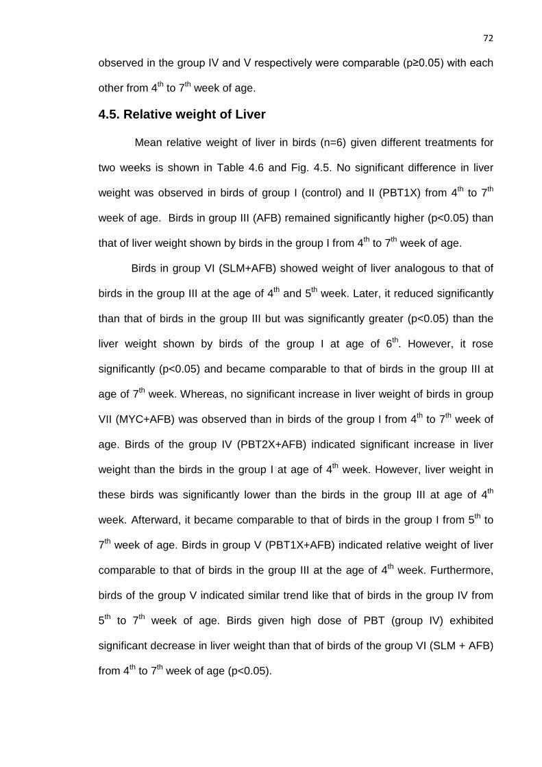
72
observed in the group IV and V respectively were comparable (p≥0.05) with each
other from 4th to 7th week of age.
4.5. Relative weight of Liver
Mean relative weight of liver in birds (n=6) given different treatments for
two weeks is shown in Table 4.6 and Fig. 4.5. No significant difference in liver
weight was observed in birds of group I (control) and II (PBT1X) from 4th to 7th
week of age. Birds in group III (AFB) remained significantly higher (p<0.05) than
that of liver weight shown by birds in the group I from 4th to 7th week of age.
Birds in group VI (SLM+AFB) showed weight of liver analogous to that of
birds in the group III at the age of 4th and 5th week. Later, it reduced significantly
than that of birds in the group III but was significantly greater (p<0.05) than the
liver weight shown by birds of the group I at age of 6th. However, it rose
significantly (p<0.05) and became comparable to that of birds in the group III at
age of 7th week. Whereas, no significant increase in liver weight of birds in group
VII (MYC+AFB) was observed than in birds of the group I from 4th to 7th week of
age. Birds of the group IV (PBT2X+AFB) indicated significant increase in liver
weight than the birds in the group I at age of 4th week. However, liver weight in
these birds was significantly lower than the birds in the group III at age of 4th
week. Afterward, it became comparable to that of birds in the group I from 5th to
7th week of age. Birds in group V (PBT1X+AFB) indicated relative weight of liver
comparable to that of birds in the group III at the age of 4th week. Furthermore,
birds of the group V indicated similar trend like that of birds in the group IV from
5th to 7th week of age. Birds given high dose of PBT (group IV) exhibited
significant decrease in liver weight than that of birds of the group VI (SLM + AFB)
from 4th to 7th week of age (p<0.05).

73
Table-4.6
Mean relative weight of liver (g/Kg) of birds taking different therapies for two
weeks
Treatments Week 4 Week 5 Week 6 Week 7
Group I (Control) 32.12 ± 1.48 c 27.76 ± 1.48 b 22.42 ± 1.47 c 26.82 ± 0.88a
Group II (PBT1X) 31.33 ± 0.83 c 27.97 ± 2.02 b 27.39 ± 0.90 c 27.36 ± 1.25 a
Group III (AFB) 43.51 ± 2.69 a 34.67 ± 1.86 a 33.30 ± 2.98 a 35.95 ± 1.13 b
Group IV (PBT2X+AFB) 39.54 ± 1.33 b 27.00 ± 1.08 b 27.33 ± 0.71 c 29.07 ± 0.54 a
Group V (PBT1X+AFB) 40.22 ± 1.94 a 28.12 ± 1.30 b 25.56 ± 1.11 c 29.32 ± 1.10 a
Group VI (SLM + AFB) 45.87 ± 4.80 a 32.32 ± 0.88 a 28.41 ± 1.42 b 34.61 ± 0.89 b
Group VII (MYC + AFB ) 36.1 4± 1.17 c 27.24 ± 1.85 b 23.91 ± 1.58 c 29.05 ± 1.60 a
Each value represent mean of relative weight of liver (g/Kg) ± Standard Error (n=6).AFB: Aflatoxin B1, PBT: Probiotics, SLM: Silymarin, MYC: Mycosorb. The figures in the same column having similar superscript are not significantly different (p>0.05).
Moreover, birds given low dose PBT (group V) indicated no significant decrease
in liver weight than that of birds in the group VI at 4th week of age. Afterward,
significant decrease was shown in liver weight of these birds than the birds of the
group V when compared to liver weight in birds of the group VI. Moreover, birds
in the group IV & V indicated significant rise (p<0.05) in liver weight than that of
bird in the group VII at 4th week of age. Thereafter, the liver weight observed in
these birds was comparable with birds of the group VII. However, significant rise
(p<0.05) was observed in liver weight of birds in the group V than weight of liver
in birds of the group IV at the age of 4th week. Afterward, no significant difference
was seen in birds of the either group.
4.6. Total Body Weight (TBW)
Mean total body weight of birds given various treatments is shown
in Table 4.7 and Fig. 4.6.

74
Each value represent mean of relative weight of Liver (g/Kg) ± Standard Error (n=6).AFB: Aflatoxin B1,PBT: Probiotics, SLM: Silymarin, MYC: Mycosorb. The groups in the same week having similar numerical superscript are not significantly different (p>0.05).
Fig. 4.5

75
Table-4.7
Mean total body weight (Kg) of birds taking different therapies for two weeks
Treatments Week 4 Week 5 Week 6 Week 7
Group I (Control) 1.02 ± 0.03 a 1.34 ± 0.05 a 1.56 ± 0.08 a 1.94 ± 0.07a
Group II (PBT1X) 1.06 ± 0.02 a 1.42 ± 0.05 a 1.64 ± 0.07 a 1.94 ± 0.10a
Group III (AFB) 0.72 ± 0.02 b 1.00 ± 0.03 b 1.17 ± 0.06 b 1.55 ± 0.04b
Group IV (PBT2X+AFB) 0.88 ± 0.03 a 1.26 ± 0.03 a 1.48 ± 0.02 a 1.97 ± 0.03a
Group V (PBT1X+AFB) 0.89 ± 0.04 a 1.32 ± 0.04 a 1.51 ± 0.07 a 1.93 ± 0.08a
Group VI (SLM + AFB) 0.87 ± 0.03 b 1.22 ± 0.03 a 1.48 ± 0.06 a 1.79 ± 0.05a
Group VII (MYC + AFB ) 0.97 ± 0.04 a 1.32 ± 0.06 a 1.46 ± 0.08 a 1.71 ± 0.09b
Each value represent mean total body weight (Kg) ± Standard Error (n=6).AFB: Aflatoxin B1, PBT: Probiotics, SLM: Silymarin, MYC: Mycosorb. The figures in the same column having similar superscript are not significantly different (p>0.05)..
No significant decrease in TBW was shown by birds of the group I
(Control) and group II (PBT1X). Birds in group III (AFB) exhibited significant
reduction (p<0.05) in TBW than that of birds in the group I from 4th to 7th week of
age. Birds taking SLM with AFB (group VI) indicated significant reduction
(p<0.05) in TBW than that of birds in the group I at the age of 4th week.
Furthermore, no significant decrease of TBW was observed in birds of the group
VI when compared to birds in the group I from 5th to 7th week of age. Birds given
MYC with AFB contaminated feed (group VII) displayed TBW comparable to that
of birds in the group I from 4th to 6th week of age (p≥0.05). However, birds
showed significant decline in TBW than the birds of the group I at the age of 7th
week.
Birds receiving both high and low dose of PBT with AFB contaminated
feed (group IV & V respectively) depicted comparable TBW to that of observed in
birds of the group I from 4th to 7th week of age. The body weight indicated by
birds in these two group was significantly higher than that of birds given SLM
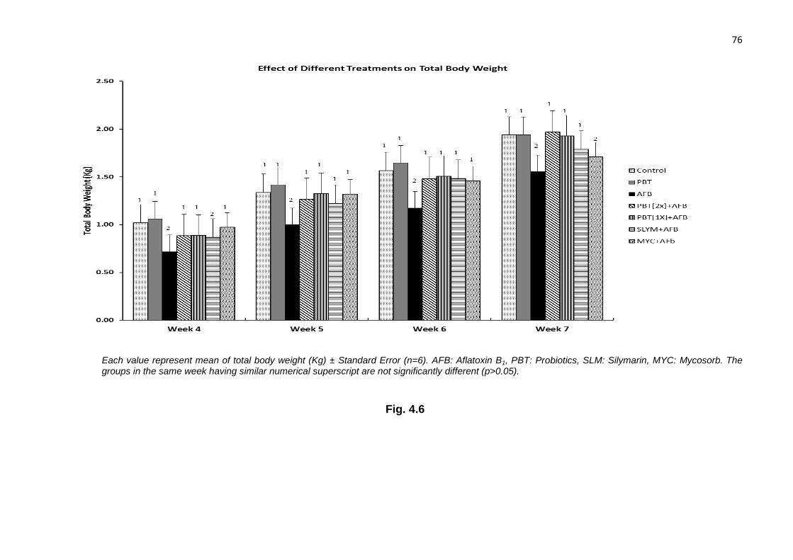
76
Each value represent mean of total body weight (Kg) ± Standard Error (n=6). AFB: Aflatoxin B1, PBT: Probiotics, SLM: Silymarin, MYC: Mycosorb. The groups in the same week having similar numerical superscript are not significantly different (p>0.05).
Fig. 4.6

77
(group VI) at the age of 4th week (p<0.05). Afterward, it became comparable to
TBW in birds of the group VI from 5th to 7th week of age. In comparison to birds
given MYC, the body weight observed in PBT treated birds was comparable to
that of birds in the group VII from 4th to 6th week of age. However, TBW was
significantly higher (p>0.05) than MYC treated birds at the age of 7th week
4.7. Total Serum Protein (TSP)
Mean total serum protein (g/dl) of birds receiving various treatments for
two weeks is shown in Table-4.8 and Fig.4.7. Total serum protein concentration
in birds of the group II (PBT1X) was significantly (p<0.05) lower than that of birds
in the group I (Control) at the age of 6th week. While it remained comparable to
birds of the group I on 4th, 5th and 7th week of age. TSP concentration was
significantly lower in birds fed with AFB contaminated feed (group III) than that of
birds received basal feed (group I) during study except at 5th week of age
(p>0.05).
Table-4.8
Mean total serum protein level (g/dl) of birds taking different therapies for two weeks
Treatments Week 4 Week 5 Week 6 Week 7
Group I (Control) 2.5 ± 0.12 a 2.8 ± 0.15 a 3.7 ± 0.14 a 4.1 ± 0.15 a
Group II (PBT1X) 2.6 ± 0.42 a 3.2 ± 0.17 a 2.7 ± 0.16 b 3.6 ± 0.07 a
Group III (AFB) 1.8 ± 0.17 b 1.5 ± 0.11 b 1.8 ± 0.20 c 2.3 ± 0.18 c
Group IV (PBT2X+AFB) 3.1 ± 0.41 a 2.4 ± 0.11 a 3.5 ± 0.33 a 3.7 ± 0.21 a
Group V (PBT1X+AFB) 3.0 ± 0.23 a 2.8 ± 0.25 a 3.0 ± 0.37 b 3.3 ± 0.14 b
Group VI (SLM + AFB) 2.2 ± 0.27 b 2.9 ± 0.30 a 2.5 ± 0.24 b 3.4 ± 0.27 a
Group VII (MYC + AFB ) 2.7 ± 0.21 a 3.2 ± 0.15 a 3.4 ± 0.14 a 3.5 ± 0.12 a
Each value represent mean total serum protein (g/100ml) ± Standard Error (n=6). AFB: Aflatoxin B1, PBT: Probiotics, SLM: Silymarin, MYC: Mycosorb. The figures in the same column having similar superscript are not significantly different (p>0.05)

78
Each value represent mean of total serum protein (g/100ml) ± Standard Error (n=6).AFB: Aflatoxin B1, PBT: Probiotics, SLM: Silymarin, MYC: Mycosorb. The groups in the same week having similar numerical superscript are not significantly different (p>0.05)
Fig. 4.7

79
While on 5th week of age the TSP was comparable to that of birds in the
group I (p≥0.05).Birds given SLM with AFB contaminated feed (Group VI)
indicated comparable TSP concentration to that of birds in the group I on 5th and
7th week of age. While at 4th week of age it was similar to the concentration
recorded in birds of the group III (p≥0.05). However, it was significantly higher
(p<0.05) than birds of the group III on 6th week of age although it was not
equivalent to the birds of the group I. The birds receiving MYC with AFB
contaminated feed (group VII) indicated comparable TSP concertation to that of
birds in the group I from 4th to 7th week of age.
Birds given high dose of PBT with AFB contaminated feed (group IV)
indicated the comparable total serum protein concentration to that of birds in the
group I from 4th to 7th week of age. The serum protein concentration in these
birds was equivalent to that of birds given MYC (group VII). In comparison to
birds of the group VI, no significant decrease was observed in TSP concentration
at the age of 4th, 5th and 7th week. However, TSP concentration was significantly
(p<0.05) higher than birds of the group VI at 6th week of age.
Birds given low dose PBT with AFB (group V) indicated significant
(p<0.05) rise in TSP than that of birds in the group III. TSP concentration in these
birds was analogue to the concentration observed in birds of the group I & VII on
4th and 5th week of age. However, on 6th and 7th week of age, serum protein
concentration indicated significant decrease in TSP concentration as compared
to that birds in the group I and VII. Moreover, results recorded in these birds were
significantly higher than the birds given SLM on 4th week of age. Later, the
protein concentration in these birds became comparable to birds of the group VI
at age of 5th and 6th week. . Later, TSP concentration in birds of the group V

80
declined and became significantly lesser than that of birds given other
interventions.
4.8. Serum Albumin
Mean serum albumin concentration in birds given different treatments for
two weeks is shown in Table 4.9 and Fig. 4.8. The serum albumin level in birds of
the group II (PBT1X) was comparable to that of birds given basal feed (group I) at
the age of 4th and 6th week. However, serum albumin concentration was
significantly higher than the birds of the group I at the age of 5th and 7th week.
Birds receiving AFB contaminated feed (group III) depicted significant (p<0.05)
decrease in serum albumin concentration than that of observed in bird fed basal
feed (group I) from 4th to 7th age of week.
Table-4.9
Mean serum albumin level (g/100ml) of birds taking different therapies for two weeks Treatments Week 4 Week 5 Week 6 Week 7
Group I (Control) 1.9 ± 0.07 b 1.9 ± 0.02 b 2.0 ± 0.09 a 2.2 ± 0.10 b
Group II (PBT1X) 2.0 ± 0.34 b 2.6 ± 0.17 a 1.9 ± 0.02 a 2.8 ± 0.07 a
Group III (AFB) 1.3 ± 0.15 c 1.0 ± 0.15 c 1.0 ± 0.14 b 1.4 ± 0.12 c
Group IV (PBT2X+AFB) 2.7 ± 0.31 a 1.0 ± 0.18 c 1.2 ± 0.26 b 1.9 ± 0.09 b
Group V (PBT1X+AFB) 1.7 ± 0.18 c 1.5 ± 0.12 c 1.0 ± 0.16 b 1.6 ± 0.08 b
Group VI (SLM + AFB) 1.6 ± 0.16 c 1.4 ± 0.22 c 1.2 ± 0.38 b 1.6 ± 0.20 b
Group VII (MYC + AFB ) 1.9 ± 0.12 b 1.1 ± 0.17 c 1.5 ± 0.15 a 2.4 ± 0.19 b
Each value represent mean serum albumin (g/100ml) ± Standard Error (n=6).AFB: Aflatoxin B1, PBT: Probiotics, SLM: Silymarin, MYC: Mycosorb. The figures in the same column having similar superscript are not significantly different (p>0.05).
Birds taking SLM with AFB contaminated feed (group VI) indicated serum
albumin concentration comparable to birds in the group III on 4th and 5th week of
age. Later, serum albumin concentration in these birds raised significantly
(p<0.05) than that of birds in the group III but was not equivalent

81
Each value represent mean of serum albumin (g/dl) ± Standard Error (n=6);AFB: Aflatoxin B1, PBT: Probiotics, SLM: Silymarin, MYC: Mycosorb. The groups in the same week having similar numerical superscript are not significantly different (p>0.05)
Fig. 4.8

82
to serum albumin concentration in birds of the group I. Birds given MYC with AFB
contaminated feed (group VII) showed serum albumin concentration analogue to
that of birds in the group I at the age of 4th, 6th and 7th week. However, at the age
of 5th week the serum albumin concertation in these birds indicated decline and
was comparable to that of birds in the group III. The birds given high dose PBT
with AFB (group IV) indicated serum albumin level significantly (p<0.05) higher
than that of shown by birds given basal feed (group I), SLM +AFB (group VI) and
MYC+AFB (group VII) at the age of 4th week. However, serum albumin
concertation in these birds declined and became comparable to that of birds
given AFB (group III) on 5th and 6th week of age (p≥0.05). Later, on 7th week of
age it raised significantly (p<0.05) higher than serum albumin level shown by
birds in the group III and became analogue to that of birds in the group I.
Likewise, birds receiving low dose PBT with AFB contaminated feed (group V)
showed serum albumin level equivalent to that of birds in the group III from 4th to
6th week of age. However, it became comparable to the serum concentration
observed in birds of the group I on 7th week of age.
4.9. Serum Glutamate-pyruvate Transferase (SGPT)
SGPT concentration (U/L) is depicted in Table 4.10 and Fig. 4.9. No
significant rise was observed in birds of the group II when compared to SGPT
concentration in birds of the group I. SGPT concentration in birds given AFB
contaminated feed (group III) was significantly (p<0.05) higher than that of birds
given basal feed only (group I) from 4th to 7th week of age. Birds given SLM with
AFB (group VI) showed SGPT concentration analogue to that of birds in the
group III on 4th, 6th and 7th week of age (p≥0.05). However, at 5th week of age, it
was significantly lower level than birds of the group III but was not analogous to
birds in the group I (p<0.05).

83
Table-4.10
Mean SGPT level (U/L) of birds taking different therapies for two weeks
Treatments Week 4 Week 5 Week 6 Week 7
Group I (Control) 13.60 ± 0.50 b 14.52 ± 1.18 c 13.73 ± 0.72 c 12.55 ± 0.74 b
Group II (PBT1X) 14.87 ± 1.94 b 12.05 ± 0.59 c 17.08 ± 0.82 c 11.23 ± 2.91 b
Group III (AFB) 28.40 ± 4.54 a 42.28 ± 1.53 a 37.82 ± 1.71 a 33.25 ± 1.08 a
Group IV (PBT2X+AFB) 15.78 ± 2.04 b 20.25 ± 1.64 c 22.38 ± 2.03 b 18.57 ± 1.24 b
Group V (PBT1X+AFB) 17.15 ± 1.93 b 23.10 ± 5.49 b 25.67 ± 5.08 b 14.77 ± 1.45 b
Group VI (SLM + AFB) 24.03 ± 1.50 a 34.57 ± 5.25 b 32.38 ± 1.27 a 29.65 ± 7.16 a
Group VII (MYC + AFB ) 12.92 ± 0.49 b 27.78 ± 1.84 b 31.92 ± 0.94 a 27.58 ± 1.07 a
Each value represent mean serum GPT (U/L) ± Standard Error (n=6). AFB: Aflatoxin B1, PBT: Probiotics, SLM: Silymarin, MYC: Mycosorb. The figures in the same column having similar superscript are not significantly different (p>0.05)
Birds given MYC with AFB (group VII) exhibited significantly (p<0.05)
lower SGPT concentration than that of birds in the group III at the age of 4th and
5th week. However on 6th and 7th week of age, increase in SGPT concentration in
these birds was comparable to that of birds in the group III. The Birds receiving
high dose of PBT with AFB (group IV) indicated comparable concentration of
SGPT to the birds receiving basal feed (group I) at the age of 4th, 5th and 7th
week. Whereas, at the age of 6th week, it was significantly (p<0.05) lower than
that of birds in the group III although was not comparable to birds in the group I.
The SGPT concentration in these birds was comparable to birds given MYC
(group VII) on 4th week of age. Thereafter, it became significantly lower than that
of birds receiving MYC (p<0.05). Moreover, protective effect of PBT in these birds
was significantly (p<0.05) lower than the birds fed SLM with AFB (group VI) from
4th to 7th week of age.
Birds given low dose PBT (group V) indicated significant (p<0.05)
reduction in SGPT concentration than that of birds of the group III from 4th to 7th

84
Each value represent mean of SGPT (U/l) ± Standard Error (n=6). AFB: Aflatoxin B1, PBT: Probiotics, SLM: Silymarin, MYC: Mycosorb. The groups in the same week having similar numerical superscript are not significantly different (p>0.05).
Fig. 4.9

85
week of age. However, the SGPT level in these birds was comparable to that of
birds in the group I on 4th, 7th week of age. The SGPT concentration in such birds
was comparable to birds given SLM with AFB at the age of 5th week. Moreover,
there was significant decrease in SGPT concentration in these birds than that of
birds in the group VI on 4th, 6th and 7th week of age. Furthermore, birds of the
group V indicated SGPT concentration similar to that of birds in the group VII on
4th and 5th week of age. Whereas, on 6th and 7th week of age, it was significantly
lower than the birds of the group VII. Birds receiving PBT either low or high dose
exhibited comparable SGPT concentration at the age of 4th, 6th and 7th week. At
5th week of age, SGPT concentration in birds of the group V was significantly
higher than the birds in the group IV.
4.10. Serum Bilirubin
Serum bilirubin level of broilers received different treatments for two weeks
is shown in Table 4.11 and graphically in Fig. 4.10.
Table-4.11
Mean serum bilirubin level (mg/dl) of birds taking different therapies for two weeks Treatments Week 4 Week 5 Week 6 Week 7
Group I (Control) 0.35 ± 0.01 b 0.38 ± 0.02 b 0.37 ± 0.02 b 0.36 ± 0.02 a
Group II (PBT1X) 0.27 ± 0.03 b 0.36 ± 0.05 b 0.40 ± 0.02 b 0.40 ± 0.03 a
Group III (AFB) 0.71 ± 0.10 a 0.77 ± 0.08 a 0.63 ± 0.02 a 0.52 ± 0.02 a
Group IV (PBT2X+AFB) 0.33 ± 0.02 a 0.39 ± 0.06 b 0.38 ± 0.06 b 0.36 ± 0.06 a
Group V (PBT1X+AFB) 0.31 ± 0.04 b 0.38 ± 0.09 b 0.38 ± 0.08 b 0.36 ± 0.05 a
Group VI (SLM + AFB) 0.61 ± 0.02 a 0.49 ± 0.08 b 0.31 ± 0.05 b 0.29 ± 0.03 a
Group VII (MYC + AFB ) 0.43 ± 0.06 b 0.46 ± 0.11 b 0.41 ± 0.10 b 0.38 ± 0.01 a
Each value represent mean serum bilirubin level (mg/dl) ± Standard Error (n=6). AFB: Aflatoxin B1, PBT: Probiotics, SLM: Silymarin, MYC: Mycosorb. The figures in the same column having similar superscript are not significantly different (p>0.05).

86
No significant rise in serum bilirubin level was observed in birds of the
group II (PBT1X) as compared to birds in group I (Control). Significantly (p<0.05)
elevated concentration of serum bilirubin was measured in birds fed AFB with
basal feed (group III) as compared to birds given basal feed (group I) from 4th to
6th week of age. However it was normalized at age of 7th week. Birds taking SLM
with AFB (group VI) indicated serum bilirubin level analogous to birds in the
group III at the age 4th week. Later it normalized and became comparable to that
of birds in the group I. No significant increase in serum bilirubin was exhibited in
birds of group VII (MYC+AFB) than that of birds in the group I from 4th to 6th week
of age.
Birds given high dose PBT with AFB (group IV) displayed serum bilirubin
concentration equivalent to that of birds in the group I, VI and VII on 5th and 6th
week of age. However on 4th week of age the serum bilirubin concentration was
analogue to birds fed AFB contaminated feed (group III). While birds receiving
low dose PBT (group VI) indicated serum bilirubin concentration comparable to
that birds in the group I and VII from 4th to 6th week of age. In comparison to SLM
taking birds (group VI) serum bilirubin level was significantly (p<0.05) lower at the
age of 4th week. However, it was comparable to that of birds in the group VI at the
age of 5th and 6th week. Moreover, serum bilirubin concentration in birds given
high or low dose PBT exhibited non-significant difference at from 5th to 6th week
of age. Furthermore, significant rise in serum bilirubin level was observed in birds
of the group IV than that of birds in the group V on 4th week of age. At the age of
7th week, no significant difference was shown in serum bilirubin level among birds
receiving various interventions.

87
Each value represent mean of serum bilirubin (mg/dl) ± Standard Error (n=6) AFB: Aflatoxin B1,PBT: Probiotics, SLM: Silymarin, MYC: Mycosorb. The groups in the same week having similar numerical superscript are not significantly different (p>0.05)
Fig. 4.10

88
4.11. Serum Blood Urea Nitrogen
Effect of various treatments on serum blood urea nitrogen (BUN) is shown
in Table 4.12 and graphically in Fig.4.11. Significant difference was observed
among birds given various treatments on 4th and 5th week of age (p<0.05). Birds
given PBT1X (group II) indicated serum BUN level analogous to birds receiving
basal feed (group I) at 4th and 5th week of age. Birds fed AFB with basal feed
(group III) indicated significant decline in serum BUN than the birds in the group I
at 4th and 5th week of age. The Birds given SLM with AFB (group VI) displayed
equivalent serum BUN concentration to that of birds in the group III at the age of
4th week. While, birds given MYC with AFB (group VII) showed significantly
(p<0.05) higher serum BUN concentration than birds in the group III at the 4th
week of age but it was not analogue to the serum BUN concentration of birds in
the group I.
Table-4.12
Mean serum BUN level (mg/dl) of birds taking different therapies for two weeks
Treatments Week 4 Week 5 Week 6 Week 7
Group I (Control) 6.15 ± 1.10 a 6.20 ± 0.34 a 5.03 ± 0.88 a 4.47 ± 0.74 a
Group II (PBT1X) 6.10 ± 0.70 a 6.21 ± 0.41 a 5.11 ± 0.85 a 4.38 ± 0.61 a
Group III (AFB) 4.06 ± 0.40 c 3.43± 0.34 b 4.02 ± 0.94 a 4.19 ± 0.61 a
Group IV (PBT2X+AFB) 5.81 ± 0.63 a 3.24 ± 0.48 b 4.75 ± 0.42 a 4.83 ± 0.29 a
Group V (PBT1X+AFB) 6.57 ± 0.57 a 4.31 ± 0.32 b 4.71 ± 0.27 a 4.95 ± 0.28 a
Group VI (SLM + AFB) 4.53 ± 1.03 c 2.42 ± 0.40 b 3.88 ± 0.29 a 4.05 ± 0.61 a
Group VII (MYC + AFB ) 6.10 ± 0.89 b 4.31 ± 0.28 b 4.83 ± 0.36 a 4.40 ± 0.40 a
Each value represent mean serum BUN level (mg/dl) ± Standard Error (n=6). AFB: Aflatoxin B1, PBT: Probiotics, SLM: Silymarin, MYC: Mycosorb, BUN: Blood Urea Nitrogen The figures in the same column having similar superscript are not significantly different (p>0.05).
Birds given PBT (both high and low dose) showed serum BUN

89
Each value represent mean of serum BUN (mg/dl) ± Standard Error (n=6) AFB: Aflatoxin B1, PBT: Probiotics, SLM: Silymarin, MYC: Mycosorb, BUN:
Blood Urea Nitrogen. The groups in the same week having similar numerical superscript are not significantly different (p>0.05).
Fig. 4.11
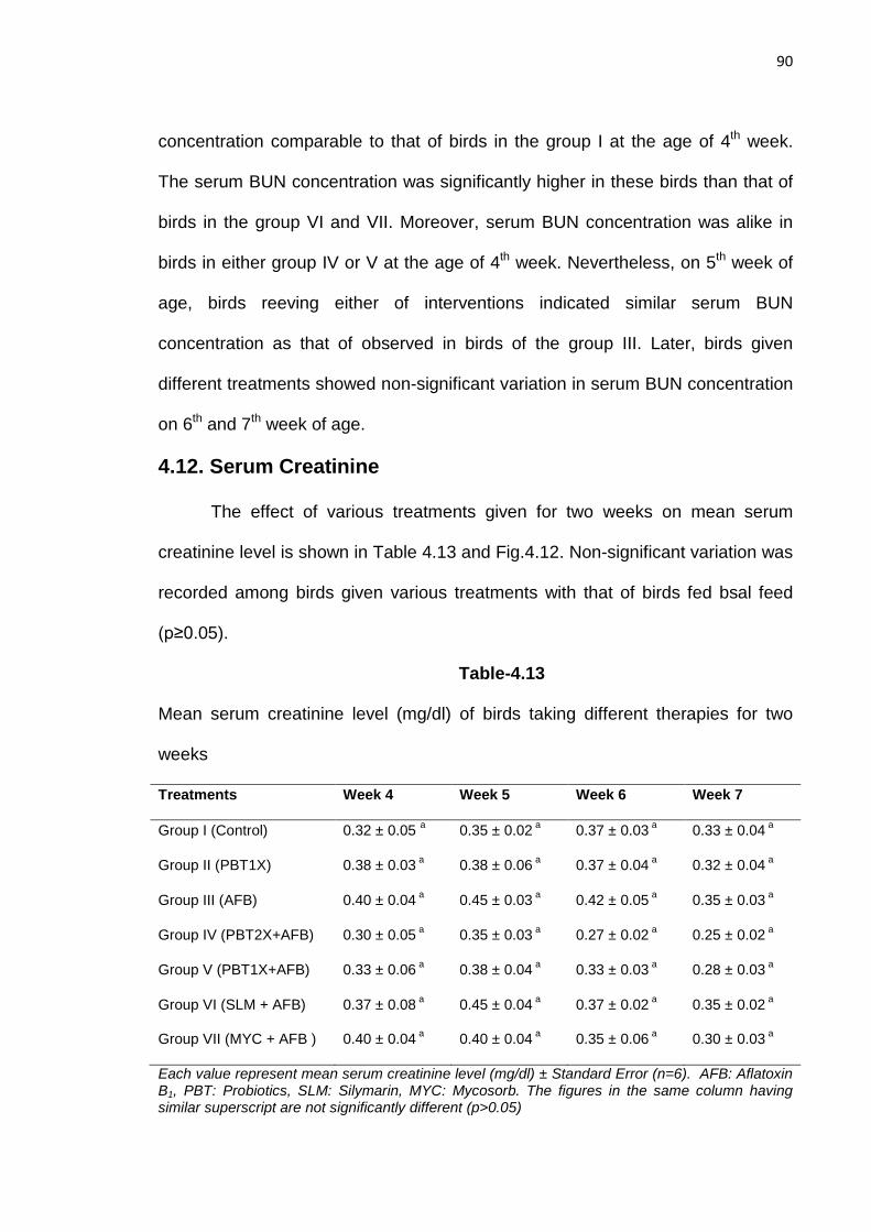
90
concentration comparable to that of birds in the group I at the age of 4th week.
The serum BUN concentration was significantly higher in these birds than that of
birds in the group VI and VII. Moreover, serum BUN concentration was alike in
birds in either group IV or V at the age of 4th week. Nevertheless, on 5th week of
age, birds reeving either of interventions indicated similar serum BUN
concentration as that of observed in birds of the group III. Later, birds given
different treatments showed non-significant variation in serum BUN concentration
on 6th and 7th week of age.
4.12. Serum Creatinine
The effect of various treatments given for two weeks on mean serum
creatinine level is shown in Table 4.13 and Fig.4.12. Non-significant variation was
recorded among birds given various treatments with that of birds fed bsal feed
(p≥0.05).
Table-4.13
Mean serum creatinine level (mg/dl) of birds taking different therapies for two
weeks
Treatments Week 4 Week 5 Week 6 Week 7
Group I (Control) 0.32 ± 0.05 a 0.35 ± 0.02 a 0.37 ± 0.03 a 0.33 ± 0.04 a
Group II (PBT1X) 0.38 ± 0.03 a 0.38 ± 0.06 a 0.37 ± 0.04 a 0.32 ± 0.04 a
Group III (AFB) 0.40 ± 0.04 a 0.45 ± 0.03 a 0.42 ± 0.05 a 0.35 ± 0.03 a
Group IV (PBT2X+AFB) 0.30 ± 0.05 a 0.35 ± 0.03 a 0.27 ± 0.02 a 0.25 ± 0.02 a
Group V (PBT1X+AFB) 0.33 ± 0.06 a 0.38 ± 0.04 a 0.33 ± 0.03 a 0.28 ± 0.03 a
Group VI (SLM + AFB) 0.37 ± 0.08 a 0.45 ± 0.04 a 0.37 ± 0.02 a 0.35 ± 0.02 a
Group VII (MYC + AFB ) 0.40 ± 0.04 a 0.40 ± 0.04 a 0.35 ± 0.06 a 0.30 ± 0.03 a
Each value represent mean serum creatinine level (mg/dl) ± Standard Error (n=6). AFB: Aflatoxin B1, PBT: Probiotics, SLM: Silymarin, MYC: Mycosorb. The figures in the same column having similar superscript are not significantly different (p>0.05)

91
Each value represent mean of serum creatinine (mg/dl) ± Standard Error (n=6) AFB: Aflatoxin B1,PBT: Probiotics, SLM: Silymarin, MYC: Mycosorb. The groups in the same week having similar numerical superscript are not significantly different (p>0.05)
Fig. 4.12

92
4.13. AFB Residue in liver
Table 4.14 and Fig. 4.13 depicted the mean AFB residue in liver of birds
given various treatments for two weeks. Birds in group I (Control) and group II
(PBT1X) did not indicate any detectable residue in liver from 4th to 7th week of
age. While, Birds given AFB contaminated feed (group III) exhibited detectable
level of AFB residue in the liver from 4th to 7th week of age.
Table-4.14
Mean of AFB residue (ng/g) in livers of birds taking different therapies for two weeks
Treatments 4th Week 5th Week 6th Week 7th Week
Group I (Control) ND ND ND ND
Group II (PBT1X) ND ND ND ND
Group III (AFB) 2.46±0.09 a 9.22±0.8 a 7.65±0.56 a 5.52± 0.6 a
Group IV (PBT2X+AFB) ND ND ND ND
Group V (PBT1X+AFB) ND 0.77±0.02 b ND ND
Group VI (SLM + AFB) 1.98±0.06 a 4.49±0.25 b 2.06±0.65 b ND
Group VII (MYC+ AFB ) ND 0.77±0.04 b 0.66±0.04 b ND
Each value represents mean of AFB residue in livers ± Standard Error (n=4). AFB: Aflatoxin B1, PBT: Probiotics, SLM: Silymarin, MYC: Mycosorb, ND: Non detectable. The figures in the same column having similar superscript are not significantly different (p>0.05).
Birds taking SLM with AFB (group VI) indicated measurable AFB residue
from 4th to 6th week of age. Whereas, birds fed MYC with AFB (group VII)
indicated measurable level of AFB in liver on 5th and 6th week of age. The
measureable level in birds of both the groups was significantly (p<0.05) lower
than the birds of the group III. Birds given high dose of PBT with AFB (group IV)
did not show any detectable level of AFB in liver from 4th to 7th week of age.
However, birds given low level of PBT (group V) displayed detectable level of
AFB in liver on 5th week of study only which was equivalent to that of birds in the
group VI and VII (p≥0.05).

93
Each value represents mean of AFB residue in livers ± Standard Error (n=4.). AFB: Aflatoxin B1, PBT: Probiotics, SLM: Silymarin, MYC: Mycosorb The groups in the same week having similar numerical superscript are not significantly different (p>0.05).
Fig. 4.1

94
4.14. Histopathological Examination
4.14.1. Liver
At 4th week of age, intake of AFB cause moderate degenerative changes
in liver of birds (group III) as compared to that of birds given basal feed only
(group I). Microscopic examination revealed vacuolar degeneration and early sign
of bile duct hyperplasia. Further exposure resulted in sever degenerative
changes in liver parenchyma (Plate i and j), vacuolar degeneration, pycnotic
nuclei and hepatocellular degeneration. Bile duct hyperplasia along with loss of
hepatic plate structure was also observed in these birds. These degenerative
changes persisted even at 6th week of age. However, at 7th week of age, the
hepatic tissues showed little recovery: from sever to moderate degenerative
changes.
Birds given SLM with AFB (group VI) and MYC +AFB (group VII) indicated
moderate degree of damage on 4th, 5th, 6th and 7th weeks of age. Liver of birds
given both high and low dose PBT (groups IV and V respectively) showed
moderate damage on 4th week of age. However on 5th, 6th and 7th weeks of age,
liver of the birds in these groups exhibited mild hepatocellular changes besides
normal parenchyma of cells (Plate k& l).
4.14.2. Bursa of Fabricius (BF)
On 4th week of age bursa of Fabricius of birds in the group III (AFB)
showed mild lymphoid degenerative changes as compared with the BF in the
group I and II. Later on 5th week of age BF showed depletion of bursal follicles,
loss of cortical cells and decreased cellular density. These atrophic changes were
also present on 6th week of birds in the group III (Plate a & b). Birds in group VI
(SLM +AFB) and group VII (MYC+AFB) showed moderate damage on 4th and 5th

95
week of age whereas, mild degenerative changes were observed on 6th and 7th
week of age (Plate c and d). Similarly, birds given high or low dose of PBT
(groups IV, V respectively) indicated mild loss of bursal follicle on 5th week of age
but indicated non-significant damage on 4th, 6th and 7th week of age i.e. Cells in
cortex remained intact,.
4.14.3 Spleen
On spleen, effect was mild but lymphocytic degeneration was also seen in
birds fed AFB (group III) on 4th and 5th week of age. On week 6th and 7th of age,
damage is not significant. Spleen of birds given various treatments exhibited non-
significant damage.
4.14.4. Kidney
Kidney of birds taking AFB (group III) showed moderated degree of
damage with atrophy of bowman corpuscle, loss of luminal limning and
desquamation of luminal epithelial cells in tubules from 4th to 6th week of age.
However, On 7th week of age, kidney showed regenerative changes with mild
degree of restoration in tubular lining cells in the birds of the group III (plates e
and f ) Birds in the groups given SLM or MYC with AFB (group VI and VII
respectively) showed atrophied glomerulus on 5th of age. Whereas, Birds in the
group IV (PBT2X+AFB) and V(PBT1X+AFB) indicated intact luminal lining of
tubules and improved appearance of bowman corpuscles on 5th week of age
(Plate g)..

96
(a) (b)
(c) (d)
a. Bursal follicle (10X) in PBT treated Birds for two weeks b. Bursal follicle (40X) in PBT treated Birds for two weeks c. Depleted Bursal follicle (10X) in AFB treated Birds for two weeks d. Depleted Bursal follicle (40X) in AFB treated Birds for two weeks
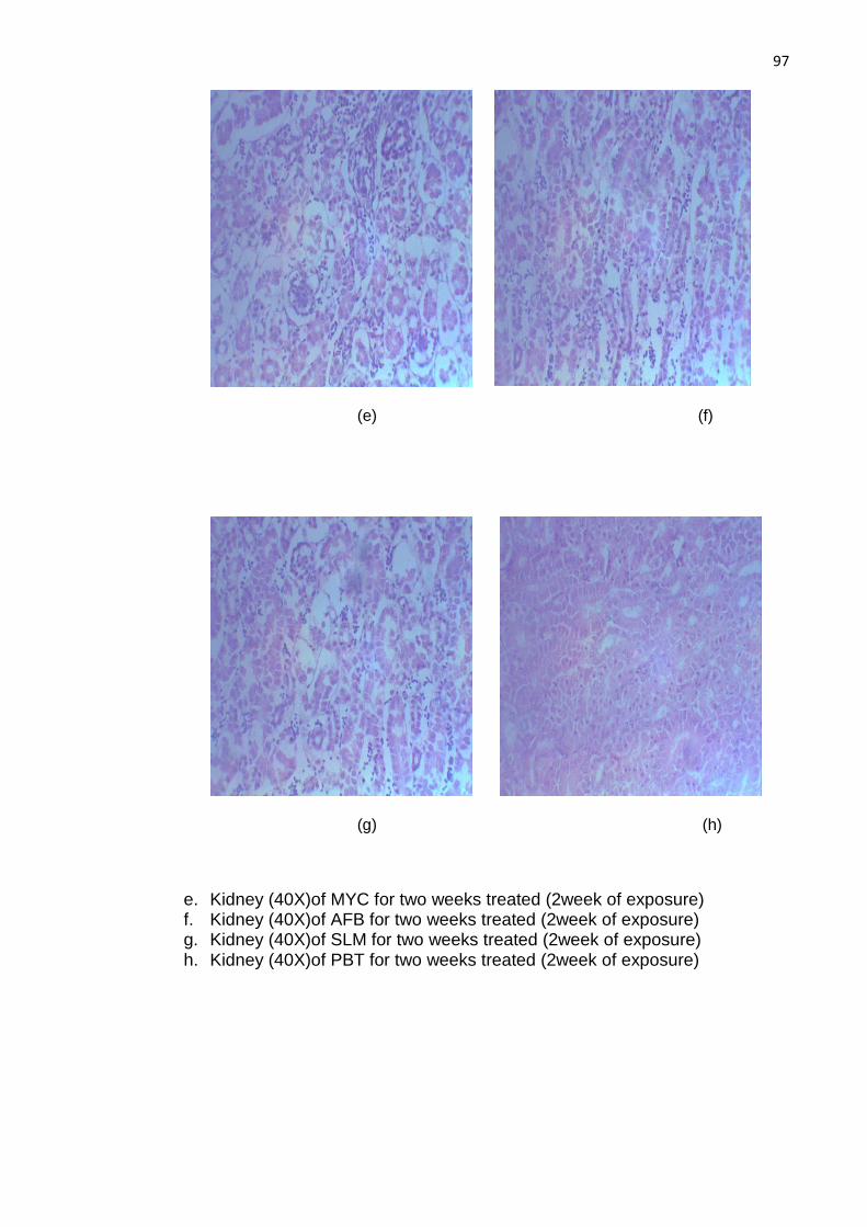
97
(e) (f)
(g) (h)
e. Kidney (40X)of MYC for two weeks treated (2week of exposure) f. Kidney (40X)of AFB for two weeks treated (2week of exposure) g. Kidney (40X)of SLM for two weeks treated (2week of exposure) h. Kidney (40X)of PBT for two weeks treated (2week of exposure)

98
(i) (j)
(k) (l)
i. Liver of AFB treated birds for two weeks (10X) j. Liver of AFB treated birds for two weeks (40X) k. Liver of PBT treated birds for two weeks (10X) l. Liver of PBT treated birds for two weeks (40X)
99
5. Discussions & Conclusion
Aflatoxin B1 (AFB), a secondary metabolite produced by many species of
Aspergillus, is classified as group I carcinogen by IARC (Wei and Jong, 1986;
IARC, 2002). AFB to be used in study was produced through rice fermentation.
The rice fermentation by Asperigllus flavus on flask shaker produced a level of
700 to 800 ppb of the toxin, which is in line of observations reported by Shotwell
et al. (1966) and Yunus and Böhm (2011).
To assess the immunosuppression serum NDV-antibody titer, leukocytes
count, relative weight of lymphoid organ such as bursa of Fabricius and spleen
were determined. Initially an increase in NDV antibody titer was observed after
one week of AFB exposure (at week 4 of age) in bird of the group III. This
temporary rise in humoral response by AFB was reported and described by
various researchers (Giambrone et al., 1985; Yunus et al., 2011b). The exact
mechanism of the initial increase in immune response is not known and may be
due to body’s protective response to the intoxication. However, AFB induced a
strong immuno-suppressive effect as observed by significant low titer values of
the NDV-antibody titer in birds of the group III after two weeks of exposure (week
5 of age) and even one week after cessation of exposure (week 6 of age). The
suppressed NDV-antibody level even after stoppage of intoxication may be due
toxicity of preformed AFB guanine adduct. The observed immuno-toxicity of AFB
is in line with previous reports (Meissonnier et al., 2008a; Chen et al., 2013b). A
rise in antibody titer in AFB treated birds after two weeks of stopping the
ingestion of AFB (week 7 of age) shows recovery of the birds from the

100
immunosuppression within two weeks after termination of exposure which
suggests that immunosuppressive effect of AFB is reversible.
Silymarin is an established hepato-protectant through its antioxidant action
(Muriel and Mourelle, 1990). It does not offer any protective effect on NDV-
antibody response, against AFB on week 4 of age in birds. The lack of protective
action at week 4 of age (1st week of exposure) indicates that SLM does not affect
the absorption of AFB. But SLM showed protective action on NDV-antibody titer
on 5-, 6- and 7-week of age. The later improved responses in birds of the group
VI although significantly lesser than PBT treated birds, can be explained through
its neutralizing effect via scavenging of free radicals and restoration of normal
glutathione level (Rastogi et al., 2001; Tedesco et al., 2004a; Chand et al., 2011).
MYC showed a protective role on NDV antibody response against
deleterious effects of AFB during the exposure by adsorption in birds of the group
VII. However, the protective effect was lost during the first week of terminating
the exposure (week 6 of age). It suggests that continuous intake of MYC may be
required to maintain protective effects of the MYC. These findings were
supported by observation of other researchers (Ghahri et al., 2009; Hasan et al.,
2010; Verbrugghe et al., 2012; Mogadam and Azizpour, 2013)
Both concentrations of PBT (1X and 2X) ameliorated the immuno-
suppressive effects of AFB in birds of the group IV and V as demonstrated by
significantly elevated NDV-antibody titer values in birds of these groups than the
birds in the group III. Secondly comparable NDV- antibody titer of birds in the
group II with that of birds in the group I, reflected lack of immunomodulation, if
given alone. It clearly reveals that higher NDV antibody titer observed in birds of
the group IV and V is due to the toxin adsorptive properties of PBT. The data
clearly demonstrate the adsorptive properties of Lactobacilli against AFB as

101
reported earlier (Peltonen et al., 2001a; Fazeli et al., 2009a; Salminen et al.,
2010b; Kumar et al., 2011). The protective action of PBT was equivalent to that of
MYC and SLM. The restoration of NDV antibody titer at the 6th week indicates
protective effect persisted even after stopping of intake of PBT. This effect could
be result of either decline in absorption during exposure or enhancing the
elimination of toxic metabolites.
Leukocytes plays central role in generation of immune response either
humoral or cellular. The data of present study show that administration of low
dose of AFB to the broilers for two week causes mild leukopenia which is in
accordance to previously published reports (Samuel et al., 2009). The AFB-
induced leukopenia is reversible and subsides as AFB-intake is stopped. Co-
administration of PBT (Lactobacilli) caused complete recovery from the AFB-
induced reduction in leukocyte count. The protective effect of lactobacilli during
the AFB exposure results in detoxification of AFB presumably via adsorption
(Fazeli et al., 2009b; Salminen et al., 2010a; Kumar et al., 2011) and beneficial
effect of PBT on leukocyte count as reported previously (Apata, 2008; Salim et
al., 2013). Intake of SLM ameliorates the leukopenia caused by AFB which is
attributed to its antioxidant properties reported by different researchers (Tedesco
et al., 2004a; Lutsenko et al., 2008; Lee et al., 2010). Similarly addition of MYC in
AFB contaminated diet prevents the negative effect of AFB on leukocytes count
which is line with observation of other workers (Basmacioglu et al., 2005; Che et
al., 2011; Xu et al., 2011)
Bursa of Fabricius is an important lymphoid organ in broilers which is
usually regressed slowly with age and vanishes upon maturity. An abrupt
reduction in weight of bursa of Fabricius was observed in birds of the group III
compared to that of birds in the group I. The atrophied changes in bursa of

102
Fabricius were not recovered during one week post-exposure period (week 6 of
age).These findings are supported by the observation of various researchers
(Smith et al., 1992; Sur and Celİk, 2003; Wang et al., 2009; Chen et al., 2014a).
Likewise, intake of MYC also exhibited improvement in the bursal weight of birds
of the group VII on week-4 and -6 which are in line of the previous studies
(Karaman et al., 2005; Girish and Devegowda, 2006; He et al., 2013b). Feeding
of SLM to birds also ameliorated the negative effect of AFB which are related to
its antioxidant abilities supported by the findings of other researchers (Abidin et
al., 2011; Chand et al., 2011).
PBT administration in diet significantly prevented the AFB-induced
regression and prevented cellular damage in bursa of Fabricius. The bursal
weight recorded in birds of the group IV and V was appeared to be better than
that of birds in the group VI and VIII. Birds in these groups indicated mild
degenerative changes in bursa of Fabricius. The ameliorative effect of PBT at
one week of exposure can be explained on basis of adsorptive properties of the
bacterial cell wall as reported by other scientists (Gratz et al., 2007; Hernandez-
Mendoza et al., 2009a; Hernandez-Mendoza et al., 2011; Bovo et al., 2014). The
suppressive actions on leukocyte count and bursa of Fabricius is also supported
by significant decrease in NDV antibody titer measured in these birds. Moreover,
protection after stoppage of intake can be explained through colonization of
Lactobacilli in intestine of birds and their positive effect on immune system.
Spleen an important organ of reticuloendothelial system involved in
immunity (white pulp). AFB reversibly causes reduction in spleen weight which
returns to normal within two weeks after stopping the toxin intake. The reversible
suppression of spleen weight was supported by findings of other researchers
(Chaytor et al., 2011; Chen et al., 2014b). Histopathological findings suggest

103
resistance of spleen to AFB’s negative effect. Similarly comparable spleen weight
in birds of the group VII on week-5 of age (week-1 exposure) indicated the toxin
binding properties of MYC as reported by other workers (Raju and Devegowda,
2000b; Basmacioglu et al., 2005; Armando et al., 2011; Zoghi et al., 2014;
Gonçalves et al., 2015). Lack of protective effect after stopping the MYC
ingestion necessitates continuous intake of MYC for protective action (Girish and
Devegowda, 2006; Matur et al., 2011; Mogadam and Azizpour, 2013). Likewise,
SLM administration produced better impact on spleen weight on week-5 of age.
The improved value in birds of the group VI indicated beneficial effect of SLM in
AFB toxicity on spleen which is attributed to protective action against oxidative
stress produced by AFB as reported earlier (Tedesco et al., 2004b; Kalorey et al.,
2005; Chand et al., 2011).
Feeding of PBT with contaminated diet did not prevent reduction in relative
weight of spleen during the AFB exposure; however, PBT accelerated the
recovery of spleen weight during post-exposure period indicated by normalized
spleen weight on week-6 of age. This beneficial effect on week-1 of exposure (5th
week of age) was linked to reduced bioavailability of AFB through adsorption by
the Lactobacilli. The reduced absorption results in decrease in formation of the
toxic metabolites. These findings are supported by observation of various
investigators (Fazeli et al., 2009b; Bovo et al., 2014; Zoghi et al., 2014). Positive
effect at 6th week of age suggests impact of Lactobacilli on either AFB activation
or elimination of epoxide metabolite.
Total body weight is an important parameter to assess growth and
nutritional consequences in AFB-induced toxicity. AFB ingestion caused
reduction in total body weight in birds which remain significantly lower from 4th to
7th week of age. The decline in total body weight in the birds of the group III is

104
linked with decreased feed intake due to AFB toxicity. These findings are in line
of previous studies conducted in various species by various scientists (He et al.,
2013a; Indresh et al., 2013; Kumar et al., 2013; Weaver et al., 2013; Chen et al.,
2014d).
MYC intake in birds significantly guards against AFB toxicity during
exposure and one week post exposure. But it fails to exhibit any protection after
two week of stopping the ingestion (week-7 of age). These findings are in line
with findings of researchers in broilers as well as other species (Karaman et al.,
2005; Indresh et al., 2013; Zychowski et al., 2013; Selim et al., 2014). SLM upon
ingestion showed shielding effect against AFB on week-5, -6 and -7 of age. The
lack of effect on toxin absorption can be reason for the absence of protection on
week 4 of age (one week-exposure). But later significant ameliorative action is
attributed to its anti-oxidative properties as studied by different scientists
(Tedesco et al., 2004a; Chand et al., 2011; Dumari et al., 2014; Kamali and
Mostafaei, 2014).
Co-administration of PBT ameliorated the harmful effect of AFB during the
exposure and post-exposure. The protective action from 4th to 5th week of age
was comparable to birds in the group VII can be explained via adsorptive
properties of these Lactobacilli. The adsorptive abilities were reported by various
researchers (Thongsong et al.; Peltonen et al., 2001b; Fazeli et al., 2009b;
Hernandez-Mendoza et al., 2009a; Nikbakht Nasrabadi et al., 2013). However,
amelioration from cumulative effect at the age of 6th to 7th week of age suggests
mechanism other than adsorption.
The liver is major site of AFB activation and more susceptible to AFB
toxicity (Wild and Turner, 2002). Ingestion of AFB to birds resulted in significant
rise in relative weight of liver (hepatomegaly) during the exposure and it remained
105
higher even after stopping the toxin intake. These observation are in line with
previous findings (Vekiru et al.; Magnoli et al., 2011; Chen et al., 2014c; Fowler et
al., 2014). Administration of PBT (Group IV) in high dose combination prevented
the hepatomegaly during exposure (4 to 5 week of age) as well as post-exposure
(6 to 7 week of age). However low dose PBT combination (Group V) showed
protection after week-2 of exposure (5th week of age) and maintained this effect
till 7th week of age. The persistent rise after the exposure shows cumulative
toxicity of AFB on hepatomegaly. Feeding of PBT with basal diet (group II) has
no significant influence on liver weight in hosts. The beneficial effect of PBT in
presence of toxin can be explained through its AFB binding abilities which have
been studied by various researches (Gratz et al., 2006; Fazeli et al., 2009b;
Hernandez-Mendoza et al., 2009a; Hernandez-Mendoza et al., 2010; Hathout et
al., 2011; Zoghi et al., 2014).
Intake of silymarin does not indicate any beneficial AFB during the
exposure. These finding demonstrates that SLM does not affect the absorption of
AFB. These observations are in line with previous reports (Tedesco et al., 2004a;
Suchý et al., 2008). Protective action recorded at week-6 of age indicates the
recovery from hepatomegaly but rise in liver weight at week-7 indicates lack of
protection against the cumulative action of AFB. SLM showed the ameliorative
action against the AFB toxicity through its antioxidant actions and scavenging of
free radical as reported by different scientists (Tedesco et al., 2004b; Kalorey et
al., 2005; Chand et al., 2011). Administration of MYC significantly prevented the
deleterious effect of the AFB on hepatomegaly during exposure (week 4 to 5) and
week one post-exposure (week-6 of age). However, lack protection at week-7 of
age proposes the continuous intake to provide amelioration. These observation
are supported by outcomes of several researchers (Raju and Devegowda, 2000a;

106
Aravind et al., 2003; Yildirim et al., 2011; Verbrugghe et al., 2012; Cao and
Wang, 2014)
Serum protein and albumin are an early indicator of hepatic injury during
aflatoxicosis (Huff et al., 1986). Both total serum protein and albumin has been
decreased in birds of the group III. The decline was significant during exposure
(week-4 to 5) and in post-exposure (week-6 to 7). The hypoproteinemia and
hypoalbuminemia in hosts was in line with previous findings (Karabacak et al.;
Manafi et al., 2012; Shamsudeen and Shrivastava, 2013; Chen et al., 2014d;
Chibanga et al., 2014).. The protective action in birds given PBT can be ascribed
to the toxin neutralizing properties of Lactobacilli presumably via adsorption.
These adsorptive /neutralizing properties were studied by several researchers
(Fazeli et al., 2009b; Hernandez-Mendoza et al., 2009a; Hernandez-Mendoza et
al., 2011; Bagherzadeh Kasmani et al., 2012; Zoghi et al., 2014). The protective
action of PBT on TSP and serum albumin was less than MYC.
Similarly protection offered by SLM was significantly lower than that of
PBT treated birds. Similarly early failure of the protective effect is associated with
negligible effect on the toxin adsorption and hence absorption. However, delayed
and weak protection in SLM fed birds, is due to its action against oxidative stress
and free radicals. These findings have also been studied in other species and
reported by various scientists (Rastogi et al., 2000; Saller et al., 2001; Tedesco et
al., 2003; Tedesco et al., 2004b; Dumari et al., 2014). Ingestion pf MYC displayed
amelioration against negative effect AFB on total serum protein and albumin
level. The protection is based on its effect to reduce the toxin bioavailability. This
adsorptive action of MYC was previously reported by other researchers (Aravind
et al., 2003; Karaman et al., 2005; Ghahri et al., 2009; Hasan et al., 2010; Yildirim
et al., 2011; Verbrugghe et al., 2012). Likewise, lower proteins concentration in
107
post-exposure time period demands continuous feeding of such detoxifying
agents and shows delayed toxicity of AFB even after intake was stopped.
SGPT is one of the transaminase present in hepatocytes which enters the
blood /serum during hepatic necrosis. Bilirubin is an important product of
hepatocytes which may enters the circulation during biliary tract obstruction. AFB
intake results in rise of both SGPT and bilirubin level as sign of hepatic injury.
The AFB produced rise in the level of SGPT and bilirubin was observed during
the toxin intake and even after stopping the ingestion of the toxin. These
observations are in line with previous reports of other investigators (Petkov et al.,
1985; Valchev et al., 2014). Same is true for other species (Cheng et al., 2001;
Rastogi et al., 2001). Histopathological examination of liver revealed severs
degenerative changes including vacuolar degeneration, karyolysis, bile duct
hyperplasia and loss of hepatic plate. Theses finding are supported by raised
concentration of bilirubin and SGPT. These histopathological findings are
supported by studies of different scientists (Denli et al., 2009; Yang et al., 2012;
Weaver et al., 2013).
Ingestion of SLM failed to show any protection after week-1 of exposure
(week-4 of age) on SGPT and bilirubin level like that of serum albumin and total
proteins. Failure of the protection on early phase indicates lack of effect on toxin
adsorption and so its bioavailability. However, later amelioration on week-5 and -
6 was due to its effect on oxidative stress caused by AFB. The observed
antioxidant action of SLM in the study is previously reported by various scientists
(Rastogi et al., 2000; Saller et al., 2001; Dumari et al., 2014). Similarly, MYC an
established mycotoxin adsorbent showed protection during the exposure but
protective action was lost after stopping the intake. The protective action seen
was due to absorption and is in line with previous findings of other researchers

108
(Basmacioglu et al., 2005; Karaman et al., 2005; Girish and Devegowda, 2006;
Yildirim et al., 2011). PBT intake resulted in normalized level of SGPT and
bilirubin during exposure as well as post exposure. Similarly mild to moderate
damage was observed in liver of birds in the group IV and V on week-5,-6 and -7
of age. Protective action of PBT was better than MYC or SLM. The significant
amelioration of the AFB toxicity by ingestion of PBT in the birds through feed can
be associated with the toxin adsorption during exposure by such bacteria. The
adsorptive properties are reported previously by several researchers (Peltonen et
al., 2001b; Gratz et al., 2006; Hernandez-Mendoza et al., 2010; Salminen et al.,
2010a; Hathout et al., 2011). However, protection in post-exposure phase
suggests some additional mechanism along with adsorption. Moreover, non-
significant impact of feeding PBT with basal feed (group II) also endorses the
above findings.
Serum creatinine and blood urea nitrogen were used to investigate
toxic effects on kidney by AFB and hence its amelioration. Lack of any significant
effect on serum creatinine by AFB during the exposure indicates that kidney is
not the main target of AFB toxicity. Blood urea nitrogen is an indicator of liver and
kidney function in early age of birds. Ingestion of AFB for two weeks produce
decline in serum BUN but this action was reversible upon stoppage of ingestion.
The decline in the BUN was reported by different researchers (Raju and
Devegowda, 2000a; Aravind et al., 2003; Li et al., 2012). AFB ingestion produced
atrophied bowman corpuscle, loss of luminal limning and desquamation of
luminal epithelial cells in tubules which were mildly recovered upon stopping of
the toxin ingestion. These findings were supported by Kamel (Kamel, 2013).
SLM administration does not show any protection on one week exposure.
However, later observed shielding effect can be explained by its anti-oxidant role.

109
The anti-oxidant role is reported previously by various investigators (Rastogi et
al., 2000; Tedesco et al., 2004b; Lutsenko et al., 2008). The protective action of
MYC during exposure on serum BUN is linked to its adsorptive action which is in
line with previous findings (Girish and Devegowda, 2006; Ghahri et al., 2009;
Hasan et al., 2010; Yildirim et al., 2011; Verbrugghe et al., 2012). However,
Kidney of birds in the group VI and VII showed atrophied glomeruli.
PBT treatments indicate improvement in histological feature on week-5 of
age and produced beneficial impact which was linked to the adsorptive ability of
Lactobacilli. It reduced the toxin absorption and hence the toxicity. The adsorptive
action of Lactobacilli is supported by studies of different researchers (El-Nezami
et al., 1998; Haskard et al., 2001; Peltonen et al., 2001b; Fazeli et al., 2009b;
Salminen et al., 2010a; Zhong et al., 2014).
5.1. Conclusions
The data of the current study demonstrate that live Lactobacilli present in
Pakistani traditional food significantly ameliorated the AFB immuno-toxicity as
well as hepatotoxicity presumably through its adsorptive action thus reducing the
toxin bioavailability. Moreover, persistent protective action after stopping the
intake shows advantage over other intervention (SLM or MYC). Furthermore, it
also suggests some additional mechanisms for neutralization of absorbed toxin or
its toxic metabolite rather than simple adsorption.
It is also suggested that addition of Dhai (source of PBT) in food or feed
decrease the likelihood of aflatoxicosis in human or birds in such regions where
prevention of fungal contamination is not practically feasible due to either cost or
unawareness of community. Moreover, addition of such probiotics in the diet of
patients suffering from hepatitis C or B patients results in delay of progression
toward HCC in such regions. Furthermore, lack of interaction with micronutrients,

110
low cost, ease of availability and control of Lactobacilli make such source of PBT
as an economical and practical remedy to reduce toxicity of the aflatoxins in
under-developed (African countries) and developing countries like Pakistan etc.
In future, further research is needed to elucidate the any additional
mechanism for AFB neutralization besides adsorption of the toxin by Lactobacilli.

111
6. References
1. Abidin, Z., Khatoon, A., Numan, M., 2011. Mycotoxins in broilers:
pathological alterations induced by aflatoxins and ochratoxins, diagnosis
and determination, treatment and control of mycotoxicosis. World's Poultry
Science Journal 67, 485-496.
2. Al-Hammadi, S., Marzouqi, F., Al-Mansouri, A., Shahin, A., Al-Shamsi, M.,
Mensah-Brown, E., Souid, A.K., 2014. The cytotoxicity of aflatoxin b1 in
human lymphocytes. Sultan Qaboos Univ Med J 14, e65-e71.
3. Allan, W., Gough, R., 1974. A standard haemagglutination inhibition test
for Newcastle disease. (1). A comparison of macro and micro methods.
Veterinary Record 95, 120-123.
4. Alvarez-Olmos, M.I., Oberhelman, R.A., 2001. Probiotic agents and
infectious diseases: a modern perspective on a traditional therapy. Clin
Infect Dis 32, 1567-1576.
5. Anjum, M., Sahota, A., Akram, M., Ali, I., 2011. Prevalence of mycotoxins
in poultry feeds and feed ingredients in Punjab (Pakistan). The Journal of
Animal and Plant Sciences 2, 117-120.
6. Apata, D.F., 2008. Growth performance, nutrient digestibility and immune
response of broiler chicks fed diets supplemented with a culture of
Lactobacillus bulgaricus. Journal of the Science of Food and Agriculture
88, 1253-1258.
7. Aravind, K., Patil, V., Devegowda, G., Umakantha, B., Ganpule, S., 2003.
Efficacy of esterified glucomannan to counteract mycotoxicosis in naturally
contaminated feed on performance and serum biochemical and
hematological parameters in broilers. Poultry Science 82, 571-576.

112
8. Armando, M., Dogi, C., Pizzolitto, R., Escobar, F., Peirano, M., Salvano,
M., Sabini, L., Combina, M., Dalcero, A., Cavaglieri, L., 2011.
Saccharomyces cerevisiae strains from animal environment with in vitro
aflatoxin B<sub>1</sub> binding ability and anti-pathogenic
bacterial influence. World Mycotoxin Journal 4, 59-68.
9. Aslam, N., Tipu, M.Y., Ishaq, M., Cowling, A., McGill, D., Warriach, H.M.,
Wynn, P., 2016. Higher Levels of Aflatoxin M1 Contamination and Poorer
Composition of Milk Supplied by Informal Milk Marketing Chains in
Pakistan. Toxins 8.
10. Aslam, S., Qazi, J.I., 2010. Isolation of acidophilic lactic acid bacteria
antagonistic to microbial contaminants. Pakistan Journal of Zoology 42,
567-573.
11. Aso, Y., Akaza, H., Kotake, T., Tsukamoto, T., Imai, K., Naito, S., 1995.
Preventive effect of a Lactobacillus casei preparation on the recurrence of
superficial bladder cancer in a double-blind trial. The BLP Study Group.
Eur Urol 27, 104-109.
12. Aziz, T., Khan, H., Bakhtair, S., Naurin, M., 2009. Incidence and relative
abundance of lactic acid bacteria in raw milk of buffalo, cow and sheep. J
Anim Plant Sci 19, 168-173.
13. Azziz-Baumgartner, E., Lindblade, K., Gieseker, K., Rogers, H.S., Kieszak,
S., Njapau, H., Schleicher, R., McCoy, L.F., Misore, A., DeCock, K., 2005.
Case–control study of an acute aflatoxicosis outbreak, Kenya, 2004.
Environmental Health Perspectives 113, 1779.
14. Bagherzadeh Kasmani, F., Karimi Torshizi, M.A., Allameh, A.,
Shariatmadari, F., 2012. A novel aflatoxin-binding Bacillus probiotic:

113
Performance, serum biochemistry, and immunological parameters in
Japanese quail. Poultry Science 91, 1846-1853.
15. Bajaj, J.S., Saeian, K., Christensen, K.M., Hafeezullah, M., Varma, R.R.,
Franco, J., Pleuss, J.A., Krakower, G., Hoffmann, R.G., Binion, D.G.,
2008. Probiotic Yogurt for the Treatment of Minimal Hepatic
Encephalopathy. Am J Gastroenterol 103, 1707-1715.
16. Bankcroft, J., Gamble, M., 2007. Theory and Practice of Histological
Technique. 5th ed. Churchill Livingstone,London.
17. Basmacioglu, H., Oguz, H., Ergul, M., Col, R., Birdane, Y., 2005. Effect of
dietary esterified glucomannan on performance, serum biochemistry and
haematology in broilers exposed to aflatoxin. Czech Journal of Animal
Science 50, 31-39.
18. Bedard, L.L., Massey, T.E., 2006. Aflatoxin B1-induced DNA damage and
its repair. Cancer Lett 241, 174-183.
19. Besselink, M.G., van Santvoort, H.C., Buskens, E., Boermeester, M.A.,
van Goor, H., Timmerman, H.M., Nieuwenhuijs, V.B., Bollen, T.L., van
Ramshorst, B., Witteman, B.J., Rosman, C., Ploeg, R.J., Brink, M.A.,
Schaapherder, A.F., Dejong, C.H., Wahab, P.J., van Laarhoven, C.J., van
der Harst, E., van Eijck, C.H., Cuesta, M.A., Akkermans, L.M., Gooszen,
H.G., 2008. Probiotic prophylaxis in predicted severe acute pancreatitis: a
randomised, double-blind, placebo-controlled trial. Lancet 371, 651-659.
20. Bhatnagar, D., Cary, J.W., Ehrlich, K., Yu, J., Cleveland, T.E., 2006.
Understanding the genetics of regulation of aflatoxin production and
Aspergillus flavus development. Mycopathologia 162, 155-166.
21. Bintvihok, A., Kositcharoenkul, S., 2006. Effect of dietary calcium
propionate on performance, hepatic enzyme activities and aflatoxin

114
residues in broilers fed a diet containing low levels of aflatoxin B1. Toxicon
47, 41-46.
22. Bovo, F., Franco, L.T., Rosim, R.E., Trindade, C.S.F., De Oliveira, C.A.F.,
2014. The Ability of Lactobacillus rhamnosus in Solution, Spray-Dried or
Lyophilized to Bind Aflatoxin B1. Journal of Food Research 3, p35.
23. Brisbin, J.T., Gong, J., Orouji, S., Esufali, J., Mallick, A.I., Parvizi, P.,
Shewen, P.E., Sharif, S., 2011. Oral Treatment of Chickens with
Lactobacilli Influences Elicitation of Immune Responses. Clinical and
Vaccine Immunology 18, 1447-1455.
24. Brown, R., Chen, Z.-Y., Cleveland, T., Russin, J., 1999. Advances in the
development of host resistance in corn to aflatoxin contamination by
Aspergillus flavus. Phytopathology 89, 113-117.
25. Brugh, M., Jr., 1978. A simple method for recording and analyzing
serological data. Avian Dis 22, 362-365.
26. Bruneau, J.C., Stack, E., O'Kennedy, R., Loscher, C.E., 2012. Aflatoxins
B(1), B(2) and G(1) modulate cytokine secretion and cell surface marker
expression in J774A.1 murine macrophages. Toxicol In Vitro 26, 686-693.
27. Bullerman, L.B., Bianchini, A., 2007. Stability of mycotoxins during food
processing. International Journal of Food Microbiology 119, 140-146.
28. Cao, J., Wang, W., 2014. Effects of astaxanthin and esterified
glucomannan on hematological and serum parameters, and liver
pathological changes in broilers fed aflatoxin-B1-contaminated feed.
Animal Science Journal 85, 150-157.
29. Cary, J.W., Rajasekaran, K., Brown, R.L., Luo, M., Chen, Z.-Y.,
Bhatnagar, D., 2011. Developing Resistance to Aflatoxin in Maize and
Cottonseed. Toxins 3, 678-696.
115
30. Çelýk, K., Denlý, M., Savas, T., 2003. Reduction of toxic effects of
aflatoxin B1 by using baker yeast (Saccharomyces cerevisiae) in growing
broiler chicks diets. Revista Brasileira de Zootecnia 32, 615-619.
31. Chand, N., Muhammad, D., Durrani, F., Qureshi, M.S., Ullah, S.S., 2011.
Protective effects of milk thistle (Silybum marianum) against aflatoxin B1 in
broiler chicks. Asian-Australasian Journal of Animal Sciences 24, 1011-
1018.
32. Chaytor, A.C., See, M.T., Hansen, J.A., de Souza, A.L.P., Middleton, T.F.,
Kim, S.W., 2011. Effects of chronic exposure of diets with reduced
concentrations of aflatoxin and deoxynivalenol on growth and immune
status of pigs1. J. Anim. Sci. 89, 124-135.
33. Che, Z., Liu, Y., Wang, H., Zhu, H., Hou, Y., Ding, B., 2011. The protective
effects of different mycotoxin adsorbents against blood and liver
pathological changes induced by mold-contaminated feed in broilers.
Asian–Aust J Anim Sci 24, 250-257.
34. Chen, J., Chen, K., Yuan, S., Peng, X., Fang, J., Wang, F., Cui, H., Chen,
Z., Yuan, J., Geng, Y., 2013a. Effects of aflatoxin B1 on oxidative stress
markers and apoptosis of spleens in broilers. Toxicol Ind Health 4, 4.
35. Chen, K., Fang, J., Peng, X., Cui, H., Chen, J., Wang, F., Chen, Z., Zuo,
Z., Deng, J., Lai, W., Zhou, Y., 2014a. Effect of selenium supplementation
on aflatoxin B1-induced histopathological lesions and apoptosis in bursa of
Fabricius in broilers. Food and Chemical Toxicology 74, 91-97.
36. Chen, K., Peng, X., Fang, J., Cui, H., Zuo, Z., Deng, J., Chen, Z., Geng,
Y., Lai, W., Tang, L., 2014b. Effects of dietary selenium on
histopathological changes and T cells of spleen in broilers exposed to
116
aflatoxin B1. International journal of environmental research and public
health 11, 1904-1913.
37. Chen, K., Shu, G., Peng, X., Fang, J., Cui, H., Chen, J., Wang, F., Chen,
Z., Zuo, Z., Deng, J., Geng, Y., Lai, W., 2013b. Protective role of sodium
selenite on histopathological lesions, decreased T-cell subsets and
increased apoptosis of thymus in broilers intoxicated with aflatoxin B(1).
Food. Chem. Toxicol 59, 446-454.
38. Chen, X., Horn, N., Applegate, T.J., 2014c. Efficiency of hydrated sodium
calcium aluminosilicate to ameliorate the adverse effects of graded levels
of aflatoxin B1 in broiler chicks. Poultry Science.
39. Chen, X., Horn, N., Cotter, P., Applegate, T., 2014d. Growth, serum
biochemistry, complement activity, and liver gene expression responses of
Pekin ducklings to graded levels of cultured aflatoxin B1. Poultry Science,
PS3904.
40. Chen, X., Horn, N., Cotter, P.F., Applegate, T.J., 2014e. Growth, serum
biochemistry, complement activity, and liver gene expression responses of
Pekin ducklings to graded levels of cultured aflatoxin B1. Poult Sci 93,
2028-2036.
41. Cheng, C.T., 1992. Perak, Malaysia, mass poisoning. Tale of the Nine
Emperor Gods and rat tail noodles. Am J Forensic Med Pathol 13, 261-
263.
42. Cheng, Y.H., Shen, T.F., Pang, V.F., Chen, B.J., 2001. Effects of aflatoxin
and carotenoids on growth performance and immune response in mule
ducklings. Comp Biochem Physiol C Toxicol Pharmacol 128, 19-26.
43. Chiavaro, E., Cacchioli, C., Berni, E., Spotti, E., 2005. Immunoaffinity
clean-up and direct fluorescence measurement of aflatoxins B1 and M1 in
117
pig liver: comparison with high-performance liquid chromatography
determination. Food Addit Contam 22, 1154-1161.
44. Chibanga, J.F., Banda-Nyirenda, D., Simbaya, J., 2014. Effects of
Supplementing Different Levels of Vitamin A to Aflatoxin B1 Contaminated
Dietson the Performance of Broiler Chickens. Asian Journal of Natural &
Applied Sciences Vol 3, 4.
45. Chung, T.K., Erdman, J.W., Jr., Baker, D.H., 1990. Hydrated sodium
calcium aluminosilicate: effects on zinc, manganese, vitamin A, and
riboflavin utilization. Poult Sci 69, 1364-1370.
46. Costanzo, P., Santini, A., Fattore, L., Novellino, E., Ritieni, A., 2014.
Toxicity of aflatoxin B1 towards the vitamin D receptor (VDR). Food Chem
Toxicol 4, 00498-00490.
47. De Groote, M.A., Frank, D.N., Dowell, E., Glode, M.P., Pace, N.R., 2005.
LACTOBACILLUS RHAMNOSUS GG BACTEREMIA ASSOCIATED WITH
PROBIOTIC USE IN A CHILD WITH SHORT GUT SYNDROME. The
Pediatric Infectious Disease Journal 24, 278-280.
48. Denli, M., Blandon, J.C., Guynot, M.E., Salado, S., Perez, J.F., 2009.
Effects of dietary AflaDetox on performance, serum biochemistry,
histopathological changes, and aflatoxin residues in broilers exposed to
aflatoxin B1. Poultry Science 88, 1444-1451.
49. Diaz, G.J., Murcia, H.W., Cepeda, S.M., 2010. Cytochrome P450 enzymes
involved in the metabolism of aflatoxin B1 in chickens and quail. Poult Sci
89, 2461-2469.
50. Dimitri, R.A., Gabal, M.A., 1996. Immunosuppressant activity of aflatoxin
ingestion in rabbits measured by response to Mycobacterium bovis

118
antigen I. Cell mediated immune response measured by skin test reaction.
Vet Hum Toxicol 38, 333-336.
51. Dumari, M.A., Sarir, H., Makki, O.F., Afzali, N., 2014. Effect of Milk Thistle
(Silybum marianum L.) on Biochemical Parameters and Immunity of
Broiler Chicks Fed Aflatoxin B1 after Three Weeks. Iranian Journal of
Toxicology Volume 8.
52. Eaton, D.L., Gallagher, E.P., 1994. Mechanisms of aflatoxin
carcinogenesis. Annu Rev Pharmacol Toxicol 34, 135-172.
53. El-Nezami, H., Kankaanpaa, P., Salminen, S., Ahokas, J., 1998. Ability of
dairy strains of lactic acid bacteria to bind a common food carcinogen,
aflatoxin B-1. Food and chemical toxicology 36, 321-323.
54. El-Nezami, H., Nicoletti, G., Neal, G., Donohue, D., Ahokas, J., 1995.
Aflatoxin M< sub> 1</sub> in human breast milk samples from Victoria,
Australia and Thailand. Food and chemical toxicology 33, 173-179.
55. Escamilla, J., Lane, M.A., Maitin, V., 2012. Cell-Free Supernatants from
Probiotic Lactobacillus casei and Lactobacillus rhamnosus GG Decrease
Colon Cancer Cell Invasion In Vitro. Nutrition and Cancer 64, 871-878.
56. Etzel, R.A., 2002. MYcotoxins. JAMA 287, 425-427.
57. Farah, N., Sadia, M, Shakira, G.S.S, Tahir, N, Rauf, M.I., et al., 2016.
Isolation, characterization and application of indigenous lactic acid bacteria
in milk fermentation. Int. J. Biosci 9, 415-430.
58. Fazeli, M.R., Hajimohammadali, M., Moshkani, A., Samadi, N., Jamalifar,
H., Khoshayand, M.R., Vaghari, E., Pouragahi, S., 2009a. Aflatoxin B1
binding capacity of autochthonous strains of lactic acid bacteria. J. Food
Prot 72, 189-192.

119
59. Fazeli, M.R., Hajimohammadali, M., Moshkani, A., Samadi, N., Jamalifar,
H., Khoshayand, M.R., Vaghari, E., Pouragahi, S., 2009b. Aflatoxin B1
binding capacity of autochthonous strains of lactic acid bacteria. J Food
Prot 72, 189-192.
60. Fernández, A., Verde, M.T., Gascón, M., Ramos, J.J., Gómez, J., 1994.
Aflatoxin and its metabolites in tissues from laying hens and broiler
chickens fed a contaminated diet. Journal of the Science of Food and
Agriculture 65, 407-414.
61. Fowler, H.M., Barrientos-Velazquez, A., Deng, Y., Bailey, C., 2014.
Utilization of a Spray-Applied Calcium Bentonite Clay to Ameliorate the
Effects of Low-Levels of Aflatoxinin Starter Broiler Diets Containing DDGS.
Natural Products Chemistry & Research.
62. Gabal, M.A., Dimitri, R.A., 1998. Humoral immunosuppressant activity of
aflatoxin ingestion in rabbits measured by response to Mycobacterium
bovis antigens using enzyme-linked immunosorbent assay and serum
protein electrophoresis. Mycoses 41, 303-308.
63. Geissmann, F., Manz, M.G., Jung, S., Sieweke, M.H., Merad, M., Ley, K.,
2010. Development of monocytes, macrophages, and dendritic cells.
Science 327, 656-661.
64. Ghahri, H., Talebi, A., Chamani, M., Lotfollahian, H., Afzali, N., 2009.
Ameliorative effect of esterified glucomannan, sodium bentonite, and
humic acid on humoral immunity of broilers during chronic aflatoxicosis.
Turkish Journal of Veterinary and Animal Sciences 33, 419-425.
65. Giambrone, J.J., Diener, U.L., Davis, N.D., Panangala, V.S., Hoerr, F.J.,
1985. Effects of aflatoxin on young turkeys and broiler chickens. Poult. Sci
64, 1678-1684.

120
66. Girish, C., Devegowda, G., 2006. Efficacy of glucomannan-containing
yeast product (Mycosorb®) and hydrated sodium calcium aluminosilicate
in preventing the individual and combined toxicity of aflatoxin and T-2 toxin
in commercial broilers. ASIAN AUSTRALASIAN JOURNAL OF ANIMAL
SCIENCES 19, 877.
67. Gonçalves, B.L., Rosim, R.E., de Oliveira, C.A.F., Corassin, C.H., 2015.
The in vitro ability of different Saccharomyces cerevisiae – Based products
to bind aflatoxin B1. Food Control 47, 298-300.
68. Gong, Y.Y., Cardwell, K., Hounsa, A., Egal, S., Turner, P.C., Hall, A.J.,
Wild, C.P., 2002. Dietary aflatoxin exposure and impaired growth in young
children from Benin and Togo: cross sectional study. Bmj 325, 20-21.
69. Gratz, S., Taubel, M., Juvonen, R.O., Viluksela, M., Turner, P.C.,
Mykkanen, H., El-Nezami, H., 2006. Lactobacillus rhamnosus strain GG
modulates intestinal absorption, fecal excretion, and toxicity of aflatoxin B-
1 in rats. Applied and environmental microbiology 72, 7398-7400.
70. Gratz, S., Wu, Q.K., El-Nezami, H., Juvonen, R.O., Mykkanen, H., Turner,
P.C., 2007. Lactobacillus rhamnosus strain GG reduces aflatoxin B-1
transport, metabolism, and toxicity in caco-2 cells. Applied and
environmental microbiology 73, 3958-3964.
71. Gross-Steinmeyer, K., Eaton, D.L., 2012. Dietary modulation of the
biotransformation and genotoxicity of aflatoxin B1. Toxicology 299, 69-79.
72. Gupta, P., Andrew, H., Kirschner, B.S., Guandalini, S., 2000. Is
Lactobacillus GG helpful in children with Crohn's disease? Results of a
preliminary, open-label study. Journal of pediatric gastroenterology and
nutrition 31, 453-457.
73. Gupta, V., Garg, R., 2009. Probiotics. Indian J Med Microbiol 27, 202-209.

121
74. Hartley, R., Nesbitt, B.F., O'kelly, J., 1963. Toxic metabolites of Aspergillus
flavus. Nature 198, 1056-1058.
75. Hasan, G., Toloei, T., Habibi, M., 2010. Efficacy of esterified
glucomannan, sodium bentonite and humic acid to counteract
experimental aflatoxicosis on antibody titers against Newcastle disease in
broilers. African Journal of Biotechnology 9, 4127-4131.
76. Haskard, C.A., El-Nezami, H.S., Kankaanpaa, P.E., Salminen, S., Ahokas,
J.T., 2001. Surface binding of aflatoxin B-1 by lactic acid bacteria. Applied
and environmental microbiology 67, 3086-3091.
77. Hathout, A.S., Aly, S.E., 2014. Biological detoxification of mycotoxins: a
review. Annals of Microbiology 64, 905-919.
78. Hathout, A.S., Mohamed, S.R., El-Nekeety, A.A., Hassan, N.S., Aly, S.E.,
Abdel-Wahhab, M.A., 2011. Ability of Lactobacillus casei and Lactobacillus
reuteri to protect against oxidative stress in rats fed aflatoxins-
contaminated diet. Toxicon 58, 179-186.
79. He, J., Zhang, K.Y., Chen, D.W., Ding, X.M., Feng, G.D., Ao, X., 2013a.
Effects of maize naturally contaminated with aflatoxin B1 on growth
performance, blood profiles and hepatic histopathology in ducks. Livestock
Science 152, 192-199.
80. He, J., Zhang, K.Y., Chen, D.W., Ding, X.M., Feng, G.D., Ao, X., 2013b.
Effects of vitamin E and selenium yeast on growth performance and
immune function in ducks fed maize naturally contaminated with aflatoxin
B1. Livestock Science 152, 200-207.
81. Hell, K., Cardwell, K.F., Setamou, M., Poehling, H., 2000. The influence of
storage practices on aflatoxin contamination in maize in four

122
agroecological zones of Benin, west Africa. J Stored Prod Res 36, 365-
382.
82. Hell, K., Mutegi, C., Fandohan, P., 2010. Aflatoxin control and prevention
strategies in maize for Sub-Saharan Africa. Julius-Kühn-Archiv, S. 534.
83. Hendrickse, R.G., 1997. Of sick turkeys, kwashiorkor, malaria, perinatal
mortality, heroin addicts and food poisoning: research on the influence of
aflatoxins on child health in the tropics. Ann Trop Med Parasitol 91, 787-
793.
84. Hernandez-Mendoza, A., Garcia, H., Steele, J., 2009a. Screening of< i>
Lactobacillus casei</i> strains for their ability to bind aflatoxin B< sub>
1</sub>. Food and chemical toxicology 47, 1064-1068.
85. Hernandez-Mendoza, A., Guzman-de-Peña, D., Garcia, H.S., 2009b. Key
role of teichoic acids on aflatoxin B1 binding by probiotic bacteria. Journal
of Applied Microbiology 107, 395-403.
86. Hernandez-Mendoza, A., Guzman-De-Peña, D., González-Córdova, A.F.,
Vallejo-Córdoba, B., Garcia, H.S., 2010. In vivo assessment of the
potential protective effect of Lactobacillus casei Shirota against aflatoxin
B1. Dairy Sci. Technol. 90, 729-740.
87. Hernandez-Mendoza, A., Rivas-Jimenez, L., Garcia, H.S., 2011.
Assessment of Aflatoxin B1 Binding to Lactobacillus reuteri by Microscopy
and Fluorescence Techniques. Food Biotechnology 25, 140-150.
88. Hitchins, A.D., McDonough, F.E., 1989. Prophylactic and therapeutic
aspects of fermented milk. Am J Clin Nutr 49, 675-684.
89. Hoyos, A.B., 1999. Reduced incidence of necrotizing enterocolitis
associated with enteral administration of< i> Lactobacillus acidophilus</i>

123
and< i> Bifidobacterium infantis</i> to neonates in an intensive care unit.
International journal of infectious diseases 3, 197-202.
90. Huebner, H.J., Herrera, P., Phillips, T.D., 2008. Clay-Based Interventions
for the Control of Chemical and Microbial Hazards in Food and Water,
Preharvest and Postharvest Food Safety, Blackwell Publishing, pp. 389-
402.
91. Huff, W., Kubena, L., Harvey, R., Corrier, D., Mollenhauer, H., 1986.
Progression of aflatoxicosis in broiler chickens. Poultry Science 65, 1891-
1899.
92. IARC, 2002. Some traditional herbal medicines, some mycotoxins,
naphthalene and styrene, IARC Monographs on the Evaluation of
Carcinogenic Risk of Chemicals to Man, World Health Organization, pp. 1-
556.
93. Indresh, H., Devegowda, G., Ruban, S.W., Shivakumar, M., 2013. Effects
of high grade bentonite on performance, organ weights and serum
biochemistry during aflatoxicosis in broilers. Vet World 6, 313-317.
94. Jard, G., Liboz, T., Mathieu, F., Guyonvarc'h, A., Lebrihi, A., 2011. Review
of mycotoxin reduction in food and feed: from prevention in the field to
detoxification by adsorption or transformation. Food Addit Contam Part A
Chem Anal Control Expo Risk Assess 28, 1590-1609.
95. JECFA, 2001. Joint FAO/WHO Expert Committee on Food Additives.
Safety
96. evaluation of certain food additives and contaminants. World Health
Organization.

124
97. Jespersen, L., 2003. Occurrence and taxonomic characteristics of strains
of Saccharomyces cerevisiae predominant in African indigenous
fermented foods and beverages. FEMS Yeast Research 3, 191-200.
98. Jin, L.Z., Marquardt, R.R., Zhao, X., 2000. A strain of Enterococcus
faecium (18C23) inhibits adhesion of enterotoxigenic Escherichia coli K88
to porcine small intestine mucus. Appl Environ Microbiol 66, 4200-4204.
99. Kabak, B., Dobson, A.D.W., Var, I., 2006. Strategies to Prevent Mycotoxin
Contamination of Food and Animal Feed: A Review. Critical Reviews in
Food Science and Nutrition 46, 593-619.
100. Kalorey, D., Kurkure, N., Ramgaonkar, J., Sakhare, P., Warke, S.,
Nigot, N., 2005. Effect of polyherbal feed supplement “Growell “during
induced aflatoxicosis, ochratoxicosis and combined mycotoxicoses in
broilers. Asian-Australasian J. Anim. Sci 18, 375-383.
101. Kamali, M., Mostafaei, A., 2014. Hepatoprotective effect of Silybum
marianum on experimental hepatotoxicity in broilers. Comparative Clinical
Pathology 23, 967-973.
102. Kamdem, L.K., Meineke, I., Godtel-Armbrust, U., Brockmoller, J.,
Wojnowski, L., 2006. Dominant contribution of P450 3A4 to the hepatic
carcinogenic activation of aflatoxin B-1. Chemical Research in Toxicology
19, 577-586.
103. Kamel, E.S., 2013. Histological study on the effect of aflatoxin B1
on the renal tubules of adult rats. Egyptian Journal of Histology 36, 246-
252.
104. Karabacak, M., Eraslan, G., Kanbur, M., Sarıca, Z.S., Effects of
Tarantula cubensis D6 on aflatoxin-induced injury in biochemical
parameters in rats. Homeopathy.

125
105. Karaman, M., Basmacioglu, H., Ortatatli, M., Oguz, H., 2005.
Evaluation of the detoxifying effect of yeast glucomannan on aflatoxicosis
in broilers as assessed by gross examination and histopathology. Br Poult
Sci 46, 394-400.
106. Karimi Torshizi, M.A., Moghaddam, A.R., Rahimi, S., Mojgani, N.,
2010. Assessing the effect of administering probiotics in water or as a feed
supplement on broiler performance and immune response. British Poultry
Science 51, 178-184.
107. Kelly, J.D., Eaton, D.L., Guengerich, F.P., Coulombe, R.A., Jr.,
1997. Aflatoxin B1 activation in human lung. Toxicol Appl Pharmacol 144,
88-95.
108. Kew, M.C., 2011. Hepatitis B virus x protein in the pathogenesis of
hepatitis B virus-induced hepatocellular carcinoma. Journal of
Gastroenterology and Hepatology 26, 144-152.
109. Khan, S.H., Shamsul, H., Rozina, S., Muhammad, A., 2011.
Occurrence of aflatoxin B1 in poultry feed and feed ingredients in
Pakistan. International Journal for Agro Veterinary and Medical Sciences
5, 30-42.
110. Khatoon, A., Zargham Khan, M., Khan, A., Saleemi, M.K., Javed, I.,
2013. Amelioration of Ochratoxin A-induced immunotoxic effects by
silymarin and vitamin E in White Leghorn cockerels. Journal of
Immunotoxicology 10, 25-31.
111. Kirk, G.D., Lesi, O.A., Mendy, M., Szymanska, K., Whittle, H.,
Goedert, J.J., Hainaut, P., Montesano, R., 2005. 249(ser) TP53 mutation
in plasma DNA, hepatitis B viral infection, and risk of hepatocellular
carcinoma. Oncogene 24, 5858-5867.

126
112. Kochan, P., Chmielarczyk, A., Szymaniak, L., Brykczynski, M.,
Galant, K., Zych, A., Pakosz, K., Giedrys-Kalemba, S., Lenouvel, E.,
Heczko, P.B., 2011. Lactobacillus rhamnosus administration causes
sepsis in a cardiosurgical patient—is the time right to revise probiotic
safety guidelines? Clinical Microbiology and Infection 17, 1589-1592.
113. Krishnamachari, K.A., Bhat, R.V., Nagarajan, V., Tilak, T.B., 1975a.
Hepatitis due to aflatoxicosis. An outbreak in Western India. Lancet 1,
1061-1063.
114. Krishnamachari, K.A.V.R., Nagarajan, V., Bhat, R., Tilak, T.B.G.,
1975b. HEPATITIS DUE TO AFLATOXICOSIS: An Outbreak in Western
India. The Lancet 305, 1061-1063.
115. Kshirsagar, A., Ingawale, D., Vyawahare, N., Ashok, P., 2009.
Silymarin: A Comprehensive Review.
116. Kumar, D.S., Rao, S., Satyanaryana, M., Kumar, P.P., Anitha, N.,
2013. Amelioration of hepatotoxicity induced by aflatoxin using citrus fruit
oil in broilers (Gallus domesticus). Toxicology and Industrial Health.
117. Kumar, M., Verma, V., Nagpal, R., Kumar, A., Behare, P., Singh, B.,
Aggarwal, P., 2012a. Anticarcinogenic effect of probiotic fermented milk
and chlorophyllin on aflatoxin-B1-induced liver carcinogenesis in rats.
British Journal of Nutrition 107, 1006-1016.
118. Kumar, M., Verma, V., Nagpal, R., Kumar, A., Behare, P.V., Singh,
B., Aggarwal, P.K., 2012b. Anticarcinogenic effect of probiotic fermented
milk and chlorophyllin on aflatoxin-B1-induced liver carcinogenesis in rats.
British Journal of Nutrition 107, 1006-1016.
119. Kumar, M., Verma, V., Nagpal, R., Kumar, A., Gautam, S.K.,
Behare, P.V., Grover, C.R., Aggarwal, P.K., 2011. Effect of probiotic
127
fermented milk and chlorophyllin on gene expressions and genotoxicity
during AFB(1)-induced hepatocellular carcinoma. Gene 490, 54-59.
120. Lee, S.H., Lillehoj, H.S., Hong, Y.H., Jang, S.I., Lillehoj, E.P.,
Ionescu, C., Mazuranok, L., Bravo, D., 2010. In vitro effects of plant and
mushroom extracts on immunological function of chicken lymphocytes and
macrophages. British Poultry Science 51, 213-221.
121. Lesbros-Pantoflickova, D., Corthesy-Theulaz, I., Blum, A.L., 2007.
Helicobacter pylori and probiotics. J Nutr 137, 812S-818S.
122. Lewis, S.J., Freedman, A.R., 1998. Review article: the use of
biotherapeutic agents in the prevention and treatment of gastrointestinal
disease. Alimentary Pharmacology & Therapeutics 12, 807-822.
123. Li, Y., Liu, Y.H., Yang, Z.B., Wan, X.L., Chi, F., 2012. The efficacy
of clay enterosorbent to ameliorate the toxicity of aflatoxin B1 from
contaminated corn (Zea mays) on hematology, serum biochemistry, and
oxidative stress in ducklings. The Journal of Applied Poultry Research 21,
806-815.
124. Lilly, D.M., Stillwell, R.H., 1965. Probiotics: Growth-Promoting
Factors Produced by Microorganisms. Science 147, 747-748.
125. Lin, H.-C., Hsu, C.-H., Chen, H.-L., Chung, M.-Y., Hsu, J.-F., Lien,
R.-i., Tsao, L.-Y., Chen, C.-H., Su, B.-H., 2008. Oral probiotics prevent
necrotizing enterocolitis in very low birth weight preterm infants: a
multicenter, randomized, controlled trial. Pediatrics 122, 693-700.
126. Liu, B.H., Yu, F.Y., Chan, M.H., Yang, Y.L., 2002. The effects of
mycotoxins, fumonisin B1 and aflatoxin B1, on primary swine alveolar
macrophages. Toxicol Appl Pharmacol 180, 197-204.

128
127. Liu, Y., Wu, F., 2010. Global burden of aflatoxin-induced
hepatocellular carcinoma: a risk assessment. Environ Health Perspect
118, 818-824.
128. Luongo, D., Russo, R., Balestrieri, A., Marzocco, S., Bergamo, P.,
Severino, L., 2013. In vitro study of AFB and AFM effects on human
lymphoblastoid Jurkat T-cell model. J Immunotoxicol 22, 22.
129. Luper, S., 1998. A review of plants used in the treatment of liver
disease: part 1. Alternative medicine review: a journal of clinical
therapeutic 3, 410.
130. Lutsenko, S., Kashnikova, T., Khmyrov, A., Ledeshkova, O.,
Fel’dman, N., Luzhnov, N., Lutsenko, E., 2008. Study of the effect of a
liposomal form of silymarin on biochemical indices of the blood serum and
productivity of broiler chicks. Russian Agricultural Sciences 34, 415-417.
131. Magnoli, A.P., Monge, M.P., Miazzo, R.D., Cavaglieri, L.R.,
Magnoli, C.E., Merkis, C.I., Cristofolini, A.L., Dalcero, A.M., Chiacchiera,
S.M., 2011. Effect of low levels of aflatoxin B1 on performance,
biochemical parameters, and aflatoxin B1 in broiler liver tissues in the
presence of monensin and sodium bentonite. Poultry Science 90, 48-58.
132. Makarova, K., Slesarev, A., Wolf, Y., Sorokin, A., Mirkin, B., Koonin,
E., Pavlov, A., Pavlova, N., Karamychev, V., Polouchine, N., Shakhova,
V., Grigoriev, I., Lou, Y., Rohksar, D., Lucas, S., Huang, K., Goodstein,
D.M., Hawkins, T., Plengvidhya, V., Welker, D., Hughes, J., Goh, Y.,
Benson, A., Baldwin, K., Lee, J.H., Díaz-Muñiz, I., Dosti, B., Smeianov, V.,
Wechter, W., Barabote, R., Lorca, G., Altermann, E., Barrangou, R.,
Ganesan, B., Xie, Y., Rawsthorne, H., Tamir, D., Parker, C., Breidt, F.,
Broadbent, J., Hutkins, R., O'Sullivan, D., Steele, J., Unlu, G., Saier, M.,

129
Klaenhammer, T., Richardson, P., Kozyavkin, S., Weimer, B., Mills, D.,
2006. Comparative genomics of the lactic acid bacteria. Proceedings of
the National Academy of Sciences of the United States of America 103,
15611-15616.
133. Manafi, M., Umakantha, B., Mohan, K., Swamy, H.N., 2012.
Synergistic effects of two commonly contaminating mycotoxins (aflatoxin
and T-2 toxin) on biochemical parameters and immune status of broiler
chickens. World Appl. Sci. J. 17, 364-367.
134. Marin, D.E., Taranu, I., Bunaciu, R.P., Pascale, F., Tudor, D.S.,
Avram, N., Sarca, M., Cureu, I., Criste, R.D., Suta, V., Oswald, I.P., 2002.
Changes in performance, blood parameters, humoral and cellular immune
responses in weanling piglets exposed to low doses of aflatoxin. Journal of
Animal Science 80, 1250-1257.
135. Marroquín-Cardona, A., Deng, Y., Garcia-Mazcorro, J.F., Johnson,
N.M., Mitchell, N.J., Tang, L., Robinson, A., Taylor, J.F., Wang, J.S.,
Phillips, T.D., 2011. Characterization and safety of uniform particle size
NovaSil clay as a potential aflatoxin enterosorbent. Applied Clay Science
54, 248-257.
136. Matur, E., Ergul, E., Akyazi, I., Eraslan, E., Inal, G., Bilgic, S.,
Demircan, H., 2011. Effects of Saccharomyces cerevisiae extract on
haematological parameters, immune function and the antioxidant defence
system in breeder hens fed aflatoxin contaminated diets. British Poultry
Science 52, 541-550.
137. Mechoud, M.A., Juarez, G.E., de Valdez, G.F., Rodriguez, A.V.,
2012. Lactobacillus reuteri CRL 1098 and Lactobacillus acidophilus CRL

130
1014 differently reduce in vitro immunotoxic effect induced by Ochratoxin
A. Food and Chemical Toxicology 50, 4310-4315.
138. Mehrzad, J., Devriendt, B., Baert, K., Cox, E., 2014. Aflatoxin B1
interferes with the antigen-presenting capacity of porcine.
139. Meissonnier, G.M., Pinton, P., Laffitte, J., Cossalter, A.M., Gong,
Y.Y., Wild, C.P., Bertin, G., Galtier, P., Oswald, I.P., 2008a.
Immunotoxicity of aflatoxin B1: impairment of the cell-mediated response
to vaccine antigen and modulation of cytokine expression. Toxicol. Appl.
Pharmacol 231, 142-149.
140. Meissonnier, G.M., Pinton, P., Laffitte, J., Cossalter, A.M., Gong,
Y.Y., Wild, C.P., Bertin, G., Galtier, P., Oswald, I.P., 2008b.
Immunotoxicity of aflatoxin B1: impairment of the cell-mediated response
to vaccine antigen and modulation of cytokine expression. Toxicol Appl
Pharmacol 231, 142-149.
141. Menkir, A., Brown, R., Bandyopadhyay, R., Chen, Z.-y., Cleveland,
T., 2006. A USA–Africa collaborative strategy for identifying,
characterizing, and developing maize germplasm with resistance to
aflatoxin contamination. Mycopathologia 162, 225-232.
142. Merad, M., Sathe, P., Helft, J., Miller, J., Mortha, A., 2013. The
dendritic cell lineage: ontogeny and function of dendritic cells and their
subsets in the steady state and the inflamed setting. Annual review of
immunology 31.
143. Miazzo, R., Rosa, C.A.R., De Queiroz Carvalho, E.C., Magnoli, C.,
Chiacchiera, S.M., Palacio, G., Saenz, M., Kikot, A., Basaldella, E.,
Dalcero, A., 2000. Efficacy of synthetic zeolite to reduce the toxicity of
aflatoxin in broiler chicks. Poultry Science 79, 1-6.

131
144. Micco, C., Miraglia, M., Onori, R., Brera, C., Mantovani, A., Ioppolo,
A., Stasolla, D., 1988. Long-term administration of low doses of
mycotoxins to poultry. 1. Residues of aflatoxin B1 and its metabolites in
broilers and laying hens. Food Addit Contam 5, 303-308.
145. Mitchell, N.J., Kumi, J., Aleser, M., Elmore, S.E., Rychlik, K.A.,
Zychowski, K.E., Romoser, A.A., Phillips, T.D., Ankrah, N.-A., 2014. Short-
Term Safety and Efficacy of Calcium Montmorillonite Clay (UPSN) in
Children. The American Journal of Tropical Medicine and Hygiene 91,
777-785.
146. Mobeen, A., 2011. Aflatoxins B1 and B2Contamination of Peanut
and Peanut Products and Subsequent Microwave Detoxification. Journal
of Pharmacy and Nutrition Sciences 1.
147. Mogadam, N., Azizpour, A., 2013. Ameliorative effect of
glucomannan-containing yeast product (Mycosorb) and sodium bentonite
on performance and antibody titers against Newcastle disease in broilers
during chronic aflatoxicosis. African Journal of Biotechnology 10, 17372-
17378.
148. Mohamadi Sani, A., Gholampour Azizi, E., Ataye Salehi, E., Rahimi,
K., 2012. Reduction of aflatoxin in rice by different cooking methods.
Toxicology and Industrial Health.
149. Morazzoni, P., Montalbetti, A., Malandrino, S., Pifferi, G., 1993.
Comparative pharmacokinetics of silipide and silymarin in rats. European
Journal of Drug Metabolism and Pharmacokinetics 18, 289-297.
150. Muhammad, D., Chand, N., Khan, S., Sultan, A., Mushtaq, M.,
2012. Hepatoprotective role of milk thistle (Silybum marianum) in meat

132
type chicken fed aflatoxin B1 contaminated feed. Pakistan Veterinary
Journal 32, 443-446.
151. Mulder, J.E., Bondy, G.S., Mehta, R., Massey, T.E., 2014. Up-
regulation of nucleotide excision repair in mouse lung and liver following
chronic exposure to aflatoxin B1 and its dependence on p53 genotype.
Toxicology and Applied Pharmacology 275, 96-103.
152. Muriel, P., Mourelle, M., 1990. Prevention by silymarin of
membrane alterations in acute CCl4 liver damage. J Appl Toxicol 10, 275-
279.
153. Mushtaq, M., Sultana, B., Anwar, F., Khan, M.Z., Ashrafuzzaman,
M., 2012. Occurrence of aflatoxins in selected processed foods from
Pakistan. International journal of molecular sciences 13, 8324-8337.
154. Nabavi, S., Rafraf, M., Somi, M.-h., Homayouni-Rad, A., Asghari-
Jafarabadi, M., 2015. Probiotic yogurt improves body mass index and
fasting insulin levels without affecting serum leptin and adiponectin levels
in non-alcoholic fatty liver disease (NAFLD). Journal of Functional Foods
18, Part A, 684-691.
155. Neeff, D.V., Ledoux, D.R., Rottinghaus, G.E., Bermudez, A.J.,
Dakovic, A., Murarolli, R.A., Oliveira, C.A.F., 2013. In vitro and in vivo
efficacy of a hydrated sodium calcium aluminosilicate to bind and reduce
aflatoxin residues in tissues of broiler chicks fed aflatoxin B1. Poultry
Science 92, 131-137.
156. Neish, A.S., 2009. Microbes in gastrointestinal health and disease.
Gastroenterology 136, 65-80.

133
157. Nesbitt, B.F., O'Kelly, J., Sargeant, K., Sheridan, A., 1962.
Aspergillus flavus and turkey X disease. Toxic metabolites of Aspergillus
flavus. Nature 195, 1062-1063.
158. Ngindu, A., Johnson, B.K., Kenya, P.R., Ngira, J.A., Ocheng, D.M.,
Nandwa, H., Omondi, T.N., Jansen, A.J., Ngare, W., Kaviti, J.N., Gatei, D.,
Siongok, T.A., 1982. Outbreak of acute hepatitis caused by aflatoxin
poisoning in Kenya. Lancet 1, 1346-1348.
159. Nikbakht Nasrabadi, E., Jamaluddin, R., Abdul Mutalib, M.S.,
Khaza'ai, H., Khalesi, S., Mohd Redzwan, S., 2013. Reduction of aflatoxin
level in aflatoxin-induced rats by the activity of probiotic Lactobacillus casei
strain Shirota. Journal of Applied Microbiology 114, 1507-1515.
160. Obuseh, F.A., Jolly, P.E., Kulczycki, A., Ehiri, J., Waterbor, J.,
Desmond, R.A., Preko, P.O., Jiang, Y., Piyathilake, C.J., 2011. Aflatoxin
levels, plasma vitamins A and E concentrations, and their association with
HIV and hepatitis B virus infections in Ghanaians: a cross-sectional study.
J Int AIDS Soc 14, 1758-2652.
161. Pandey, G., 2014. SILYMARIN, A HERBAL DRUG AGAINST
MULTIPLE DISORDERS. INTERNATIONAL JOURNAL OF ANIMAL,
VETERINARY, FISHERY AND ALLIED SCIENCES ISSN-2394-4498 1.
162. Peltonen, K., El-Nezami, H., Haskard, C., Ahokas, J., Salminen, S.,
2001b. Aflatoxin B-1 binding by dairy strains of lactic acid bacteria and
bifidobacteria. Journal of dairy science 84, 2152-2156.
163. Petkov, R., Tacheva, T., Borisova, L., 1985. [Paraclinical research
on broiler chickens treated with aflatoxin B1 and microbiological studies of
the meat and liver obtained from them]. Vet Med Nauki 22, 58-64.

134
164. Phillips, T., Afriyie-Gyawu, E., Williams, J., Huebner, H., Ankrah, N.-
A., Ofori-Adjei, D., Jolly, P., Johnson, N., Taylor, J., Marroquin-Cardona,
A., 2008a. Reducing human exposure to aflatoxin through the use of clay:
a review. Food Additives and Contaminants 25, 134-145.
165. Phillips, T.D., Afriyie-Gyawu, E., Williams, J., Huebner, H., Ankrah,
N.A., Ofori-Adjei, D., Jolly, P., Johnson, N., Taylor, J., Marroquin-Cardona,
A., Xu, L., Tang, L., Wang, J.S., 2008b. Reducing human exposure to
aflatoxin through the use of clay: A review. Food Additives &
Contaminants: Part A 25, 134-145.
166. Phillips, T.D., Lemke, S.L., Grant, P.G., 2002a. Characterization of
clay-based enterosorbents for the prevention of aflatoxicosis. Adv Exp
Med Biol 504, 157-171.
167. Phillips, T.D., Lemke, S.L., Grant, P.G., 2002b. Characterization of
clay-based enterosorbents for the prevention of aflatoxicosis. Advances in
experimental medicine and biology 504, 157-171.
168. Pizzolitto, R.P., Armando, M.R., Salvano, M.A., Dalcero, A.M.,
Rosa, C.A., 2013. Evaluation of Saccharomyces cerevisiae as an
antiaflatoxicogenic agent in broiler feedstuffs. Poultry Science 92, 1655-
1663.
169. Poorbaghi, S.L., Dadras, H., Gheisari, H.R., Mosleh, N., Firouzi, S.,
Roohallazadeh, H., 2014. Effects of Lactobacillus acidophilus and inulin on
faecal viral shedding and immunization against H9N2Avian influenza virus.
Journal of Applied Microbiology 116, 667-676.
170. Prandini, A., Tansini, G., Sigolo, S., Filippi, L., Laporta, M., Piva, G.,
2009. On the occurrence of aflatoxin M< sub> 1</sub> in milk and dairy
products. Food and chemical toxicology 47, 984-991.

135
171. Probst, C., Bandyopadhyay, R., Price, L.E., Cotty, P.J., 2010.
Identification of Atoxigenic Aspergillus flavus Isolates to Reduce Aflatoxin
Contamination of Maize in Kenya. Plant Disease 95, 212-218.
172. Probst, C., Njapau, H., Cotty, P.J., 2007. Outbreak of an acute
aflatoxicosis in Kenya in 2004: Identification of the causal agent. Applied
and environmental microbiology 73, 2762-2764.
173. Qazi, J.I., Fayyaz, Z., 2006. Aflatoxin contaminated foods and
health risk perspective for Pakistani population. Mycopathol 4, 27-34.
174. Quigley, E.M.M., 2010. Prebiotics and probiotics; modifying and
mining the microbiota. Pharmacological Research 61, 213-218.
175. Qureshi, H., Zuberi, S.J., Jafarey, N.A., Zaidi, S.H., 1990.
Hepatocellular carcinoma in Karachi. J Gastroenterol Hepatol 5, 1-6.
176. Raju, M., Devegowda, G., 2000a. Influence of esterified-
glucomannan on performance and organ morphology, serum biochemistry
and haematology in broilers exposed to individual and combined
mycotoxicosis (aflatoxin, ochratoxin and T-2 toxin). British poultry science
41, 640-650.
177. Raju, M.V.L.N., Devegowda, G., 2000b. Influence of esterified-
glucomannan on performance and organ morphology, serum biochemistry
and haematology in broilers exposed to individual and combined
mycotoxicosis (aflatoxin, ochratoxin and T-2 toxin). British Poultry Science
41, 640-650.
178. Rao, S.B.N., Chopra, R.C., 2001. Influence of sodium bentonite and
activated charcoal on aflatoxin M1 excretion in milk of goats. Small
Ruminant Research 41, 203-213.

136
179. Rashid, N., Bajwa, M., Rafeeq, M., Khan, M., Ahmad, Z., Tariq, M.,
Wadood, A., Abbas, F., 2012. Prevalence of aflatoxin B1 in finished
commercial broiler feed from west central Pakistan. J Anim Plant Sci 22, 6-
10.
180. Rashid, N., Bajwa, M.A., Rafeeq, M., Tariq, M.M., Abbas, F., Awan,
M.A., Khan, M.A., Shahzad, I., Rehman, A., Ahmad, Z., 2013. Prevalence
of aflatoxicosis in broiler chickens in Quetta, Pakistan. Pakistan Journal of
Zoology 45.
181. Rastogi, R., Srivastava, A.K., Rastogi, A.K., 2001. Biochemical
changes induced in liver and serum of aflatoxin B1-treated male wistar
rats: preventive effect of picroliv. Pharmacol Toxicol 88, 53-58.
182. Rastogi, R., Srivastava, A.K., Srivastava, M., Rastogi, A.K., 2000.
Hepatocurative Effect of Picroliv and Silymarin Against Aflatoxin B~ 1
Induced Hepatotoxicity in Rats. Planta medica 66, 709-713.
183. Rawal, S., Bauer, M.M., Mendoza, K.M., El-Nezami, H., Hall, J.R.,
Kim, J.E., Stevens, J.R., Reed, K.M., Coulombe Jr, R.A., 2014.
Aflatoxicosis chemoprevention by probiotic Lactobacillius and lack of effect
on the major histocompatibility complex. Research in Veterinary Science
97, 274-281.
184. Rawal, S., Kim, J.E., Coulombe, R., 2010. Aflatoxin B 1 in poultry:
toxicology, metabolism and prevention. Research in veterinary science 89,
325-331.
185. Regulation, E.C.E.C., 2006. European Commission (EC)
Commission Regulation (EC) No 1881/2006 of 19 December 2006 setting
maximum levels for certain contaminants in foodstuffs. Off. J. Eur. Union L
364, 5-24.

137
186. Rizzello, V., Bonaccorsi, I., Dongarra, M.L., Fink, L.N., Ferlazzo, G.,
2011. Role of natural killer and dendritic cell crosstalk in
immunomodulation by commensal bacteria probiotics. BioMed Research
International 2011.
187. Rossi, G.A., Zocchi, E., Sacco, O., Balbi, B., Ravazzoni, C.,
Damiani, G., 1986. Alveolar macrophage stimulation of T-cell proliferation
in autologous mixed lymphocyte reactions. Role of HLA-DR antigens. Am
Rev Respir Dis 133, 78-82.
188. Saad-Hussein, A., Abdalla, M.S., Shousha, W.G., Moubarz, G.,
Mohamed, A.H., 2014. Oxidative Role of Aflatoxin B1 on the Liver of
Wheat Milling Workers. Open Access Macedonian Journal of Medical
Sciences [previously Macedonian Journal of Medical Sciences] 2, 6.
189. Salim, H.M., Kang, H.K., Akter, N., Kim, D.W., Kim, J.H., Kim, M.J.,
Na, J.C., Jong, H.B., Choi, H.C., Suh, O.S., Kim, W.K., 2013.
Supplementation of direct-fed microbials as an alternative to antibiotic on
growth performance, immune response, cecal microbial population, and
ileal morphology of broiler chickens. Poultry Science 92, 2084-2090.
190. Saller, R., Meier, R., Brignoli, R., 2001. The Use of Silymarin in the
Treatment of Liver Diseases. Drugs 61, 2035-2063.
191. Salminen, S., Nybom, S., Meriluoto, J., Collado, M.C., Vesterlund,
S., El-Nezami, H., 2010b. Interaction of probiotics and pathogens--benefits
to human health? Curr. Opin. Biotechnol 21, 157-167.
192. Samuel, A.O., Olubukola, O., Matthew, A.O., 2009. Hematological
and immunological effect on chicken exposed to aflatoxin. Vet. World 2, 5-
7.
193. Schoental, R., 1967. Aflatoxins. Annu Rev Pharmacol 7, 343-356.

138
194. Selim, K., El-hofy, H., Khalil, R., 2014. The efficacy of three
mycotoxin adsorbents to alleviate aflatoxin B1-induced toxicity in
Oreochromis niloticus. Aquaculture International 22, 523-540.
195. Shamsudeen, P., Shrivastava, H., 2013. Interaction of zinc with
aflatoxin with reference to serum biochemical profile of broiler chicken.
Indian Journal of Field Veterinarians (The) 9, 39-42.
196. Shantha, T., 1999. Fungal degradation of aflatoxin B1. Natural
toxins 7, 175-178.
197. Shen, H., Lv, P., Xing, X., Xing, L., Yan, X., Wang, J., Zhang, X.,
2012. Impairment of alveolar type-II cells involved in the toxicity of
Aflatoxin G1 in rat lung. Food and chemical toxicology 50, 3222-3228.
198. Shetty, P.H., Hald, B., Jespersen, L., 2007. Surface binding of
aflatoxin B1 by Saccharomyces cerevisiae strains with potential
decontaminating abilities in indigenous fermented foods. International
Journal of Food Microbiology 113, 41-46.
199. Shotwell, O.L., Hesseltine, C., Stubblefield, R., Sorenson, W., 1966.
Production of aflatoxin on rice. Applied Microbiology 14, 425-428.
200. Smela, M.E., Hamm, M.L., Henderson, P.T., Harris, C.M., Harris,
T.M., Essigmann, J.M., 2002. The aflatoxin B(1) formamidopyrimidine
adduct plays a major role in causing the types of mutations observed in
human hepatocellular carcinoma. Proc Natl Acad Sci U S A 99, 6655-
6660.
201. Smith, E.E., Kubena, L.F., Braithwaite, C.E., Harvey, R.B., Phillips,
T.D., Reine, A.H., 1992. Toxicological evaluation of aflatoxin and
cyclopiazonic acid in broiler chickens. Poult Sci 71, 1136-1144.

139
202. Smith, J.E., Henderson, R.S., 1991. Mycotoxins And Animal Foods.
CRC Press, Boca Raton, FL.
203. Sohooa, R., Khana, A.U., Ameena, K., Rafia-Munire, A., Saleemb,
F., 2015. Outbreak of aflatoxicosis on a local cattle farm in Pakistan.
Veterinaria 3, 13-17.
204. Sporn, M.B., Suh, N., 2002. Chemoprevention: an essential
approach to controlling cancer. Nat Rev Cancer 2, 537-543.
205. Suchý, P., Straková, E., Kummer, V., Herzig, I., Písaříková, V.,
Blechová, R., Mašková, J., 2008. Hepatoprotective effects of milk thistle
(Silybum marianum) seed cakes during the chicken broiler fattening. Acta
Veterinaria Brno 77, 31-38.
206. Sur, E., Celİk, İ., 2003. Effects of aflatoxin B1 on the development
of the bursa of Fabricius and blood lymphocyte acid phosphatase of the
chicken. British Poultry Science 44, 558-566.
207. Tandon, H.D., Tandon, B.N., Ramalingaswami, V., 1978. Epidemic
of toxic hepatitis in India of possible mycotoxic origin. Arch Pathol Lab Med
102, 372-376.
208. Tang, L., Xu, L., Afriyie-Gyawu, E., Liu, W., Wang, P., Tang, Y.,
Wang, Z., Huebner, H., Ankrah, N.-A., Ofori-Adjei, D., 2009. Aflatoxin–
albumin adducts and correlation with decreased serum levels of vitamins A
and E in an adult Ghanaian population. Food additives and contaminants
26, 108-118.
209. Tedesco, D., Steidler, S., Galletti, S., Tameni, M., Sonzogni, O.,
Ravarotto, L., 2004a. Efficacy of silymarin-phospholipid complex in
reducing the toxicity of aflatoxin B1 in broiler chicks. Poult Sci 83, 1839-
1843.

140
210. Tedesco, D., Tameni, M., Steidler, S., Galletti, S., Di Pierro, F.,
2003. Effect of silymarin and its phospholipid complex against AFM1
excretion in an organic dairy herd. Milchwissenschaft 58, 416-419.
211. Thongsong, B., Kalandakanond-Thongsong, S., Chavananikul, V.,
Effects of the addition of probiotic containing both bacteria and yeast or an
antibiotic on performance parameters, mortality rate and antibiotic residue
in broilers.
212. Tumolo, T., Lanfer-Marquez, U.M., 2012. Copper chlorophyllin: A
food colorant with bioactive properties? Food Research International 46,
451-459.
213. Valchev, I., Kanakov, D., Hristov, T., Lazarov, L., Binev, R.,
Grozeva, N., Nikolov, Y., 2014. INVESTIGATIONS ON THE LIVER
FUNCTION OF BROILER CHICKENS WITH EXPERIMENTAL
AFLATOXICOSIS. Bulgarian Journal of Veterinary Medicine 17, 302-313.
214. Vekiru, E., Fruhauf, S., Rodrigues, I., Ottner, F., Krska, R.,
Schatzmayr, G., Ledoux, D.R., Rottinghaus, G.E., Bermudez, A.J., In vitro
binding assessment and in vivo efficacy of several adsorbents against
aflatoxin B<sub>1</sub>. World Mycotoxin Journal.
215. Verbrugghe, E., Croubels, S., Vandenbroucke, V., Goossens, J., De
Backer, P., Eeckhout, M., De Saeger, S., Boyen, F., Leyman, B., Van
Parys, A., Haesebrouck, F., Pasmans, F., 2012. A modified glucomannan
mycotoxin-adsorbing agent counteracts the reduced weight gain and
diminishes cecal colonization of Salmonella Typhimurium in T-2 toxin
exposed pigs. Research in Veterinary Science 93, 1139-1141.
216. Verma, J., Johri, T.S., Swain, B.K., Ameena, S., 2004. Effect of
graded levels of aflatoxin, ochratoxin and their combinations on the

141
performance and immune response of broilers. British Poultry Science 45,
512-518.
217. Viegas, S., Veiga, L., Malta-Vacas, J., Sabino, R., Figueredo, P.,
Almeida, A., Viegas, C., Carolino, E., 2012. Occupational exposure to
aflatoxin (AFB(1)) in poultry production. J Toxicol Environ Health A 75,
1330-1340.
218. Wang, G.H., Xue, C.Y., Chen, F., Ma, Y.L., Zhang, X.B., Bi, Y.Z.,
Cao, Y.C., 2009. Effects of combinations of ochratoxin A and T-2 toxin on
immune function of yellow-feathered broiler chickens. Poultry Science 88,
504-510.
219. Wang, P., Afriyie-gyawu, E., Tang, Y., Johnson, N.M., Xu, L., Tang,
L., Huebner, H.J., Ankrah, N.A., Ofori-adjei, D., Ellis, W., Jolly, P.E.,
Williams, J.H., Wang, J.S., Phillips, T.D., 2008. NovaSil clay intervention in
Ghanaians at high risk for aflatoxicosis: II. Reduction in biomarkers of
aflatoxin exposure in blood and urine. Food Additives & Contaminants:
Part A 25, 622-634.
220. Weaver, A.C., See, M.T., Hansen, J.A., Kim, Y.B., De Souza, A.L.,
Middleton, T.F., Kim, S.W., 2013. The use of feed additives to reduce the
effects of aflatoxin and deoxynivalenol on pig growth, organ health and
immune status during chronic exposure. Toxins 5, 1261-1281.
221. Weaver, M.A., Abbas, H.K., Falconer, L.L., Allen, T.W., Pringle,
H.L., Sciumbato, G.L., 2015. Biological control of aflatoxin is effective and
economical in Mississippi field trials. Crop Protection 69, 52-55.
222. Wei, D.-L., Jong, S.-C., 1986. Production of aflatoxins by strains of
the Aspergillus flavus group maintained in ATCC. Mycopathologia 93, 19-
24.

142
223. Wild, C., Turner, P., 2002. The toxicology of aflatoxins as a basis
for public health decisions. Mutagenesis 17, 471-481.
224. Williams, J.H., Phillips, T.D., Jolly, P.E., Stiles, J.K., Jolly, C.M.,
Aggarwal, D., 2004. Human aflatoxicosis in developing countries: a review
of toxicology, exposure, potential health consequences, and interventions.
Am J Clin Nutr 80, 1106-1122.
225. Wolzak, A., Pearson, A.M., Coleman, T.H., Pestka, J.J., Gray, J.I.,
Chen, C., 1986. Aflatoxin carryover and clearance from tissues of laying
hens. Food Chem Toxicol 24, 37-41.
226. Wu, F., 2008. Field evidence: Bt corn and mycotoxin reduction. ISB
News Report Available: http://www. isb. vt. edu/news/2008/feb08. pdf
[February 2008].
227. Wu, F., Khlangwiset, P., 2010. Health economic impacts and cost-
effectiveness of aflatoxin reduction strategies in Africa: Case studies in
biocontrol and postharvest interventions. Food additives & contaminants.
Part A, Chemistry, analysis, control, exposure & risk assessment 27, 496-
509.
228. Wu, Q., Jezkova, A., Yuan, Z., Pavlikova, L., Dohnal, V., Kuca, K.,
2009. Biological degradation of aflatoxins. Drug Metab Rev 41, 1-7.
229. Xu, L., Eicher, S., Applegate, T., 2011. Effects of increasing dietary
concentrations of corn naturally contaminated with deoxynivalenol on
broiler and turkey poult performance and response to lipopolysaccharide.
Poultry science 90, 2766-2774.
230. Yang, J., Bai, F., Zhang, K., Bai, S., Peng, X., Ding, X., Li, Y.,
Zhang, J., Zhao, L., 2012. Effects of feeding corn naturally contaminated

143
with aflatoxin B1 and B2 on hepatic functions of broilers. Poultry Science
91, 2792-2801.
231. Yildirim, E., Yalçinkaya, L., Kanbur, M., Çinar, M., Oruc, E., 2011.
Effects of yeast glucomannan on performance, some biochemical
parameters and pathological changes in experimental aflatoxicosis in
broiler chickens. Rev. Med. Vet.(Toulouse) 162, 413-420.
232. Yunus, A., Böhm, J., 2011. A simple method for producing aflatoxin
B1 on rice medium for use in experimental animal feeds. The Journal of
Animal and Plant Sciences (Pakistan) 21, 303-304.
233. Yunus, A.W., Razzazi-Fazeli, E., Bohm, J., 2011a. Aflatoxin B1 in
affecting broiler’s performance, immunity, and gastrointestinal tract: A
review of history and contemporary issues. Toxins 3, 566-590.
234. Zhong, L., Zhang, X., Covasa, M., 2014. Emerging roles of lactic
acid bacteria in protection against colorectal cancer. World journal of
gastroenterology: WJG 20, 7878.
235. Zoghi, A., Khosravi-Darani, K., Sohrabvandi, S., 2014. Surface
Binding of Toxins and Heavy Metals by Probiotics. Mini Reviews in
Medicinal Chemistry 14, 84-98.
236. Zychowski, K.E., Hoffmann, A.R., Ly, H.J., Pohlenz, C., Buentello,
A., Romoser, A., Gatlin, D.M., Phillips, T.D., 2013. The Effect of Aflatoxin-
B1 on Red Drum (Sciaenops ocellatus) and Assessment of Dietary
Supplementation of NovaSil for the Prevention of Aflatoxicosis. Toxins 5,
1555-1573.

144
A1 Approval letter of Animal and Ethical Committee