Embed Size (px)
Citation preview

Albrecht v. Graefes Arch. klin. exp. Ophthal. 193,245~252 (1975) �9 by Springer-Verlag 1975
Investigations on Laser Coagulated Rat Eyes by Fluorescence Angiography and Microscopy*
I-I. Baurmann , K. Sasaki, and G. ChiorMia
Universit~its-Augenklinik Bonn (Direktor: Prof. Dr. W. Best)
Received October 10, 1974
Summary. Laser coagulation using 50 and 100 mW of power, a spot of 50 microns and an exposure time of 0,2 seconds were performed on rat eye fundi. We observed the effects which were produced in the retina and choroid utilizing both fluorescence photomicroscopy with incident excitation light and fluorescence angiography. No distinct differences were noticed between the effects produced by 50 and by 100 roW. Both energies damaged choroid and retina, especially the outer part of the retina. Moreover, fluorescein angiography within 24 hours gave us effective information for estimating the applied laser effect.
Zusammen/assung. An Rattenfundi wurden Laser-Koagulationen mit einer Intensiti~t yon 50 und 100 voW, einem Spot yon 50 Mikron und einer Belichtungszeit yon 0,2 sec durchgefiihrt. Die erzielten Effekte wurden sowohl mit Fluorescenzenzangiographie als auch Fluorescenz- photomicroscopie mit Auflicht beobachtet und dargestellt. Es wurden keine wesentlichen Unterschiede zwischen den mit 50 mW und 100 m}V verursachten Effekten beobachtet. Mit beiden Energien wurden Netzhaut und Aderhant gesch~digt, insbesondere die i~ugeren Schichten der Netzhaut. Die 24 Stunden nach der Laser-Koagulation durchgefflhrte Fluores- cenzangiographie vermittelte wichtige Informationen f/Jr die Auswertung der Lasereffekte.
Introduction
The exper imenta l research on ret inal and choroidM changes after in jury by
argon laser has been described by m a n y authos. Addi t ional observat ions by
fluoreseein angiography af ter t r e a t m e n t have been also published. Unt i l now only
a few fluorescence-microscopic invest igat ions on X e n o n coagulat ion have been
conducted [6], [8]. The purpose of our inves t iga t ion was to demonst ra te , uti l izing
both fluorescence angiography and fluorescence microscopy, the diffusion of the
in jec ted dye in the coagulated areas at various stages under a constant laser power.
Materials and Methods
The retinas of twenty normal pigmented rat eyes (BDE strain, 150-250 grams body weight) were irradiated with an argon laser photocoagulator (Coherent Radiation). The coagulations were performed under the following two conditions : Group A was treated with 50 mW of laser power, Group B with 100 mW. The other conditions were the same for both groups: spot size 50 microns, exposure time 0.2 seconds. The irradiations were performed under general anesthesia with Ketamine hydrochloride 10 mg per Kg. The animals were fixed during both laser treat- ment and the angiography by an instillation which was designed by V. Dragomirescu (Bonn) especially for these investigations. The argon laser coagulations were performed using a Gold- mann contact lens, through which 18 to 20 laser lesions were produced in each eye. Fluorescence angiographic and fluorescence microscopic observations were made one hour, twenty four hours, three days, and one week later. Before enucleation, an angiography was made with
* This investigation was in part reported on the Symposium "Laser and Eye", Albi (France), May 20 th-24 th, 1974.
18 Albrecht v. Graefes Arch. klin. exp. Ophthal. , Bd. 193

246 H. Baurmann et al.
Topcon Retinal Camera (TRC-F 3). For the angiography we injected 0.1 to 0.18 ml of an 1% sodium fluorescein solution into a femoral vein. These dosages correspond to those usually applied in humans, on a perweight basis. The enucleated eyes were freeze-dried in a vacuum of 0.01 Tort at --40 ~ C for 20 hours, then embedded in paraffin and cut into 6 to 10 tz sections [3], [5], [7]. These paraffin sections were observed under a Zeiss Fluorescence Photomicroscope with incident excitation light. We used a BG 12 filter (or occasionally an UV filter) as the exiter filter and a no 50 (or no 41 . . . 58 with UV) as the barrier filter. The photographic re- cord was obtained on Ektachrome high speed film (ASA 160) utilizing the automatic exposure system of the microscope.
Results
1. Fundus Appearances
One hour after the irradiation, the coagulated parts of the retina showed round white spots, the diameter of which varied from somewhat larger than that of the papilla (laser power of 50 mW) to even 11/2 to 2 papilla diameter (100 mW power). Sometimes hemorrhages of varying size were noticeable in the middle of the white spots. After 24 hours, the margins of these white spots became somewhat indefinite, the whiteness of the entire lesion becoming fainter after three days. One week after irradiations, the white spots disappeared and pigment deposits of nearly one-half papilla diameter surrounded by an area of depigmentation were observed in the lesion.
2. Fluorescence Microscopical Findings
i) One hour after the laser coagulation. The entire retina was somewhat swollen in the region of the lesion. The injected fluorescine diffused through the damaged pigment epithelium into the retina. A marked diffusion was observed in the central part of the lesion. Fluorescein extended into the internuclear layer. On the other hand, at the periphery of the lesion, a diffusion of the dye was noticeable mainly in the photoreceptor cell layer. These particular dye diffusions remained, for the most part, within the lesions. The inner parts of the retina showed, if sometimes only slight, fluorescence microscopical changes. The photoreeeptor cell layer showed various irregularities or disruption. In some parts, the outer segments seemed to have disappeared. In the center of the coagulation, a disruption of the pigment epithelium and occasionally even an extensive destruction of more deeply located structures were noticed. The diffusion sometimes resembled a geyser or fountain formation just at the center of the lesion, phenomena which were also observed 24 hours after the coagulation (Fig. 1). The brilliant fluorescein usually seen in the choriocapillaris and in the choroidal vessels of non-coagulated parts was obscure, often disappearing in and near the coagulated parts.
ii) 24 hours after the laser coagulation. The entire retina was of nearly normal thickness in the injured parts. However, at some lesions, the swelling slightly remained. Especially after the coagulation with 100 mW of power, this swelling was more distinct than that produced by 50 roW. Irregularity or disruption of the photoreceptor cell layer was the same as that of one hour (Fig. 2). A fluorescein diffusion was clearly observed in the lesion. The dye spread up to the outer nuclear layer and at some areas up to the inner nuclear layer. The degree and shape of the fluorescein diffusion in the center of the coagulation were different from that in its periphery. Near the center a saddle-like form was produced (Fig. 3). At the peri- phery the diffusion appeared to be pyramidal or more plateau-like (Fig. 4). The

Fluorescence Angiography and Microscopy 247
Fig. 1. One hour after the laser coagulation. (50 raW, 0.2 sec, 50 mic.) Center of lesion. Geyser- like fluorescein diffusion into the retina. • 220. r retina, C Choroid, S sclera
Fig. 2 .24 hours after the laser coagulation. (100 mW, 0.2 sec, 50 mic.) The photoreceptor cell layer showed various irregularities. At the r ight side the central par t of the lesion is seen (T).
• 220. r retina, C Choroid, S sclera
18"

2~8 H. Bwarmaan et, al.
Fig. 3. The center of the lesion 24 hours after the coagulation. (i00 roW, 0.2 sec, 50 mic.) Fluorescein diffused into ~he outer part of ~he ~t/na. A saddle-like form may be seen in the middle of the picture (~'}. Choroidal fluorescein in the coagulated par~s is very slight, x 95.
r retina, C C horoid, ~q selera
Fig. 4. 24 hours after the eoagulation. (100 roW, 0.2 see, 50 mie.) Plateau-like lluoreseein diffusion w~s observed in the outer part of the r~it~,~ (~r}. ;~ 155, r retina, 0 Choro.id, s selet-~
fluoreseein difYusior~ ~re~s after 1 O0 r a ~ eoag~d~tion seemed soraewh~t ]~rger ~h~n those of 50 raW, and intr~retin~I heraorrhages were deteet~ble in some p~rts, being localized between the pigraen~- ep i the l ium ~ndthe, inte~'nuelear ~ y e r ; however,
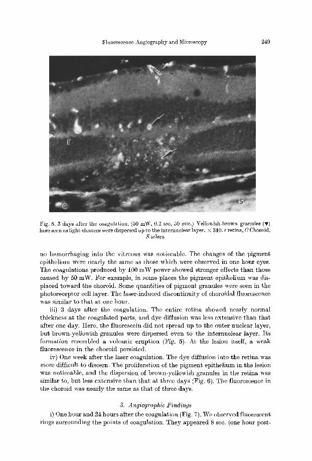
Fluorescence Angiography and Microscopy 249
Fig. 5.3 days after the coagulation. (50 mW, 0.2 see, 50 mic.) Yellowish-brown granules (V) here seen as light clusters were dispersed up to the internuclear layer. • 310. r retina, C Choroid,
S sclera
no hemorrhaging into the vitreous was noticeable. The changes of the pigment epithelium were nearly the same as those which were observed in one hour eyes. The coagulations produced by 100 mW power showed stronger effects than those caused by 50 mW. For example, in some places the pigment epithelium was dis- placed toward the choroid. Some quantities of pigment granules were seen in the photoreceptor cell layer. The laser-induced discontinuity of choroidM fluorescence was similar to tha t at one hour.
iii) 3 days after the coagulation. The entire retina showed nearly normal thickness at the coagulated parts, and dye diffusion was less extensive than that after one day. Here, the fluorescein did not spread up to the outer nuclear layer, but brown-yellowish granules were dispersed even to the internuclear layer. I t s formation resembled a volcanic eruption (Fig. 5). At the lesion itself, a weak fluorescence in the ehoroid persisted.
iv) One week after the laser coagulation. The dye diffusion into the retina was more difficult to discern. The proliferation of the pigment epithelium in the lesion was noticeable, and the dispersion of brown-yellowish granules in the retina was similar to, but less extensive than that at three days (Fig. 6). The fluorescence in the choroid was nearly the same as that of three days.
3. Angiographic Findings i) One hour and 24 hours after the coagulation (Fig. 7). We observed fluorescent
rings surrounding the points of coagulation. They appeared 8 sec. (one hour post-

250 H. Baurmann et al.
Fig. 6. One week after the coagulation (50 mW, 0.2 see, 50 mic.). Proliferation of the pigment epithelium and dispersion of granules (V) are seen. • 265. r retina, C Choroid, S selera
Fig. 7. Fluoreseein angiography (24 hours: 100 mW, 0.2 see, 50 mic.) ging-formation (~) , snow-ball-like diffusion (~v ~v) and point fluorescein leakages through the capillaries (v ) can be
observed
op.) and 6 see. (24 hours post-op.) a f te r the dye inject ion. Abou t 5 see. a f te r i ts appearance , this f luorescent r ing was enlarged bo th outs ide as well as inside, and thereaf ter , the r ings changed s lowly into snow-bal l - l ike format ions which were well d is t inguishable 150 see. af ter in jec t ion (24 hours post-op.) . I n the one hour

Fluorescence Angiography and Microscopy 251
post-op angiographies these appeared later, and dark areas within the ring still remained at 5 minutes. Thereafter this confluent fluorescence was noticeable through the late phase of the angiography. Leakage from the capillaries was in only some lasered areas discernable.
ii) 3 days after the coagulation. Granular, fluorescence staining in the coagula- ted areas appeared during the early phase (about 5-6 see.), and its intensity increasing gradually, but not uniformly, throughout the entire coagulated area. The ring-shaped fluorescence around the lesion could also be discerned, but it was not as distinct as that mentioned above.
iii) One week after the coagulation. A granular fluorescence was observed in the coagulated areas, but the intensity of the fluorescence decreased markedly.
Discussion
We can not compare our results directly with those of others who used human, monkey or rabbit eyes because we used rats as our experimental model. Histo- pathologically, there were the same disruptions of the outer retinal layer and the pigment epithelium as are usually observed by other investigators, but the pathological changes in the rat eyes in our investigations seemed to be stronger than those reported using other experimental materials treated with comparable laser power. However, the basic changes should be nearly the same, and rat eyes lend themselves readily to experimental laser coagulation.
In this series, we performed laser coagulation with two different laser powers: 50 mW and 100 mW. No significant differences between these two different powers were observed, the main changes after the coagulation being nearly the same. At the central parts of the lesions, disruption of the pigment epithelium and occasionally even, severe destruction were usually observed, sometimes accompanied by intra- retinal hemorrhages which we Mso considered to be important changes. Under our experimental conditions, the most important laser-induced changes visible under the fluorescence microscope were mainly localized in the outer part of the retina. Formation of saddle, plauteau-like or pyramidal fluorescein diffusion is due to the degree to which the photoreceptor cell layer is destroyed. To avoid complete retinal destruction in the rat eye, an applied laser dosage of 100 mW for 0.2 sec. seemed to be the maximal, greater dosages having produced severe changes (a report on which to be published at a later date).
The control by angiography at the early stages, for example, within 24 hours after the coagulation will be more effective for the estimation of applied laser power. The so-called "ring formation" (or "halo") at the early stages after the dye injection and the "snow-ball-like" fluorescence at the later stage have also observed other authors [1], [2], [4]. I t seems that these findings should appear only during the first few days after the treatment, their duration depending upon the species of the experimental animal and the intensity of applied laser power. Moreover, this ring formation should be caused by such disturbances of the choroidal circulation as constrictions or occlusions which are due to the direct in- fluence of the laser energy. Snow-ball-like fluorescence will be caused by the dye which comes from the periphery or surroundings of the lesion and is retained within the area of the retinal lesions. Sometimes this snow-ball-like formation is

252 H. Baurmann et al.
enhanced by ano ther factor , t h a t is, f luorescence by vascular effect. This fluores- cence is d is t inguished as a po in t fluorescence wi thin the f luorescent r ing in the ear ly phases of angiography.
Ac]cnowledgments. The authors would like to express deep gratitude to Mr. Ph. Hendrickson and Mr. V. Dragomirescu. Great appreciation should also be expressed to Mr. K. Fujita general manager of Topcon Europe N. V., for his invaluable assistance. This research was generally supported by the Alexander yon ttumboldt-Stiftung.
References
1. Aoki, A.: Fluorescein angiographic and electron microscopic findings of the retinas of pigmented rabbits immediately after ruby laser photocoagulation. Folia ophthal. Jap. 23, 707-720 (1972)
2. Appel, D. J., Goldberg, M. F., Wyhinny, G. : Histopathology and ultrastructure of the argon laser lesion in human retinal and choroidal vasculature. Amer. J. Ophthal. 75, 595-609 (1973)
3. Baurmann, I-I. : Grundlagen der Fluoreseenzangiographie des Augenhintergrundes. Advanc. Ophthal. 24, 204-263 (1971)
4. I-Iosoya, S.: Ophthalmoscopic and fluorescein angiographic and histological study of chorioretinal lesion produced by ruby laser on rabbit. Acta Soc. ophthM. Jap. 75, 2127-2136 (1971)
5. Mizuno, K., Sasaki, K., Otsuki, K.: Histochemical identification of fluorescein in ocular tissue. Proceed. ISFA Tokyo 1972, Igaku Shoin, p. 221-225 (1974)
6. Ota, M. : Fluorescein microscopic studies of experimental light coagulation in the rabbit eye. Folia OphthM. Jap. 24, 499-504 (1973)
7. Sasaki, K., Otsuki, K., Mizuno, K. : I-Iistochemical identification of sodium fluorescein in normal retina. Jap. J. ophthM. 17, 323-334 (1973)
8. Tsukahara, I., Ota, M. : Angiographic-histologic study on the location of sodium fluorescein in the fundus. Proceed. ISFA Tokyo 1972, Igaku Shoin, p. 230-234 (1974)
Prof. Dr. H. Baurmann Universit~ts-Augenklinik D-5300 Bonn-Venusberg Abb~straBe 2 Federal Republic of Germany