Embed Size (px)
Citation preview
HAL Id: hal-00540078https://hal.archives-ouvertes.fr/hal-00540078
Submitted on 26 Nov 2010
HAL is a multi-disciplinary open accessarchive for the deposit and dissemination of sci-entific research documents, whether they are pub-lished or not. The documents may come fromteaching and research institutions in France orabroad, or from public or private research centers.
L’archive ouverte pluridisciplinaire HAL, estdestinée au dépôt et à la diffusion de documentsscientifiques de niveau recherche, publiés ou non,émanant des établissements d’enseignement et derecherche français ou étrangers, des laboratoirespublics ou privés.
Investigations on the aetiology of pinching off syndromein four white-tailed sea eagles (Haliaeetus albicilla) from
GermanyKerstin Müller, Elvira Schettler, Helga Gerlach, Leo Brunnberg, Hafez
Mohamed Hafez, Kim Hattermann, Reimar Johne, Rainer Kollmann, OliverKrone, Michael Lierz, et al.
To cite this version:Kerstin Müller, Elvira Schettler, Helga Gerlach, Leo Brunnberg, Hafez Mohamed Hafez, et al..Investigations on the aetiology of pinching off syndrome in four white-tailed sea eagles (Hali-aeetus albicilla) from Germany. Avian Pathology, Taylor & Francis, 2007, 36 (03), pp.235-243.�10.1080/03079450701338748�. �hal-00540078�
For Peer Review O
nly
Investigations on the aetiology of pinching off syndrome in four white-tailed sea eagles (Haliaeetus albicilla) from
Germany
Journal: Avian Pathology
Manuscript ID: CAVP-2006-0115.R2
Manuscript Type: Original Research Paper
Date Submitted by the Author:
23-Jan-2007
Complete List of Authors: Müller, Kerstin; Small Animal Clinic, Free University of Berlin
Schettler, Elvira; Leibniz Institute for Zoo and Wildlife Research; Wildlife Park Eekholt Gerlach, Helga Brunnberg, Leo; Small Animal Clinic, Free University of Berlin Hafez, Hafez; Institute for Poultry Diseases, Free University of Berlin Hattermann, Kim; Robert Koch Institute Johne, Reimar; Institute for Virology, Faculty of Veterinary Medicine, University of Leipzig Kollmann, Rainer; Project Group for the Protection of the White-tailed Sea Eagle
Krone, Oliver; Leibniz Institute for Zoo and Wildlife Research Lierz, Michael; Institute for Poultry Diseases, Free University of Berlin Linke, Sonja; Robert Koch Institute Lüschow, Dörte; Institute for Poultry Diseases, Free University of Berlin Mankertz, Anette; Robert Koch Institute Müller, Hermann; Institute for Virology, Faculty of Veterinary Medicine, University of Leipzig Prusas, Christina; Institute for Poultry Diseases, Free University of Berlin Raue, Rüdiger; Institute for Virology, Faculty of Veterinary
Medicine, University of Leipzig Soike, Dirk; Landesamt für Verbraucherschutz und Landwirtschaft
E-mail: [email protected] URL: http://mc.manuscriptcentral.com/cavp
Avian Pathology
For Peer Review O
nly
Speck, Stephanie; Leibniz Institute for Zoo and Wildlife Research Wolf, Petra; Department of Animal Nutrition, University of Veterinary Medicine Hannover Frölich, Kai; Leibniz Institute for Zoo and Wildlife Research
Keywords: birds of prey, white-tailed sea eagles, pinching off syndrome, feather abnormality
Page 1 of 43
E-mail: [email protected] URL: http://mc.manuscriptcentral.com/cavp
Avian Pathology
For Peer Review O
nly
Formatted: Footer
Cavp-2006-0115.R2
Investigations on the aetiology of pinching off syndrome in four white-tailed sea eagles
(Haliaeetus albicilla) from Germany
Kerstin Müller1*,**, Elvira Schettler2,3**, Helga Gerlach4, Leo Brunnberg1, Hafez
Mohamed Hafez5, Kim Hattermann6, Reimar Johne7, Rainer Kollmann8, Oliver Krone2,
Michael Lierz5, Sonja Linke6, Dörte Lueschow5, Annette Mankertz6, Hermann Müller7,
Christina Prusas5, Rüdiger Raue7, Dirk Soike9, Stephanie Speck2, Petra Wolf10 and Kai
Frölich2
1Small Animal Clinic, Free University of Berlin, Oertzenweg 19b, 14163 Berlin,
Germany, 2Leibniz Institute for Zoo and Wildlife Research, Alfred-Kowalke-Str. 17,
10315 Berlin, Germany, 3Wildlife Park Eekholt, 24623 Großenaspe, Germany,
4Grosshesseloherstr. 23, 81479 München, Germany, 5Institute for Poultry Diseases, Free
University of Berlin, Königsweg 63, 14163 Berlin, Germany, 6Robert Koch Institute,
Nordufer 20, 13353 Berlin, Germany, 7Institute for Virology, Faculty of Veterinary
Medicine, University of Leipzig, An den Tierkliniken 29, 04103 Leipzig, Germany,
8Project Group for the Protection of the White-tailed Sea Eagle, Olshausenstraße 40,
24118 Kiel, Schleswig-Holstein, Germany, 9Landesamt für Verbraucherschutz und
Landwirtschaft, Pappelallee 20, 14469 Potsdam, and 10Department of Animal Nutrition,
University of Veterinary Medicine Hannover, Bischofsholer Damm 15, 30173 Hannover,
Germany
Deleted: Manuscript 1
Style Definition: Normal: Font:
Times New Roman, Bold, Left
Formatted: Section start:
Continuous
Page 2 of 43
E-mail: [email protected] URL: http://mc.manuscriptcentral.com/cavp
Avian Pathology
For Peer Review O
nly
Formatted: Footer
*To whom correspondence should be addressed. Tel: 0049 30 83862388. Fax: 0049 30
83862521. E-mail: [email protected]
**First two authors contributed equally to this publication.
Deleted: Manuscript 1
Page 3 of 43
E-mail: [email protected] URL: http://mc.manuscriptcentral.com/cavp
Avian Pathology
For Peer Review O
nly
Formatted: Footer
Abstract
The purpose of this study was to investigate the aetiology of the pinching off syndrome
(POS), a generalised feather abnormality affecting free-living fledglings of the white-
tailed sea eagle (Haliaeetus albicilla) (WTSE) in Europe. For the first time, extensive
clinical, haematological, biochemical, virological, bacteriological, nutritional,
histopathological, parasitological and electron microscopical examinations were
performed on three females and one male suffering from POS. Early and increased
cytokeratin formation at the base of regenerating feathers and their follicle was
observed in affected birds. Ultrathin sections of the feather papillae revealed an
extended stratum transitivum and a compact, thickened keratinised stratum corneum.
The transitional cells in POS feathers contained vacuoles often associated with the
nucleus. Lipofuscin accumulations in neurons, glial cells
and islet cells of the pancreas were found in all examined birds. It was not clear if there
is an association between the occurrence of lipofuscin and POS.
No evidence was found to suggest that infectious agents (parasites, bacteria, fungi or
viruses), malnutrition or hormonal imbalances are involved in the aetiology of POS in
WTSEs. It remains unclear whether there is a genetic background of POS.
Deleted: Manuscript 1
Page 4 of 43
E-mail: [email protected] URL: http://mc.manuscriptcentral.com/cavp
Avian Pathology
For Peer Review O
nly
Formatted: Footer
Key words
birds of prey, feather abnormality, feather loss, feather dystrophy, pinching off
syndrome, white-tailed sea eagles, Haliaeetus albicilla
Introduction
Due to intense human persecution the German population of the white-tailed sea eagle
(WTSE) was believed to consist of only 20 breeding pairs at the beginning of the 20th
century (Hauff 2004). The population increased slowly after the species was protected
but decreased again between 1950 and 1970 due to environmental contamination with
DDT (Hauff 1998). The numbers remained relatively constant until the end of the
seventies. Between 1970 and 1980 the German population of WTSEs consisted of
approximately 100 breeding pairs (Hauff 2004). At this time (1975) the first case of a
generalised feather abnormality was found in a WTSE fledgling from Schleswig-
Holstein, Germany (Rüger 1981). From 1975 to 2006, a total of 17 fledglings of free-
living WTSEs with a feather abnormality (pinching off syndrome, POS) were found
near their nesting sites in Germany. In Europe, altogether 32 cases are documented
during that period (Müller, et al. in press).
At the time of capture all primaries, secondaries and rectrices of affected WTSEs were
malformed, and many feathers were already lost. The coverts were involved to various
degrees. No affected bird was able to fly. Some birds were kept in captivity for several
years but feather quality continued to worsen.
Deleted: Manuscript 1
Page 5 of 43
E-mail: [email protected] URL: http://mc.manuscriptcentral.com/cavp
Avian Pathology
For Peer Review O
nly
Formatted: Footer
A wide variety of reasons for generalised skin and feather abnormalities in birds are
known, including parasitic, bacterial, fungal and viral infections, trauma, chemical or
toxic substances, inadequate nutrition, genetic, neoplastic, immune-mediated diseases,
allergy or irradiation, behavioural disorders (Cooper & Harrison 1994) as well as
hormonal imbalances (Oglesbee 1992). The cause of POS is unknown and no therapy is
available to date. The aim of the present study of four birds with POS was to investigate
the aetiology of POS and to describe the clinical appearance, the pathology and
histopathology of feathers and organs of affected birds.
Materials and Methods
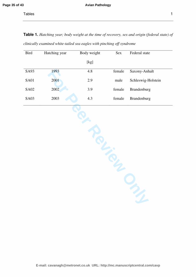
Animals. The study was conducted between 2001 and 2004 on four German WTSEs
found in the wild with generalised feather abnormalities. The investigated birds were
from three different German federal states Schleswig-Holstein (SA01), Brandenburg
(SA02; SA03) and Saxony-Anhalt (SA93) (Table 1). The three females and one male
were recently fledged at the time of capture. They were found on the ground near their
nests in the months July (n=2) and August (n=2). At the time of the first clinical
investigations three WTSEs were fledglings (SA01, SA02, SA03) and approximately 15
to 20 weeks old. The fourth bird (SA93), already 10 years old, was found in 1993 and
kept in captivity until 2003. SA01 was observed during the nestling period and showed
visible alterations of the flight feathers by the age of 10 weeks. The feather condition was
clinically evaluated different times in SA01 and SA02, and the birds were kept for 31
and 17 months, respectively. SA03 was kept for only two months.
Deleted: Manuscript 1
Page 6 of 43
E-mail: [email protected] URL: http://mc.manuscriptcentral.com/cavp
Avian Pathology
For Peer Review O
nly
Formatted: Footer
In all birds the primaries, secondaries and rectrices were either lost or malformed;
hence all birds were never able to fly.
Clinical examination and laboratory tests. Initial diagnostic procedures included
evaluation of the feather status and lateral and ventrodorsal radiographs of the whole
body. Endoscopy of the coelom was conducted in SA01 and SA02 some days after
admission using routine methods (Taylor 1994). Growing abnormal primary and
secondary feathers, and rectrices were obtained under isoflurane inhalation anaesthesia
from three birds (SA01, SA02, SA03). Blood samples for virological examination,
haematology, biochemistry and heavy metal analysis were obtained from the Vena
ulnaris. Haematocrit was determined with the microhaematocrit method and
haemoglobin was measured spectrophotometrically using the cyanmethaemoglobin
method. Analysis of blood chemistry was carried out on lithium heparin plasma
immediately after blood sampling by Konelab 30® (Thermo Electron GmbH, Dreieich,
Germany) at 37°C and included potassium, sodium, total calcium, anorganic
phosphorus, alanine aminotransferase (ALT), aspartate aminotransferase (AST),
glutamate dehydrogenase (GLDH), lactate dehydrogenase (LDH), glucose, urea, uric
acid, alkaline phosphatase (AP), creatine kinase (CK), total protein (TP, biuret method),
and cholinesterase. Albumin was measured via electrophoresis. Blood lead levels were
measured by atomic absorption spectrometry in a federal laboratory (Federal
Environmental Agency Berlin, Germany).
For investigation of thyroid gland function, a TSH-stimulation test was conducted in all
four birds and one control bird without feather abnormalities. After blood sampling for
baseline plasma thyroxine level, 1IU thyroid-stimulating-hormone (TSH, Sigma-Aldrich
Deleted: Manuscript 1
Page 7 of 43
E-mail: [email protected] URL: http://mc.manuscriptcentral.com/cavp
Avian Pathology
For Peer Review O
nly
Formatted: Footer
Chemie GmbH, Munich, Germany) was injected in the Musculus supracoracoideus and
a second blood sample was taken six hours later. Plasma was separated and stored at -
20°C. Radioimmunoassay analysis was performed in a commercial laboratory.
Pathology and parasitology. All four WTSEs were euthanised, because the feather
quality deteriorated and release into the wild was not possible. The birds were measured
and weighed prior to dissection. Detailed pathological techniques including the
assessment of the body condition have been described previously (Krone, et al. 2003).
Postmortem examination concentrated on signs of specific diseases, organ alterations,
parasites and specific alterations of feathers and skin. For histopathology, formalin fixed
samples of the heart, liver, lungs, airsacs, kidneys, spleen, ovary or testes, oviduct,
proventriculus, ventriculus, duodenum, jejunum, different parts of the brain (cerebrum,
cerebellum), thyroid glands, parathyroid glands, pancreas, adrenal glands, bursa
cloacalis, thymus, large blood vessels and growing feathers (primaries, secondaries and
rectrices) were embedded in paraffin and cut at 3 µm. Standard stains included
haematoxylin and eosin (HE), periodic acid-Schiff reaction (PAS), and Turnbull blue
(TB).
Tissue samples from growing feathers from biopsies and samples collected from
euthanised birds (primaries, secondaries and rectrices) including the feather follicles as
well as various organs were stored in 7% formalin for histopathology, in cold modified
Karnowsky medium and 3% glutaraldehyde for electron microscopy and at -20°C and -
80°C for negative staining.
Growing contour feathers still containing pulp of all birds were examined for quill mites
by standard methods (Clayton & Wather 1997).
Deleted: Manuscript 1
Page 8 of 43
E-mail: [email protected] URL: http://mc.manuscriptcentral.com/cavp
Avian Pathology
For Peer Review O
nly
Formatted: Footer
Electron microscopy. In search of an infectious aetiology of POS negative staining for
electron microscopy was performed. Tissue homogenates of the bursa cloacalis, spleen,
liver, skin, thymus, intestinal mucosa, caecal tonsils, brain and feather follicle were
diluted 1:5 in PBS, sonicated for 40 s at 20 kHz and clarified for 15 min at 3000g.
Supernatant was used for direct preparation and particle enrichment. For particle
enrichment the supernatant was centrifuged 12 min at 120,000g directly on the
microscopic grid using an air-driven ultracentrifuge (Beckman Airfuge, Global Medical
Instrumentation, Ramsey, USA). Specimens were stained with 2% phosphotungstic acid
(PTA).
Tissue samples of the bursa cloacalis, spleen, liver and caecal tonsils of one juvenile bird
(SA03) with POS were prepared for ultrastructural analysis according to standard
procedure: glutardialdehyde-fixation, osmium tetroxide postfixation, dehydration,
epoxy resin embedding, staining with uranyl acetat and lead citrate. Specimens were
examined with a Jeol JEM 1010 electron microscope at 80 kV.
For fine structural analysis basal parts (2-3 mm in diameter) of growing primaries and
secondaries of two WTSEs with POS (SA01, SA03) and one control bird without feather
abnormalities were prepared. Chemical fixation, dehydration and embedding of
specimens for light- and electron microscopy were performed according to Elias &
Friends (1975) and Pfeiffer et al. (2000). Areas of cross sectioned basal parts of
regenerating feathers and its follicles were preselected in the light microscope using
semithin sections (60-70 nm). Fine structural analysis was performed in the stratum
germinativum of the epidermal collar and the follicle wall (terminology as described by
Deleted: Manuscript 1
Page 9 of 43
E-mail: [email protected] URL: http://mc.manuscriptcentral.com/cavp
Avian Pathology
For Peer Review O
nly
Formatted: Footer
Lucas & Stettenheim, 1972). Ultrathin sections were examined with a Philips CM 10
electron microscope.
Bacteriology. Sterile 0.9 mm cotton swabs (Hain Diagnostika, Nehren, Germany) were
used to take samples from feather follicles and feather pulp of five primaries of every
bird, collected aseptically during feather biopsies and necropsies. Swabs were stored in
transport medium (Hain Diagnostika) and kept at room temperature up to 5 hours until
plated onto Columbia agar (5% sheep blood), Chocolate agar, Gassner medium,
Sabouraud agar and MacConkey II agar (Oxoid, Wesel, Germany). All agar plates were
incubated aerobically at 37°C for 24-72 h; Columbia agar and Chocolate agar plates in
5% CO2 (AnaeroJar 2.5l, Oxoid). In addition, Sabouraud agar plates were incubated at
room temperature. Swabs were also cultured for 24 hours in trypticase soy broth for
enrichment and then subcultured only if the enrichment broth appeared cloudy.
Primary identification of microbial isolates was based on Gram staining, cellular
morphology, catalase and oxidase reaction. Commercially available analytical profile
index (API) from bioMérieux (Nürtingen, Germany) were applied for the identification
of staphylococci and micrococci (API Staph), streptococci (API 20 Strep), coryneform
bacteria (API Coryne), and gram-negative nonfermenting bacteria (API 20 NE).
Conventional biochemical tests (Bisping & Amtsberg 1988; Quinn, et al. 2000) were also
used.
Virus cultivation. Liver, lung, spleen, bursa cloacalis homogenates and cloacal swabs
were inoculated on primary chicken embryo liver cell monolayer (CEL), prepared from
11 day old specified pathogen free embryonated chicken eggs (Valo, Lohmann Tierzucht
Deleted: Manuscript 1
Page 10 of 43
E-mail: [email protected] URL: http://mc.manuscriptcentral.com/cavp
Avian Pathology
For Peer Review O
nly
Formatted: Footer
GmbH, Germany). Tissue samples from liver, spleen, feather follicle, and buffy coat
were used to inoculate confluent monolayers of chicken embryo fibroblast (CEF).
Inoculated cell cultures were incubated at 37°C and examined daily for cytopathic effect
(CPE) for 5-7days. Three blind passages were carried out for each sample before it was
judged to be negative for virus propagation.
Polymerase chain reactions (PCRs). DNA for reticuloendotheliosis virus detection
(REV) was extracted from tissue, buffy coat and from the 3rd passage of CEF cell
suspension by the QIAamp DNA Mini Kit (Qiagen GmbH, Hilden Germany). Two
different primer pairs were chosen for detection of REV proviral DNA amplifying a 291
bp fragment of the REV long terminal repeat (LTR) region (Aly, et al. 1993; Diallo, et al.
1998) and a 642 bp fragment of the envelope gene (Singh, et al. 2000) respectively.
The presence of DNA of avian polyomavirus (APV), beak and feather disease virus
(BFDV) and pigeon circovirus (PiCV) was investigated using DNA isolated from EDTA
blood, feather follicle tissue, spleen, kidney, liver, heart, skin, bursa cloacalis, thymus,
intestinal mucosa, and the caecal tonsil of all birds by PCR as described by Johne &
Müller (1998), Raue et al. (2004) and Raue et al. (2005), respectively. These PCRs were
performed with DNA isolated from the samples using the DNeasy Tissue Kit (Qiagen,
Hilden, Germany) and Taq DNA polymerase (PeqLab, Germany).
Additionally, for circovirus consensus PCR, capable of detecting different avian
circoviruses (Todd et al., 2001), DNA was prepared from EDTA blood and feather
follicle tissue (Qiagen DNeasy tissue kit; Qiagen, Hilden/Germany). Initially, samples
were tested in a control PCR (Hattermann, et al. 2004), then circovirus consensus PCR
was performed according to Todd et al. (2001).
Deleted: Manuscript 1
Page 11 of 43
E-mail: [email protected] URL: http://mc.manuscriptcentral.com/cavp
Avian Pathology
For Peer Review O
nly
Formatted: Footer
All PCRs were controlled by the use of a
negative control reaction to which no
sample-DNA was added. y Plasmids containing the specific PCR products were used as
positive controls. PCR products were separated on ethidium bromide-stained agarose
gels and visualised by UV transillumination.
Serology. Antibodies against West Nile virus (WNV) were detected using two different
methods. For the neutralisation test plasma samples were diluted in Dulbecco's modified
eagle medium (DMEM) and 25 µL, which had been mixed with the same volume of a
virus suspension WNV genotype 1 containing 50 TCID50/mL, were added to a 96-well
microtiter plate and the mixture was incubated for 2 hours at 37°C. Vero cells (104 cells
in 100 µL medium) were added to each well and incubated for 3 days at 37°C. All
specimens were initially tested in duplicate at a dilution of 1:10. Reactive samples were
retested at dilutions of 1:10-1:320.
Immunofluorescence test was conducted for detection of IgG plasma antibodies against
WNV. Commercially available immunofluorescence test slides (EUROIMMUN AG,
Lübeck, Germany) coated with WNV-infected cells and a commercial antibird FITC
labelled antibody (Bethyl, Inc., Montgomery, Texas, U.S.) were used. Following the
manufacturer’s protocol, the diluted plasma (1:10, 1:50) was incubated with the cells for
30 min. The slides were washed, the conjugate was added and they were incubated again
for 30 min. After a second washing step the biochips were embedded and the slides were
analysed under a fluorescence microscope.
Deleted: Manuscript 1
Page 12 of 43
E-mail: [email protected] URL: http://mc.manuscriptcentral.com/cavp
Avian Pathology
For Peer Review O
nly
Formatted: Footer
Determination of amino acid levels in feathers and plasma. Feather contents of at least
two removed or shed deformed primaries or secondaries of the four WTSEs with POS
and three feathers of a WTSE without feather abnormalities were analysed. After
washing the feathers with acetone they were dried and ground. Amino acids were
analysed after oxidation to determine levels of cystine and methionine and after
hydrolysis in 6M HCL at 110°C to determine levels of other amino acids. Determination
of the individual amino acids for feathers and plasma was carried out by ionic
exchanger chromatography (amino acids analyzer, model LC 3000, Biotronic, Berlin,
Germany) using different time zone elutions of individual amino acids with different
buffers. Afterwards measurements of the several tapes were done photometrically by a
colour reaction with ninhydrine.
Determination of trace elements in feathers and plasma. To estimate levels of the trace
elements copper and zinc, ground feathers were mixed with 20 ml nitric acid (HNO3,
65%) and 1 ml hydrogen peroxide (H2O2, 30%) and dissolved by microwave acid
digestion in a high performance rotor (mls 1200 mega, Milestone, Sorisole, Italy). A
„high performance rotor“ (HPR-1000/6, Milestone, Sorisole, Italy) was utilised to
pulverise feathers. Hereby organic matter of feathers is destroyed and the residues
comprise among others the inorganic trace elements copper and zinc. These elements in
feathers and plasma were detected by atomic absorption spectrometry (Unicam solaar
969, Unicam, Cambridge, United Kingdom).
Results
Deleted: Manuscript 1
Page 13 of 43
E-mail: [email protected] URL: http://mc.manuscriptcentral.com/cavp
Avian Pathology
For Peer Review O
nly
Formatted: Footer
Clinical examination. At the first clinical presentation all birds were infested with
feather lice. In SA01, a few quill mites belonging to the Family Ascouracaridae (Gaud &
Atyeo 1976) were detected once in the calamus of a lost feather. Treatment with
ivermectin (Ivomec®, 0.4 mg/kg i.m. once a week over three weeks) eliminated the mites,
but caused no improvement in feather quality.
All birds had wounds on one or both wings medial of the carpal joint caused by
mechanical irritation through wing flapping. Pruritus was not observed.
In bird SA93 a slight body shivering were observed at all times. Radiographs and
endoscopy revealed no significant alterations. The feather condition deteriorated during
the time of captivity in all birds.
Apart from this and the feather abnormalities no other clinical findings could be
observed.
Description of the feather abnormalities. All WTSEs were unable to fly. The appearance
of the syndrome differed slightly between all four birds. Main characteristics of the
flight and tail feathers were as follows:
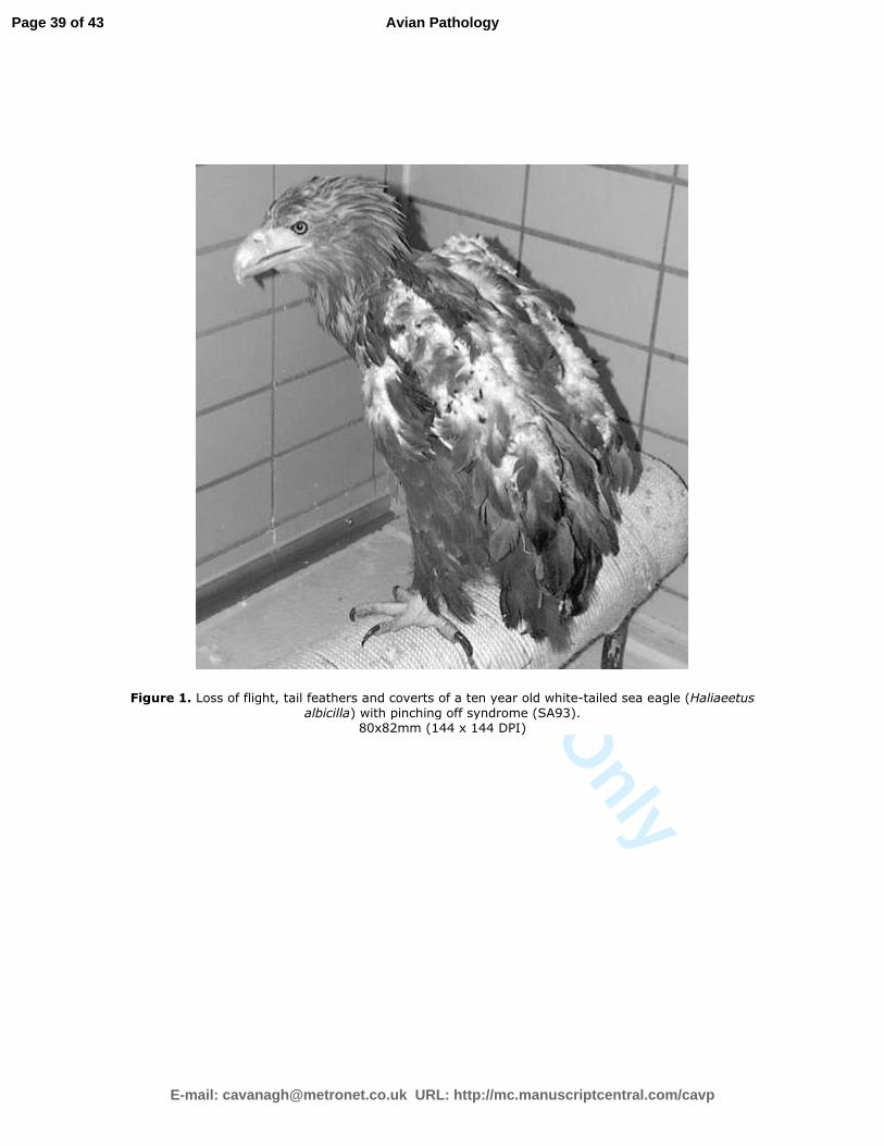
Feather loss and malformation (Figure 1)
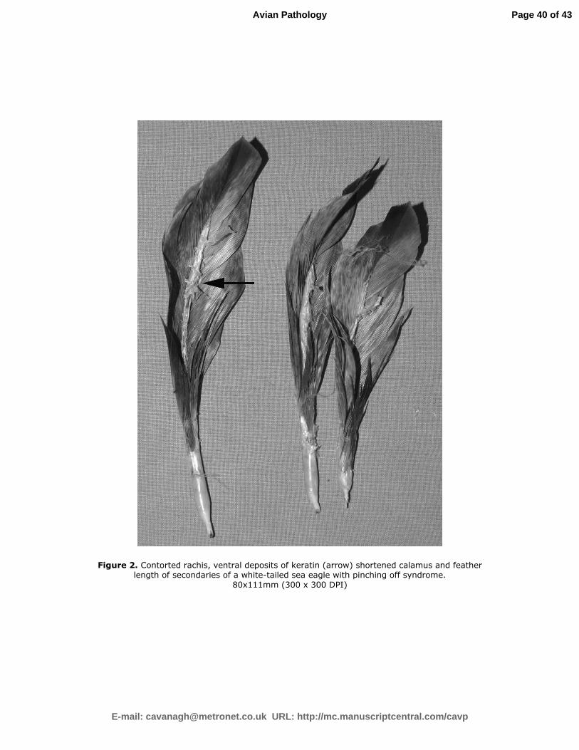
Rachis contorted or/and ventral open, ventral deposits of keratin (Figure 2)
Vane flabby and curved ventrally (Figure 2)
Calamus scaly and shortened (Figure 2)
Proximal umbilicus constricted, irregular, with feather sheath remains
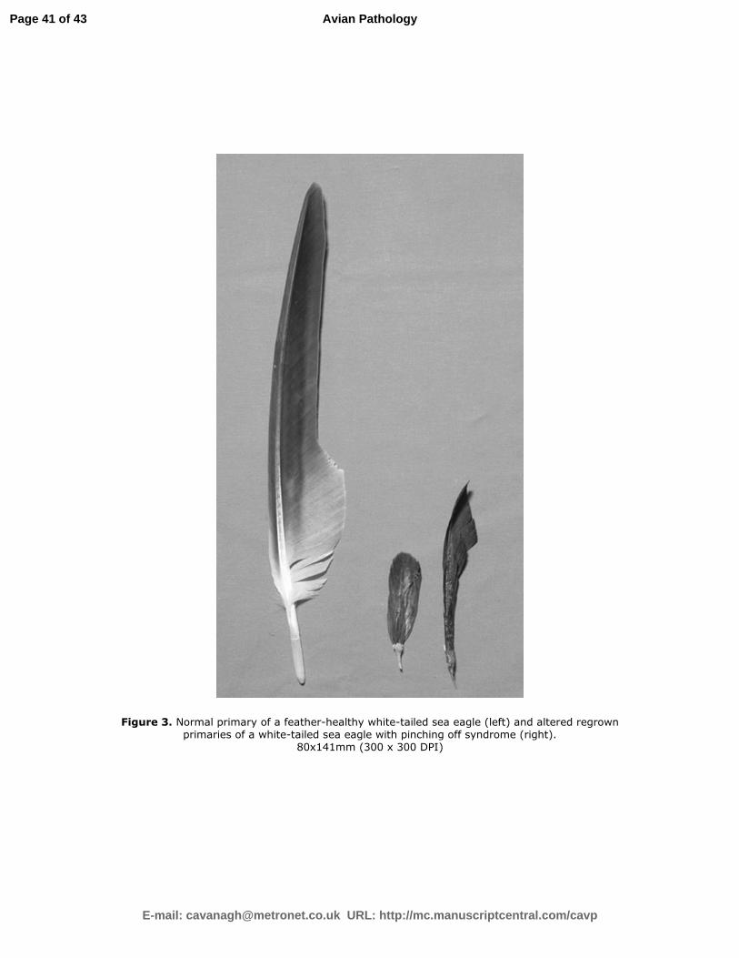
Feather length normal to extremely shortened (Figure 2, 3).
Deleted: Manuscript 1
Formatted: No bullets or
numbering
Page 14 of 43
E-mail: [email protected] URL: http://mc.manuscriptcentral.com/cavp
Avian Pathology
For Peer Review O
nly
Formatted: Footer
The coverts were modified in all but one bird (SA01) with ventral scaly rachis and
reduced quantity (Figure 1). The new developing feathers were extremely short and
abnormal in appearance (Figure 3). The feather quality worsened over time. In birds
kept in captivity for years (SA01 31 months, SA02 17 months, SA93 10 years,) no
moulting period were observed, the birds lost and replaced their feathers continuously.
Blood analysis. The blood chemistry and haematology values were similar to those of
healthy nestlings (Müller, unpublished). Blood lead and mercury levels ranged between
0.013-0.045 ppm and 0.001-0.250 ppm, respectively and are considered as background
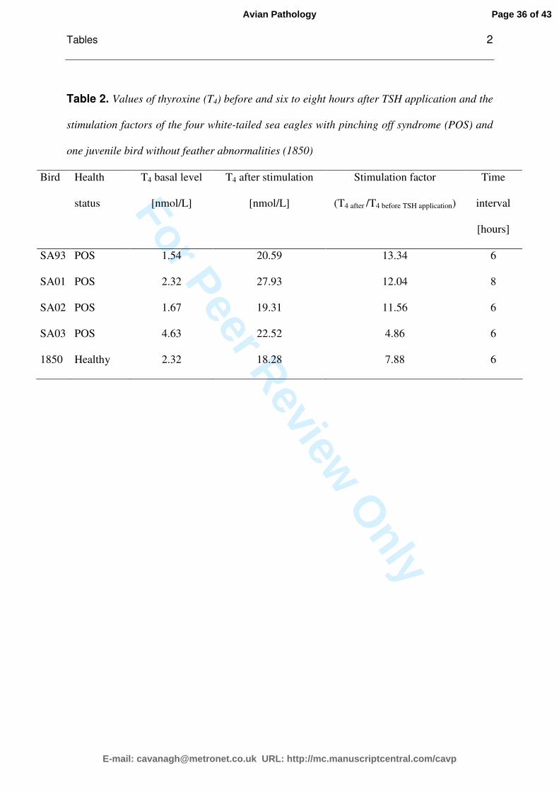
levels (Church, et al. 2006; Jagoe, et al. 2002). TSH stimulation test showed an evident
increase of thyroxine (T4) level (Table 2).
Histopathology of the feathers. 52 primaries, secondaries and rectrices of all four
WTSEs with POS were histologically examined (SA93 n=18; SA01 n=3; SA02 n=9; SA03
n=22). Although various numbers of feathers were investigated per bird the proportion
of feathers altered as described below did not differ between three individuals (SA93,
SA02, SA03). Only three suitable feathers could be examined from SA01. In these
feathers some alterations were not diagnosed, but this is most probably contributeddue
to the low number of examined feathers. In 27 feathers (51.9% of feathers examined,
n=4 birds) deposits of keratin were observed in the papilla between the epithelium of the
feather follicle and the epithelium of the pulp. In the pulp epithelium dyskeratosis
(23.1%, 12 feathers in 3 birds) and degeneration associated with vacuoles in the
cytoplasm as well as necrosis were diagnosed (40.4%, n=3 birds). In 32.7% of the
investigated feathers (n=4 birds) degeneration and necrosis of the epithelia appeared in
Deleted: Manuscript 1
Comment [M1]: As lead is never normal in living organisms, low lead levels are called background levels. This is the valid term used in studies dealing with heavy metal intoxications in birds. (E.g., Church et al. 2006)
Page 15 of 43
E-mail: [email protected] URL: http://mc.manuscriptcentral.com/cavp
Avian Pathology
For Peer Review O
nly
Formatted: Footer
the epidermal collar and the papilla. The epithelia, which accompanied the developing
feathers, were modified in a similar way. The epithelium of the feather sheath was
altered due to degeneration and necrosis (17.3% of feathers, n=3 birds) as well as
dyskeratosis (19.2% of feathers, n=4 birds).
The epithelium of the feather follicle was affected to a lesser extent: in 11.5% (n=2 birds)
degeneration and necrosis, in 11.5% (n=2 birds) dyskeratosis. Pathological proliferation
was mainly observed at the pulp epithelium (19.2%, n=3 birds).
Due to alterations of the epithelium in the pulp and feather sheath also the development
of the ramogenic columns and in consequentially vane formation was impaired. A
change of the growth direction of the rami (13.5%, n=3 birds) was leading to a
disordered appearance of the vanes. Damage to keratin formation resulted in 13.5%
(n=3 birds) in missing barbs or the rami consisted of few cell layers. In 23.1% (n=4
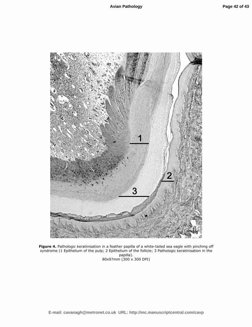
birds) of feathers, premature keratinisation of the ramogenic column inhibited further
growth (Figure 4). In 7.7% of the feathers (n=2 birds), dyskeratosis in the ramogenic
column was observed.
In 75% of the examined growing feathers (n=4 birds) a marked heterophil leucocytosis
in the pulp was observed. The papilla and the epidermal collar were affected in 17.3%,
but the epithelium of the feather sheath was affected in only 3.8%. Leucocytes were not
detected in the epithelium of the feather follicle.
Heterophil leucocytes were also visible between the ramogenic columns and the more
proximally growing rami in 17.3% of feathers (n=3 birds). In only one case high
numbers of bacteria were detected between the rami. The bacteria were small and it was
Deleted: Manuscript 1
Page 16 of 43
E-mail: [email protected] URL: http://mc.manuscriptcentral.com/cavp
Avian Pathology
For Peer Review O
nly
Formatted: Footer
not possible to determine their morphology. Haemorrhages within the pulp were
detected in 65.4% of the feathers (n=3 birds).
Histopathology of the skin. Dyskeratosis was observed focally in the epidermis of all
birds. Deposits of fibrin were detected in two birds. Proliferation of the stratum
germinativum (n=1 bird, SA93) and degenerate nuclei (n=1 bird, SA93) were, however,
rare.
Other alterations of the skin were necrotising dermatitis (n=1 bird, SA93), perivascular
inflammation (n=3 birds), degeneration of the smooth muscles (n=3 birds), hyaline
degeneration (n=1 bird, SA93), subendothelial atherosclerosis-like deposits in dermal
blood vessels as well as brightly stainable and enlarged nuclei infiltrating the walls of
these vessels (n=1 bird, SA93).
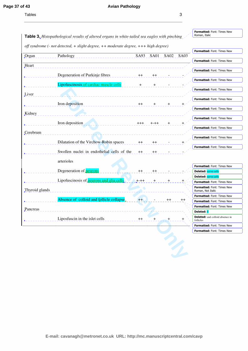
Histopathology of the organs. Lipofuscin was detected at varying levels in all birds
(Table 3).
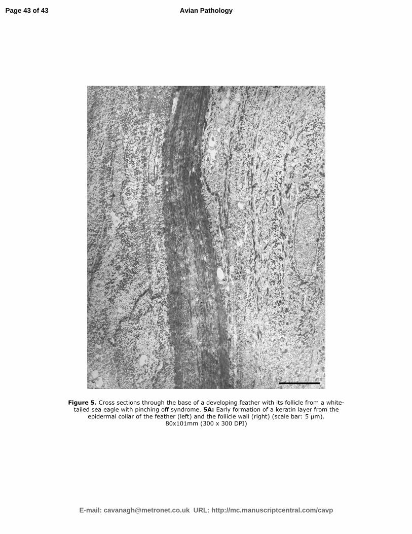
Ultrastructure. Differences within the fine structure of the germinative layer were found
between the two POS birds and the healthy bird. In control feathers the stratum
germinativum was well stratified - from pulp to follicle - by one layer of stratum basale
and several layers of stratum intermedium and stratum transitivum. Only a thin
keratinised outer layer (stratum corneum) was tightly attached to the thin stratum
corneum of the follicle. In affected feathers, the cell layers of the stratum germinativum
were less easily distinguishable. Main characteristics were an extended stratum
transitivum and a compact, several microns thick keratinised layer representing the
Deleted: Manuscript 1
Page 17 of 43
E-mail: [email protected] URL: http://mc.manuscriptcentral.com/cavp
Avian Pathology
For Peer Review O
nly
Formatted: Footer
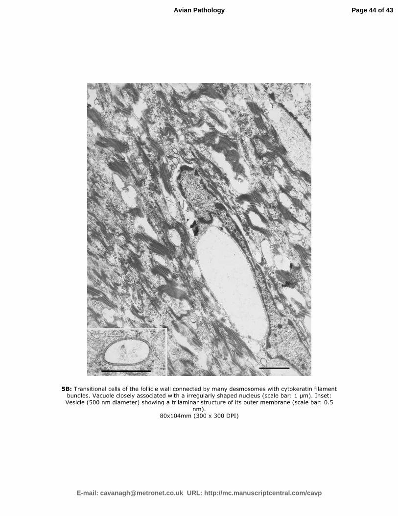
stratum corneum (Figure 5A). Cells of the stratum transitivum in affected feathers were
connected by many desmosomes. Numerous bundles of cytokeratin filaments extended
from the desmosomes throughout the cells (Figure 5B). The transitional cells contained
vacuoles with dense inclusions often closely associated with the nucleus and small
vesicles (200-500 nm) in the cytoplasm (Figure 5B). The outer membrane of the small
vesicles in cross-sectional profiles often displayed a trilaminar structure with
intramembrane particles. Such vesicles were not observed in control feathers.
Incidentally, melanocytes with many melanosomes and cell extensions into the stratum
transitivum were found in the stratum intermedium of one POS feather (SA03), but not in
the other birds. The ultrastructural appearance corresponded well with the
histopathological findings. Ultrastructurally, neither the feather papillae nor the
examined organs showed any evidence of an infectious cause of POS. Negative contrast
staining also failed to reveal an infectious agent in the growing feathers and in the
organs of all birds.
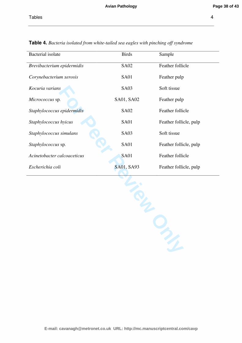
Bacteriology. Moderate numbers of ten different bacteria were recovered from the
feather follicle, feather pulp and the soft tissue around the feather follicle (Table 4).
Most of the isolated bacteria belonged to the group of Gram-positive cocci and Gram-
positive coryneform rods. Two species comprised Gram-negative rods. The predominant
genus found was Staphylococcus. Fungi were not detected in any of the samples.
Virology. All PCRs were negative for REV, APV, BFDV and PiCV. Also, the consensus
PCR, which is capable of detecting a broad range of different circoviruses, was negative
Deleted: Manuscript 1
Page 18 of 43
E-mail: [email protected] URL: http://mc.manuscriptcentral.com/cavp
Avian Pathology
For Peer Review O
nly
Formatted: Footer
in all cases. In addition no virus was cultivated by passage in CEL. No antibodies against
WNV were detected.
Trace elements and amino acids in feathers and blood plasma. No significant differences
between control birds and POS birds were detected in the trace element and amino acid
content of the analysed feathers and plasma.
Discussion
The four investigated birds belong to a group of free-living WTSEs that were found in
Germany between 1975 and 2006 showing a generalised feather loss and feather
malformation known as POS. Only nestlings of both sexes were affected.
Most case reports about feather abnormalities in birds of prey describe pinching off as a
loss of one or a few abnormal premature primaries or rectrices (Cooper 1972; Glasier
1980; Grünhagen 1988; Mavrogordato 1960). Typical characteristics of the affected
feathers are constricted calamus and pinched appearance (Cooper 1972; Glasier 1980;
Grünhagen 1988). Diminished flying and hunting abilities were not described by those
workers and a normal feather will regrow after some days (Glasier 1980; Grünhagen
1988; Mavrogordato 1960).
In the Netherlands, a generalised feather abnormality was reported in fledglings of the
northern goshawk (Accipiter gentilis) (Bijlsma, et al. 1994; Ottens, et al. 1997; Van
Geneijgen, et al. 1995; Vedder 2000). No investigations into the aetiology have been
performed.
Deleted: Manuscript 1
Page 19 of 43
E-mail: [email protected] URL: http://mc.manuscriptcentral.com/cavp
Avian Pathology
For Peer Review O
nly
Formatted: Footer
In our study, extensive examinations of affected birds were conducted.
Quill mites (Syringophilidae) were reported to be associated with feather loss in birds of
prey (Philips 2000). Quill mites of the Family Ascouracaridae were identified in one bird.
However, feather abnormalities did not improve after treatment with ivermectin. No
quill mite infestation in any of the other birds was observed. Quill mites are therefore
not thought to be the main cause of POS in WTSEs.
Malley & Whitbread (1996) reported that avian skin is fairly resistant to primary bacterial
infection. In our study, only a limited number of bacteria commonly identified as normal skin
flora in other animals were isolated. Gram-positive cocci predominated which is consistent
with investigations regarding the normal bacterial flora in common buzzards (Buteo buteo)
(Pérez, et al. 1994). Among the Gram-negative bacteria isolated were Acinetobacter species,
which are typically detected in soil and water. Little is known, however, about their
pathogenicity for wild animals (Quinn, et al. 2000). Escherichia coli was isolated from two
birds but these are regarded as contamination because only a few bacteria were detected
histopathologically in the feather papillae and only in one case. In contrast to reports about
bacterial polyfolliculitis (Oglesbee & Oglesbee 1994; Rosskopf, et al. 1983) our birds never
revealed signs of pruritus nor feather follicle swelling as seen by Bayer et al. (1976).
Mycotic infections of feathers in pigeons and psittacine birds can lead to feather loss in
several parts of the body especially at the breast. In some cases, pruritus and feather
pulling were also observed (Gartrell, et al. 2005; Tudor 1983). Feather loss and
hyperkeratotic short feathers were symptoms of fungal infections in Gouldian finches
(Erythrura gouldiae) and Java sparrows (Padda oryzivora) (Pass 1989). Spores as well as
hyphae were present in the keratinised layers (Pass 1989), but could not be detected in
the birds of the present study.
Circovirus (Latimer, et al. 1991; Pass, et al. 1994; Raidal & Riddoch 1997; Stewart, et al.
2006), polyomavirus (Bernier, et al. 1981, 1984), adenovirus (Cooper & Harrison 1994),
parvovirus (Cooper & Harrison 1994; Jestin, et al. 1991), REV (Koyama, et al. 1975;
Deleted: Manuscript 1
Formatted: Font: Not Bold
Page 20 of 43
E-mail: [email protected] URL: http://mc.manuscriptcentral.com/cavp
Avian Pathology
For Peer Review O
nly
Formatted: Footer
Koyama, et al. 1980; Tajima, et al. 1977) and WNV (P. Redig, personal communication)
are known to cause generalised feather loss and/or feather malformation in various bird
species. The PCR tests applied yielded no evidence that circoviruses, APV or REV are
involved in the development of POS. Also, no viruses were detected by inoculation of the
samples onto cultures of CEL cells. However, the possibility cannot be excluded that
presently unknown viruses which could not be detected by the used PCR assays or
which could not be propagated in CEL cells are involved in POS.
Several reports exist about feather abnormalities in undernourished poultry.
Deficiencies in vitamin B6 (Gehle & Balloun 1965), selenium and vitamin E (Supplee
1966), zinc (Supplee, et al. 1961; Young, et al. 1958) and amino acids like arginine
(Anderson & Warnick 1967), glycine (Robel 1977), valin, leucine, isoleucine or
phenylalanine and tyrosine (Anderson & Warnick 1967; Robel 1977) are described. The
present study, however, revealed no abnormalities of amino acid and trace element
composition. Since the general physical condition in all birds was normal and stress
marks were absent in affected remiges and rectrices, it therefore seems unlikely that
malnutrition caused POS.
In poultry the oral intake of T-2 toxin produced by Fusarium spp. (Hoerr, et al. 1981;
Wyatt, et al. 1975) may also cause feather alterations. In this case, however, it is unlikely
that T-2 toxin was involved, because affected birds typically suffer a reduced growth
rate (Wyatt, et al. 1975) and other organ lesions like gastroenteritis and hepatocellular
degeneration occur (Mishra, et al. 1987). Such signs were not observed in the present
study.
Hypothyroidism was associated with feather loss in a scarlet macaw (Ara macao)
(Oglesbee 1992). In all four WTSEs with POS, thyroxine basal levels were comparable
Deleted: Manuscript 1
Page 21 of 43
E-mail: [email protected] URL: http://mc.manuscriptcentral.com/cavp
Avian Pathology
For Peer Review O
nly
Formatted: Footer
with these of scarlet macaws (1.72±0.66 nmol/L) and African grey parrots (Psittacus
erithacus) (1.83±0.57 nmol/L) (Lothrop, et al. 1985), but much lower than reported for
pigeons (16.5-26.6 nmol/L) (Lumeij & Westerhof 1988). An increase 5 to 13 times of
thyroxine basal level was observed six or eight hours after TSH application, which is
comparable to investigations in psittacine species by Lothrop et al. (1985) and pigeons by
Lumeij & Westerhof (1988). However, histopathology revealed a moderate follicle
collapse and absence of colloid from follicles of the thyroid glands in three birds.
Although it is not possible to explain this apparent discrepancy at present,
hypothyroidism is considered unlikely to be the primary cause of POS in WTSEs since
histopathological alterations were moderate, the thyroid glands could be adequately
stimulated with TSH and no other organ alterations were found.
There was no histopathological indication that adrenal gland degeneration caused
feather loss as described by Onderka and Claffey (1987) in a blue and gold macaw (Ara
ararauna).
Some inherited feather abnormalities are known in poultry. Dysplastic remiges of
chicken, that may follow an autosomal recessive genetic expression (Urrutia, et al. 1983),
seem to be very similar to POS found in WTSEs. The WTSE population suffered two
crises during the last 150 years caused by human persecution until the beginning of the
20th century and by a reduced breeding success due to the accumulation of DDT in the
fifties, sixties and seventies (Hauff 1998). Inbreeding during these periods might have
contributed to the appearance of POS.
This is the first description of histopathology and ultrastructure of POS in WTSEs and
two major findings seem to be important. The early and increased cytokeratin
formation at the base of a regenerating feather and its follicle seems to interrupt the
Deleted: Manuscript 1
Formatted: Adjust space
between Latin and Asian text,
Adjust space between Asian text
and numbers
Page 22 of 43
E-mail: [email protected] URL: http://mc.manuscriptcentral.com/cavp
Avian Pathology
For Peer Review O
nly
Formatted: Footer
normal development leading to the altered appearance of the feather. Lipofuscin
accumulation in neurons, glial cells and pancreas was found in all investigated birds.
One of these birds (SA93, ten years old) had tremors. This might be associated with
lipofuscin accumulation, since lipofuscin is known to cause degenerative neurological
diseases in humans (Gray & Woulfe 2005), horses (Hartley, et al. 1982), sheep (Jolly, et
al. 1990), cattle (Bianchi 1979), dogs (Borras, et al. 1999), cats (Bildfell, et al. 1995;
Green & Little 1974) and was also reported in a nine-month old peach-faced love bird
(Agapornis roseicollis) (Reece & Macwhirter 1988). Lipofuscin is a lipoprotein that
linearly accumulates intralysosomally in postmitotic cells, especially in neurons and
cardiac myocytes. It is a degradation product of cell constituents and is considered to be
a normal manifestation of aging (Porta 1991). Lipofuscin has been found in neurons of
human infants at the age of four months (Porta 2002). It still remains unknown why
lipofuscin is not completely degraded by the lysosomes (Porta 2002). The appearance of
a comparatively high amount of lipofuscin especially in the two young WTSEs and
whether there is an association between the occurrence of lipofuscin and POS needs
further evaluation, although in the authors' view, the latter seems unlikely.
In conclusion, our investigations have given no indication that infectious agents
(parasites, bacteria, fungi or viruses), malnutrition or hormonal imbalances play a role
in the aetiology of POS in WTSEs. Moreover, the clinical and histological examinations
did not reveal that trauma, chemical or toxic substances, neoplasia or allergy were likely
to be the cause of POS. Feather loss and malformation seem to be a consequence of
increased and premature cytokeratin formation in the growing feather. It remains
unclear whether there is a genetic background of POS.
Deleted: Manuscript 1
Page 23 of 43
E-mail: [email protected] URL: http://mc.manuscriptcentral.com/cavp
Avian Pathology
For Peer Review O
nly
Formatted: Footer
Acknowledgement
We would like to thank H. Pendl for performing the blood cell counts, S. Pfeiffer and M.
Beese for assistance with the ultrastructural examination of feather material and I.
Nehlmeier for help by testing for West Nile virus infection.. We also thank D. Haas, P.
Sömmer, H. Freymann and A. Valentin for their support and J. R. Philips as well as
Carol Ksiazek for their helpful comments.
This study was partly funded by the Brandenburg State Office for Environment and the
German Nature Conservation Union (NABU), Section Berlin.
References
Deleted: Manuscript 1
Formatted: Line spacing: single
Formatted: Indent: Left: 0 pt,
First line: 0 pt, Line spacing:
single
Formatted: Indent: Left: 0 pt,
First line: 0 pt
Page 24 of 43
E-mail: [email protected] URL: http://mc.manuscriptcentral.com/cavp
Avian Pathology
For Peer Review O
nly
Formatted: Footer
Aly, M.M., Smith, E.J. & Fadly, A.M. (1993). Detection of reticuloendotheliosis virus
infection using the polymerase chain reaction. Avian Pathology, 22, 543-554.
Anderson, J.O. & Warnick, R.E. (1967). Gross abnormalities in chicks fed amino acid
deficient diets. Poultry Science, 46, 856-862.
Bayer, R.C., Muir, F.V., Chawan, C.B. & Bryan, T.A. (1976). Infected feather follicles in
cage reared broilers. Poultry Science, 55, 1194-1200.
Bernier, G., Morin, M. & Marsolais, G. (1981). A generalized inclusion body disease in
the budgerigar (Melopsittacus undulatus) caused by a papovavirus-like agent. Avian
Diseases, 25, 1083-1092.
Bernier, G., Morin, M. & Marsolais, G. (1984). Papovavirus induced feather
abnormalities and skin lesions in the budgerigar: clinical and pathological findings.
Canadian Veterinary Journal, 25, 307-310.
Bianchi, M. (1979). Lipofuscine in the neurons of large herbivores during growth and
old age. Atti del Convegno Nazionale Societa Italiana delle Scienze Veterinarie, 33, 137.
Bijlsma, R.G., Speelman, R., Ottens, H.J., Hasper, H. & van Manen, W. (1994). Een
veerafwijking bij een nestjonge Havik Accipiter gentilis. De Takkeling, 2, 38-40.
Bildfell, R., Matwichuk, C., Mitchell, S. & Ward, P. (1995). Neuronal ceroid-
lipofuscinosis in a cat. Veterinary Pathology, 32, 485-488.
Bisping, W. & Amtsberg, G. (1988). Colour atlas for the diagnosis of bacterial pathogens
in animals. Berlin, Hamburg: Paul Parey Scientific Publishers.
Borras, D., Ferrer, I. & Pumarola, M. (1999). Age-related changes in the brain of the
dog. Veterinary Pathology, 36, 202-211.
Church, M.E., Gwiazda, R., Risebrough, R.W., Sorenson, K., Chamberlain, C.P., Farry,
S., Heinrich, W., Rideout, B.A. & Smith, D.R. (2006). Ammunition is the principal
Deleted: Manuscript 1
Formatted: Indent: Left: 0 pt,
First line: 0 pt, Line spacing:
single
Page 25 of 43
E-mail: [email protected] URL: http://mc.manuscriptcentral.com/cavp
Avian Pathology
For Peer Review O
nly
Formatted: Footer
source of lead accumulated by California condors re-introduced to the wild.
Environmental Science & Technology, 40, 6143-6150.
Clayton, D.H. & Wather, B.A. (1997). Collection and quantification of arthropod
parasites of birds. In D.H. Clayton, & J. Moore. (1997). Host-parasite evolution. General
principles and avian models. (pp419-440). Oxford: Oxford University Press.
Cooper, J.E. (1972). Feather conditions in birds of prey. Journal of the North American
Falconers' Association, 11, 39-44.
Cooper, J.E. & Harrison, G.J. (1994). Dermatology. In B.W. Ritchie, G.J. Harrison, &
L.R. Harrison. (1994). Avian medicine: principles and application (pp607-639). Lake
Worth, Florida: Wingers Publishing.
Diallo, I.S., MacKenzie, M.A., Spradbrow, P.B. & Robinson, W.F. (1998). Field isolates
of fowlpox virus contaminated with reticuloendotheliosis virus. Avian Pathology, 27, 60-
66.
Elias, P.M. & Friends, D.S. (1975). The permeability barrier in mamalian epidermis.
The Journal of Cell Biology, 65, 185-189.
Gartrell, B.D., Rogers, L. & Alley, M.R. (2005). Eosinophilic dermatitis associated with
Trichosporon asahii in a cockatiel (Nymphicus hollandicus). Journal of Avian Medicine
and Surgery, 19, 25-29.
Gaud, J. & Atyeo, W.T. (1976). Ascouracarinae, n. sub-fam. des Syringobiidae,
sarcoptiformes plumicioles. Acarologia, 18, 143-162.
Gehle, M.H. & Balloun, S.L. (1965). Selected hemocytological effects of vitamin B6
deficiency in chicks. The Journal of Nutrition, 87, 197-201.
Glasier, P. (1980). Moulting, imping and coping. London: BT Batsford.
Deleted: Manuscript 1
Page 26 of 43
E-mail: [email protected] URL: http://mc.manuscriptcentral.com/cavp
Avian Pathology
For Peer Review O
nly
Formatted: Footer
Gray, D.A. & Woulfe, J. (2005). Lipofuscin and aging: a matter of toxic waste. Sciences
of Aging Knowledge Environment, 2005, re1.
Green, P.D. & Little, P.B. (1974). Neuronal ceroid-lipofuscin storage in Siamese cats.
Canadian Journal of Comparative Medicine, 38, 207-212.
Grünhagen, H. (1988). Federanomalien bei Greifvögeln. Greifvögel und Falknerei, 73-76.
Hartley, W.J., Kuberski, T., LeGonidec, G. & Daynes, P. (1982). The pathology of
Gomen disease: a cerebellar disorder of horses in New Caledonia. Veterinary Pathology,
19, 399-405.
Hattermann, K., Maerz, A., Slanina, H., Schmitt, C. & Mankertz, A. (2004). Assessing
the risk potential of porcine circoviruses for xenotransplantation: consensus primer-
PCR-based search for a human circovirus. Xenotransplantation, 11, 547-50.
Hauff, P. (1998). Bestandsentwicklung des Seeadlers Haliaeetus albicilla in Deutschland
seit 1980 mit Rückblick auf die vergangenen 100 Jahre. Vogelwelt, 119, 47-63.
Hauff, P. (2004). Seeadler (Haliaeetus albicilla). In K. Gedeon, A. Mitschke, & C.
Sudfeldt. (2004). Brutvögel in Deutschland (pp8-9). Hohenstein-Ernstthal: Stiftung
Vogelmonitoring Deutschland.
Hoerr, F.J., Carlton, W.W. & Yagen, B. (1981). Mycotoxicosis caused by a single dose of
T-2 toxin or diacetoxyscirpenol in broiler chickens. Veterinary Pathology, 18, 652-664.
Jagoe, C.H., Bryan, A.L., Jr., Brant, H.A., Murphy, T.M. & Brisbin, I.L., Jr. (2002).
Mercury in bald eagle nestlings from South Carolina, USA. Journal of Wildlife Diseases,
38, 706-712.
Jestin, V., Bras, M.O.l., Cherbonnel, M., Gall, G.l. & Bennejean, G. (1991).
Demonstration of highly pathogenic parvovirus (Derzsy's disease virus) in flocks of
muscovy ducks. Recueil de Medecine Veterinaire, 167, 849-857.
Deleted: Manuscript 1
Page 27 of 43
E-mail: [email protected] URL: http://mc.manuscriptcentral.com/cavp
Avian Pathology
For Peer Review O
nly
Formatted: Footer
Johne, R. & Müller, H. (1998). Avian polyomavirus in wild birds: genome analysis of
isolates from Falconiformes and Psittaciformes. Archives of Virology, 143, 1501-1512.
Jolly, R.D., Martinus, R.D., Shimada, A., Fearnley, R.M. & Palmer, D.N. (1990). Ovine
ceroid-lipofuscinosis is a proteolipid proteinosis. Canadian Journal of Veterinary
Research, 54, 15-21.
Koyama, H., Nagashima, T., Ohwada, Y. & Saito, Y. (1975). Cause of "nakanuke" in
chickens. II. Isolation of C-type virus from material infected with turkey herpesvirus.
The Kitasato Archives of Experimental Medicine, 48, 93-105.
Koyama, H., Sasaki, T., Ohwada, Y. & Saito, Y. (1980). The relationship between
feathering abnormalities ("nakanuke") and tumour production in chickens inoculated
with reticuloendotheliosis virus. Avian Pathology, 9, 331-340.
Krone, O., Langgemach, T., Sömmer, P. & Kenntner, N. (2003). Causes of mortality in
white-tailed sea eagles from Germany.In B. Helander, M. Marquiss, & W. Bowerman
(Eds). Proccedings of the Sea Eagle 2000 (pp211-218). Björkö, Sweden.
Latimer, K.S., Rakich, P.M., Niagro, F.D., Ritchie, B.W., Steffens, W.L., Compagnoli,
R.P., Pesti, D.A. & Lukert, P.D. (1991). An updated review of psittacine beak and
feather disease. Journal of the Association of Avian Veterinarians, 5, 211-221.
Lothrop, C.D., Jr., Loomis, M.R. & Olsen, J.H. (1985). Thyrotropin stimulation test for
evaluation of thyroid function in psittacine birds. Journal of the American Veterinary
Medical Association, 186, 47-48.
Lucas, A.M. & Stettenheim, P.R. (1972). Avian Anatomy. Integument. Part II.
Washington, D.C.: Gov. Print. Off.
Lumeij, J.T. & Westerhof, I. (1988). Clinical evaluation of thyroid function in racing
pigeons (Columba livia domestica). Avian Pathology, 17, 63-70.
Deleted: Manuscript 1
Page 28 of 43
E-mail: [email protected] URL: http://mc.manuscriptcentral.com/cavp
Avian Pathology
For Peer Review O
nly
Formatted: Footer
Malley, A.D. & Whitbread, T.J. (1996). The integument. In P.H. Beynon, N. Forbes, &
N.H. Harcourt-Brown. (1996). Manual of raptors, pigeons and waterfowl (pp129-139).
Cheltenham: British Small Animal Veterinary Association.
Mavrogordato, J.G. (1960). A hawk for the bush. London: HF & G Witherby.
Mishra, U.K., Dwarkanath, P.K. & Hossain, M.I. (1987). Clinical manifestation and
hepatic trace minerals in growing chickens as influenced by T-2 toxin. Indian Journal of
Animal Sciences, 57, 1069-1074.
Müller, K., Altenkamp, R., Brunnberg, L., Fašungová, L., Freymann, H., Frölich, K.,
Kollmann, R., Krone, O., Literák, I., Mizera, T., Sömmer, P. & Schettler, E. (In press).
Pinching off syndrome in free-living white-tailed sea eagles (Haliaeetus albicilla) from
Europe - frequency and distribution of a generalized feather abnormality. Journal of
Avian Medicine and Surgery, In press.
Oglesbee, B.L. (1992). Hypothyroidism in a scarlet macaw. Journal of the American
Veterinary Medical Association, 201, 1599-1601.
Oglesbee, B.L. & Oglesbee, M.J. (1994). Feather dystrophy in a cockatiel (Nymphicus
hollandicus). Journal of the Association of Avian Veterinarians, 8, 16-20.
Onderka, D.K. & Claffey, F.P. (1987). Adrenal degeneration associated with feather loss
in a macaw. Canadian Veterinary Journal, 28, 193-194.
Ottens, H.J., Jansman, H. & Speelman, R. (1997). Genetische afwijking
hoogstwaarschijnlijk andermaal oorzaak van veerafwijking bij nestjonge Havik
Accipiter gentilis. De Takkeling, 5, 12-16.
Pass, D.A. (1989). The pathology of the avian integument: a review. Avian Pathology, 18,
2-72.
Deleted: Manuscript 1
Page 29 of 43
E-mail: [email protected] URL: http://mc.manuscriptcentral.com/cavp
Avian Pathology
For Peer Review O
nly
Formatted: Footer
Pass, D.A., Plant, S.L. & Sexton, N. (1994). Natural infection of wild doves (Streptopelia
senegalensis) with the virus of psittacine beak and feather disease. Australian Veterinary
Journal, 71, 307-308.
Pérez, J.M., Extremera, A.L. & Ruiz, I. (1994). Bacteriological study of the feathers and
lice of captive common buzzards (Buteo buteo). Avian Pathology, 23, 163-168.
Pfeiffer, S., Vielhaber, G., Vietzke, J.-P., Wittern, K.-P., Hintze, U. & Wepf, R. (2000).
High-pressure freezing provides new information on human epidermis: simultaneous
protein antigen and lamellar lipid structure preservation. Study on human epidermis by
cryoimmobilization. The Journal of Investigative Dermatology, 114, 1030-1038.
Philips, J.R. (2000). A review and checklist of the parasitic mites (Acarina) of the
Falconiformes and Strigiformes. Journal of Raptor Research, 34, 210-231.
Porta, E.A. (1991). Advances in age pigment research. Archives of Gerontology and
Geriatrics, 12, 303-320.
Porta, E.A. (2002). Pigments in aging: an overview. Annals of the New York Academy of
Sciences, 959, 57-65.
Quinn, P.J., Carter, M.E., Markey, B. & Carter, G.R. (2000). Clinical Veterinary
Microbiology. London: Mosby.
Raidal, S.R. & Riddoch, P.A. (1997). A feather disease in senegal doves (Streptopelia
senegalensis) morphologically similar to psittacine beak and feather disease. Avian
Pathology, 26, 829-836.
Raue, R., Johne, R., Crosta, L., Bürkle, M., Gerlach, H. & Müller, H. (2004). Nucleotide
sequence analysis of a C1 gene fragment of psittacine beak and feather disease virus
amplified by real-time polymerase chain reaction indicates a possible existence of
genotypes. Avian Pathology, 33, 41-50.
Deleted: Manuscript 1
Page 30 of 43
E-mail: [email protected] URL: http://mc.manuscriptcentral.com/cavp
Avian Pathology
For Peer Review O
nly
Formatted: Footer
Raue, R., Schmidt, V., Freick, M., Reinhardt, B., Johne, R., Kamphausen, L., Kaleta,
E.F., Müller, H. & Krautwald-Junghanns, M.-E. (2005). A disease complex associated
with pigeon circovirus infection, young pigeon disease syndrome. Avian Pathology, 34,
418-425.
Reece, R.L. & MacWhirter, P. (1988). Neuronal ceroid lipofuscinosis in a lovebird. The
Veterinary Record, 122, 187.
Robel, E.J. (1977). A feather abnormality in chicks fed diets deficient in certain amino
acids. Poultry Science, 56, 1968-1971.
Rosskopf, W.J., Jr., Woerpel, R.W., Sievers, M.J. & Pater, C. (1983). Treatment of
feather folliculitis in a lovebird. Modern Veterinary Practice, 64, 923.
Rüger, A. (1981). Bestandsstützung durch Adoptionsverfahren - Erfahrungen mit
Seeadlern in Schleswig-Holstein. Natur und Landschaft, 56, 133-135.
Singh, P., Kim, T.J. & Tripathy, D.N. (2000). Re-emerging fowlpox: evaluation of
isolates from vaccinated flocks. Avian Pathology, 29, 449-455.
Stewart, M.E., Perry, R. & Raidal, S.R. (2006). Identification of a novel circovirus in
Australian ravens (Corvus coronoides) with feather disease. Avian Pathology, 35, 86-92.
Supplee, W.C. (1966). Feather abnormality in poults fed a diet deficient in vitamin E
and selenium. Poultry Science, 45, 852-854.
Supplee, W.C., Creek, R.D., Combs, G.F. & Blamberg, D.L. (1961). The zinc
requirements of poults receiving pracitcal diets. Poultry Science, 40, 171-176.
Tajima, M., Nunoya, T. & Otaki, Y. (1977). Pathogenesis of abnormal feathers in
chickens inoculated with reticuloendotheliosis virus. Avian Diseases, 21, 77-89.
Deleted: Manuscript 1
Page 31 of 43
E-mail: [email protected] URL: http://mc.manuscriptcentral.com/cavp
Avian Pathology
For Peer Review O
nly
Formatted: Footer
Taylor, M. (1994). Endoscopic examination and biopsy techniques. In B.W. Ritchie, G.J.
Harrison, & L.R. Harrison. (1994). Avian medicine: principles and application (pp327-
354). Lake Worth, Florida: Wingers Publishing.
Todd, D., Weston, J.H., Soike, D. & Smyth, J.A. (2001). Genome sequence
determinations and analyses of novel circoviruses from goose and pigeon. Virology, 286,
354-362.
Tudor, D.C. (1983). Mycotic infection of feathers as the cause of feather-pulling in
pigeons & psittacine birds. Veterinary Medicine, Small Animal Clinician, 78, 249-253.
Urrutia, M.S., Crawford, R.D. & Classen, H.L. (1983). Dysplastic remiges, a genetic
abnormality reducing feathering in the domestic fowl. The Journal of Heredity, 74, 101-
104.
van Geneijgen, P., van Nie, G.J. & de Smid, T. (1995). Veerafwijking bij nestjonge
Havik. De Takkeling, 3, 91.
Vedder, O. (2000). Veerafwijking bij nestjonge Havik Accipiter gentilis, en mogelijnk
oorzaak. De Takkeling, 8, 221-222.
Wyatt, R.D., Hamilton, P.B. & Burmeister, H.R. (1975). Altered feathering of chicks
caused by T-2 toxin. Poultry Science, 54, 1042-1045.
Young, R.J., Edwards, H.M. & Gillis, M.B. (1958). Studies in zinc in poultry nutrition.
II. Zinc requirements and deficiency symptoms of chicks. Poultry Science, 37, 1100-1107.
Deleted: Manuscript 1
Formatted: Line spacing: single
Page 32 of 43
E-mail: [email protected] URL: http://mc.manuscriptcentral.com/cavp
Avian Pathology
For Peer Review O
nly
Formatted: Footer
Figure 1. Loss of flight, tail feathers and coverts of a ten year old white-tailed sea eagle
(Haliaeetus albicilla) with pinching off syndrome (SA93).
Figure 2. Contorted rachis, ventral deposits of keratin (arrow) shortened calamus and
feather length of secondaries of a white-tailed sea eagle with pinching off syndrome.
Figure 3. Normal primary of a feather-healthy white-tailed sea eagle (left) and altered
regrown primaries of a white-tailed sea eagle with pinching off syndrome (right).
Figure 4. Pathological keratinisation in a feather papilla of a white-tailed sea eagle with
pinching off syndrome (1 Epithelium of the pulp; 2 Epithelium of the follicle; 3
Pathological keratinisation in the papilla).
Figure 5. Cross sections through the base of a developing feather with its follicle from a
white-tailed sea eagle with pinching off syndrome. 5A: Early formation of a keratin
layer from the epidermal collar of the feather (left) and the follicle wall (right) (scale
bar: 5 µm). 5B: Transitional cells of the follicle wall connected by many desmosomes
with cytokeratin filament bundles. Vacuole closely associated with an irregularly shaped
nucleus (scale bar: 1 µm). Inset: Vesicle (500 nm diameter) showing a trilaminar
structure of its outer membrane (scale bar: 0.5 nm).
Deleted: Manuscript 1
Formatted: Line spacing: single
Formatted: Indent: Left: 0 pt,
First line: 0 pt, Line spacing:
single
Formatted: Indent: Left: 0 pt,
First line: 0 pt, Line spacing:
single
Formatted: Indent: Left: 0 pt,
First line: 0 pt, Line spacing:
single
Page 33 of 43
E-mail: [email protected] URL: http://mc.manuscriptcentral.com/cavp
Avian Pathology
For Peer Review O
nly
Formatted: Footer
Deleted: Manuscript 1
Page 34 of 43
E-mail: [email protected] URL: http://mc.manuscriptcentral.com/cavp
Avian Pathology
For Peer Review O
nly
Tables 1
Table 1. Hatching year; body weight at the time of recovery, sex and origin (federal state) of
clinically examined white-tailed sea eagles with pinching off syndrome
Bird Hatching year Body weight
[kg]
Sex Federal state
SA93 1993 4.8 female Saxony-Anhalt
SA01 2001 2.9 male Schleswig-Holstein
SA02 2002 3.9 female Brandenburg
SA03 2003 4.3 female Brandenburg
Page 35 of 43
E-mail: [email protected] URL: http://mc.manuscriptcentral.com/cavp
Avian Pathology
For Peer Review O
nly
Tables 2
Table 2. Values of thyroxine (T4) before and six to eight hours after TSH application and the
stimulation factors of the four white-tailed sea eagles with pinching off syndrome (POS) and
one juvenile bird without feather abnormalities (1850)
Bird Health
status
T4 basal level
[nmol/L]
T4 after stimulation
[nmol/L]
Stimulation factor
(T4 after /T4 before TSH application)
Time
interval
[hours]
SA93 POS 1.54 20.59 13.34 6
SA01 POS 2.32 27.93 12.04 8
SA02 POS 1.67 19.31 11.56 6
SA03 POS 4.63 22.52 4.86 6
1850 Healthy 2.32 18.28 7.88 6
Page 36 of 43
E-mail: [email protected] URL: http://mc.manuscriptcentral.com/cavp
Avian Pathology
For Peer Review O
nly
Tables 3
Table 3. Histopathological results of altered organs in white-tailed sea eagles with pinching
off syndrome (- not detected, + slight degree, ++ moderate degree, +++ high degree)
Organ Pathology SA93 SA01 SA02 SA03
Heart
Degeneration of Purkinje fibres ++ ++ - -
Lipofuscinosis of cardiac muscle cells + + - -
Liver
Iron deposition ++ + + +
Kidney
Iron deposition +++ +-++ + +
Cerebrum
Dilatation of the Virchow-Robin spaces ++ ++ - +
Swollen nuclei in endothelial cells of the
arterioles
++ ++ - -
Degeneration of neurons ++ ++ - -
Lipofuscinosis of neurons and glia cells +-++ + + +
Thyroid glands
Absence of colloid and follicle collapse ++ - ++ ++
Pancreas
Liposfuscin in the islet cells ++ + + +
Formatted: Font: Times New
Roman, Italic
Formatted: Font: Times New
Formatted: Font: Times New
Formatted: Font: Times New
Formatted: Font: Times New
Formatted: Font: Times New
Formatted: Font: Times New
Formatted: Font: Times New
Formatted: Font: Times New
Formatted: Font: Times New
Formatted: Font: Times New
Formatted: Font: Times New
Formatted: Font: Times New
Formatted: Font: Times New
Formatted: Font: Times New
Roman, Not Italic
Formatted: Font: Times New
Formatted: Font: Times New
Formatted: Font: Times New
Formatted: Font: Times New
Formatted: Font: Times New
Deleted: nerve cells
Deleted: nerve cells
Deleted: F
Deleted: and colloid absence in
follicles
Page 37 of 43
E-mail: [email protected] URL: http://mc.manuscriptcentral.com/cavp
Avian Pathology
For Peer Review O
nly
Tables 4
Table 4. Bacteria isolated from white-tailed sea eagles with pinching off syndrome
Bacterial isolate Birds Sample
Brevibacterium epidermidis SA02 Feather follicle
Corynebacterium xerosis SA01 Feather pulp
Kocuria varians SA03 Soft tissue
Micrococcus sp. SA01, SA02 Feather pulp
Staphylococcus epidermidis SA02 Feather follicle
Staphylococcus hyicus SA01 Feather follicle, pulp
Staphylococcus simulans SA03 Soft tissue
Staphylococcus sp. SA01 Feather follicle, pulp
Acinetobacter calcoaceticus SA01 Feather follicle
Escherichia coli SA01, SA93 Feather follicle, pulp
Page 38 of 43
E-mail: [email protected] URL: http://mc.manuscriptcentral.com/cavp
Avian Pathology
For Peer Review O
nly
Figure 1. Loss of flight, tail feathers and coverts of a ten year old white-tailed sea eagle (Haliaeetus
albicilla) with pinching off syndrome (SA93). 80x82mm (144 x 144 DPI)
Page 39 of 43
E-mail: [email protected] URL: http://mc.manuscriptcentral.com/cavp
Avian Pathology
For Peer Review O
nly
Figure 2. Contorted rachis, ventral deposits of keratin (arrow) shortened calamus and feather length of secondaries of a white-tailed sea eagle with pinching off syndrome.
80x111mm (300 x 300 DPI)
Page 40 of 43
E-mail: [email protected] URL: http://mc.manuscriptcentral.com/cavp
Avian Pathology
For Peer Review O
nly
Figure 3. Normal primary of a feather-healthy white-tailed sea eagle (left) and altered regrown primaries of a white-tailed sea eagle with pinching off syndrome (right).
80x141mm (300 x 300 DPI)
Page 41 of 43
E-mail: [email protected] URL: http://mc.manuscriptcentral.com/cavp
Avian Pathology
For Peer Review O
nly
Figure 4. Pathologic keratinisation in a feather papilla of a white-tailed sea eagle with pinching off syndrome (1 Epithelium of the pulp; 2 Epithelium of the follicle; 3 Pathologic keratinisation in the
papilla). 80x97mm (300 x 300 DPI)
Page 42 of 43
E-mail: [email protected] URL: http://mc.manuscriptcentral.com/cavp
Avian Pathology
For Peer Review O
nly
Figure 5. Cross sections through the base of a developing feather with its follicle from a white-tailed sea eagle with pinching off syndrome. 5A: Early formation of a keratin layer from the
epidermal collar of the feather (left) and the follicle wall (right) (scale bar: 5 µm). 80x101mm (300 x 300 DPI)
Page 43 of 43
E-mail: [email protected] URL: http://mc.manuscriptcentral.com/cavp
Avian Pathology
For Peer Review O
nly
5B: Transitional cells of the follicle wall connected by many desmosomes with cytokeratin filament bundles. Vacuole closely associated with a irregularly shaped nucleus (scale bar: 1 µm). Inset: Vesicle (500 nm diameter) showing a trilaminar structure of its outer membrane (scale bar: 0.5
nm). 80x104mm (300 x 300 DPI)
Page 44 of 43
E-mail: [email protected] URL: http://mc.manuscriptcentral.com/cavp
Avian Pathology