Embed Size (px)
Citation preview

INV ITEDP A P E R
Noninvasive Neural ProsthesesUsingMobile andWireless EEGA system using micro-electro-mechanical sensors has been used to
detect motion sickness and help participants maintain alertness
while using an automobile driving simulator.
By Chin-Teng Lin, Fellow IEEE, Li-Wei Ko, Jin-Chern Chiou, Member IEEE,
Jeng-Ren Duann, Member IEEE, Ruey-Song Huang, Sheng-Fu Liang,
Tzai-Wen Chiu, and Tzyy-Ping Jung, Senior Member IEEE
ABSTRACT | Neural prosthetic technologies have helped many
patients by restoring vision, hearing, or movement and
relieving chronic pain or neurological disorders. While most
neural prosthetic systems to date have used invasive or
implantable devices for patients with inoperative or malfunc-
tioning external body parts or internal organs, a much larger
population of Bhealthy[ people who suffer episodic or
progressive cognitive impairments in daily life can benefit
from noninvasive neural prostheses. For example, reduced
alertness, lack of attention, or poor decision-making during
monotonous, routine tasks can have catastrophic conse-
quences. This study proposes a noninvasive mobile prosthetic
platform for continuously monitoring high-temporal resolution
brain dynamics without requiring application of conductive
gels on the scalp. The proposed system features dry micro-
electromechanical system electroencephalography sensors,
low-power signal acquisition, amplification and digitization,
wireless telemetry, online artifact cancellation, and signal
processing. Its implications for neural prostheses are examined
in two sample studies: 1) cognitive-state monitoring of
participants performing realistic driving tasks in the virtual-
reality-based dynamic driving simulator and 2) the neural
correlates of motion sickness in driving. The experimental
results of these studies provide new insights into the under-
standing of complex brain functions of participants actively
performing ordinary tasks in natural body positions and
situations within real operational environments.
KEYWORDS | Cognitive-state monitoring; electroencephalogra-
phy (EEG); motion sickness; neural prosthesis; noninvasive
mobile prosthetic platform
I . INTRODUCTION
Prosthesis refers to a surrogate or replacement of a body
part such as a tooth, an eye, a facial bone, an ear, a hip, a
knee or other joint, a leg, or an arm. Prostheses may bedesigned for functional or cosmetic purposes or both.
Advances in biomedical science and technology have
enabled integration of some prostheses with body tissues,
including the central nervous system. These neuroprosthe-ses can now respond to commands from the brain. During
the current decade, hundreds of thousands of patients have
already been helped by neuroprosthetic technologies that
restore vision [1], hearing [2] or movement [3] and relievechronic pain or neurological disorders [4]. These neuro-
prostheses usually involve invasive or implantable devices
for patients with inoperative or malfunctioning external
body members or internal body organs [5]. However, a
much larger population of Bhealthy[ people who suffer
momentary, episodic, or progressive cognitive impair-
ments in daily life have, ironically, been overlooked. For
Manuscript received April 20, 2007; revised February 12, 2008. This work was
supported by the National Science Council, Taiwan, R.O.C., under Contracts
NSC 96-2627-E-009-001 and NSC 96-2218-E-009-001; the BAiming for the Top
University Plan,[ National Chiao-Tung University; the Ministry of Education, Taiwan,
under Contract 96W803; and the Defense Advanced Research Projects Agency under
Grant NBCH1060010.
C.-T. Lin, L.-W. Ko, and J.-C. Chiou are with the Brain Research Center and
Department of Electrical and Control Engineering, National Chiao-Tung University,
Hsinchu, Taiwan, R.O.C. (e-mail: [email protected]; [email protected];
J.-R. Duann and T.-P. Jung are with the Brain Research Center, National Chiao-Tung
University, Hsinchu, Taiwan, R.O.C. They are also with the Institute for Neural
Computation, University of California San Diego, La Jolla, CA 92093 USA
(e-mail: [email protected]; [email protected]).
R.-S. Huang is with the Institute for Neural Computation, University of California
San Diego, La Jolla, CA 92093 USA (e-mail: [email protected]).
S.-F. Liang is with the Brain Research Center, National Chiao-Tung University, Hsinchu,
Taiwan, R.O.C. He is also with the Department of Computer Science and Information
Engineering, National Cheng-Kung University, Tainan, Taiwan, R.O.C.
(e-mail: [email protected]).
T.-W. Chiu is with the Brain Research Center, National Chiao-Tung University, Hsinchu,
Taiwan, R.O.C. (e-mail: [email protected]; [email protected]).
Digital Object Identifier: 10.1109/JPROC.2008.922561
Vol. 96, No. 7, July 2008 | Proceedings of the IEEE 11670018-9219/$25.00 �2008 IEEE

example, as the last 60 years of research in human vigilanceand attention has shown, humans are not well suited for
maintaining alertness and attention under monotonous
conditions, particularly during the normal sleep phase of
their circadian cycle [6]. Catastrophic errors can result
from momentary lapses in alertness and attention during
periods of relative inactivity. Many people, not just
patients, can benefit from a prosthetic system that
continuously monitors fluctuations in cognitive states inthe workplace and/or the home. However, to be practical
for routine clinical or occupational use, the prosthetic
system must be noninvasive, nonintrusive, lightweight,
battery-powered, and easy to don and doff. Further, it must
enable a full range of head, eye, and body movements. The
only possible brain-imaging modality fulfilling these criteria
is electroencephalography (EEG). EEG is a powerful
noninvasive tool widely used for both medical diagnosisand neurobiological research because it can provide high
temporal resolution in milliseconds that directly reflects
the dynamics of the generating cell assemblies. Electroen-
cephalography is also the only brain-imaging modality that
can be performed without fixing the head/body. Substantial
research [7]–[11] has shown that many features of EEG
dynamics index the current state of subject alertness,
arousal, and attention. However, data collection in mostEEG studies requires skin preparation and gel application to
ensure good electrical conductivity between sensor and
skin. These procedures are time consuming, uncomfort-
able, and even painful for participants since skin prepara-
tion usually involves abrasion of the outer skin layer.
Repeated skin preparation and gel application for EEG may
also cause allergic reactions or infections. Further, the
signal quality may degrade over time as the skin regeneratesand the conductive gel dries. Advance electrode designs are
needed to overcome these requirements and complications
of adhesive contacts between EEG electrodes and the skin
surface before routine EEG monitoring can be feasible in
real-world environments.
Another major challenge for EEG monitoring in
operational environments is extracting meaningful and
informative event-related brain dynamics from the re-corded signals that are often subject to severe artifacts
from head/body motion or other ocular or muscle
movement. The recorded information generally has value
only in proportion to the sophistication and reliability of
the analytical method employed. Sophisticated signal-
processing techniques are often computationally expensive
and implemented either online or offline using high-end
personal computers (PCs). The substantial processingrequirements hinder the wearability, portability, and
practical use of the systems in operational environments.
However, given the recent development of embedded
system and signal-processing techniques, it is now
practical to implement these sophisticated algorithms in
embedded systems for online EEG monitoring and/or
brain–computer interface (BCI). These real-time embed-
ded systems, in conjunction with wireless transmission,have proven useful in applications such as diagnosis and
homecare systems [12] because they provide maximum
portability and wearability.
In short, the unavailability of EEG monitoring systems
that do not require application of conductive gels to the
scalp and are capable of high-definition recording, online
signal processing, and artifact cancellation has long
thwarted applications of EEG monitoring in other thanwell-controlled laboratory conditions. To make the non-
invasive prosthetic system practical for routine use, it is
essential to provide a quickly and easily donned and doffed
EEG acquisition system for implementing online signal-
processing techniques to continuously and accurately
extract meaningful and informative information. The
system should also be a convenient size, rugged, and
lightweight and have low power consumption to meet therequirements of wearability, portability, and durability.
This study details the design, development, and testing
of a noninvasive mobile prosthetic platform for continu-
ously monitoring high-temporal resolution brain dynamics
without requiring conductive gels applied to the scalp. The
system employs dry microelectromechanical system
(MEMS) EEG sensors, low-power signal acquisition,
amplification and digitization, wireless telemetry, onlineartifact cancellation, and real-time signal processing. Its
implications in neuroprostheses are also demonstrated
through two sample studies: 1) cognitive-state monitoring
of participants performing realistic driving tasks in a
virtual reality–based dynamic driving environment and
evaluation of the efficacy of the system in delivering
stimulating feedback to help maintain optimal task
performance and 2) identification of neural correlates ofmotion sickness in driving.
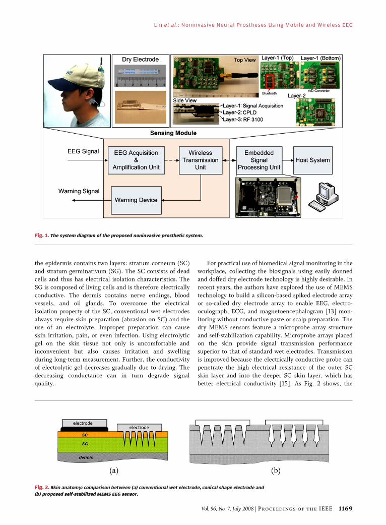
II . NONINVASIVE PROSTHETIC SYSTEM
The study reported in this paper designed and developed a
noninvasive mobile prosthetic platform (Fig. 1) consisting
of self-stabilized MEMS sensors, signal amplification and
digitization, wireless transmission/receiving, and a real-time embedded system to assess human physiological and
mental information in operational environments.
A. Self-Stabilized MEMS SensorElectrodes for biopotential measurement transduce
biosignals from skin tissue to the amplifier circuit. They
are used extensively in biomedical applications to
measure biosignals such as EEG and electrocardiogram(ECG) signals [13]. The most important task in designing
and fabricating the biopotential electrode is ensuring low
electrode/skin interface impedance so that the measured
biosignals can be bridged into the front-end circuits with
minimal attenuation and noise interference [13]. Skin
anatomy can be divided into three layers: the epidermal,
dermal, and subcutaneous layers [14]. As Fig. 2(a) shows,
Lin et al.: Noninvasive Neural Prostheses Using Mobile and Wireless EEG
1168 Proceedings of the IEEE | Vol. 96, No. 7, July 2008
the epidermis contains two layers: stratum corneum (SC)
and stratum germinativum (SG). The SC consists of dead
cells and thus has electrical isolation characteristics. The
SG is composed of living cells and is therefore electrically
conductive. The dermis contains nerve endings, blood
vessels, and oil glands. To overcome the electrical
isolation property of the SC, conventional wet electrodes
always require skin preparation (abrasion on SC) and theuse of an electrolyte. Improper preparation can cause
skin irritation, pain, or even infection. Using electrolytic
gel on the skin tissue not only is uncomfortable and
inconvenient but also causes irritation and swelling
during long-term measurement. Further, the conductivity
of electrolytic gel decreases gradually due to drying. The
decreasing conductance can in turn degrade signal
quality.
For practical use of biomedical signal monitoring in the
workplace, collecting the biosignals using easily donned
and doffed dry electrode technology is highly desirable. In
recent years, the authors have explored the use of MEMS
technology to build a silicon-based spiked electrode array
or so-called dry electrode array to enable EEG, electro-
oculograph, ECG, and magnetoencephalogram [13] mon-
itoring without conductive paste or scalp preparation. Thedry MEMS sensors feature a microprobe array structure
and self-stabilization capability. Microprobe arrays placed
on the skin provide signal transmission performance
superior to that of standard wet electrodes. Transmission
is improved because the electrically conductive probe can
penetrate the high electrical resistance of the outer SC
skin layer and into the deeper SG skin layer, which has
better electrical conductivity [15]. As Fig. 2 shows, the
Fig. 2. Skin anatomy: comparison between (a) conventional wet electrode, conical shape electrode and
(b) proposed self-stabilized MEMS EEG sensor.
Fig. 1. The system diagram of the proposed noninvasive prosthetic system.
Lin et al.: Noninvasive Neural Prostheses Using Mobile and Wireless EEG
Vol. 96, No. 7, July 2008 | Proceedings of the IEEE 1169
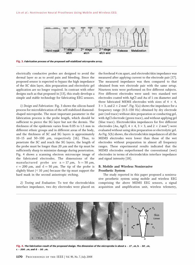
electrically conductive probes are designed to avoid the
dermal layer so as to avoid pain and bleeding. Since the
proposed sensor is expected to bypass the high impedance
of the SC skin layer, skin preparation and electrolytic gel
application are no longer required. In contrast with other
designs such as that proposed in [13], this study develops a
simple and stable technology for fabricating EEG sensors.
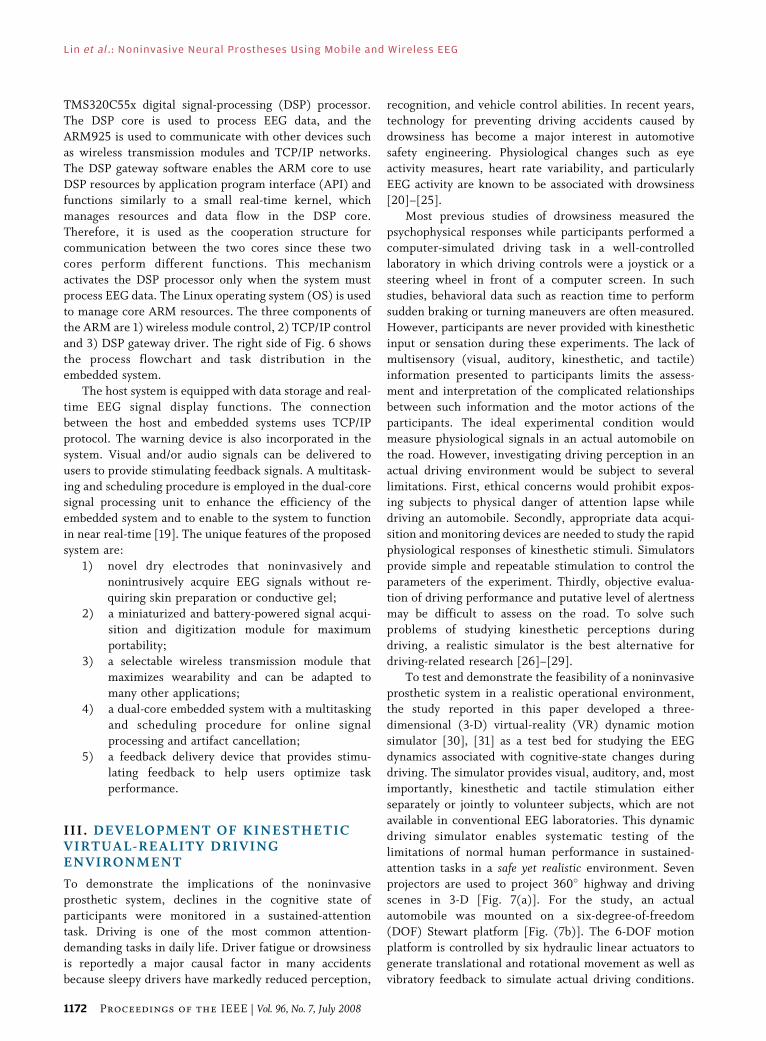
1) Design and Fabrication: Fig. 3 shows the silicon-basedprocess for microfabrication of the self-stabilized diamond-
shaped microprobe. The most important parameter in the
fabrication process is the probe length, which should be
sufficient to pierce the SG layer but not the dermis. The
thickness of the epidermis varies from 0.05 to 1.5 mm in
different ethnic groups and in different areas of the body,and the thickness of SC and SG layers is approximately
10–15 and 50–100 �m, respectively [16]. Thus, to
penetrate the SC and reach the SG layers, the length of
the probe must be longer than 20 �m and the tip must be
sufficiently sharp to minimize damage during penetration.
Fig. 4 shows a scanning electron microscope image of
the fabricated electrodes. The dimensions of the
manufactured probe are a ¼ 17 �m, b ¼ 50 �m,c ¼ 200 �m, and d ¼ 50 �m. The tip of the probe is
slightly blunt (G 10 �m) because the tip must support the
hard mask in the second anisotropic etching.
2) Testing and Evaluation: To test the electrode/skin
interface impedance, two dry electrodes were placed on
the forehead 4 cm apart, and electrode/skin impedance was
measured after applying current to the electrode pair [17].
The measured impedance was then compared to that
obtained from wet electrode pair with the same setup.
Nineteen tests were performed on five different subjects.
Five different electrodes were used: two standard wet
electrodes coated with AgCl and Au of 1 cm diameter andthree fabricated MEMS electrodes with sizes of 4 � 4,
3 � 3, and 2� 2 mm2. Fig. 5(a) shows the impedance for a
frequency range (0.5–150 Hz) obtained by dry electrode
pair (red trace) without skin preparation or conducting gel,
with AgCl electrode (green trace), and without applying gel
(blue trace). Electrode/skin impedances for five different
electrodes (Au, AgCl, 4 � 4, 3 � 3, and 2 � 2 mm2) were
evaluated without using skin preparation or electrolytic gel.As Fig. 5(b) shows, the electrode/skin impedances of all the
MEMS electrodes were lower than those of the wet
electrodes without preparation in almost all frequency
ranges. These experimental results indicated that the
MEMS electrodes outperformed the conventional (wet)
electrodes in terms of electrode/skin interface impedance
and signal intensity [18].
B. Mobile and Wireless NoninvasiveProsthetic System
The study reported in this paper proposed a noninva-
sive prosthetic system using mobile and wireless EEG
comprising the above MEMS EEG sensors, a signal
acquisition and amplification unit, wireless telemetry,
Fig. 4. The fabrication result of the proposed design. The dimension of the microprobe is about a ¼ 17 �m, b ¼ 50 �m,
c ¼ 200 �m, and d ¼ 50 �m.
Fig. 3. Fabrication process of the proposed self-stabilized microprobe array.
Lin et al.: Noninvasive Neural Prostheses Using Mobile and Wireless EEG
1170 Proceedings of the IEEE | Vol. 96, No. 7, July 2008

and online signal processing modules. The system analyzes
EEG signals in near real-time to monitor human cognition
states and delivers arousing feedback to help maintain
subject alertness. The left side of Fig. 6 shows the detailed
system architecture of the noninvasive prosthetic system,which consists of five major units: (a) signal acquisition
and amplification units, (b) wireless data transmission
unit, (c) dual-core processing unit, (d) host system for data
storage and real-time display, and (e) warning device. The
EEG signals acquired by MEMS electrodes are first
amplified by the signal acquisition and amplification
unit. The wireless data transmission unit consists of an
analog-to-digital converter, a complex programmable logic
device, and a wireless module. The wireless module can be
either a radio-frequency (RF) module (RF3100/3105) or aBluetooth module depending on transmission range
requirements. The heart of the mobile and wireless non-
invasive prosthetic system is a dual-core processing unit
based on Texas Instruments’ Open Multimedia Architec-
ture Platform 1510 featuring an ARM925 processor and a
Fig. 6 (Left) Detailed system architecture of the mobile and wireless prosthetic system. (Right) The structure
of the embedded system and the data processing flowchart.
Fig. 5. Comparisons of impedance spectra for the test biopotential electrodes.
Lin et al.: Noninvasive Neural Prostheses Using Mobile and Wireless EEG
Vol. 96, No. 7, July 2008 | Proceedings of the IEEE 1171
TMS320C55x digital signal-processing (DSP) processor.The DSP core is used to process EEG data, and the
ARM925 is used to communicate with other devices such
as wireless transmission modules and TCP/IP networks.
The DSP gateway software enables the ARM core to use
DSP resources by application program interface (API) and
functions similarly to a small real-time kernel, which
manages resources and data flow in the DSP core.
Therefore, it is used as the cooperation structure forcommunication between the two cores since these two
cores perform different functions. This mechanism
activates the DSP processor only when the system must
process EEG data. The Linux operating system (OS) is used
to manage core ARM resources. The three components of
the ARM are 1) wireless module control, 2) TCP/IP control
and 3) DSP gateway driver. The right side of Fig. 6 shows
the process flowchart and task distribution in theembedded system.
The host system is equipped with data storage and real-
time EEG signal display functions. The connection
between the host and embedded systems uses TCP/IP
protocol. The warning device is also incorporated in the
system. Visual and/or audio signals can be delivered to
users to provide stimulating feedback signals. A multitask-
ing and scheduling procedure is employed in the dual-coresignal processing unit to enhance the efficiency of the
embedded system and to enable to the system to function
in near real-time [19]. The unique features of the proposed
system are:
1) novel dry electrodes that noninvasively and
nonintrusively acquire EEG signals without re-
quiring skin preparation or conductive gel;
2) a miniaturized and battery-powered signal acqui-sition and digitization module for maximum
portability;
3) a selectable wireless transmission module that
maximizes wearability and can be adapted to
many other applications;
4) a dual-core embedded system with a multitasking
and scheduling procedure for online signal
processing and artifact cancellation;5) a feedback delivery device that provides stimu-
lating feedback to help users optimize task
performance.
III . DEVELOPMENT OF KINESTHETICVIRTUAL-REALITY DRIVINGENVIRONMENT
To demonstrate the implications of the noninvasive
prosthetic system, declines in the cognitive state of
participants were monitored in a sustained-attention
task. Driving is one of the most common attention-
demanding tasks in daily life. Driver fatigue or drowsiness
is reportedly a major causal factor in many accidents
because sleepy drivers have markedly reduced perception,
recognition, and vehicle control abilities. In recent years,technology for preventing driving accidents caused by
drowsiness has become a major interest in automotive
safety engineering. Physiological changes such as eye
activity measures, heart rate variability, and particularly
EEG activity are known to be associated with drowsiness
[20]–[25].
Most previous studies of drowsiness measured the
psychophysical responses while participants performed acomputer-simulated driving task in a well-controlled
laboratory in which driving controls were a joystick or a
steering wheel in front of a computer screen. In such
studies, behavioral data such as reaction time to perform
sudden braking or turning maneuvers are often measured.
However, participants are never provided with kinesthetic
input or sensation during these experiments. The lack of
multisensory (visual, auditory, kinesthetic, and tactile)information presented to participants limits the assess-
ment and interpretation of the complicated relationships
between such information and the motor actions of the
participants. The ideal experimental condition would
measure physiological signals in an actual automobile on
the road. However, investigating driving perception in an
actual driving environment would be subject to several
limitations. First, ethical concerns would prohibit expos-ing subjects to physical danger of attention lapse while
driving an automobile. Secondly, appropriate data acqui-
sition and monitoring devices are needed to study the rapid
physiological responses of kinesthetic stimuli. Simulators
provide simple and repeatable stimulation to control the
parameters of the experiment. Thirdly, objective evalua-
tion of driving performance and putative level of alertness
may be difficult to assess on the road. To solve suchproblems of studying kinesthetic perceptions during
driving, a realistic simulator is the best alternative for
driving-related research [26]–[29].
To test and demonstrate the feasibility of a noninvasive
prosthetic system in a realistic operational environment,
the study reported in this paper developed a three-
dimensional (3-D) virtual-reality (VR) dynamic motion
simulator [30], [31] as a test bed for studying the EEGdynamics associated with cognitive-state changes during
driving. The simulator provides visual, auditory, and, most
importantly, kinesthetic and tactile stimulation either
separately or jointly to volunteer subjects, which are not
available in conventional EEG laboratories. This dynamic
driving simulator enables systematic testing of the
limitations of normal human performance in sustained-
attention tasks in a safe yet realistic environment. Sevenprojectors are used to project 360� highway and driving
scenes in 3-D [Fig. 7(a)]. For the study, an actual
automobile was mounted on a six-degree-of-freedom
(DOF) Stewart platform [Fig. (7b)]. The 6-DOF motion
platform is controlled by six hydraulic linear actuators to
generate translational and rotational movement as well as
vibratory feedback to simulate actual driving conditions.
Lin et al.: Noninvasive Neural Prostheses Using Mobile and Wireless EEG
1172 Proceedings of the IEEE | Vol. 96, No. 7, July 2008

The 360� projection of driving scenery is updated
synchronously with deviations caused by wheel/paddle
movement by the subjects or by road conditions such thatsubjects feel the car moving as if they are driving in real-
world conditions. The motion platform also provides
physiological and behavioral response recordings to not
only to evaluate driving performance and behavior but also
examine the brain dynamics in response to the kinesthetic
stimulation generated by the motion platform. Therefore,
this test environment provides an interactive, safe, and
realistic environment at very low cost, and the outcomes ofthe study should be highly applicable to real-life driving
safety research.
IV. CASE STUDY: DROWSINESSDETECTION IN LONG-TERM DRIVING
The goal of the study was to examine the efficacy of a
noninvasive prosthetic system for monitoring declines incognitive function during a sustained-attention task. By
combining EEG power spectrum estimation, principal
component analysis (PCA), and linear regression of
artificial neural networks, the authors previously reported
that continuous, accurate, noninvasive, and near real-time
estimation of alertness of an operator is feasible [30], [31].
The study employed dry MEMS sensors and mobile
wireless EEG monitoring technologies to extend previouslaboratory studies of EEG-based drowsiness detection to a
simulated operational environment using a realistic
kinesthetic VR driving simulator.
A. Method and MaterialsTen subjects (aged 20–40 years; mean 29.8 years;
standard deviation �5.9) participated in the VR-based
highway driving experiments, which were all conducted inthe early afternoon after lunch. The VR scenes simulated
driving at a constant speed (100 km/h) on a highway with
the car randomly drifting away from the center of the
cruising lane to simulate driving on nonideal road surfaces
or with poor alignment [32]. Other than a straight and
monotonous road, no traffic or other stimuli appeared in
the VR scene, which was intended to simulate a driving
situation likely to induce drowsiness.
All participants completed informed consent formsbefore being briefed on the task requirements. All subjects
practiced keeping the car in the cruising lane using the
steering wheel for 15–30 min until they reached a
performance asymptote for the task. After the practice
session, conventional wet electrodes and MEMS EEG
sensors were prepared and placed on the forehead of each
subject (Fig. 8), who then performed the driving task for
1 h. Each participant returned on a different day tocomplete a second 1-h driving session. Driving perfor-
mance was measured by the distance of lane deviation,
which was small when the subject was alert, and vice versa.
The driving parameters (lane position and wheel rotation)
were in sync with the EEG acquisition system.
Fig. 8 shows the placements of theMEMS/conventional
electrode pairs at the five forehead locations. The first
and fifth MEMS EEG sensors were placed at Fp1 and Fp2according to the international 10–20 electrode placement
system [33]. Three additional MEMS EEG sensors were
also evenly positioned between these two MEMS sensors
Fig. 7. (a) Kinesthetic VR-based driving environment; (b) driving cabin simulator mounted on a 6-DOF dynamic Stewart motion platform.
Fig. 8. Forehead positions of (circle) conventional wet electrodes and
(square) MEMS EEG sensors.
Lin et al.: Noninvasive Neural Prostheses Using Mobile and Wireless EEG
Vol. 96, No. 7, July 2008 | Proceedings of the IEEE 1173
and labeled as MEMS2, MEMS3, and MEMS4.Corresponding conventional wet electrodes were placed
1cm above the MEMS EEG sensors. The contact imped-
ance between the MEMS/wet electrode and skin tissue
was calibrated to less than 5 k�. The EEG signals were
recorded from these five MEMS and five wet sensors, then
referenced against linked mastoids (A1, A2) by the
proposed noninvasive prosthetic system and Neuroscan
NuAmps Express system (Compumedics Ltd., VIC,Australia), respectively. The frequency range was selected
to bandpass from 0.5 to 100 Hz with a 60-Hz notch filter,
recorded with 16-bit quantization level at a sampling rate
of 500 Hz and downsampled to 250 Hz to simplify signal
processing.
B. ResultsFig. 9 plots the raw EEG signals measured by the
noninvasive mobile wireless prosthetic system and tradi-
tional EEG system (only the leftmost and rightmost
MEMS/wet pairs are shown). The figure shows that the
EEG signals recorded by the MEMS sensors are virtually
identical to those obtained by the corresponding wet
electrodes. Fig. 10 overplots the EEG power spectra of EEG
data collected by five MEMS/wet electrode pairs. The plot
shows their similarity, especially in low frequency bands(1–30 Hz), indicating that EEG signals obtained by the
proposed noninvasive prosthetic system using MEMS
sensors approximated those recorded by the conventional
EEG system using conventional wet electrodes.
Fig. 11 shows data analysis procedures for estimating
the drowsiness level from EEG power spectra. The
acquired EEG signals were fed into an EEG-based
drowsiness estimation program [31] to estimate the driverdrowsiness level during long-term driving. First, artifacts
were rejected before further analysis. A low-pass filter with
a cutoff frequency of 50 Hz was applied to EEG data to
remove line noise and other high-frequency noises. Thetime series of recorded behavior data (driving perfor-
mance) and EEG signals were smoothed using a causal 90-s
square moving-averaged filter advancing at 2-s steps to
eliminate variance at cycle lengths shorter than 1–2 min
since driving performance becomes erratic at cycle lengths
of 4 min and longer [8], [9]. After moving-average power
spectral analysis, an EEG log power spectrum was obtained
for the five MEMS (or wet) electrodes. Logarithmic scalinglinearizes the expected multiplicative effects of subcortical
systems involved in wake–sleep regulation on EEG
amplitudes [34]. Karhunen–Loeve PCA was then applied
to the resultant EEG log spectrum between 1–40 Hz to
extract the directions of largest variance for each session.
Finally, projections of the EEG log spectral data (PCA
features mapping) along the subspace formed by the
eigenvectors corresponding to the largest 50 eigenvalueswere used as inputs to a multiple linear regression model
[35] to estimate the time course of driving error for each
subject [36]. The features of each model were trained and
extracted only from the training session and tested on the
data from a separate testing session.
Figs. 12–15 compare the estimation performance
obtained by MEMS EEG sensors and wet electrodes in
the sustained-attention driving tasks. In each figure, theblue and red traces represent the acquired and estimated
driving errors, respectively, and all of the figures show test
data results. Fig. 12(a) and (b) shows the estimated driving
error for Subject 1 in Session #2 using the EEG signals
recorded by the conventional wet electrodes and the
MEMS EEG sensors, respectively. The estimators were
trained with the EEG signals from Session #1 to estimate
the driving errors in Session #2 of Subject 1 (the bluetraces in Fig. 12). Conversely, Fig. 13(a) and (b) shows the
estimated driving error of Subject 1 using EEG data
from Session #2 as the training dataset and those from
Fig. 9. EEG signal recording contrast MEMS sensors with wet electrodes.
Lin et al.: Noninvasive Neural Prostheses Using Mobile and Wireless EEG
1174 Proceedings of the IEEE | Vol. 96, No. 7, July 2008

Fig. 10. Comparison of EEG power spectra between five dry (MEMS)/wet electrode pairs. (a) Dry1 (MEMS1)/Wet1 electrode pair.
(b) Dry2 (MEMS2)/Wet2 electrode pair. (c) Dry3 (MEMS3)/Wet3 electrode pair. (d) Dry4 (MEMS4)/Wet4 electrode pair.
(e) Dry5 (MEMS5)/Wet5 electrode pair.
Lin et al.: Noninvasive Neural Prostheses Using Mobile and Wireless EEG
Vol. 96, No. 7, July 2008 | Proceedings of the IEEE 1175

Session #1 as the testing dataset. Similarly, Figs. 14 and 15
show estimated and actual driving errors made by another
subject (Subject 2). Table 1 compares correlation coeffi-
cients between the actual and estimated driving error time
series using MEMS EEG sensors and conventional wet
electrodes for ten different subjects. As Figs. 12–15 and
Table 1 show, the estimated driving errors based on EEGspectra matched well with actual errors, which was
consistent with a recent report by the authors in which
the same driving tasks were analyzed by whole-head
32-channel EEG [30], [31]. These analytical results
demonstrate the feasibility of accurately estimating subject
task performance based on EEG signals collected by dry
MEMS sensors positioned on frontal areas. Further, the
estimation accuracy based on the EEG collected by theMEMS EEG sensor is comparable to that of signals
collected by conventional wet electrodes, which indicates
that the proposed noninvasive prosthetic system does not
require skin preparation or conductive gels to acquire high-
quality EEG signals in operational environments.
V. CASE STUDY: MOTION SICKNESSWHILE DRIVING ON WINDING ROADS
Motion sickness can be induced when humans are exposed
to vestibular or visual motion stimuli. Symptoms may
differ according to the vehicle or environment, such as car
sickness, sea sickness, air sickness, and space sickness.
Motion sickness can induce symptoms including eye
strain, headache, pallor, sweating, vertigo, ataxia, nausea,
and vomiting. Many previous studies indicate that motionsickness can sometimes impair cognitive and response
Fig. 11. Flowchart for processing the EEG signals. 1) A low-pass filter was used to remove the line noise and higher frequency (> 50 Hz) noise.
2) Moving-averaged spectral analysis was used to calculate the EEG log power spectrum of each channel advancing at 2-s steps. 3) Two EEG
channels with higher correlation coefficients between the subject’s driving performance and EEG log power spectrum were further selected.
4) PCA was trained and used to decompose selected features and extract the representative PCA components as the input vectors for the linear
regression models. 5) The linear regression models were trained in one training session and used to continuously estimate and predict the
individual subject’s driving performance in the testing session.
Fig. 12. Estimated (red traces) and actual (blue traces) driving error of Session #2 of Subject 1 using the EEG signals recorded by
(a) the conventional wet electrodes and (b) MEMS EEG sensors, respectively. The estimators were trained with the EEG signals
from Session #1 to estimate the driving error of Session #2.
Lin et al.: Noninvasive Neural Prostheses Using Mobile and Wireless EEG
1176 Proceedings of the IEEE | Vol. 96, No. 7, July 2008

ability [37], [38]. Thus, numerous studies have attempted
to elucidate motion sickness and its symptoms. Two main
theories of the cause of motion sickness are the sensory
conflict theory [39] and the postural instability theory [40].
Studies have employed various methods of inducingmotion sickness, such as rotary chair [41], circular vection
drum [42], and off-axis yaw oscillator [43]. According to
the sensory conflict theory, subjects get sick in a virtual
environment because of the conflict between the visual
and vestibular system. Thus, conflict can be induced or
reduced by using a motion platform in a VR environment.
The syndrome of sickness should be induced or totally
eliminated if the motion of the platform is misaligned or
perfectly aligned with the VR scene, respectively. The goal
of the study reported in this paper was to demonstrate the
potential use of a noninvasive prosthetic system for
predicting the onset of motion sickness while driving by
exploring the EEG correlates of motion sickness system-atically induced/reduced by the aforementioned kines-
thetic VR driving platform.
A. Method and MaterialsA three-stage driving task was designed. Each subject
was given a 10-min practice session before each experi-
ment to familiarize himself with the VR environment and
vehicle controls. Each experiment started with a 10-min
Fig. 13. Estimated (red traces) andactual (blue traces)drivingerrorofSession#1 Subject 1 using theEEGsignals recordedby (a)wetand (b)MEMS
electrodes, respectively. The estimators were trained with the EEG signals of Session #2 to estimate the driving error of Session #1.
Fig. 14. Estimated (red traces) and actual (blue traces) driving error of Session #2 of Subject 2 using the EEG signals recorded by (a) wet and
(b)MEMS electrodes, respectively. The estimators were trained with the EEG signals from Session #1 to estimate the driving error of Session #2.
Lin et al.: Noninvasive Neural Prostheses Using Mobile and Wireless EEG
Vol. 96, No. 7, July 2008 | Proceedings of the IEEE 1177

period of straight road driving followed by 40 min of
winding-road driving to induce motion sickness, followed
by 15 min of straight-road driving for recovery. Fig. 16
shows the experimental scheme. Data for the first 10-min
portion of each experiment were regarded as baseline data.
The subjects were expected to experience motion sickness
during and after the 40-min winding-road driving session.
The physiological signals collected during the Bmotion
sickness[ session were then compared with those in the
Bbaseline[ session. The VR scene was designed to simulate
Fig. 15. Estimated (red traces) and actual (blue traces) driving error of Session #1 of Subject 2 using the EEG signals recorded by (a) wet and
(b) proposed electrodes, respectively. The estimators were trainedwith the EEG signals of Session #2 to estimate the driving error of Session #1.
Table 1 Testing Patterns for Electrode-Skin-Electrode Impedance (ESEI) Measurement
Fig. 16. Protocol of the designed VR scene to induce motion sickness.
Lin et al.: Noninvasive Neural Prostheses Using Mobile and Wireless EEG
1178 Proceedings of the IEEE | Vol. 96, No. 7, July 2008

Fig. 17. The EEG correlates of motion sickness induced by a dynamic VR driving simulator. (a) ERSP of a sample independent component.
(b) Mean and individual scalp maps of a centroparietal component cluster. (c) The mean log alpha power as a function of subjective
motion-sickness level.
Lin et al.: Noninvasive Neural Prostheses Using Mobile and Wireless EEG
Vol. 96, No. 7, July 2008 | Proceedings of the IEEE 1179

driving in a tunnel, which is known to induce motionsickness.
Ten healthy volunteers (six males and four females;
aged 18–26 years old; average age 22 years old) with no
history of gastrointestinal, cardiovascular, or vestibular
disorders participated in the experiments. All subjects had
been instructed to avoid tobacco, caffeine, drugs, or
alcohol for a week prior to the experiment to minimize
influences on the central and autonomic nervous systems.A motion-sickness questionnaire was also designed based
on several related studies to evaluate the behavioral
performance of subjects during varying severity of motion
sickness [44]. To fully explore the EEG correlates of
motion sickness in the kinesthetic VR environment, this
preliminary study employed a whole-head 32-channel EEG
cap (Neuroscan, Inc.) to measure subject EEG data in the
motion-sickness experiments. All channels werereferenced to the right mastoid with input impedance
lower than 5 k�. Subjective sickness level was continu-
ously reported by subjects using a joystick synchronized
with EEG acquisition. After artifact removal, the EEG data
were processed by independent component analysis [45]–
[48] and time-frequency analysis to assess EEG correlates
of motion sickness. Time series in each epoch k were
transformed into time-frequency matrix Fkðf ; tÞ using 1-smoving-window fast Fourier transforms. Log power
spectra were estimated at 50 linear-spaced frequencies
from 0.5 to 25 Hz, then normalized by subtracting the log
mean power spectrum in the baseline (first 10-min
straight-road driving). The resulting event-related spectral
perturbation (ERSP) images were constructed to show
potentially significant spectral perturbations (log power
differences) from the mean power spectral baseline(pG 0:01, not corrected for multiple comparisons) [49].
The power spectra of independent component (IC) time
courses were also calculated and correlated with the
subjective motion-sickness level to select motion-sickness-
related IC(s).
B. ResultsFig. 17(a) shows the ERSP of a typical Bnausea-related[
independent component with an equivalent dipole source
located in the centroparietal area. This component
exhibited strong alpha-band (8–12 Hz) power increases
when the subjects experienced motion sickness plotted
above the ERSP image. The spectra of equivalent
components of some subjects even exhibited parallel
spectral increases at 18�20 Hz. To test the consistency
of component activities, clustering analysis of equivalentICs was performed across sessions and subjects. The ICs
from all experiments were grouped semiautomatically
based on their scalp maps and power spectral baselines
[50]. Fig. 17(b) shows the mean scalp map for the
centroparietal component cluster. Individual maps in this
cluster resembled the cluster mean map, indicating the
component scalp projections, putatively equivalent dipole
locations, and component activities were acceptably stableacross sessions and subjects. Fig. 17(c) shows the mean
(and þ=� standard deviation) alpha power of the
centroparietal components across sessions and subjects
as a function of subjective motion-sickness level. Alpha
band power increased monotonically with subjective
motion-sickness level.
These experimental results suggest that continuous,
accurate, noninvasive, and near real-time estimation of thephysical state (in this example, motion sickness) of the
driver is feasible using EEG measures recorded from
the scalp.
VI. DISCUSSIONS AND CONCLUSIONS
While most neural prosthetic systems to date have focused
on invasive or implantable devices for patients withinoperative or malfunctioning body parts or organs, a
much larger population of otherwise healthy people often
suffer momentary, episodic, or progressive cognitive
impairments in daily life and can benefit from noninvasive
neural prosthesis. This paper proposes a mobile, wireless,
and noninvasive prosthetic system that allows continuous
monitoring of EEG and other physiological signals and
prosthesis for monitoring physical and cognitive states.The unavailability of an EEG monitoring system capable of
high-definition recording, online signal processing, and
artifact cancellation without the need to apply conductive
gels to the scalp has long limited the use of EEG
monitoring in operational environments. The proposed
mobile and wireless EEG system composed of dry MEMS
EEG sensors, low-power signal acquisition, amplification
and digitization, wireless telemetry, online artifact can-cellation, and signal processing enables continuous and
accurate monitoring of physiological signals in real
operational environments. This paper also reports a
dynamic VR motion platform integrating technologies of
information science and electrical and mechanical control
to gather multisensory (visual, auditory, kinesthetic, and
tactile) information from participants in a safe yet realistic
environment. The platform allows repetitive and system-atic assessment of the complicated coupling between such
information and the motor actions applied to the subjects
and vehicle.
This paper also demonstrated the applications of
this mobile and wireless noninvasive technology in
neuroscience and neurotechnology through two case
studies: 1) cognitive-state monitoring of participants
performing realistic driving tasks in the VR-based dynamicdriving simulator and 2) neural correlates of motion
sickness in winding-road driving. The experimental results
of these studies provide new insights into the understand-
ing of complex brain functions of participants actively
performing ordinary tasks in natural body positions and
situations in operational environments. Such data would
be difficult or impossible to obtain in a standard EEG
Lin et al.: Noninvasive Neural Prostheses Using Mobile and Wireless EEG
1180 Proceedings of the IEEE | Vol. 96, No. 7, July 2008

laboratory where participants are asked to limit their eyeblinks, teeth clenching, or other head/ body movements.
These experimental results may also be applied in future
studies to elucidate the limitations of normal human
performance in repetitive task environments and may
inspire more detailed study of changes in cognitive
dynamics in brain-damaged, diseased, or genetically
abnormal individuals. The proposed system has many
potential applications in clinical research and practice in
such diverse fields as neurology, psychiatry, gerontology,and rehabilitative medicine. h
Acknowledgment
The authors would like to greatly thank Y. C. Chen,
C. W. Chang, C. F. Chao, J. L. Jeng, Y. H. Lin, and
S. W. Chuang for their developing and operating the
experiments.
REFERENCES
[1] E. Margalit, M. Maia, J. Weiland,R. Greenberg, G. Fujii, G. Torres,D. Piyathaisere, T. O’Hearn, W. Liu,G. Lazzi, G. Dagnelie, D. Scribner,E. de Juan, Jr., and M. Humayun,BRetinal prosthesis for the blind,[ Surv.Ophthal., vol. 47, no. 4, pp. 335–356, 2002.
[2] B. S. Wilson, D. T. Lawson, J. M. Muller,R. S. Tyler, and J. Kiefer, BCochlear implants:Some likely next steps,[ Annu. Rev. Biomed.Eng., vol. 5, pp. 207–249, 2003.
[3] X. Navarro, T. B. Krueger, N. Lago, S. Micera,T. Stieglitz, and P. Dario, BA critical reviewof interfaces with the peripheral nervoussystem for the control of neuroprosthesesand hybrid bionic systems,[ J. Periph. NervousSyst., vol. 10, no. 3, pp. 229–258, 2005.
[4] S. Siegel, E. Paszkiewicz, C. Kirkpatrick,B. Hinkel, and K. Oleson, BSacral nervestimulation in patients with chronicintractable pelvic pain,[ J. Urol., vol. 166,no. 5, pp. 1742–1745, 2001.
[5] M. Mojarradi, D. Binkley, B. Blalock,R. Andersen, N. Ulshoefer, T. Johnson, andL. Del Castillo, BA miniaturizedneuroprosthesis suitable for implantation intothe brain,[ IEEE Trans. Neural Syst. Rehab.Eng., vol. 11, no. 1, pp. 38–42, 2003.
[6] N. H. Mackworth, BThe breakdown ofvigilance during prolonged visual search,[Ergonomics, vol. 3, pp. 133–153, 2005.
[7] T. Pilutti and G. Ulsoy, BIdentificationof driver state for lane-keeping tasks,[IEEE Trans. Syst., Man, Cybern. A, Syst.Humans, vol. 29, pp. 486–502, Sep. 1999.
[8] T. P. Jung, S. Makeig, M. Stensmo, andT. J. Sejnowski, BEstimating alertness fromthe EEG power spectrum,[ IEEE Trans.Biomed. Eng., vol. 44, no. 1, pp. 60–69, 1997.
[9] S. Makeig and T. P. Jung, BChangesin alertness are a principal component ofvariance in the EEG spectrum,[ Neuroreport,vol. 7, no. 1, pp. 213–216, 1995.
[10] H. J. Eoh, M. K. Chung, and S. H. Kim,BElectroencephalographic study of drowsinessin simulated driving with sleep deprivation,[Int. J. Ind. Ergon., vol. 35, pp. 307–320, 2005.
[11] R. S. Huang, T. P. Jung, A. Delorme, andS. Makeig, BTonic and phasicelectroencephalographic dynamics duringcontinuous compensatory tracking,[Neuroimage, 2008, in press.
[12] N. G. Page and M. A. Gresty, BMotorist’svestibular disorientation syndrome,[J. Neurol. Neurosurg. Psych., vol. 48,pp. 729–735, 1985.
[13] H. A. Miller and D. C. Harrison, BiomedicalElectrode Technology. New York: Academic,1974.
[14] A. P. Spence, Basic Human Anatomy.Redwood City, CA: Benjamin Cumming,1990.
[15] P. Griss, P. Enoksson, H. K. Tolvanen-Laakso,P. Merilainen, S. Ollmar, and G. Stemme,BMicromachined electrodes for biopotentialmeasurements,[ J. Microelectromech. Syst.,vol. 10, no. 1, pp. 10–16, 2001.
[16] A. P. Spence, Basic Human Anatomy.Redwood City, CA: Benjamin Cumming,1990.
[17] J. G. Webster, Medical InstrumentationApplication and Design, 3rd ed. New York:Wiley, 1998.
[18] J. C. Chiou, L. W. Ko, C. T. Lin, T. P. Jung,S. F. Liang, J. L. Jeng, and C. T. Hong, BUsingnovel MEMS EEG sensors in detectingdrowsiness application,[ in Proc. IEEE Biomed.Circuits Syst. Conf. (BioCAS 2006), London,U.K., Nov. 29–Dec. 1, 2006.
[19] C. T. Lin, H. Y. Hsieh, L. W. Ko, M. Lin,S. F. Liang, and C. T. Hong, BDevelopmentof wireless brain computer interface withembedded multi-task scheduling,[ in Proc.IEEE Biomed. Circuits Syst. Conf. (BioCAS2006), London, U.K., Nov. 29–Dec. 1, 2006.
[20] R. S. Huang, C. J. Kuo, L. L. Tsai, andO. T. C. Chen, BEEG pattern recognitionarousal states detection and classification,[ inProc. IEEE Int. Conf. Neural Netw., 1996,vol. 2, pp. 641–646.
[21] S. Makeig and M. Inlow, BLapses in alertness:Coherence of fluctuations in performance andEEG spectrum,[ Electroencephal. Clin.Neurophysiol., vol. 86, pp. 23–35, 1993.
[22] A. Vuckovic, V. Radivojevic, A. C. N. Chen,and D. Popovic, BAutomatic recognition ofalertness and drowsiness from EEG by anartificial neural network,[ Med. Eng. Phys.,vol. 24, pp. 349–360, 2002.
[23] S. Roberts, I. Rezek, R. Everson, H. Stone,S. Wilson, and C. Alford, BAutomatedassessment of vigilance using committees ofradial basis function analysers,[ Proc. Inst.Elect. Eng. Sci., Meas. Technol., vol. 147, no. 6,pp. 333–338, Nov. 2000.
[24] K. B. Khalifa, M. H. Bedoui, R. Raytchev, andM. Dogui, BA portable device for alertnessdetection,[ in Proc. 1st Annu. Int. Conf.Microtechnol. Med. Biol., 2000, pp. 584–586.
[25] J. Wilson and T. D. Bracewell, BAlertnessmonitor using neural networks for EEGanalysis,[ in Proc. IEEE Signal Process. Soc.Workshop Neural Netw. Signal Process., 2000,vol. 2, pp. 814–820.
[26] W. Wierville, J. G. Casali, and B. S. Repa,BDriver steering reaction time to abrupt-onsetcrosswind, as measured in a moving-basedriving simulator,[ Human Factors, vol. 25,no. 1, pp. 103–116, 1983.
[27] G. Reymond, A. Kemeny, J. Droulez, andA. Berthoz, BRole of lateral acceleration incurve driving: Driver model and experimentson a real vehicle and a driving simulator,[Human Factors, vol. 43, pp. 483–495, 2001.
[28] E. L. Groen, I. P. Howard, and B. S. Cheung,BInfluence of body roll on visually inducedsensation of self-tilt and rotation,[ Perception,vol. 28, pp. 287–297, 1999.
[29] C. T. Lin, I. F. Chung, L. W. Ko, Y. C. Chen,S. F. Liang, and J. R. Duann, BEEG-basedassessment of driver cognitive responses in adynamic virtual-reality driving environment,[IEEE Trans. Biomed. Eng., vol. 54, no. 7,pp. 1349–1352, 2007.
[30] C. T. Lin, R. C. Wu, S. F. Liang, W. H. Chao,Y. J. Chen, and T. P. Jung, BEEG-baseddrowsiness estimation for safety driving usingindependent component analysis,[ IEEETrans. Circuits Syst. I, Regular Papers, vol. 52,pp. 2726–2738, Dec. 2005.
[31] C. T. Lin, R. C. Wu, T. P. Jung, S. F. Liang,and T. Y. Huang, BEstimating alertness levelbased on EEG spectrum analysis,[ EURASIPJ. Appl. Signal Process., vol. 2005, no. 19,pp. 3165–3174, Mar. 2005.
[32] R. S. Huang, T. P. Jung, and S. Makeig,BMulti-scale EEG brain dynamics duringsustained attention tasks,[ in Proc. IEEEInt. Conf. Acoust., Speech Signal Process., 2007,vol. 4, pp. 1173–1176.
[33] N. V. Thakor, Biopotentials and Electro-Physiology Measurement. Baltimore, MD:Johns Hopkins School of Medicine, 1999.
[34] M. Steriade, BCentral core modulationof spontaneous oscillations and sensorytransmission in thalamocortical systems,[Current Opinion Neurobiol., vol. 3, no. 4,pp. 619–625, 1993.
[35] S. Chatterjee and A. S. Hadi, BInfluentialobservations, high leverage points, andoutliers in linear regression,[ Statist. Sci.,pp. 379–416, 1986.
[36] C. M. Bishop, Neural Networks for PatternRecognition. Oxford, U.K.: Oxford Univ.Press, 1995.
[37] S. Hu, K. A. McChesney, K. A. Player,A. M. Bahl, J. B. Buchanan, andJ. E. Scozzafava, BSystematic investigation ofphysiological correlates of motion sicknessinduced by viewing an optokinetic rotatingdrum,[ Aviation, Space, Environ. Med., vol. 70,no. 8, pp. 759–765, 1999.
Lin et al.: Noninvasive Neural Prostheses Using Mobile and Wireless EEG
Vol. 96, No. 7, July 2008 | Proceedings of the IEEE 1181

[38] B. Cheung and P. Vaitkus, BPerspectivesof electrogastrography and motionsickness,[ Brain Res. Bull., vol. 47, no. 5,pp. 421–431, 1998.
[39] J. T. Reason and J. J. Brand, MotionSickness. London, U.K.: Academic, 1975.
[40] G. E. Riccio and T. A. Stoffregen,BAn ecological theory of motion sicknessand postural instability,[ Ecol. Psychol., vol. 3,no. 3, pp. 195–240, 1991.
[41] T. Russomano, D. F. G. de Azevedo,L. Piedade, F. S. Glock, M. Tello,M. A. dos Santos, F. P. Falcao, andJ. L. Giongo, BDevelopment and validation ofan electrically controlled rotatory chair to beused as a simulator for spatial disorientationand motion sickness,[ in Proc. 25th Annu. Int.Conf. IEEE Eng. Med. Biol. Soc., 2003, vol. 4,pp. 3306–3308.
[42] M. J. Williamson, M. J. Thomas, andR. M. Stern, BThe contribution ofexpectations to motion sickness symptoms
and gastric activity,[ J. Psychosom. Res.,vol. 56, no. 6, pp. 721–726, 2004.
[43] S. J. Wood, BHuman otolith-ocular reflexesduring off-vertical axis rotation: Effect offrequency on tilt-translation ambiguityand motion sickness,[ Neurosci. Lett., vol. 323,no. 1, pp. 41–44, 2002.
[44] R. S. Kennedy, N. E. Lane, K. S. Berbaum, andM. G. Lilienthal, BSimulator sicknessquestionnaire: An enhanced method forquantifying simulator sickness,[ Int. J.Aviation Psychol., vol. 3, pp. 203–220, 1993.
[45] A. J. Bell and T. J. Sejnowski,BAn information-maximization approach toblind separation and blind deconvolution,[Neural Comput., vol. 7, pp. 1129–1159, 1995.
[46] S. Makeig, A. J. Bell, T. P. Jung, andT. J. Sejnowski, BIndependent componentanalysis of electroencephalographic data,[ inAdvances in Neural Information ProcessingSystems, D. Touretzky, M. Mozer, andM. Hasselmo, Eds. Cambridge, MA:MIT Press, 1996, vol. 8, pp. 145–151.
[47] S. Makeig, T.-P. Jung, D. Ghahremani,A. J. Bell, and T. J. Sejnowski, BBlindseparation of auditory event-related brainresponses into independent components,[Proc. Nat. Acad. Sci. USA, vol. 94,pp. 10979–10984, 1997.
[48] T. P. Jung, S. Makeig, M. J. Mckeown,A. J. Bell, T. W. Lee, and T. J. Sejnowski,BImaging brain dynamics using independentcomponent analysis,[ Proc. IEEE, vol. 89,no. 7, pp. 1107–1122, 2001.
[49] S. Makeig, BAuditory event-related dynamicsof the EEG spectrum and effects of exposureto tones,[ Electroenceph. Clin. Neurophys.,vol. 86, pp. 283–293, 1993.
[50] T. P. Jung, S. Makeig, M. Westerfield,J. Townsend, E. Courchesne, andT. J. Sejnowski, BAnalysis and visualization ofsingle-trial event-related potentials,[ HumanBrain Map., vol. 14, pp. 166–185, 2001.
ABOUT THE AUTHORS
Chin-Teng (CT) Lin (Fellow, IEEE) received the B.S.
degree from National Chiao-Tung University
(NCTU), Taiwan, R.O.C., in 1986 and the Ph.D.
degree in electrical engineering from Purdue
University, West Lafayette, IN, in 1992.
He is currently the Chair Professor of Electri-
cal and Computer Engineering, Dean of Academic
Affairs, and Director of the Brain Research
Center at NCTU. He is coauthor of Neural Fuzzy
SystemsVA Neuro-Fuzzy Synergism to Intelli-
gent Systems (Englewood Cliffs, NJ: Prentice Hall, 1996) and the author
of Neural Fuzzy Control Systems with Structure
and Parameter Learning (Singapore: World Sci-
entific, 1994). He is an Associate Editor of the
International Journal of Speech Technology.
Dr. Lin was a Distinguished Lecturer of the IEEE Circuits and Systems
Society from 2003 to 2005. He has been President of the Asia Pacific
Neural Network Assembly since 2004. He was named one of the 38th Ten
Outstanding Rising Stars in Taiwan (2000). He currently is an Associate
Editor of IEEE TRANSACTIONS ON CIRCUITS AND SYSTEMSVPART I: REGULAR
PAPERS and PART II: ANALOG AND DIGITAL SIGNAL PROCESSING; IEEE TRANSAC-
TIONS ON SYSTEMS, MAN, AND CYBERNETICS, IEEE TRANSACTIONS ON FUZZY
SYSTEMS.
Li-Wei Ko received the B.S. degree in mathemat-
ics from National Chung Cheng University, Chiayi,
Taiwan, R.O.C., in 2001, the M.S. degree in
educational measurement and statistics from
National Taichung University, Taichung, Taiwan,
in 2004, and the Ph.D. degree in electrical and
control engineering from National Chiao-Tung
University (NCTU), Hsinchu, Taiwan, in 2007.
He is currently an Executive Officer with the
Brain Research Center, NCTU. His research inter-
ests are in the areas of machine learning, brain computer interface,
cognitive neural engineering, and biomedical signal processing.
Jin-Chern Chiou (Member, IEEE) received the
M.S. and Ph.D. degrees in aerospace engineering
science from the University of Colorado at
Boulder, Boulder, in 1986 and 1990, respectively.
Before joining the Department of Electrical and
Control Engineering, National Chiao-Tung Univer-
sity (NCTU), Taiwan, R.O.C., in 1992, he was with
the Center for Space Structure and Control,
University of Colorado, as a Research Associate
(1991–1992). His research interests include micro-
electromechanical systems (MEMS), biomedical chips, gas sensors, and
modeling and control of multibody dynamic systems. He has published
more than 50 journal papers. He is the coauthor of advanced reference
books on MEMS technology and application, CD-ROM system technology,
and mechanics and control of large flexible structures. He has received
five U.S. patents with three pending and eight R.O.C. patents with three
pending.
Dr. Chiou has received several awards from the Acer Foundation, Y. Z.
Hsu Foundation, Taiwan Information Storage Association, Institute for
Biotechnology and Medicine Industry, NCTU, and National Science
Council, R.O.C., for his outstanding MEMS, biomedical device, and control
research.
Jeng-Ren Duann (Member, IEEE) received the B.S.
and M.S. degrees in biomedical engineering and
the Ph.D. degree in physics from Chung Yuan
University, Taiwan, R.O.C., in 1990, 1992, and 1999,
respectively.
He was a Research Associate with the Compu-
tational Neurobiology Laboratory, The Salk Insti-
tute, San Diego, CA. He is currently an Assistant
Project Scientist with the Institute for Neural
Computation, University of California, San Diego.
He is the author of FMRLAB, a freely downloadable Matlab toolbox for
functinal neuroimaging data analysis using independent component
analysis. His research interests are in the areas of biomedical signal and
image processing, biosystem simulation and modeling, structural and
functional human brain mapping and applications in cognitive neurosci-
ence, and functional cardiac imaging.
Lin et al.: Noninvasive Neural Prostheses Using Mobile and Wireless EEG
1182 Proceedings of the IEEE | Vol. 96, No. 7, July 2008

Ruey-Song Huang received the B.S. and M.S.
degrees in electrical engineering from National
Chung Cheng University, Chiayi, Taiwan, R.O.C., in
1996 and 1998, respectively, and the M.S. and
Ph.D. degrees in cognitive science from the
University of California San Diego (UCSD), La Jolla,
CA, in 2003 and 2006, respectively.
He is currently a Postdoctoral Fellow with the
Swartz Center for Computational Neuroscience,
UCSD. His research interests are in the areas of
cognitive neuroscience, human brain mapping, and multimodal neuro-
imaging (fMRI and EEG).
Sheng-Fu Liang was born in Tainan, Taiwan,
R.O.C., in 1971. He received the B.S. and M.S.
degrees in control engineering and the Ph.D.
degree in electrical and control engineering from
National Chiao-Tung University (NCTU), Taiwan, in
1994, 1996, and 2000, respectively.
From 2001 to 2005, he was a Research
Assistant Professor in the Department of Electrical
and Control Engineering, NCTU. In 2005, he joined
the Department of Biological Science and Tech-
nology, NCTU, where he was an Assistant Professor. Currently, he is an
Assistant Professor in the Department of Computer Science and
Information Engineering, National Cheng-Kung University, Tainan, and
a Collaborative Researcher with the Brain Research Center, NCTU. His
current research interests are biomedical engineering, biomedical
signal/image processing, machine learning, and multimedia signal
processing.
Tzai-Wen Chiu received the B.S. degree in nursing
from National Taiwan University, Taipei, Taiwan,
R.O.C., in 1995 and the M.S. and Ph.D. degrees in
physiology from the Medical College, National
Cheng Kung University, Tainan, Taiwan, in 1997
and 2004, respectively.
From 2005 to 2006, she was a Research
Associate with the Bat Laboratory, Department
of Psychology, Art and Science College, University
of Washington, Seattle. In 2006, she joined the
Brain Research Center, National Chiao-Tung University, as a Research
Assistant Professor. Her research interests are biosignal processing,
cognitive neuroscience, neural plasticity, neural mechanisms of sound
coding, and tinnitus.
Tzyy-Ping Jung (Senior Member, IEEE) received
the B.S. degree in electronics engineering from
National Chiao-Tung University, Taiwan, R.O.C., in
1984 and the M.S. and Ph.D. degrees in electrical
engineering from The Ohio State University,
Columbus, in 1989 and 1993, respectively.
He was a Research Associate with the Compu-
tational Neurobiology Laboratory, The Salk Insti-
tute, San Diego, CA. He is currently an Associate
Research Scientist with the Institute for Neural
Computation, University of California San Diego. He is also Associate
Director of the Swartz Center for Computational Neuroscience, UCSD. His
research interests are in the areas of biomedical signal processing,
cognitive neuroscience, machine learning, time-frequency analysis of
human EEG, functional neuroimaging, and brain–computer interfaces
and interactions.
Dr. Jung received the 2008 ICA Unsupervised Learning Pioneer Award
from the Society for Photo-Optical Instrumentation Engineers in 2008.
Lin et al.: Noninvasive Neural Prostheses Using Mobile and Wireless EEG
Vol. 96, No. 7, July 2008 | Proceedings of the IEEE 1183


![[Gustavo Colonnetti Medal invited paper]](https://img.pdfslide.net/doc/110x75/6283deb95f820d75351483a3/gustavo-colonnetti-medal-invited-paper.jpg)