Embed Size (px)
Citation preview

(CANCER RESEARCH 52, 5317-5322, October 1, 1992)
Involvement of the Spleen in Preleukemic Development of a MurineRetrovirus-induced Promonocytic Leukemia
Kathryn Mason-Bure henal1 and Linda Wolff2
Biology Department, The Catholic University of America, Washington, DC 20064 [K. N-B.], and the Laboratory of Genetics, National Cancer Institute, NIH,Bethesda, Maryland 20892 [L. W.]
ABSTRACT
An acute myeloid leukemia can result from the inoculation of Molo-ney murine leukemia virus into BALB/c mice undergoing a 2,6,10,14-tetramethylpentadecane-induced chronic inflammatory response in theperitoneal cavity. This leukemia is ultimately observed in the peritonealcavity as an ascites with cells infiltrating the granulomatous tissue. Ithas been proposed, however, that hematopoietic organs such as thespleen and bone marrow are involved in preleukemic development ofMoloney murine leukemia. Therefore, to determine if the spleen plays arole in this development, mice were splenectomized at various timesrelative to virus inoculation. When splenectomies were performed 3 daysbefore and 2, 4, 6, and 8 weeks after virus inoculation there was, in allcases, a decreased death rate compared to sham-splenectomized controls. The greatest difference in death rate due to promonocytic leukemia was observed when mice were splenectomized at 4 weeks after virusinoculation. The decrease in disease incidence observed as a result ofsplenectomy was not caused by decreased virus spread in hematopoieticorgans or an alteration in the profile of the cellular infiltrate in thegranuloma. It was found, however, that the spleens of 2,6,10,14-tetra-methylpentadecane-treated mice, relative to those of normal mice, havea significantly increased number of granulocyte-macrophage colony-forming cells and a slightly increased number of multipotential colony-forming cells. These observations suggest that a population of targetcells for transformation, consisting of granulocyte-macrophage precursor cells, may reside in the spleen. Alternatively, partially transformedcells may reside temporarily in the spleen during the developmentalstages of the disease process.
INTRODUCTIONWhen mice receive i.p. injections of pristane,3 a chronic in
flammatory response occurs which is evidenced by the presenceof extensive granulomatous tissue throughout the peritonealcavity. In BALB/c mice this inflammation in combination withcertain retroviruses, such as Moloney MuLV, results in theformation of promonocytic leukemias (1, 2). These leukemiaswere previously named MML for Moloney MuLV-induced myeloid leukemia. Analyses of the phenotype of leukemic cellshave shown that they have many characteristics in commonwith monocytes or macrophages. For example, they are positivefor cell surface markers Mac-1 and Mac-2, they are adherentand phagocytic, and they produce lysozyme (1,2). The leukemiccells as they appear in the late stages of disease in ascites aremorphologically similar to the transformed cells of humanacute myeloid leukemia AML-M5 (3, 4).
Received 4/27/92; accepted 7/27/92.The costs of publication of this article were defrayed in part by the payment of
page charges. This article must therefore be hereby marked advertisement in accordance with 18 U.S.C. Section 1734 solely to indicate this fact.
1Present address: Laboratory of Molecular Medicine, Memorial Sloan-Ketter-ing Cancer Center, New York, NY 10021.
2 To whom requests for reprints should be addressed, at Building 37, Room2B04, Laboratory of Genetics, National Cancer Institute, NIH, Bethesda, MD20892.
3 The abbreviations used are: pristane, 2,6,10,14-tetramethylpentadecane;MuLV, murine leukemia virus; MML, Moloney murine leukemia; PFU, plaqueforming unit; GM-CFC, granulocyte-macrophage colony-forming cells; GEM(M)-CFC, granulocyte, erythroid, macrophage (megakaryocyte) colony-forming cells;CSF, colony-stimulating factor.
Molecular studies have demonstrated that 100% of MMLcells examined have an altered arrangement of sequences in thec-myb locus due to proviral integration. This insertional mu-tagenic event invariably occurs in the 5' end of the gene, and the
viral LTR promotes transcription from a site downstream ofthe normal promotor. Leukemic cells which have undergoneinsertional mutagenesis express an aberrant myb protein that isNH3-terminally truncated (5, 6).
Acute disease is manifested in the peritoneal cavity in the latestages of the leukemia at 3-4 months after virus inoculation,
but it has not been determined if all neoplastic developmentoccurs at this site or if some preleukemic development occurs atother sites as well. The fact that i.v. inoculation but not i.p.inoculation is effective in inducing disease suggests that otherhematopoietic organs such as the spleen or bone marrow maybe involved in the disease process.
The present study indicates that the spleen may play a role inthe early development of the neoplastic disease by promotinghematopoiesis along the myeloid lineage during inflammation.The data show that the pristane-induced chronic inflammation,
which is required for ascites development, increases the myeloidprecursor cells in the spleen and that splenectomy can reducethe incidence of disease.
MATERIALS AND METHODS
Animal Experiments. Female BALB/cAnPt (5-8 weeks old), bredand housed in a conventional colony at Hazleton Laboratories (Rock-ville, MD) (National Cancer Institute Contract NOI CB-710-85), received i.p. injections of 0.5 ml pristane (Aldrich Chemical Co., Milwaukee, WI). Three weeks later they were inoculated with 0.5 mlMoloney MuLV (>106 PFU/ml) by injection into the tail vein. Mice
were monitored for disease by examining smears of ascites cells thatwere stained using Diff-Quick (Baxter Scientific Products). If mice werepositive for promonocytic leukemic cells and moribound they weresacrificed. The remaining animals were terminated 6 months after virusinfection. All experimental animals were autopsied.
For splenectomies, mice were anesthetized with 10 mg/kg sodiumpentobarbital solution in 0.2 ml injected i.p. A vertical incision wasmade below the left costal margin, the spleen was removed after theveins and arteries were hemostatically clipped with medium 10hemoclips (Edward Week & Co., Research Triangle Park, NC), and theincision was clipped shut with 9-mm Autoclips (Clay Adams, Parsip-pany, NJ). Sham splenectomies were performed on control mice bymobilizing the spleen outside of the peritoneal cavity and replacing it.Time points reported for splenectomies are relative to the time of virusinoculation, which was always 3 weeks after pristane injection. TheFisher-Irwin exact test was used for single time points (7), and theMantel-Haenszel test was used for a combined analysis over all timepoints (7).
Virus and Measurement of Virus Infectivity. Moloney MuLV-infected NIH 3T3 cells were prepared as described previously (1).Twenty-four-h supernatant was collected from a confluent producerline and the titer was determined using the XC plaque assay (8). Todetermine the relative number of in vivo derived hematopoietic cellsthat were producing infectious virus, single cell suspensions of bone
5317
Research. on February 1, 2019. © 1992 American Association for Cancercancerres.aacrjournals.org Downloaded from

ROLE FOR SPLEEN IN PRELEUKEMIA
marrow, spleen, and liver were overlayed onto NIH 3T3 cells, and thenumber of cells producing infectious virus (infectious centers) was determined by the XC plaque assay. Whole spleens and whole livers werehomogenized by pressing the tissue through a piece of sterile gauze andresuspending the homogenate in KI'Ml 1640. Prior to counting, cells
were diluted in 3% acetic acid to lyse red cells.Colony Formation of Hematopoietic Cells. Assays for committed
GM-CFCs and multipotential GEM(M)-CFCs were a modification ofthe method of Metcalf (9) and were carried out in RPM1 1640 containing 20% selected fetal calf serum and 0.35% low-melting-point agarose(SeaPlaque; FMC, Rockland, ME). Bone marrow cells (a pool of thetibiae and femora from each mouse) were assayed at 5 x IO4 cells/ml,spleen cells at 5 x 10⁵cells/ml, and liver cells at 5 x 10⁵cells/ml. Spleen
and liver cells were prepared as described above. All samples wereplated in duplicate. Cell aggregates having >50 cells were scored ascolonies. For the determination of GM-CFC, 50 units/ml GM-CSF(Genzyme, Boston, MA) were used, and colonies were counted on day7. For the determination of GEM(M)-CFC, medium was supplementedwith 2 x 10~4 M hemin/ml, 2 units erythropoietin/ml (step3 column-
purified sheep Epo; Connaught Labs, ON, Canada), 100 units inter-leukin 3/ml (Genzyme), and 1 x 10~4 M 2-mercaptoethanol/ml. Colo
nies were counted on day 14 and scored if there was evidence of botherythroid and myeloid components (with or without visible megakary-
ocytes). Positive control tissues for the GEM(M) assay were taken frommice that had received i.v. injections of 150 mg/kg 5-fluorouracil (Sigma Chemical Co., St. Louis, MO) 5 days prior to assay (data notshown).
For the determination of the proportion of cells in S phase, cellsamples were set up in duplicate: one sample was incubated for 20 minat 37°Cwith 50 ^Ci of ['HJthymidine/ml (specific activity, 6.7 Ci/
iniiiol: New England Nuclear) in RPMI 1640, and the control samplewas incubated with RPMI 1640 (10). After one wash at 4°Cin excess
cold thymidine and multiple washings in RPMI 1640, cells were platedas for GM-CFC and GEM(M)-CFC assays described above. On appropriate days colonies were scored. The percentage of cells in S phase wasdetermined by calculating the reduction in the number of colonies produced after samples were treated with [3H]thymidine as compared to
untreated samples.An assay for erythroid colony-forming cells used a 2-day plasma clot
culture system (11). The clots were stained with benzidine and hema-toxylin; benzidine-positive colonies were scored.
Preparation of Granuloma for Histology. Sections of the granulo-matous tissue were excised, fixed in Fekete's modified Tellyesniczky's
fluid, and stored in 70% ethanol. Samples were embedded in paraffinblocks, sliced, mounted on slides, and stained in Wright-Giemsa (BakerHistology Laboratory, Great Falls, VA).
RESULTS
Splenectomy Decreases the Incidence of MML. Since inflammation is required for the induction of leukemic ascites andthe spleen is an accessory organ during inflammation (12), itwas of interest to determine if the spleen played a role in thedevelopment of this leukemia. In order to assess the involvement of the spleen, splenectomies were performed on pristane-treated mice at —¿�3days and +2, +4, +6, and +8 weeks relative
to Moloney MuLV inoculation, and the mice were monitoredfor the presence of leukemic ascites. At all time points the deathrate due to promonocytic leukemia was decreased in splenecto-mized versus sham-splenectomized mice (Fig. 1). However,removal of the spleen at 4 weeks after virus infection was themost effective (P < 0.01). In establishing a control group, aprevious experiment (data not shown) demonstrated that inmice receiving pristane and virus the incidence of MML was thesame whether they were subjected to a sham operation or nooperation.
0.7
0 <.
aDo
0.3
0.2
0.1-2 0 2 4 6 Õ
Weeks post-virus infection10
Fig. 1. Difference in the death rate of splenectomized and control mice. At—¿�3weeks, BALB/c mice received i.p. injections of 0.5 ml pristane, and at day 0they were inoculated i.v. with 0.5 ml Moloney MuLV (>106 PFU/ml). D, sham-splenectomized mice: the number of mice in each group that was sham-splenectomized was 33, 18, 17, 35, and 22 at -3 days and +2. +4, +6, and +8 weeks,respectively, relative to virus infection. •¿�,splenectomized mice: the number ofmice in each group was 35, 18, 19, 31, and 24 at -3 days and +2, +4, +6, and +8weeks, respectively. The one-tailed /"-values for the Fisher-Irwin exact test were0.24,0.24,0.009, 0.46, and 0.73 at -3 days and 2,4. 6, and 8 weeks, respectively.The Mantel-Haenszel test was used to combine the analysis over all tin- timepoints (P = 0.0034).
Splenectomy Does Not Overtly Affect Pristane-inducedGranuloma Formation. To determine if Splenectomy affectedthe development of the pristane-induced granuloma in the peritoneal cavity and, thus, indirectly affected a developing leukemia in this region, granuloma tissue from the peritoneal cavityof comparatively treated mice was examined. Mice were pris-tane-treated for 3 weeks, received injections of virus, and splenectomized at —¿�3days or +4 weeks relative to the virus inoc
ulation. Granuloma samples were collected at 6 and 10.5 weeksafter virus inoculation and fixed for histológica! preparation.Morphological observation of tissue sections indicated thatsplenectomy had no obvious effects on the content or proportion of monocyte-macrophage cells, neutrophils, or lymphocytes.
Splenectomy Does Not Affect Virus Spread to Other Hematopoietic Organs. An explanation for the decreased incidence of leukemia in splenectomized mice could be that theremoval of the spleen affects the levels of replicating virus inthe remaining hematopoietic tissues. It was of interest, therefore, to determine the level of virus replication by comparingthe number of infected cells in the hematopoietic organs of micethat received virus and pristane but did or did not undergosplenectomy.
A determination was initially made of the time course ofinfection in several hematopoietic organs in the absence of splenectomy (Fig. 2). Using the XC plaque assay, cells producingvirus were quantitated in the bone marrow, spleen, and liver ofpristane-treated mice at 1, 2, 4, 6, 8, 10, and 20 weeks afterMoloney MuLV inoculation. The results are presented in Fig.2, with the tissues shown on three different scales to emphasizethat while the number of infected cells (infectious centers) arehighest in the bone marrow and lowest in the liver, the kineticsare the same in all three tissues. At each time point the organsfrom four mice (two mice at 20 weeks) were assayed, and thenumber of infectious centers for each organ from each mouse
5318
Research. on February 1, 2019. © 1992 American Association for Cancercancerres.aacrjournals.org Downloaded from

ROLE FOR SPLEEN IN PRELEUKEMIA
LU
OS
üo
oLUU-z
4U3020100-10,o
en¿ou200150100500-50(i
nnfiIUUU800600400200nLIVER
0000
0 °i
°°@go oglili)
5 10 1520SPLEEN
o-0o_
000-
o°.08
8 8«lili5
10 1520BONE
MARROWOOO0
°fio8o0
0Tae080
gì„¿� I II05 10 15 20222Õ
WEEKS
Fig. 2. Viremia in hematopoietic organs of Moloney MuLV-inoculated mice.At —¿�3weeks, BALB/c received i.p. injections of 0.5 ml pristane, and on day 0,0.5ml Moloney MuLV (>106 PFU/ml) was injected i.v. At the time points indicated,cells were examined from each of 4 mice (2 mice at 20 weeks) with the XC plaqueassay.
1ZSO-.1000
—¿�!
7Mma¿
500-»,260
.0
-aAnID
Aa8a
AADD1aAPristane1PrislaneVirusbsplcntvKiniv(-.1¿ass]'1
Vin»-O
<weeksA
11.5w.ekl-0DDDO
DDA
—¿�saPrista«
FrisureVirusVirusSplcneclotm(
+4»eeks)'Ag
APrislaneS
pic nomum1-.1(W\
irti sg
APristaneVirusS
pi enéelunu1+4weeks)Bone
marrow Liver
Fig. 3. Replication of Moloney MuLV in the bone marrow and liver of sple-nectomized mice. At -3 weeks, BALB/c received i.p. injections of 0.5 ml pristane,and on day 0, they were inoculated i.v. with 0.5 ml Moloney MuLV (>106PFU/ml). At 6 and 10.5 weeks, cells from mice were examined with the XCplaque assay. The splenectomies at -3 days and +4 weeks are times relative tovirus inoculation. PFU is an infectious center or represents one infected cell. Eachpoint represents one mouse.
was determined. Virus replication was highest in all three tissues at 10 weeks after virus inoculation, although the tissueshad a substantial number of infected cells through 20 weeks. Itis noted that at all assay time points organs were from mice thatwere apparently disease-free, and mice surviving at 20 weekspost virus inoculation were, naturally, from the 50% that didnot succumb to the disease.
To address the possibility that removal of the spleen influences virus spread, XC assays were performed on bone marrow
and liver from mice that were splenectomized at either —¿�3days
or +4 weeks (the time point at which splenectomy exhibited thegreatest inhibitory effect on disease) relative to virus inoculation. Fig. 3 shows that, regardless of whether the spleen wasremoved before or after virus inoculation, both 6 and 10.5weeks later, there was no significant difference in the number ofinfectious centers in the bone marrow or the liver of splenectomized mice as compared to sham-splenectomized mice.
Peritoneal Inflammation Does Not Increase Virus Replication but Causes an Increase in Splenic Myelopoiesis. Sincepristane has been shown to be necessary for the induction of theacute phase of M ML, we wanted to see if the presence of agranuloma or the activity of forming or sustaining the granuloma increase number of infected cells in hematopoietic organs.The number of infected cells (infectious centers) in the bonemarrow, spleen, and liver were compared in virus-inoculatedmice that either received pristane or did not receive pristane.The data in Fig. 4 indicate that at 6 weeks there is little difference in the number of infectious centers obtained for pristane-treated or untreated mice. But by 10.5 weeks there was a substantial difference between the two groups, which was evidentfrom the infectious center data obtained for both the bone marrow and the spleen. However, instead of increasing the numberof infected cells in these organs, pristane decreased the number.It can be concluded from this experiment that the inflammatoryresponse is not required for leukemia induction simply becauseit increases virus spread.
Since pristane is required for the induction of leukemic as-cites and our data have shown that hematopoietic organ(s) areinvolved in disease development, we thought it was importantto determine the effects of pristane on myelopoiesis in the bonemarrow, spleen, and liver. To examine the impact of pristane,or the inflammatory response, on the myeloid compartment,colony assays were performed on cells derived from pristane-treated or untreated mice. Myeloid precursor cells from the
3000—¿�2500
-_.
2000—¿�15"sP
'**>-tEuX1000
—¿�soo--0
—¿�AoA
21°0'2000
—¿�500-A400
•¿�0
à O
300-200
-nn
A 100-3«R'aiDA
B 0—¿�PristineVBTJIVruiBone
marrowA-/ADAAD
0.D
ftII
gDo t׿�Prilline
Vini!Vini!SpleenO
tweeksA10 < »cekaA
AOAAyaosPrimate
VirtùVirtùLiver
Fig. 4. A comparison of virus replication in the bone marrow, spleen, and liverof mice with and without pristane. At —¿�3weeks, BALB/c mice received i.p.injections of 0.5 ml pristane. and on day 0 they were inoculated i.v. with 0.5 mlMoloney MuLV (>!06 PFU/ml). At 6 and 10.5 weeks, cells were examined fromeach of six mice by the XC plaque assay. The data presented here were preparedsimultaneously with those shown in Fig. 3; therefore the data for the control miceare the same for Figs. 3 and 4.
5319
Research. on February 1, 2019. © 1992 American Association for Cancercancerres.aacrjournals.org Downloaded from
ROLE FOR SPLEEN IN PRELEUKEMIA
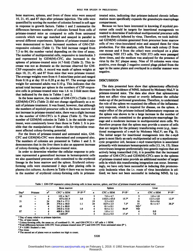
bone marrows, spleens, and livers of these mice were assayed10, 21, 45, and 87 days after pristane injection. The cells werequantified by scoring the number of colonies formed in soft agarin response to growth factors. Tables 1 and 2 summarize thedifferences between numbers of colonies formed from cells ofpristane-treated mice as compared to cells from untreated
controls which were age matched and assayed in parallel inseveral different experiments. Spleens of pristane-treated micewere demonstrated to have increased numbers of GM-CSF-responsive colonies (Table 1). The fold increase ranged from7.3 to 84; the number varied depending on the time of assay.The number of multipotential cells, responsive to interleukin 3and represented by GEM(M)-CFC, also increased in thespleens of pristane-treated mice (4.7-fold) (Table 2). This increase was not as dramatic as the increase observed for GM-CSF-responsive cells. A record was made of spleen weights atdays 10, 21, 45, and 87 from mice that were pristane treated.The average weights were from 4-5 mice/time point and rangedfrom 0.16 g at day 10 to 0.20 g at day 87. The average spleenweight for untreated adults was 0.10 g. This indicates that theactual total increase per spleen in the numbers of CSF-respon-sive cells in pristane-treated mice was 1.6- to 2-fold more than
that indicated by the data in Tables 1 and 2.In the bone marrow, the number of GM-CFCs (Table 1) and
GEM(M)-CFCs (Table 2) did not change significantly as a re
sult of pristane treatment. It was found, however, that althoughthe numbers of myeloid precursor cells in the bone marrow didnot increase in pristane-treated mice, there was a slight increasein the number of GM-CFCs in S phase (Table 3). The total
number of GEM(M) colonies in Table 3, in the suicide experiment, were consistently lower than those in Table 2. It is possible that the manipulations of these cells for thymidine treatment affected colony-forming potential.
For the livers of pristane-treated and untreated mice, GM-CFC and GEM(M)-CFC were determined and compared also.
The numbers of colonies are presented in Tables 1 and 2. Itdemonstrates that in the liver there is also an apparent increaseof colony-forming cells in pristane-treated mice.
In order to determine whether the cellular response to pristane represented a generalized increase in hematopoietic cellswe also quantitated precursor cells committed to the erythroidlineage in the bone marrow and the spleen. Erythroid colony-
forming cells were enumerated 2 days after plating cells inplasma clot cultures. As shown in Table 4 there was no increasein the number of erythroid colony-forming cells in pristane-
treated mice, indicating that pristane-induced chronic inflammation more specifically expands the granulocyte-macrophagecompartment.
Because we have been interested in knowing if myeloid precursor cells could be targets for insertional mutagenesis wewanted to determine if individual multipotential precursor cellscould be directly infected by virus. Therefore, we took individual GEM(M) colonies generated from precursor bone marrowcells of two virus-inoculated mice and assayed them for virusproduction. For this analysis, cells from each colony (5 fromone mouse and 4 from the other) were overlayed on a platecontaining NIH 3T3 cells. The NIH 3T3 cells were passagedthree times, and the medium was analyzed for the presence ofvirus by the XC plaque assay. Nine of 10 colonies were viruspositive, even though 2 negative control plugs picked from theagar in the same plates and overlayed in a similar manner werenegative.
DISCUSSION
The data presented here show that splenectomy effectivelydecreases the incidence of MML induced by Moloney MuLV inpristane-treated mice. The data also show that splenectomydoes not affect virus spread or overtly influence the cellularmake-up of the peritoneal granuloma. Therefore, to understandthe role of the spleen we examined the effects of the inflammatory response, which is required for disease, on the spleen. Amajor effect of the pristane-induced inflammatory response onthe spleen was shown to be a large increase in the number ofprecursor cells committed to the granulocyte-macrophage lineage and a moderate increase in multipotential stem cells. Wetherefore propose that the spleen may provide a source of cellsthat are targets for the primary transforming event (e.g., insertional mutagenesis of c-myb by Moloney MuLV; see Fig. 5).The initial target for insertional mutagenesis into the c-mybgene is most likely an early multipotential cell or a myelomono-cytic progenitor cell, because c-myb transcription is associatedprimarily with immature hematopoietic cells (13, 14, 15). Sinceretroviruses integrate preferentially into genetic regions that areactively being transcribed (16), it is possible that the increasednumber of GM-CFCs and GEM(M)-CFCs seen in the spleensof pristane-treated mice provide an additional number of targetcells in which this transforming integration can occur. Interestingly, we have only been successful in inducing this promono-cytic leukemia when the i.v. route of virus inoculation is utilized; we have not been successful in inducing MML by i.p.
Table 1 GM-CSF-responsive colony-forming cells in bone marrow, spleen, and liver of pristane-treated and untreated mice
Day"10214587Pristanen*4+
424+
284+
44+
2Bone
marrowCFC'
±SEM155
±36.7457±161.9336.5
+27.2370.4±40.0241.
2±80.0250.0±53.8408.0±58.0>405.0*P+/P-*
n53.0
4191.1
2441.0
443SpleenCFC
±SEM7.0
±4.7110.0+65.02.3±0.516.9
±2.5O.I±0.18.4±3.11.8
±1.131.4+ 9.5P+/P-15.7'7.3'84.0'17.4'n541615ND^ND41LiverCFC
±SEM1.0
±1.012.5±6.00.4
±0.68.1±4.70.07.0P+/P-12.5'20.2'
" Day of assay relative to pristane injection.* No. of mice tested.r Colony-forming cells, the mean no. of combined G-, M-, and GM-CFC/5 x 10* cells ±l SEM.d Fold differences between GM-CFC from pristane-treated mice (P~*)and GM-CFC from untreated mice (P~).' P < 0.001 Student's t test.^Not done.*The second set of plates were at numbers too high to count.
5320
Research. on February 1, 2019. © 1992 American Association for Cancercancerres.aacrjournals.org Downloaded from
ROLE FOR SPLEEN IN PRELEUKEMIA
Table 2 Interleukin 3 and EPO-responsive CEM(M) colony-forming cells in the bone marrow, spleen, and liver of prislane-treated and untreated mice"
Pristane+n»24
29Bone
marrowCFCC
+SEM81.5±19.1
116.9±16.9PVP-1.4n28 21SpleenCFC
+SEM1.9±0.4
9.0±2.2P+/P-4.7*n24 21LiverCFC
+SEM0.1
±0.071.7±0.4P*/P-17.0
" This assay was performed 21 days post-pristane injection.* Total no. of mice tested.""Colony-forming cells; represents the mean number of GEM(M) colonies/S x IO5cells ±SEM.d0.05</'<0.1.
Table 3 Bone marrow cells in S phase: pristane-treated and untreated mice
CFC"
CFCtypeGMCGEM(M)dPristanen*8+
1312+
16-I'HIThy421.9
±53.5443.8 ±49.042.1
±12.258.1 ±11.3+[3H]Thy256.9
±40.3190.0±37.545.4
±12.257.2 ±13.1%
inS39.057.2-8.0
1.5" Colony-forming cells; the cells were assayed at 3-4 weeks post-pristane injec
tion.* Total no. of mice.c Colonies that formed in response to GM-CSF; the mean number of combined
G-CFC, M-CFC, and GM-CFC/5 x IO5 cells ±SEM.''Colonies that formed in response to interleukin 3; the mean number of
GEM(M) colonies/5 x IO5 cells ±SEM.
Table 4 Colony-forming units-erythroid in bone marrow and spleen of mice 21days post-pristane treatment
Pristane+n"9
9Bone
marrowCFC*
±SEM1922±244
1783±257P+/P-0.9Cn9 9SpleenCFC
±SEM466±37
515 ±69PVP-1.1«" Total no. of mice. The assay was performed at 21 days post-pristane injection.* Colony-forming units-erythroid/5 x IO5 cells. Each number represents the
mean ±SEM.CP>0.5.
should be noted that, in MML, transformed cells eventuallytravel from the spleen to the peritoneal cavity because leukemiccells are found in the late stages of the disease as ascites and inthe granulomatous tissue induced by pristane (1, 5). It is wellestablished that monocytes in inflammatory exudates are recruited from cells in peripheral circulation (12), and more specifically, it has been observed that cells of the monocytic lineageare attracted to factors released by cells involved in an inflammatory response in the peritoneal cavity (12). The pristane-induced inflammatory granuloma is most likely providing afavorable environment for the recruitment and proliferation ofpreleukemic or leukemic cells; further transforming events mayoccur there as well.
In support of our data presented here, which suggest that atsome time during leukemia development, transformed or partially transformed cells reside in the spleen, we have recentlydemonstrated that cells with insertional activation of c-myb canreside in the spleen as early as 3 weeks after virus inoculation(this is the subject of another manuscript in preparation).
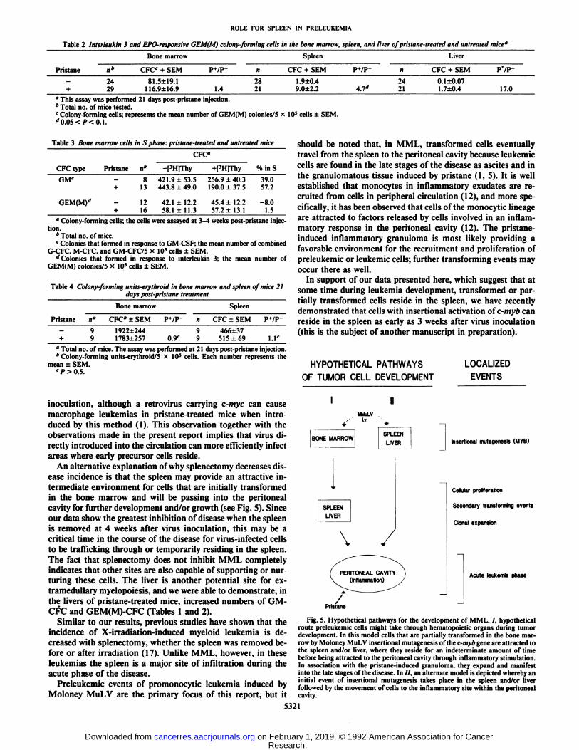
HYPOTHETICAL PATHWAYS LOCALIZED
OF TUMOR CELL DEVELOPMENT EVENTS
inoculation, although a retrovirus carrying c-myc can causemacrophage leukemias in pristane-treated mice when introduced by this method (1). This observation together with theobservations made in the present report implies that virus directly introduced into the circulation can more efficiently infectareas where early precursor cells reside.
An alternative explanation of why splenectomy decreases disease incidence is that the spleen may provide an attractive intermediate environment for cells that are initially transformedin the bone marrow and will be passing into the peritonealcavity for further development and/or growth (see Fig. 5). Sinceour data show the greatest inhibition of disease when the spleenis removed at 4 weeks after virus inoculation, this may be acritical time in the course of the disease for virus-infected cellsto be trafficking through or temporarily residing ¡nthe spleen.The fact that splenectomy does not inhibit MML completelyindicates that other sites are also capable of supporting or nurturing these cells. The liver is another potential site for ex-tramedullary myelopoiesis, and we were able to demonstrate, inthe livers of pristane-treated mice, increased numbers of GM-CFC and GEM(M)-CFC (Tables 1 and 2).
Similar to our results, previous studies have shown that theincidence of X-irradiation-induced myeloid leukemia is decreased with splenectomy, whether the spleen was removed before or after irradiation (17). Unlike MML, however, in theseleukemias the spleen is a major site of infiltration during theacute phase of the disease.
Preleukemic events of promonocytic leukemia induced byMoloney MuLV are the primary focus of this report, but it
I
BONE MARROWSPLEEN
LIVER
SPLEEN
LIVER
\
Insertional mulagenesis (MYB)
Cellular proliferation
Secondary transforming events
Clonal expansion
Acute leukemia phase
Pristane
Fig. 5. Hypothetical pathways for the development of MML. /, hypotheticalroute preleukemic cells might take through hematopoietic organs during tumordevelopment. In this model cells that are partially transformed in the bone marrow by Moloney MuLV insertional mutagenesis of the c-myb gene are attracted tothe spleen and/or liver, where they reside for an indeterminate amount of timebefore being attracted to the peritoneal cavity through inflammatory stimulation.In association with the pristane-induced granuloma, they expand and manifestinto the late stages of the disease. In //, an alternate model is depicted whereby aninitial event of insertional mutagenesis takes place in the spleen and/or liverfollowed by the movement of cells to the inflammatory site within the peritonealcavity.
5321
Research. on February 1, 2019. © 1992 American Association for Cancercancerres.aacrjournals.org Downloaded from

ROLE FOR SPLEEN IN PRELEUKEMIA
ACKNOWLEDGMENTS
We thank Michael Potter for developing our interest in the biologyof the pristane-induced inflammatory response and its contribution totumor development. In addition we would like to thank him for hiscontinued support of these studies and useful discussions. We alsothank Jonathan Keller for advice on performing colony assays, JudithWax for help with the mice, Robert Tarone for performing statisticalanalyses, and Sandra Ruscelli for critical review of the manuscript.
REFERENCES1. Wolff, L., Mushinski, J. F., Shen-Ong, G. L. C, and Morse, H. C, III. A
chronic inflammatory response: its role in supporting the development ofc-myb and c-myc related promonocytic and monocytic tumors in BALB/Cmice. J. Immunol., 141: 681-689, 1988.
2. Wolff, L., and Nason-Burchenal, K. Retrovirus-induced tumors whose development is facilitated by a chronic immune response: a comparison of twotumors committed to the monocytic lineage. Curr. Top. Microbiol. Immunol., 149: 79-87, 1989.
3. Bennett, J. M., Catovsky, D., Daniel, M. T., Flandrin, G., Galton, D. A. G.,Gralnick, H. R., and Sultan, C. Proposals for the classification of the acuteleukemias. Br. J. Hematol., 33: 451-458, 1976.
4. Huhn, D. Morphology, cytochemistry, and ultrastructure of leukemic cellswith regard to the classification of leukemias. Recent Results Cancer Res., 93:51-68, 1984.
5. Shen-Ong, G. L. C., and Wolff, L. Moloney murine leukemia virus-inducedmyeloid tumors in adult Balb/c mice: requirement of c-myb activation butlack of v-abl involvement. J. Virol., 61: 3721-3725, 1987.
6. Shen-Ong, G. L. C., Luscher, B., and Eisenman, R. N. A second c-mybprotein is translated from an alternatively spliced niRNA expressed fromnormal and 5'-disrupted myb loci. Mol. Cell. Biol., 9: 5456-5463, 1989.
7. Gart, J. J., Krewski, D., Lee, P. N., Tarone, R. E., and Wahrendorf, J.
Statistical Methods in Cancer Research, Vol. 3. Lyon: International Agencyfor Research on Cancer, 1986.
8. Rowe, W. P., Pugh, W. E., and Hartley, J. W. Plaque assay techniques formurine leukemia viruses. Virology, 42: 1136-1139, 1970.
9. Metcalf, D. The Hemopoietic Colony Stimulating Factors. New York:Elsevier, 1984.
10. Broxmeyer, H. E., Williams, D. E., Hangoc, G., Cooper, S., Gillis, S., Shad-duck, R. K., and Bicknell, D. C. Synergistic myelopoietic actions in vivo afteradministration to mice of combinations of purified natural murine colony-stimulating factor 1, recombinant murine interleukin 3, and recombinantmurine granulocyte/macrophage colony-stimulating factor. Proc. Nati. Acad.Sci. USA, 84: 3871-3875, 1987.
11. Stephenson, J. R., Axelrad, A. A., McLeod, D. L., and M. M. Shreeve, M. M.Induction of colonies of hemoglobin-synthesizing cells by erythropoietin invitro. Proc. Nati. Acad. Sci. USA, 68: 1542-1546, 1971.
12. van Furth, R., and Cohn, Z. A. The origin and kinetics of mononuclearphagocytes. J. Exp. Med., 128: 415-435, 1968.
13. Gonda, T. J., Sheiness, D. K., and Bishop, J. M. Transcripts from the cellularhomologs of retroviral oncogenes: distribution among chicken tissues. Mol.Cell. Biol., 2:617-624, 1982.
14. Westin, E. H., Gallo, R. C, Arya, S. K., Eva, A., Souza, L. M., Baluda, M.A., Aaronson, S. A., and Wong-Staal, F. Differential expression of the armgene in human hematopoietic cells. Proc. Nati. Acad. Sci. USA, 79: 2194-2198, 1982.
15. Gonda, T. J., and Metcalf, D. Expression of myb, myc, and/os proto-onco-genes during the differentiation of a murine myeloid leukemia. Nature(Lond.), 310: 249-251, 1984.
16. Rohdewohld, H., Weiher, H., Reik, R., Jaenisch, R., and Breindl, M. Ret-rovirus integration and eliminatili structure: Moloney murine leukemiaproviral integration sites map near DNase I-hypersensitive sites. J. Virol., 61:336-343, 1987.
17. Upton, A. C., Wolff, F. F., Furth, J., and Kimball, A. W. A comparison of theinduction of myeloid and lymphoid leukemias in x-radiated RF mice. CancerRes., 18: 842-848, 1958.
5322
Research. on February 1, 2019. © 1992 American Association for Cancercancerres.aacrjournals.org Downloaded from

1992;52:5317-5322. Cancer Res Kathryn Nason-Burchenal and Linda Wolff Murine Retrovirus-induced Promonocytic LeukemiaInvolvement of the Spleen in Preleukemic Development of a
Updated version
http://cancerres.aacrjournals.org/content/52/19/5317
Access the most recent version of this article at:
E-mail alerts related to this article or journal.Sign up to receive free email-alerts
Subscriptions
Reprints and
To order reprints of this article or to subscribe to the journal, contact the AACR Publications
Permissions
Rightslink site. Click on "Request Permissions" which will take you to the Copyright Clearance Center's (CCC)
.http://cancerres.aacrjournals.org/content/52/19/5317To request permission to re-use all or part of this article, use this link
Research. on February 1, 2019. © 1992 American Association for Cancercancerres.aacrjournals.org Downloaded from