Embed Size (px)
Citation preview
Ion-induced stacking of photosensitizermolecules can remarkably affect theluminescence detection of singlet oxygenin Candida albicans cells
Ariane FelgenträgerFernanda Pereira GonzalesTim MaischWolfgang Bäumler
Downloaded From: https://www.spiedigitallibrary.org/journals/Journal-of-Biomedical-Optics on 5/11/2018 Terms of Use: https://www.spiedigitallibrary.org/terms-of-use
Ion-induced stacking of photosensitizer molecules canremarkably affect the luminescence detection of singletoxygen in Candida albicans cells
Ariane Felgenträger, Fernanda Pereira Gonzales, Tim Maisch, and Wolfgang BäumlerRegensburg University Hospital, Department of Dermatology, 93053 Regensburg, Germany
Abstract. Singlet oxygen (1O2) is an important reactive intermediate in photodynamic reactions, particularly inantimicrobial PDT (aPDT). The detection of 1O2 luminescence is frequently used to elucidate the role of 1O2
in various environments, particularly in microorganisms and human cells. When incubating the fungus,Candida albicans, with porphyrins XF73 (5,15-bis-[4-(3-Trimethylammonio-propyloxy)-phenyl]-porphyrin) orTMPyP (5,10,15,20-Tetrakis(1-methyl-4-pyridinio)-porphyrin tetra(p-toluenesulfonate)), the 1O2 luminescence sig-nals were excellent for TMPyP. In case of XF73, the signals showed strange rise and decay times. Thus, 1O2 gen-eration of XF73 was investigated and compared with TMPyP. Absorption spectroscopy of XF73 showed a change inabsorption cross section when there was a change in the concentration from 1 × 10⁻⁶ M to 1 × 10⁻³ M indicatingan aggregation process. The addition of phosphate buffered saline (PBS) substantially changed 1O2 luminescence inXF73 solution. Detailed experiments provided evidence that the PBS constituents NaCl and KCl caused the changeof 1O2 luminescence. The results also indicate that Cl− ions may cause aggregation of XF73 molecules, which inturn enhances self-quenching of 1O2 via photosensitizer molecules. These results show that some ions, e.g., thosepresent in cells in vitro or added by PBS, can considerably affect the detection and the interpretation of time-resolved luminescence signals of 1O2, particularly in in vitro and in vivo. These effects should be consideredfor any other photosensitizer used in photodynamic processes. © The Authors. Published by SPIE under a Creative Commons
Attribution 3.0 Unported License. Distribution or reproduction of this work in whole or in part requires full attribution of the original publication, including
its DOI. [DOI: 10.1117/1.JBO.18.4.045002]
Keywords: porphyrin; photodynamic; singlet oxygen; luminescence; aggregation.
Paper 12512RR received Aug. 10, 2012; revised manuscript received Feb. 7, 2013; accepted for publication Mar. 14, 2013; publishedonline Apr. 3, 2013.
1 IntroductionThe fast development of multiresistant patterns against antibiot-ics of many species of bacteria has led to novel antibacterialstrategies like the antibacterial photodynamic therapy (aPDT).1,2
A lot of work has been done to develop molecular structures andtheir derivatives that are able to generate reactive oxygen species(ROS), which are the active agents for killing microorganisms.3–7
The search for photosensitizers (PSs) for aPDT has caused thesynthesis of various porphyrin molecules, which have beeninvestigated regarding their photophysics and antimicrobialactivity.4,8,9 Naturally occurring porphyrins can be found endog-enously, e.g., the protoporphyrin IX that is in the prostheticgroup of the hemoglobin or the chlorophylls based on the chlo-rine structure. Some endogenous porphyrins in bacteria are usedto treat acne, where Propionibacterium acnes is a causative ofthe inflammatory processes.10 The porphyrin TMPyP has beenfrequently used for cell staining in order to investigate genera-tion and decay of 1O2.
11–13
Different PSs are considered to localize in different compart-ments or regions in the eukaryotic or prokaryotic cell due to theirnumber of positive charges and structure of the side chain. Inorder to determine the subcellular localization of PS and
hence the site of 1O2 generation, fluorescence microscopy isapplied by exciting the respective PSs. Since the resolutionof light microscopy is limited, this procedure should fail withsmall bacteria and fungus cells with a diameter of about 1 μm.The direct measurement of 1O2 luminescence at 1270 nm mightbe an alternative candidate to elucidate the cellular action of 1O2
because the rise and decay time of 1O2 luminescence dependcritically on its adjacency.14,15 In addition, singlet oxygen lumi-nescence can provide information about the photodynamic proc-ess in bacteria during irradiation.
XF73 is a newly synthesized porphyrin molecule that alreadyshowed a high potential in antimicrobial PDT against gram-neg-ative and gram-positive bacteria.16,17 However, principal data arelacking regarding its use in 1O2 detection in vitro. Thus, it is thegoal of the present study to investigate the photophysical proper-ties of XF73 and its potential to monitor photodynamic action inmicroorganisms. Exemplarily 1O2 luminescence detection wasanalyzed in vitro in Candida albicans cells. The well-knownTMPyP was used for reference experiments.
2 Material and Methods
2.1 Chemicals
Thecationicdiporphyrin-based5,15-bis-[4-(3-Trimethylammonio-propyloxy)-phenyl]-porphyrin (also referred to herein as XF73)with a molar mass of M ¼ 765.81 g∕mol, including the counterion, was synthesized by Xiangdong Feng (Solvias Company,
Address all correspondence to: Ariane Felgenträger, Regensburg UniversityHospital, Department of Dermatology, Franz-Josef-Strauss-Allee 11, 93053Regensburg, Germany. Tel: 0049‐941‐944‐9650; Fax: 0049‐941‐944‐8943; E-mail: [email protected]
Journal of Biomedical Optics 045002-1 April 2013 • Vol. 18(4)
Journal of Biomedical Optics 18(4), 045002 (April 2013)
Downloaded From: https://www.spiedigitallibrary.org/journals/Journal-of-Biomedical-Optics on 5/11/2018 Terms of Use: https://www.spiedigitallibrary.org/terms-of-use
Basel, Switzerland) and kindly provided by Destiny PharmaLtd. (Brighton, United Kingdom).
The 5,10,15,20-Tetrakis(1-methyl-4-pyridinio)-porphyrintetra(p-toluenesulfonate) (also referred to herein as TMPyP)with a molar mass of M ¼ 1363.63 g∕mol, purity 97%, NaN3
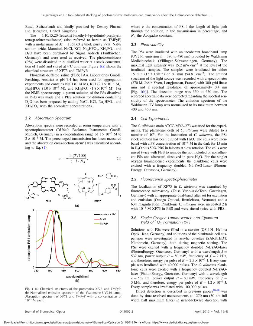
sodium azide, Mannitol, NaCl, KCl, Na2HPO4, KH2PO4, andD2O have been purchased by Sigma Aldrich (Taufkirchen,Germany), and were used as received. The photosensitizers(PSs) were dissolved in bi-distilled water at a stock concentra-tion of 1 mM and stored at 4°C until use. Figure 1(a) shows thechemical structure of XF73 and TMPyP.
Phosphate-buffered saline (PBS; PAA Laboratories GmbH,Pasching, Austria) at pH 7.4 has been used for aggregationexperiments and contains NaCl (0.14 M), KCl (2.7 × 10⁻³ M),Na2HPO4 (1.0 × 10⁻² M), and KH2PO4 (1.8 × 10⁻³ M). Forthe NMR spectroscopy, a parent solution of the PSs dissolvedin D2O was made and a PBS solution for dilution containingD2O has been prepared by adding NaCl, KCl, Na2HPO4, andKH2PO4 with the accordant concentrations.
2.2 Absorption Spectrum
Absorption spectra were recorded at room temperature with aspectrophotometer (DU640, Beckman Instruments GmbH,Munich, Germany) in a concentration range of 1 × 10⁻⁶ M to2 × 10−3 M. The percentaged transmission has been measuredand the absorption cross-section σðcm2Þ was calculated accord-ing to Eq. (1):
σ ¼ −lnðT∕100Þc · l · NA
; (1)
where c the concentration of PS, l the length of light paththrough the solution, T the transmission in percentage, andNA the Avogadro constant.
2.3 Photostability
The PSs were irradiated with an incoherent broadband lamp(UV236; emission λ ¼ 380 to 480 nm) provided by WaldmannMedizintechnik (Villingen-Schwenningen, Germany). Themaximal light intensity was 15.2 mWcm−2 at the level of theirradiated samples. The samples were irradiated for either15 min (13.7 J cm−2) or 60 min (54.8 J cm−2). The emittedspectrum of the light source was recorded with a spectrometer(270 M, Jobin Yvon, Longjumeau, France) with 300 grid lines/mm and a spectral resolution of approximately 0.4 nm[Fig. 1(b)]. The detection range was 350 to 650 nm. Therecorded spectral data were corrected regarding the spectral sen-sitivity of the spectrometer. The emission spectrum of theWaldmann UV lamp was normalized to its maximum between400 and 450 nm.
2.4 Cell Experiments
The C. albicans strain ATCC-MYA-273 was used for the experi-ments. The planktonic cells of C. albicans were diluted to anumber of 106. For the incubation of C. albicans, the PSsstock solution has been diluted with H2O. The cells were incu-bated with a PS concentration of 10−4 M in the dark for 15 minin H2O plus 50% PBS in falcons at slow rotation. The cells wererinsed twice with PBS to remove the not included or nonadher-ent PSs and afterward dissolved in pure H2O. For the singletoxygen luminescence experiments, the planktonic cells wereexcited with a frequency doubled Nd:YAG-Laser (Photon-Energy, Ottensoos, Germany).
2.5 Fluorescence Spectrophotometer
The localization of XF73 in C. albicans was examined byfluorescence microscopy (Zeiss Vario-AxioTech, Goettingen,Germany) with an appropriate dual-band filter set for excitationand emission (Omega Optical, Brattleboro, Vermont) and a63× magnification. Planktonic C. albicans were incubated 2 hwith 10−4 M XF73 in PBS and were rinsed twice with PBS.
2.6 Singlet Oxygen Luminescence and QuantumYield of 1O2 Formation (ΦΔ)
Solutions with PSs were filled in a cuvette (QS-101, HellmaOptik, Jena, Germany) and solutions of the planktonic cell sus-pension were investigated in acrylic cuvettes (SARSTEDT,Nümbrecht, Germany), both during magnetic stirring. ThePSs were excited with a frequency doubled Nd:YAG-laser(PhotonEnergy, Ottensoos, Germany) with a wavelength λ ¼532 nm, power output P ¼ 50 mW, frequency of f ¼ 2 kHz,and therefore, energy per pulse of E ¼ 2.5 × 10⁻⁵ J. Every sam-ple was irradiated with 40,000 pulses. The C. albicans plank-tonic cells were excited with a frequency doubled Nd:YAG-laser (PhotonEnergy, Ottensoos, Germany) with a wavelengthλ ¼ 532 nm, power output P ¼ 60 mW, frequency of f ¼5 kHz, and therefore, energy per pulse of E ¼ 1.2 × 10⁻⁵ J.Every sample was irradiated with 100,000 pulses.
Direct detection as described in previous papers18–20 wasdone by time resolved measurements at 1270 nm (30 nm fullwidth half maximum filter) in near-backward direction with
Fig. 1 (a) Chemical structures of the porphyrins XF73 and TMPyP.(b) Normalized emission spectrum of the Waldmann-UV236 lamp.Absorption spectrum of XF73 and TMPyP with a concentration of10−5 M each.
Journal of Biomedical Optics 045002-2 April 2013 • Vol. 18(4)
Felgenträger et al.: Ion-induced stacking of photosensitizer molecules can remarkably affect the luminescence detection. . .
Downloaded From: https://www.spiedigitallibrary.org/journals/Journal-of-Biomedical-Optics on 5/11/2018 Terms of Use: https://www.spiedigitallibrary.org/terms-of-use
respect to the exciting beam using an infrared-sensitive photo-multiplier (R5509-42, Hamamatsu Photonics DeutschlandGmbH, Herrsching, Germany) with using an additional 950 nmcut-off-filter. The luminescence intensity is given by
IðtÞ ¼ CtR−1 − tD−1
�exp
�−
ttD
�− exp
�−
ttR
��; (2)
where C ¼ ½T1�t¼0 kT1Δ ½3O2� was used to fit the singlet oxygenluminescence signal, describing the deactivation of the excitedtriplet state T1 of the photosensitizer by oxygen in its groundstate (3O2).
20 tR and tD are the rise and decay times, whichis the excited triplet state decay time τT1 of the photosensitizerand the decay time of singlet oxygen τΔ. The attribution of τT1and τΔ depends on the oxygen concentration in the system; athigh oxygen concentrations, usually the decay time τD of thesignal describes the decay time of singlet oxygen τΔ. Inorder to determine the rise and decay times, the Levenberg-Marquardt-algorithm of Mathematica (Wolfram Research,Champaign) was used. The luminescence signal was spectrallyresolved using interference filters in front of the photomultipliertube at wavelengths ranging from 1150 to 1400 nm or a mono-chromator (Horiba, Yobin Yvon Inc., USA) from 1200 to1350 nm at 10 nm regular steps (XF73 in pure H2O). The valuesshow the integrated luminescence signals detected at a certainwavelength and are normalized to the maximal value. ALorentz-shaped curve has been fitted through the measurementpoints, with the maximum at λ ¼ 1270 nm, referring to themaximal value in H2O.
For the determination of ΦΔ of XF73 in H2O, it is comparedwith the ΦΔ of TMPyP, which is reported in literature being0.7421 and 0.77� 0.0412 in aqueous solution. Therefore, fiveprobes of each PS of different concentrations (between 30%and 70% absorption at a wavelength of λ ¼ 532 nm) are irradi-ated and the emitted 1O2-photons are determined with the inte-gral over the luminescence curve, given with the fit routinementioned.
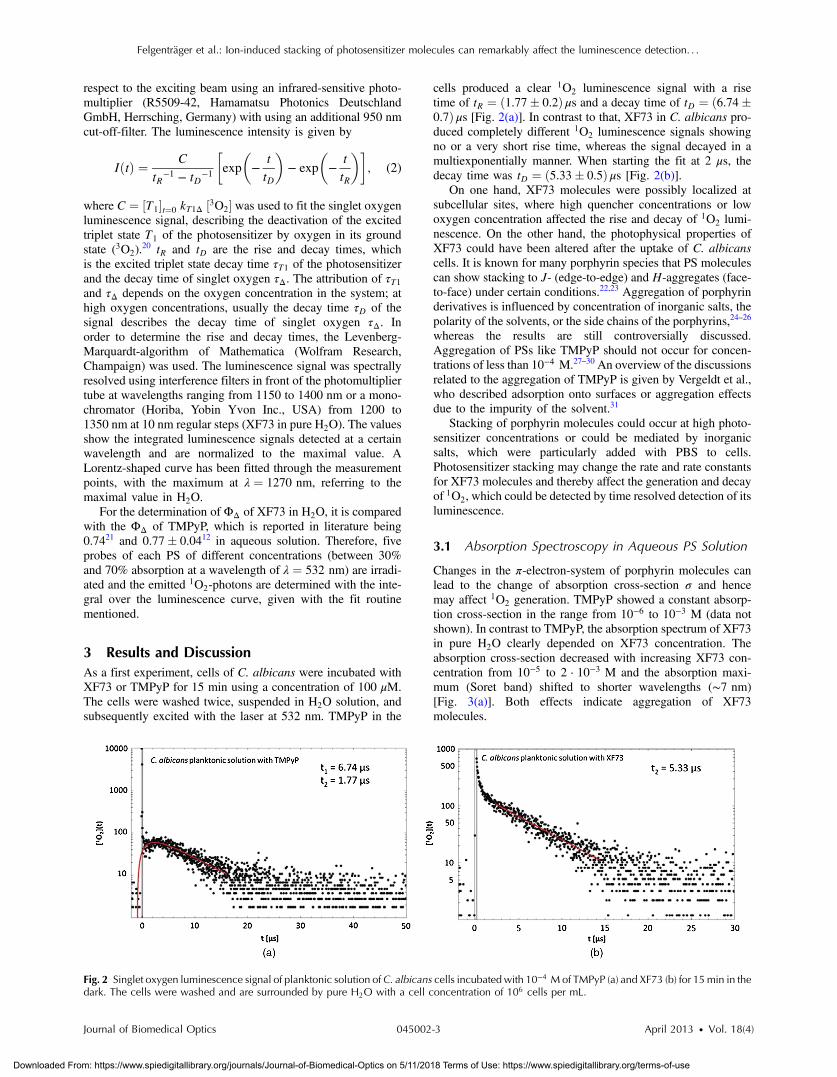
3 Results and DiscussionAs a first experiment, cells of C. albicans were incubated withXF73 or TMPyP for 15 min using a concentration of 100 μM.The cells were washed twice, suspended in H2O solution, andsubsequently excited with the laser at 532 nm. TMPyP in the
cells produced a clear 1O2 luminescence signal with a risetime of tR ¼ ð1.77� 0.2Þ μs and a decay time of tD ¼ ð6.74�0.7Þ μs [Fig. 2(a)]. In contrast to that, XF73 in C. albicans pro-duced completely different 1O2 luminescence signals showingno or a very short rise time, whereas the signal decayed in amultiexponentially manner. When starting the fit at 2 μs, thedecay time was tD ¼ ð5.33� 0.5Þ μs [Fig. 2(b)].
On one hand, XF73 molecules were possibly localized atsubcellular sites, where high quencher concentrations or lowoxygen concentration affected the rise and decay of 1O2 lumi-nescence. On the other hand, the photophysical properties ofXF73 could have been altered after the uptake of C. albicanscells. It is known for many porphyrin species that PS moleculescan show stacking to J- (edge-to-edge) and H-aggregates (face-to-face) under certain conditions.22,23 Aggregation of porphyrinderivatives is influenced by concentration of inorganic salts, thepolarity of the solvents, or the side chains of the porphyrins,24–26
whereas the results are still controversially discussed.Aggregation of PSs like TMPyP should not occur for concen-trations of less than 10−4 M.27–30 An overview of the discussionsrelated to the aggregation of TMPyP is given by Vergeldt et al.,who described adsorption onto surfaces or aggregation effectsdue to the impurity of the solvent.31
Stacking of porphyrin molecules could occur at high photo-sensitizer concentrations or could be mediated by inorganicsalts, which were particularly added with PBS to cells.Photosensitizer stacking may change the rate and rate constantsfor XF73 molecules and thereby affect the generation and decayof 1O2, which could be detected by time resolved detection of itsluminescence.
3.1 Absorption Spectroscopy in Aqueous PS Solution
Changes in the π-electron-system of porphyrin molecules canlead to the change of absorption cross-section σ and hencemay affect 1O2 generation. TMPyP showed a constant absorp-tion cross-section in the range from 10−6 to 10−3 M (data notshown). In contrast to TMPyP, the absorption spectrum of XF73in pure H2O clearly depended on XF73 concentration. Theabsorption cross-section decreased with increasing XF73 con-centration from 10−5 to 2 · 10−3 M and the absorption maxi-mum (Soret band) shifted to shorter wavelengths (∼7 nm)[Fig. 3(a)]. Both effects indicate aggregation of XF73molecules.
Fig. 2 Singlet oxygen luminescence signal of planktonic solution of C. albicans cells incubated with 10−4 Mof TMPyP (a) and XF73 (b) for 15min in thedark. The cells were washed and are surrounded by pure H2O with a cell concentration of 106 cells per mL.
Journal of Biomedical Optics 045002-3 April 2013 • Vol. 18(4)
Felgenträger et al.: Ion-induced stacking of photosensitizer molecules can remarkably affect the luminescence detection. . .
Downloaded From: https://www.spiedigitallibrary.org/journals/Journal-of-Biomedical-Optics on 5/11/2018 Terms of Use: https://www.spiedigitallibrary.org/terms-of-use
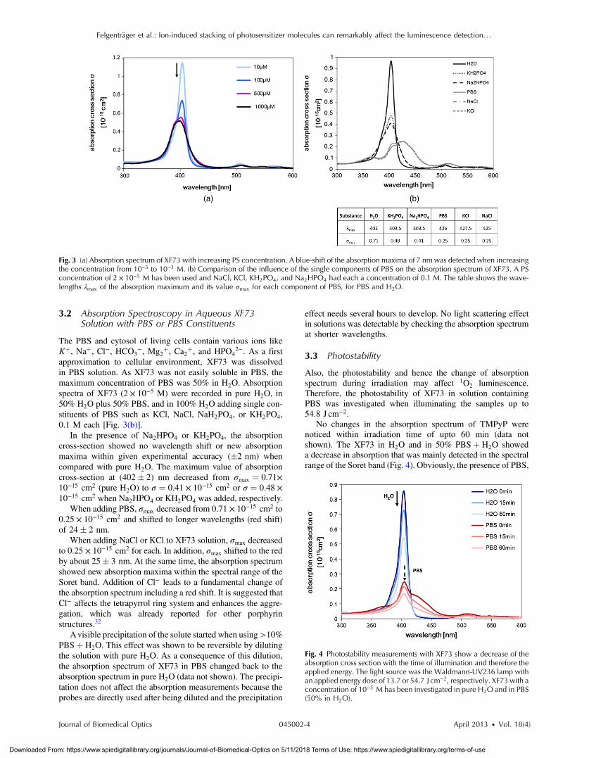
3.2 Absorption Spectroscopy in Aqueous XF73Solution with PBS or PBS Constituents
The PBS and cytosol of living cells contain various ions likeKþ, Naþ, Cl−, HCO3
−, Mg2þ, Ca2þ, and HPO4
2−. As a firstapproximation to cellular environment, XF73 was dissolvedin PBS solution. As XF73 was not easily soluble in PBS, themaximum concentration of PBS was 50% in H2O. Absorptionspectra of XF73 (2 × 10⁻⁵ M) were recorded in pure H2O, in50% H2O plus 50% PBS, and in 100% H2O adding single con-stituents of PBS such as KCl, NaCl, NaH2PO4, or KH2PO4,0.1 M each [Fig. 3(b)].
In the presence of Na2HPO4 or KH2PO4, the absorptioncross-section showed no wavelength shift or new absorptionmaxima within given experimental accuracy (�2 nm) whencompared with pure H2O. The maximum value of absorptioncross-section at (402� 2) nm decreased from σmax ¼ 0.71×10⁻¹⁵ cm2 (pure H2O) to σ ¼ 0.41 × 10⁻¹⁵ cm2 or σ ¼ 0.48 ×10⁻¹⁵ cm2 when Na2HPO4 or KH2PO4 was added, respectively.
When adding PBS, σmax decreased from 0.71 × 10⁻¹⁵ cm2 to0.25 × 10−15 cm2 and shifted to longer wavelengths (red shift)of 24� 2 nm.
When adding NaCl or KCl to XF73 solution, σmax decreasedto 0.25 × 10⁻¹⁵ cm2 for each. In addition, σmax shifted to the redby about 25� 3 nm. At the same time, the absorption spectrumshowed new absorption maxima within the spectral range of theSoret band. Addition of Cl− leads to a fundamental change ofthe absorption spectrum including a red shift. It is suggested thatCl− affects the tetrapyrrol ring system and enhances the aggre-gation, which was already reported for other porphyrinstructures.32
Avisible precipitation of the solute started when using>10%PBSþ H2O. This effect was shown to be reversible by dilutingthe solution with pure H2O. As a consequence of this dilution,the absorption spectrum of XF73 in PBS changed back to theabsorption spectrum in pure H2O (data not shown). The precipi-tation does not affect the absorption measurements because theprobes are directly used after being diluted and the precipitation
effect needs several hours to develop. No light scattering effectin solutions was detectable by checking the absorption spectrumat shorter wavelengths.
3.3 Photostability
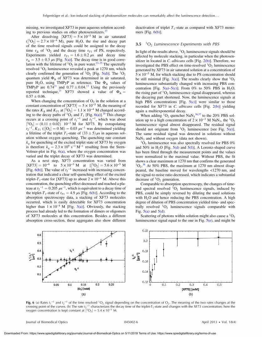
Also, the photostability and hence the change of absorptionspectrum during irradiation may affect 1O2 luminescence.Therefore, the photostability of XF73 in solution containingPBS was investigated when illuminating the samples up to54.8 J cm−2.
No changes in the absorption spectrum of TMPyP werenoticed within irradiation time of upto 60 min (data notshown). The XF73 in H2O and in 50% PBSþ H2O showeda decrease in absorption that was mainly detected in the spectralrange of the Soret band (Fig. 4). Obviously, the presence of PBS,
Fig. 3 (a) Absorption spectrum of XF73 with increasing PS concentration. A blue-shift of the absorption maxima of 7 nm was detected when increasingthe concentration from 10−5 to 10−3 M. (b) Comparison of the influence of the single components of PBS on the absorption spectrum of XF73. A PSconcentration of 2 × 10⁻⁵ M has been used and NaCl, KCl, KH2PO4, and Na2HPO4 had each a concentration of 0.1 M. The table shows the wave-lengths λmax of the absorption maximum and its value σmax for each component of PBS, for PBS and H2O.
Fig. 4 Photostability measurements with XF73 show a decrease of theabsorption cross section with the time of illumination and therefore theapplied energy. The light source was the Waldmann-UV236 lamp withan applied energy dose of 13.7 or 54.7 J cm−2, respectively. XF73 with aconcentration of 10−5 M has been investigated in pure H2O and in PBS(50% in H2O).
Journal of Biomedical Optics 045002-4 April 2013 • Vol. 18(4)
Felgenträger et al.: Ion-induced stacking of photosensitizer molecules can remarkably affect the luminescence detection. . .
Downloaded From: https://www.spiedigitallibrary.org/journals/Journal-of-Biomedical-Optics on 5/11/2018 Terms of Use: https://www.spiedigitallibrary.org/terms-of-use

i.e., its ions, can additionally reduce radiation absorption ofXF73. These effects may also affect the use of XF73 whenapplied for photodynamic inactivation of microorganisms.
In case of 1O2 experiments (see below), XF73 solutions wereirradiated with 1 J of laser energy (532 nm). It is expected that σvalues do not significantly change under these experimentalconditions.
3.4 1O2 Luminescence Experiments without PBS
Incubation of bacteria or human cells with XF73 and subsequentirradiation yielded effective cell killing by means of 1O2 gener-ation, which was confirmed by adding 1O2 quencher NaN3 thatsignificantly reduced the cell toxicity.16 Since detailed studies on1O2 generation of the novel porphyrin molecule XF73 were
Fig. 5 (a)–(c) 1O2 luminescence signals of ½XF73� ¼ 5 × 10⁻⁵ M with different PBS concentrations in H2O with an oxygen concentration of½3O2� ¼ 2.7 × 10⁻⁴ M. (d) Spectroscopically resolved 1O2 luminescence signal, generated by XF73 in H2O with an oxygen concentration of½O2� ¼ 2.7 × 10⁻⁴ M. A Lorentz-shaped curve has been fitted through the measurement points. (e) 1O2 luminescence generated by XF73 in H2Oþ20%PBS at 1270 nmwith 2 × 10⁻³ MNaCl in solution. (f) Spectroscopically resolved 1O2 luminescence signal, generated by XF73 in 30% PBSþH2Owith an oxygen concentration of ½O2� ¼ 2.7 × 10⁻⁴ M. A Lorentz-shaped curve has been fitted through the measurement points.
Journal of Biomedical Optics 045002-5 April 2013 • Vol. 18(4)
Felgenträger et al.: Ion-induced stacking of photosensitizer molecules can remarkably affect the luminescence detection. . .
Downloaded From: https://www.spiedigitallibrary.org/journals/Journal-of-Biomedical-Optics on 5/11/2018 Terms of Use: https://www.spiedigitallibrary.org/terms-of-use
missing, we investigated XF73 in pure aqueous solution accord-ing to previous studies on other photosensitizers.27
After dissolving ½XF73� ¼ 5 × 10⁻⁵ M in air saturated(½3O2� ¼ 2.7 × 10⁻⁴ M), pure H2O, the rise and decay partof the time resolved signals could be assigned to the decaytime τΔ of 1O2 and the decay time τT1 of PS, respectively.Experiments yielded τT1 ¼ 1.6� 0.2 μs and decay timeτΔ ¼ 3.5� 0.3 μs [Fig. 5(a)]. The decay time is in good corre-lation with the lifetime of 1O2 in pure water.33–35 The spectrallyresolved 1O2 luminescence revealed a peak at 1270 nm, whichclearly confirmed the generation of 1O2 [Fig. 5(d)]. The 1O2
quantum yield ΦΔ of XF73 was determined in air saturated,pure H2O, using TMPyP as reference. The ΦΔ values ofTMPyP are 0.7421 and 0.77� 0.04.13 Using the previouslyreported technique,21 XF73 showed a value of ΦΔ ¼0.57� 0.06.
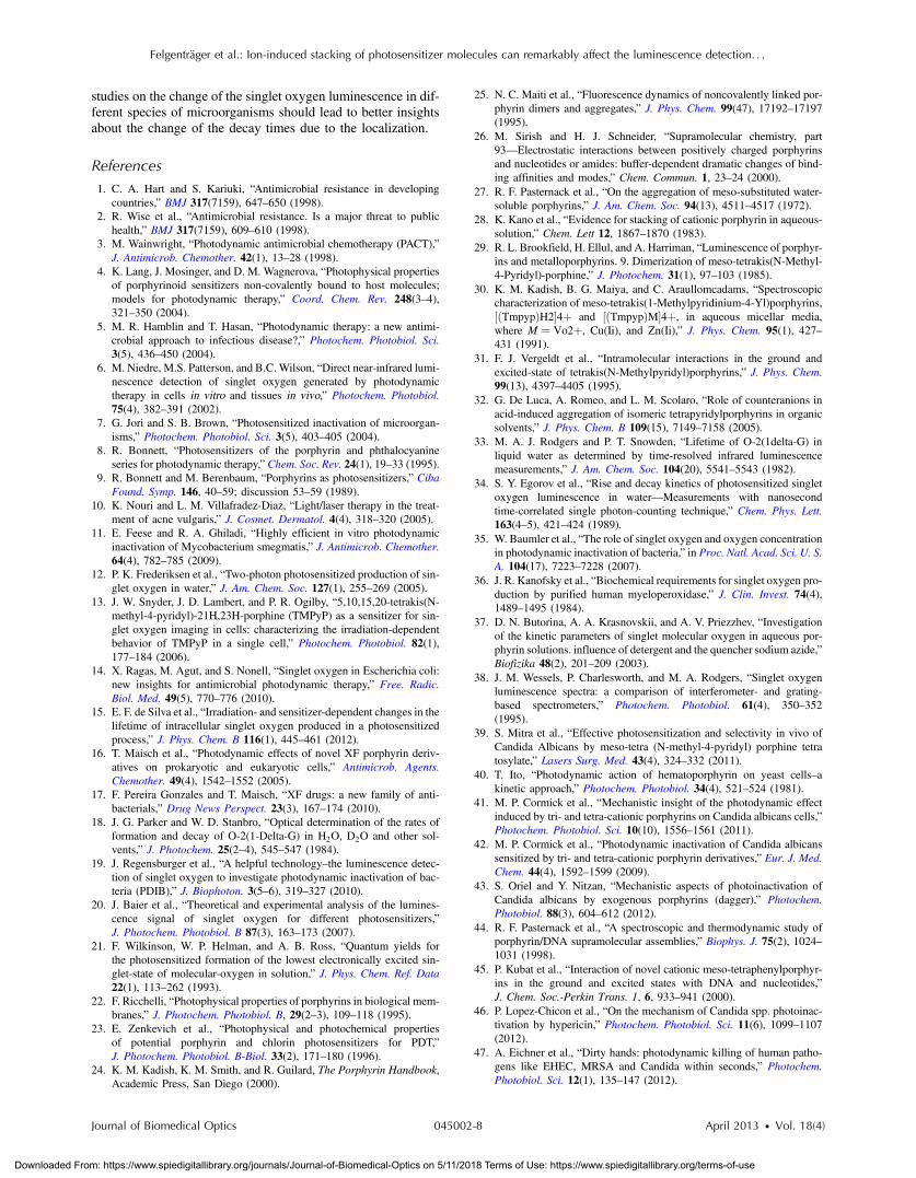
When changing the concentration of O2 in the solution at aconstant concentration of ½XF73� ¼ 5 × 10⁻⁵ M, the meaning ofthe rates KΔ and KT1 at ½3O2� ¼ 1.1 × 10⁻⁴ M changed accord-ing to the decay paths of 1O2 and T1 [Fig. 6(a)].
20 This changeoccurs at a crossing point of t1−1 and t2−1, which was about½3O2� ¼ ð0.11� 0.02Þ 10−3 M for XF73. By extrapolatingt2−1, KT1 (ð½O2� ¼ 0 MÞ ¼ 0.03 μs−1 was determined yieldinga lifetime of the triplet T1-state of ð33� 5Þ μs in aqueous sol-ution without oxygen quenching. The quenching rate constantkq for quenching of the excited triplet state of XF73 by oxygenis therefore kq ¼ 2.3 × 10⁹ s−1 M−1 resulting from the Stern-Volmer-plot in Fig. 6(a), where the oxygen concentration wasvaried and the triplet decay of XF73 was determined.
As a next step, XF73 concentration was varied from½XF73� ¼ 10−6 to 5 × 10⁻³ M at [½3O2� ¼ 5.6 × 10⁻⁵ M[Fig. 6(b)]. The value of t2−1 increased with increasing concen-tration that indicated a clear self-quenching effect of the excitedtriplet-T1-state for [XF73] up to about 2 × 10⁻⁴ M. Above thisconcentration, the quenching effect decreased and reached a pla-teau at t2−1 ¼ 0.205 μs−1, which is equivalent to a decay time ofthe triplet-T1-state of tT1 ¼ 4.9 μs [Fig. 6(b)]. According to theabsorption spectroscopy data, a stacking of XF73 moleculesoccurred, which is easily detectable for XF73 concentrationhigher than 1 × 10⁻⁴ M [Fig. 3(a)]. Obviously, the stackingprocess had already led to the formation of dimers or oligomersof XF73 molecules at this concentration. Besides a differentabsorption cross-section, these aggregates also show different
deactivation of triplet T1-state as compared with XF73 mono-mers [Fig. 6(b)].
3.5 1O2 Luminescence Experiments with PBS
In light of the results above, 1O2 luminescence signals should beaffected by molecule stacking, in particular when the photosen-sitizer in located in C. albicans cells [Fig. 2(b)]. Therefore, weinvestigated the PBS effect on time-resolved 1O2 luminescencegenerated by XF73 in air saturated solution at a concentration of5 × 10−5 M, for which stacking due to PS concentration shouldbe still minimal [Fig. 3(a)]. The results clearly show that 1O2
luminescence substantially changed with increasing PBS con-centration [Fig. 5(a)–5(c)]. From 0% to 50% PBS in H2O,the rising part of 1O2 luminescence signal disappeared, whereasthe decaying part shortened. Now, the luminescence signals athigh PBS concentrations [Fig. 5(c)] were similar to thoserecorded for XF73 in C. albicans cells [Fig. 2(b)] yieldingagain a multiexponential decay.
When adding 1O2 quencher NaN336,37 to the 20% PBS sol-
ution up to a high concentration of 2 × 10⁻³ M NaN3, the 1O2
luminescence signal almost disappeared. The residual signalshould not originate from 1O2 luminescence [see Fig. 5(e)].The same residual signal was detected in solutions withoutNaN3 and without oxygen (data not shown).
1O2 luminescence was also spectrally resolved for PBS 0%and 50% in H2O [Fig. 5(d) and 5(f)]. A Lorentz-shaped curvehas been fitted through the measurement points and the valueswere normalized to the maximal value. Without PBS, the fitshows a clear maximum at 1270 nm that confirms the generated1O2.
38 At 50% PBS, the maximum at 1270 nm almost disap-peared, the baseline moved for wavelengths <1270 nm, andthe signal-to-noise ratio decreased, which indicates a substantialdecrease of 1O2 generation.
Comparable to absorption spectroscopy, the changes of time-and spectral resolved 1O2 luminescence signals, induced byPBS, could be simply reversed by diluting the used solutionswith H2O and hence reducing the PBS concentration. A highdegree of dilution of PBS concentration yielded time- and spec-trally resolved 1O2 luminescence signals comparable withFig. 5(a) and 5(d).
Scattering of photons within solution might also cause a 1O2
luminescence signal equal to the one in Fig. 5(e), and might be
Fig. 6 (a) Rates t1−1 and t2−1 of the time resolved 1O2 signal depending on the concentration of O2. The meaning of the two rates changes at thecrossing point of the curves. (b) The rate t2−1 characterizes the decay time of the triplet-T1-state and changes with the XF73 concentration; here theoxygen concentration is kept constant at ½3O2� ¼ 5.4 × 10⁻⁵ M.
Journal of Biomedical Optics 045002-6 April 2013 • Vol. 18(4)
Felgenträger et al.: Ion-induced stacking of photosensitizer molecules can remarkably affect the luminescence detection. . .
Downloaded From: https://www.spiedigitallibrary.org/journals/Journal-of-Biomedical-Optics on 5/11/2018 Terms of Use: https://www.spiedigitallibrary.org/terms-of-use
originating from precipitation due to the stacking of the porphyr-ins. To exclude any scattering effects, the scattering agent SiO2
was added to aqueous solutions containing 5 × 10⁻⁵ M XF73 orTMPyP. No effect on the shape of the 1O2 luminescence signaland no change of the rise and decay times were detected for bothphotosensitizers. Additionally there was no scattering effect vis-ible in the absorption spectrum of XF73 in H2Oþ 50% PBS.
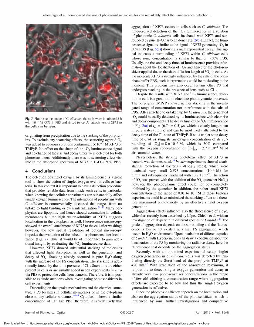
4 ConclusionsThe detection of singlet oxygen by its luminescence is a greattool to show the action of singlet oxygen even in cells or bac-teria. In this context it is important to have a detection procedurethat provides reliable data from inside such cells, in particularwhen knowing that cellular constituents can substantially affectsinglet oxygen luminescence. The interaction of porphyrins withC. albicans is controversially discussed that ranges from nouptake to tight binding or even internalization.39–43 Many por-phyrins are lipophilic and hence should accumulate in cellularmembranes but the high water-solubility of XF73 suggestslocalization in the cytoplasm as well. Fluorescence microscopyshowed the overall attachment of XF73 to the cell after washing;however, the low spatial resolution of optical microscopyimpedes the evaluation of the subcellular photosensitizer locali-zation (Fig. 7). Thus, it would be of importance to gain addi-tional insight by evaluating the 1O2 luminescence data.
However, XF73 showed substantial stacking of moleculesthat affected light absorption as well as the generation anddecay of 1O2. Stacking already occurred in pure H2O alongwith the increase of the PS concentration. The stacking is addi-tionally forced by the ionic pressure of Cl−. Such ions are eitherpresent in cells or are usually added in cell experiments in vitrovia PBS to protect the cells from osmosis. Therefore, it is impos-sible to exclude such ions when investigating photosensitizers incell experiments.
Depending on the uptake mechanisms and the chemical struc-ture, a PS localizes in cellular membranes or in the cytoplasmclose to any cellular structures.44,45 Cytoplasm shows a similarconcentration of Cl− like PBS; therefore, it is very likely that
aggregation of XF73 occurs in cells such as C. albicans. Thetime-resolved detection of the 1O2 luminescence in a solutionof planktonic C. albicans cells incubated with XF73 and sur-rounded by pureH2O has been done [Fig. 2(b)]. In fact, the lumi-nescence signal is similar to the signal of XF73 generating 1O2 in30% PBS [Fig. 5(c)] showing a multiexponential decay. This sig-nal indicates a surrounding of XF73 within C. albicans cellswhose ionic concentration is similar to that of >30% PBS.Usually, the rise and decay times of luminescence provides infor-mation about the localization of 1O2 and hence of the photosen-sitizer applied due to the short diffusion length of 1O2 in cells. Asthe molecule XF73 is strongly influenced by the salts of the phos-phate buffer PBS, such interpretations could be misleading at themoment. This problem may also occur for any other PS thatundergoes stacking in the presence of ions such as Cl−.
Despite the results with XF73, the 1O2 luminescence detec-tion in cells is a great tool to elucidate photodynamic processes.The porphyrin TMPyP showed neither stacking in the investi-gated range of concentration nor interference with the salts ofPBS. After attached to or taken up by C. albicans, the generated1O2 could be easily detected by its luminescence with clear riseand decay components. The decay time of the 1O2 luminescencein Fig. 2(a) of tD ¼ ð6.74� 0.5Þ μs, which is clearly longer thanin pure water (3.5 μs) and can be most likely attributed to thedecay time of the T1-state of TMPyP. If so, a triplet state decaytime of 6.74 μs suggests an oxygen concentration of its sur-rounding of ½O2� ¼ 8 × 10⁻⁵ M, which is 30% comparedwith the oxygen concentration of ½O2�sat ¼ 2.7 × 10⁻⁴ M ofair saturated water.
Nevertheless, the striking phototoxic effect of XF73 inbacteria was demonstrated.16 In vitro experiments showed a sub-stantial reduction of bacteria (∼8 log10 steps), which wereincubated very small XF73 concentrations (10−8 M) for5 min and subsequently irradiated with 13.7 J cm−2. The actionof 1O2 was proven with the addition of the 1O2 quencher NaN3;however, the photodynamic effect could not be completelyinhibited by the quencher. In addition, the rather small XF73concentration in the range of 0.01 to 10 μM in those bacteriaexperiments could have minimized the stacking effect and there-fore maximized phototoxicity by an effective singlet oxygengeneration.
Aggregation effects influence also the fluorescence of a dye,which has recently been described by López-Chicón et al. with aninvestigation of Hypericin in different species of Candida.46 Thegrade of aggregation depends on the surrounding and the fluores-cence is low or not existent at a high PS aggregation, whichoccurs in H2O-environment. Upon incubation of different speciesof Candida with Hypericin, one can draw a conclusion about thelocalization of the PS by monitoring the radiative decay, here thefluorescence that depends on the aggregation status.
Recently, with an optimized experimental setup singletoxygen generation in C. albicans cells was detected by irra-diating directly the Soret-band of the porphyrin TMPyP at420 nm.47 With irradiation of the absorption maximum, itis possible to detect singlet oxygen generation and decay atalready very low photosensitizer concentrations in the rangeof few μM offering a concentration range where aggregationeffects are expected to be low and thus the singlet oxygengeneration is effective.
Since the phototoxic efficacy depends on the localization andalso on the aggregation status of the photosensitizer, which isinfluenced by ions, further investigations and comparative
Fig. 7 Fluorescence image of C. albicans; the cells were incubated 2 hwith 10−4 M XF73 in PBS and rinsed twice. An attachment of XF73 tothe cells can be seen.
Journal of Biomedical Optics 045002-7 April 2013 • Vol. 18(4)
Felgenträger et al.: Ion-induced stacking of photosensitizer molecules can remarkably affect the luminescence detection. . .
Downloaded From: https://www.spiedigitallibrary.org/journals/Journal-of-Biomedical-Optics on 5/11/2018 Terms of Use: https://www.spiedigitallibrary.org/terms-of-use
studies on the change of the singlet oxygen luminescence in dif-ferent species of microorganisms should lead to better insightsabout the change of the decay times due to the localization.
References1. C. A. Hart and S. Kariuki, “Antimicrobial resistance in developing
countries,” BMJ 317(7159), 647–650 (1998).2. R. Wise et al., “Antimicrobial resistance. Is a major threat to public
health,” BMJ 317(7159), 609–610 (1998).3. M. Wainwright, “Photodynamic antimicrobial chemotherapy (PACT),”
J. Antimicrob. Chemother. 42(1), 13–28 (1998).4. K. Lang, J. Mosinger, and D. M. Wagnerova, “Photophysical properties
of porphyrinoid sensitizers non-covalently bound to host molecules;models for photodynamic therapy,” Coord. Chem. Rev. 248(3–4),321–350 (2004).
5. M. R. Hamblin and T. Hasan, “Photodynamic therapy: a new antimi-crobial approach to infectious disease?,” Photochem. Photobiol. Sci.3(5), 436–450 (2004).
6. M. Niedre, M.S. Patterson, and B.C. Wilson, “Direct near-infrared lumi-nescence detection of singlet oxygen generated by photodynamictherapy in cells in vitro and tissues in vivo,” Photochem. Photobiol.75(4), 382–391 (2002).
7. G. Jori and S. B. Brown, “Photosensitized inactivation of microorgan-isms,” Photochem. Photobiol. Sci. 3(5), 403–405 (2004).
8. R. Bonnett, “Photosensitizers of the porphyrin and phthalocyanineseries for photodynamic therapy,” Chem. Soc. Rev. 24(1), 19–33 (1995).
9. R. Bonnett and M. Berenbaum, “Porphyrins as photosensitizers,” CibaFound. Symp. 146, 40–59; discussion 53–59 (1989).
10. K. Nouri and L. M. Villafradez-Diaz, “Light/laser therapy in the treat-ment of acne vulgaris,” J. Cosmet. Dermatol. 4(4), 318–320 (2005).
11. E. Feese and R. A. Ghiladi, “Highly efficient in vitro photodynamicinactivation of Mycobacterium smegmatis,” J. Antimicrob. Chemother.64(4), 782–785 (2009).
12. P. K. Frederiksen et al., “Two-photon photosensitized production of sin-glet oxygen in water,” J. Am. Chem. Soc. 127(1), 255–269 (2005).
13. J. W. Snyder, J. D. Lambert, and P. R. Ogilby, “5,10,15,20-tetrakis(N-methyl-4-pyridyl)-21H,23H-porphine (TMPyP) as a sensitizer for sin-glet oxygen imaging in cells: characterizing the irradiation-dependentbehavior of TMPyP in a single cell,” Photochem. Photobiol. 82(1),177–184 (2006).
14. X. Ragas, M. Agut, and S. Nonell, “Singlet oxygen in Escherichia coli:new insights for antimicrobial photodynamic therapy,” Free. Radic.Biol. Med. 49(5), 770–776 (2010).
15. E. F. de Silva et al., “Irradiation- and sensitizer-dependent changes in thelifetime of intracellular singlet oxygen produced in a photosensitizedprocess,” J. Phys. Chem. B 116(1), 445–461 (2012).
16. T. Maisch et al., “Photodynamic effects of novel XF porphyrin deriv-atives on prokaryotic and eukaryotic cells,” Antimicrob. Agents.Chemother. 49(4), 1542–1552 (2005).
17. F. Pereira Gonzales and T. Maisch, “XF drugs: a new family of anti-bacterials,” Drug News Perspect. 23(3), 167–174 (2010).
18. J. G. Parker and W. D. Stanbro, “Optical determination of the rates offormation and decay of O-2(1-Delta-G) in H2O, D2O and other sol-vents,” J. Photochem. 25(2–4), 545–547 (1984).
19. J. Regensburger et al., “A helpful technology–the luminescence detec-tion of singlet oxygen to investigate photodynamic inactivation of bac-teria (PDIB),” J. Biophoton. 3(5–6), 319–327 (2010).
20. J. Baier et al., “Theoretical and experimental analysis of the lumines-cence signal of singlet oxygen for different photosensitizers,”J. Photochem. Photobiol. B 87(3), 163–173 (2007).
21. F. Wilkinson, W. P. Helman, and A. B. Ross, “Quantum yields forthe photosensitized formation of the lowest electronically excited sin-glet-state of molecular-oxygen in solution,” J. Phys. Chem. Ref. Data22(1), 113–262 (1993).
22. F. Ricchelli, “Photophysical properties of porphyrins in biological mem-branes,” J. Photochem. Photobiol. B, 29(2–3), 109–118 (1995).
23. E. Zenkevich et al., “Photophysical and photochemical propertiesof potential porphyrin and chlorin photosensitizers for PDT,”J. Photochem. Photobiol. B-Biol. 33(2), 171–180 (1996).
24. K. M. Kadish, K. M. Smith, and R. Guilard, The Porphyrin Handbook,Academic Press, San Diego (2000).
25. N. C. Maiti et al., “Fluorescence dynamics of noncovalently linked por-phyrin dimers and aggregates,” J. Phys. Chem. 99(47), 17192–17197(1995).
26. M. Sirish and H. J. Schneider, “Supramolecular chemistry, part93—Electrostatic interactions between positively charged porphyrinsand nucleotides or amides: buffer-dependent dramatic changes of bind-ing affinities and modes,” Chem. Commun. 1, 23–24 (2000).
27. R. F. Pasternack et al., “On the aggregation of meso-substituted water-soluble porphyrins,” J. Am. Chem. Soc. 94(13), 4511–4517 (1972).
28. K. Kano et al., “Evidence for stacking of cationic porphyrin in aqueous-solution,” Chem. Lett 12, 1867–1870 (1983).
29. R. L. Brookfield, H. Ellul, and A. Harriman, “Luminescence of porphyr-ins and metalloporphyrins. 9. Dimerization of meso-tetrakis(N-Methyl-4-Pyridyl)-porphine,” J. Photochem. 31(1), 97–103 (1985).
30. K. M. Kadish, B. G. Maiya, and C. Araullomcadams, “Spectroscopiccharacterization of meso-tetrakis(1-Methylpyridinium-4-Yl)porphyrins,½ðTmpypÞH2�4þ and ½ðTmpypÞM�4þ, in aqueous micellar media,where M ¼ Vo2þ, Cu(Ii), and Zn(Ii),” J. Phys. Chem. 95(1), 427–431 (1991).
31. F. J. Vergeldt et al., “Intramolecular interactions in the ground andexcited-state of tetrakis(N-Methylpyridyl)porphyrins,” J. Phys. Chem.99(13), 4397–4405 (1995).
32. G. De Luca, A. Romeo, and L. M. Scolaro, “Role of counteranions inacid-induced aggregation of isomeric tetrapyridylporphyrins in organicsolvents,” J. Phys. Chem. B 109(15), 7149–7158 (2005).
33. M. A. J. Rodgers and P. T. Snowden, “Lifetime of O-2(1delta-G) inliquid water as determined by time-resolved infrared luminescencemeasurements,” J. Am. Chem. Soc. 104(20), 5541–5543 (1982).
34. S. Y. Egorov et al., “Rise and decay kinetics of photosensitized singletoxygen luminescence in water—Measurements with nanosecondtime-correlated single photon-counting technique,” Chem. Phys. Lett.163(4–5), 421–424 (1989).
35. W. Baumler et al., “The role of singlet oxygen and oxygen concentrationin photodynamic inactivation of bacteria,” in Proc. Natl. Acad. Sci. U. S.A. 104(17), 7223–7228 (2007).
36. J. R. Kanofsky et al., “Biochemical requirements for singlet oxygen pro-duction by purified human myeloperoxidase,” J. Clin. Invest. 74(4),1489–1495 (1984).
37. D. N. Butorina, A. A. Krasnovskii, and A. V. Priezzhev, “Investigationof the kinetic parameters of singlet molecular oxygen in aqueous por-phyrin solutions. influence of detergent and the quencher sodium azide,”Biofizika 48(2), 201–209 (2003).
38. J. M. Wessels, P. Charlesworth, and M. A. Rodgers, “Singlet oxygenluminescence spectra: a comparison of interferometer- and grating-based spectrometers,” Photochem. Photobiol. 61(4), 350–352(1995).
39. S. Mitra et al., “Effective photosensitization and selectivity in vivo ofCandida Albicans by meso-tetra (N-methyl-4-pyridyl) porphine tetratosylate,” Lasers Surg. Med. 43(4), 324–332 (2011).
40. T. Ito, “Photodynamic action of hematoporphyrin on yeast cells–akinetic approach,” Photochem. Photobiol. 34(4), 521–524 (1981).
41. M. P. Cormick et al., “Mechanistic insight of the photodynamic effectinduced by tri- and tetra-cationic porphyrins on Candida albicans cells,”Photochem. Photobiol. Sci. 10(10), 1556–1561 (2011).
42. M. P. Cormick et al., “Photodynamic inactivation of Candida albicanssensitized by tri- and tetra-cationic porphyrin derivatives,” Eur. J. Med.Chem. 44(4), 1592–1599 (2009).
43. S. Oriel and Y. Nitzan, “Mechanistic aspects of photoinactivation ofCandida albicans by exogenous porphyrins (dagger),” Photochem.Photobiol. 88(3), 604–612 (2012).
44. R. F. Pasternack et al., “A spectroscopic and thermodynamic study ofporphyrin/DNA supramolecular assemblies,” Biophys. J. 75(2), 1024–1031 (1998).
45. P. Kubat et al., “Interaction of novel cationic meso-tetraphenylporphyr-ins in the ground and excited states with DNA and nucleotides,”J. Chem. Soc.-Perkin Trans. 1, 6, 933–941 (2000).
46. P. Lopez-Chicon et al., “On the mechanism of Candida spp. photoinac-tivation by hypericin,” Photochem. Photobiol. Sci. 11(6), 1099–1107(2012).
47. A. Eichner et al., “Dirty hands: photodynamic killing of human patho-gens like EHEC, MRSA and Candida within seconds,” Photochem.Photobiol. Sci. 12(1), 135–147 (2012).
Journal of Biomedical Optics 045002-8 April 2013 • Vol. 18(4)
Felgenträger et al.: Ion-induced stacking of photosensitizer molecules can remarkably affect the luminescence detection. . .
Downloaded From: https://www.spiedigitallibrary.org/journals/Journal-of-Biomedical-Optics on 5/11/2018 Terms of Use: https://www.spiedigitallibrary.org/terms-of-use