Embed Size (px)
Citation preview
IP-10, a -C-X-C- Chemokine, Elicits a Potent Thymus-dependent Antitumor Response In Vivo By Andrew D. Luster and Philip Leder
From the Department of Genetics, Harvard Medical School, Howard Hughes Medical Institute, Boston, Massachusetts 02115
S u m m a r y
IP-10 is a member of the -C-X-C- chemokine superfamily of proinflammatory cytokines whose secretion is induced by interferon 3' (IFN-y) and lipopolysaccharide (LPS). To date no function has been described for IP-10. We have genetically engineered tumor cells to secrete high levels of murine IP-10 and demonstrate that while IP-10 has no effect on the growth of these tumor cells in culture, it elicits a powerful host-mediated antitumor effect in vivo. The IP-10 antitumor response is T lymphocyte dependent, non-cell autonomous, and appears to be mediated by the recruitment of an inflammatory infiltrate composed of lymphocytes, neutrophils, and monocytes. These results document an important biologic property of IP-10 and raise the possibility that some of the T cell-directed effects of IFN-3, and LPS may be mediated by this chemokine.
T he observation that some cancer patients who developed concurrent bacterial infections also experienced remis-
sions of their malignant disease led Coley over 100 years ago to treat cancer patients with a preparation of heat-kiUed Gram- negative and Gram-positive bacteria, now commonly referred to as "Coley's toxins" (1, 2). The study of this phenomenon in animal models led to the identification of LPS as an active component of the toxins that induces a cytokine-mediated antitumor response in vivo (3). As IP-10 is a cytokine that is dramatically induced by LPS and IFN-y, we sought to de- termine if IP-10 could elicit an antitumor effect in vivo.
IP-10 was originally isolated as a predominant mRNA in- duced by IFN-3/(4) or LPS (5, 6) in monocytes, and its ex- pression has been detected in vivo during the development of a delayed-type hypersensitivity cellular immune response by keratinocytes, endothelial cells, and infiltrating mononuclear cells (7). In addition, IP-10 expression has been seen in the epidermis and dermis in cutaneous lesions of psoriasis (8), tuberculoid leprosy (7), and fixed DRUG eruptions (9).
IP-10 is a member of the chemokine superfamily and is ,v30% homologous to IL-8 and platelet factor (PF4). 1 The chemokines are secreted proteins induced by inflammatory stimuli and are involved in orchestrating the selective migra- tion, diapedesis, and activation of blood-borne leukocytes that mediate the inflammatory response (10, 11). This superfamily has been subdivided into two classes based on the positions of the four invariant cysteine residues found in the primary structure of these molecules. IP-10 belongs to the so-caUed o~, or -C-X-C-, class of chemokines that map to a cluster
1Abbreviations used in this paper: M, murine; MCP-1, monocyte chemotactic protein 1; MIP-I~/, macrophage inflammatory protein 1B; PF4, platelet factor 4.
on human chromosome 4 (q12-21), and includes IL-8, GRO, and the platelet ol-granule basic proteins PF4 and ~/-thrombo- globulin (flTG). The fl, or -C-C-, class maps to human chro- mosome 17 (q11-32), and includes monocyte chemotactic pro- tein 1 (MCP-1), R.ANTES, macrophage inflammatory protein lfl (MIP-I~), and 1-309. The chemokines seem to affect different, yet overlapping, leukocytes subsets. For example, IL-8 is specific for neutrophils (12) and CD45RO + memory T cells (13, 14), RANTES is specific for monocytes, eosinophils (15), and a subset of CD4 + T cells (16); MIP-lfl is specific for monocytes and CD8 + T cells (17); and MIP-loe is specific for monocytes, basophils, and mast cells (18). Although IP-10 was one of the first chemokines to be identified, its physiologic function remains unknown. We therefore under- took the present study to gain insight into the biological ac- tivities of IP-10 by analyzing the cells that respond to IP-10 in vivo and to determine if IP-10 possesses an antitumor effect in vivo.
Materials and Methods
Cell Lines and Cell Culture. J558L is a heavy chain loss variant of the BALB/c plasmacytoma line J558 (19). The K485 mammary adenocarcinoma line was derived from a mammary tumor in a trans- genic mouse carrying an activated c-myc oncogene (20). TheJ558L cell line was grown in RPMI 1640 supplemented with 10% FCS, 50 U/ml penicillin, 50 #g/ml streptomycin, 2 mM t-glutamine, and 57 #M 2-ME. K485 and RAW 264.7 cell lines were main- tained in DME supplemented with 10% FCS and penicillin and streptomycin in the concentrations noted above.
Construction of Ig-IPlO and Moloney (Mo)LTR-IPIO Expression Vectors. The murine (m)IP-10 cDNA was cloned from the mu- rine macrophage cell line RAW 264.7 (American Type Culture Col- lection, Rockville, MD). RNA was isolated from RAW 264.7 cells that were treated for 3 h with 200 U/m1 rmIFN-3, (Genentec, Inc.,
1057 J. Exp. Med. �9 The Rockefeller University Press �9 0022-1007/93/09/1057/09 $2.00 Volume 178 September 1993 1057-1065
San Francisco, CA). This RNA was then used as a template for first-strand cDNA synthesis. The complete mlP-10 coding sequence was cloned from this cDNA using Taq DNA polymerase, standard PCR conditions, a 5' sense oligonucleotide AAGCGCTTCATC- CACCG, and a 3' antisense oligonucleotide GCGTGGCTTCTC- TCCAG based on sequences 1-17 and 362-379, respectively, from the sequence of mlP-10 (5). The PCR product was purified by agarose gel electrophoresis, blunt-ended with Klenow, phos- phorylated with T4 DNA kinase, and then blunt-end ligated into the XhoI site of pIgTE/N and the EcoRI site of a MoLTR-SV40 I/pA expression vectors that had been flushed with Klenow. Se- quence analysis of the cloned PCR product confirmed the sequence of mlP-10. The Ig control sequences used in the generation of the plasmid plgTE/N were derived from the plasmid pTARY (22), placed 5' of the Ig promoter at the unique XbaI site in pBluescript (Stratagene, La Jolla, CA). Both vectors contain an SV40 intron and splice and polyadenylation signals (23) after the cDNA cloning site. The MoLTR was isolated as a 0.6-kb ClaI-XmaI fragment of pZip-NeoSV (X)-I plasmid (24), which includes the 3' LTR of the Moloney murine leukemia virus.
Transfection of Tumor Cell Lines. Transfection of J558L ceils was performed by electroporation (25). 20/~g of linearized Ig-IP10 or MoLTR-IP10 expression vector plasmid DNA and 1/~g of linear- ized neomycin resistance plasmid pSV7Neo (26) were used per trans- fection of 5 x 10⁶ cells. After 48 h in RPMI, cells were cen- trifuged and resuspended in selective media containing 0.8 mg/ml of G418 (as calculated for 100% antibiotic activity; Genticin; GIBCO BRL, Gaithersburg, MD) and plated in serial dilutions into 288 wells (6.4-ram diameter) to clone by limiting dilution. G418-resistant cells from single wells were expanded, and a second round of cloning by limiting dilution in selective media was per- formed to ensure donality. Transfection of K485 cells was performed by electroporation using 30/~g of linearized MoLTR-IP10 expres- sion vector DNA and 1.5 #g ofpSV7Neo DNA per 100-mm plate containing 6 x 10 s cells. 24 h after transfection, cells were split 1:3 in DME; G418 at a concentration 0.4 mg/ml was added after an additional 24 h. Individual G418-resistant colonies were picked after 10-12 d, and then to insure clonality, single cell clones were then isolated by limiting dilution in selective media. In vitro dou- bling times were determined by serial cell counts using a hemocytometer over 96 h.
R N A Blots. RNA was isolated by lysing cells in guanidine hy- drochloride and pelleting the RNA through a 5.7-M CsC12 cushion (27). RNA was fractionated on a 1.2% agarose gel con- taining 0.2 M formaldehyde and then transferred to gene screen (DuPont Co., Wilmington, DE) and hybridized with [32p]dCTP Klenow-labeled mlP-10 cDNA or a [32p]dCTP Klenow-labeled plasmid containing a 279-bp XholI-DraI fragment encoding the ribosomal protein rp L32 (28) as a control for RNA loading.
Protein Expression and Purification. rmlP-10 beginning with the putative mature NH2-terminal Ile and terminating with the COOH-terminal Pro was expressed in the Escherichia coli strain M15 using the Qiaexpress vectors pQE12 and pQE8 (Qiagen Inc., Chats- worth, CA). Expression of IP-10 in pQE12 results in a fusion pro- tein containing a six-histidine COOH-terminal tag, and expres- sion of IP-10 in pQE8 results in a fusion protein containing an NH2-terminal six-histidine tag. IP-10 was purified to apparent homogeneity (a single peak on HPLC) by sedimentation of inclu- sion bodies through sucrose, affmity chromatography on nickel agarose (Qiagen Inc.), and then FPLC on the cation exchange resin Mono-S (Pharmacia Biotech Inc., Piscataway, NJ) eluting with a NaC1 gradient or reverse-phase HPLC (Waters Associates, Milford, MA) eluting with an acetonitrile gradient. The concentration of
purified protein was determined using a Bradford assay (Bio-Rad Laboratories, Richmond, CA) with IgG or bovine gamma globulin as the known standard.
AntibodyPretmration. For immunizations, IP-10 was purified as above except that the eluent from the nickel-agarose chromatog- raphy column was separated on a denaturating SDS-polyacrylamide gel. The region of the gel containing IP-10 was then emulsified with CFA for the primary immunization and with IFA for subse- quent immunizations. Approximately 200 /xg of the COOH- terminal-tagged protein (IP-10-[His]6) was injected subcutaneously into each of three, 8-wk-old, female New Zealand white rabbits. The rabbits were boosted twice, at l-too intervals, with 100/~g of the NH2-terminal-tagged protein ([His]6-IP-10) per rabbit to ensure the generation of antibodies recognizing the native NH2 and COOH-termini of IP-10. 10 d after the second boost, the three rabbits were bled and serum was isolated and pooled for further studies.
Immunoprecipitation. 107 cells were metabolically labeled in 1 ml for suspension cells and 2 ml for adherent cells of methionine- free DME supplemented with 5% dialyzed serum and 0.5 mCi of [3SS]methionine for 6 h at 37~ The media were collected, cen- trifuged at 1,000 g, and the protease inhibitors (Boehringer Mann- heim Biochemicals, Indianapolis, IN) at the following concentra- tions were added to the supernatant: leupeptin (0.3 ng/ml), aprotinin (10 ng/ml), PMSF (20 ng/ml), and pepstatin (0.8 ng/ml). The su- pernatant was then centrifuged at 10,000 g for 30 min at 4~ and then precleared for 2 h at 4~ with 10/~1 of pooled normal rabbit serum and 100/xl of a 1:1 slurry of protein A-Sepharose (PAS; Phar- macia Fine Chemicals, Piscataway, NJ). The PAS was then spun out at 10,000 g in eppendorf tubes, the supernatant was split and transferred to new tubes, and then 2/xl of pooled immune serum or preimmune serum was added to each sample for 3 h at 4~ 50/~1 of a 1:1 slurry of PAS was added to each sample, incubated another 4 h at 4~ and then centrifuged at 10,000 g for 10 rain. The PAS beads were then washed three times with 1 ml of RIPA buffer (0.15 M NaC1, 1% NP-40, 0.1% SDS, 0.5% deoxycholate, 0.05 M Tris, pH 8.0) transferred to new tubes and then boiled for 3 rain in 50/,1 of sample buffer containing 0.3 M 2-ME, 4% SDS, and 0.01% Pyronin Y. 15/~1 was then analyzed on a 15% SDS- polyacrylamide gel using a Tris/Tricine buffer system (29) that has good resolution in the low molecular weight region.
ELISA. Conditioned medium was collected for 48 h from 2 x 10⁶ cells/ml in serum-free medium and 100/~1 was then ad-
sorbed in duplicate to microtiter wells (3911 Micro Test III; Falcon Labware, Oxnard, CA) for 4 h. Nonspecific binding sites were blocked for 2 h at room temperature with 200/~l/well of a PBS solution containing 2% goat serum and 2% BSA (blocking solu- tion). This was followed by 100 ttl/well of anti-IP-10 antiserum at a 1:1,000 dilution in blocking solution for 2 h at room tempera- ture. The plates were then washed four times with water, incubated for I h at room temperature with a 1:10,000 dilution of an alkaline phosphatase-conjugated goat anti-rabbit IgG (Jackson Im- munoResearch, West Grove, PA), washed four more times with water, and treated with 100/~l/well of the substrate p-nitrophenyl phosphate in diethanolamine (Pierce, Rockford, IL). The o ~ mOD405/min was measured on a Om~ kinetic microplate reader /Molecular Devices, Menlo Park, CA).
Animal Studies. Tumor cell injections were carried out using freshly prepared suspensions at a concentration of 107 cells/ml in PBS. The total number of tumor cells injected was 2 x 10⁶, ex- cept in mixed tumor transplantation experiments, in which 2 x 10⁶ cells of each type were mixed and injected. All injections were performed subcutaneously in the right lower abdominal quadrant
1058 IP-10 Elicits a Thymus-dependent Antitumor Response In Vivo
via 25- or 27-gauge needles. BALB/c, BALB/c nu/nu, Swiss nu/nu, and FVB mice were obtained from Taconic Farms (Germantown, NY) and were cared for according to the guidelines provided by the Harvard Medical Area Animal Research Center.
Histology. Tissue at the site of tumor cell inoculation was fixed in OptimalFix (American Histology Reagent Co., Stockton, CA). Standard paraffin-embedding, sectioning, and staining with hema- toxylin and eosin was performed by the Transgenic Pathology Lab- oratory (University of California, Davis, CA).
Results Generation of IP-lO-expressing Tumor Lines. To establish
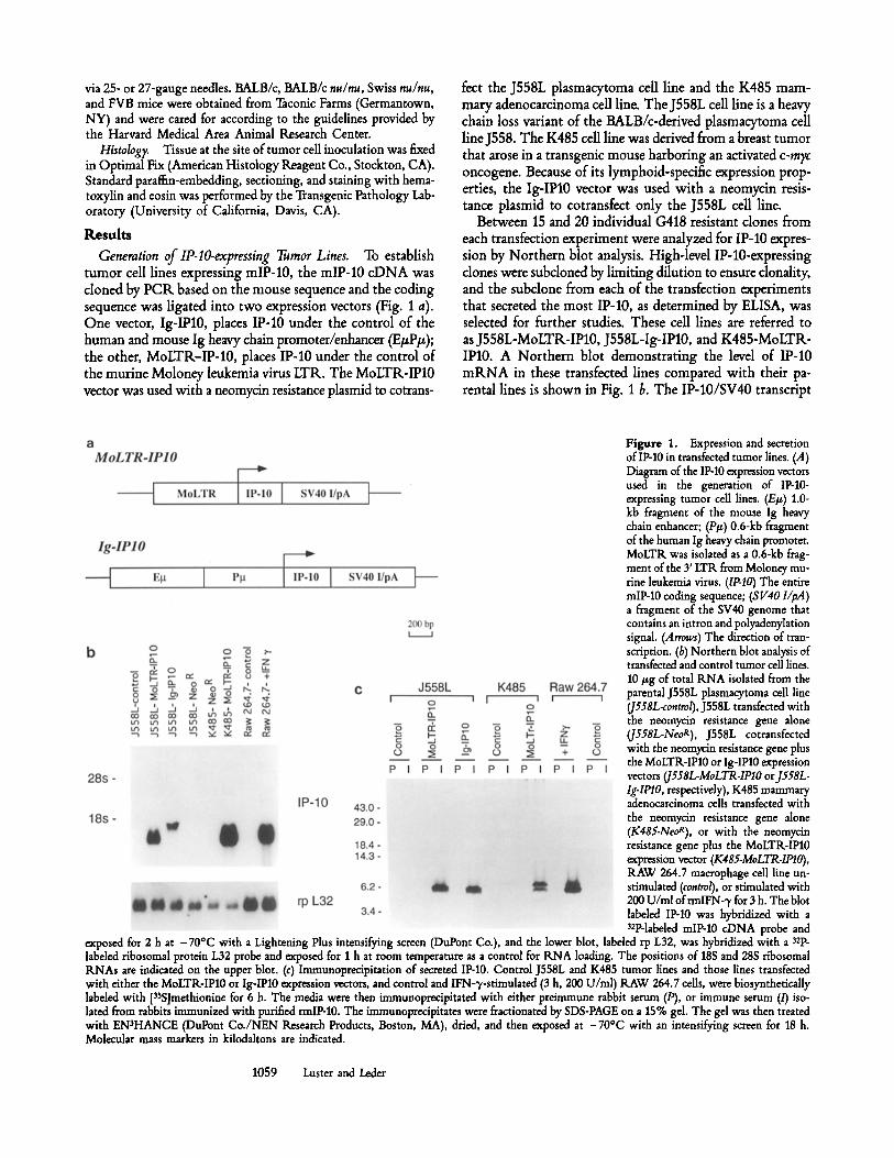
tumor ceil lines expressing mlP-10, the mIP-10 c D N A was cloned by P C R based on the mouse sequence and the coding sequence was ligated into two expression vectors (Fig. 1 a). One vector, Ig-IPl0, places IP-10 under the control of the human and mouse Ig heavy chain promoter/enhancer (E#P#); the other, MoLTR-IP-10, places IP-10 under the control of the routine Moloney leukemia virus LTR. The MoLTR-IP10 vector was used with a neomycin resistance plasmid to cotrans-
fect the J558L plasmacytoma cell line and the K485 mam- mary adenocarcinoma cell line. The J558L cell line is a heavy chain loss variant of the BALB/c-derived plasmacytoma cell line J558. The K485 cell line was derived from a breast tumor that arose in a transgenic mouse harboring an activated c-m?c oncogene. Because of its lymphoid-specific expression prop- erties, the Ig-IPl0 vector was used wi th a neomycin resis- tance plasmid to cotransfect only the J558L cell line.
Between 15 and 20 individual G418 resistant clones from each transfection experiment were analyzed for IP-10 expres- sion by Northern blot analysis. High-level IP-10-expressing clones were subdoned by limiting dilution to ensure donality, and the subclone from each of the transfection experiments that secreted the most IP-10, as determined by ELISA, was selected for further studies. These cell lines are referred to as J558L-MoLTR-IP10, J558L-Ig-IP10, and K485-MoLTR- IPl0. A Northern blot demonstrating the level of IP-10 m R N A in these transfected lines compared with their pa- rental lines is shown in Fig. 1 b, The IP-10/SV40 transcript
Figure 1. Expression and secretion of IP-10 in transfeeted tumor lines. (A) Diagram of the IP-10 expression vectors used in the generation of IP-10- expressing tumor cell lines. (E#) 1.0- kb fragment of the mouse Ig heavy chain enhancer; (P/~) 0.6-kb fragment of the human Ig heavy chain promoter. MoLTR was isolated as a 0.6-kb frag- ment of the 3' LTR from Moloney mu- rine leukemia virus. (IP-IO) The entire mlP-10 coding sequence; (8 V40 l/pA) a fragment of the SV40 genome that contains an intron and polyadenylation signal (Arrows) The direction of tran- scription. (b) Northern blot analysis of transfeeted and control tumor cell lines. 10 #g of total RNA isolated from the parental J558L plasmacytoma call line (J558L-control), J558L transfected with the neomycin resistance gene alone (J558L-NeoR), J558L cotransfected with the neomycin resistance gene plus the MoLTR-IP10 or Ig-IPl0 expression vectors (J558L-MoLTR-IPlO or J558L- Ig-IPlO, respectively), K485 mammary adenocarcinoma cells transfected with the neomycin resistance gene alone (K485.NeoR), or with the neomycin resistance gene plus the MoLTR-IPl0 expression vector (K485-MoLTR-IPIO), RAW 264.7 macrophage cell line un- stimulated (control), or stimulated with 200 U/nil ofrmlFN-3, for 3 h. The blot labeled IP-10 was hybridized with a 32p-labded mIP-10 cDNA probe and
exposed for 2 h at -70~ with a Lightening Plus intensifying screen (DuPont Co.), and the lower blot, labeled rp L32, was hybridized with a 32p_ labeled ribosomal protein L32 probe and exposed for I h at room temperature as a control for RNA loading. The positions of 18S and 28S ribosomal RNAs are indicated on the upper blot. (c) Immunoprecipitation of secreted IP-10. Control J558L and K485 tumor lines and those lines transfected with either the MoLTR-IPl0 or Ig-IPl0 expression vectors, and control and IFN-q,-stimulated (3 h, 200 U/ml) RAW 264.7 cells, were biosynthetically labded with [3SS]methionine for 6 h. The media were then immunopreeipitated with either preimmune rabbit serum (P), or immune serum (/) iso- lated from rabbits immunized with purified rmlP-10. The immunoprecipitates were fractionated by SDS-PAGE on a 15% gel. The gel was then treated with EN~HANCE (DuPont Co./NEN Research Products, Boston, MA), dried, and then exposed at -70~ with an intensifying screen for 18 h. Molecular mass markers in kilodaltons are indicated.
1059 Luster and Leder
present in cells expressing the transfected Ig-IP10 gene is larger than the transcript seen in cells expressing the transfected MoLTIk-IP10 gene. This is presumably due to a more prox- imal site of RNA transcription initiation as determined by the different promotors.
To ensure that these clones secreted authentic IP-10 pro- tein, secreted IP-10 was visualized by immunoprecipitation and SDS-PAGE (Fig. 1 c). Since the previously characterized rabbit anti-human IP-10 antiserum (30) does not recognize the murine homologue, mlP-10 was expressed and purified from E. coli and used to generate a monospecific polydonal rabbit antiserum. Various control and IP-10-expressing J558L and K485 cell lines were metabolically labeled with [3SS]me- thionine, and IP-10 was immunoprecipitated from the media using this rabbit anti-murine IP-10 antiserum. The protein immunoprecipitated from the transfected tumor cell lines and from the IFN-~/-treated murine macrophage cell line, RAW 264.7, used as a positive control, migrated as a doublet be- tween 6 and 7 kD under reducing conditions (Fig. 1 c). A doublet of the same size had been seen when human IP-10 was immunoprecipitated from endothelial cells, keratinocytes, and monocytes (30). This size heterogeneity probably reflects proteolytic processing. In fact, IP-10 purified from a stimu- lated tumor line revealed heterogeneity at the NH2 terminus (31). This type of NH2-terminal processing is a character- istic feature of the ot chemokines. Several NH2-terminally processed forms of IL-8 (32) and ffl'G (33) have been identified that differ in their biological activities. The IP-10 protein im- munoprecipitated from the transfected tumor cell lines comigrated with the protein immunoprecipitated from IFN- 3,-stimulated RAW 264.7 cells (Fig. I c). This confirms that the transfected tumor cells are constitutively secreting authentic IP-10.
To quantify the amount of IP-10 secreted from these trans- fected tumor cell lines, a solid-phase ELISA was developed using the rabbit anti-murine IP-10 antiserum. The amount of IP-10 collected in serum-free media was compared with a standard curve generated using known amounts of IP-10 purified from E. coli (data not shown). The IP-10-expressing J558L and K485 clones used in this study secreted *20 and -150 ng/ml/2 x 106 cells per 48 h, respectively (Table 1). Comparable amounts of IP-10 are secreted from equivalent numbers of transfected tumor cells and from IFN-~/-stimulated RAW 264.7 ceils (Fig. 1 c). IP-10 expression could not be detected in the parental J558L cell line by Northern blot, immunoprecipitation, or ELISA. Although the ELISA did not detect IP-10 in the medium collected from the parental K485 cell line (lower limit of detection, - 5 ng/ml), low levels of baseline IP-10 mRNA could be detected by Northern blot and immunoprecipitation (at least 100-fold less than after trans- fection with the MoLTK-IP10 vector).
Inhibition of Tumor Growth In Viva Both parental cell lines chosen for this study readily form tumors when injected sub- cutaneously into appropriate inbred strains of mice, with a latency of 10-14 d for J558L in BALB/c and m28 d for K485 in FVB. The transfection and expression of IP-10 in the J558L and K485 cell lines did not alter their growth properties in culture as assayed by doubling time or morphology (data not
Table 1. Quantitation of Secreted IP-IO by ELISA
Cell line m O D / m i n Concentration
ng/ml J558L transfected with:
None 3.54 _+ 0.1 - MoLTR.-IP10 29.03 _+ 0.5 ~20 Ig-IP10 24.31 _+ 1.4 ~20
K485 transfected with: None 9.08 _+ 0.8 - MoLTR-IP10 91.01 _+ 1.3 ~150
The approximate concentration of secreted IP-10 from stably transfected cell lines was determined by comparing the ELISA value obtained from the media of 2 x 10⁶ cells grown for 48 h in serum-free conditions to a standard curve obtained using purified rmlP-10.
shown). However, the expression of IP-10 did have a pro- found effect on the growth of these tumor cells in vivo. As is shown in Fig. 2 A and Table 2, the IP-10-producing J558L tumor cells failed to grow when they were injected subcutane- ously into BALB/c mice. In contrast, both the parental J558L line and clones ofJ558L transfected with the neomycin resis- tance gene alone grew rapidly and formed large local masses 10-14 d after subcutaneous injection into BALB/c mice. The antitumor effect was observed with both the MoLTR-IP10 and Ig-IP10 vectors, demonstrating that the effect is not vector specific.
That the effect is not restricted to a single tumor type, or to a single inbred genetic background of mice, was shown by demonstrating IP-10's antitumor effect on the K485 mam- mary adenocarcinoma cell line in FVB mice. Fig. 2 B and Table 3 demonstrate the inhibition of tumor formation of a K485 clone cotransfected with MoLTR-IP10 and a neomycin resistance gene, compared with a control K485 clone trans-
Table 2. Tumorigenicity of J558L Control and Transfectants in Syngeneic BALB/c and Nude Mice
J558L transfected with:
Tumor occurrence
BALB/c nu/nu
None 21/21 7/8 Neo g done 4/5 5/5 MoLTR-IP10 0/14 12/16 Ig-IP10 2/16 12/16
Mice were autopsied 10-14 d after 2 x 10⁶ cells were injected subcutane- ously into the right lower abdominal quadrant. The weight of the extir- pated tumor or residual scar was determined and a tumor mass was considered positive if it weighed ~0.5 g. This table is a compilation of seven different experiments; control and experimental animals for any given experiment were always autopsied on the same day.
1060 IP-10 Elicits a Thymus-dependent Antitumor Response In Vivo
Figure 2. Mice injected with control and IP-10-secreting tumors. (A) 12 d after subcutaneous fight lower quadrant injection of control (untransfected), MoLTR-IFI0, and Ig-IP10J558L tumor lines into syngeneic BALB/c mice. (B) 28 d after subcutaneous fight lower quadrant injection of control (Neo R alone) and MoLTR-IP10 K485 tumor lines into FVB mice. 2 x 10 ~ tumor cells were injected per animal.
fected with the neomycin resistance gene alone. K485 con- trol tumor cells subcutaneously injected into FVB mice ini- tiaUy grew as a local tumor mass that, by 1 mo after injection, locally invaded through the peritoneum and led to malig- nant ascites (Fig. 2/3). However, IP-10-expressing K485 tumor cells were inhibited from growing and did not invade and metastasize, even though a small (<0.5-g) tumor mass was occasionally present at 1 mo.
IP-10's Antitumor Effect Is Thymus Dependent. The in vivo antitumor effect of IP-10 was dependent on thymus- derived cells as was evidenced by the rapid growth of all tumor lines (IP-10 expressors and nonexpressors) in both BALB/c
Table 3. Effect of lP-lO on the Tumorigenicity of Mammary Adenocarcinoma Cells in FVB Mice
K485 transfected with: Tumor occurrence
Neo R alone 5/6
MoLTR-IP10 0/8
nu/nu and Swiss nu/nu mice (Table 2). This implies that the antitumor response evoked by IP-10 was T cell mediated.
Coinjection of lP-I O Producers and Nonproducers Demonstrates the Non-cell Autonomous Effect of lP-lO. Mixing producer and nonproducer cells and transplanting them in a 1:1 ratio into syngeneic mice demonstrated that the effect of IP-10 was non-ceU autonomous (that is, did not require that IP-10 be produced by all tumor cells). As shown in Table 4, when a J558L neomycin resistant IP-10 nonproducer clone was in- jected subcutaneously into BALB/c mice, it formed tumors
Table 4. IP-lO's Effect in Mixed Tumor Cell Transplantation
Cell Tumor occurrence
J558L-Neo R alone 4/5
1:1 mix of J558L-Neo ~ and
J558L transfected with
MoLTR-IP10 0/5
Ig-IP10 1/5
Mice were autopsied 28 d after 2 x 10~ cells were injected subcutane- ously into the fight lower abdominal quadrant. Tumors were associated with a tense bloody malignant ascites.
1061 Luster and Leder
Mice were autopsied 14 d after 2 x 10⁶ of each cell type was injected subcutaneously into the fight lower abdominal quadrant. Tumors were extirpated and considered positive if they weighed ;~r-0.5 g.
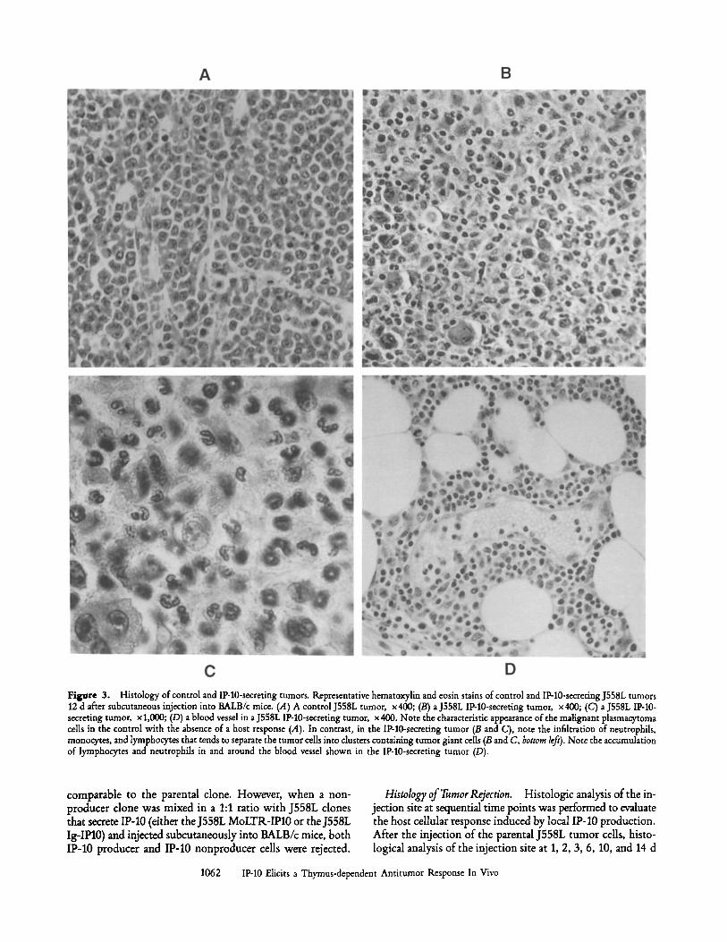
Figure 3. Histology of control and IP-10-secreting tumors. Representative hematoxylin and eosin stains of control and IP-10-secreting J558L tumors 12 d after subcutaneous injection into BALB/c mice. (A) A control J558L tumor, x400; (B) a J558L IP-10-secreting tumor, x400; (C) a J558L IP-10- secreting tumor, x 1,000; (D) a blood vessel in a J558L IP-10-secreting tumor, x 400. Note the characteristic appearance of the malignant plasmacytoma cells in the control with the absence of a host response (.4). In contrast, in the IP-10-secreting tumor (B and C), note the infiltration of neutrophils, monocytes, and lymphocytes that tends to separate the tumor cells into clusters containing tumor giant cells (B and C, bottom left). Note the accumulation of lymphocytes and neutrophils in and around the blood vessel shown in the IP-10-secreting tumor (D).
comparable to the parental clone. However, when a non- producer clone was mixed in a 1:1 ratio with J558L clones that secrete IP-I0 (either theJ558L MoLTR-IP10 or theJ558L Ig-IP10) and injected subcutaneously into BALB/c mice, both IP-10 producer and IP-10 nonproducer cells were rejected.
Histology of Tumor Rejection. Histologic analysis of the in- jection site at sequential time points was performed to evaluate the host cellular response induced by local IP-10 production. After the injection of the parental J558L tumor cells, histo- logical analysis of the injection site at 1, 2, 3, 6, 10, and 14 d
1062 IP-10 Elicits a Thymus-dependent Antitumor Response In Vivo
revealed progressive growth of the characteristic malignant plasmacytoma cells with large pleomorphic hyperchromatic nuclei, prominent nudeoli, and abundant amphophilic cyto- plasm (Fig. 3 A). Abundant mitotic figures were usually evi- dent. An occasional peripheral neutrophil, monocyte, or thin connective tissue capsule was seen. In contrast, the injection site of IP-10-expressing J558L cells, analyzed at the same time points as the controls, revealed the infiltration of monocytes, lymphocytes, and neutrophils by day 3 after injection. At later time points, relatively more neutrophils were observed, and by day 12, this leukocytic infiltrate seemed to invade and sep- arate the tumor cells into clusters, with a tendency of the residual tumor cells to form tumor giant cells (Fig. 3, B and C). Lymphocyte and neutrophil accumulation was seen sur- rounding blood vessels in the IP-10-secreting tumors, sug- gesting recent diapedesis (Fig. 3 D). In addition, a dense con- nective tissue response surrounding the infiltrated residual tumor was evident by day 12 (data not shown). In accor- dance with these histological changes, the gross appearance of these injection sites often consisted of a small white patch of scar tissue. At no time point were eosinophils observed, unlike the antitumor infiltrate elicited by J558L and K485 cells that were transfected with IL-4, which contained pre- dominantly eosinophils (34). Of note, however, IL-2 induced an inflammatory infiltrate that also showed a predominance of neutrophils even though its antitumor effect in tumor cell gene transfer experiments was also T cell dependent (35).
In marked contrast to this vigorous cellular infiltration mounted by a syngeneic immunocompetent host, histolog- ical analysis of IP-10-producing tumors in congenitally athymic nu/nu mice revealed essentially no host inflammatory response (data not shown). There was no appreciable difference in the histological appearance of the IP-10 producers and non- producers. This correlated with the gross appearance of pal- pable tumors in these mice. In the few cases (1/26 for J558L into BALB/c, 1/6 K485 into FVB, and 1/13 J558L into nudes) in which no tumors formed after the injection of control tumor cells, no evidence of scar or histologic evidence of a host re- sponse was seen, suggesting the leakage of cells from the in- jection site. Likewise, in the 8 of 32 instances when IP-10 producers failed to grow in nude mice, there was no evidence suggesting rejection. While we cannot rule out a partial effect in nude mice, we feel the data are most consistent with failure to successfully implant the tumor. Contrarily, in the rare in- stances (total of 3/48) when IP-10 producers formed tumors >0.5 g, histologic analysis revealed one area of the tumor with a typical inflammatory infiltrate, but another area without the infiltration of host-immune cells. This may have been due to the inability of the inflammatory response to reach that part of the tumor, or to the outgrowth of IP-10 nonex- pressors in that area.
Discussion
An analysis of the growth properties and immune response to transplanted tumor cells overexpressing a cytokine gene is a useful approach for exploring and biological potential of a cytokine with unknown function and unknown cell
1063 Luster and Leder
targets, such as IP-10. This approach is also useful for studying better characterized cytokines, since their in vitro activities do not always predict their predominant in vivo effects. For example, the fact that IL-4 elicits an antitumor infiltrate that is dependent on eosinophils (36) was not predicted by in vitro studies. In this study we demonstrate that IP-10 can elicit an antitumor inflammatory response that is capable of in- hibiting the growth of a plasmacytoma and mammary ade- nocarcinoma in immunocompetent mice. This effect was shown to be thymus dependent, providing the first indica- tion that IP-10 might act on T lymphocytes. The neutro- phil, monocyte, and lymphocyte accumulation, seen as a re- sult of the in vivo production of IP-10 in immunocompetent mice, is not seen in nu/nu mice. It has previously been demon- strated in G-CSF tumor cell gene transfer experiments, how- ever, that neutrophils from nu/nu mice are capable of inducing an antitumor response (37). This suggests that the neutro- phil and monocyte accumulation seen after implantation of IP-10-secreting tumor cells in immunocompetent mice may be the result of secondary T cell product(s) induced by IP- 10. The fact that IP-10 appears to have no direct effect on neutrophils in vivo was quite unexpected, since all other -C-X-C- chemokines have been shown to be chemotactic for neutrophils. IP-10's inability to affect neutrophils has recently been corroborated in vitro using chemically synthesized IP- 10. (38)
To date, one member of the -C-X-C- family, IL-8 (13), and two members of the -C-C- family, tLANTES and MIP- 1~, have been shown to be chemotactic for T cells. R.ANTES has been shown to be specific for CD4+/CD45RO +- prestimulated memory T ceils (16), and MIP-I~8 has been shown to attract and induce binding of CD8 + T ceils to the vascular cell adhesion molecule (VCAM-1) (17). It is an in- triguing possibility that, in addition to controlling the selec- tive migration of monocytes and granulocytes, the chemokine superfamily is also involved in controlling the trafficking of T cell subsets. In this regard, the T cell-directed effects of LPS and IFN-3' may be mediated by IP-10. For example, it has been demonstrated in gene transfer tumor cell transplan- tation experiments that IFN-'y elicits an antitumor response that is T cell dependent (39). Furthermore, although paren- teral administration of endotoxin causes hemorrhagic necrosis of tumors, complete tumor regression after endotoxin injec- tion was shown to be indirect, T cell dependent, and limited by severe toxicities (40).
The chemokines may be useful clinically as combination anticancer agents because they seem to have different antitumor mechanisms and seem to be well tolerated at high doses. For example, intralesional injection of PF4 has been shown to inhibit the growth of the B-16 melanoma in syngeneic mice and the HCT-116 human colon carcinoma in nu/nu mice, without direct inhibitory activity on either cell type (41). PF4 has been shown to inhibit angiogenesis in a chorioallan- toic membrane assay and inhibit proliferation of endothelial cells in culture (42). It has therefore been proposed that the in vivo antitumor mechanism for PF4 is suppression of an- giogenesis. Another chemokine, MCP-1, a member of the -C-C- family, has been shown to elicit an antitumor response
with a different mechanism of action than either IP-10 or PF4. The MCP-1 antitumor response is effective in nude mice and seems to be mediated by the recruitment of an inflam- matory infiltrate composed of eosinophils and monocytes, with a striking absence of neutrophils and lymphocytes (43). It is interesting to note that both MCP-1 and IP-10 are induced by LPS, an active component of Coley's toxins. Mice har- boring tumor cells secreting IP-10 or MCP-1 show no signs of systemic toxicities or cachexia. This is in contrast to the cachectic state induced in mice injected with tumor cells secreting another LPS-inducible cytokine, TNF (44).
A greater understanding of cytokine control of the im- mune response will lead to more efficacious and better toler- ated anticancer immunotherapy. One can envision combina- tion cytokine therapy using cytokines with synergistic anticancer action to improve upon the antitumor effects of Coley's toxins but with fewer toxic side effects. The chemo- kines may be good candidates since they seem to lack the systemic toxicities of LPS, TNF, IL-1, IL-2, and IL-4, and have the potential of recruiting many types of leukocytes into the inflammatory process.
We are especially grateful to Dr. R. Cardiff and the Transgenic Pathology laboratory at the University of California'at Davis for their expertise in the preparation and interpretation of the histological sections. We would like to thank the members of the Leder laboratory, especially Drs. D. Seldin and H. Oettgen, for their helpful discussions and review of this manuscript.
A. D. Luster is supported by a postdoctoral fellowship from the Damon lkunyon-Walter Winchell Cancer Fund.
Address correspondence to Philip Leder, Department of Genetics, Harvard Medical School, Howard Hughes Medical Institute, 200 Longwood Avenue, Boston, MA 02115.
Received for publication 19 April 1993 and in revised form 25 May 1993.
Note added in proof Recently, Tanb et al. (45) demonstrated that human IP-10 is a chemoattractant for human T lymphocytes and monocytes, and promotes T cell adhesion to endothelial cells.
R~ferences 1. Coley, W.B. 1893. The treatment of malignant tumors by
repeated inoculations of erysipelas: with a report of ten original cases. Am. J. Med. Sci. 105:487.
2. Starnes, C.O. 1992. Coley's toxins in perspective. Nature (Lond.). 357:11.
3. Shear, M.J. 1943. Chemical treatment of tumors. IX. Reac- tions of mice with primary subcutaneous tumors to injection of a hemorrhagic-producing bacterial polysaccharide. J. Natl. Cancer Inst. 4:461.
4. Luster, A.D., J.C. Unkeless, andJ.V. Ravetch. 1985. Gamma- interferon transcriptionally regulates an early-response gene con- taining homology to plateht proteins. Nature (Lond.). 315:672.
5. Vanguri, P., and J.M. Farber. 1990. Identification of CRG-2. An interferon-inducible mRNA predicted to encode a murine monokine. J. Biol. Chem. 265:15049.
6. Ohmori, Y., and T.A. Hamilton. 1990. A macrophage LPS- inducible early gene encodes the murine homologue of IP-10. Biochem. Biophys. Res. Commun. 168:1261.
7. Kaplan, G., A.D. Luster, G. Hancock, and Z.A. Cohn. 1987. The expression of a y interferon-induced protein (IP-10) in delayed immune responses in human skin. J. Exit Med. 166:1098.
8. Gottlieb, A.B., A.D. Luster, D.N. Posnett, and D.M. Carter. 1988. Detection of a 3' interferon-induced protein IP-10 in psori- atic plaques. J. Exit Med. 168:941.
9. Smoller, B.R., A.D. Luster, J.F. Krane, J. Krueger, M.H. Gray, N.S. McNutt, A. Hsu, and A.B. Gottlieb. 1991. Fixed drug eruptions: evidence for a cytokine-mediated process. J. Cuta-
neous Pathol. 18:13. 10. Oppenheim, J.J., C.O. Zachariae, N. Mukaida, and K. Mat-
sushima. 1991. Properties of the novel proinflammatory su- pergene "intercrine" cytokine family. Annu. Reu Immunol. 9:617.
11. Schall, T.J. 1991. Biology of the RANTES/SIS cytokine family. Cytokine. 3:165.
12. Matsushima, K., C.G. Larsen, G.C. DuBois, and J.J. Oppen- heim. 1989. Purification and characterization of a novel mono- cyte chemotactic and activating factor produced by a human myelomonocytic cell line. J. Exit Med. 169:1485.
13. Larsen, C.G., A.O. Anderson, E. Appella, J.J. Oppenheim, and K. Matsushima. 1989. The neutrophil-activating protein (NAP-l) is also chemotactic for T lymphocytes. Science (Wash. DC). 243:1464.
14. Wilkinson, PC., and I. Newman. 1992. Identification of Ib8 as a locomotor attractant for activated human lymphocytes in mononuclear cell cultures with anti-CD3 or purified protein derivative of Mycobacterium tuberculosis. J. lmmunol. 149:2689.
15. Rot, A., M. Krieger, T. Brnnner, S.C. Bischoff, T.J. Schall, and C.A. Dahinden. 1992. R.ANTES and macrophage inflam- matory protein lol induce the migration and activation of normal human eosinophil granulocytes.J. Exit Med. 176:1489.
16. Schall, T.J., K. Bacon, K.J. Toy, and D.V. Goeddel. 1990. Selec- tive attraction of monocytes and T lymphocytes of the memory phenotype by cytokine RANTES. Nature (Lond.). 347:669.
17. Tanaka, Y., D.H. Adams, S. Hubscher, H. Hirano, U. Sieben- list, and S. Shaw. 1993. T-cell adhesion induced by proteoglycan- immobilized cytokine MIP-lb. Nature (Lond.). 361:79.
1064 IP-10 Elicits a Thymus-dependent Antitumor Response In Vivo
18. Alam, R., P.A. Forsythe, S. Stafford, B.M. Lett, andJ.A. Grant. 1992. Macrophages inflammatory protein lc~ activates basophils and mast cells. J. Extx Med. 176:781.
19. Oi, V.T., S.L. Morrison, L.A. Herzenberg, and P. Berg. 1983. Immunoglobulin gene expression in transformed lymphoid cells. Prog Natl. Acad. Sci, USA. 80:825.
20. Leder, A., P.K. Pattengale, A. Kuo, T.A. Stewart, and P. Leder. 1986. Consequences of widespread deregulation of the c-myc gene in transgenic mice: multiple neoplasms and normal de- velopment. Cell. 45:485.
21. Danner, D., and P. Leder. 1985. Role of an RNA cleavage/poly(A) addition site in the production of membrane- bound and secreted IgM mRNA. Proa Natl. Acad. Sci. USA. 82:8658.
22. Banerji, J., L. Olson, and W. Schaffner. 1983. A lymphocyte- specific cellular enhancer is located downstream of the joining region in immunoglobulin heavy chain genes. Cell. 33:729.
23. Seed, B., and A. Aruffo. 1987. Molecular cloning of the CD2 antigen, the T-cell erythrocyte receptor, by a rapid immunoselec- tion procedure. Proa Natl. Acad. Sci. USA. 84:3365.
24. Cepko, C.L., B.E. Roberts, and R.C. Mulligan. 1984. Con- struction and applications of a highly transmissible murine retrovirus shuttle vector. Cell. 37:1053.
25. Potter, H., L. Weir, and P. Leder. 1984. Enhancer-dependent expression of human kappa immuonglobulin genes introduced into mouse pre-B lymphocytes by electroporation. Proa Natl. Acad. Sci. USA. 81:7161.
26. Murphy, W., J. Sarid, R. Taub, T. Vasicek, J. Battey, G. Lenoir, and P. Leder. 1986. A translocated human c-myc oncogene is altered in a conserved coding sequence. Proc. Natl. Acad. Sci. USA. 83:2939.
27. Chirgwin, J.M., A.E. Przybyla, R.J. MacDonald, and W.J. Rutter. 1979. Isolation of biologically active ribonucleic acid from sources rich in ribonuclease. Biochemistry. 18:5294.
28. Shen, M.M., and P. Leder. 1992. Leukemia inhibitory factor is expressed by the preimplantation uterus and selectively blocks primitive ectoderm formation in vitro. Proc. Natl. Acad. Sci. USA. 89:8240.
29. Schagger, H., and G. vonJagow. 1987. Tricine-sodium dodecyl sulfate-polyacrylamide gel electrophoresis for the separation of proteins in the range from I to 100 kDa. Anal. Biochem. 166:368.
30. Luster, A.D., and J.V. Ravetch. 1987. Biochemical character- ization of a gamma interferon-inducible cytokine (IP-10). j . Exl~ Med. 166:1084.
31. Proost, P., C. De Wolf-Peeters, R. Conings, G. Opdenakker, A. Billiau, and J. Van Damme. 1993. Identification of a novel granulocyte chemotactic protein (GCP-2) from human tumor cells. J. lmmunoL 150:1000.
32. Yoshimura, T., E. Robinson, E. Appella, K. Matsushima, S.D. Showalter, A. Skeel, and E.J. Leonard. 1989. Three forms of monocyte-derived neutrophil chemotactic factor (MDNCF) dis- tinguished by different lengths of the amino-terminal sequence. )VIOL Immunol. 26:87.
33. Walz, A., B. Dewald, V. yon Tscharner, and M. Baggiolini.
1989. Effects of the neutrophil-activating peptide NAP-2, platelet basic protein, connective tissue-activating peptide III, and platelet factor 4 on human neutrophils, j . EXl~ Med. 170:1745.
34. Tepper, R.I., P.K. Pattengale, and P. Leder. 1989. Murine interleukin-4 displays potent anti-tumor activity in vivo. Cell. 57:503.
35. Cavallo, F., M. Giovarelli, A. Gulino, A. Vacca, A. Stoppac- ciaro, A. Modesti, and G. Forni. 1992. Role of neutrophils and CD4 § T lymphocytes in the primary and memory re- sponse to nonimmunogenic murine mammary adenocarcinoma made immunogenic by I1.,2 gene. J. Immunol. 149:3627.
36. Tepper, R.I., R.L. Coffman, and P. Leder. 1992. An eosinophil- dependent mechanism for the antitumor effect ofinterleukin-4. Science (Wash. DC). 247:548.
37. Colombo, M.P., G. Ferrari, A. Stoppacciaro, M. Parenza, M. Rodolfo, F. Mavilio, and G. Parmiani. 1991. Granulocyte colony- stimulating factor gene transfer suppresses tumorigenicity of a murine adenocarcinoma in vivo. J. Exp. Med. 173:889.
38. Dewald, B., B. Moser, L. Barella, C. Schumacher, M. Bag- giolini, and I. Clark-Lewis. 1992. IP-10, a 3'-interferon-indudble protein related to interleukin-8, lacks neutrophil activating properties. Immunol. Lett. 32:81.
39. Watanabe, Y., K. Kuribayashi, S. Miyatake, K. Nishihara, E. Nakayama, T. Taniyama, and T. Sakata. 1989. Exogenous ex- pression of mouse interferon 3' cDNA in mouse neuroblas- toma C1300 cells results in reduced tumorigenicity by aug- mented anti-tumor immunity. Proa Natl. Acad. Sci. USA. 86:9456.
40. Berendt, M.J., R.J. North, and D.P. Kirstein. 1978. The im- munological basis of endotoxin-induced tumor regression. Re- quirement for a pre-existing state of concomitant anti-tumor immunity. J. Exp. Med. 148:1560.
41. Sharpe, R.J., H.R. Byers, C.F. Scott, S.I. Bauer, and T.E. Maione. 1990. Growth inhibition of murine melanoma and human colon carcinoma by recombinant human platelet factor 4. J. Natl. Cancer Inst. 82:848.
42. Maione, T.E, G.S. Gray, J. Petro, A.J. Hunt, A.L. Donner, S.I. Bauer, H.F. Carson, and R.J. Sharpe. 1990. Inhibition of angiogenesis by recombinant human platelet factor-4 and related peptides. Science (Wash. DC). 247:77.
43. Rollins, B.J., and M.E. Sunday. 1991. Suppression of tumor formation in vivo by expression of the JE gene in malignant cells. Mol. Cell. Biol. 11:3125.
44. Oliff, A., D. Defeo-Jones, M. Boyer, D. Martinez, D. Kiefer, G. Vuocolo, A. Wolfe, and S. Socher. 1987. Tumors secreting human TNF/cachectin induce cachexia in mice. Cell. 555.
45. Taub, D.D., A.R. Lloyd, K. Conlon, J.M. Wang, J.R. Or- taldo, A. Harada, K. Matsushima, D.J. Kelvin, and J.J. Op- penheim. 1993. Recombinant human interferon-inducible pro- tein 10 is a chemoattractant for human monocytes and T lymphocytes and promotes T cell adhesion to endothelial cells. J. Exl~ Med. 177:1809.
1065 Luster and Leder

![chemokine/chemokine receptor pair ccL20/ccR6 in human ... · pancreas, stomach, prostate, testis, uterine cervix and skin[11]. The chemokine receptor CCR6 was originally described](https://img.pdfslide.net/doc/110x75/5f9ac7b0798b75658905651c/chemokinechemokine-receptor-pair-ccl20ccr6-in-human-pancreas-stomach-prostate.jpg)