Embed Size (px)
Citation preview

American Mineralogist, Volume 74, pages 610419, 1989
Iron distribution in staurolite at room and low temperatures
Vnr,rNo.t D. Ar-nxlNnnnDepartment of Geology, Brigham Young University, Provo, Utah 84602, U.S.A.
AssrRAcr
X-ray diffraction and Miissbauer spectroscopy at room and liquid-N2 temperatureshave been used to examine the Fe distribution in a staurolite with an approximate com-position of
FerrrMgoroZno urTio onMno o8Al1r rosir 260o, r(OH)o r.
The space group is C2/m, and lattice parameters at room and low temperatures, respec-tively, are a -- 7 .865(2) and 7 .87l(2) A, b : 16.580(4) and 16.587(5) A, c : 5.668(3) and5.661(3) A, and B : 90.38(5)" and 90.39(7)". Optical properties measured for this stauroliteare d. : 1.738(2), P : 1.743(2), 7 : 1.747(2), and 2V*: 89.5(7f. The Mdssbauer spectraare fit to four doublets, resulting in 12 values of 1.2 and 1.3 for the room- and low-temperature spectra, respectively. The room- and low-temperature structure refinementsresulted in R values of0.03l and 0.032, respectively.
The following distribution of Fe is derived solely from structure-refrnement results: 920loin larFe, 60lo in 16lAl(3) , and 2o/o in l6lu. Mtissbauer spectra indicate that 5-100/o of the Fe istrivalent. The three largest M6ssbauer doublets are attributed to Fe2+ in energeticallynonequivalent I4lFe sites. Positional disorder at the Fe site is attributed to variations inthe crystal-field energy at the Fe site. Intensities of the Mdssbauer doublets and of theelectron-density peaks at the Fe site exhibit no definite temperature dependence.
INrnonucrroN
The crystal structure of staurolite has been a subject ofstudy for nearly 50 years. Various investigators, usingX-ray-difraction or neutron-difraction techniques, havedetermined locations and approximate occupancies forall sites and approximate site distributions for most cat-ions (Naray-Szab6, 1929; N6ray-Szab6 and Sasv6ri, I 958;Hanisch, 1966; Smith, 1968; Tak6uchi et al., 1972;Tagaiand Joswig, 1985; Bringhurst and Grifen, 1986). Thestructure of staurolite consists of layers of kyaniteJikestructure and composition alternating with monolayers,one atom thick, containing sites occupied mostly by Fe,Al, and Mg (Smith, 1968). The monolayers also accom-modate elements such as Mn, Zn, Ti, and Co (Smith,1968; Gritren, l98l; Ward, 1984b; Bringhurst and Grif-fen, 1986). The tetrahedral Si site and the octahedralA(1A), Al(1B), and Al(2) sites are located in the kyanitelayer and are all nearly fully occupied. (Site names are inbold-face type to distinguish them from the symbols forchemical elements.) The tetrahedral Fe site and the par-tially occupied octahedral Al(3A), Al(3B), U(1), and U(2)sites are located in the monolayer (Fig. l). The protonsites, P(lA) and P(lB), are located near O(1A) and O(lB)in the faces of the Al(3A) and Al(3B) octahedra (Tak6uchiet al., 1972).
In spite ofall the effort that has been expended on thismineral, its crystal chemistry is still not completelyunderstood. One unresolved question centers around thedistribution of Fe in the structure. M6ssbauer spectros-
0003404x/89/0506-06 I 0$02.00
copy has proved invaluable in determining valence statesand coordination numbers of Fe atoms in the crystalstructures of many minerals and has been applied to stau-rolite several times. The first Mdssbauer spectrum ofstaurolite was reported by De Coster et al. (1963). Betterresults were obtained later by Bancroft et al. (1967); theyresolved the spectrum into two doublets and concludedthat most Fe in staurolite is divalent. On the basis of theMdssbauer parameters of the two doublets in the spec-trum, they assigned the outer doublet to tetrahedrally co-ordinated Fe and the inner doublet to octahedrally co-ordinated Fe. This interpretation was accepted by somelater workers (Smith, 1968; Tak6uchi et al., 1972; Phillipsand Griffen, 1986) who partitioned 2L25o/o of the Fe intosites other than the tetrahedral Fe site on the basis of thepeak intensities of Mcissbauer spectra taken from theirparticular staurolite specimens. In a Mcissbauer study ofsingle crystals of staurolite at diferent temperatures, Reg-nard (1976) fit his spectra with three Fe'z+ doublets andone (small) Fe3+ doublet. He also followed the Bancroftet al. (1967) interpretation by assigning the two innerdoublets to octahedrally coordinated Fe and the outerdoublet to tetrahedrally coordinated Fe.
Other workers have proposed different interpretations,however. Dowty (1972) first noted the large temperaturedependence of the inner doublet (see Dowty's Fig. 2) andsuggested that the Fe may be partitioned among two ormore subsites within the Fe site and may not be in theoctahedral sites at all. An electron-density difference mapof the Fe site published by Smith (1968) shows as many
6 1 0

ALEXANDER: IRON DISTRIBUTION IN STAUROLITE 6 1 1
as four possible subsites within the Fe site and supportsDowty's idea, although Smith followed the Bancroft et al.(1967) interpretation. Scorzelli et al. (1976) attributed thetemperature dependence of the inner doublet to electronexchange between Fe2+ and electrophilic anion vacanciesnecessitated by substitution of Fe2+ for Al3* in the octa-hedral sites. Dickson and Smith (1976) investigated theeffects of low temperature on Mdssbauer spectra of stau-rolite and reached "no firm conclusion regarding the pres-ence of octahedral Fe2+." In a study of the temperaturedependence of the Mdssbauer spectra of 15 staurolites,Dzhemats and Nikitina (1977) fit the spectra with up tofive doublets and concluded that the Fe is distributedamong octahedral sites as well as energetically nonequiv-alent tetrahedral sites. In yet another temperature-depen-dence study, Varma and Varma (1986) resolved theirspectra into four doublets, two of which they attributedto divalent Fe in tetrahedral sites and the other two totrivalent Fe in octahedral sites.
In addition to Mdssbauer spectroscopy, other methodshave been used in attempts to solve the Fe-distributionproblem in staurolite. Griffen and Ribbe (1973) appliedprincipal-component analysis to two sets of chemicalanalyses of staurolites. They concluded that Fe exists inboth octahedral and tetrahedral coordination and that Aland Zn are more important than Mg as substituents atthe Fe site. This conclusion was later supported by Ward(1984a), who examined the relationships between unit-cell parameters and Mg contents in some high-Mg stau-rolites. A different Fe-distribution scheme was proposedby Tagai and Joswig (1985) who refined the crystal struc-ture from neutron-diffraction data. A small amount (80/o)of the Fe was assigned to the Si site, on the basis of theneutron-scattering lengths ofthe cations in the site, andthe remaining Fe was assigned to the Fe and U sites onthe basis of the interatomic distances in the Al(3) octa-hedra. Holdaway et al. (1986b) also addressed the Fe-distribution problem. From their chemical analyses of 3lstaurolites and Smith's (1968) structure refinement, theyconcluded that the kyanite-layer sites are occupied main-ly by Si and Al, the Al(3) sites by Al and Fe3+, the U sitesby Mn and Fe2+, and the Fe site by Fe2+, Mg, Ti, Li, andZn. Their results differed from those of Griffen and Ribbe(1973) in that they found Mg to be more important thanAl as a substituent in the Fe site. They attributed thedifference to the lack of accurate H* analyses in the Grif-fen and Ribbe (1973) study.
The present study addresses the Fe-distribution prob-Iem by focusing on two related issues: (1) the distributionof Fe among various possible sites and (2) the significanceofapparent positional and/or temporal disorder at the Fesite. A sample of staurolite from Franklin County, NorthCarolina (USNM 106038), was obtained from D. T. Grif-fen. Crystal-structure refinements at room and nominalliquid-N, temperatures were compared with M0ssbauerspectra, also obtained at room and liquid-N2 tempera-tures, in order to determine the distribution of Fe in thisparticular staurolite.
Fig. l. The crysral structure of the monolayer in staurolite,(a) showing cation and oxygen sites and (b) showing locations ofthe H sites [P(lA) and P(rB)]. After Holdaway et al. (1986b).
Pnocnlunps AND REsuLTs
Crystal structure
Data collection. The crystal selected for collection ofX-ray{iffraction data was roughly tetrahedral in shape,0.4 mm on a side. A Nicolet P3 automated four-circlesingle-crystal diffractometer was used with MoKa X-ra-diation. During low-temperature data collection, N, gas,cooled by passing through liquid Nr, was constantly blownover the crystal. The actual temperature attained by thecryogenic device is estimated to be 123 K. The same setof reflections was collected under identical machine set-tings at both room and low temperatures, with the excep-tion ofabout 50 reflections that were outside the physicalrange of the low-temperature device. Based on space groupC2/m, a set of unique reflections and their Friedel coun-terparts were collected. R factors for equivalent reflec-
o(\,
5)
b

6r2
Trsue 1. Lattice parameters
ALEXANDER: IRON DISTRIBUTION IN STAUROLITE
Parameter Room temoerature Low temoerature
a (A)b (A)c (A)B C )v(A.)
The refinement of the low-temperature data proYed tobe more sensitive to the site-occupancy model represent-ed by the scattering factors. Since the scattering power ofa site depends on the type of atom in the site, the scat-tering power ofa site containing several different speciescan be approximated by the scattering factor of an "av-erage" atom determined by A : (2 a,n,)/N, where ,4 isthe atomic number of the average atom, a' the atomicnumber of each species, n, the number of atoms of eachspecies at the site, and N the total number of atoms inthe site. Various scattering factors representing differentcation-site occupancy models were tried in the structurerefinement. The final site occupancy model is listed inTable 2. Problems were encountered when the scatteringfactors necessary to avoid nonpositive-definite tempera-ture factors for some sites required heavier model atomsthan the chemical formula, determined by microprobeanalysis, provided. Use of ionic scattering factors for allsites eliminated the problem. Once an acceptable site-occupancy model was established, absorption correctionswere made on both data sets using appropriate scatteringfactors and the method of Walker and Stuart (1983).
A difference-Fourier map (Fig. 2) showed three distinctpeaks around the Fe site similar to those noted by Smith(1968) and Bringhurst and Griffen (1986). The peak po-sitions on this map are nearly the same as Smith's (1968),but the relative peak heights more closely resemble thoseof Bringhurst and Griffen (1986) in that Fel (Smith's C)is the largest and Fe3 (Smith's B) is the smallest. Thepositions of these peaks were used as starting parametersfor dividing the Fe site into three subsites, resulting in adecrease of 0.002 in the conventional residuals. Occu-pancies for the Fe subsites are listed in Table 3. Althoughdisorder at the Fe site appears to be an intrinsic featureof staurolite, a model with three Fe subsites does notrepresent a significant improvement over the model withone Fe site for two reasons: First, the precision of thestructure refinement is questionable for such closely spacedsites, as evidenced by correlation coefficients of 0.9 orgreater between some positional parameters and the Fe-subsite occupancies. Second, the number of Fe subsites
7.865(2)1 6.s80(4)s.668(3)
90.38(5)739.1 (3)
7.871(2)16.587(5)s.661(3)
e0.39(7)739.1 (4)
A/ote: Numbers in parentheses are esd's.
tions, which were automatically averaged during the datareduction, were 0.009 for the room-temperature data and0.010 for the low-temperature data. A d-2d scan modewas used with an upper 2d limit of 60'. The scan rate andwidth were varied depending on difraction intensity andBragg angle, respectively. Three check reflections, col-lected once in every 100 reflections, showed variations inintensity of less than 60/o, and the crystal was automati-cally recentered every 1000 reflections. Background andLorentz-polarization corrections were made following datacollection.
Lattice parameters shown in Table I were determinedby least-squares refinement of the 25 reflections used tocenter the crystal for data collection. The same reflectionswere used in calculating lattice parameters at both roomand low temperatures.
Crystal-structure refinement. The crystal structure wasrefined using the program sHELX-26 (Sheldrick, 1976).Starting parameters were taken from Smith (1968), scat-tering-factor coefficients from Cromer and Mann (1968),and anomalous scattering factors and mass-absorptioncoefficients from Cromer and Liberman (1970). The re-finement was begun with the room-temperature data andneutral-atom scattering factors for Fe, Si, Al, and O. Onlypositional parameters were allowed to vary during thefirst few cycles, then isotropic temperature factors wererefined. Next, anisotropic temperature factors were intro-duced for all sites except the low-occupancy U(1) andU(2) sites. The temperature factors were allowed to varyalternately with the cation-site occupancies for a few cyclesbefore all parameters were refined together.
TABLE 2. Cation-site occuoancies
Cation site
Observed occupancy.*Fractional
occupancytPostulated occupancy(in numbers of atoms)
Model-atomoccupancy' Room temp. Low temp.
Fe
siA(1A)A (18 )A(2)A(3A)Ar(38)u(1 )u(2)
0.07 Al, 0.09 Ti, 0.65 Zn,0 93 Mg, 2.14 Fe,*
7.22 Si, 0.78 Al3.79 Al3.84 Al7.79 Al1.145 At , 0.095 Fe3*0.595 Al, 0.045 Fe3*0.05 Mn, 0.03 Fe,*0.03 Mn, 0.01 Fe'p*
0.97 (V+)
0.99 (Si.)0.95 (AF-)0.96 (A13.)0.e7 (AF.)0.62 (Si"-)0.32 (Si4.)0.04 (Mna)0.02 (Mn'?.)
0.966(4)
0.es5(4)0.942(5)0 9s7(5)0.e64(s)0.614(7)0.318(6)0.042(3)0.022(3)
0.976(4)
1.002(4)0.945(5)0.961(6)0.971(5)0.622(7)0.318(6)0.043(3)0.024(3)
0 9 7
1.000.950 9 6
0.620.320.040.02
. Occupancy based on scattering power of model atom (listed in parentheses)..- Site occupancy from crystal-structure refinements.t Number of atoms (of any species) divided by number of sites.

ALEXANDER: IRON DISTRIBUTION IN STAUROLITE 6r3
Trele 3. Occupancies of the Fe subsites
x
@
Observed occupancy.Percent of totalFe-site cations
LTRTL IoaI
€ 2 8c5,c
.E
20
0.46 0.490.18 0 .180.36 0.33
Fe1Fe2Fe3
0.45(8)o.17(4)0.35(8)
0.48(9)0.1 7(5)0.32(8)
' Site occupancy from crystal-structure refinement at room temperature(RT) and low temperature (LT)
has not been well established. A diference-Fourier mapthrough the Fe site of the refinement for three Fe subsitesshowed some anomalous electron density (possibly dueto the necessity of using isotropic temperature factors forthe Fe subsites) that may indicate the presence of other(minor) occupied positions in the Fe tetrahedron in ad-dition to the three previously observed subsites. For thesereasons, all data presented in this paper will be from themodel with one Fe site unless otherwise noted.
Both room- and low-temperature refinements werecompleted using absorption-corrected data and aniso-tropic temperature factors for all sites except the low-occupancy U sites and the strongly overlapping Fesubsites. Final conventional residuals for the room-tem-perature refinements, with three Fe subsites and one Fesite were 0.029 and 0.03 I, respectively, for the 1093 re-flections with 1 > 3o(I); for the low-temperature refine-ments, they were 0.030 and 0.032, respectively, for 1097reflections with 1 > 3o(1). Observed and calculated struc-ture factors are listed in Table 4.' Table 5 contains thefinal positional parameters and temperature-factor coef-ficients. Bond lengths and angles are listed in Table 6.
Optical properties
The crystal used for X-ray data collection was mountedon a spindle stage for determination of optical properties.Indices of refraction, measured in Na, light with standardoil-immersion techniques, are d : 1.738, p : 1.743, and7 : 1.747 (all +0.002). The computer program ExcALTBR(Bloss, l98l) was used to determine ttre 2V, angle of89. 5(7)' from extinction-curve data, in reasonable agree-ment with the 2V, angle calculated from the indices ofrefraction (83.6).
This staurolite is optically negative and contains rela-tively high concentrations of Zn (Table 7). Griffen (1981)also found a synthetic Zn end-member staurolite to beoptically negative. Bringhurst and Grifen (1986) notedthe same property in their cobaltoan staurolite and at-tributed it to low numbers of transition-metal atoms ratherthan to the presence of Co. The ionic refractivity of Zn
' A copy of Table 4 may be ordered as Document AM-89-405from the Business Office, Mineralogical Society of America, 1625I Street, N.W., Suite 414, Washington, D.C. 20006, U.S.A. Pleaseremit $5.00 in advance for the microfiche.
38X In hundredths
4 6
1 230 38 46
X in hundr€dtho
Fig.2. (a) Room-temperature and (b) low-temperature elec-tron-density difference maps of staurolite (USNM 106038) at y: 0 in the vicinity of the Fe site. Refined positions of the Fesubsites are indicated by x's. Contour interval is approximately0.3 electrons per cubic angstrom.
is about two-thirds that of Fe (Batsanov, 1959, p. 60), so2.33 Fe atoms combined with 0.65 Zn atom would beoptically equivalent to about 2.7 Fe atoms, which yieldsa 2V, of about 87' from Figure I of Griffen and Ribbe(1973), in good agreement with the measured 2V*of 89.5.
Microprobe analysis
The crystal was mounted in epoxy and analyzed usingthe nnr- microprobe at the University of Utah. Standardsused were anorthite for Si and Al, olivine for Mg and Fe,pyroxene for Mn, titanium oxide for Ti, chromite for Cr,vanadinite for V, synthetic sphalerite for Zn, and Co metalfor Co. The raw data were reduced to oxide weight per-centages using ZAF corrections.
Chemical formulas were calculated according to twodiferent estimates of the Ht content, one from an earlierchemical analysis of this staurolite and the other based
l 2
36
30
oCE
€ r tcts.sN 2 0

6t4 ALEXANDER: IRON DISTRIBUTION IN STAUROLITE
TraLe 5, Final atomic positional parameters and temperature factor coefficients ( x 104)
Site
Room temperatureFe
Fe1 * *
Fe2*'Fe3* '
siA(14)A (18 )A(2)A(3A)A(38)u(l)u(2)o(1A)o(18)o(2A)o(2B)o(3)o(4)o(s)
FeF e 1 . *Fe2"Fe3'"
0.00.00 .00 .00.1 662(0)0.1 751(1 )0.1 749(1 )0.4104(0)0.00.00.00.00.00.00 .1 614(1)0.1 61 0(1 )0.0889(1 )0.2493(1 )0.0996(1 )
0.00.00.00.00.1 662(0)0 .1751(1)0.1 749(1 )0.41 04(0)0.00.00.00.00.00.00 .1614(1)0.1 609(1 )0.0889(1)0.2492(110.0996(1 )
8o(1 o)89(1 1 )87(7)88(7)
1 00(7)83(7)s0(7)
48(3)
56(3)35(5)44(s)48(3)67(10)46(1 8)
1 64(3)
40(3)e(4)
18(4)52(3)88(1 1 )81 (20)
75(1 1 )83(1 1 )61(7)56(7)
1 19(7)59(7)46(7)
1 37(3)
31(3)3(s)
1 1(s)37(4)79(1 1 )67(21)
57(1 1 )64(12],53(7)48(7)e7(8)s0(7)33(7)
0.3897(1 )o.3778(20)0.4027(2210.3994(20)0.1 342(1 )0.50.50.2628(1 )0.00.00.50.50.2328(3)0.23s8(3)0.2558(2)0.2545(210.001 5(2)0.021 8(2)0.5270(2)
0.3901 (1 )0.3789(20)0.4031 (25)0.4001(2210.1 342(1 )
0 .50.2627(1\0 00.00.5u,c0.2332(3)0.2361 (3)0.25s9(2)o.2s47(2)0.001 6(2)0.0217(2)0.527O(2)
0.2471(11 150(3)0.2497(16) 73(10)0.2783(43) 39(23)0.2291(31) 67(14)0.2482(11 40(3)0.0 29(410.5 38(4)0.2518(1) 3s(3)0.0 78(10)0.5 52(18)0.0 19(40)0.5 175(107)0.9628(4) 126(11)0.5339(4) 153(11)0.0152(3) 71(7)0.4835(3) 79(7)0.2447(31 63(7)0.2493(3) 72(710.2495(3) 49(71
Low temperature0.2472(1) 129(3)0.2496(16) 57(9)0.2765(471 26(24)0.22e0(3s) 44(16)0.2482(1) 31(3)0.0 17(4)0.5 28(4)0.2518(1) 27(3)0.0 79(10)0.5 43(18)0.0 10(40)0.5 176(100)0.9628(4) 107(11)0.5339(4) 130(11)0.0150(3) 51(7)0.4837(3) 60(7)0.2446(3) 57(710.2492(3) 56(7)0.2496(3) 40(71
65(3)
63(3)48(5)58(5)60(3)86(1 0)65(18)
-1(2)001(3)00
00
-s(6)e(5)
-4(6)3(6)0(6)
-1(21000(2)00
0(2)001(3)00
000(6)4(6)0(6)2(6)
-3(6)
6e(1 0)78(1 1 )74(7)68(7)87(7)6s(7)80(7)
-3(2) 0
0(2) 0(2)-2(31 0
6(3) 0-1(2) -2(2126(71 0
-31(13) 0
16(8) 02(e) 0
-2(5) 3(5)4(5) -3(5)0(6) 3(6)
-1(5) 7(6)-1 (5) =11(5)
-3(2) 0
siA(1A)A (18 )A(2)A(3A)A(38)u(1)u(2)o(1A)o(18)o(2A)o(28)o(3)o(4)o(5)
0(2)0(3)3(3)0(2)
23(71-21(14)
1 7(e)2(s)
-3(6)5(6)
-4(6)-2(6)-4(5)
005(5)
-2(5)4(6)s(6)
-14(5)
- 4 are coefficients in the expression exp[-2"2(a-ru11h2 + t2u4k2 + d2uel2 + 2a.b'uehk + 2a*dunhl + 2tcu2skl)].
.- Fe-subsite parameters are taken from the refinement with three Fe sites. All others come from the refinement with one Fe site.
on the observed site occupancies of the present structurerefinement. Previous chemical analyses of this staurolitefound 3.023 Ht and 0.01I Li* ions (Dutrow et al., 1986;Holdaway et al., 1986a, 1986b). Table 7 lists the oxideweight percentages, the chemical formula resulting whenthese amounts of H+ and Li* are assumed, and the chem-ical formula resulting when the maximum H+ content(according to the present refinement) is assumed. Themaximum H+ content was estimated following the rea-soning of Tak6uchi et al. (1972) that a proton site canonly be occupied when its adjacent Al(3) site is not. Thefractional occupancies of the Al(3A) and Al(3B) sites are0.62 and 0.32, respectively. This leaves 2.12 yacancies inthe Al(3) octahedra or a maximum of 4.24 occupied pro-ton sites per 48-oxygen formula unit. Holdaway et al.(1986b) have suggested more accurate methods for esti-mating the H* and Lit contents of staurolite based onassociations with other minerals. However, completemineral associations for this staurolite are unknown. sothese methods could not be applied.
The chemical formulas were first calculated with theassumption that all Fe is Fe2+. Later, when the crystal-structure refinements were complete and the Mdssbauerspectra were obtained, the suggestion of Holdaway et al.(1986b)-that all Fe in the Al(3) sites is Fe3+-was fol-lowed. The chemical formulas were recalculated on thebasis of 60/o of the Fe being Fe3+, the amount of Fe as-signed to the Al(3) sites in the site-occupancy model forthe crystal-structure refinement. This is supported byMdssbauer data, presented below, which indicate that 5-l0o/o of the Fe is Fe3*.
The number of cations in the chemical formula withall Fe as Fe2+ turned out to be about 0.90lo more than thesite occupancies (determined during the structure refine-ment) could accommodate. This number changed to 0.7o/owhen 60lo of the Fe was assumed to be Fe3*. In order todistribute the discrepancy evenly among the cation speciesbefore making final cation-site assignments, a correctionfactor was determined that was based on the relative scat-tering powers of the ions in the chemical formulas and

ALEXANDER: IRON DISTRIBUTION IN STAUROLITE
Trele 6. Interatomic distances and angles
6 1 5
Distances (A) Distances (A) Angles (')
Atoms LTRTLTLTRT Atoms RT
Fe tetrahedrono(1A)-o(18) 3.237(3) 1 05.9 105.8
1O9.7 x2 109.6 x2108 .9 x2 108 .9 x21 13 .6 1 13 .8
108.5 108.4109 .0 109 01 1 1 , 2 1 1 1 . 3109 .4 109 .41 10.6 110.7108 .2 108 .1
97.4 x2 97.4 x29 1 . 5 x 2 9 1 . 5 x 289 .8 x2 89 .8 x281.4 x2 81 .3 x 297.2 97.082.6 x2 82.8 x2o 7 A Q 7 6
Fe-O(1A)-o(18)-o(5)
Mean
si-o(24)-o(28)-o(3)-o(4)
Mean
A(1A)-o(2A)-o(4)-o(5)
Mean
Ar(1B)-o(28)-o(4)-o(5)
Mean
2023(212.033(2)1.973(2) x22 001
1.638(2)1.632(2)1.6s3(2)1.637(2)1.640
1.937(21 x21 .896(2) x 21 .900(2) x 21 . 9 1 1
2.022(2)2.032(311.972(2) x22.000
1.638(2)1.633(2)1.653(2)1.637(2)1.640
1.937(21 x21.895(2) x21 .899(2) x 21 .910
-o(s)o(1B)-O(5)o(s)-o(5)Mean
-o(5)-o(5)
o(4)-o(4)-o(5)
o(5)-o(5)Mean
o(28)-o(4)-o(4)-o(5)-o(5)
o(4)-o(4)-o(5)
o(5)-o(5)Mean
-o(s)o(5)-o(5)
-o(5)Mean
3.267(31 x23 259(3) x23.303(3)3.265
2.707(21 x22.5O1\21 x22.845(3)2.506(2) x22.857(3)2.698
3 233(3)3.265(3) x 23.258(3) x 23.304(3)3.264
2.653(2)2.679(2)2.704(2\2.682(2)2 690(2)2.664(212.679
2.880(21 x22.745(21 x22.707(21 x22.500(2\ x22.840(3)2.508(21 x22.8s5(3)2.698
2.905(21 x22.752(2) x22724(2) x22.496(21 x22.862(3)2.508(21 x22.870(3)2.709
2.428(3)2.6ee(2)2.634(3)2787(2)2.688(2)2.6s8(3)2.771(2)3 008(2)2.770(2)2.500(2)2.811(2)2.496(2)2.691
2.843(3) x42.634(3) x42.949(3) x22.769(3) x22.779
2.867(31 x42.698(3) x 42.949(3) x22.892(31 x22.829
3.265(2) x42.787(2) x43.304(3) x 22.855(3) x23.044
3.258(2) x42.771(2) x43 304(3) x 22.870(3) x23.039
Si tetrahedrono(2A)-O(28) 2.654(21
-o(3) 2.678(2)-o(4) 2.703(2)
o(2B)-O(3) 2.680(2)-o(4) 2 688(2)
o(3)-o(4) 2.664(2)Mean 2.678
Al(1A) octahed.onO(2A)-O(4) 2880(2) x2
-o(4) 2.74s(21 xz
Al(1 B) octahedron
Ar(2)-O(1A)-o(1B)-o(2A)-o(28)-o(3)-o(5)
Mean
1.946(21 x21.906(2) x 21.904(21 x21 .919
1.921(2)1 .919(2)1.930(2)1.917(2)1.878(2)1.862(2)1.905
1.946(2) x21 .906(2) x 21.902(2) x21 . 918
1.920(2)1 .918(2)1.928(2)1.914(2)1.881(2)1.863(2)1.904
1.849(2) x22.023(2) x41 965
1 867(2\ x22.065(21 x41.999
2.109(2) x22.183(21 x42.158
2.087(21 x22.188(21 x42 ' t 5 4
2.904(2) x22751(2) x22.725(2) x22.499(2) \22.865(3)2.506(21 x22.874(312.709
97.8 x29 1 . 1 x 290 .1 x28 1 0 x 29 7 582.3 x298.0
93.6 x486.5 x49 1 . 1 x 289.0 x2
97.9 x291 .2 x290.1 x 28 0 9 x 297.382.4 x297.9
Al(2) octahedrono(1AFO(18) 2.431(3)
-o(2A) 2.6e8(2)-o(3) 2.631(2)-o(5) 2788(2)
o(18)-0(28) 2.689(2)-o(3) 2.6e5(2)-o(s) 2.772(2)
o(2A)-O(28) 3 014(2)
78.6 78.589.0 89.187.7 87.795.0 94.989.0 89.190.4 90.594.3 94.2
103 .1 103 .193.3 93.382.5 82.595.6 95.682.8 82.7
94.4 x4 94 4 x485 .6 x4 85 .6 x493 .5 x2 93 .6 x286.5 x2 86.4 x2
-o(3)-o(5)
o(28)-O(3)-o(5)
Mean
Al(34) octahedronO(1A)-o(3) 2.841(2) x4
-O(3) 2631(2) x4O(3)-O(3) 2.948(3) x2
-O(3) 2.774(3) x2Mean 2.778
Al(3B) octahedron
2.77O(212.5O1(212.810(212.499(2)2.692
o(18)-o(3)-o(3)
o(3)-o(3)-o(3)
Mean
U(1 ) octahedronO(1A)-O(5) 3267(2) x4
-o(s) 2.788(21 x4o(s)-o(s) 3.303(3) x2
-o(5) 2.857(31 x2Mean 3 045
u(2) octahedronO(18)-O(5) 3.259(21 x4
Ar(3A)-o(1A)-o(3)
Mean
A(38)-O(1B)-o(3)
Mean
u(1)-o(1A)-o(5)
Mean
u(2)-o(18)-o(5)
Mean
1.845(2) x22.024(2) x41 964
1 863(2) x 22.066(2) x41 .998
2.111(2) x22.184(2\ x42.160
2.088(21 x22.189(21 xa2.155
2.866(2) x42.695(2\ x42.948(31 x22.894(3) x22.827
2.772(2\ x43.303(3) x 22.874(31 x23 040
93.5 x486.5 x491 .1 x288.9 x 2
99 .0 x4 99 .0 x48 1 . 0 x 4 8 1 0 x 4983 x2 98 .3 x281.7 x2 81.7 x2
99.2 x4 99.3 x480.8 x4 80.8 x497 .9 x2 98 .1 x282.1 x2 82O x2

6t6 ALEXANDER: IRON DISTRIBUTION IN STAUROLITE
TABLE 8, M6ssbauer parameters and peak areas
CS (mm/s) QS (mm/s) | (mm/s) A(%l
RT LT RT LT RT LT
A (Fe4)B (Fe4)C (Fe'?-)D (Fest)
E
.9coaoo
Cooo4
cIcoooo
coI
oc
LT
0.960.980.920.60
1.071.041.000.27
2.50 2.932.13 2.521 . 1 7 1 . 6 00.83 0.58
0.29 0.30 23.30.46 0.44 39.50.66 0.83 30.80.29 0.52 6.4
20.746.023.99.4
volocity (mm/sec)
Fig. 3. Mrissbauer spectra of staurolite (USNM 106038) at(a) room temperature and (b) liquid-N, temperature. Metallic Feis the reference material.
the refined site occupancies. Scattering powers were de-termined by the following equations:
S"rut'*-fro"^"nt :2 K,Au
where S is the scattering power, ,( is the occupancy ofeach site, and Ai is the atomic number of the averageatom for that site, and
Sctremica fomuta : 2 nrA,,
TABLE 7. Microprobe analysis
/Vofej CS is the center shift, OS is the quadrupole splitting, I is the linewidth, and A is the relative area inside each doublet.
where n, is the number of atoms of each tlpe and a, isthe atomic number of each type of atom. Since the scat-tering power of the "maximum H+" formula (with 60loFe3+) was closest to that determined by the structure re-finement, this chemical formula was multiplied by a fac-tor of 0.995 from the ratio
S.*ou." *fro"-*t/Sctremical fomula'
All references to the chemical composition of this stau-rolite are made based on the conected maximum H*chemical formula.
Miissbauer spectroscopy
Miissbauer spectra at room and liquid N, temperatures(293 K and 77 K, respectively) were obtained from apoydered portion of the staurolite sample (Fig. 3). Thespectra are fit to three Fe2+ doublets and one Fe3+ doubletwith x2 : |.2 and 1.3 for the room- and low-temperaturespectra, respectively. Fits with only two Fe2+ doubletsresulted in 12 values of 2. I and 4.6 for the two spectra,respectively. M6ssbauer parameters and relative peak areasare listed in Table 8. The dips and widths of the com-ponents of doublets A, C, and D were constrained to beequal. Doublet B was left unconstrained as a test of thevalidity of an eight-peak fit. The high- and low-velocitypeaks ofdoublet B showed only slight diferences in area(2o/o at room temperature and 4o/o at low temperature),indicating that the number of peaks fit to the spectra isnot unreasonable. Estimated minimum errors for theMdssbauer parameters were calculated using the methodof Dollase (1975) and are listed in Tables 9 and 10. Threelimitations on the interpretation ofthese M0ssbauer spec-tra should be noted. First, doublet D (Fe3+) is not verywell defined, as is indicated by the unreasonably largechanges in line width, center shift, and quadrupole split-ting with temperature (Table 8). The estimated errors inpeak areas for this doublet are also very large (Table l0).Although a small amount of Fe3* (-5-l0o/o of the Fe)does seem to be present in this staurolite, nothing morecan be derived from Mdssbauer data concerning Fe3+ inthis specimen. Second, changes in the relative areas oftheFe2* peaks with temperature are within the estimated errorsofthe peak areas (Table l0), and therefore the apparenttemperature dependence ofthe relative areas ofthese peaksmay be an artifact of the curve fitting. Third, the largeline widths and the increase in line widths with temper-
Oxide'
Cations forCations for Li : 0.011
H(max ) : 4 . 2 H :3 .023*
MgoAlro3sio,Tio,MnOFeOFerO"zno
Total
2.3256.5926.740.420.349.660.693.24
100.00
0.94 0.9518.10 18 .337.26 7.350.09 0.090.08 0.08219 2.220 . 1 4 0 . 1 40.65 0.66
. V, Cr, and Co were not found in detectable amounts.'- Li and H values from Holdaway et at. (1986b).
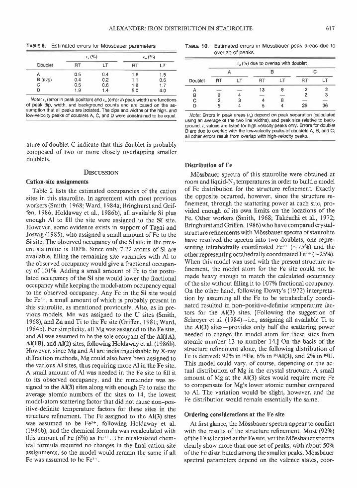
ALEXANDER: IRON DISTRIBUTION IN STAUROLITE 617
TABLE 9. Estimated errors for Mossbauer parameters
e, (%) ,* (o/")
TABLE 10, Estimated errors in Mossbauer peak areas due tooverlap of peaks
t. (%) due to overlap with doublet
Doublet RT
Nofe. Errors in peak areas G,) depend on peak separation (calculatedusing an average of the two line widths), and peak size relative to back-ground. €, values are listed for high-velocity peaks only. Errors for doubletD are due to overlap with the low-velocity peaks of doublets A, B, and C;all other errors result from overlap with high-velocity peaks.
Distribution of Fe
Mdssbauer spectra of this staurolite were obtained atroom and liquid-N, temperatures in order to build a modelof Fe distribution for the structure refinement. Exactlythe opposite occurred, however, since the structure re-finement, through the scattering power at each site, pro-vided enough of its own limits on the locations of theFe. Other workers (Smith, 1968; Tak6uchi et al., 19721'Bringhurst and Griffen, 1986) who have compared crystal-structure refinements with M6ssbauer spectra of staurolitehave resolved the spectra into two doublets, one repre-senting tetrahedrally coordinated p"z+ 1-75o/o) and theother representing octahedrally coordinated Fe2+ (- 250/o).When this model was used with the present structure re-finement, the model atom for the Fe site could not bemade heavy enough to match the calculated occupancyof the site without filling it to l07o/o fractional occupancy.On the other hand, following Dowty's (1972) interpreta-tion by assuming all the Fe to be tetrahedrally coordi-nated resulted in non-positive-definite temperature fac-tors for the A(3) sites. [Following the suggestion ofSchreyer et al. (1984)-i.e., assigning all available Ti tothe Al(3) sites-provides only half the scattering powerneeded to change the model atom for these sites fromatomic number 13 to number l4.l On the basis of thestructure refinement alone, the following distribution ofFe is derived: 92o/o in ta!'F e, 60/o in 16rAl(3), and 2o/o in r6ru.
This model could vary, of course, depending on the ac-tual distribution of Mg in the crystal structure. A smallamount of Mg at the Al(3) sites would require more Feto compensate for Mg's lower atomic number comparedto Al. The variation would be slight, however, and theFe distribution would remain essentially the same.
Ordering considerations at the Fe site
At first glance, the Mossbauer spectra appear to conflictwith the results of the structure refinement. Most (920lo)ofthe Fe is located at the Fe site, yet the M6ssbauer spectraclearly show more than one set of peaks, with about 500/oof the Fe distributed among the smaller peaks. Mdssbauerspectral parameters depend on the valence states, coor-
LTLT
AB (avg)
D
0.5 0.40.4 0.2u.c u.b1 9 1 . 4
t . o'l .11 . 65.0
0.61 . 74.0
LT
2 22 3
29 36
1 3 I
4 8c 4
9 42 35 4
AB
Divofe.' (, (error in peak position) and 6. (error in peak width) are functions
of peak dip, width, and background counts and are based on the as-sumption that all peaks are isolated. The dips and widths of the high- andlow-velocity peaks of doublets A, C, and D were constrained to be equal.
ature ofdoublet C indicate that this doublet is probablycomposed of two or more closely overlapping smallerdoublets.
DrscussroN
Cation-site assignments
Table 2 lists the estimated occupancies of the cationsites in this staurolite. In agreement with most previousworkers (Smith, 1968; Ward, 1984a; Bringhurst and Grif-fen, 1986; Holdaway et al., 1986b), all available Si plusenough A1 to fill the site were assigned to the Si site.However, some evidence exists in support of Tagai andJoswig (1985), who assigned a small amount of Fe to theSi site. The observed occupancy ofthe Si site in the pres-ent staurolite is 1000/0. Since only 7.22 atoms of Si areavailable, filling the remaining site vacancies with Al tothe observed occupancy would give a fractional occupan-cy of 10lo/0. Adding a small amount of Fe to the postu-lated occupancy of the Si site would lower the fractionaloccupancy while keeping the model-atom occupancy equ alto the observed occupancy. Any Fe in the Si site wouldbe Fe3+, a small amount of which is probably present inthis staurolite, as mentioned previously. Also, as in pre-vious models, Mn was assigned to the U sites (Smith,1968), and Zn and Ti to the Fe site (Griffen, l98l; Ward,1984b). For simplicity, all Mg was assigned to the Fe site,and Al was assumed to be the sole occupant of the Al(1A),A(lB), and Al(2) sites, following Holdaway et al. (1986b).However, since Mg and Al are indistinguishable by X-raydiffraction methods, Mg could also have been assigned tothe various Al sites, thus requiring more Al in the Fe site.A small amount of Al was needed in the Fe site to fill itto its observed occupancy, and the remainder was as-signed to the A(3) sites along with enough Fe to raise theaverage atomic numbers of the sites to 14, the lowestmodel-atom scattering factor that did not cause non-pos-itive-definite temperature factors for these sites in thestructure refinement. The Fe assigned to the Al(3) siteswas assumed to be Fe3+, following Holdaway et al.(1986b), and the chemical formula was recalculated withthis amount of Fe (60/o) as Fe3+. The recalculated chem-ical formula required no changes in the final cation-siteassignments, so the model would remain the same if allFe was assumed to be Fe2+.

6 1 8 ALEXANDER: IRON DISTRIBUTION IN STAUROLITE
dination numbers, and crystal-field splittings at the Fesites (Hawthorne, 1983). Most, if not all, the Fe in thepresent staurolite is Fe2+, as previously noted, so valencestates will not have much efect on the Mcissbauer param-eters. Coordination numbers have been established throughthe crystal-structure refinement and indicate that all threeFe2* Mcissbauer doublets arise from the l4lFe site. Thisleaves crystal-field variations to explain the number ofMdssbauer doublets that are present. If each Mdssbauerdoublet arises from Fe in a different crystal-field environ-ment, then there must be three major types of Fe sitesand possibly several minor types of Fe sites, as well asminor octahedral Fe sites, as indicated by the large linewidths of doublet C.
The positional disorder indicated by the electron-den-sity difference map (Fig. 2) from the structure refinementalso suggests that there are at least three types ofFe sites.Bringhurst and Grifen (1986) summarized three possibleexplanations for positional disorder at the Fe site in stau-rolite: (l) cation-cation repulsion between the Fe and Usites, (2) metal-metal bonding attraction between the Feand U sites, and (3) proton-cation repulsion between theP and Fe sites (Fig. 1). The major shortcoming of the firsttwo proposals is the low occupancy of the U sites. The Fesite could only be affected ifone or both neighboring Usites were occupied, which is true for, at most, 60/o of theFe sites (Table 2). The most highly occupied Fe subsite(Fel) contains about 500/o of the cations in the Fe site(Table 3), implying that at least half of the cations in theFe site are being influenced by whatever mechanism iscausing the positional disorder at the Fe site.
The proton-cation repulsion explanation [also proposedby Holdaway et al. (1986b)l implies control of the Fesubsite occupancies by occupancy ofone or both P sites.In other words, when both P(lA) and P(lB) are vacant,the Fel subsite is occupied; and when P(lA) or P(lB) isoccupied, the Fe2 or Fe3 subsite is occupied, respectively(Figs. I and2). The occupancies ofthe Fe subsites supporta proton-cation repulsion mechanism in that Fe3 has ahigher occupancy than Fe2. The P sites are located in thefaces of the Al(3) octahedra, implying that when a P siteis occupied, its corresponding Al(3) site is not; so, sinceAl(3B) is less occupied than Al(3A), P(lB) should be moreoccupied than P(lA). Ifthe proton-cation repulsion con-cept is correct, the occupancies ofFe2 and Fe3 could beused as estimates for the occupancies of P(lA) and P(lB),respectively. An estimate of H* content could then bemade from these occupancies. Using the occupancies ofthe Fe subsites from the low-temperature structure re-finement, the number of H* ions per formula unit for thisstaurolite would be between 1.96 and 2.20, depending onwhether the P sites are occupied when the Fe site is vacant.This is an unreasonably low amount of H* for staurolite[the lowest H+ content found by Holdaway et al. (1986a)is 2.68 atomsl. However, the large errors for the Fe-subsiteoccupancies could bring the estimated P-site occupancyto as high as 2.80, a very reasonable number. More ac-
curate and precise Fe-subsite occupancies would be need-ed to make reliable estimates of P-site occupancies.
If the proton-cation repulsion idea is taken one stepfurther to include next-nearest neighbors ofthe Fe site, abetter explanation for both the positional disorder andthe crystal-field variations at the Fe site can be made. Twofactors will affect the crystal-field energy at the Fe site.First, ifoccupancy ofthe P site could affect the energy atthe Fe site, then occupancy or vacancy of other neigh-boring sites (A(3), U, Fe, and kyanite-layer sites) couldhave an effect on the energy at the Fe site (Fig. l). Second,the energy at the Fe site will depend on the identities ofthe cations occupying neighboring sites. Thus, the threemajor Miissbauer doublets can be attributed to the threemost probable occupancy distributions around the Fe site:Al in both A(3A) and At(3B), Al in Al(3A) and H inP(lB), and H in P(f A) and Al in Al(3B). Other occupancydistributions (including vacancies and/or substitution ofminor cation species in some sites) are reflected in theMiissbauer spectra by line-broadening of the major dou-blets.
Positional disorder at the Fe site is probably a result ofvariations in the crystal field, since variations in the crystalfield could cause changes in the location ofthe energeticminimum at the site. The location of the energetic min-imum could also depend on the identity of the cation inthe Fe site; some cations may prefer one subsite overanother. Comparison of Mdssbauer peak areas with struc-ture-refinement subsite occupancies could determine ifthis is the case. However, for the present study, the sta-tistical errors would mask any differences between the twotypes of data. If a random distribution of Fe-site cationspecies is assumed, then M6ssbauer and electron-densitypeaks can be tentatively correlated. On the basis of Mdss-bauer doublet areas and subsite occupancies alone (Tables8 and 3), Fel would correlate with doublet B, Fe2 withdoublet A, and Fe3 with doublet C. Assuming all octa-hedrally coordinated Fe to be Fe3* would result in a cor-relation between doublet D and the Al(3) sites.
Effects of temperature
The occupancies of the Fe subsites, determined fromX-ray diffraction data, show essentially no change be-tween room and low temperatures, since the differencesare all within the range ofthe statistical errors, in agree-ment with the Miissbauer results. An apparent tempera-ture dependence in Mdssbauer spectra of staurolite hasbeen noted by other workers (Dowty, 1972;Regnard,197 6;Scorzelli et. al., 197 6; Varma and Varma, 1986); however,no errors for peak parameters are listed, so the validityofthe temperature dependence in these spectra cannot beevaluated. For the present staurolite, the apparent changein relative Mdssbauer peak areas with temperature is mostlikely an artifact produced by the difficulty of separatingoverlapping peaks. Peak separations could be affected byslight changes with temperature in the electric-field gra-dient around the Fe-occupied sites, which would cause

slight changes in the positions ofthe corresponding peaksin the Mdssbauer spectra.
CoNcr-usroNs
L The Fe distribution for the staurolite of this study,derived from structure-refinement data, is 920lo in the Fesite, 60/0 in the Al(3) sites, and 2o/o in the U sites. There-fore, the Mdssbauer spectra for this staurolite are inter-preted as representing 92o/o of the Fe in tetrahedral co-ordination and 80/o in octahedral coordination.
2. The three resolved Fe2+ Mdssbauer doublets are at-tributed to energetically nonequivalent Fe sites resultingfrom variations in the crystal-field energy at the site.Crystal-field variations are attributed to occupancy andvacancy distributions and cation substitutions in theneighborhood of the Fe site. Line broadening of theMdssbauer doublets (especially doublet C) is attributedto small unresolved doublets arising from less likely vari-ations in the crystal-field energy at the Fe site and tosmall amounts of octahedrally coordinated Fe2+ in the Usrtes.
3. Neither the electron density at the Fe site nor theMdssbauer spectra exhibit any definite temperature de-pendence since all variations with temperature are withinthe range of statistical errors.
AcxNowr.rocMENTS
This project was done in fulfillment of the thesis requirement for anM.S. degree at Brigham Young University. Financial support was provid-ed by a departmental fellowship and by a research grant from the Asso-ciated Students of BYU
Rnrnnnxcns crrnnBancroft, G.M., Maddock, A.G., and Burns, R.G. (1967) Applications of
the M6ssbauer effect to silicate mineralogy-I. Iron silicates of knowncrystal structure. Geochimica et Cosmochimica Acta, 31, 2219-2246.
Batsanov, S.S. (1959) Strukturnaya Refraktometriya. Uniyersity Press,Moscow (translated in P.P. Sutton, Refractometry and chemical struc-ture, 250 p. Consultants Bureau, New York, 1961).
Bloss, F.D. (l 98 l) The spindle stage: Principles and practice, 340 p. Cam-bridge University Press, Cambridge, England.
Bringhurst, K.N., and Griffen, D.T. (1986) Staurolite-lusakite series. II.Crystal structure and optical properties of a cobaltoan staurolite. Amer-ican Mineralogisl, 7 l, | 466-1 47 2.
Cromer, D.T , and Liberman, D (1970) Relativistic calculation of anom-alous scattering factors for X-rays. Journal of Chemical Physics, 53,I 89 l - 1 898 .
Cromer, D.T., and Mann, J.B. (1968) X-ray scattering factors computedfrom numerical Hartree-Fock wave functions. Acta Crystallographica,A^24,32t-324-
De Coster, M., Pollack, H., and Amelinckx, S (1963) A study of Mdss-bauer absorption in iron silicates. Physica Status Solidi, 3, 283-288.
Dickson, B.L., and Smith, G. (1976) Low-temperature optical absorptionand Mdssbauer spectra ofstaurolite and spinel. Canadian Mineralogist,t4.206-215
619
Dollase, W.L. (1975) Statistical limitations of Miissbauer spectral fitting.American Mineralogist, 60, 257 -264.
Dowty, E. (1972) Site distribution ofiron in staurolite. Earth and Plane-tary Science LetIerc, 15,72-74.
Dutrow, B.L., Holdaway, M.J., and Hinton, R.W. (1986) Lithium in stau-rolites: Its petrologic significance. Contributions to Mineralogy and Pe-trology,94, 496-506.
Dzhemats, A.C., and Nikitina, L.P (1977) Mdssbauer spectroscopy ofstaurolite. Leningrad Universitet Vestnik, Geologiia i Geografira, 24,4246.
Griffen, D.T. (1981) Synthetic FelZn staurolites and the ionic radius ofIvZn2+. American Mineralogist, 66, 932-937.
Griffen, D.T., and Ribbe, P.H. (1973) The crystal chemistry of staurolite.American Journal of Science, 273-4, 479495.
Hanisch, K. ( I 966) Zur Kenntnis der Kristallstruktur von Staurolith. NeuesJahrbuch fiir Mineralogie Monatshefte, 362-366.
Hawthome, F.C. (l 983) Quantitative characterization of site-occupanciesin minerals American Mineralogist, 68, 287 -306.
Holdaway, M J., Dutrow, B L., Borthwick, J., Shore, P., Harmon, R.S.,and Hinton, R.W. (1986a) H content of staurolite as determined by Hextraction line and ion microprobe. American Mineralogist, 7 I , I 1 3 5-I l 4 l .
Holdaway, M.J., Dutrow, B.L., and Shore, P. (1986b) A model for thecrystal chemistry of staurolite. American Mineralogist, 71, 1142-1159'
N6ray-Szab6, I. (1929) The structure of staurolite. Zeitschrift fiir Kristal-lographie, 71, 103-l 16.
Niray-Szab6, I., and Sasvdri, K. (1958) On the structure of staurolite,HFerAlrSinOzr. Acta Crystallographica, I 1, 862-865.
Phillips, L.V., and Griffen, D.T. (1986) Stauolitelusakite series. I. Syn-thetic Fe-Co staurolites. American Mineralogist, 7 l, 146l-1465
Regnard, J.R. (1976) Mdssbauer study of natural crystals of staurolite.Journal de Physique, Colloque, 37, C6-797-C6'800.
Schreyer, W., Horrocks, P C, and Abraham, K. (1984) High-magnesiumstaurolite in a sapphirine-garnet rock from the Limpopo Belt, SouthernAfrica. Contributions to Mineralogy and Petrology, 86, 200-207
Scorzelli, R.B., Baggio-Saitovitch, E., and Danon, J. (1976) Mdssbauerspectra and electron exchange in tourmaline and staurolite. Journal dePhysique, Colloque, 37, C6-801-C6-805.
Sheldrick, G.M. (1976) sHELx-76: A programme for crystal structure de-termination. University of Cambridge, Cambridge, England.
Smith, J.V. (1968) The crystal structure of staurolite. American Miner-alogist , 53, I 139-1 155.
Tagai, T, and Joswig, W ( I 985) Untersuchungen der Kationenveneilungim Staurolith durch Neutronenbeugung bei I 00K. Neues Jahrbuch IiirMineralogie Monatshefte, 97-l 07.
Tak6uchi, Y., Aikawa, N., and Yamamoto, T. (1972) The hydrogen lo-cations and chemical composition ofstaurolite. Zeitschrift fiir Kristal-lographie, 136, l-22.
Varma, H.V., and Varma, J. (1986) Mrissbauer effect study of naturalstaurolite Physica Status Solidi A, 97,275-278.
Walker, N., and Stuart, D. (1983) An empirical method for correctingdiffractometer data for absorption effects. Acta Crystallographica, A39,158 -166
Ward, C.M. (1984a) Magnesium staurolite and green chromian staurolitefrom Fiordland, New Zealand. American Mineralogist, 69, 531-540.
-(1984b) Titanium and the color of staurolite American Mineral-ogist ,69, 541-545.
MANUscRrpr RECETvED JtNs 10, 1988MANUscRrpr AccEprED Jemue,nv I l. 1 989
ALEXANDER: IRON DISTRIBUTION IN STAUROLITE