Embed Size (px)
Citation preview
Iron Metabolism and Storage
Dr Graham Jones
Staff Specialist in Chemical Pathology
St Vincent’s Hospital, Sydney
UNSW Lecture, May 2005
Summary
• Overview• Iron absorption• Iron distribution• Iron storage• Iron loss• Testing for iron status
• Highlight: – Physiological principles– Clinical conditions– Available tools for patient management
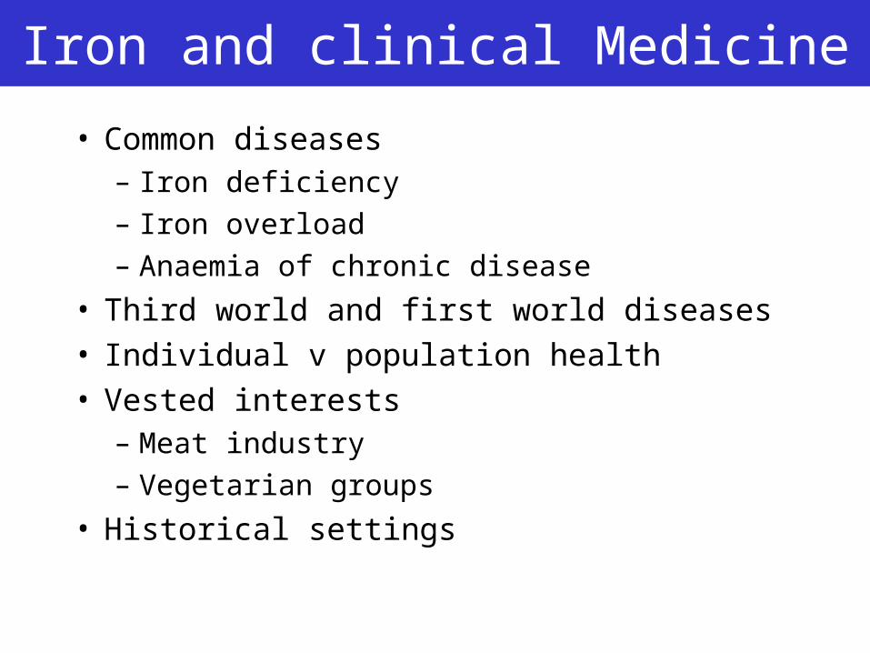
Iron and clinical Medicine
• Common diseases– Iron deficiency
– Iron overload
– Anaemia of chronic disease
• Third world and first world diseases• Individual v population health• Vested interests
– Meat industry
– Vegetarian groups
• Historical settings
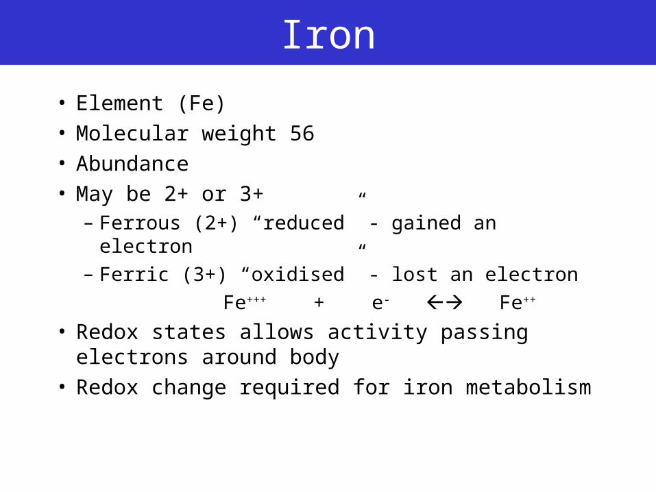
Iron
• Element (Fe)• Molecular weight 56• Abundance• May be 2+ or 3+
– Ferrous (2+) “reduced” - gained an electron
– Ferric (3+) “oxidised” - lost an electron
Fe+++ + e- Fe++
• Redox states allows activity passing electrons around body
• Redox change required for iron metabolism
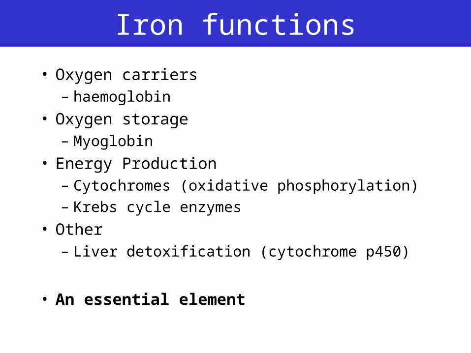
Iron functions
• Oxygen carriers– haemoglobin
• Oxygen storage– Myoglobin
• Energy Production– Cytochromes (oxidative phosphorylation)– Krebs cycle enzymes
• Other– Liver detoxification (cytochrome p450)
• An essential element
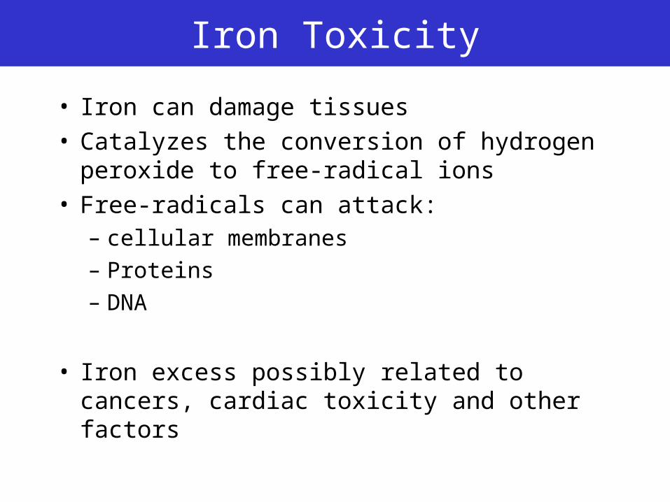
Iron Toxicity
• Iron can damage tissues• Catalyzes the conversion of hydrogen
peroxide to free-radical ions• Free-radicals can attack:
– cellular membranes
– Proteins
– DNA
• Iron excess possibly related to cancers, cardiac toxicity and other factors
Principle
• Bodies require the right amount of substance• Too much or too little of any required
substance may be detrimental
• “There is no substance, which taken in sufficient excess, is not toxic to the body”
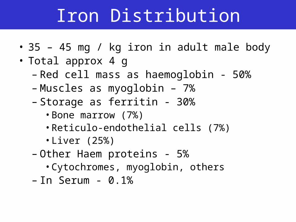
Iron Distribution
• 35 – 45 mg / kg iron in adult male body• Total approx 4 g
– Red cell mass as haemoglobin - 50%– Muscles as myoglobin – 7%– Storage as ferritin - 30%
• Bone marrow (7%)• Reticulo-endothelial cells (7%)• Liver (25%)
– Other Haem proteins - 5%• Cytochromes, myoglobin, others
– In Serum - 0.1%
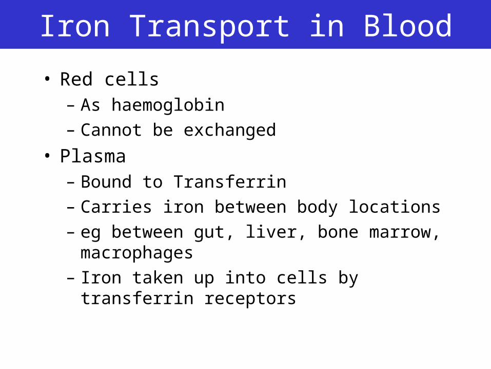
Iron Transport in Blood
• Red cells– As haemoglobin
– Cannot be exchanged
• Plasma– Bound to Transferrin
– Carries iron between body locations
– eg between gut, liver, bone marrow, macrophages
– Iron taken up into cells by transferrin receptors
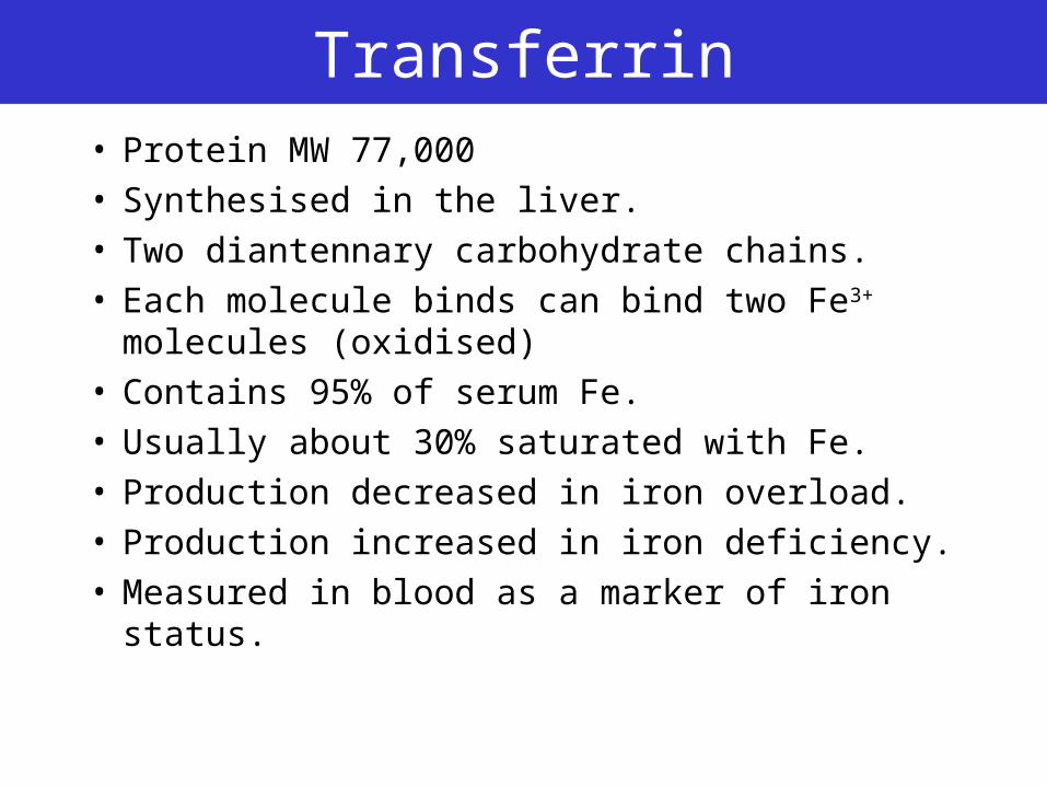
Transferrin• Protein MW 77,000• Synthesised in the liver.• Two diantennary carbohydrate chains. • Each molecule binds can bind two Fe3+
molecules (oxidised)• Contains 95% of serum Fe.• Usually about 30% saturated with Fe.• Production decreased in iron overload.• Production increased in iron deficiency.• Measured in blood as a marker of iron status.
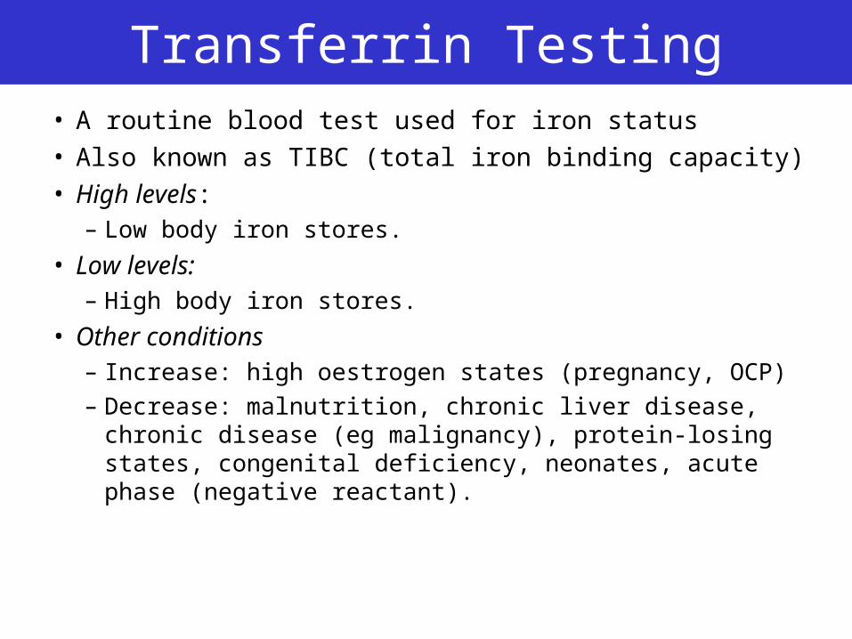
Transferrin Testing• A routine blood test used for iron status• Also known as TIBC (total iron binding capacity)• High levels:
– Low body iron stores.
• Low levels: – High body iron stores.
• Other conditions– Increase: high oestrogen states (pregnancy, OCP)
– Decrease: malnutrition, chronic liver disease, chronic disease (eg malignancy), protein-losing states, congenital deficiency, neonates, acute phase (negative reactant).
Principle
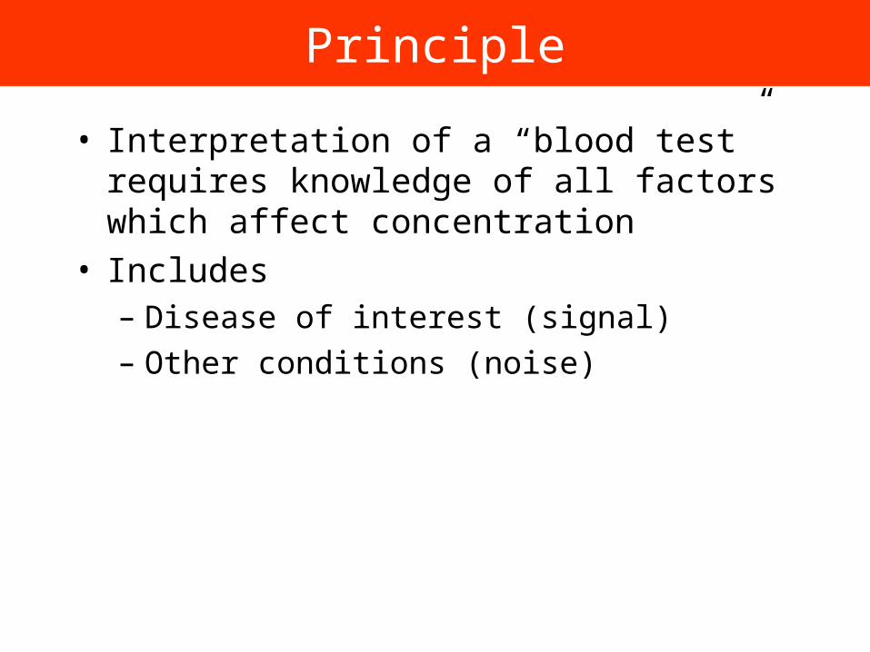
• Interpretation of a “blood test” requires knowledge of all factors which affect concentration
• Includes– Disease of interest (signal)
– Other conditions (noise)
Transferrin Receptors
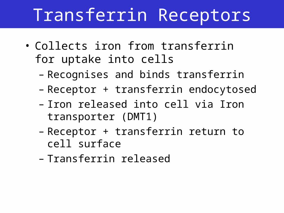
• Collects iron from transferrin for uptake into cells– Recognises and binds transferrin
– Receptor + transferrin endocytosed
– Iron released into cell via Iron transporter (DMT1)
– Receptor + transferrin return to cell surface
– Transferrin released
Soluble Transferrin Receptors
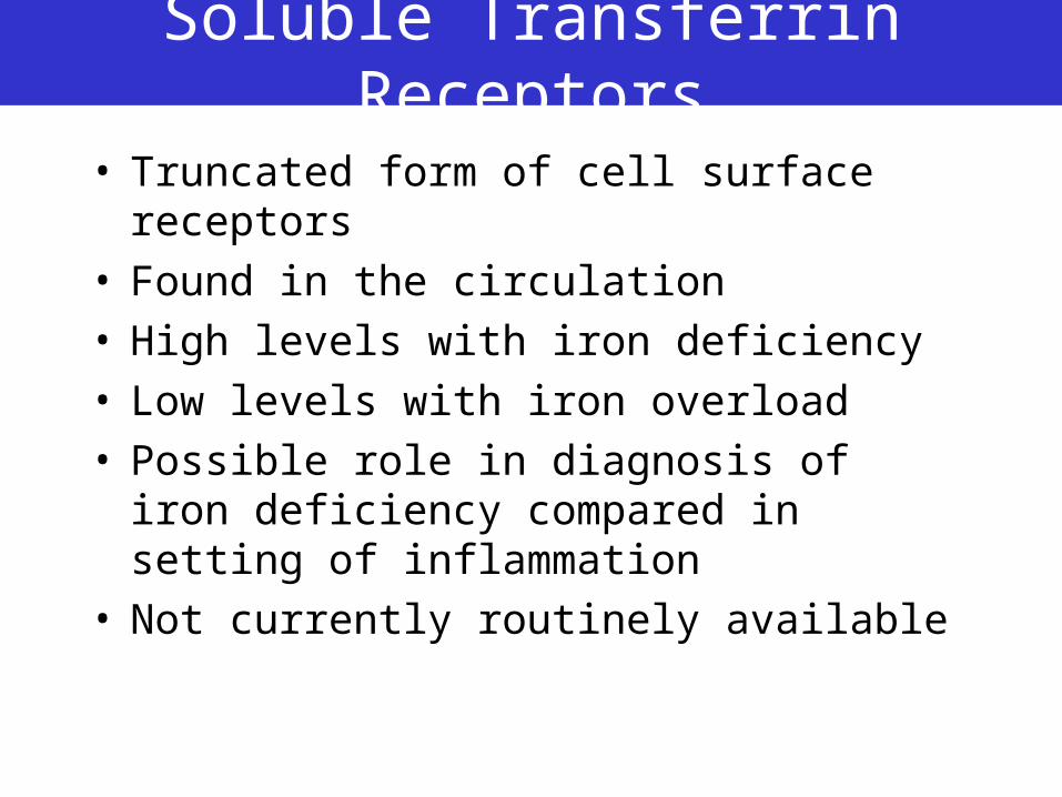
• Truncated form of cell surface receptors• Found in the circulation• High levels with iron deficiency• Low levels with iron overload• Possible role in diagnosis of iron deficiency
compared in setting of inflammation• Not currently routinely available
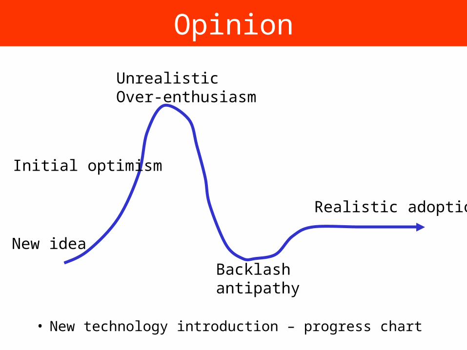
Opinion
• New technology introduction – progress chart
UnrealisticOver-enthusiasm
Initial optimism
Backlashantipathy
Realistic adoption
New idea
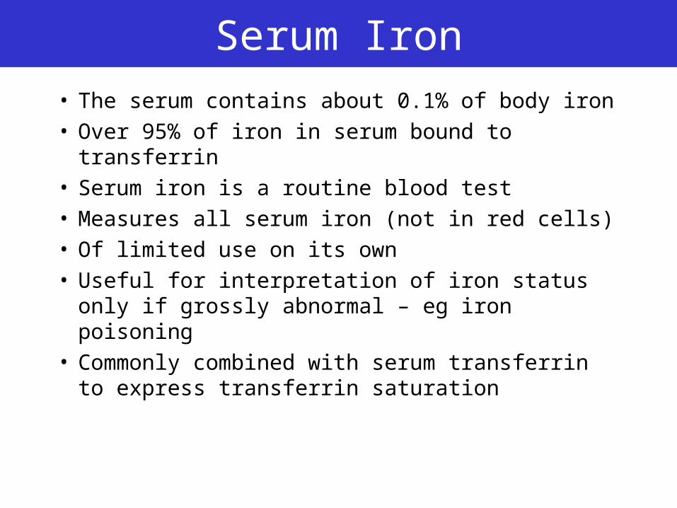
Serum Iron
• The serum contains about 0.1% of body iron• Over 95% of iron in serum bound to
transferrin• Serum iron is a routine blood test• Measures all serum iron (not in red cells)• Of limited use on its own• Useful for interpretation of iron status only if
grossly abnormal – eg iron poisoning• Commonly combined with serum transferrin
to express transferrin saturation
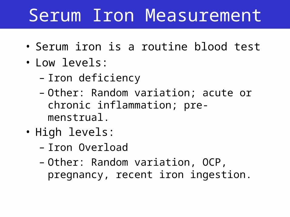
Serum Iron Measurement
• Serum iron is a routine blood test• Low levels:
– Iron deficiency
– Other: Random variation; acute or chronic inflammation; pre-menstrual.
• High levels: – Iron Overload
– Other: Random variation, OCP, pregnancy, recent iron ingestion.
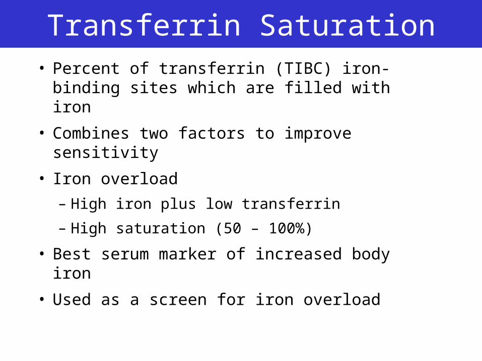
Transferrin Saturation• Percent of transferrin (TIBC) iron-binding
sites which are filled with iron
• Combines two factors to improve sensitivity
• Iron overload
– High iron plus low transferrin
– High saturation (50 – 100%)
• Best serum marker of increased body iron
• Used as a screen for iron overload
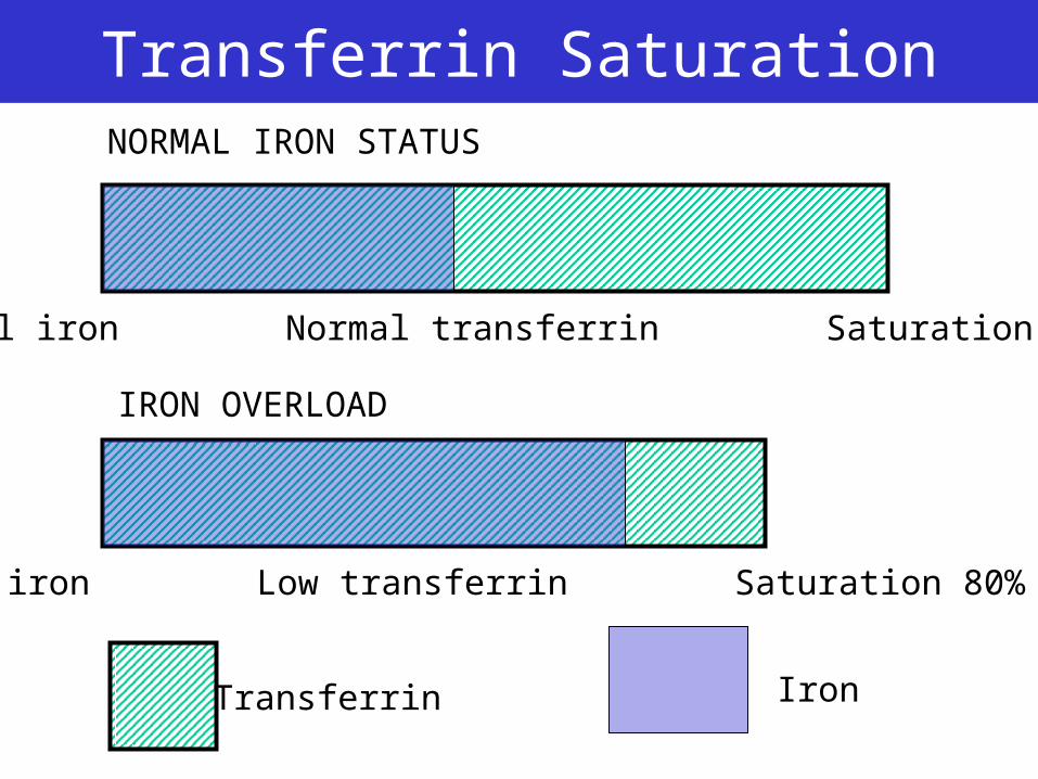
Transferrin Saturation
Normal iron Normal transferrin Saturation 40%
High iron Low transferrin Saturation 80%
Transferrin Iron
IRON OVERLOAD
NORMAL IRON STATUS
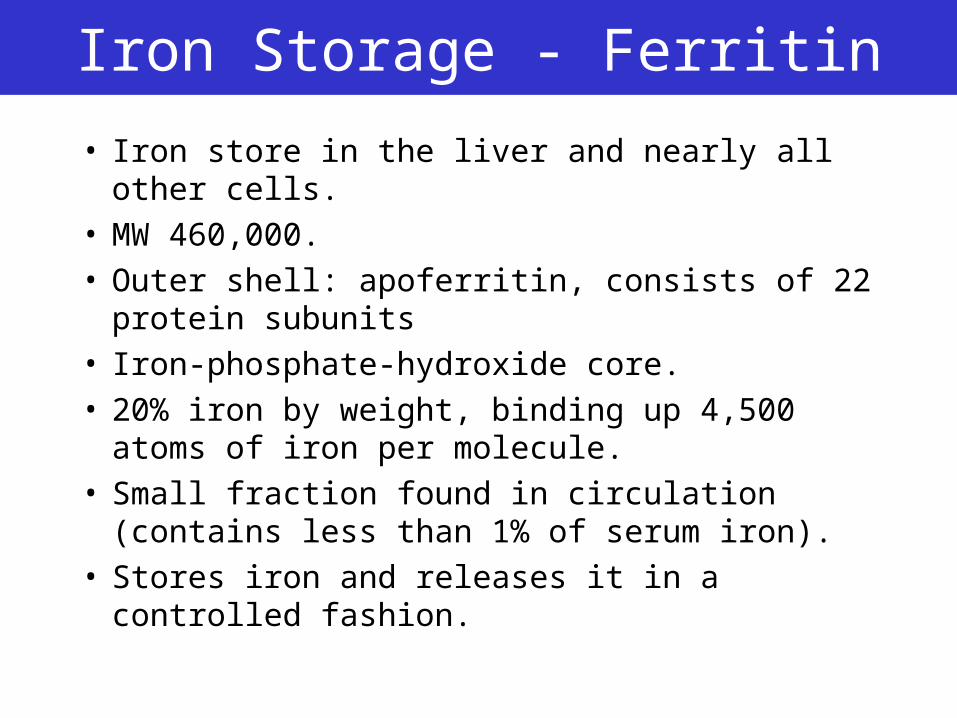
Iron Storage - Ferritin
• Iron store in the liver and nearly all other cells. • MW 460,000. • Outer shell: apoferritin, consists of 22 protein
subunits • Iron-phosphate-hydroxide core. • 20% iron by weight, binding up 4,500 atoms of
iron per molecule. • Small fraction found in circulation (contains less
than 1% of serum iron).• Stores iron and releases it in a controlled fashion.
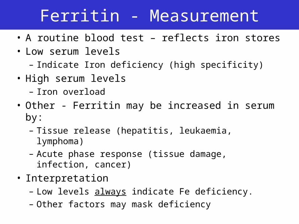
Ferritin - Measurement• A routine blood test – reflects iron stores• Low serum levels
– Indicate Iron deficiency (high specificity)
• High serum levels– Iron overload
• Other - Ferritin may be increased in serum by:– Tissue release (hepatitis, leukaemia, lymphoma)– Acute phase response (tissue damage, infection,
cancer)
• Interpretation– Low levels always indicate Fe deficiency. – Other factors may mask deficiency
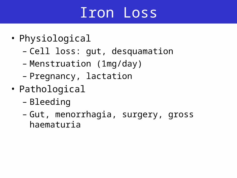
Iron Loss
• Physiological– Cell loss: gut, desquamation
– Menstruation (1mg/day)
– Pregnancy, lactation
• Pathological– Bleeding
– Gut, menorrhagia, surgery, gross haematuria
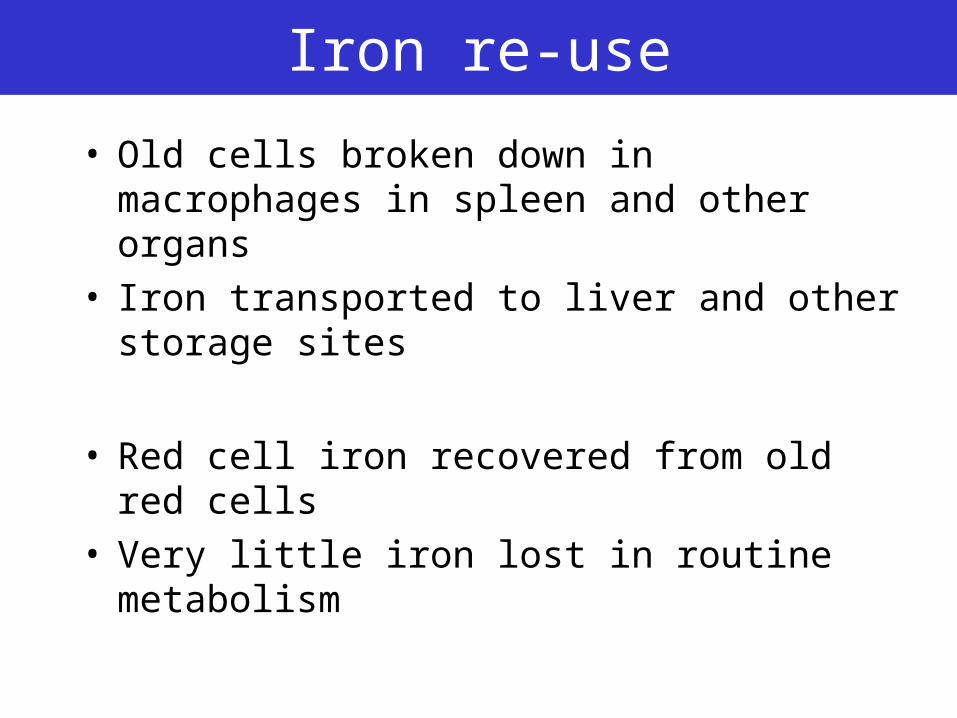
Iron re-use
• Old cells broken down in macrophages in spleen and other organs
• Iron transported to liver and other storage sites
• Red cell iron recovered from old red cells• Very little iron lost in routine metabolism
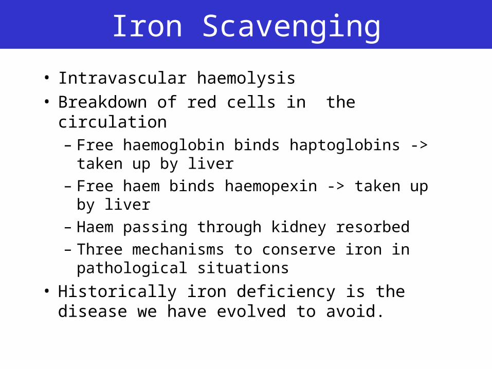
Iron Scavenging
• Intravascular haemolysis• Breakdown of red cells in the circulation
– Free haemoglobin binds haptoglobins -> taken up by liver
– Free haem binds haemopexin -> taken up by liver
– Haem passing through kidney resorbed
– Three mechanisms to conserve iron in pathological situations
• Historically iron deficiency is the disease we have evolved to avoid.
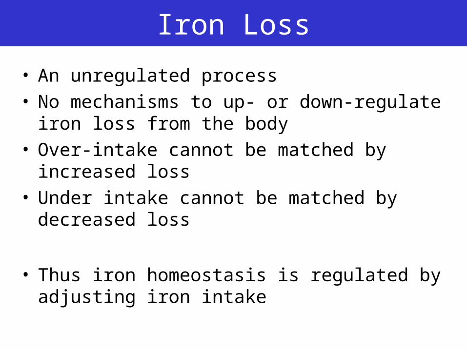
Iron Loss
• An unregulated process• No mechanisms to up- or down-regulate iron loss
from the body• Over-intake cannot be matched by increased loss• Under intake cannot be matched by decreased
loss
• Thus iron homeostasis is regulated by adjusting iron intake
Principle
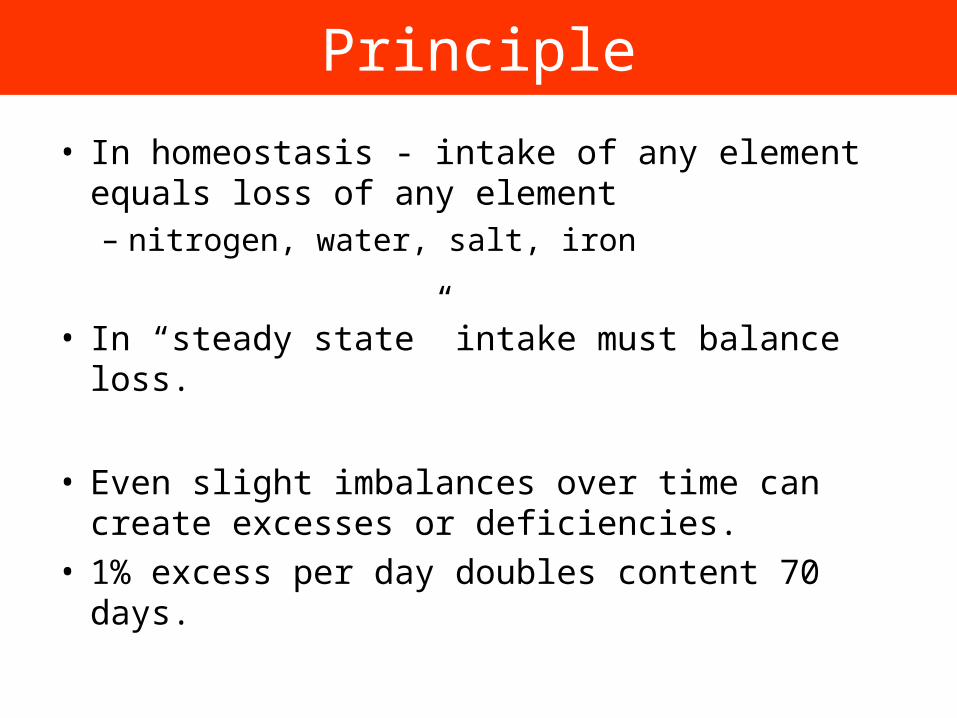
• In homeostasis - intake of any element equals loss of any element– nitrogen, water, salt, iron
• In “steady state” intake must balance loss.
• Even slight imbalances over time can create excesses or deficiencies.
• 1% excess per day doubles content 70 days.
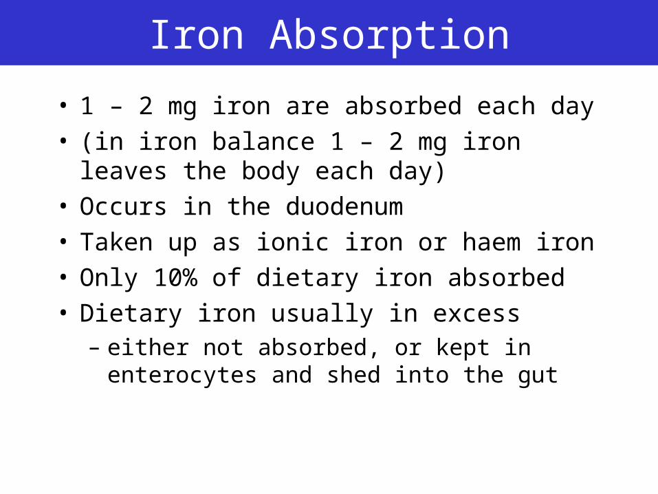
Iron Absorption
• 1 – 2 mg iron are absorbed each day• (in iron balance 1 – 2 mg iron leaves the body
each day)• Occurs in the duodenum• Taken up as ionic iron or haem iron• Only 10% of dietary iron absorbed• Dietary iron usually in excess
– either not absorbed, or kept in enterocytes and shed into the gut
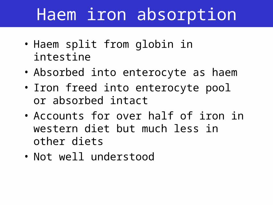
Haem iron absorption
• Haem split from globin in intestine• Absorbed into enterocyte as haem• Iron freed into enterocyte pool or absorbed
intact• Accounts for over half of iron in western diet
but much less in other diets• Not well understood
Iron Absorption
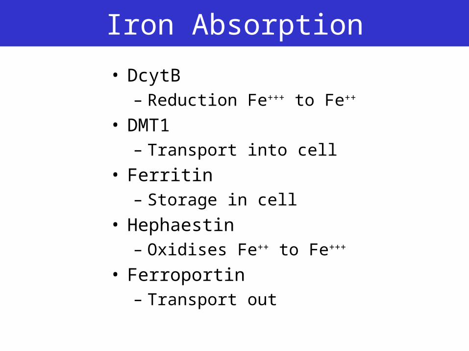
• DcytB– Reduction Fe+++ to Fe++
• DMT1– Transport into cell
• Ferritin– Storage in cell
• Hephaestin– Oxidises Fe++ to Fe+++
• Ferroportin– Transport out
Principles
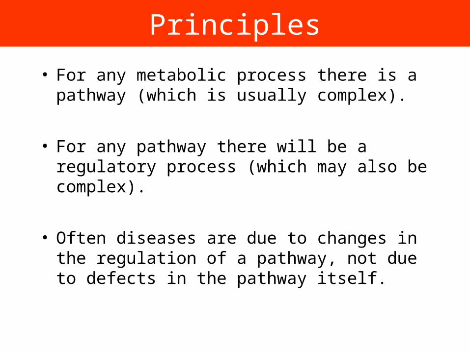
• For any metabolic process there is a pathway (which is usually complex).
• For any pathway there will be a regulatory process (which may also be complex).
• Often diseases are due to changes in the regulation of a pathway, not due to defects in the pathway itself.
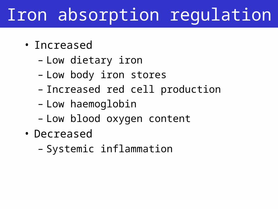
Iron absorption regulation
• Increased– Low dietary iron
– Low body iron stores
– Increased red cell production
– Low haemoglobin
– Low blood oxygen content
• Decreased– Systemic inflammation
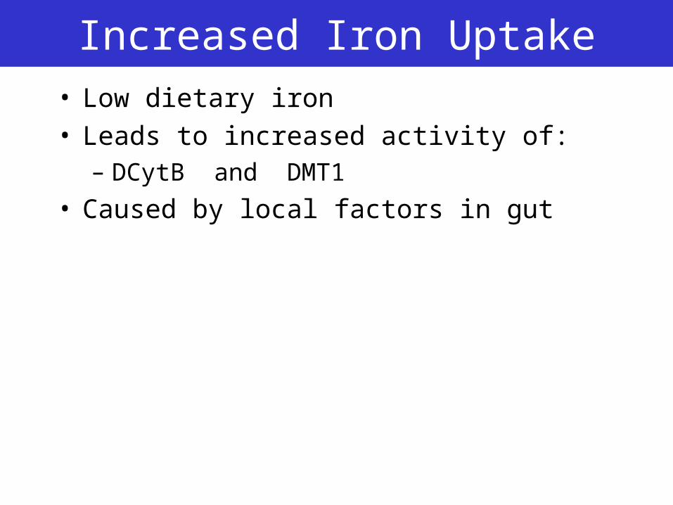
Increased Iron Uptake• Low dietary iron• Leads to increased activity of:
– DCytB and DMT1
• Caused by local factors in gut
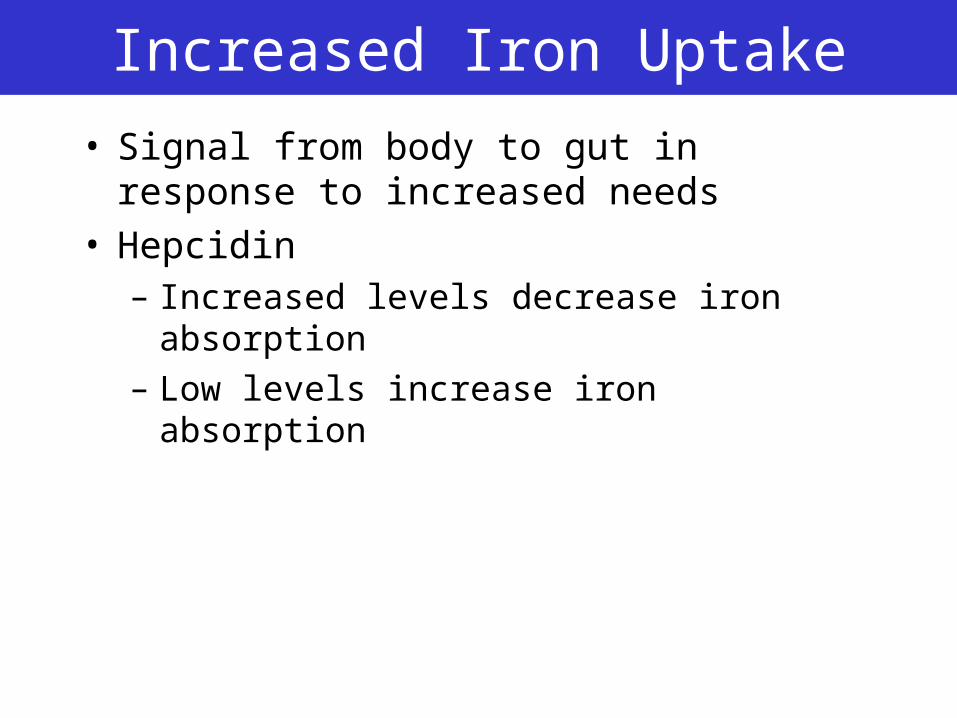
Increased Iron Uptake
• Signal from body to gut in response to increased needs
• Hepcidin– Increased levels decrease iron absorption
– Low levels increase iron absorption
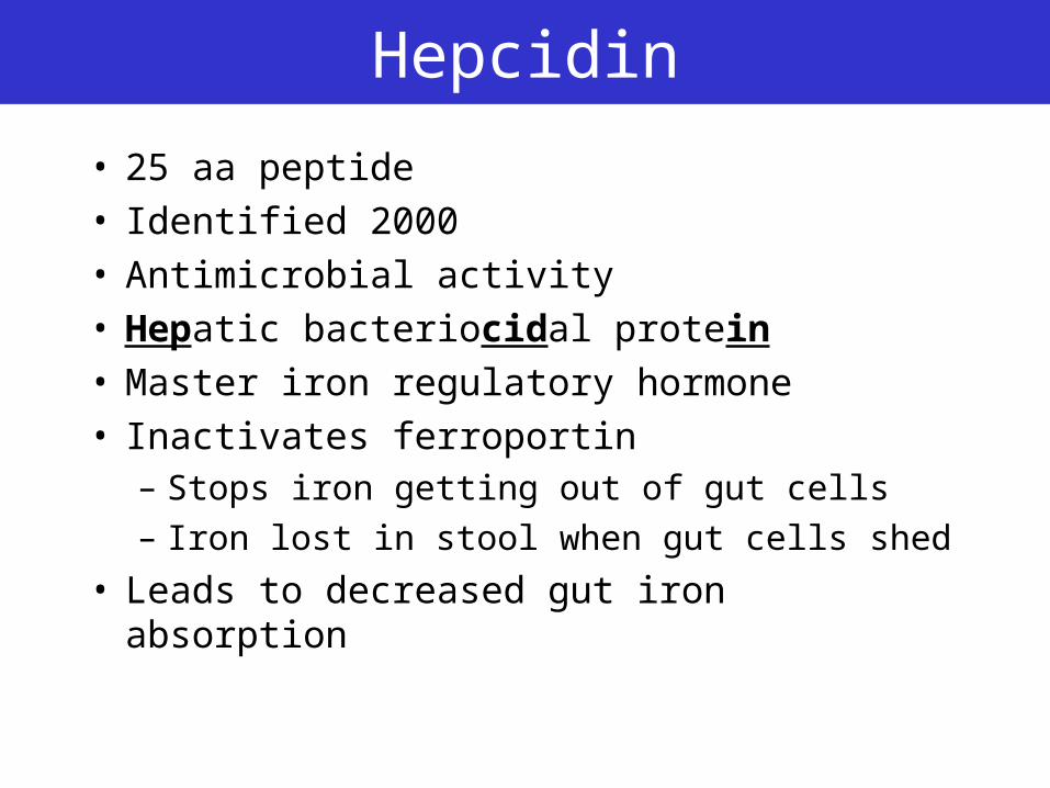
Hepcidin
• 25 aa peptide• Identified 2000• Antimicrobial activity• Hepatic bacteriocidal protein• Master iron regulatory hormone• Inactivates ferroportin
– Stops iron getting out of gut cells
– Iron lost in stool when gut cells shed
• Leads to decreased gut iron absorption
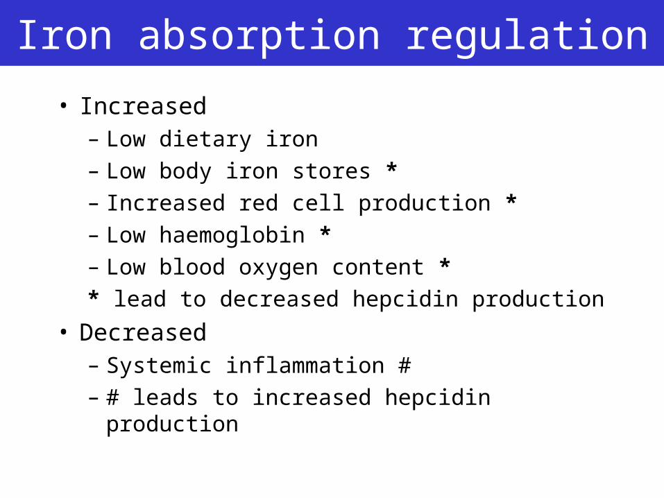
Iron absorption regulation
• Increased– Low dietary iron
– Low body iron stores *
– Increased red cell production *
– Low haemoglobin *
– Low blood oxygen content *
* lead to decreased hepcidin production
• Decreased– Systemic inflammation #
– # leads to increased hepcidin production
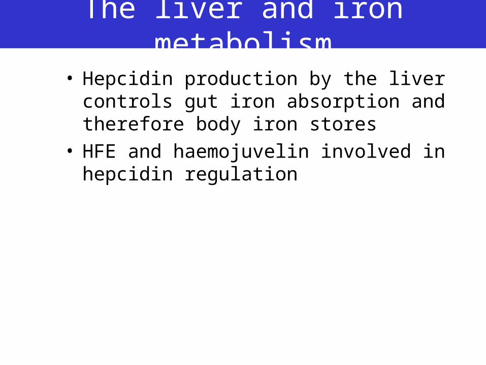
The liver and iron metabolism
• Hepcidin production by the liver controls gut iron absorption and therefore body iron stores
• HFE and haemojuvelin involved in hepcidin regulation
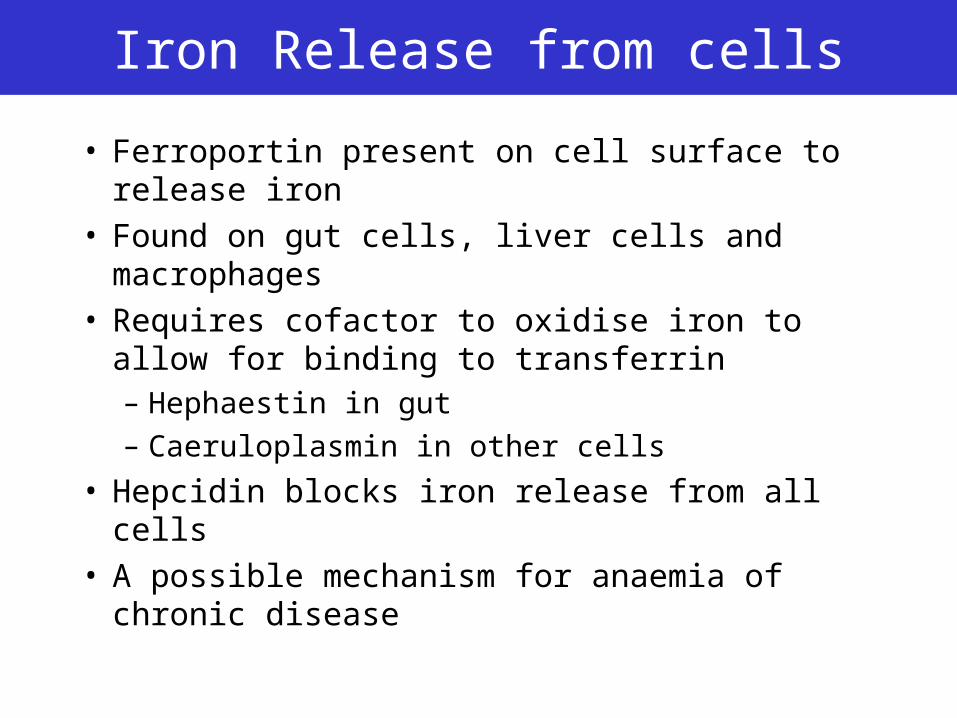
Iron Release from cells
• Ferroportin present on cell surface to release iron
• Found on gut cells, liver cells and macrophages• Requires cofactor to oxidise iron to allow for
binding to transferrin– Hephaestin in gut– Caeruloplasmin in other cells
• Hepcidin blocks iron release from all cells• A possible mechanism for anaemia of chronic
disease
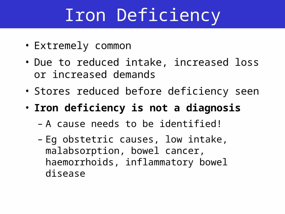
Iron Deficiency
• Extremely common
• Due to reduced intake, increased loss or increased demands
• Stores reduced before deficiency seen
• Iron deficiency is not a diagnosis
– A cause needs to be identified!
– Eg obstetric causes, low intake, malabsorption, bowel cancer, haemorrhoids, inflammatory bowel disease
Iron Deficiency
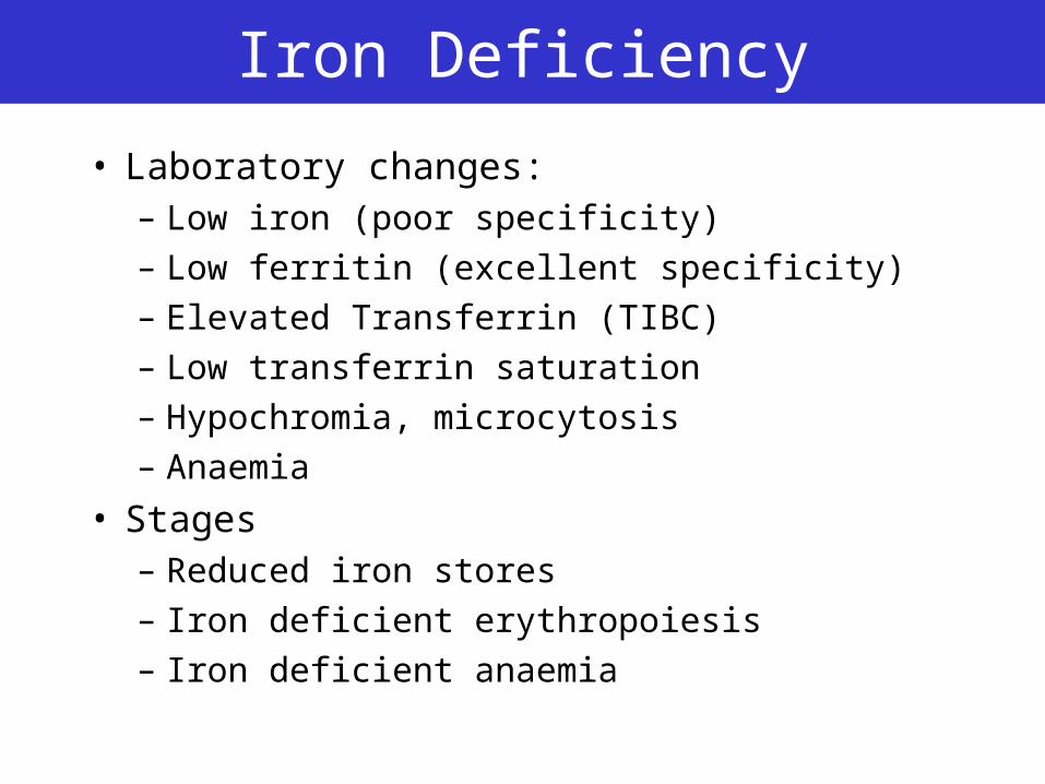
• Laboratory changes:– Low iron (poor specificity)– Low ferritin (excellent specificity)– Elevated Transferrin (TIBC)– Low transferrin saturation– Hypochromia, microcytosis– Anaemia
• Stages– Reduced iron stores– Iron deficient erythropoiesis– Iron deficient anaemia
Anaemia of chronic disease
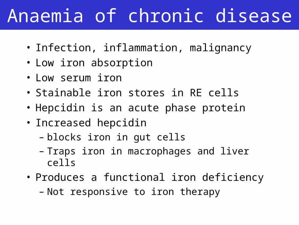
• Infection, inflammation, malignancy• Low iron absorption• Low serum iron• Stainable iron stores in RE cells• Hepcidin is an acute phase protein• Increased hepcidin
– blocks iron in gut cells
– Traps iron in macrophages and liver cells
• Produces a functional iron deficiency– Not responsive to iron therapy
Anaemia of chronic disease
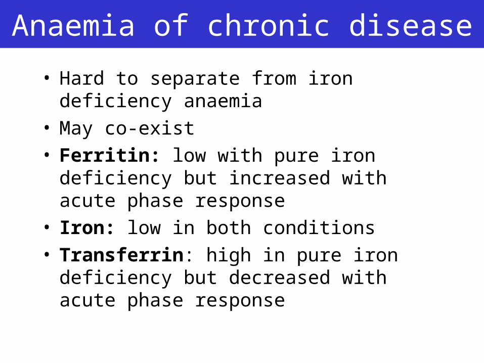
• Hard to separate from iron deficiency anaemia
• May co-exist• Ferritin: low with pure iron deficiency but
increased with acute phase response• Iron: low in both conditions• Transferrin: high in pure iron deficiency but
decreased with acute phase response
Genetic haemochromatosis
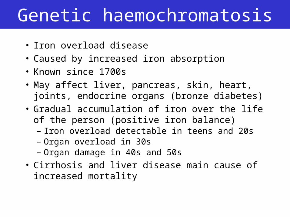
• Iron overload disease• Caused by increased iron absorption• Known since 1700s• May affect liver, pancreas, skin, heart, joints,
endocrine organs (bronze diabetes)• Gradual accumulation of iron over the life of the
person (positive iron balance)– Iron overload detectable in teens and 20s– Organ overload in 30s– Organ damage in 40s and 50s
• Cirrhosis and liver disease main cause of increased mortality
Genetic Haemochromatosis
• >95% defect in HFE gene (C282Y)• Associated with low hepcidin• Leads to overactivity of ferroportin
– Increased gut absorption of iron
• Also other mechanisms– Increased DMT1 and DcytB activity
– Not related to hepcidin
• Limited penetrance (1 – 50%)– May require other genes to be involved
Observation
• “We’ve found the genetic defect!!”
• “We’ve found 10 genetic defects!”
• “We’ve found 100’s of genetic defects”
• Genetic understanding hugely increases our understanding of diseasebut
• Genetic testing rarely simplifies our diagnostic testing but may improve testing in right circumstances
Genetic Haemochromatosis
HFE-Related• Type 1 – HFE defects
Non HFE Related• Type 2a – Haemojuvelin defects• Type 2b – Hepcidin defects• Type 3 – Transferrin receptor defects• Type 4 – Ferroportin defects
Other tests related to iron status
• Haemoglobin– Low with iron deficiency, anaemia of chronic
disease
• Mean Cell Volume– Low with iron deficiency, thallassaemia
• Liver iron– High with iron overload
– Better marker for GH when corrected for age
– (Hepatic iron index)
• Bone marrow iron– Low with iron deficiency
Future possibilities
• Treatment with hepcidin for iron overload• Blocking of hepcidin for anaemia of chronic
disease• Diagnostic tests based on hepcidin
Conclusions
• Iron related diseases are common and clinically important
• Recent advances have changed our understanding
• Groups of tests “Iron studies” are the best first line investigation
• New tests and therapies will follow the new understandings.
Reading
• Andrews NC. Medical Progress: disorders of iron metabolism. NEJM 1999;341:1986-95
• Pietrangelo A. haemochromatosis – a new look at an old disease. NEJM 2004;350:2383-97.
• Weiss G, Goodnough LT. Medical Progress: Anaemia of chronic disease. NEJM 2005;352:1011-1023
• Fleming RE, Bacon BR. Orchestration of iron homeostasis. NEJM 2005;352:1741-4