Embed Size (px)
Citation preview

Osteoarthritis and Cartilage xxx (2015) 1e9
Iron overload in a murine model of hereditary hemochromatosisis associated with accelerated progression of osteoarthritis undermechanical stress
A. Camacho y z k *, M. Sim~ao k ¶, H.-K. Ea # yy, M. Cohen-Solal # yy, P. Richette # yy,J. Branco zz xx, M.L. Cancela x ky Department of Orthopedics, Centro Hospitalar Lisboa Central, Lisboa, Portugalz PhD Program in Medicine, NOVA Medical School, University Nova de Lisboa, Lisbon, Portugalx Department of Biomedical Sciences and Medicine (DCBM), University of Algarve, Faro, Portugalk Centre of Marine Sciences (CCMAR), University of Algarve, Faro, Portugal¶ PhD Program in Biomedical Sciences, University of Algarve, Faro, Portugal# Inserm 1132, Hopital Lariboisi�ere, Paris, Franceyy Universit�e Paris Diderot, UFR m�edicale, Assistance Publique-Hopitaux de Paris, Hopital Lariboisi�ere, F�ed�eration de Rhumatologie, Paris, Francezz Department of Rheumatology, Hospital Egas Moniz, Centro Hospitalar Lisboa Ocidental EPE, Lisbon, Portugalxx CEDOC e Chronic Diseases Research Center, NOVA Medical School, University Nova de Lisboa, Lisbon, Portugal
a r t i c l e i n f o
Article history:Received 8 June 2015Accepted 11 September 2015
Keywords:Iron overloadMouse modelHereditary hemochromatosisOsteoarthritisMechanical stress
* Address correspondence and reprint requests to:Orthopedics, Centro Hospitalar Lisboa Central, Rua daLisboa, Portugal. Tel: 351-217-924-200.
E-mail address: [email protected]
http://dx.doi.org/10.1016/j.joca.2015.09.0071063-4584/© 2015 Osteoarthritis Research Society In
Please cite this article in press as: Camachaccelerated progression of osteoarthritisj.joca.2015.09.007
s u m m a r y
Objective: Hereditary hemochromatosis (HH) is a disease caused by mutations in the Hfe gene charac-terised by systemic iron overload and associated with an increased prevalence of osteoarthritis (OA) butthe role of iron overload in the development of OA is still undefined. To further understand the molecularmechanisms involved we have used a murine model of HH and studied the progression of experimentalOA under mechanical stress.Design: OAwas surgically induced in the knee joints of 10-week-old C57BL6 (wild-type) mice and Hfe-KOmice. OA progression was assessed using histology, micro CT, gene expression and immunohistochem-istry at 8 weeks after surgery.Results: Hfe-KO mice showed a systemic iron overload and an increased iron accumulation in the kneesynovial membrane following surgery. The histological OA score was significantly higher in the Hfe-KOmice at 8 weeks after surgery. Micro CT study of the proximal tibia revealed increased subchondral bonevolume and increased trabecular thickness. Gene expression and immunohistochemical analysis showeda significant increase in the expression of matrix metallopeptidase 3 (MMP-3) in the joints of Hfe-KOmice compared with control mice at 8 weeks after surgery.Conclusions: HH was associated with an accelerated development of OA in mice. Our findings suggestthat synovial iron overload has a definite role in the progression of HH-related OA.
© 2015 Osteoarthritis Research Society International. Published by Elsevier Ltd. All rights reserved.
Introduction
Osteoarthritis (OA) is a disease of the joints characterized byprogressive articular cartilage destruction and subchondral bonechanges. Several factors influence OA development, including ge-netic background, past joint injuries, mechanical overload andageing1,2. The pathologic process that leads to cartilage destruction
A. Camacho, Department ofBeneficencia, n.º 8, 1069-166
.pt (A. Camacho).
ternational. Published by Elsevier L
o A, et al., Iron overload inunder mechanical stress
involves a disruption of the normal resting state of chondrocytes,leading to an increased production of both matrix proteins andmatrix degrading enzymes such as matrix metallopeptidase (MMP)and a disintegrin and metallopeptidase with thrombospondinmotifs (ADAMTS)3.
Hereditary hemochromatosis (HH), a chronic disease caused bymutations in the Hfe gene4 and characterized by systemic ironoverload that causes damage in the liver, heart and endocrine or-gans5, is associated with an increased incidence of OA6 and jointreplacement surgery7e9. Although liver and heart complicationsare the main causes of mortality in patients with HH, arthropathyhas the greatest impact on the quality of life and rarely benefits
td. All rights reserved.
a murine model of hereditary hemochromatosis is associated with, Osteoarthritis and Cartilage (2015), http://dx.doi.org/10.1016/

A. Camacho et al. / Osteoarthritis and Cartilage xxx (2015) 1e92
from therapeutic phlebotomies10. Studies of human joint tissuehave shown hemosiderin accumulation in the cartilage and syno-vial tissue and histological features reminiscent of OA and ofrheumatoid arthritis (RA)11,12 and increased ferritin levels in thesynovial fluid13 of affected joints, but it is unclear how the ironoverload damages the joints and which HH patients will developmusculoskeletal complications14.
Previous reports have shown that murine models of HH can beuseful in studying the pathogenesis of HH-related liver disease15,HH-related diabetes16 and HH-related changes in bone meta-bolism17, but there are no studies using a murine model of HH toinvestigate the effect of iron overload in articular cartilage.
In order to investigate the role of iron overload in the develop-ment of OAwe induced OA18 in a mouse model of HH19 and studiedthe subsequent morphological, histological and genetic changesoccurring in the cartilage and subchondral bone of the knee joint.
Materials and methods
Details of additional data, methods, primers and reagents areavailable in the Supplementary information material.
Animal model, experimental procedure, feeding and housing
TheHfe-KOmice in a C57BL/6 background19 (HFE-KO) were usedas a model for human HH. C57Bl/6 mice from the University ofAlgarve Animal Facility in-house colony were used as wild-typecontrols. Distribution of animals between groups and tasks isfurther explained in Supplementary fig. 1. Animals were main-tained in specific pathogen-free conditions and had access to waterand food ad-libitum fromweaning up to time of euthanasia. OAwasinduced in the right knee joint of 10-week-old Hfe-KO and wild-type mice by sectioning the medial collateral ligament andexcising of the medial meniscus (MNX) using a microscope18. Theleft knee was sham-operated (SHAM). All the animals wereeuthanized 8 weeks after surgery and bilateral knee joints werecollected, processed and used as described below. All the pro-cedures were approved by the Portuguese National Authority forAnimal Health (Ref. 0421/000/000/2013) and by the Animal Facilityof University of Algarve.
Assessment of iron accumulation
For the assessment of iron accumulation in the liver and blood,samples of six animals of each strain were used. At the time ofeuthanasia blood was collected by intra-cardiac puncture fordetermination of serum iron, serum ferritin and serum transferrinsaturation; liver tissue was collected to determine the hepatic non-heme iron concentration20.
Grading of osteoarthritic changes
Bilateral knee joints from ten animals in each group were iso-lated, cleaned of adherent soft tissues in ice-cold PBS and fixed for24 hrs in 4% paraformaldehyde in PBS pH 7.4, followed by decal-cificationwith 0.5 M Ethylenedinitrilotetraacetic acid (EDTA) in PBSpH 7.4 for 3 weeks. After dehydration through graded alcohols andinclusion in paraffin, 5 mm sagittal sections were cut from themedial compartment of the joints and stained with Safranin-O/FastGreen/Meyer's Hematoxylin. Two separate observers, blinded tothe strain and intervention, graded the cartilage lesions (grade0e6) using the Osteoarthritis Research Society International(OARSI) scoring system for murine OA21. The medial tibial plateauand the medial femoral condyle were graded separately. The finalscore for each joint was the average of both observers score.
Please cite this article in press as: Camacho A, et al., Iron overload inaccelerated progression of osteoarthritis under mechanical stressj.joca.2015.09.007
Morphological characterization of the knee joint by micro-CT
Micro-CT was performed on bilateral knee joints of five animalsin each group with a Skyscan 1172 X-ray computed microtomo-graph (Bruker, Belgium). For the image acquisition the followingparameters were used: X-ray tube potential 70 kVp, X-ray tubecurrent 100 mA, 0.5 mm Al filter, rotation step 0.4�, isotropic voxelsize 5 mm3, integration time 500 ms, frame averaging ¼ 6. Theproximal epiphysis of the tibia was selected as region of interest(ROI). To quantify the characteristics of the subchondral bone22 theepiphysis was further divided in a cortical part (subchondral boneplate) and a trabecular part. The following 3D morphometric pa-rameters were used to describe the bone of the trabecularcompartment: Bone volume fraction (BV/TV, in %) is the ratio of thesegmented bone volume to the total volume of the region of in-terest; Connective density (Conn.D, in mm�3) is a measure of theaverage number of trabeculae per unit of volume; Trabecularthickness (Tb.Th, in mm) is the mean thickness of trabeculae. Todescribe the subchondral bone plate we measured the averagethickness (Sb.Th, in mm).
Histological evaluation of undecalcified samples
Following micro-CT the samples were included in methylmethacrylate (MMA) at 4�C. From these undecalcified samples,5 mm sagittal sections were cut from the medial knee compartmenton a heavy-duty microtome equipped with tungsten carbide kni-ves. Sections were stained with Perl's and counterstained withNeutral Red in order to assess iron deposition. The regions of in-terest for this analysis were the knee joint and the proximalepiphysis of the tibia. The total area of hemosiderin deposits in thesynovial membrane was measured as previously described11.
Evaluation of gene expression
The bilateral knee joints of ten mice from each strain were usedfor RNA isolation. Immediately after euthanasia both knee jointswere immersed in RNALater at 4�C and cleaned of soft tissues, inorder to isolate the tibial and femoral cartilage and subchondralbone of the medial knee compartment. RNA was extracted usingTRI Reagent (SigmaeAldrich), purified using the High Pure RNAIsolation Kit (Roche) and quantified with an Experion RNA analysissystem (Bio-Rad). The reverse transcription reaction (RT) was car-ried out using 0.5 mg of total RNA per reaction with the M-MLVreverse transcriptase (Invitrogen) according to the manufacturerindications. Real-time PCR was carried out in a Bio-Rad CFX-96machine, using the parameters specified by the manufacturer forthe combination of machine and master mix. The Rpl13a gene wasused as control of endogenous gene expression23 and theWT.SHAM group as the reference condition.
Immunohistochemistry
Bilateral knee joints of three mice of each strain were used forimmunohistochemical localization. After dewaxing and rehydra-tion, epitope retrieval was performed and the sections were incu-bated with polyclonal rabbit antibodies against mouse MMP-3,MMP-13 (Proteintech, England), ADAMTS-5 (Abcam ab41037) at adilution of 1:40 and against type X collagen (Abcam ab58632) at adilution of 1:20 for 24 h at 4�C. The sections were then treated with0.3% hydrogen peroxide to block endogenous peroxidases, incu-bated with horseradish peroxidase-conjugated goat antibodiesagainst rabbit IgG (SigmaeAldrich) at a 1:100 dilution. Afterrinsing, the sections were incubated with a 0.05% 3,30-dia-minobenzidine solution to visualize the location of the target
a murine model of hereditary hemochromatosis is associated with, Osteoarthritis and Cartilage (2015), http://dx.doi.org/10.1016/

A. Camacho et al. / Osteoarthritis and Cartilage xxx (2015) 1e9 3
protein-primary-secondary antibodies complex and counter-stained with Mayer's haematoxylin.
Statistical analysis
Unless stated otherwise, results are expressed as the mean andcorresponding 95% Confidence Interval (95% CI). The Student's-ttest was used to compare continuous variables between differentstrains. The Mann-Whitney U test was used when comparingordinal variables or when the assumptions for a parametric testwere not met. Pairwise comparisons using paired t-tests withHolm's P-value adjustment were used to compare continuous var-iables between different groups, in order to account for the corre-lation between joints of the same animal. The Wilcoxon paired testwith Holm's P-value adjustment was used for ordinal variables. Atwo-tailed P-value < 0.05 was considered statistically significant.All analysis and plotting were conducted using the R v3.1.1 (RFoundation for Statistical Computing, Vienna, Austria) software.
Results
All the animals initially operated were included in the finalanalysis; the experimental procedure had no adverse effects on themice.
Fig. 1. Iron parameters in WT and Hfe-KO mice (A) Bar plots for the different serum iron pathe knee synovial membrane to observe the hemosiderin deposits (blue) and the quantificathe Hfe-KO mice. Scale bar 50 mm. (C) Real time PCR, using mRNA isolated from the medial kthe Hfe-KO and control animals (n ¼ 5 per group). The results confirm that the Hfe gene ismRNA isolated from the medial knee compartment of 18 weeks-old animals, to detect the expthe Tfrc gene and that the medial meniscectomy leads to an overexpression of this gene. Da
Please cite this article in press as: Camacho A, et al., Iron overload inaccelerated progression of osteoarthritis under mechanical stressj.joca.2015.09.007
Mice not expressing the Hfe allele have increased iron accumulationin the serum, liver and knee synovial membrane
The Hfe-KOmice had a plasmatic iron overload (n¼ 6 per group,Fig. 1(A)) and up to five fold increase in hepatic iron content whencompared to their controls [1121, 95% CI (927, 1316) mg iron/mg ofwet liver weight vs 282, 95% CI (196, 367) mg iron/mg of wet liverweight; P-value ¼ 0.002, respectively, n ¼ 6 per group].
To evaluate the iron accumulation in the joints of Hfe-KO micewe used undecalcified, MMA-embedded sections of the knee,stained with Perl's. We observed hemosiderin deposits in the sy-novial membrane of the knee but not in the cartilage(Supplementary fig. 2). These occupied a greater area in the Hfe-KOmice, although there was no significant difference between theMNX and the SHAM operated sides (n ¼ 5 per group, Fig. 1(B)).Unpublished data from our research team (Sim~ao M) showed thatin non-operated knees of Hfe-KO mice there is no accumulation ofhemosiderin in the synovial membrane at 18 weeks of age(Supplementary fig. 3). These observations suggest that the he-mosiderin deposits originate from the blood that enters the jointfollowing the arthrotomy. The higher iron content of the Hfe-KOmice blood could explain the greater hemosiderin accumulation.
Using RNA extracted from the subchondral bone and articularcartilagewe examined theHfe and TfrcmRNA expression. Real-timePCR analysis confirmed the decreased Hfe expression in the Hfe-KO
rameters. (B) Perl's staining of MMA-embedded knee joint, high magnification view oftion of their area showing a greater accumulation of iron in the synovial membrane ofnee compartment of 18 weeks-old animals, to detect the expression of the Hfe gene innot expressed in the bone and cartilage of the Hfe-KO mice. (D) Real time PCR, usingression of the Tfrc gene. The results show that in Hfe-KO there is an underexpression ofta are shown as the individual values (shapes) with the corresponding 95% CI (range).
a murine model of hereditary hemochromatosis is associated with, Osteoarthritis and Cartilage (2015), http://dx.doi.org/10.1016/

A. Camacho et al. / Osteoarthritis and Cartilage xxx (2015) 1e94
mice (n ¼ 5 per group, Fig. 1(C)). The Tfrc expression was decreasedin the Hfe-KO SHAM-operated knees and was elevated in bothstrains on the MNX-operated knees. (n ¼ 5 per group, Fig. 1(D)).
Hfe-KO mice have increased cartilage degeneration and subchondralbone volume following the surgical induction of OA
The Hfe-KO mice were morphologically indistinguishable fromtheir WT counterparts and had similar body weight at the time ofeuthanasia [24.5, 95% CI (23.4, 25.6) g vs 25.2, 95% CI (24.3, 26.1) grespectively; P-value ¼ 0.33; n ¼ 28 per strain].
To evaluate the effect of the iron overload in the development ofOA we surgically induced OA in Hfe-KO mice and in WT controls,using a previously described technique18. At 8 weeks after theintervention theMNX-operated knees of the Hfe-KOmice showed ahigher level of cartilage destruction [Fig. 2(A)], resulting in a highersummed tibial and femoral OARSI scores for the medial femoralcondyle and medial tibial plateau (n ¼ 10 per group, Fig. 2(B)). Weassessed the articular calcified cartilage (ACC) and the hyalinearticular cartilage (HAC) of the tibial plateau in the undecalcified,MMA-embedded knee sections. The MNX operated knees had alower ACC thickness, lower HAC/ACC ratio and greater subchondralbone height (n ¼ 5 per group, Supplementary fig. 4).
To better evaluate the subchondral bone architecture of themedial tibial plateau, we performed micro-CT scans of the kneejoints [Fig. 3(A)]. The MNX-operated knees in both strains showedan increase in bone volume, in trabecular thickness and in sub-chondral plate thickness, but these increases were greater in theHfe-KO mice (n ¼ 5 per group, Fig. 3(B)).
Fig. 2. Cartilage changes following meniscectomy in WT and Hfe-KO mice (A) Haematoxycontrol (n ¼ 10 per group) and Hfe-KO mice (n ¼ 10 per group) at 8 weeks after surgical i(OARSI) scores for the medial femoral condyle and medial tibial plateau of control and Hfe-Ksections from the lateral, middle and medial one-third of the medial knee compartment fr
Please cite this article in press as: Camacho A, et al., Iron overload inaccelerated progression of osteoarthritis under mechanical stressj.joca.2015.09.007
Using RNA extracted from the subchondral bone and articularcartilage we examined Bmp6 mRNA expression, a signalling mole-cule involved in iron homeostasis24 and bone formation25 andfound it to be increased in the Hfe-KO MNX operated kneescompared to their WT controls (n ¼ 5 per group, Fig. 3(C)). Thesefindings suggest that the Hfe-KO animals have an acceleratedprogression of the experimental OA, when compared to their WTcontrols.
The expression of a gene related to cartilage degradation isincreased in knee joints of the Hfe-KO mice
To have a better understanding of the accelerated progression ofthe articular damage in the Hfe-KO mice, we investigated theexpression of genes known to be involved in the acute inflamma-tory response and in the development of OA, namely genes codingfor components of the extracellular matrix, chondrocyte hyper-trophic markers, factors that promote bone formation, cartilage-degrading enzymes and pro-inflammatory cytokines.
Although cytokines Il1b and Il6 showed no meaningful changein expression at 8-weeks post surgery (Supplementary fig. 5), theremaining genes were up regulated in all MNX-operated knees(n¼ 5 per group, Fig. 4). OnlyMmp3, a cartilage-degrading enzyme,was up regulated in the Hfe-KO, MNX operated knees whencompared with their WT.MNX controls, at 8 weeks after surgery(n ¼ 5 per group, Fig. 4).
In addition, we evaluated the localization of some of the pro-teins encoded by these genes using immunohistochemical staining.Eight weeks after surgery both the Hfe-KO and the WT MNX
lin-Fast Green-Safranin-O staining of the medial femoral condyle and tibial plateau ofnduction of OA. Scale bar 50 mm. (B) The Osteoarthritis Research Society InternationalO mice (SHAM and MNX operated knees) were obtained by summing the score of threeom each mouse. Data are shown as the mean (bar) and corresponding 95% CI (range).
a murine model of hereditary hemochromatosis is associated with, Osteoarthritis and Cartilage (2015), http://dx.doi.org/10.1016/
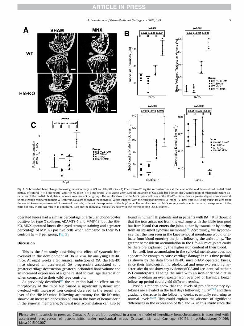
Fig. 3. Subchondral bone changes following meniscectomy in WT and Hfe-KO mice (A) Knee micro-CT sagittal reconstructions at the level of the middle one-third medial tibialplateau of control (n ¼ 5 per group) and Hfe-KO mice (n ¼ 5 per group) at 8 weeks after surgical induction of OA. Scale bar 500 mm (B) Quantification of microarchitecture pa-rameters of the medial tibial plateau of mice knees (n ¼ 5 per group). The results show that the MNX operated knees of the Hfe-KO animals have a greater degree of subchondralsclerosis when compared to their WT controls. Data are shown as the individual values (shapes) with the corresponding 95% CI (range) (C) Real time PCR, using mRNA isolated fromthe medial knee compartment of 18 weeks-old animals, to detect the expression of the Bmp6 gene. The results show that MNX surgery leads to an increase in the expression of thegene but only in Hfe-KO mice is it significant. Data are the individual values (shapes) with the corresponding 95% CI (range).
A. Camacho et al. / Osteoarthritis and Cartilage xxx (2015) 1e9 5
operated knees had a similar percentage of articular chondrocytespositive for type X collagen, ADAMTS-5 and MMP-13, but the Hfe-KO, MNX operated knees displayed stronger staining and a greaterpercentage of MMP-3 positive cells when compared to their WTcontrols (n ¼ 3 per group, Fig. 5).
Discussion
This is the first study describing the effect of systemic ironoverload in the development of OA in vivo, by analysing Hfe-KOmice. At eight weeks after surgical induction of OA, the Hfe-KOmice showed an accelerated OA progression associated to agreater cartilage destruction, greater subchondral bone volume andan increased expression of a gene related to cartilage degradationwhen compared to their wild-type controls.
As previously described19, the mutation had no effect on themorphology of the mice but caused a significant systemic ironoverload with increased iron content observed in the serum andliver of the Hfe-KO mice. Following arthrotomy the Hfe-KO miceshowed an increased deposition of iron in the form of hemosiderinin the synovial membrane. Synovial iron accumulation can also be
Please cite this article in press as: Camacho A, et al., Iron overload inaccelerated progression of osteoarthritis under mechanical stressj.joca.2015.09.007
found in human HH patients and in patients with RA11. It is thoughtthat the iron arises not from the exchange with the labile iron poolbut from blood that enters the joint, either by trauma or by oozingfrom an inflamed synovial membrane26. Accordingly, we hypothe-sise that the iron seen in the knee synovial membrane would orig-inate from blood entering the joint following the arthrotomy. Thegreater hemosiderin accumulation in the Hfe-KO mice joints couldbe therefore explained by the higher iron content of their blood.
By itself, iron accumulation in the synovial membrane does notappear to be enough to cause cartilage damage in this time period,as shown by the data from Hfe-KO mice SHAM-operated knees,since their histological, morphological and gene expression char-acteristics do not show any evidence of OA and are identical to theirWT counterparts. Feeding the mice with an iron-enriched diet inorder to obtain an even greater iron overload or having a longerfollow-up period could yield different results.
Previous reports show that the levels of proinflammatory cy-tokines are elevated in the first days following injury27,28 and thengradually decrease in the following weeks, eventually returning tonormal levels29,30. This could explain the absence of significantdifferences in the expression of Il1b and Il6 in this study since the
a murine model of hereditary hemochromatosis is associated with, Osteoarthritis and Cartilage (2015), http://dx.doi.org/10.1016/

Fig. 4. Changes in gene expression following surgical induction of OA. Real time PCR, using mRNA isolated from the medial knee compartment of animals euthanized 8 weeks aftersurgery, to detect the expression of selected target genes (n ¼ 5 per group). The results confirm that MNX surgery induces the expression of genes related with matrix degradationand chondrocyte hypertrophy and that this is more pronounced in the Hfe-KO mice. Data are shown as the individual values (shapes) with the corresponding 95% CI (range).
A. Camacho et al. / Osteoarthritis and Cartilage xxx (2015) 1e96
mice were evaluated at 8 weeks after the initial injury, when thedisease process is already in a quiescent phase31.
As previously reported in the literature, we found that surgicallyinduced mechanical instability of the knee joint resulted inderegulation of genes related to extracellular matrix production,matrix degradation and chondrocyte hypertrophy31e33, as well ascartilage degeneration34,35 and subchondral bone sclerosis36,37. Allof these were observable in both WT and Hfe-KO mice meniscec-tomized knees but differences were more evident in the latter(Figs. 1e4) providing further evidence for the increased suscepti-bility for OA in the Hfe-KOmice. On the other hand, the presence ofexcess iron in the synovial tissue could also account for the accel-erated OA progression in the meniscectomized knees of the Hfe-KOmice since intra- and extra-cellular overload of iron in the tissueinduces the generation of free radicals and reactive oxygen species(ROS)38e40. These can lead to hyper-activation of MMPs41, a crucialstep in many normal and pathological biological processes42. Weobserved that, following the surgical induction of OA, the Hfe-KOmice displayed an increased expression of MMP-3 whencompared to their WT controls. MMP-3 is an enzyme capable ofdegrading several components of the extracellular matrix, makingit less resilient to the mechanical stress and accelerating the pro-gression of OA33,43. Therefore we hypothesise that the increasediron content in the synovial membrane contributes to an increasedgeneration of ROS after an initial pro-inflammatory signal, such asjoint trauma or surgical intervention, causing an up-regulation ofMMP-3 expression, contributing to the greater cartilage damageobserved in the Hfe-KO mice knees.
Please cite this article in press as: Camacho A, et al., Iron overload inaccelerated progression of osteoarthritis under mechanical stressj.joca.2015.09.007
The precise role of subchondral bone in the initiation and pro-gression of OA is still unclear44,45. A recent review46 proposes thatthe non-physiologic strain on the joint causes micro damage to thesubchondral bone leading to increased resorption and a decrease inthickness in the early-stages of OA, progressing to subchondraltrabecular sclerosis and increased calcified cartilage thickness inthe late-stage OA. These changes in subchondral bone architectureare likely to accompany cartilage damage that could, in turn, in-fluence subchondral bone degradation, resulting in a vicious cycle.Accordingly, in our study we observed a greater increase in sub-chondral bone volume and calcified cartilage thickness in the jointswith more pronounced cartilage damage, which was in agreementto data previously reported by Chappard et al.47, Hayami et al.48 andBotter et al36.
The present study has some limitations. In humans, HH-relatedOA develops in the fourth and fifth decades of life whereas we usedyoung mice (10-week-old) to study the development of OA there-fore this model may not accurately represent the evolution of thehuman disease. The observed changes in cartilage and subchondralbone were restricted to a single time-point making it difficult toconclude about the entire sequence of changes leading to OA, butthis could be addressed in future studies. The time point waschosen since the cartilage and subchondral bone changes arealready well defined18 and the disease process is in a quiescentphase31. Our selection of target genes was geared towards the maincomponents of the extracellular matrix49, the main proteolyticenzymes involved in matrix degradation43,50 and the main mole-cules related with chondrocyte hypertrophy51. It is possible that
a murine model of hereditary hemochromatosis is associated with, Osteoarthritis and Cartilage (2015), http://dx.doi.org/10.1016/
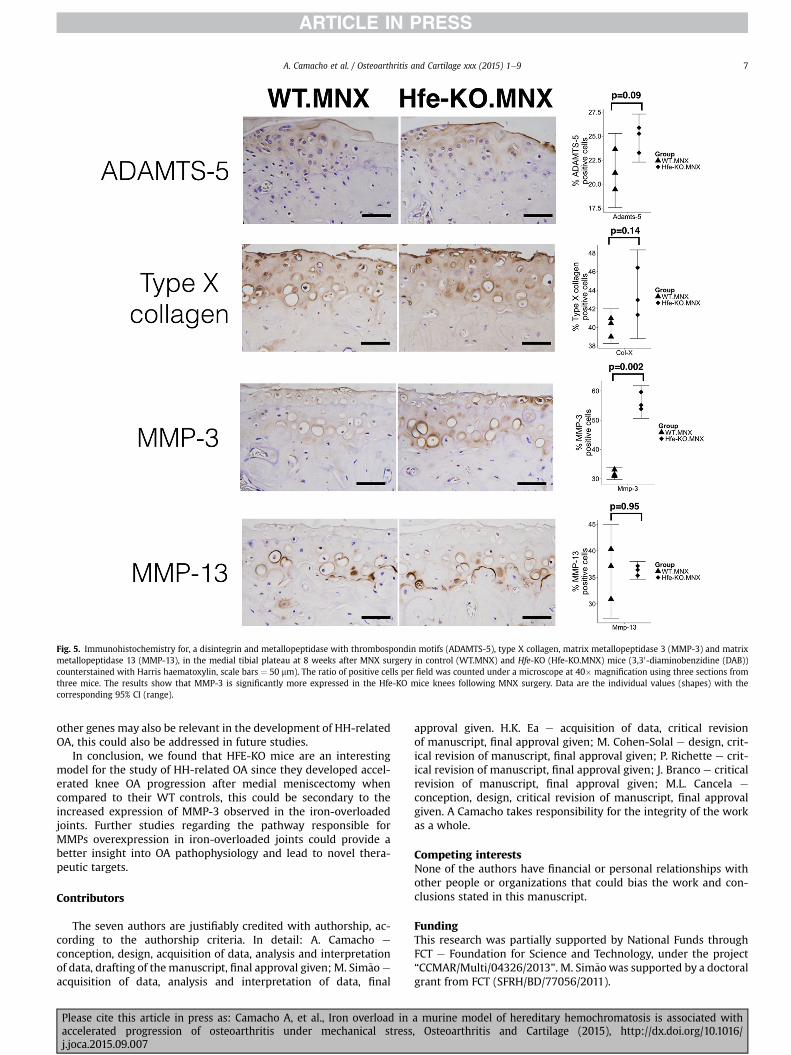
Fig. 5. Immunohistochemistry for, a disintegrin and metallopeptidase with thrombospondin motifs (ADAMTS-5), type X collagen, matrix metallopeptidase 3 (MMP-3) and matrixmetallopeptidase 13 (MMP-13), in the medial tibial plateau at 8 weeks after MNX surgery in control (WT.MNX) and Hfe-KO (Hfe-KO.MNX) mice (3,30-diaminobenzidine (DAB))counterstained with Harris haematoxylin, scale bars ¼ 50 mm). The ratio of positive cells per field was counted under a microscope at 40� magnification using three sections fromthree mice. The results show that MMP-3 is significantly more expressed in the Hfe-KO mice knees following MNX surgery. Data are the individual values (shapes) with thecorresponding 95% CI (range).
A. Camacho et al. / Osteoarthritis and Cartilage xxx (2015) 1e9 7
other genes may also be relevant in the development of HH-relatedOA, this could also be addressed in future studies.
In conclusion, we found that HFE-KO mice are an interestingmodel for the study of HH-related OA since they developed accel-erated knee OA progression after medial meniscectomy whencompared to their WT controls, this could be secondary to theincreased expression of MMP-3 observed in the iron-overloadedjoints. Further studies regarding the pathway responsible forMMPs overexpression in iron-overloaded joints could provide abetter insight into OA pathophysiology and lead to novel thera-peutic targets.
Contributors
The seven authors are justifiably credited with authorship, ac-cording to the authorship criteria. In detail: A. Camacho e
conception, design, acquisition of data, analysis and interpretationof data, drafting of the manuscript, final approval given; M. Sim~aoe
acquisition of data, analysis and interpretation of data, final
Please cite this article in press as: Camacho A, et al., Iron overload inaccelerated progression of osteoarthritis under mechanical stressj.joca.2015.09.007
approval given. H.K. Ea e acquisition of data, critical revisionof manuscript, final approval given; M. Cohen-Solal e design, crit-ical revision of manuscript, final approval given; P. Richette e crit-ical revision of manuscript, final approval given; J. Branco e criticalrevision of manuscript, final approval given; M.L. Cancela e
conception, design, critical revision of manuscript, final approvalgiven. A Camacho takes responsibility for the integrity of the workas a whole.
Competing interestsNone of the authors have financial or personal relationships withother people or organizations that could bias the work and con-clusions stated in this manuscript.
FundingThis research was partially supported by National Funds throughFCT e Foundation for Science and Technology, under the project“CCMAR/Multi/04326/2013”. M. Sim~aowas supported by a doctoralgrant from FCT (SFRH/BD/77056/2011).
a murine model of hereditary hemochromatosis is associated with, Osteoarthritis and Cartilage (2015), http://dx.doi.org/10.1016/

A. Camacho et al. / Osteoarthritis and Cartilage xxx (2015) 1e98
The funding source had no influence on the study design, datacollection, analysis and interpretation, nor in the writing of themanuscript or the decision to submit it.
Acknowledgments
We thank Caroline Marty at INSERM 1132 and Paulo Gavaia atCCMAR for their technical expertise with histochemical techniquesand Agn�es Ostertag at INSERM 1132 for assistance with the micro-CT scanner and software. Also the authors thank the late Jorge Pintoat Instituto de Biologia Molecular e Celular (IBMC) in Porto forproviding the initial colony of Hfe-KO mice.
Supplementary data
Supplementary data related to this article can be found at http://dx.doi.org/10.1016/j.joca.2015.09.007.
References
1. Johnson VL, Hunter DJ. The epidemiology of osteoarthritis. BestPract Res Clin Rheumatol 2014;28:5e15.
2. Neogi T, Zhang Y. Epidemiology of osteoarthritis. Rheum DisClin North Am 2013;39:1e19.
3. Loeser RF, Goldring SR, Scanzello CR, Goldring MB. Osteoar-thritis: a disease of the joint as an organ. Arthritis Rheum2012;64:1697e707.
4. Zhou XY, Tomatsu S, Fleming RE, Parkkila S, Waheed A, Jiang J,et al. HFE gene knockout produces mouse model of hereditaryhemochromatosis. Proc Natl Acad Sci 1998;95:2492e7.
5. Pietrangelo A. Hereditary hemochromatosis: pathogenesis,diagnosis, and treatment. Gastroenterology 2010;139:393e408. 408.e391-392.
6. Richette P, Ottaviani S, Vicaut E, Bardin T. Musculoskeletalcomplications of hereditary hemochromatosis: a case-controlstudy. J Rheumatol 2010;37:2145e50.
7. Elmberg M, Hultcrantz R, Simard JF, Carlsson Å, Askling J.Increased risk of arthropathies and joint replacement surgeryin patients with genetic hemochromatosis: a study of 3,531patients and their 11,794 first-degree relatives. Arthritis CareRes (Hoboken) 2013;65:678e85.
8. Sahinbegovic E, Dallos T, Aigner E, Axmann R, Engelbrecht M,Sch€oniger-Hekele M, et al. Hereditary hemochromatosis as arisk factor for joint replacement surgery. Am J Med 2010;123:659e62.
9. Wang Y, Gurrin LC, Wluka AE, Bertalli NA, Osborne NJ,Delatycki MB, et al. HFE C282Y homozygosity is associatedwith an increased risk of total hip replacement for osteoar-thritis. Semin Arthritis Rheum 2012;41:872e8.
10. von Kempis J. Arthropathy in hereditary hemochromatosis.Curr Opin Rheumatol 2001;13:80e3.
11. Heiland GR, Aigner E, Dallos T, Sahinbegovic E, Krenn V,Thaler C, et al. Synovial immunopathology in haemochroma-tosis arthropathy. Ann Rheum Dis 2010;69:1214e9.
12. Schumacher HR. Articular cartilage in the degenerativearthropathy of hemochromatosis. Arthritis Rheum 1982;25:1460e8.
13. Carroll GJ, Sharma G, Upadhyay A, Jazayeri JA. Ferritin con-centrations in synovial fluid are higher in osteoarthritis pa-tients with HFE gene mutations (C282Y or H63D). Scand JRheumatol 2010;39:413e20.
14. Husar-Memmer E, Stadlmayr A, Datz C, Zwerina J. HFE-relatedhemochromatosis: an update for the rheumatologist. CurrRheumatol Rep 2014;16:393.
Please cite this article in press as: Camacho A, et al., Iron overload inaccelerated progression of osteoarthritis under mechanical stressj.joca.2015.09.007
15. Nicolas G, Viatte L, Lou D-Q, Bennoun M, Beaumont C, Kahn A,et al. Constitutive hepcidin expressionprevents iron overload inamouse model of hemochromatosis. Nat Genet 2003;34:97e101.
16. Cooksey RC, Jouihan HA, Ajioka RS, Hazel MW, Jones DL,Kushner JP, et al. Oxidative stress, beta-cell apoptosis, anddecreased insulin secretory capacity in mouse models of he-mochromatosis. Endocrinology 2004;145:5305e12.
17. Guggenbuhl P, Fergelot P, Doyard M, Libouban H, Roth M-P,Gallois Y, et al. Bone status in a mouse model of genetic he-mochromatosis. Osteoporos Int 2011;22:2313e9.
18. Kamekura S, Hoshi K, Shimoaka T, Chung U, Chikuda H,Yamada T, et al. Osteoarthritis development in novel experi-mental mouse models induced by knee joint instability.Osteoarthritis Cartilage 2005;13:632e41.
19. Levy JE, Montross LK, Cohen DE, Fleming MD, Andrews NC. TheC282Y mutation causing hereditary hemochromatosis doesnot produce a null allele. Blood 1999;94:9e11.
20. Rebouche CJ, Wilcox CL, Widness JA. Microanalysis of non-heme iron in animal tissues. J Biochem Biophys Methods2004;58:239e51.
21. Glasson SS, Chambers MG, Van Den Berg WB, Little CB. TheOARSI histopathology initiative e recommendations for his-tological assessments of osteoarthritis in the mouse. Osteoar-thritis Cartilage 2010;18(Suppl 3):S17e23.
22. Botter SM, van Osch GJVM, Waarsing JH, Day JS, Verhaar JAN,Pols HAP, et al. Quantification of subchondral bone changes ina murine osteoarthritis model using micro-CT. Biorheology2006;43:379e88.
23. Curtis KM, Gomez LA, Rios C, Garbayo E, Raval AP,Perez-Pinzon MA, et al. EF1alpha and RPL13a represent normal-ization genes suitable for RT-qPCR analysis of bone marrowderived mesenchymal stem cells. BMC Mol Biol 2010;11:61.
24. Andriopoulos Jr Billy, Corradini E, Xia Y, Faasse SA, Chen S,Grgurevic L, et al. BMP6 is a key endogenous regulator ofhepcidin expression and iron metabolism. Nat Genet 2009;41:482e7.
25. Kugimiya F, Kawaguchi H, Kamekura S, Chikuda H, Ohba S,Yano F, et al. Involvement of endogenous bone morphogeneticprotein (BMP) 2 and BMP6 in bone formation. J Biol Chem2005;280:35704e12.
26. Muirden KD, Senator GB. Iron in the synovial membrane inrheumatoid arthritis and other joint diseases. Ann Rheum Dis1968;27:38e48.
27. Haslauer CM, Proffen BL, Johnson VM, Hill A, Murray MM.Gene expression of catabolic inflammatory cytokines peakbefore anabolic inflammatory cytokines after ACL injury in apreclinical model. J Inflamm (Lond) 2014;11:34.
28. Irie K, Uchiyama E, Iwaso H. Intraarticular inflammatory cy-tokines in acute anterior cruciate ligament injured knee. Knee2003;10:93e6.
29. Bigoni M, Sacerdote P, Turati M, Franchi S, Gandolla M,Gaddi D, et al. Acute and late changes in intraarticular cytokinelevels following anterior cruciate ligament injury. J Orthop Res2013;31:315e21.
30. Ward BD, Furman BD, Huebner JL, Kraus VB, Guilak F, Olson SA.Absence of posttraumatic arthritis following intraarticularfracture in the MRL/MpJ mouse. Arthritis Rheum 2008;58:744e53.
31. Loeser RF, Olex AL, McNulty MA, Carlson CS, Callahan M,Ferguson C, et al. Disease progression and phasic changes ingene expression in a mouse model of osteoarthritis. PLoS One2013;8:e54633.
32. Appleton CTG, Pitelka V, Henry J, Beier F. Global analyses ofgene expression in early experimental osteoarthritis. ArthritisRheum 2007;56:1854e68.
a murine model of hereditary hemochromatosis is associated with, Osteoarthritis and Cartilage (2015), http://dx.doi.org/10.1016/
A. Camacho et al. / Osteoarthritis and Cartilage xxx (2015) 1e9 9
33. Bateman JF, Rowley L, Belluoccio D, Chan B, Bell K, Fosang AJ,et al. Transcriptomics of wild-type mice and mice lackingADAMTS-5 activity identifies genes involved in osteoarthritisinitiation and cartilage destruction. Arthritis Rheum 2013;65:1547e60.
34. Burleigh A, Chanalaris A, Gardiner MD, Driscoll C, Boruc O,Saklatvala J, et al. Joint immobilization prevents murine oste-oarthritis and reveals the highly mechanosensitive nature ofprotease expression in vivo. Arthritis Rheum 2012;64:2278e88.
35. Glasson SS, Blanchet TJ, Morris EA. The surgical destabilizationof the medial meniscus (DMM) model of osteoarthritis in the129/SvEv mouse. Osteoarthritis Cartilage 2007;15:1061e9.
36. Botter SM, Glasson SS, Hopkins B, Clockaerts S, Weinans H, vanLeeuwen JPTM, et al. ADAMTS5-/- mice have less subchondralbone changes after induction of osteoarthritis through surgicalinstability: implications for a link between cartilage and sub-chondral bone changes. Osteoarthritis Cartilage 2009;17:636e45.
37. Hayami T, Pickarski M, Wesolowski GA, McLane J, Bone A,Destefano J, et al. The role of subchondral bone remodeling inosteoarthritis: reduction of cartilage degeneration and pre-vention of osteophyte formation by alendronate in the ratanterior cruciate ligament transection model. Arthritis Rheum2004;50:1193e206.
38. Brissot P, Ropert M, Le Lan C, Lor�eal O. Non-transferrin boundiron: a key role in iron overload and iron toxicity. BiochimBiophys Acta 2012;1820:403e10.
39. Morris CJ, Blake DR, Wainwright AC, Steven MM. Relationshipbetween iron deposits and tissue damage in the synovium: anultrastructural study. Ann Rheum Dis 1986;45:21e6.
40. Sullivan JL. Is stored iron safe? J Lab Clin Med 2004;144:280e4.
41. Mairuae N, Connor JR, Cheepsunthorn P. Increased cellular ironlevels affect matrix metalloproteinase expression and phago-cytosis in activated microglia. Neurosci Lett 2011;500:36e40.
Please cite this article in press as: Camacho A, et al., Iron overload inaccelerated progression of osteoarthritis under mechanical stressj.joca.2015.09.007
42. Nagase H, Woessner JF. Matrix metalloproteinases. J Biol Chem1999;274:21491e4.
43. Blom AB, van Lent PL, Libregts S, Holthuysen AE, van derKraan PM, van Rooijen N, et al. Crucial role of macrophages inmatrix metalloproteinase-mediated cartilage destructionduring experimental osteoarthritis: involvement of matrixmetalloproteinase 3. Arthritis Rheum 2007;56:147e57.
44. Burr DB. The importance of subchondral bone in the pro-gression of osteoarthritis. J Rheumatol Suppl 2004;70:77e80.
45. Oegema Jr TR, Carpenter RJ, Hofmeister F, Thompson Jr RC. Theinteraction of the zone of calcified cartilage and subchondralbone in osteoarthritis. Microsc Res Tech 1997;37:324e32.
46. Li G, Yin J, Gao J, Cheng TS, Pavlos NJ, Zhang C, et al. Sub-chondral bone in osteoarthritis: insight into risk factors andmicrostructural changes. Arthritis Res Ther 2013;15:223.
47. Chappard C, Peyrin F, Bonnassie A, Lemineur G, Brunet-Imbault B, Lespessailles E, et al. Subchondral bone micro-architectural alterations in osteoarthritis: a synchrotronmicro-computed tomography study. Osteoarthritis Cartilage2006;14:215e23.
48. Hayami T, Pickarski M, Zhuo Y, Wesolowski GA, Rodan GA,Duong LT. Characterization of articular cartilage and sub-chondral bone changes in the rat anterior cruciate ligamenttransection and meniscectomized models of osteoarthritis.Bone 2006;38:234e43.
49. Martin I, Jakob M, Sch€afer D, Dick W, Spagnoli G, Heberer M.Quantitative analysis of gene expression in human articularcartilage from normal and osteoarthritic joints. OsteoarthritisCartilage 2001;9:112e8.
50. Verma P, Dalal K. ADAMTS-4 and ADAMTS-5: key enzymes inosteoarthritis. J Cell Biochem 2011;112:3507e14.
51. Zheng Q, Zhou G, Morello R, Chen Y, Garcia-Rojas X, Lee B.Type X collagen gene regulation by Runx2 contributes directlyto its hypertrophic chondrocyte-specific expression in vivo.J Cell Biol 2003;162:833e42.
a murine model of hereditary hemochromatosis is associated with, Osteoarthritis and Cartilage (2015), http://dx.doi.org/10.1016/


![In Vivo Correction of Murine Tyrosinemia Type I by DNA ...web.stanford.edu/group/markkaylab/publications/12498772.pdf · model of hereditary tyrosinemia type 1 (HT1) [19], be-cause](https://img.pdfslide.net/doc/110x75/5fadaef398034276572c5af9/in-vivo-correction-of-murine-tyrosinemia-type-i-by-dna-web-model-of-hereditary.jpg)


![NON-HFE IRON OVERLOAD: IS PHLEBOTOMY THE ANSWER · do not correlate with serum ferritin levels in Hereditary Haemochromatosis (>7.1kPa) [24]. A longitudinal study in transfusion-independent](https://img.pdfslide.net/doc/110x75/5f068b947e708231d4188542/non-hfe-iron-overload-is-phlebotomy-the-answer-do-not-correlate-with-serum-ferritin.jpg)