Embed Size (px)
Citation preview

INFECrION AND IMMUNITY, Sept. 1994, p. 4021-40270019-9567/94/$04.00+0Copyright X 1994, American Society for Microbiology
Iron Release from Transferrin by Pyoverdin and Elastasefrom Pseudomonas aeruginosa
CHRISTIANE WOLZ,1 KARIN HOHLOCH,' AYDIN OCAKTAN,2 KEITH POOLE,3 ROBERT W. EVANS,4NATACHA ROCHEL,S ANNE-MARIE ALBRECHT-GARY,5 MOHAMED A. ABDALLAH 2
AND GERD DORING'*Department of General and Environmental Hygiene, Hygiene Institute, University of Tubingen, Tubingen, Germany';Laboratoire de Chimie Microbienne, URA 31 Centre National de la Recherche Scientifique,2 and Laboratoire deChimie Physique Bioinorganique, Centre de Recherche de Chimie, Universite Louis Pasteur, Strasbourg, France;
Department of Microbiology and Immunology, Queens University, Kingston, Ontario, Canada3;and Division of Biochemistry, UMDS, Guy's Hospital, London, United Kingdom4
Received 4 April 1994/Returned for modification 13 May 1994/Accepted 20 June 1994
Pseudomonas aeruginosa produces the siderophores pyoverdin and pyochelin as well as receptors forsiderophores in response to iron deprivation. Previously, it has been shown in vitro that at neutral pH purifiedpyoverdin acquires iron from transferrin only in the presence of P. aeruginosa elastase (LasB), whichproteolytically degrades transferrin. We constructed a LasB-negative mutant, PAO1E, by insertional mutagen-esis to investigate whether this mutant differs in growth from the parental strain PA01 in an iron-depletedmedium supplemented with transferrin or human serum. PAO1 and PAOIE did not differ in growth with 1.25,uM Fe2-transferrin as the only iron source. Urea gel electrophoresis indicated iron release from intacttransferrin during the logarithmic growth phase of PAO1 and PAO1E. A total of 333 ,uM LasB was synthesizedfrom PAOI after onset of stationary-phase growth. Quantification of pyoverdin by spectroscopy revealed thatup to 900 ,uM pyoverdin was produced during growth of the strains in medium supplemented withFe2-transferrin or 10%o human serum. Incubation of Fe2-transferrin and purified pyoverdin in concentrationssimilar to those found in the culture supernatant resulted in release of iron from transferrin after 10 h at 37°C.However, LasB significantly enhanced the rate constant for iron acquisition of pyoverdin from transferrin. Weconclude that P. aeruginosa can use transferrin as an iron source without further need of LasB or pH changes.This is further supported by experiments with P. aeruginosa K437, which has a defective iron uptake system,and its LasB-negative mutant, K437E. Though K437 and K437E did not differ in growth with Fe2-transferrinas the only iron source, their growth was significantly reduced relative to that of PAO1 and PAO1E.
In spite of the virtual absence of freely available iron in thehuman body, pathogenic bacteria can successfully multiply invivo to establish infection. The iron transport proteins trans-ferrin and lactoferrin are thought to serve as the iron sourcefor bacteria in the human body fluids (16). Pseudomonasaeruginosa, like other pathogens, produces siderophores dur-ing iron deprivation which bind Fe3" and deliver the iron tothe bacterial cell by high-affinity uptake systems (6). However,there are conflicting results concerning the role of the P.aeruginosa siderophores pyoverdin and pyochelin in iron re-lease from the transferrin molecule. Both siderophores pro-moted growth of P. aeruginosa when added to a medium withFe2-transferrin or human sera as the iron source (1). But,because of the higher iron binding constant of pyoverdin (1032)(8) compared with that of pyochelin (105) (7), pyoverdin issuggested to be more effective. Accordingly, a pyochelinmutant was still able to grow in human sera whereas a
pyoverdin mutant was deficient in growth (1).However, incubation of the purified compounds pyoverdin
and Fe2-transferrin showed that the transfer of iron fromFe2-transferrin to pyoverdin is detectable only under certainexperimental conditions. The reduction of pH after growth ofbacteria in medium containing glucose is one possible mecha-nism to release iron from transferrin (26). Other results
* Corresponding author. Mailing address: Department of Generaland Environmental Hygiene, Hygiene Institute, University of Tubin-gen, Wilhelmstrasse 31, D-72072 Tiibingen, Germany. Phone: 49 7071292069. Fax: 49 7071 293011.
implied that proteolytic cleavage of transferrin may be neces-sary for iron release (12). P. aeruginosa produces severalextracellular proteases, one of which, elastase (LasB), has beenshown to cleave transferrin into small peptides (12). Interest-ingly, it was reported that the synthesis of LasB was enhancedin iron-deficient media (2, 5, 27). Thus, the synthesis oftransferrin-hydrolyzing enzymes in concert with siderophoresmay allow the pathogens to overcome iron limitations duringinfection. To test this hypothesis, we examined the ability ofLasB-negative mutants of P. aeruginosa to release iron fromFe2-transferrin during growth. The LasB structural genes fromstrains PAO1 and K437, a derivative of PAO1 defective inpyoverdin, pyochelin, and pyoverdin uptake, were interruptedby insertional mutagenesis, resulting in strains PAO1E andK437E, respectively. We then reinvestigated the reaction be-tween purified pyoverdin and transferrin with and withoutLasB in vitro. The LasB mutants were still able to release ironfrom the Fe2-transferrin at a rate comparable with that of theproteolytically competent parental strain in transferrin media.Purified pyoverdin scavenges iron from transferrin withoutneed for LasB.
MATERIALS AND METHODS
Bacterial strains and mutagenesis. P. aeruginosa PAO1 andthe LasB-negative mutant PAO1E have been described else-where (28). Strain K437 is deficient in pyoverdin, pyochelin,and pyoverdin uptake (25). Strain K437E, the LasB-negativemutant derived from K437, was constructed by insertional
4021
Vol. 62, No. 9
on April 1, 2019 by guest
http://iai.asm.org/
Dow
nloaded from

4022 WOLZ ET AL.
0o
0
10 1
a
00
time (h) time (h)FIG. 1. Growth of P. aeruginosa PAO1 (A) and PAO1E (B) in CDM with 25 ,uM FeSO4 (0), 1.25 ,M apotransferrin (O), 1.25 JIM
Fe2-transferrin (0), or 10% heat-inactivated human serum (U).
mutagenesis, and the insertion of the mobilization vectorpBRMOB-LasB was confirmed by Southern hybridization asdescribed previously (28).
Chemicals and glassware. Fe2-transferrin and apotrans-ferrin were purchased from Sigma; LasB was purchased fromNagase Biochemicals. For all iron-restricted-growth experi-ments, the glassware was rinsed in 1 mM EDTA overnight andthen once with 0.1 N HCl and six times with deionized water(dH20).Growth conditions and media. A chemically defined me-
dium (CDM) which was optimized for LasB production (19)(14.4 mM K2PO4, 92 mM Na-glutamate, 24 mM valine, 8 mMphenylalanine, 70 mM glucose, 1.33 mM MgSO4, 0.14 mMCaCl2, 0.0085 mM ZnSO4) was used. Free iron was removedwith Chelex-100 (Bio-Rad Laboratories); 3x concentratedstock solutions of NaCl, K2PO4, Na-glutamate, valine, andphenylalanine and a 1OX concentrated stock solution of glu-cose were stirred with Chelex-100 (5% [wt/vol]) for 2 h andsterile filtered into iron-free, sterile glassware. MgSO4, CaCl2,and ZnSO4 were prepared as 100x stock solutions, autoclaved,and added to the media. The iron concentration of the mediumwas 44 ,ug/ml as determined by atomic absorption spectros-copy. In some experiments, iron was added from a 45 mMFeSO4 solution which had been freshly prepared and sterilefiltered. Human serum was freshly prepared and heat inacti-vated (30 min, 56°C). Apotransferrin or Fe2-transferrin wasadded to a final concentration of 1.25 ,uM to the medium.Bacteria were inoculated in 25 ml of medium in 100-ml flaskswith an initial optical density value at 480 nm (OD480) of 0.01from an overnight culture in the same medium.Radioimmunoassay, gel electrophoresis, and immunoblot-
ting. Radioimmunoassay for LasB was carried out as describedpreviously (24). Sodium dodecyl sulfate-polyacrylamide gelelectrophoresis (SDS-PAGE) was performed according to themethod of Laemmli (20) with 12% T-3% C. The gels werestained with colloidal Coomassie G (23) or with silver (4).Urea gel electrophoresis (21) was performed with some mod-ifications in the Mini-Protean II gel apparatus (Bio-Rad Lab-oratories). The gel was prepared as follows: 4.5 g of urea-625,ul of 20x concentration of TBE (2 M Tris, 0.2 M boric acid,0.032 M EDTA; pH 8.4), was added to 2.7 ml of acrylamide
stock solution (2.6% C-30% T), the volume was adjusted to12.5 ml with dH2O, and the gel was polymerized with 10 mg ofammonium persulfate and 20 p1 of TEMED (N,N,N',N'-tetramethylethylenediamine). The gel was overlaid with acomb gel (1.3 ml of 20X TBE, 1.3 ml of acrylamide stocksolution [2.6% C, 30% T], 6.1 ml of dH20, 10 ,ul of ammoniumpersulfate, 50 RI of TEMED). TBE (lx, pH 8.4) was used aselectrophoresis buffer. Proteins were freshly prepared in sam-ple buffer (10% glycerol, 0.2% bromphenol blue, lx TBE).For samples containing serum, 100 pI of culture supernatantwas mixed with 800 pI of Rivanol solution (25% glycerol,0.38% Rivanol in lx TBE) and left for 5 min, and theprecipitated serum proteins were removed by centrifugation.Electrophoresis was carried out for 5 h at 100 V. For immu-noblotting, proteins were transferred to nitrocellulose (Schlei-cher and Schuill) by semidry blotting in electrophoresis buffer.Blots were developed with polyclonal primary antibodies andperoxidase-conjugated secondary antibodies, and bands werevisualized with chemiluminescence (ECL Kit from AmershamBuchler) according to the instructions of the manufacturer.
Pyoverdin purification and quantification. Pyoverdin wasisolated from the culture supernatant of P. aeruginosa ATCC15692 (9) and purified according to the hydrophobic chroma-tography method (8). The spectrophotometric measurementswere performed with a spectrophotometer (Pharmacia, Freiburg,Germany). Iron-free pyoverdin has an absorption maximum at400 nm, whereas binding of iron leads to a characteristicshoulder at 465 nm. Since the spectrum of pyoverdin as a freeligand is pH dependent, the quantification of pyoverdin wasperformed at pH 5.0. The supernatants were diluted (1:10)with 0.5 M acetic acid-sodium acetate buffer, pH 5.0. At thispH, the spectrophotometric characteristics of pyoverdin areXmax = 380 nm (Emax = 16,500 M x L-1) (8). Upon acidifica-tion of the supernatants, the peak shifted from 400 to 380 nmas described for purified pyoverdin (22).The fluorescence spectrum measurements were carried out
with a Deltascan 4000 spectrofluorimeter (Photon TechnologyInternational, Inc.). The concentration of pyoverdin in theculture supernatant was determined with a calibration curveobtained from the fluorescence emission spectra of purepyoverdin at pH 5.0 (in 0.5 M acetic acid-sodium acetate
INFECT. IMMUN.
on April 1, 2019 by guest
http://iai.asm.org/
Dow
nloaded from

P. AERUGINOSA IRON ACQUISITION 4023
TABLE 1. Cell density (OD480), pyoverdin, and LasB synthesis of P. aeruginosa PA01 and PA0lE in stationary phase
OD480, pyoverdin, and LasB synthesis of Pseudomonas strainsb:Growth PA01 PA01E
conditionssOD480 (24 h) Pyoc (P±M) LasBd (PM) OD480 (24 h) Pyo (PM) LasB (pM)
Apotransferrin 1.1 ± 0.3 126 + 20 13.6 ± 6 1.4 ± 0.6 120 ± 14 NDeFe2-transferrin 10.9 ± 1.8 582 ± 140 342 ± 30 9.1 ± 0.9 174 ± 35 NDSerum 8.6 ± 1.2 855 + 430 396 ± 121 5.4 ± 1.4 990 ± 126 NDFeSO4 15.2 ± 2.2 ND 324 ± 90 13.2 ± 1.2 ND ND
a Iron-depleted CDM with apotransferrin (1.25 P,M), Fe2-transferrin (1.25 PM), 10% human serum, or 25 p,M FeSO4.b Mean ± standard deviation of at least three determinations.c Pyo, pyoverdin. Molarity was calculated by measuring A380 at pH 5.0; E = 16,500 M` cm-'.d Determined by radioimmunoassay.e ND, not detectable (<1 ng of LasB per ml).
buffer; excitation wavelength, 400 nm; emission, 455 nm). Sinceboth methods for pyoverdin quantification led to comparableresults, pyoverdin concentrations in the culture supematantwere normally measured by spectroscopy.
Kinetics. Pyoverdin (1.8 mM) was incubated with 8 ,uMFe2-transferrin in the absence or presence of LasB (3 mM) in0.1 M Tris-HCl, pH 7.4. The ferric solution was prepared withFe(C104)3 x 9H20. The kinetic measurements were done byclassical absorption spectrophotometry at 465 nm (Kontron;Uvikon 860). The fastest step observed in the presence of LasBwas checked by a rapid mixing technique at 465 nm (Stopped-Flow Spectrophotometer; Applied Photophysics). The temper-ature was kept at 25.0 ± 0.1°C for all the experiments. Thekinetic data have been fitted by statistical method (Biokine V3.0 software; Bio-Logic Co., Echirolles, France).
RESULTS
Characterization of P. aeruginosa strains grown in a chem-ically defined medium. To investigate the role of LasB in ironacquisition from transferrin, we compared the LasB-negativemutants (PA01E and K437E) with the proteolytic wild-typestrains. The PA01 derivative K437, a strain defective inpyoverdin and pyochelin synthesis as well as in pyoverdinuptake (25), was mutagenized by insertional mutagenesis,resulting in the LasB-negative strain K437E. Mutagenesis wasconfirmed by Southern hybridization of BglII-digested chromo-somal DNA with lasB. The size of the restriction fragmentcarrying lasB increased in the mutants K437E and PA0lE (28)by the size of the inserted plasmid (data not shown). LasB wasproduced only in PA01 and K437, not in the mutants PAOlEand K437E, as shown by a radioimmunoassay which has asensitivity limit of 1 ng/ml.For growth experiments, an iron-depleted medium (CDM)
was used, which was optimized for LasB production. All strainsgrew equally well in CDM supplemented with 20 ,uM FeSO4.Without added iron, PA01 and PA01E showed growth inhi-bition and produced pyoverdin in equal concentrations (100,uM) (data not shown). The strains K437 and K437E did notgrow at all without additional iron. The pH in the growthmedia was maintained at pH 8 throughout the experiments.Growth of P. aeruginosa PAO1 and PAO1E with transferrin.
Strains were grown in CDM with Fe2-transferrin, apotrans-ferrin, or human sera. Whereas apotransferrin has no growth-stimulating effect on the strains, addition of 1.25 puM Fe2-transferrin or 10% human serum resulted in growth promotionsimilar to that of addition of free iron. PA01 and PA0lE didnot differ in growth in CDM with Fe2-transferrin (Fig. 1; Table1). However, addition of 10% human serum resulted in higher
cell density in the stationary phase with PAO1 compared withPA0lE (Table 1). Under these conditions, the pH decreasedto pH 5.0 in PAO1 but remained stable in PA0lE.
Cleavage of transferrin by LasB during growth of P. aerugi-nosa PAO1 and PAOIE. The proteins of the culture superna-tants were analyzed by SDS-PAGE (Fig. 2), and transferrinand LasB were identified by immunoblotting (data not shown).As expected in PA01, LasB was produced after 24 h of growthand transferrin was cleaved in the stationary phase. In PAOlE,LasB was undetectable and transferrin was still intact evenafter 36 h of growth (Fig. 2, lane 5). In media supplementedwith human sera, besides transferrin other proteins were alsocleaved in PA01 (Fig. 2, lane 2).
Iron release from transferrin by P. aeruginosa PAO1 andPAO1E. Since the transferrin molecule was still intact after 36h of growth of the LasB-negative mutant, we examined the ironload of the transferrin by urea gel electrophoresis. Because ofthe increase in stability as iron is bound to transferrin, fourmolecular forms of transferrin are separated with gels with 6 Murea: iron-free apotransferrin, iron bound only to the carboxyl
1 2 3 4 5 6
MEOO IUhmmn
FIG. 2. SDS-PAGE of proteins from the culture supernatant (10,ul) collected after 24 h of growth of P. aeruginosa PA01 and PAGlEin CDM with 10% human serum or Fe2-transferrin (1.25 ,M). Tf,transferrin; L, elastase. Lane 1, 10% human serum in CDM after 24 hat 37°C without bacteria; lane 2, PAO1 grown with human serum; lane3, PAOlE grown with human serum; lane 4, PA01 grown withFe2-transferrin; lane 5, PA0lE grown with Fe2-transferrin; lane 6,molecular mass marker (94, 67, 43, and 30 kDa).
VOL. 62, 1994
on April 1, 2019 by guest
http://iai.asm.org/
Dow
nloaded from

4024 WOLZ ET AL.
6 h 12h 18h 246h 30h
M WT Mu WT Mu |WT Mu WT Mu WT Mu
Tf-
Tf-Fe
Fe--Tf-*
Tf Fe2 w e
FIG. 3. Urea-polyacrylamide gel from culture supernatant col-lected during growth of P. aeruginosa PAO1 (WT) and PAO1E (Mu).M, mixture of Fe2-transferrin, N-terminal monoferric transferrin (Fe-Tf), C-terminal monoferric transferrin (Tf-Fe), and apotransferrin.
(C)-terminal site of transferrin, iron bound only to the amino(N)-terminal site of transferrin, and iron bound to both ironbinding sites (14). Whereas incubation of Fe2-transferrin inCDM without bacteria at 37°C for up to 8 days in the mediumresulted in no iron release from Fe2-transferrin, transferrin lostthe iron when bacteria were grown in the media as seen by theappearance of apotransferrin after onset of bacterial growth.This was independent of LasB, since growth of strain PAO1Ealso led to iron release from Fe2-transferrin (Fig. 3).When the bacterial strains were inoculated in media with
10% human serum, similar results were found. The bandsrepresenting the four molecular forms of transferrin in the serawere identified by immunoblotting with antitransferrin anti-bodies (data not shown). Again, P. aeruginosa PAO1 andPAO1E used the iron bound to the transferrin, since aftergrowth only apotransferrin (PAO1E) or no detectable trans-ferrin (PAO1) was present in the media.
Pyoverdin production of P. aeruginosa PAO1 and PAOIEunder diferent growth conditions (Fig. 1; Table 1). Addition offree iron resulted in no detectable pyoverdin. With apotrans-ferrin in the media, growth was minimal but pyoverdin wasproduced. When the iron in the medium was bound totransferrin (Fe2-transferrin or 10% human serum), up to 990,uM pyoverdin was detected. The absorption and fluorescencespectra of the culture supernatants were identical to thespectra of purified iron-free pyoverdin.
106 io-r 10i4 10-3 10-2 10-1 1 M
FIG. 4. Urea-polyacrylamide gel after incubation (10 h, 37°C) ofFe2-transferrin (12.5 jiM) with increasing concentrations of pyoverdin(0.749 nM to 0.749 mM). M, mixture of Fe2-transferrin, N-terminalmonoferric transferrin (Fe-Tf), C-terminal monoferric transferrin (Tf-Fe), and apotransferrin.
TABLE 2. Incubation of Fe2-transferrin and pyoverdin with andwithout LasB in phosphate-buffered salinea
Filtrate6Incubation
A400 A465
Transferrin-Fe2 0.001 0.000Transferrin-Fe2 + LasB 0.008 0.003Pyoverdin 0.603 0.008Transferrin-Fe2 + pyoverdin + LasB 0.578 0.207Transferrin-Fe2 + pyoverdin 0.601 0.137
a Incubation was carried out for 10 h at 37°C; amounts were as follows:Fe2-transferrin (40 ,uM); pyoverdin (40 pLM); LasB (9 mM).
b After incubation, pyoverdin was separated from transferrin and LasB byultrafiltration, and A400 and A465 were determined in the filtrate.
Incubation of pyoverdin and Fe2-transferrin with and with-out LasB. From the observation that the LasB mutant was ableto release iron from Fe2-transferrin, we assumed that thesiderophores produced by this strain are able to get the irondirectly from transferrin. Therefore, purified pyoverdin andFe2-transferrin were incubated at 37°C, and the moleculeswere separated by ultrafiltration. The iron saturation of trans-ferrin was monitored by urea gel electrophoresis (Fig. 4), andthe iron acquisition of pyoverdin was monitored by spectros-copy (Table 2). With increasing pyoverdin concentrations, ironwas lost from transferrin, and the spectrum of the pyoverdinshowed the typical shoulder at 465 nm, indicating iron acqui-sition. This iron transfer was more efficient when, at lowerpyoverdin concentrations, LasB was added to the incubationmixture. The transferrin was cleaved within 10 h of incubationat 37°C (Fig. 5, lane 5), and the pyoverdin bound more iron inthe presence of LasB (Table 2).
In order to investigate the reaction between Fe2-transferrinand pyoverdin with and without LasB in more detail, a kineticanalysis was performed. The kinetic measurements of theiron(III) exchange reaction between Fe2-transferrin andpyoverdin in the absence of LasB (Fig. 6A) showed anexponential increase of the absorption versus time with apseudo-first-order rate constant = (3.6 + 0.4) x 10-5 S-1.
1 2 3 4 5 6
Tf
Tf-Fe
Fe-Tf
b9
FIG. 5. Urea gel electrophoresis after incubation (10 h, 37°C) ofFe2-transferrin (40 ,uM) and pyoverdin (40 ,uM) with (9 mM) andwithout LasB. Lane 1, mixture of Fe2-transferrin, N-terminal mono-ferric transferrin (Fe-Tf), C-terminal monoferric transferrin (Tf-Fe),and apotransferrin; lane 2, Fe2-transferrin; lane 3, Fe2-transferrin withLasB; lane 4, Fe2-transferrin with LasB and pyoverdin; lane 5, pyover-din; lane 6, Fe2-transferrin with pyoverdin.
INFECF. IMMUN.
kw--, i,iz
,W;,.4... .1it.zl..9f:,
if
on April 1, 2019 by guest
http://iai.asm.org/
Dow
nloaded from

P. AERUGINOSA IRON ACQUISITION 4025
0.34 -
032-
a 030-
0.28-
0.26
1000 1500 2000Time (mn)
T2500 3000
B
I I I I I I I I I I50 100 ISO 200 250
TIlme (mD)
uoliIIt
0.28
0.26
C
0 10 20 30 40Time (mn)
FIG. 6. Kinetics of iron(III) exchange performed in 0.1 M Tris-HCl (pH 7.4, 25.0 ± 0.1°C, A = 465 nm). (A) Fe2-transferrin (8 ,uM) withpyoverdin (1.8 mM); (B) Fe2-transferrin (8 jiM) with pyoverdin (1.8 mM) and LasB (3 mM); (C) Fe (CIO4)3 (16 jiM) with pyoverdin (1.8 mM).
Assuming a maximum iron saturation (1.6 x 10-' M iron) ofpyoverdin during the reaction, a maximum increase ofA465 of0.06 can be calculated with the known extinction coefficients ofthe reactants: pyoverdin (free ligand), e465 M-1 cm = 140(3); pyoverdin (ferric complex), s465 M- cm-' = 6,500 (3);apotransferrin, e465 M-1 cm-' = 0 (15); Fe2-transferrin, 6465M-1 cm-' = 4,800 (15). This is in good agreement with theincrease in absorption measured.
In the presence of LasB (Fig. 6B), three steps were observedat 465 nm: a fast decay of the absorption and a fast increaseand a second slow increase of absorption. The pseudo-first-order rate constants for the second and third step are (1.20.1) X 10-' s-1 and (5.0 ± 0.5) x 10-5 s-', respectively.The rate constant of the reaction between free iron and
pyoverdin was (2 ± 0.2) 10-3 S-1 (Fig. 6C). Therefore, weattributed the fast increase of absorption in Fig. 6B to theacquisition of free iron by pyoverdin. Control experiments withFe2-transferrin and LasB without pyoverdin (data not shown)showed that the initial decay in Fig. 6B is due to cleavage oftransferrin, which leads to the release of free iron. Apparently,
cleavage of transferrin is not completed, since the rate constantdecreased during the reaction to (5.0 ± 0.5) x 10-5 s-',comparable to the rate constant of the reaction of pyoverdinwith Fe2-transferrin (Fig. 6A). This was confirmed by SDS andurea gel electrophoresis showing uncleaved apotransferrinafter overnight incubation at room temperature.Growth and iron release from transferrin by P. aeruginosa
K437 and K437E. Since from the above experiments weassumed that pyoverdin is probably effective in iron releasewithout LasB, we further investigated this notion with thesiderophore-deficient derivative K437 of P. aeruginosa PAO1.K437 is defective in synthesis of pyoverdin, pyochelin, andpyoverdin uptake. After a variable long lag phase (1 to 7 days),this strain was able to grow in CDM with Fe2-transferrin as theiron source. Analysis of the culture supernatant revealed thatafter onset of growth K437 started to produce a fluorescentgreen pigment. The fluorescence spectrum was identical to thatof purified pyoverdin. The concentration, however, was re-duced to 10% of the PA01 pyoverdin concentration. K437produced LasB, and therefore, the cleavage of the transferrin
A0.34
032
* 030i
h.
023
0.26 -
0
' I
300
VOL. 62, 1994
on April 1, 2019 by guest
http://iai.asm.org/
Dow
nloaded from
4026 WOLZ ET AL.
00.1
0.010 5 10 15 20 25 30 35 40 45 50
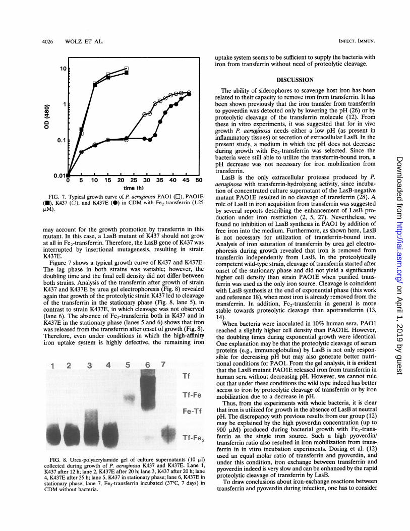
time (h)FIG. 7. Typical growth curve of P. aeruginosa PAO1 (L), PAO1E
(U), K437 (0), and K437E (0) in CDM with Fe2-transferrin (1.25AIM).
may account for the growth promotion by transferrin in thismutant. In this case, a LasB mutant of K437 should not growat all in Fe2-transferrin. Therefore, the LasB gene of K437 was
interrupted by insertional mutagenesis, resulting in strainK437E.
Figure 7 shows a typical growth curve of K437 and K437E.The lag phase in both strains was variable; however, thedoubling time and the final cell density did not differ betweenboth strains. Analysis of the transferrin after growth of strainK437 and K437E by urea gel electrophoresis (Fig. 8) revealedagain that growth of the proteolytic strain K437 led to cleavageof the transferrin in the stationary phase (Fig. 8, lane 5), incontrast to strain K437E, in which cleavage was not observed(lane 6). The absence of Fe2-transferrin both in K437 and inK437E in the stationary phase (lanes 5 and 6) shows that ironwas released from the transferrin after onset of growth (Fig. 8).Therefore, even under conditions in which the high-affinityiron uptake system is highly defective, the remaining iron
1 2 3 4 5 7
Su
FIG. 8. Urea-polyacrylamide gel of culture supernatants (10 ,ul)collected during growth of P. aeruginosa K437 and K437E. Lane 1,K437 after 12 h; lane 2, K437E after 20 h; lane 3, K437 after 20 h; lane4, K437E after 35 h; lane 5, K437 in stationary phase; lane 6, K437E instationary phase; lane 7, Fe2-transferrin incubated (37°C, 7 days) inCDM without bacteria.
uptake system seems to be sufficient to supply the bacteria withiron from transferrin without need of proteolytic cleavage.
DISCUSSION
The ability of siderophores to scavenge host iron has beenrelated to their capacity to remove iron from transferrin. It hasbeen shown previously that the iron transfer from transferrinto pyoverdin was detected only by lowering the pH (26) or byproteolytic cleavage of the transferrin molecule (12). Fromthese in vitro experiments, it was suggested that for in vivogrowth P. aeruginosa needs either a low pH (as present ininflammatory tissues) or secretion of extracellular LasB. In thepresent study, a medium in which the pH does not decreaseduring growth with Fe2-transferrin was selected. Since thebacteria were still able to utilize the transferrin-bound iron, apH decrease was not necessary for iron mobilization fromtransferrin.LasB is the only extracellular protease produced by P.
aeruginosa with transferrin-hydrolyzing activity, since incuba-tion of concentrated culture supernatant of the LasB-negativemutant PA01E resulted in no cleavage of transferrin (28). Arole of LasB in iron acquisition from transferrin was suggestedby several reports describing the enhancement of LasB pro-duction under iron restriction (2, 5, 27). Nevertheless, wefound no inhibition of LasB synthesis in PAO1 by addition offree iron into the medium. Furthermore, as shown here, LasBis not necessary for utilization of transferrin-bound iron.Analysis of iron saturation of transferrin by urea gel electro-phoresis during growth revealed that iron is removed fromtransferrin independently from LasB. In the proteolyticallycompetent wild-type strain, cleavage of transferrin started afteronset of the stationary phase and did not yield a significantlyhigher cell density than strain PAO1E when purified trans-ferrin was used as the only iron source. Cleavage is coincidentwith LasB synthesis at the end of exponential phase (this workand reference 18), when most iron is already removed from thetransferrin. In addition, Fe2-transferrin in general is morestable towards proteolytic cleavage than apotransferrin (13,14).When bacteria were inoculated in 10% human sera, PAO1
reached a slightly higher cell density than PAO1E. However,the doubling times during exponential growth were identical.One explanation may be that the proteolytic cleavage of serumproteins (e.g., immunoglobulins) by LasB is not only respon-sible for decreasing pH but may also generate better nutri-tional conditions for PAO1. From the gel analysis, it is evidentthat the LasB mutant PAO1E released iron from transferrin inhuman sera without decreasing pH. However, we cannot ruleout that under these conditions the wild type indeed has betteraccess to iron by proteolytic cleavage of transferrin or by ironmobilization due to a decrease in pH.
Thus, from the experiments with whole bacteria, it is clearthat iron is utilized for growth in the absence of LasB at neutralpH. The discrepancy with previous results from our group (12)may be explained by the high pyoverdin concentration (up to900 ,uM) produced during bacterial growth with Fe2-trans-ferrin as the single iron source. Such a high pyoverdin/transferrin ratio also resulted in iron mobilization from trans-ferrin in in vitro incubation experiments. Doring et al. (12)used an equal molar ratio of transferrin and pyoverdin, andunder this condition, iron exchange between transferrin andpyoverdin indeed is very slow and can be enhanced by the rapidproteolytic cleavage of transferrin by LasB.To draw conclusions about iron-exchange reactions between
transferrin and pyoverdin during infection, one has to consider
INFECr. IMMUN.
on April 1, 2019 by guest
http://iai.asm.org/
Dow
nloaded from

P. AERUGINOSA IRON ACQUISITION 4027
concentrations of the reactants. However, little is known aboutthe concentrations of pyoverdin and LasB produced duringinfections. Although pyoverdin (17) and LasB transcription(27) were detected in sputa from patients with cystic fibrosisinfected with P. aeruginosa, quantitative measurement wasperformed only for LasB. Depending on the strain used,between 2.4 and 24 puM LasB was detected in an animal model(10). Detection of LasB in sputa of P. aeruginosa-infectedcystic fibrosis patients was successful only in the absence ofspecific antibodies (11). However, other transferrin-hydrolyz-ing enzymes derived from the host or other microorganismsmay be present during infection. Under conditions of highproteolytic activity combined with low siderophore activity, thecleavage of the transferrin molecule may have a more pro-found effect on iron acquisition for bacteria. Therefore, weused a P. aeruginosa strain (K437) with a highly defective ironuptake system (25) to investigate whether under these condi-tions the cleavage of LasB plays a role in iron acquisition. Thisstrain showed diminished growth compared with PAO1. Re-gardless, the strain was still able to utilize iron bound totransferrin even when lasB was interrupted by insertionalmutagenesis. Urea gel analysis again showed that in theproteolytically competent strain the transferrin is cleaved andin the LasB mutant the iron is released from the transferrin.Onset of growth in both strains was accompanied by pyoverdinsynthesis. This was probably due to a regulatory mechanismand not to spontaneous mutation since subculturing of coloniesgrown with Fe2-transferrin as the only iron source showed thesame long lag phases for pyoverdin synthesis and growth.Although the genetic background of this regulatory processremains unclear, we conclude that a strain defective in ironuptake as well as in LasB synthesis was able to chelate ironfrom transferrin. Therefore, the iron uptake system of P.aeruginosa may also be sufficient during infection to supplybacteria with this essential ion.
ACKNOWLEDGMENTS
This study was supported by a grant (Do 249/6) from the DeutscheForschungsgemeinschaft to K.H.We thank the Chemical Laboratory of the Hygiene Institute,
University of Tubingen, for determination of iron by atom absorptionspectroscopy.
REFERENCES1. Ankenbauer, R., S. Sriyosachati, and C. D. Cox. 1985. Effects of
siderophores on growth of Pseudomonas aeruginosa in humanserum and transferrin. Infect. Immun. 49:132-140.
2. Bjorn, M. J., P. A. Sokol, and B. H. Iglewski. 1979. Influence ofiron on extracellular products in Pseudomonas aeruginosa cultures.J. Bacteriol. 138:193-200.
3. Blanc-Parasote, S. 1989. Etude physico-chimique d'ionophoresnaturels: antibiotiques carboxyliques et siderophores bacteriens.Doctoral thesis. l'Universite Louis Pasteur, Strasbourg, France.
4. Blum, H., H. Beier, and H. J. Gross. 1987. Improved silver stainingof plant proteins, RNA and DNA in polyacrylamide gels. Electro-phoresis 8:93-99.
5. Brumlik, M. J., and D. G. Storey. 1992. Zinc and iron regulatetranslation of the gene encoding Pseudomonas aeruginosa elastase.Mol. Microbiol. 6:337-344.
6. Cox, D. C., and P. Adams. 1985. Siderophore activity of pyoverdinfor Pseudomonas aeruginosa. Infect. Immun. 48:130-138.
7. Cox, D. C., and R. Graham. 1979. Isolation of an iron-bindingcompound from Pseudomonas aeruginosa. J. Bacteriol. 137:357-364.
8. Demange, P., S. Wendenbaum, A. Bateman, A. Dell, and M. A.Abdallah. 1987. Bacterial siderophores: structure and physio-chemical properties of pyoverdins and related compounds, p.
167-187. In G. Winkelmann, D. Van der Helm, and J. B. Neilands(ed.), Iron transport in microbes, plants and animals. VCHVerlagsgesellschaft, Weilheim, Germany.
9. Demange, P., S. Wendenbaum, C. Linget, C. Mertz, M. T. Cung, A.Dell, and M. A. Abdallah. 1990. Bacterial siderophores: structureand NMR assignment of pyoverdins Pa, siderophores of Pseudo-monas aeruginosa ATCC 15692. Biol. Metals 3:155-170.
10. Doring, G., A. Dalhof, 0. Vogel, H. Brunner, U. Droge, and K.Botzenhart. 1984. In vivo activity of proteases of Pseudomonasaeruginosa in a rat model. J. Infect. Dis. 149:532-537.
11. Doring, G., H.-J. Obernesser, K. Botzenhart, B. Flehmig, N.H0iby, and A. J. Hofnann. 1983. Proteases of Pseudomonasaeruginosa in cystic fibrosis. J. Infect. Dis. 147:744-750.
12. Doring, G., M. Pfestorf, K. Botzenhart, and M. A. Abdallah. 1988.Impact of proteases on iron uptake of Pseudomonas aeruginosapyoverdin from transferrin and lactoferrin. Infect. Immun. 56:291-293.
13. Esparaza, I., and J. H. Brock. 1980. The effect of trypsin digestionon the structure and iron-donation properties of transferrins fromseveral species. Biochim. Biophys. Acta 622:297-307.
14. Evans, R., and J. Williams. 1978. Studies of the binding ofdifferent iron donors to human serum transferrin and isolation ofiron-binding fragments from the N- and C-terminal regions of theprotein. Biochem. J. 173:543-552.
15. Frieden, E., and P. Aisen. 1980. Forms of iron transferrin. TrendsBiochem. Sci. 5:xi.
16. Griifiths, E. 1987. The iron-uptake systems of pathogenic bacteria,p. 69-137. In J. J. Bullen and E. Griffiths (ed.), Iron and infection.John Wiley and Sons Ltd., Chichester, United Kingdom.
17. Haas, B., J. Kraut, J. Marks, S. C. Zanker, and D. Castignetti.1991. Siderophore presence in sputa of cystic fibrosis patients.Infect. Immun. 59:3997-4000.
18. Iglewski, B. H., L Rust, and R. A. Bever. 1990. Molecular analysisof Pseudomonas aeruginosa elastase, p. 36-43. In S. Silver, A. M.Chakrabarty, B. Iglewski, and S. Kaplan (ed.), Pseudomonas:biotransformations, pathogenesis, and evolving biotechnology.American Society for Microbiology, Washington, D.C.
19. Jensen, S. E., L. T. Fecycz, and J. N. Campbell. 1980. Nutritionalfactors controlling exocellular protease production by Pseudomo-nas aeruginosa. J. Bacteriol. 144:844-847.
20. Laemmli, U. K. 1970. Cleavage of structural proteins during theassembly of the head of bacteriophage T4. Nature (London)227:680-685.
21. Makey, D. G., and U. S. Seal. 1976. The detection of fourmolecular forms of human transferrin during the iron bindingprocess. Biochim. Biophys. Acta 453:250-256.
22. Meyer, J. M., and M. A. Abdallah. 1978. The fluorescent pigmentof Pseudomonas fluorescens: biosynthesis, purification and physi-cochemical properties. J. Gen. Microbiol. 107:319-328.
23. Neuhoff, V., R Stamm, and J. Eibl. 1985. Clear background andhighly sensitive protein staining with Coomassie blue dyes inpolyacrylamide gels: a systematic analysis. Electrophoresis 6:427-448.
24. Obernesser, H. J., and G. Doring. 1982. Extracellular toxins ofPseudomonas aeruginosa. IV. Radioimmunoassay for detection ofelastase. Zentralbl. Bakteriol. Mikrobiol. Hyg. (A) 252:248-256.
25. Poole, K., S. Neshat, and D. Heinrichs. 1990. Pyoverdine-mediatediron transport in Pseudomonas aeruginosa: involvement of a high-molecular-mass outer membrane protein. FEMS Microbiol. Lett.78:1-6.
26. Sriyosachati, S., and C. D. Cox. 1986. Siderophore-mediated ironacquisition from transferrin by Pseudomonas aeruginosa. Infect.Immun. 52:885-891.
27. Storey, D. G., E. E. Ujack, and H. R Rabin. 1992. Populationtranscript accumulation of Pseudomonas aeruginosa exotoxin Aand elastase in sputa from patients with cystic fibrosis. Infect.Immun. 60:4687-4694.
28. Wolz, C., E. Hellstern, M. Haug, D. R Galloway, M. L. Vasil, andG. Doring. 1991. Pseudomonas aeruginosa LasB mutant con-structed by insertional mutagenesis reveals elastolytic activity dueto alkaline proteinase and the LasA fragment. Mol. Microbiol.5:2125-2131.
VOL. 62, 1994
on April 1, 2019 by guest
http://iai.asm.org/
Dow
nloaded from