Embed Size (px)
Citation preview
© 2014 Dental Press Journal of Orthodontics Dental Press J Orthod. 2014 Sept-Oct;19(5):136-49136
special article
Is there a consensus for CBCT use in Orthodontics?
Daniela G. Garib1, Louise Resti Calil2, Claudia Resende Leal3, Guilherme Janson4
How to cite this article: Garib DG, Calil LR, Leal CR, Janson G. Is there a consensus for CBCT use in Orthodontics? Dental Press J Orthod. 2014 Sept-Oct;19(5):136-49. DOI: http://dx.doi.org/10.1590/2176-9451.19.5.136-149.sar
Submitted: August 11, 2014 - Revised and accepted: August 28, 2014
» Patients displayed in this article previously approved the use of their facial and intraoral photographs.
Contact address: Daniela G. GaribAlameda Octávio Pinheiro Brisola, 9-75Bauru, São Paulo - Brazil — CEP 17.012-901E-mail: [email protected]
» The author reports no commercial, proprietary or financial interest in the prod-ucts or companies described in this article.
1 Associate professor of Orthodontics at the Hospital for Rehabilitation of Craniofacial Anomalies/USP and School of Dentistry — University of São Paulo/Bauru.
2 Masters student in Orthodontics, School of Dentistry — University of São Paulo/Bauru.
3 Masters student in Science of Rehabilitation, Hospital for Rehabilitation of Craniofacial Anomalies/USP.
4 Full professor, Department of Orthodontics, School of Dentistry — University of São Paulo/Bauru.
This article aims to discuss current evidence and recommendations for cone-beam computed tomography (CBCT) in Orthodontics. In comparison to conventional radiograph, CBCT has higher radiation doses and, for this reason, is not a standard method of diagnosis in Orthodontics. Routine use of CBCT in substitution to conventional ra-diograph is considered an unaccepted practice. CBCT should be indicated with criteria only after clinical examina-tion has been performed and when the benefits for diagnosis and treatment planning exceed the risks of a greater radiation dose. It should be requested only when there is a potential to provide new information not demonstrated by conventional scans, when it modifies treatment plan or favors treatment execution. The most frequent indica-tion of CBCT in Orthodontics, with some evidence on its clinical efficacy, includes retained/impacted permanent teeth; severe craniofacial anomalies; severe facial discrepancies with indication of orthodontic-surgical treatment; and bone irregularities or malformation of TMJ accompanied by signs and symptoms. In exceptional cases of adult patients when critical tooth movement are planned in regions with deficient buccolingual thickness of the alveolar ridge, CBCT can be indicated provided that there is a perspective of changes in orthodontic treatment planning.
Keywords: Orthodontics. Cone-beam computed tomography. Recommendations.
DOI: http://dx.doi.org/10.1590/2176-9451.19.5.136-149.sar
O presente artigo visa discutir as evidências e recomendações atuais concernentes à indicação da tomografia com-putadorizada de feixe cônico (TCFC) em Ortodontia. Devido à dose de radiação mais elevada em relação às ra-diografias, a TCFC não é o método padrão de diagnóstico em Ortodontia. O seu uso rotineiro, em substituição à documentação convencional, é considerado uma prática inaceitável. A TCFC deve ser indicada com muito critério, e somente após uma análise clínica, quando os benefícios para o diagnóstico e tratamento superarem os riscos de uma dose mais elevada de radiação. Deve ser requisitada estritamente quando houver um potencial de prover novas informações não demonstradas em exames radiográficos convencionais, modificando o plano de tratamento ou fa-cilitando a sua execução. As indicações mais frequentes em Ortodontia, que demonstram algum nível de evidência sobre sua eficácia clínica, podem ser resumidas em casos de dentes permanentes retidos; anomalias craniofaciais complexas; discrepâncias faciais severas com indicação de tratamento ortodôntico-cirúrgico; e malformações ou irregularidades ósseas na ATM acompanhadas de sinais e sintomas. Em casos excepcionais, em pacientes adultos em que se planeja movimentos dentários críticos em áreas com espessura óssea vestibulolingual deficiente, a TCFC pode ser indicada, desde que se vislumbre uma perspectiva de alteração no plano de tratamento ortodôntico.
Palavras-chave: Ortodontia. Tomografia Computadorizada de Feixe Cônico. Recomendações.
© 2014 Dental Press Journal of Orthodontics Dental Press J Orthod. 2014 Sept-Oct;19(5):136-49137
Garib DG, Calil LR, Leal CR, Janson G special article
INTRODUCTIONWe have currently been through modern times in
Orthodontics. In a retrospective view of our science and art, we envisage a Classical era from the end of the XIX century until the 60s with the legacy of Ed-ward Hartley Angle and his eminent pupils, includ-ing Charles Tweed, Broadbent and Brodie.49,50 After the Classical era, a Contemporary era started in the 70s not only with the development of specific occlu-sal objectives and the Straigth-Wire appliance by An-drews, but also with the development of orthognatic surgery and facial analysis for orthodontic diagno-sis.51,52 When we look to the present, we see our time being highlighted by two major vanguard advents: tridimensional images and skeletal anchorage.
Cone-beam computed tomography (CBCT) to-gether with digital dental models and 3D facial pho-tographs personify the modernity of the present. In-troduced in 1998,39 CBCT is in its adolescence, but has contributed with over seven hundred international publications in Orthodontics, according to a search at Pubmed database. Evidence related to CBCT have provided important development in three levels: orth-odontic diagnosis; orthodontic or orthodontic-surgical treatment planning; and knowledge of treatment out-comes. It is not difficult to fall in love for CBCT scans, once they allow three-dimensional visualization of the morphology of the face and cranium, and demonstrate one’s anatomy in multiplanar sections with adequate resolution and sharpness.21 CBCT presents high accu-racy and precision, sensibility and specificity, as well as absence of image amplification.6,7,9,11,17,27,28,33-36,38,41,43 Faced with these advantages, the following question recurrently arises: Can CBCT be indicated as a rou-tine in Orthodontics?
As every light has its shadows, a method does not have advantages, only. CBCT has the drawback of having a higher radiation dose compared to conven-tional radiograph frequently requested in Orthodon-tics.3,45 Effective radiation dose is the sum of the dose received by all irradiated tissues and organs, consid-ering both tissue weight and the quality of ionizing radiation in terms of biological effects.15 Effective radiation dose represents a stochastic risk to health, in other words, the probability of carcinogenesis and genetic effects on irradiated tissues.15 During X-ray examination, millions of photons pass through
patient’s cells and can cause damage to DNA mol-ecules due to ionization.15 The majority of changes caused to genetic material is reversible and immedi-ately repaired.15 However, DNA may be rarely, yet permanently altered, thereby establishing a genetic mutation.15 Fortunately, effective dose and risks re-lated to dental radiation are very small compared to the natural risks of carcinogenesis.15,16 Nevertheless, some limited evidence on the increase of radiation-related tumor in the brain and thyroid glands requires caution and rationality before indicating X-ray ex-amination in Dentistry, including conventional ra-diographs.15 This concern is amplified in children, as they present tissues with higher radiosensitivity, greater number of cell divisions and a longer lifetime spam for carcinogenesis development.16
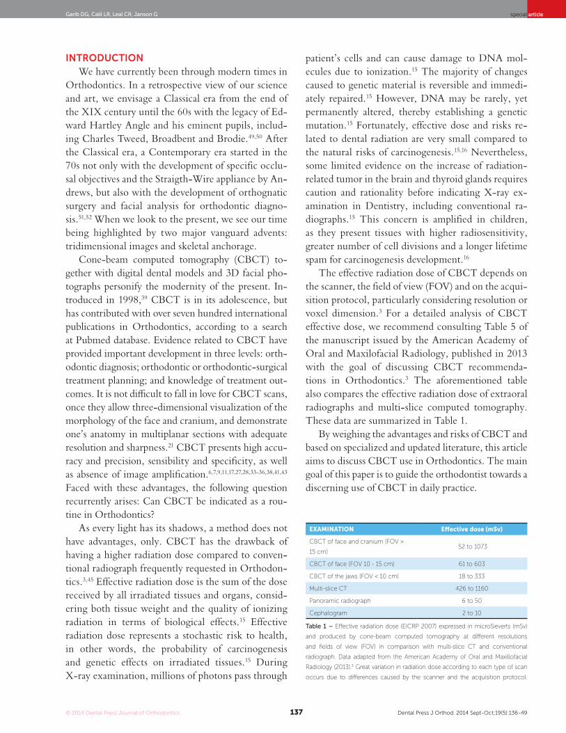
The effective radiation dose of CBCT depends on the scanner, the field of view (FOV) and on the acqui-sition protocol, particularly considering resolution or voxel dimension.3 For a detailed analysis of CBCT effective dose, we recommend consulting Table 5 of the manuscript issued by the American Academy of Oral and Maxilofacial Radiology, published in 2013 with the goal of discussing CBCT recommenda-tions in Orthodontics.3 The aforementioned table also compares the effective radiation dose of extraoral radiographs and multi-slice computed tomography. These data are summarized in Table 1.
By weighing the advantages and risks of CBCT and based on specialized and updated literature, this article aims to discuss CBCT use in Orthodontics. The main goal of this paper is to guide the orthodontist towards a discerning use of CBCT in daily practice.
Table 1 – Effective radiation dose (EICRP 2007) expressed in microSieverts (mSv)
and produced by cone-beam computed tomography at different resolutions
and fields of view (FOV) in comparison with multi-slice CT and conventional
radiograph. Data adapted from the American Academy of Oral and Maxillofacial
Radiology (2013).3 Great variation in radiation dose according to each type of scan
occurs due to differences caused by the scanner and the acquisition protocol.
EXAMINATION Effective dose (mSv)
CBCT of face and cranium (FOV >
15 cm) 52 to 1073
CBCT of face (FOV 10 - 15 cm) 61 to 603
CBCT of the jaws (FOV < 10 cm) 18 to 333
Multi-slice CT 426 to 1160
Panoramic radiograph 6 to 50
Cephalogram 2 to 10
© 2014 Dental Press Journal of Orthodontics Dental Press J Orthod. 2014 Sept-Oct;19(5):136-49138
Is there a consensus for CBCT use in Orthodontics?special article
possibility to simulate and demonstrate the therapy of choice to patients; and last but not least, the evi-dence that CBCT radiation dose is minimal in com-parison to the sum of radiation doses of panoramic radiograph, cephalometric radiograph and the full set of periapical radiographs.29
Opposing to the general use of CBCT in Ortho-dontics, it was mentioned that criteria for patients selection should be based on the ratio risk-benefit of CBCT; and that there was not enough evidence supporting CBCT efficacy for diagnosis, treatment planning or treatment outcomes in Corrective Or-thodontics.23 We invite readers to advance in the arguments raised by Dr. Halazonetis23 by carefully examining the following topics of this article.
WEIGHING RISKS AND BENEFITSThere seems to be an antithesis between what the
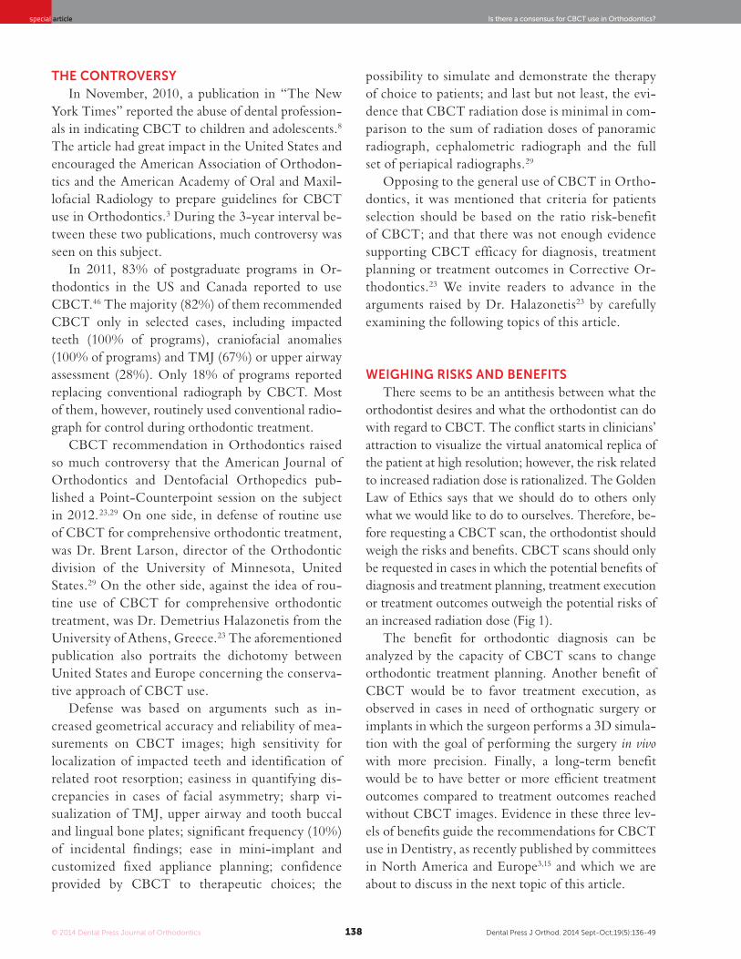
orthodontist desires and what the orthodontist can do with regard to CBCT. The conflict starts in clinicians’ attraction to visualize the virtual anatomical replica of the patient at high resolution; however, the risk related to increased radiation dose is rationalized. The Golden Law of Ethics says that we should do to others only what we would like to do to ourselves. Therefore, be-fore requesting a CBCT scan, the orthodontist should weigh the risks and benefits. CBCT scans should only be requested in cases in which the potential benefits of diagnosis and treatment planning, treatment execution or treatment outcomes outweigh the potential risks of an increased radiation dose (Fig 1).
The benefit for orthodontic diagnosis can be analyzed by the capacity of CBCT scans to change orthodontic treatment planning. Another benefit of CBCT would be to favor treatment execution, as observed in cases in need of orthognatic surgery or implants in which the surgeon performs a 3D simula-tion with the goal of performing the surgery in vivo with more precision. Finally, a long-term benefit would be to have better or more efficient treatment outcomes compared to treatment outcomes reached without CBCT images. Evidence in these three lev-els of benefits guide the recommendations for CBCT use in Dentistry, as recently published by committees in North America and Europe3,15 and which we are about to discuss in the next topic of this article.
THE CONTROVERSYIn November, 2010, a publication in “The New
York Times” reported the abuse of dental profession-als in indicating CBCT to children and adolescents.8 The article had great impact in the United States and encouraged the American Association of Orthodon-tics and the American Academy of Oral and Maxil-lofacial Radiology to prepare guidelines for CBCT use in Orthodontics.3 During the 3-year interval be-tween these two publications, much controversy was seen on this subject.
In 2011, 83% of postgraduate programs in Or-thodontics in the US and Canada reported to use CBCT.46 The majority (82%) of them recommended CBCT only in selected cases, including impacted teeth (100% of programs), craniofacial anomalies (100% of programs) and TMJ (67%) or upper airway assessment (28%). Only 18% of programs reported replacing conventional radiograph by CBCT. Most of them, however, routinely used conventional radio-graph for control during orthodontic treatment.
CBCT recommendation in Orthodontics raised so much controversy that the American Journal of Orthodontics and Dentofacial Orthopedics pub-lished a Point-Counterpoint session on the subject in 2012.23,29 On one side, in defense of routine use of CBCT for comprehensive orthodontic treatment, was Dr. Brent Larson, director of the Orthodontic division of the University of Minnesota, United States.29 On the other side, against the idea of rou-tine use of CBCT for comprehensive orthodontic treatment, was Dr. Demetrius Halazonetis from the University of Athens, Greece.23 The aforementioned publication also portraits the dichotomy between United States and Europe concerning the conserva-tive approach of CBCT use.
Defense was based on arguments such as in-creased geometrical accuracy and reliability of mea-surements on CBCT images; high sensitivity for localization of impacted teeth and identification of related root resorption; easiness in quantifying dis-crepancies in cases of facial asymmetry; sharp vi-sualization of TMJ, upper airway and tooth buccal and lingual bone plates; significant frequency (10%) of incidental findings; ease in mini-implant and customized fixed appliance planning; confidence provided by CBCT to therapeutic choices; the
© 2014 Dental Press Journal of Orthodontics Dental Press J Orthod. 2014 Sept-Oct;19(5):136-49139
Garib DG, Calil LR, Leal CR, Janson G special article
Figure 1 - Cone-beam computed tomography should only be requested in cases in which the potential benefits of diagnosis and treatment planning, treatment execution or treatment outcomes outweigh the potential risks of an increased radiation dose.
BASIC PRINCIPLES FOR CBCT RECOMMENDATION
According to the American Academy of Oral and Maxillofacial Radiology, there is neither con-vincing evidence for radiation-induced carcinogen-esis at the level of dental exposure, nor absence of evidence of such effect. Because Orthodontics is a field of health, we prudently assume there is a risk, given that there is no safe limit for ionizing radia-tion.3 Each exposure has a cumulative effect on the risk of carcinogenesis.3 In this perspective, the basic principles recommended by European and North-American guidelines aim to avoid or minimize un-necessary exposure for diagnosis purposes.
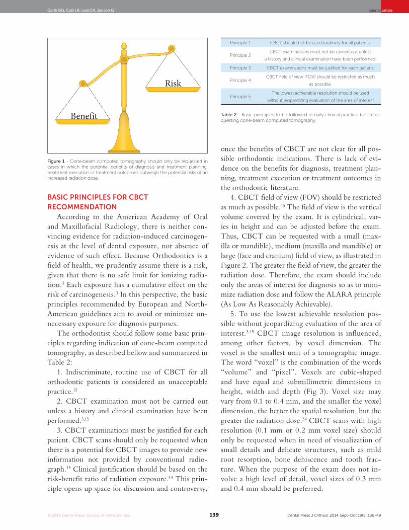
The orthodontist should follow some basic prin-ciples regarding indication of cone-beam computed tomography, as described bellow and summarized in Table 2:
1. Indiscriminate, routine use of CBCT for all orthodontic patients is considered an unacceptable practice.15
2. CBCT examination must not be carried out unless a history and clinical examination have been performed.3,15
3. CBCT examinations must be justified for each patient. CBCT scans should only be requested when there is a potential for CBCT images to provide new information not provided by conventional radio-graph.15 Clinical justification should be based on the risk-benefit ratio of radiation exposure.44 This prin-ciple opens up space for discussion and controversy,
once the benefits of CBCT are not clear for all pos-sible orthodontic indications. There is lack of evi-dence on the benefits for diagnosis, treatment plan-ning, treatment execution or treatment outcomes in the orthodontic literature.
4. CBCT field of view (FOV) should be restricted as much as possible.15 The field of view is the vertical volume covered by the exam. It is cylindrical, var-ies in height and can be adjusted before the exam. Thus, CBCT can be requested with a small (max-illa or mandible), medium (maxilla and mandible) or large (face and cranium) field of view, as illustrated in Figure 2. The greater the field of view, the greater the radiation dose. Therefore, the exam should include only the areas of interest for diagnosis so as to mini-mize radiation dose and follow the ALARA principle (As Low As Reasonably Achievable).
5. To use the lowest achievable resolution pos-sible without jeopardizing evaluation of the area of interest.3,15 CBCT image resolution is influenced, among other factors, by voxel dimension. The voxel is the smallest unit of a tomographic image. The word “voxel” is the combination of the words “volume” and “pixel”. Voxels are cubic-shaped and have equal and submillimetric dimensions in height, width and depth (Fig 3). Voxel size may vary from 0.1 to 0.4 mm, and the smaller the voxel dimension, the better the spatial resolution, but the greater the radiation dose.34 CBCT scans with high resolution (0.1 mm or 0.2 mm voxel size) should only be requested when in need of visualization of small details and delicate structures, such as mild root resorption, bone dehiscence and tooth frac-ture. When the purpose of the exam does not in-volve a high level of detail, voxel sizes of 0.3 mm and 0.4 mm should be preferred.
Table 2 - Basic principles to be followed in daily clinical practice before re-questing cone-beam computed tomography.
Principle 1 CBCT should not be used routinely for all patients.
Principle 2CBCT examinations must not be carried out unless
a history and clinical examination have been performed.
Principle 3 CBCT examinations must be justified for each patient.
Principle 4CBCT field of view (FOV) should be restricted as much
as possible.
Principle 5The lowest achievable resolution should be used
without jeopardizing evaluation of the area of interest.
Benefit
Risk
© 2014 Dental Press Journal of Orthodontics Dental Press J Orthod. 2014 Sept-Oct;19(5):136-49140
Is there a consensus for CBCT use in Orthodontics?special article
Figure 2 - Cone-beam computed tomography field of view: It is cylindrical and determined ac-cording to its vertical extent. A) Field of view of the face and cranium; B) Field of view of the face; C) Field of view of the jaws: D) Field of view of the maxilla; E) Field of view of the mandible.
Figure 3 - The voxel is cubic-shaped and is the smallest unit of a tomo-graphic image. In CBCT, voxels have equal and submillimetric dimensions in height, width and depth.
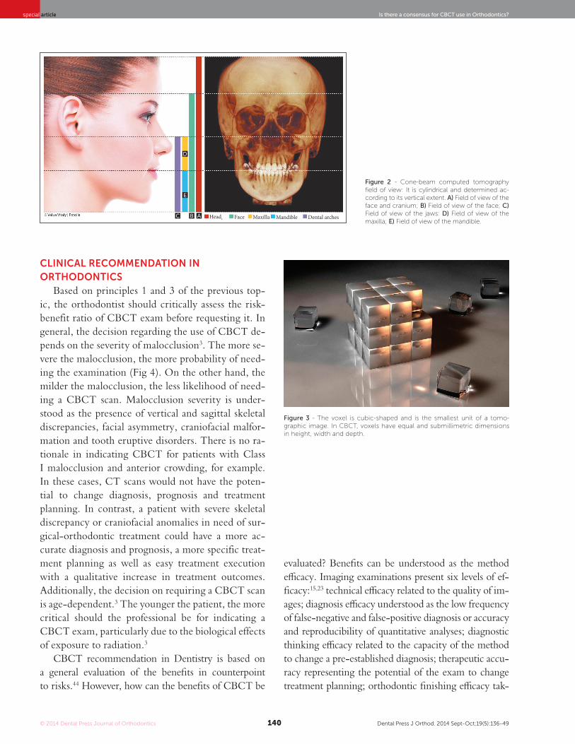
CLINICAL RECOMMENDATION IN ORTHODONTICS
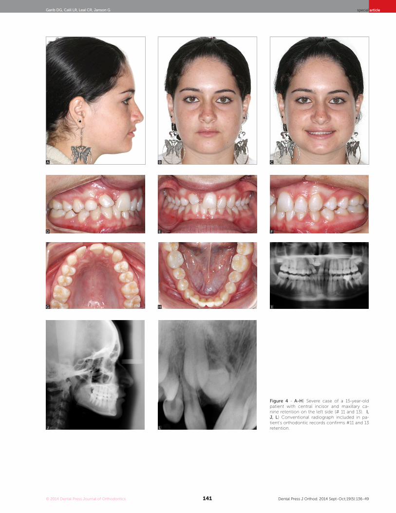
Based on principles 1 and 3 of the previous top-ic, the orthodontist should critically assess the risk-benefit ratio of CBCT exam before requesting it. In general, the decision regarding the use of CBCT de-pends on the severity of malocclusion3. The more se-vere the malocclusion, the more probability of need-ing the examination (Fig 4). On the other hand, the milder the malocclusion, the less likelihood of need-ing a CBCT scan. Malocclusion severity is under-stood as the presence of vertical and sagittal skeletal discrepancies, facial asymmetry, craniofacial malfor-mation and tooth eruptive disorders. There is no ra-tionale in indicating CBCT for patients with Class I malocclusion and anterior crowding, for example. In these cases, CT scans would not have the poten-tial to change diagnosis, prognosis and treatment planning. In contrast, a patient with severe skeletal discrepancy or craniofacial anomalies in need of sur-gical-orthodontic treatment could have a more ac-curate diagnosis and prognosis, a more specific treat-ment planning as well as easy treatment execution with a qualitative increase in treatment outcomes. Additionally, the decision on requiring a CBCT scan is age-dependent.3 The younger the patient, the more critical should the professional be for indicating a CBCT exam, particularly due to the biological effects of exposure to radiation.3
CBCT recommendation in Dentistry is based on a general evaluation of the benefits in counterpoint to risks.44 However, how can the benefits of CBCT be
evaluated? Benefits can be understood as the method efficacy. Imaging examinations present six levels of ef-ficacy:15,23 technical efficacy related to the quality of im-ages; diagnosis efficacy understood as the low frequency of false-negative and false-positive diagnosis or accuracy and reproducibility of quantitative analyses; diagnostic thinking efficacy related to the capacity of the method to change a pre-established diagnosis; therapeutic accu-racy representing the potential of the exam to change treatment planning; orthodontic finishing efficacy tak-
ABC
E
D
Head Face Maxilla Mandible Dental arches
© 2014 Dental Press Journal of Orthodontics Dental Press J Orthod. 2014 Sept-Oct;19(5):136-49141
Garib DG, Calil LR, Leal CR, Janson G special article
Figure 4 - A-H) Severe case of a 15-year-old patient with central incisor and maxillary ca-nine retention on the left side (# 11 and 13). I, J, L) Conventional radiograph included in pa-tient’s orthodontic records confirms #11 and 13 retention.
A
D
G I
B
E
H
J L
C
F
© 2014 Dental Press Journal of Orthodontics Dental Press J Orthod. 2014 Sept-Oct;19(5):136-49142
Is there a consensus for CBCT use in Orthodontics?special article
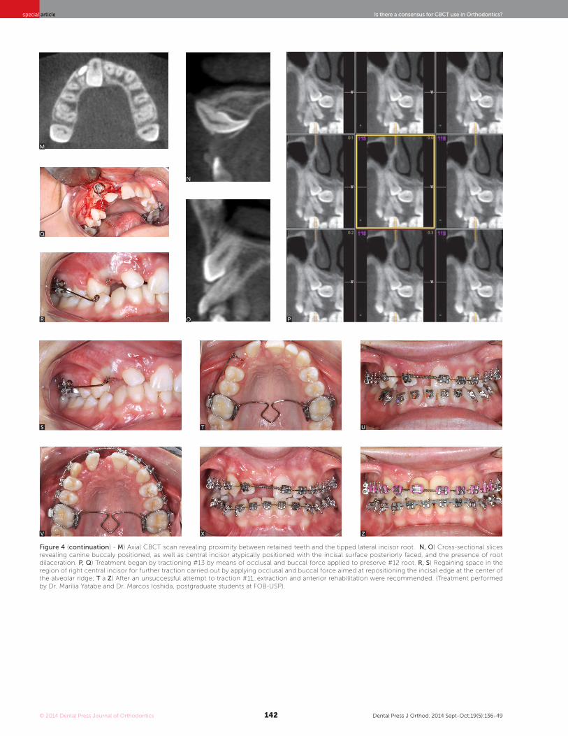
Figure 4 (continuation) - M) Axial CBCT scan revealing proximity between retained teeth and the tipped lateral incisor root. N, O) Cross-sectional slices revealing canine buccaly positioned, as well as central incisor atypically positioned with the incisal surface posteriorly faced, and the presence of root dilaceration. P, Q) Treatment began by tractioning #13 by means of occlusal and buccal force applied to preserve #12 root. R, S) Regaining space in the region of right central incisor for further traction carried out by applying occlusal and buccal force aimed at repositioning the incisal edge at the center of the alveolar ridge; T a Z) After an unsuccessful attempt to traction #11, extraction and anterior rehabilitation were recommended. (Treatment performed by Dr. Marilia Yatabe and Dr. Marcos Ioshida, postgraduate students at FOB-USP).
M
N
Q
T
X
P
S
V
OR
U
Z
© 2014 Dental Press Journal of Orthodontics Dental Press J Orthod. 2014 Sept-Oct;19(5):136-49143
Garib DG, Calil LR, Leal CR, Janson G special article
ing into account the qualitative gain of treatment re-sults; and, finally, the societal efficacy.23
In Orthodontics, there is few evidence on the CBCT potential to change the quality of treatment outcomes and no evidence of CBCT social benefits.23 Current evidence of efficacy for the other four lev-els have guided the North-American and European recommendations for CBCT use. In other words, evidence of efficacy guided the eligibility criteria of cases that justify the use of CBCT.
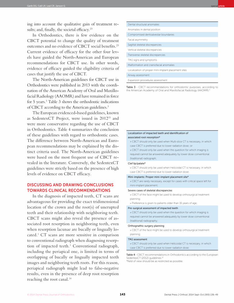
The North-American guidelines for CBCT use in Orthodontics were published in 2013 with the coordi-nation of the American Academy of Oral and Maxillo-facial Radiology (AAOMR) and have remained in force for 5 years.3 Table 3 shows the orthodontic indications of CBCT according to the American guidelines.3
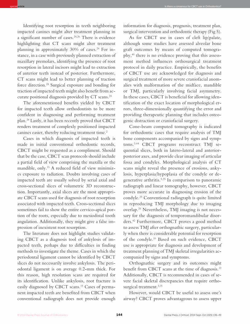
The European evidenced-based guidelines, known as SedentexCT Project, were issued in 201215 and were more conservative regarding the use of CBCT in Orthodontics. Table 4 summarizes the conclusion of these guidelines with regard to orthodontic cases. The difference between North-American and Euro-pean recommendations may be explained by the dis-tinct criteria used. The North-American guidelines were based on the most frequent use of CBCT re-vealed in the literature. Conversely, the SedentexCT guidelines were strictly based on the presence of high levels of evidence on CBCT efficacy.
DISCUSSING AND DRAWING CONCLUSIONS TOWARDS CLINICAL RECOMMENDATIONS
In the diagnosis of impacted teeth, CT scans are advantageous for providing the exact tridimensional location of the crown and the root(s) of unerupted teeth and their relationship with neighboring teeth. CBCT scans might also reveal the presence of as-sociated root resorption in neighboring teeth, even when resorption lacunae are bucally or lingually lo-cated.1 CT scans are more sensitive in comparison to conventional radiograph when diagnosing resorp-tion of impacted teeth.1 Conventional radiograph, including the periapical one, is limited in terms of overlapping of bucally or lingually impacted teeth images and neighboring teeth roots. For this reason, periapical radiograph might lead to false-negative results, even in the presence of deep root resorption reaching the root canal.14
Table 3 - CBCT recommendations for orthodontic purposes, according to the American Academy of Oral and Maxillofacial Radiology (AAOMR).3
Dental structural anomalies
Anomalies in dental position
Compromised dentoalveolar boundaries
Facial asymmetry
Sagittal skeletal discrepancies
Vertical skeletal discrepancies
Transverse skeletal discrepancies
TMJ signs and symptoms
Malformation and craniofacial anomalies
Localization of proper mini-implant placement sites
Airway assessment
Expansion procedures assessment
Table 4 - CBCT recommendations in Orthodontics according to the European SedentexCT (2012) guidelines.15 *field of view should be as restricted as possible.
Localization of impacted teeth and identification of
associated root resorption*
» CBCT should only be used when Multi-slice CT is necessary, in which
case CBCT is preferred due to lower radiation dose; or
» CBCT should only be used when the question for which imaging is
required cannot be answered adequately by lower dose conventional
(traditional) radiograph;
Clef lip/palate*
» CBCT should only be used when Helicoidal CT is necessary, in which
case CBCT is preferred due to lower radiation dose;
Mini-implants: Proper mini-implant placement site*
» CBCT are rarely necessary, except for cases with critical space left for
mini-implant placement;
Severe cases of skeletal discrepancies
» CBCT of the face might be used to develop orthosurgical treatment
planning;
» Preference is given to patients older than 16 years of age;
Pre-surgical assessment of impacted teeth
» CBCT should only be used when the question for which imaging is
required cannot be answered adequately by lower dose conventional
(traditional) radiography;
Orthognathic surgery planning
» CBCT of the face might be used to develop orthosurgical treatment
planning;
TMJ assessment
» CBCT should only be used when Helicoidal CT is necessary, in which
case CBCT is preferred due to lower radiation dose;
© 2014 Dental Press Journal of Orthodontics Dental Press J Orthod. 2014 Sept-Oct;19(5):136-49144
Is there a consensus for CBCT use in Orthodontics?special article
Identifying root resorption in teeth neighboring impacted canines might alter treatment planning in a significant number of cases.24,26 There is evidence highlighting that CT scans might alter treatment planning in approximately 30% of cases.24 For in-stance, in a case with previously planned extraction of maxillary premolars, identifying the presence of root resorption in lateral incisors might lead to extraction of anterior teeth instead of posterior. Furthermore, CT scans might lead to better planning of traction force direction.24 Surgical exposure and bonding for traction of impacted teeth might also benefit from ac-curate positional diagnosis provided by CT scans.24
The aforementioned benefits yielded by CBCT for impacted teeth allow orthodontists to be more confident in diagnosing and performing treatment plan.24 Lastly, it has been recently proved that CBCT renders treatment of complexly positioned impacted canines easier, thereby reducing treatment time.2
Cases in which diagnosis of impacted teeth is made in initial conventional orthodontic records, CBCT might be requested as a compliment. Should that be the case, CBCT scan protocols should include a partial field of view comprising the maxilla or the mandible, only.15 A reduced field of view minimiz-es exposure to radiation. Doubts involving cases of impacted teeth are usually solved by serial axial and cross-sectional slices of volumetric 3D reconstruc-tion. Importantly, axial slices are the most appropri-ate CBCT scans used for diagnosis of root resorption associated with impacted teeth. Cross-sectional slices sometimes fail to show the entire cervico-apical por-tion of the roots, especially due to mesiodistal tooth angulation. Additionally, they might give a false im-pression of inexistent root resorption.
The literature does not highlight studies validat-ing CBCT as a diagnosis tool of ankylosis of im-pacted teeth, perhaps due to difficulties in finding methods to investigate the theme. Cases in which the periodontal ligament cannot be identified by CBCT slices do not necessarily involve ankylosis. The peri-odontal ligament is on average 0.2-mm thick. For this reason, high resolution scans are required for its identification. Unlike ankylosis, root fracture is easily diagnosed by CBCT scans.15 Cases of perma-nent impacted teeth are benefited from CBCT when conventional radiograph does not provide enough
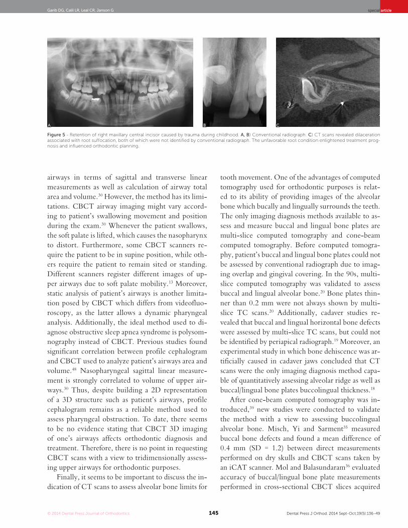
information for diagnosis, prognosis, treatment plan, surgical intervention and orthodontic therapy (Fig 5).
As for CBCT use in cases of cleft lip/palate, although some studies have assessed alveolar bone graft outcomes by means of computed tomogra-phy,40 there is no evidence proving that this assess-ment method influences orthosurgical treatment protocol in daily practice. Empirically, the benefits of CBCT use are acknowledged for diagnosis and surgical treatment of more severe craniofacial anom-alies with malformation of the midface, mandible or TMJ, particularly involving facial asymmetry. In these cases, CBCT is beneficial for allowing iden-tification of the exact location of morphological er-rors, three-dimensionally quantifying the error and providing therapeutic planning that includes osteo-genic distraction or craniofacial surgery.
Cone-beam computed tomography is indicated for orthodontic cases that require analysis of TMJ bone components accompanied by signs and symp-toms.3,44 CBCT programs reconstruct TMJ se-quential slices, both in latero-lateral and anterior-posterior axes, and provide clear imaging of articular fossa and condyles. Morphological analysis of CT scans might reveal the presence of erosions, anky-losis, hyperplasia/hypoplasia of the condyle or de-generative arthritis.5,25 In comparison to panoramic radiograph and linear tomography, however, CBCT proves more accurate in diagnosing erosion of the condyle.25 Conventional radiograph is quite limited in reproducing TMJ morphology due to imaging overlap.25 Nevertheless, TMJ imaging is not neces-sary for the diagnosis of temporomandibular disor-ders.42 Furthermore, CBCT proves a good method to assess TMJ after orthognathic surgery, particular-ly when there is considerable potential for resorption of the condyle.10 Based on such evidence, CBCT use is appropriate for diagnosis and development of treatment planning of TMJ skeletal irregularities ac-companied by signs and symptoms.
Orthognathic surgery and its outcomes might benefit from CBCT scans at the time of diagnosis.12 Additionally, CBCT is recommended in cases of se-vere facial skeletal discrepancies that require ortho-surgical treatment.3,15
However, would CBCT be useful to assess one’s airway? CBCT proves advantageous to assess upper
© 2014 Dental Press Journal of Orthodontics Dental Press J Orthod. 2014 Sept-Oct;19(5):136-49145
Garib DG, Calil LR, Leal CR, Janson G special article
tooth movement. One of the advantages of computed tomography used for orthodontic purposes is relat-ed to its ability of providing images of the alveolar bone which bucally and lingually surrounds the teeth. The only imaging diagnosis methods available to as-sess and measure buccal and lingual bone plates are multi-slice computed tomography and cone-beam computed tomography. Before computed tomogra-phy, patient’s buccal and lingual bone plates could not be assessed by conventional radiograph due to imag-ing overlap and gingival covering. In the 90s, multi-slice computed tomography was validated to assess buccal and lingual alveolar bone.20 Bone plates thin-ner than 0.2 mm were not always shown by multi-slice TC scans.20 Additionally, cadaver studies re-vealed that buccal and lingual horizontal bone defects were assessed by multi-slice TC scans, but could not be identified by periapical radiograph.19 Moreover, an experimental study in which bone dehiscence was ar-tificially caused in cadaver jaws concluded that CT scans were the only imaging diagnosis method capa-ble of quantitatively assessing alveolar ridge as well as buccal/lingual bone plates buccolingual thickness.18
After cone-beam computed tomography was in-troduced,39 new studies were conducted to validate the method with a view to assessing buccolingual alveolar bone. Misch, Yi and Sarment35 measured buccal bone defects and found a mean difference of 0.4 mm (SD = 1.2) between direct measurements performed on dry skulls and CBCT scans taken by an iCAT scanner. Mol and Balasundaram36 evaluated accuracy of buccal/lingual bone plate measurements performed in cross-sectional CBCT slices acquired
Figure 5 - Retention of right maxillary central incisor caused by trauma during childhood. A, B) Conventional radiograph. C) CT scans revealed dilaceration associated with root suffocation, both of which were not identified by conventional radiograph. The unfavorable root condition enlightened treatment prog-nosis and influenced orthodontic planning.
airways in terms of sagittal and transverse linear measurements as well as calculation of airway total area and volume.30 However, the method has its limi-tations. CBCT airway imaging might vary accord-ing to patient’s swallowing movement and position during the exam.30 Whenever the patient swallows, the soft palate is lifted, which causes the nasopharynx to distort. Furthermore, some CBCT scanners re-quire the patient to be in supine position, while oth-ers require the patient to remain sited or standing. Different scanners register different images of up-per airways due to soft palate mobility.13 Moreover, static analysis of patient’s airways is another limita-tion posed by CBCT which differs from videofluo-roscopy, as the latter allows a dynamic pharyngeal analysis. Additionally, the ideal method used to di-agnose obstructive sleep apnea syndrome is polysom-nography instead of CBCT. Previous studies found significant correlation between profile cephalogram and CBCT used to analyze patient’s airways area and volume.48 Nasopharyngeal sagittal linear measure-ment is strongly correlated to volume of upper air-ways.30 Thus, despite building a 2D representation of a 3D structure such as patient’s airways, profile cephalogram remains as a reliable method used to assess pharyngeal obstruction. To date, there seems to be no evidence stating that CBCT 3D imaging of one’s airways affects orthodontic diagnosis and treatment. Therefore, there is no point in requesting CBCT scans with a view to tridimensionally assess-ing upper airways for orthodontic purposes.
Finally, it seems to be important to discuss the in-dication of CT scans to assess alveolar bone limits for
A B C
© 2014 Dental Press Journal of Orthodontics Dental Press J Orthod. 2014 Sept-Oct;19(5):136-49146
Is there a consensus for CBCT use in Orthodontics?special article
by NewTom QR-DVT-9000. They found a mean difference of -0.23 mm between real measurements and CBCT, thereby revealing that CBCT tends to underestimate real bone loss. The mean absolute dif-ference between anatomic measurements and CBCT scans was 1.27 mm (SD = 1.43). Lower incisors had the lowest accuracy. The magnitude of the error was attributed to the use of primitive CBCT scanners which are no longer available. The devices produced unclear, low-contrast images.
Lund, Gröndahl and Gröndahl33 used cross-sectional CBCT slices of a dry scull scanned by Ac-cuitomo scanner (Morita, Kyoto, Japan) to measure buccal/lingual bone plates. The mean error for the distance between the cementoenamel junction and the bone crest was -0.04 mm (SD = 0.54), with varia-tion between -1.5 mm and +1.9 mm.
Leung et al31 assessed accuracy of natural bone dehiscence measurements and CBCT sensitivity of identifying them. The authors used 13 dry skull scans acquired by CB MercuRay (Hitachi, Medical Systems American, Ohio, USA). Their study presented some negative morphological aspects, as bone dehiscence was assessed in 3D reconstruction instead of CBCT orthogonal slices. Furthermore, they measured the distance from cuspid tips to the alveolar bone crest instead of the distance between the cementoenam-el junction and the bone crest. The authors found a mean difference of -0.2 mm (SD = 1.0) and an abso-lute difference of 0.6 mm (SD = 0.8 mm) between
real and digital measurements. They concluded that 3D reconstructions present low sensitivity (0.4), but high specificity (0.95) in identifying bone dehiscence.
Despite submillimetric accuracy revealed by CBCT, some principles must be followed when as-sessing buccal/lingual bone plates.37 Imaging spa-tial resolution is the minimal distance required to distinguish two contiguous anatomical structures.37
The smaller the anatomical structures, the higher the spatial resolution required.37 Spatial resolution is not equivalent to voxel size (the smallest tomo-graphic image), since calculation of mean partial volume, noise and artifacts negatively influence im-aging clearness.37 Mean partial volume occurs when a voxel includes two structures of different densi-ties, for instance, the periodontal ligament and the alveolar bone. Density attributed to the voxel will be equivalent to the mean density of both tissues,44 which hinders clear visualization of the limits of each structure in computed tomography.
Images acquired by iCAT scanner with voxel size of 0.2 mm have a mean spatial resolution of 0.4 mm, whereas images with voxel size of 0.3 and 0.4 mm have a spatial resolution of 0.7 mm.4 Bone plates thin-ner than the imaging spatial resolution might not be revealed by CBCT, thereby reaching a false-positive diagnosis of bone dehiscence or achieving quantita-tive assessments that underestimate the level of bone crest.47 Thus, care should be taken while drawing conclusions based on dimensions smaller than the
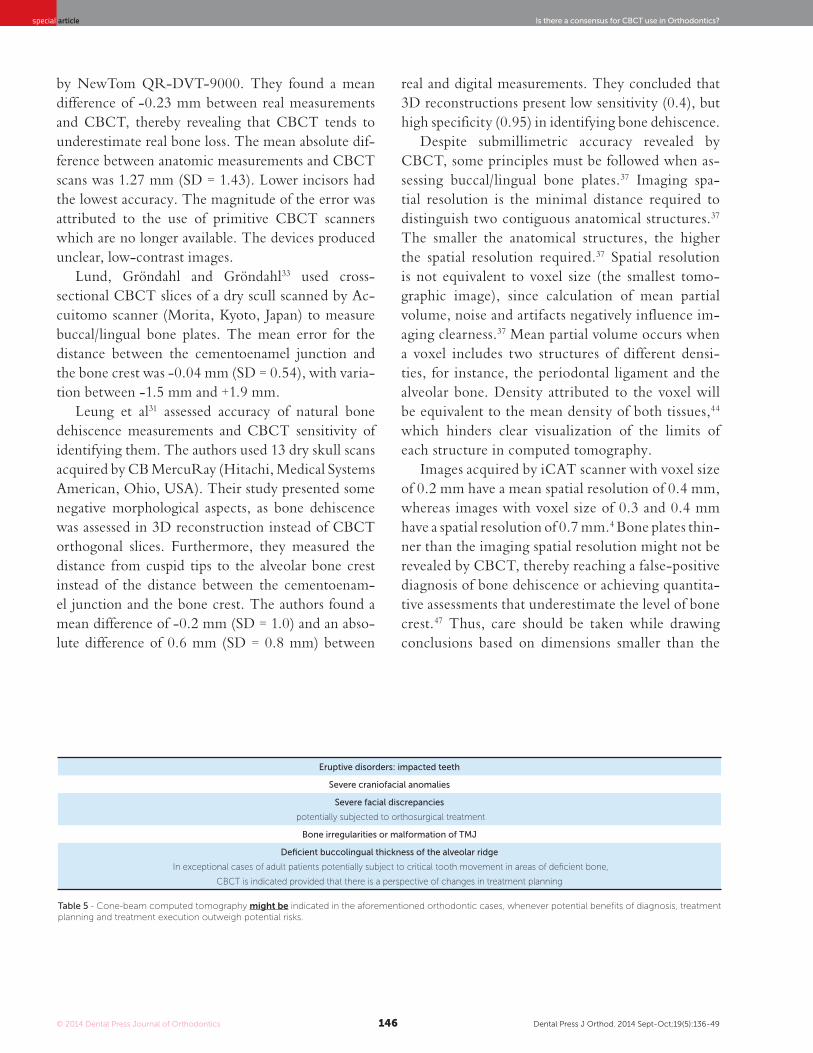
Table 5 - Cone-beam computed tomography might be indicated in the aforementioned orthodontic cases, whenever potential benefits of diagnosis, treatment planning and treatment execution outweigh potential risks.
Eruptive disorders: impacted teeth
Severe craniofacial anomalies
Severe facial discrepancies
potentially subjected to orthosurgical treatment
Bone irregularities or malformation of TMJ
Deficient buccolingual thickness of the alveolar ridge
In exceptional cases of adult patients potentially subject to critical tooth movement in areas of deficient bone,
CBCT is indicated provided that there is a perspective of changes in treatment planning
© 2014 Dental Press Journal of Orthodontics Dental Press J Orthod. 2014 Sept-Oct;19(5):136-49147
Garib DG, Calil LR, Leal CR, Janson G special article
imaging spatial resolution.37 In Orthodontics, voxel sizes of 0.4 mm and 0.3 mm are the most used.47 However, investigations aiming to assess periodontal structures before and/or after orthodontic treatment should use the smallest voxel possible.37 The small-est voxel in iCAT scanner is 0.2 mm; whereas Ac-cuitomo and PreXon scanners produce images with higher spatial resolution, as their smallest voxel is 0.1 mm32 Images with reduced voxel size are more accurate in terms of thickness and height of buccal/lingual bone plates.47
Therefore, CBCT scans are useful to assess the presence of bone dehiscence. However, CBCT scans have been restricted to investigations that guide the clinician towards the alveolar limits in cases of critical movement such as buccolingual tooth move-ment.22 In Orthodontics, CBCT should be indi-cated to assess deficiencies of buccolingual thickness in the alveolar ridge of adult patients subjected to critical tooth movement in which case absence of buccolingual bone would affect orthodontic treat-ment. In these cases, the best option would be to use high resolution (reduced voxels) and a limited field of view (FOV) (Table 5).
IMPORTANT RECOMMENDATIONS: EDUCATION AND TRAINING
According to SedentexCT guidelines,15 the prescrib-er, the clinics where the exam is taken and the medical physics expert share the responsibility over a radiographic exam. All professionals involved with CBCT, including the prescriber, should receive theoretical and practical training that includes the technical procedure of image acquisition, radiation dose, radiation protection and to-mographic reading.15 That is, the prescriber should know when and for what purpose he will request it. Further-more, he should know how to exam and fully interpret it.
FINAL CONSIDERATIONSCone-beam computed tomography is not a stan-
dard diagnosis method in Orthodontics. CBCT should be indicated with criteria, when the potential benefits for diagnosis and treatment planning out-weigh the potential risks of an increased radiation dose. The recommendations discussed in this article originate from current evidence and therefore are time-dependent. In the future, new evidence as well as technological evolution and innovation of CBCT scanners could change the current indications of CBCT in Orthodontics.
© 2014 Dental Press Journal of Orthodontics Dental Press J Orthod. 2014 Sept-Oct;19(5):136-49148
Is there a consensus for CBCT use in Orthodontics?special article
1. Alqerban A, Jacobs R, Fieuws S, Nackaerts O, SEDENTEXCT Project
Consortium, Willems G. Comparison of 6 cone-beam computed
tomography systems for image quality and detection of simulated canine
impaction-induced external root resorption in maxillary lateral incisors. Am
J Orthod Dentofacial Orthop. 2011;140(3):e129-39.
2. Alqerban A, Jacobs R, van Keirsbilck PJ, Aly M, Swinnen S, Fieuws S,
et al. The effect of using CBCT in the diagnosis of canine impaction
and its impact on the orthodontic treatment outcome. J Orthod Sci.
2014;3(2):34-40.
3. American Academy of Oral and Maxillofacial Radiology. Clinical
recommendations regarding use of cone beam computed tomography
in Orthodontics. Position statement by the American Academy of Oral
and Maxillofacial Radiology. Oral Surg Oral Med Oral Pathol Oral Radiol.
2013;116(2):238-57.
4. Ballrick JW, Palomo JM, Ruch E, Amberman BD, Hans MG. Image
distortion and spatial resolution of a commercially available cone-beam
computed tomography machine. Am J Orthod Dentofacial Orthop.
2008;134(4):573-82.
5. Barghan S, Merrill R, Tetradis S. Cone beam computed tomography
imaging in the evaluation of the temporomandibular joint. J Calif Dent
Assoc. 2010;38(1):33-9.
6. Berco M, Rigali PH Jr, Miner RM, DeLuca S, Anderson NK, Will LA. Accuracy
and reliability of linear cephalometric measurements from cone-beam
computed tomography scans of a dry human skull. Am J Orthod
Dentofacial Orthop. 2009;136(1):17.e1-9; discussion 17-8.
7. Bernardes RA, Moraes IG, Hungaro Duarte MA, Azevedo BC, Azevedo JR,
Bramante CM. Use of cone-beam volumetric tomography in the diagnosis
of root fractures. Oral Surg Oral Med Oral Pathol Oral Radiol Endod.
2009;108(2):270-7.
8. Bogdanich W, McGinty JC. Radiation worries for children in dentists’
chairs. The New York Times. 2010 Nov. 23; A1.
9. Brown AA, Scarfe WC, Scheetz JP, Silveira AM, Farman AG. Linear accuracy
of cone beam CT derived 3D images. Angle Orthod. 2009;79(1):150-7.
10. Cevidanes LH, Bailey LJ, Tucker SF, Styner MA, Mol A, Phillips CL, et al.
Three-dimensional cone-beam computed tomography for assessment of
mandibular changes after orthognathic surgery. Am J Orthod Dentofacial
Orthop. 2007;131(1):44-50.
11. Damstra J, Fourie Z, Huddleston Slater JJ, Ren Y. Accuracy of linear
measurements from cone-beam computed tomography-derived surface
models of different voxel sizes. Am J Orthod Dentofacial Orthop.
2010;137(1):16.e1-6; discussion 16-7.
12. Edwards SP. Computer-assisted craniomaxillofacial surgery. Oral
Maxillofac Surg Clin North Am. 2010;22(1):117-34.
13. Enciso R, Nguyen M, Shigeta Y, Ogawa T, Clark GT. Comparison of cone-
beam CT parameters and sleep questionnaires in sleep apnea patients
and control subjects. Oral Surg Oral Med Oral Pathol Oral Radiol Endod.
2010;109(2):285-93.
14. Ericson S, Kurol J. Incisor root resorptions due to ectopic maxillary
canines imaged by computerized tomography: a comparative study in
extracted teeth. Angle Orthod. 2000;70:276-83.
REFERENCES
15. European Commission. Cone Beam CT for dental and maxillofacial
radiology: evidence-based guidelines. Luxembourg: SEDENTEXCT; 2012.
(Radiation Protection; n. 172).
16. Farman AG. Image gently: enhancing radiation protection during
pediatric imaging. Oral Surg Oral Med Oral Pathol Oral Radiol.
2014;117(6):657-8.
17. Fernandes TM, Adamczyk J, Poleti ML, Henriques JF, Friedland B,
Garib DG. Comparison between 3D volumetric rendering and multiplanar
slices on the reliability of linear measurements on CBCT images: an in
vitro study. J Appl Oral Sci. 2014 Jul 4;0:0. [Epub ahead of print].
18. Fuhrmann RA. Three-dimensional interpretation of labiolingual bone
width of the lower incisors. Part II. J Orofac Orthop. 1996;57(3):168-85.
19. Fuhrmann RA, Bucker A, Diedrich PR. Assessment of alveolar bone
loss with high resolution computed tomography. J Periodontal Res.
1995;30(4):258-63.
20. Fuhrmann RA, Wehrbein H, Langen HJ, Diedrich PR. Assessment of the
dentate alveolar process with high resolution computed tomography.
Dentomaxillofac Radiol 1995;24:50-4.
21. Garib DG, Raymundo Junior R, Raymundo MV, Raymundo DV, Ferreira
SN. Tomografia computadorizada de feixe cônico (cone beam):
entendendo este novo método de diagnóstico por imagem com
promissora aplicabilidade na Ortodontia. Rev Dental Press Ortod Ortop
Facial. 2007;12(2):139-56.
22. Garib DG, Yatabe MS, Ozawa TO, Silva Filho OG. Morfologia alveolar
sob a perspectiva da tomografia computadorizada: definindo os limites
biológicos para a movimentação dentária. Dental Press J Orthod.
2010;15(5):192-205.
23. Halazonetis DJ. Cone-beam computed tomography is not the imaging
technique of choice for comprehensive orthodontic assessment. Am J
Orthod Dentofacial Orthop. 2012;141(4):403-7.
24. Haney E, Gansky SA, Lee JS, Johnson E, Maki K, Miller AJ, et al.
Comparative analysis of traditional radiographs and cone-beam
computed tomography volumetric images in the diagnosis and treatment
planning of maxillary impacted canines. Am J Orthod Dentofacial
Orthop. 2010;137(5):590-7.
25. Honey OB, Scarfe WC, Hilgers MJ, Klueber K, Silveira AM, Haskell BS,
et al. Accuracy of cone-beam computed tomography imaging
of the temporomandibular joint: comparisons with panoramic
radiology and linear tomography. Am J Orthod Dentofacial Orthop.
2007;132(4):429-38.
26. Katheria BC, Kau CH, Tate R, Chen JW, English J, Bouquot J.
Effectiveness of impacted and supernumerary tooth diagnosis from
traditional radiography versus cone beam computed tomography. Pediatr
Dent. 2010;32(4):304-9.
27. Kobayashi K, Shimoda S, Nakagawa Y, Yamamoto A. Accuracy in
measurement of distance using limited cone-beam computerized
tomography. Int J Oral Maxillofac Implants. 2004;19(2):228-31.
28. Lamichane M, Anderson NK, Rigali PH, Seldin EB, Will LA. Accuracy of
reconstructed images from cone-beam computed tomography scans.
Am J Orthod Dentofacial Orthop. 2009;136(2):156.e1-6; discussion 156-7.
© 2014 Dental Press Journal of Orthodontics Dental Press J Orthod. 2014 Sept-Oct;19(5):136-49149
Garib DG, Calil LR, Leal CR, Janson G special article
29. Larson B. Cone-beam computed tomography is the imaging technique
of choice for comprehensive orthodontic assessment. Am J Orthod
Dentofacial Orthop. 2012;141(4):402, 404, 406.
30. Lenza MG, Lenza MM, Dalstra M, Melsen B, Cattaneo PM. An analysis of
different approaches to the assessment of upper airway morphology: a
CBCT study. Orthod Craniofac Res. 2010;13(2):96-105.
31. Leung CC, Palomo L, Griffith R, Hans MG. Accuracy and reliability
of cone-beam computed tomography for measuring alveolar bone
height and detecting bony dehiscences and fenestrations. Am J Orthod
Dentofacial Orthop. 2010;137(4 Suppl):S109-19.
32. Liang X, Jacobs R, Hassan B, Li L, Pauwels R, Corpas L, et al. A
comparative evaluation of Cone Beam Computed Tomography (CBCT)
and Multi-Slice CT (MSCT) Part I. On subjective image quality. Eur J
Radiol. 2010;75(2):265-9.
33. Lund H, Grondahl K, Grondahl HG. Accuracy and precision of linear
measurements in cone beam computed tomography Accuitomo
tomograms obtained with different reconstruction techniques.
Dentomaxillofac Radiol. 2009;38(6):379-86.
34. Menezes CC, Janson G, Cambiaghi L, Massaro C, Garib DG.
Reproducibility of bone plate thickness measurements with cone-beam
computed tomography using different image acquisition protocols.
Dental Press J Orthod. 2010;15(5):143-9.
35. Misch KA, Yi ES, Sarment DP. Accuracy of cone beam computed
tomography for periodontal defect measurements. J Periodontol.
2006;77(7):1261-6.
36. Mol A, Balasundaram A. In vitro cone beam computed tomography
imaging of periodontal bone. Dentomaxillofac Radiol. 2008;37(6):319-24.
37. Molen AD. Considerations in the use of cone-beam computed
tomography for buccal bone measurements. Am J Orthod Dentofacial
Orthop. 2010;137(4 Suppl):S130-5.
38. Mostafa YA, El-Beialy AR, Omar GA, Fayed MS. Four curious cases of
cone-beam computed tomography. Am J Orthod Dentofacial Orthop.
2010;137(4 Suppl):S136-40.
39. Mozzo P, Procacci C, Tacconi A, Martini PT, Andreis IA. A new volumetric
CT machine for dental imaging based on the cone-beam technique:
preliminary results. Eur Radiol. 1998;8(9):1558-64.
40. Oberoi S, Chigurupati R, Gill P, Hoffman WY, Vargevik K. Volumetric
assessment of secondary alveolar bone graft using cone beam computed
tomography. Cleft Palate Craniofac J. 2009;46(5):503-11.
41. Özer SY. Detection of vertical root fractures of different thicknesses in
endodontically enlarged teeth by cone beam computed tomography
versus digital radiography. J Endod. 2010;36(7):1245-9.
42. Petersson A. What you can and cannot see in TMJ imaging: an
overview related to the RDC/TMD diagnostic system. J Oral Rehabil.
2010;37(10):771-8.
43. Pinsky HM, Dyda S, Pinsky RW, Misch KA, Sarment DP. Accuracy of three-
dimensional measurements using cone-beam CT. Dentomaxillofac Radiol.
2006;35(6):410-6.
44. Scarfe WC, Farman AG. What is cone-beam CT and how does it work?
Dent Clin North Am. 2008;52(4):707-30.
45. Silva MA, Wolf U, Heinicke F, Bumann A, Visser H, Hirsch E. Cone-
beam computed tomography for routine orthodontic treatment
planning: a radiation dose evaluation. Am J Orthod Dentofacial Orthop.
2008;133(5):640.e1-5.
46. Smith BR, Park JH, Cederberg RA. An evaluation of cone-beam computed
tomography use in postgraduate orthodontic programs in the United
States and Canada. J Dent Educ. 2011;75(1):98-106.
47. Sun Z, Smith T, Kortam S, Kim DG, Tee BC, Fields H. Effect of bone
thickness on alveolar bone-height measurements from cone-beam
computed tomography images. Am J Orthod Dentofacial Orthop.
2011;139(2):e117-27.
48. Vizzotto MB, Liedke GS, Delamare EL, Silveira HD, Dutra V, Silveira HE.
A comparative study of lateral cephalograms and cone-beam computed
tomographic images in upper airway assessment. Eur J Orthod.
2012;34(3):390-3.
49. Wahl N. Orthodontics in 3 millennia. Chapter 3: The professionalization of
orthodontics. Am J Orthod Dentofacial Orthop. 2005;127(6):749-53.
50. Wahl N. Orthodontics in 3 millennia. Chapter 4: The professionalization
of orthodontics (concluded). Am J Orthod Dentofacial Orthop.
2005;128(2):252-7.
51. Wahl N. Orthodontics in 3 millennia. Chapter 12: Two controversies:
early treatment and occlusion. Am J Orthod Dentofacial Orthop.
2006;130(6):799-804.
52. Wahl N. Orthodontics in 3 millennia. Chapter 13: Temporomandibular
joint and orthognathic surgery. Am J Orthod Dentofacial Orthop.
2007;131(2):263-7.