Embed Size (px)
Citation preview

SIŒLETAL MUSCLE PO}<ŒRED CARDIAC ASSIST
il
...
r
A Theais Submitted to the Faculty of Graduate Studies
and Research in partial fulfillment of the
requirements for the Degree of
Master of sc1.enée
(C) Dr. Garrett Lyndon Walsh, 1
Department of Surgery,
Division of Surgical Research,
MaG!ll University, Montreal,
c· Marc~ 1988.
(
..
" •
,

Permission has been granted to the National Library of Canada to microfilm this thesis and to lend or sell copies of the film.
The author (copyright own1rr> has reserved other publication rights, a~d neither the thesis nor elftens ive extracts from i t may be printed, or otherwise reproduced without his/her written p~~ission.
L'autorisation-a 6t6 accord'. à la "Bibliothèque nationale du Canada de microfilmer cette thèse et de prater ou de vendre des exemplai-ree du film.
L'auteur (titulaire 'du drQit d'auteur) se r6serve lee autres droits de publication, ni la th~se ni de longs extraits de celle-ci ne. doivent Atre imprim'e ou autrement reproduits sane Bon autorisation 'cri te.
1{
\ . ISBN 0-315-45930-1
, , ." , ... ~ -,,--~-,--__ ...... ' _'1

•
Ml '-il.;;
, r
.. '
::.
. \
-";'
....
, . l
-. .t
o.
. ..
< , 'I.~l
,.
• 1
~
SKELETAL MUSCLE POWERED CARDIAC ASStsT
)
- 1
.1
( ,-'
<> ...
. , i .' .

~ . (
'r
,0 0
. ~
-!
Q . "
"
~ ;-
t-.-_ ,
----.~---~------
ABSTRACT;
The purpose of Othis study was teS devdop a \totally implantable
cardiac a8si~evice which would use skeletal muscle as an autogenous
power source. The canine latissimus dorsi lDUscle was transforlled by
chronic stimulation to a muscle whiChE fatigue resistant and more
aerobically metabolizing as demQnstra ed histochemically and bio~
chemieally. A pulse train stimulator a used to charac,terize the
optimal electrical parameters of transformed muscles. An e~tra-
sortie balloon powered by these transformed muscles was used to
coupterpulse the aorta. Hemodynamically signifieant cardiac assist
through diastolic augmentation was achievecr:a evidenced by the
irl'crease in the subendocardial viability -index. Another series of
experiments examined the cardiomyo~lastic surgieal technique in • 11" •
. which skeletal muscle is transplanted onto the hesrt for systolif
cardiac assiste The 'use of a pericardial pa~ch as a neoendocardium
was examined.
..
, --"-
• • .. ,
/
1
/ ,.
/ .. /' 1
/ 1
#
,r
"
.~

'f
o
r ,
o
o
~
J
, 1
)
/
R1!SuMS: \ .
J 0
Le' b~ de cette étude était- de, deyeloper une p'ompe d' &ss istance
car~iac implBtntable qui pourrait utili~er le mu'sele squeletique
; " . d' • côllUne source energle. Le muscle latissimus dorsi du chiert a ~t'
transfôrmé par stimul,ation chronique pour qu'il soit re-sistant à'
. la
un
'1 "
fatigue par un metabol isme aerobique. . ~,
baÜo~ extra-aortique a étée e.~abli en
une~contr~-pulsation par
utilisant l~ muscle
transformé. Une aùgmentation de la pression diastolique a contribu'
à une -amei ioration hèmOdyna~Lue mise' en e'vidence parc une augmentation
de l'index de viabilité sous endocardique. Une autre serie
d'experiences avait pour but d'utiliser le lIIl\ISale transformé pour . i
c'J'.!ée une augmentation sy'stolique par cardiomyoplastie. L'utilisation .... l'
,du pericarde en tant que neo-endo.5!arde a été etudié • •
, " 't.'.r:,.
-,
" . ,
. \
'.
r
,
',::. , " '1 (
.. .'
.:: .... ~f(1 'i" ' .... :t I,'" ~' .... H " , lit t
. -.
... • 1
-'
,.
, ,',
"
1
; .
(
/
(
o.
. \ . i
• . ' , \
PRÈFACEI /
1 1. This t~esis represents.research which was conducted from July of
, ". \ \'''- ® 1985 until June of 1986 during my second year of general surgical
residency training. \ .
This was undertaken in the universitJ Surgical
Clinies of The Montrea) General Ho~pital under the direction of Dr. Ray , .
C. -J. Chiu. This is an ongoing project which is sponsored by the "
, G
Medical Reaearch Council of Can~a and ia specifically interested in
eke1etal 'musc le and i te potent~a1 uses in cardiac surgery. Dur log t"his.
Year, Dr. Michael Dewar:then a researeh fellow of the Canadlan Heart \ . ./ 1. ,l'
Foundation, and l worked conjoint1y on two different surgiea1
applications of skeletal, muscle in eardiae su;gery, ineluding diastolie
_and systolic modes of assistance. )
Chapter r is the historical review of the subject whieh l
researched for this thesis; ~looking at aIl of the different ways , d
skeletal muscle has been used in conjûnction with the heart since the r-"-. _
infaacyof card!ae 8urgery in the early 19JO'8. This has sinee been .~ 1 /
published as the introductory dhapter to a recènt monograph pUblished by \' -1
'the Futu~ Publishing Company of Mount Kisco, New York entitled • • <
"Biomechanfcal Cardiae Assiat: Car.diomyoplasty and Muscle Powered .. oèvices".' 1
Chapter II examines our, approach to skeletal muscle whe~ used, in.a ....
dJ..astolic mode of cardiae assiste Today, in clinical pract·ice, the ~
pneumatioally drivenin:~~a-aortie baJ:loon counterpulsator represents the ,
,
lI\ost widely implemented cardiac assist device.- It ié used for empQrary' ~ .
support for a patient in cardiogenic shock awaiting correctiv 1 1.. .~
, surgery or the post-operative patient who has had diffieult ' .
from cârdiopulmonar~ bypass and require~ ongo~g suppor~. in ~.nticip.tlon
l, /
, \
• .1
..
\ .. -' ,t,
'.

---11111!1----.,.---1IIII!if41!11 . .,...,.--------,.J>!'!'i---:-------;--"':"I.,------::---;--...,...--:~~---------
o
,0
..
1
•
,1
. i
, \
ii~
~. " . .
of myocardial recovery. We have exténded the physiologie bê'nefits of \
*ld pressure, a~gmentati~n th'rOU9: coun'terpu1sation with an in,tra- '
aprtic ballC?on to an ~xtra-aortic baUoon configurS'tion and.. used .
~kqJetal muscle as an aut~genoqs power source. To circumvent the ~
central problem of muscle fatigue, we have capi~alized on the
faécinating electr0l'hysiologic property of skeletal muscle "pl~sticity". \ , --
By chronically stimulating a mixed fibered muscle such as.latissi~us
dorsi, i.t lS.possible tè "transform" ite enzymatic rnachinery and protein
morphology to a muscle adapted to more aerobic \. m~taboli~ith slower
twi tch fibers. _ This results in ~ musçle which is less fatiguab1eland
capable o'f repetitive contractions, more sin:lilâr to cardiac muscle and , \
hence better suited for long-te~ork. To stimulat~ a.skeletal muscle to do cardiae work requi!es a
, contraction which has sufficient force and ~uration to be hemo-
~ dynamically significant.~ A pulse train stimulator has been previously
deyeloped in this project which generates~a tra~n of pulses which
" 1 •
results in a summatlon of muscle twitches and !ubsequently a smooth
muscle co~tractlon. The characterization of the optimal stimulation . ~
.par~tprs for transformed {ke1eta~ ~u.c1e and the 8urglca1 .pp~icatlon of this to balloon cQunterpu\sation is my contribut~on to the world
literatth'e. ,This seems to indicate for the first tirne that transformed,
f~ti9ue ~sistant ~uscles are capabl~ of hemodynamically significant '<
cardiae assista~ce when appropriately stimulated: My experimental
design and elucidation of the optimal stimul~tion parameters has bee~
published in the~ceedingS of the 8~ Annual co~rence of the
I.E.E.E. E~gineering~Medicin~and Biology Society. The surgieal
application by extra-~ortie balloon counterpulsation ha~been pUbliahed
.\
• \
' ..

"
c
<
(
2"'" '
•
" , Hi
"
in the Surgiesl Forum, Volume 37 in 1986 and was presented at the 72nd
Annual C1ini~a~ Congres~ of~the A~riean c;llege of Surgeons in New
Orleans in Oetomer of 1986. More reeently, wieb the enzyme and ~ . ,
morphology stùçies, this ehapter has been aecepted for p~lieation in ~
the Journal of Cardiovascular a~d Thoracie Su~ery.
Chaptet III reviews work which has been done when skeletal muselels
intended use is during dardiac syst6le. This requires mobilization and
transplantation of the muscle either into or'odto the heart. ~
This type r
of surgery has already reached the stage of clini~al application with . '
centres in France and the U.S.A. having perf~rmed cardio~yoplasties in , '
p~tients who have had either excision of èardiae tumors with 10ss of )
left ven~rièular muscle mass.or who require skéletal muscle as a ' "
"" buttress for left ventricular aneurysm repaire. Cardiomyoplasty
, technique.s also have, potential 'uses in c?n~.~nital heart surgery ~
,au~genous patehes for ~nlarging outfiow ~raets or cardiac cha~ers with ,
the potenti~l of growth with the infant. We ~ave examined one aspect of Jo ,
the cardiomyoplastic technique which occurs when by necessity th~ \
s~oth, thromboprotective endoc~rdium is broached, for instance in
• excision of a transmurai tumor on infarct. We have developed and ;
~
analyzed the use of an autogenous patch of pèricardium as a neo~ • 4
endocardium during cardiomyoplastic repaire This chapter has been
published recently in the Annala of Thqracic Surgery. D~ewar ia the
~first auth~r of this work and l am the second author. J ,
'" may
Finally, Chapter IV ls my future perspective look at what l feel
be th:l.ole skeletal muscle .... y ~l.Y 1,.cl,1n1cal card1ac a.sl.8t.n~8 g \. • , ..
and what may be the technolog~c and physiOlogie limitations of thi~ use.
I wish at this point.to acknowledge several people who have been
, {
" , .....
'h " --
J,. 1

o -
l
o
o r'
"
....--/ .' , ,
iv
,1
instrumental ~rough my researeh year with this projeet, thesis and
publications. Firs~and foremo~t, Dr. Ray Chiu, a ~rue pioneer i~
cardiac assist deviees whose gUidance and vast weaith of knowledge and
exp~rienee serves as an inspiration for me in my own ehosen surgieal
subspeciaity of eardiov~eular and thoraeic surgery. He will remain one
of my most import~nt mentors throughout my career. Dr. Michael Dewar,
co-réseareher, surgieal eolleague and,. fri,f3nd. The many hours we spent
toqether trying to make muscles twiteh or squeeze balloons, and
developing techniques to transplant muscle into the heart,wili remain
dear in my thoughts. In many respects, we feit much as the Wright
brothers must have at the turn of the ceptury trying to hopelessly mold
a machine out of wood and metal which was capable of ~lying. Dr. John
Lough, l wish to thank for the muscle analysis for myosin ATPase stains.
Dr_ David Ianuzzo from York univ~rsity for his collaboration in analysie (
of muscle samples for citrate synthase and phosphofruetokinase and gel "-
eleetrophoresis. Dr. Jim Stewar~ of the Department of Cardiology for ,
ech~cardiography in our dogs. Dr. Rick Fœaser from the Department of
o pathology for his histologie analysi~ of our pericardial patch .. techniques. The Nuclear Medicine Department of the Montreal General for
, the pyrophosphate scans. Special thanks to Mrs. Maureen Smith for her
l inyaluable time and effort as operating room director. , Dr. Eric toot,
our full-time veterinarian and part-time -8aMiac anesthetist'. Mr.
Reginald Abraham and Carolyne Desrosiers for their technieal assistance ~ ~,~
\
as perfusion!sts during our ~xperiments which required cardiopulmonary
bypass. Mr. Matthew Rosenberg, Mr. Daniel Yee, Mr. Jean Marie
Chavannes, Mr. Wojtek,Grzywacz for their daily work beyond the calI of #
dut Y in preparing and caring for the animaIs both pre- and ~
...

(
(
'1
,
----r
\' • v
post-operatively. The Medtronic Company and Dr. Aida Khalafalla who-c ---
supplied thè programmable Itrel pacemakers, special Biomer extra-aortie
balloons, and helped develop our concept of a pulse train stimulator •
. Without their interest and hi~h teehnology material input, much of this
project WOUl~ not be possible. Last but nct lèast, a special thank you
ta Mrs: Emma Lisi, indispensable, irreplaeeable for typing this
manuscript, the mos~ beautiful-w~an in the world.
CI
.. 1 r
r

o
o
•
TABLE OF CONTENTS
CHAPTER l )
SKELETAL MUSCLE FOR CARDIAC REPAIR AND ASSIST: A HlSTORICAL REVIEW
.Introduction ~
I. Skeletal Muscle For Myocardial Revascularization
II. Skeletal Muscle For Valve Const~uction And Great Vessel
Repair And Replacement
III. Skeletal Muscle For Cardiomyoplasty
IV. Skeletal Muscle As An Energy Source For Cardiac Assist:
Hydraulic Pouches And Counterpulsation
Diastolic Counterpulsation
Skeletal Muscle Fatigue
Conclus ion
., CHAPTER II
IMPLANTABL& EXTRA-AORTlC BALLOON ASSIST POWERED BY TRANSFORMED
FATIGUE RESISTANT SKELETAL MUSCLE
Introd\.ic tion
~aterials & ~OdS ~
TranSforma~~ Of The Skeletal Muscle
Charac,terization off Optimal StiÎnulation Parameters
Counterpulsation powered By Transformed Skeletal Muscle
Results
Transformation Of The Skeletal Muscle
Th~ Stimulation Parameters
Counterpulaation Powered B~ Fafigue Resistant MUBcle
Discussion
1
2
4
5
15
16
19
32
33
33
34
35
36
37
38
39

(~
'"
(~ J
1
c
CHArTER III
LEFT VENTRICULAR FULL THICKNESS CARDIOMYOPLASTY WITH PER1CARDIAL
NEO-ENDOCARDIUM: EXPERIMENTAL DEVELOPMENT OF A SURGICAL PROCEDUFcE
Introduction
Materials & Methods
Simple Full Thickness Cardiomyoplasty
Fu Il Thickness Cardiomyop lasty Over Per i~ardia 1 Patch
(Neo-endocardium)
Results
Simple Full Thickness Cardiomyoplasty
Full Thickness Cardiomyoplasty Over Pericardial Patch
(Neo-endocardium)
Discussion
CIIAl'TER IV
FUTURE PERSPECTIVES
'\
(
59
60
60
61 ~.
62 (
63
65 ..
83

CHAPTER l '.,
,.
SKELETAL MUSCLE FO,R CARDIAC REPAIR AND ASSIST:
A HISTORICAL OVERVIEW
)
,
•

, .
(
1.
INTkOD'UCTION 1
Cardiac death still remains the number one killer of North •
Americans today, although death rates from cardiovascular diseases a~e • . ..
on 8 downward trend. Medical treatments and eurgieal modalities have .,
improved morta1ity and morbidity, but there ie a growing number of
patients with congestive h~art fai1ure and end-stage cardiac disease.
It is towards this subaet of the patient population that considerable
tlme, effort and money have been spent sinee the mid 1960'5 in search
for ventricular assist devlcea with a multi-armed research approach into
biomaterials, blood pumps, energy storage and transmission(1): The use
of the intra-aortic ba11oon, the most successful ventricular aasiet
device routinely employed in cardiac centers, ie now commonp1ace
(2) f0110wing its conception by Harken in 1958 and development in the
1960s. We are also witnessing the initial clinical application of ~
artificial hearts. Orthotopic heart transplantation with aggressive 1'"
immunosuppressive therapy is an e~tablished forro of treatment for a
selected few.
The limitation of donor heart availability (3) , the thrombotic and
infective complications associated wlth 1eft ventricular ·assist
(4) • (5) devlces and difflculties with implantable power sources have
prompted many groups fa continue work using endogenous skeletal muscle t·
for cardiac repair and asslst, work whlch starte<\. aver .a half century
ago.
We will review the myriad applications of skeletal muscle,
includinq its use in -
(
r
J

0, h
'.
1
..
o
2.
~ y
1. ~evascularization of thJ heart
2. Creation of heart valves and replacement and repair of great
vessels
3 . Cardiomyoplasties
4. As an energy source to power assist devices and hydJ:aulic
poucl)es
5. Counterpulsation
We will.outline sorne of the research on muscle stimulation and on ~
the fundamental problem of skeletal muscle fatigue.
1. SKELETAL MUSCLE FOR MYOCARDIAL REVASCULARIZATION: ~
Several surgieal approaches were made to increas~ blood supply to
the heart muscle, when the understanding of coronary artery disease was .. in its infancy and prior to the development of techniques allowing
direct surgery on the coronary arteries. These included:
1. Techniques tcf utilize and enhance the normally existing extra-
. ~
coronary anastomoses between the heart and the pericardium by inducing a
sterile pericarditis, achieved by either mechanieal epicardial abra8i~ , " .
or c~emical irritatiun with various substances (inc~udinq Dakin's
, (6 7) 18) (9) (lO} solution ' , asbestos powder , carborundum sand , talc ). J
2. To fu%ther augment the e~tra-coronary anastomosis, flaps and J
- pedicled grafts were \emplOyed. Such "cardiopericardiopexies" included
usinq:
a) mediastinal tissues(9)
h) pericardiai fat (lI)

-(
(
c
3.
c) skin and subcutaneous tissue (12)
d) omentum(13) \.-
\ e) lunq (14)
f) j j (15) e unum and
g) skeletal muscle (16)
Beek, performing over 1,200 experiments in the 1aboratory batween "
1923 and 1935, was able to demonstrate that anas~omoses would re~ily
develop between skeletal and cardiac muscles. These cOllatenat: would
'" prote~t the heart from fibrillation fo110wing coronary artery 1igation
and would also diminish the subsequent infarct size. The collaterals'
not only served as a new source of b100d supply to the heart, but a1so
redistributed coronary b100d flow. The physio1ogica1 "need" of the
myocardium wi th pressure differential in arterial beds determined the '\ (17)
develop~ent of collaterals •
The first c1inica1 application of the technique was performed by "
Beek' in February 1935 on a 48 year old farmer with angina pectoris
transplanting a pedicled pectoralis ma~r flap onto his heart. The
(17) patient survived the proce~ure and had relief of symptoms •. By 1937,
• he had performed hi~ operation on over 20 patients with marked relief of . . (18)
symptoms in.those'who survived the operation • lA
Bakst as late as 19?7 combined the Beck procedure w:i;t.h Thompson' s
car<Uopericardiopexy, usin/;} mB.gnesium silicate. He was able to show a
three-fold increase in rétrograde coronary artery blood flow in animaIs
with pedicled pectoralis grafts to their hearts. He was also able to
demonstrate filling of ·the coronary circulation via the extra-coronary
collaterals with Neoprene Latex injection of the descending aorta(19).

, \
o
o
.. '
o
4.
II : SIŒLETAL MUSCLE FOR VALVE CONSTRUCTION AND GREAT VESSEL REPAIR
AND REPLACEMENT:
• Skeletfll muscle had been used in a number of novel ways for the )-
reconstruction" of valves and vessels, in times prior to the advent of &
reliable kni tted and woven grafte and dependable prosthetic cardiae
~alves. In 1959,~ the early days of cardiac valvular repair, Absolçn
noted that an ideal valve replacement should be made of a living
rnaterial with regenerative growth potential. Ingeniously usinq the
thick muscular and th in tendinous aspects of the diaphragm, he W8S able
to fashion valves which were competent against 400 mrnHg retrograde
pressure (20) • petrovsky, a Russian surgeon, used pedicled dlaphragmatie
(21) grafts to repair injuries to the aorta in humans , and Wesolowski in
1963 used the central tendon of the diaphragm as a replacement of a
segment of the descending thoracic aorta in pige (22) •
It ie kn~wn that there are several limitations in the "conduits"
used today that 'are of special interest to congenital heart surgeons. ,
These include high thrombogenieity, foreign body reaetions,' false
aneurysm formation, and their inability to gr~with the child. Yee in
1985 reported the us"e of fascial island grafta of rectus muscle with an
intact 'blood supply to, replace and patch pulmonary a'rteries in beagle
- (23) puppies. He noted no aneurysma1 dilatation or thrombus formation •
Gaines has recently used free grafts of gracilis mus,ple with micro-
vascular.anastomoses to the internaI mammary vessels to reconstruct the
(24) right ventricular outflow tract of infant swine • As reported in
detai! in their chapter in this book, at 10 weeks, there was histol~ic
evidence of endothel1alization of the grafts and it \lias hoped that
future experiments would reveal that the grafts would qrow with the.
• 1

c
c
te P'
5. 1 , infant and resist aneurysmal dilatation. This is the first reported
, case of free m\croV~B"Ular grafts used ln repair of the heart and
vesse1s. "
III. SKELETAL MUSCLE FOR CARDIOMYOPLASTYr
Various muscles have been transplanted into, enta and àround the
heart over the past 50 years in an effor\. te;) enlarge a cardiac chamber
(such as ventriculoplasty for congenital hypoplastic right ventricle),
tO,reinforce a myocardial infarct or an aneurysm or to augment cardiac
output, eitller ,by diminishing 'paradol<:ical movement in a left ventricular
aneurysm or by synchronously pacing the ske1etal muscle with cardiac
activity, making use of the musclels intrinsic contractile properties.
The followlng muscles haye been used previously: pectoralis major,
diaphragm, latissimus dorsi, rectus abdominis, intercostals, grac~li~,
sternohyoid, sternocieidomastoid, internaI oblique,.pronator tares, and
~astus lateralis.
Grafts can be classified as free, pedicled, free grafts with micro-
vascular anastomoses, innervated, denervated, paced or unpaced. )
Jesus in 1933 perhaps can be credited with the first use of a
skeletal muscle to repair a trauma tic in jury of the left ventricle in
humans (25) •
Leriche in 1933, using free muscle grafta managed bo have a canine
survivor after a 4 x 2 x 1 1/2 cm excision of the left ventr!cle with.a
~~ graft take. His ultimate intentio~.was to.use skeletal muscle ta y~~ '(26) replace infarcted myocardial tissue in humans •
~
Griffith and Bates in 1938, performing the '''Beek'' operation' foX'
revascularization of the heart, extended the musclels intended use to
:
,~-

o
\
o
• , t s, tf t 'f "j
6.
f 1
repair an iatrogenic hole in the right ventricle after an apical ~
suture had torn through the myocardium(27).
Weinstein in 1946 investigated free ~uscle grafta onto
myocarditun, pointing out the simplicity of harvesting free graft
compared to the tedious dissection required for Beck's pedicled .. ./ pectoralis grafts. 2 They sutured free ~afts, averaging 28 cm of va.tua
• lateralis~d interna~ oblique muscles around the heart of dogs. After
o 10-15 weeks, disappointingly, three out of six grafts had tétaI
absorption of the muscle elements with replacement by connective tissue.
''!'wo out of six grafta took, however, showing good fixation to the ,
underlying myocardium with no shrinkage and normal muscie histology, •
with a rich vascular network coming from the underlying ~hickened
epicardium. They conclüded that free grafts could be transplanted onto
the qeart (28) • ~
The Russian investigator Petroveky at the Leningrad Oncology'
conference in 1948, suggested using diaphragmatic muscle as a pedicled il
... or a free graft for plastic operations ont the esophagus (for ,. ,
gastroesophageal ~eflux and cardiospasm), and to close defects in wounds
(21 29) te the liver, lung, heart and gr~at vessels ' • He also su9gested -y\
using i t to rein force aneu'rysmal repairs on the h.eart, and as in Beek 18 ,
procedure, to improve myocardial blood supply. 'l'he diaphragmatiço /
l pedic~es he advocated were strong and elastic, resisted necrosis, and
. • had a serous lining on both sides. Large grafts were possible with the
ability to ~lose the defects relatively easily. By 1966, he reviewed •
his experience of u8in9 diaphragm 1n'"100 cases for eardiac aneurysms
which he classified as diffuse, sacciform or fungous. Despite bis •
different surgical approaches to each type of aneurysm, he cdhcluded
j ! i ........ h
1 1 •

..
J. / 7.
, that "each operation upon a hea"rt aneurysm must be follQwed by
r.inforcement of t~e suture 1ine and scar tissue with a f1ap made from
the muscu~ar part of the diaphragm". This remains the largest reported j
• (30) çlinica1 series of 'skeletal muscle used on the heart .
~. Kantrowitz, in 1958 also used the diaphragm (as he noted it to be a
powerful and expendab1e muscle whieh eould be mobi1ized at the periphery -
without disturbing its p4renic n~rve), and wrapppd a fIat pedicle
transversely t~ the long axis of a dog's ventricle. 8y stimulating the
phrenie nerve during systole, he was able to evoke a musc~lar
contraction, but CQuld not geneI;ate sign,ificant hemodynamic results.
More impo~t~ntly, however, as we will diseuss later, by wrapping the
graft around the distal aorta and using an apprOpri~IY timed
stimulation during diastole, he was able to significantly augment
diastolic pressure ••• the birth of skeletal muscle powered aortic
,~ (31 32) counterpulsation ~ 1
..
Nakamura and Glenn in 1964, again us{ng diaphragm, made an 8 x 10
cm pedieled graft and implanted it into the right atrium, thus doubling 1
its size. They also used a 1a~ger graft with central tendon to wrap
around the hear~. They recognized the importance of an intact nerye for ,
the viability and contractility of the graft. With an intact phrenic , 4
nerve, the graft would eontract rqythmieally with respiration and with
pacemaker stimulation. Cutting the nerve would,result in graft atrophy.
They d~monstrated that chronic pacing was possib\e over 7-10 months with
the graYt remaining'Contrac~ile and'viabie, and t~ were able to raise
right atriai pressure with st~ulation. In two dogs with muscle wrapped
around the heart, 8~ilar to Kantrowitz's preparation, they were able to
raise blood pressure over 2Q mmHg during 8 minutes of st~ulation prior
... .

o
. ,
o
o .. , ,4
• 0 _ l
8.
to the onset of muscle fatigue. Th~ was the firet demonstration of a
hemodynami'c effec~ in vivo, though short lived (33) • Ii
Terme~ in 1966, like his predecessors, found diaphragma tic flaps , l,
easy to use, but found his doqS would have respiratory difficulties
po~t-~eratively. This prompt~d the first reported use of pedicled
~ latissimus dorsi flaps which ~eemed to have the advantage of easy
dissection and minimal donor site disability. The muscle is large and
bulky so that two thirds of the graft is sufficient to encircie the l . .
ventricle. Surprisingly, after.8 months, he found the grafte were not
Adherent to the myocardium but they appeared normal in five out of seven
with fibrous replacement in remaining ~wo. However, he was able to
generate blood pressures of 60~80 mmHg over 15-20 minutes period by
stimulating the muscle after inducing ventricular fibrillation. Like 1 .'
Nakarnura, he a1so experimented with chronic pacing, wrapping the muscle
on a pieê~ of ruPber placed subcutaneously ta generate contractions over
(34) a one month period •
Shepard and Muséin 1968, stimulated by the problem of , \
congenital hypoplast c right ventricles, further addressed the question
of atrophy in denervated pedicled grafta, and noted in a series of onlay
grafts to the rigbt ventricle tbat chronic pacing of a denervated graft
would preserve the.muscle fibers and ita contracti1ity. They, like , . ~ Beek, appreciated the abundance of neovascul~ization betwe~n the
< .'
skeleta1 and card).ac::...muscles an~_the good incorporation"'8't the right
ventr~cular onlay grafts, but unfor~unately, with ~ime noted the grafta
ta become less compliant and,were, therefore, acting as mere inert
baffles (35,36) • J
Phillips in 1969, a9ain using diaphragm, noted that the snugness of

)
(
( 9.
fit of the graft and the orientation of the muscle fibers in relation to
the ventricle was important for optimizinq contraction. Despite this he ~ , .
was unable, ~ike Kantrowitz eleven years bafore, to generate significant .
hemodynamic resu1ts when wrapping it àcound t~ heart. He pointed out,
modestly that the ~keletal muscle could neVer be used to ~eplace the •
heart, but hoped that it wou Id be use fuI for supplementary cardiac
(37) assist . •
Kusaba in 1973 further addressed the specifie problem of,skeletal
muscle force generation capâbi1ities, stimulation pararneters and the
pauctty of hemodynamicalry significant results to that date. Using a
,stimulation frequency of 60-70 Hz, he was able to generate a force 57\
of that of the left ventricle, but noted that with nerve stimulation and
pedicle contraction, the entire heart was pulled in his canine . . . 1
experimenta1 model. After 3 hours, because of the musclé fatigue, no 4
further hêmodynamic benefits cou1d be-demonstrated. He hoped that the
lower rest~ng heart rate in humans would part~ally al1eviate this
central problem of fatigue (3B) •
Kopytov in 1976, continuing Petrovsky's work in the USSR, reportéd
the use of di4fhragrn and pericardial grafts to repa~r 2-4 cm ventricu1ar
de~ects. Although his gra(ts were npt pace~, ne n~ted no aneurysm
formation but some snrinkage and fibrosis took place from 1-4 months
after the procedures (39) • J
I~ 1978, Noel Thompson exper~ented with free (no microvascular
anastomoses) muscle grafts to the left ventrieIe similar to those of ~ ,
Weinstein thirty years earl!er. He outlined some of the basic criteria
for transplanting free autografts to heterotopic sites, including: •
J ,
. '

o
o
o
1)
10. ,
, An entire muscle be~ly should be transplanted to preserve
f iber length.
,-,
'\ 2) - The muscle is denervated 2 weeks prior to trans!er to decreas8 ..,
metabolic dema9ds and increase vascularization.
3) The graft i~ placed in direct contact wiih the muscle at the
recipient site, se that the motor axona1 ingrowth and ( ---
vascularization of the graft May take place.
Previous attempts at free grafte to the left ventricle h~d failed
probably in part because of the lack of soma tic motor axon
re-innervation, which ia not available from the cardiac muscle.
1nompson, therefore,/implanted the central end of the transected left -'
phrenic nerve into the free grafte of pronator teles and found after 6
month~, in spi~ of a 50-60\ ioss of volume o! the graft~1 histo
iogi~aîly g~od preservation of myocyte nucl~i and motor end plàtes • . \
They prpposed future use of the intercostal nerves, which have a high
concentrfltion of mot?r axons, rather thlfrl comprolaising diaphragmatlc
fu~ction by sacrificing the phrenic nerve(40).
Christ in 1982 was the se?ond investigator after Termet to report
the use of pedicled grafts of latissimus 'dorsi muscle to the left .
ventricle, ci~ing the musclels advantages of bulk, ease of harvesting, \
with no donor site disability. They used la~issimu8 dorsi to cover a
partial thickness·left ventricular aefect and no~iced good adherence and • \ ... Cl • . , ,
neo-vascularization, as had been shown by Beck with the pectoralis major
muscle forty-five years previously. They felt that pedicled latissimua ~
grafts may be ~seful for myocardial revascularization of the distal
coronary arterial tree in patients with diffuse coronary artery diseaae ,
not am~able to coronary artery bypass graftin9(41~.
> ,~
. .

. ,
...
.,
~ 'i"
- 1 - -.
11.
1 .. Muscle flaps h~e been used recently for the treatment of .
,/r mediastinit\s in patients with open sternal defects. -Their use was
extended to the 1eft ventricle by Schaff and 'Arnold to repair a .
potentially Iethal infected false aneurysm followinq a le ft ventricular
aneurysm repair with'Teflon felt(42) •
Severai investigators since 1981, inciuding Macoviak, Mannion and '- . .
nrmenti in Stephenson's laboratory at the University of Penn~ylvania,
have conti~ued to investigate the technical feasibility, electr~ . ,
physiol09ic and mech~ical properties of pedicied gtafts used to repair ,
a non-functioning myocardium or to replace a hypoplastic right
ventricle. They recognized, as'shèpqrd did earlier, that pacing th~ grafta would diminish subsequent graft atrophy. Graft contraction by
1 : '
simultaneous pac1ng could be shown ech~cardio9raphically by thickening
of the grafts. Non-stimulated grafts ultimately. would 'atrophy and \
• resulted in parado~ic graft movem~t during ventricular systole. The . '. ,
grafts would quickly lose thei~ abil{ty to contract if the vascular
pedicles were transected.. They also paced the heart by the direct
stimulation of the skeletal muscle(43-S0) t ~
Christopher Papp in I9859 reported the s~ of intercostal musçle
pediclea, noting that the pleural surface of the grafts enhanoe~their ~
"-
~ 8bfIity to retain suture~better than la~issimus or pectoralis muscles.
There was less~ection required f~ fiap mobilization and aft~r , . "
• ttànsplantation into ~h8'lèft ventricle fOllowing epicardial and
\ myocatdial excision, there was no aneurysmai dilatation despite not ~
-
having paced the grafta. Neo-ossification of the ,grafts (as recognized
by Shepard), al though diminishing. the
increase their ~tability(Sl) •
'p \
compliance'of the grafts, would
)
..
,

o
o
o
12.
Sola in 1985 attempted to provide a mode1 for st~dyin9 ske1etal - \ -
musclels adaptation to repetitive stretch over a period of time. He
used inlay and onlay denervated grafts of sternohyoid and sternocleido
mastoid muscles into canine right and 1eft ventricles. Intere~in91Y,
he noted no thrombus formation of grafts i~ the right ventricle, but
thrombi would form On the skeletal grafts when inlayed in the la ft
ventricle. We are at present further investigating this problem of
mural thrombus formation, hoping to obviate this potentially seriollS
• • complication by endocardial pericardiai patching prior to skeietai graft
inlay. Sola alsb noted that if the grafts were sutured into the heart
under sufficient tensio~, the paradoxic systolic motion (as noted by ~
Macoviak anQ'Shepard) çould be avoided despite the fact that the grafts
were not paced. He noted neo-vascularization of tae grafts from the
cardiac side as seen histologi~ally by sinusoid formation. This finding
l of cardiac blood nourishing a pedicled graft sheds a diff&rent light on ~ J
Beck's eariier work in which the graft was tho~ght to provide blood f10w
to the underlying ischemic myocardium(S2,53) •
Investigations in Chiu's laboratory at McGill University including
(54 55) those by Drinkwater and Dewar ' led to the development of a new
pulse train stimulator which was capable of extending the short
contractile p~riod of the skeletal muscle and augmenting its maximum
tension development. with this stimulator, they were able to show
significant augmentation in left ventricular contractility and maximum
tension generation after rect~s pedicled 9rafts were used to replace a
segment of excised myocardium approximately 25' of the 1eft ventricle.
Graft orientation, when sewn into the left ventric1e, was a1so found to
.1
b

(
c
-,
13.
, 1
be an important factor, as noted earlier by Phillips in 1969.
A clinical extension of this promising experimenta~ study was
performed by Carpentier in France in 1985 in which he used a paced ;-
latissimus dorsi graft after resection of a 1.B kg cardiac tumor(56) • " '-' By day 30 post-op after progressive ~ncrements in graft to cardiac
pacing ratios (in an effort to "train" the skeletal muscle), he noted a
23% increase in left ~e~tricular ejection fraction~ and 31' increase in
the inferior walh motion by echocardiography with graft pacina.
. This operation was repeated in North America by McGovern in
Pittsburg, using again a paced latissimus dorsi muscle to reinforce a
left ventricular aneurysm, whlch Is similar in many respects to
(57) Petrovsky 1 s earlie;r;.work . o
IV. SKELETAL MUSCLE AS AN ENERGY SOURCE FOR CARDiAC ASSIS~I l
HYDRAULIC POUCHES AND œUNTERPULSATION: . As we have seen, through nurnerous experimental surgerles and past
clin1cal application by Petrovsky and more recent~y by Carpentier and
McGovern, it is possible to use skeletal muscle grafts as viable,
vascularized endogenous tissue patches to enlarge or reinforce various
chambers of the heart. However, aside from the hrief periods of
~ (33) elevation of blood pressure reported by Nakamura with muscle
stimulation, Dewar's acute experiments with le ft ventricular inlay ,
(55) patches ,and ~arpentier's reported augmentation in ejection fraction
in his patient with a paced latissimus dorai .llap .to her left
ventricle(56l; there is little evidence to date that.skelet~l"müscle le
capable'of long-term cardiac assistance in terms of p9rf~rming useful
cardiac work., Its advantages over prosthetic mateiials when ~ted
\ ..

ca
o
• )
,.
14.
into or onto the heart may only rest in the fact that it i8 endogenous,
vascularized with diminished risk of infection or rejection and
potentially capable of growth and development in a young patient.
Vadous investiga!:ors have attempted experimentally to quantitate
the intrinsic work capabilities of skeletal muscle and have utilized ..
this~ontractile tissue in linear and pouch configurations ta Bet 8S an
auxiliary ventricle or for diastolie counterpulsation.
Kusserow in 1964 reported the use af quadriceps femoris in a linear
model attaching its tendon te a unidir~ctienal flow pump, and through
repetitive net"{e stimulation, was able to pump a stroke volume of 10-12
mIs at~60 times per minute for over 8 hours(S8) •
Il Ugolini 20 years lat~r in a similar linear arrangement, detaehing
the tendon of triceps and connectil'lg i t to a forge transducer, was able ,
to generate 2 watts/kg at 2 Hz motor nerve stimulation. They felt that
such a linear set-up with the muscle tendon detached from ita natural
insertion and reconnected to an "energy collector" would be the bast way
te harvest useful work from skeletal muscle (59) •
Spotnitz in 1974 fashioned rectus abdominis muscle into a pouch and .. was able to generate 600 mmHg pressure during isovolumetric
contractions, but noted that his hydraulic peuch required high end
diastolie filling pressures of between 50-160 mmHg to give peak
performance. The muscle fatigued very rapidly after ,lO minutes of
repeated contractions. He indicated the importance Z presarving
(60) collateral blood flow to maintaill viability of tlte muscle pouch
Va.:hon, .o&.unov and Zing9 in 1975 further addressed the apparent
direct relationship between a required high muscle preload and the
!
·,_., .... , •• ,"',L~ ______ ...i.-. ___________ ._

(
(
c
1\ 15.
subsequent pouch fatigue using diâphragmatic hydraulic pouches and
estimated the power output caJcu1a~ed from their pressure-volume curves
obtained with various stimuli(61).
~ Von .Recum in 1977 noted again the rapid muscle fatigue with
. (62) pro1onged stimulation of diaphragmatic pouches •
~ Dewar utilized rectus powered pouches to charaaterize the improved
(55) force and tinie of contraction from a train of pulses .
Brister in 1985 using a valved bal100n conduit from the left ,
ventricular apex to the thoracic aorta, powered by a rectus muscle
stimulated by timed pulse train ,lectric impulses, showed significant
. (63) augmentation of the systo11C pressures .
• John Brown in 1985 uslng ske1etal muscle powered hydraulic pouches \0
with contro11ed inflow and outflow (preload - afterload) valves,
estimated the work capabilities to be 20-25\ that of the canine left
• (64) \.vent:ricle
Stephenson' s group have improved upon pouch design \Ising 1atissimus
dorsi, and have an implantable hydraulic system, powered by stimulated
(65) latissimus dorsi pouches lasting ~or many weeks "
DIASTOLIC COUNTERPULSATION:
Perhaps. one of the more promising uses of skeletal muscle for
cardiac·assist ia its use in counterpulsation. • (31)
Kantrowitz ln 1959 , , although unab1e to show hemodynamically significant resu1ts by wrapping
~iaphragm around the heart and then stimulating i t during systole, found
that when this pedicled graft was wrapped around the mobilized distal , Q
1
aorta and stimulated to contra ct during dia'stole, it could significantly .,
raise diastolic pressure with a resulting 26.5\ rise in mean arterial
,"

o \J
o
•
~- 16.
\ pressure. Little work was conducted-in this area unt!l the concept W&S
revitalized in our laboratory after the development of our new pulse
train ~timulator. This stimulator, developed in conjunction with ~
Medtronics Incorporated of Minneapolis, allowed for accurate timing of
muscle stimulation during diastole by a burst of electric impulses. ~
Brister showed the feasibil~y of diastolic augmentation using such a
rectus powered left ventricle-to-aorta conduit (63) • Further work by '\..,. t_
Neilson in acute canine experiments using an e~tra-aortic balloon
powered by the latissimus muscle"was able to achieve significant , (66) counterpulsation for over 10 hours . Variable sensing-to-pacing
ratios diminished the fatigue, an option not av~ilable if muscle is used
for systolic assist in eardiomYoplasty. Filling of the extra-aortie
~ balloon during sy~tole also allowed for better resting muscle stretch
and hence better eontractili ty.
.. SKELETAL MUSCLE FATIGUE:
Al though bath skeletal and cardiac muscles are composed .. of
èontr~ctile proteins which are capable of transforming chemieal energy
into mechanical work, cardiac muscle has evolved into a highly fatigue
resistant, aerobic metabolizer which is capablè of generating cardiac
outputs of 3-7 liters/minute at pressures over 120 nun Hg .... for ov.er 70
years in most heaithy humans. As we have seen, severai investigators
(33) (34). (37) (38) (55) (Nakamura , Termet , Ph1llip , Kusaba , Dewar ,
,
Spotnitz(60» i~ attempting to replace or assist portions of the left
ventricle with skeletal muscle, have aIl been plagued,by rapid skeletal
muscle fatigue. What would initially seem to be a hopeless undertaking,

(
c
17 • .. \
wlth onqoing reeearch over the past 25 years into the field of muscle
'pla8ticity, there i8 now a dim light at the end of the tunnel in the
quest for a modified skeletal muscle which is capable of long-term work
'without fatigue.
It had been known for over 100 years that mammalian skeletal muscle
ranges in color from deep red to near white and that different muscles i
contract more quickly than others. Ranvier in 1873 published data on
twitch characteristics of red muscle in rabbits and found that the red . ,J (67)
muscle contracted more slowly than its white_counterpa1~ • Pauku1 in
1904 noted slow twitch muscles· such as Boleus to be a1ways ~ed, but not
. (68) aIl red muscles were necessarily "slow" • .,
Over the past 50 years, through various histological and enxymatic
analyses, muscle fibers have now been roughly classed into two t~~es.
Type l fiber~, similar-in many ~espects to cardiac muscle, utiliz~
aerobic metabolism, have a high mitochondrial content, conta in slow
contractile proteins with sUbsequent prolonged contraction times, and
are fatigue resistant. Type II, fast twitch fihers depend more on
glycolytic metabolism, are \ow in mitochondrial content and have a
higher rate of fatigue. Most of the muscles which we have seen used in
attempted cardiomyoplasties and for ventricular assist (diaphragm,
pectoralis major, latissimus dorsi) are mixtures of the Aboye two fiber
types and will fatigue as a result of the Type II fibers(69) •
The abili~y to change physiologic~d biologie properties of
skeletal muscle or muscle "plasticity" was highlighted by the pioneer , work of Buller in 1960. In his important cross re-innervation
experiments, the motor nerves to fast muscles and slow muscle nerves
were eut and cross anastomosed, resulting in altered contractile

o
o
o
rI
18.
characteristiC8 in which the fast muscles became slower and the Blow , , ' (70) muscles faster • They proposed a chemical trophic factor carried by
1
the motor nerve to the ~scle as an explanation for this transformation.
stanley Salmons and Sreter, working in the mid 19608, noted that
motoneurons supplying the slow twi~ch muscle maintained a low frequency
pattern of activity compared to the intermittent quick burst o~tput in
nerves supplying fast 4Citch muscles: They discovered that low ..
frequency chronic stimu1:ation (10 Hz) of a fast muscle' s motor nerve by
an externJl stimulator would result in transformation of the skeletal
muscle fiber type, similar to that observed in Buller's cross re
. (71) innervation work • Further research noted that this d'ramatic
transformation oœcurred in an orderly sequence, with early alterations
of the muscle sarcoplasmic reticulum, followed by changes in the enzyme
metabolism with an increase in the oxidative cycle and, a diminution in
the activity of the glycolytic p~hwayS, an increase in capillary
density as the metabolism be~)mes more aerobically dependent, and
finally changes in the heavy and light myosin chains. This total
transformation occurs over 12-20 weeks of external stimulation(72,73).
As an important extension of these basic muscle electrophysiologic
properties, mu ch work has been done by the group in Pennsylvania under
Dr. Larry Stephenson to apply such knowledge to cardiac Rurgery. They
have shown that it' ia possible to transform skeletal muscle at a lower
frequency of 2 Hz, which ia in the range of their canine model heart
rate. 'Similarly, transformation 18 possible through direct muscle
stimulation as weIl as the motor nerve. Through chr0r:'ic pacing and -
alteration in collateral blood flow, the y have been able to fashion
fatigue resistant skeletal muscl~ pouches which are capable of pumpinq
. ~j,

c Cl
0 , ?
19.
k ( 64 1 74 , 75)
over several wee s •
Through c1inica1 work attempting to correct sco1iosis in chi1dren
(76) through paravertebral muscle stimulat~on , ventilatory support in
.. (77) quadriplegics through phrenic nerve stimulation and recent attempts
to help paraplegies to walk through peripheral nerve stimulation (78) , we \
know thàt chronic pacing of human motor nerves and muscles is _
technieally feasible and well tolerated ~Y patients ant! wou1d not be an
obstacle when used for cardiac assist. 11
CONCLUSION:
As we have briefly reviewed, skeletal muscle has been used in a
variety of ways over the past fifty years to repair or assist the héart.
There are~severe l~itations in donor heart availabi1ity for biological
cardiae as~iBt through transplantation and the current cumbersome,
expensive mechanieal assi~t devices are still frought with thrombotic
and infectlve complicatioes: wjth advances in microchip and pacemaker
techno1ogy and ong01ng researeh into muscle plastieity, we'are heraldinq
the dawn of a new era in cardiae assist devices. Biomechanica1 cardiac [
assist represents a merger of years of research and clinica1 experienee
and adds a new dimension to the spectrum of aasist devices (Table I) , ~ .
which offers hope for a vast and increasinq number of patients'with
congen! ta! and end stage heart dise~ses.,
: ~t J%~,\: 'l~ ).~t
. ,

o
0
o
20.
TABLE I:
TH' "SPECTRUM" OF CARDIAC REPLACEMENT AND ASSIST OEVICES
Biblogical
Mechanical
( ,
Biomechanical
!J
Anatomical ~
Replacement
Orthotopic
Transplant
Artificial
Heart
specl:rum . Functional
.. Replacement Augmentation
(
parallel Series
HEJterotopic ./ Transplant \
,~
Biventricular ,Left Ventricular Intra-aortic
Assist Assist
Cardiomyoplasty
)
Balloon
Extra'"aot'tio •
Counterpulsation
, .
/

\ '
•
c
c
•
21.
REFERENCES:
1. Watson, J. T.: Th#r~sent and.. fut~e of cardiac assist d~vices.
Artif. Org. 9: 138, 1985.
2. Harken, O'f" Presentation at the Internationa,~ College of
Cardiology Meeting, Brussels, Belgium, 1958;
3. Griffith, B.P.: Cardiopulmonary transplantation experience in
4.
Pittsburgh. Presented at the International Symposium on Cardio-
v8seular Surgery - 1985, at the Texas Heart Institute, Houston,
Texas, Sept. 13, 1985 •
... Unger, F., Genelin, A., Kager, J. et al: Functional heart replaee-
ment with nonpulsatile assist device~. In Assisted Circulation,
~ vol. 2, pp. 363, 1984.
~ Thoma, H.: Drive and management of circulation support system • .
In ~ssisted Circulation, vol. 2, pp. 339, 1984'v
6. Beek, ç.S.: The effect of surgieal'solution of chlorinated soda
(Dakin's solution) in the'pericardial eavity. Arch. Burg. 18:
1659, 1929.
7. Beek, C.S.: prinelples unde~ying the operative approach to the
treatment of myocardial iaehemia. Ann. Surg. 118: 78S, 1~4~.
>
.J.

o
o
1 •
•
----- 22.
8. ~ Schildt, P., Stanton, E., Beck, C.S.I Communications between ,-/ .
coronary arteries produced by the application of inflammatory .
agents to the surface of the heart. Ann. Surg. 1181 34, 1943.
9. Heinbecker, P., Barton, W.A.2 Operations for development of .
collateral circulation to the myocardium. Ann. Surg. 114: 186,
1941.
10. Thompson, S.A.: Development of cardio··pericardial adhesions
fOllOwing"the ~e of talc. Proc. Soc. Exper. Biol. & Med.
40: 260, k:.
11. Beek, C-.S., Tichy, V .L.: Surgical development of a new blood • f
supply to the·heart. Am. Heart J. 101 849, 1935.
" Neumann, c.G~, J., Lord, J. et al: Revascularization 12.
of the heart by tUbed pedicled graft of skin and subcutaneous • . (.
tissue. PIast. & Reconstr. Surg. 10: 295, 1952 •
• 13. O'Shaughnessy, L.: Experimental method of providing a collateral
circulation to the heart. Br. J. Surg. ,23: 665, 1936.
14. Carter, R • .N., Gall, S.A., Wadsworth, C.L.I A experimental stùdy
~ of colla~eral coronary circulation produced cardiopneumopexy.
Surg. 25: 489, 1949 •
J
l

c
,.
(
\ 23.
~ Of t's. Key, J .A., Kergin, F .G., Martineau, Y. et al: A method of
supplementing the coronary circulation by a jejunal pedicle graft.
J. Thorac. Surg. 28: 320, 1954.
16. Vaneant, J.H., Muller, W.H.: Surgical procedures to revascularize
the heart. Am. J. Su~. 100: 572, 1969.
17. Beek, C.S.: The development of a, new blood supply to the heart by
operation. Ann. Surg. 102: 801, 1935.
, lB. Beek, C.S.I Further data on tNe establishment of a new blood
supp1y to the heart by operation. J. Thorac. Surg. 5: &04, 1936.
19'. Bakst, A.A., Boley, S.J., Morse, W. et al: Experime~tal surgicàl
treatment of occlusive ~oronary artery disease., Angiol. B: 30B,
1957.
• 20. Absolon, K.B., Hunter, S.W., Quattlebaum, F.W.: A new technique
tor cardiac valve construction trom autologous diaphragm. Surg.
461 1078, 1959 ..
21. PetrovsJcy, B. V. 1 The use of diaphragm grafts for plastic
operations in thoracic surqery. J. Thorac. & Cardiovasc. Surg.
41. '348, 1961.
22. Wesolowski, S.A., Fr!es, c.e., Domingo, R.T. et al: Fate of simple
and compound arterial prostheses: Experimental 'and human
observa.tions. In Fundamentals of vascular ~tin?, pp. 255,
McGraw ~ill, 1963.
..

,0
• . . •
lIB 24.
23. Yèe, E.S., Ebert, P.A.: Rectus myofascial flaps as replacem~nt
for pulmonary arteries in puppies. Proe. 14th meetin~ of the
24.
Neuroelectric Society, p. 40, June 16-21, 1985, Vravrona, Greece.
Gaines, W.E., G~dberg, N.H., Mergner, W.J. ét al: ~
-'
Reconstruction
of the right ventricular outflow traet with a vascularized free
flap of striated muscle. Surg. Forum 36:.250, 1995.'
<\ 25. Jesus" F.R. de: Bol. Asoc. Med. Puerto Rico 23: 380-382, 1931.
26. Leriche, R.: Essai experimentale de traitement de certains
infarctus du myocarde et de l'aneurisme du coeur par une graffe
de muscle strie. Bull. Soc. Nat. Chir. 59:~ 229, 1933 ••
27. Griffith, G.C., Bates, W.: A ventricular perforation in trans-
planting a new blood supply. New Internat. Clinics, N.S. 2: 17, ....
1938.
. 28. Weinstein, M., Shafiroff, B.G.: Grafts of free muscle t~ansplants ,
upon the myocardium. Science 104 (2705): 410, 1946.
29. Fedorova, 0.0.: Attachment of diaphragm flep to cardia in the
surgical t~~ent of cardiospasm. Acta. Chir. PIast. 6 (4): 279,
196~. "" "-..
30. Petrovsky, B.V.: Surgical treatment of eardiac aneurysms. J.
Cardiovasc. Surg. 7: 87, 1966.
•

d
(
c
25.
-,
31. Kantrowitz, A., MeKinnon, W: The experimental use of the diaphragm
as an auxiliary myocarJium. Surg. Forum 9: 266, 1959. ---
" 32. Kantrowitz, A.I Functioning autogenous muscle used experimentally
as an auxiliary ventricle. Trans. Am. -Soc. Artif. Int. Orge 6:
305, 1960.
33. Nakamura, K., Glenn, W.L.: Graft of diaphragm as a functioning
eulleti tute for myocax-dium. J. S?X-9. Res. 4: 435, 1964.
34. Terrnet, H., Chalencon, J.L., Estoux-, E. et al: T~~sPlantation sur
le myocarde d'un muscle strie excite ,par pace-maker. Ann. Chir. 1
Thor. Car. 5: 270, 1966.
35. Shepard, M.P., Diaphragmatic muscle and cardiac surgery. Ann.
Roy. Coll. Surg. En~r. 45: 212,' 1969.
36 •. Shepard, M.P., Tamaki, H., Mustard, W.T.: Experimental s1:udy of
the paced denervated diaphragmatic pedicle graft. Bri t. J. Surg. #'
55 (2): 91, 1968.
37 • Phi 11 ips, W. L., pall in, S., CrastQopol, P.: Diaphragm
transplantation. Angiol. 20: 635, 1969.
38. Kusaba, E., Schraut, "'., Sawatorfi, S.: A diaphragmatic graft for
augmenting left ventricular funetion: A feasibility study. Trans.
Am. Soc. Artif. Int. Orge 19: 251, 1973.
,
'\

~o
?
o
o
26.
\
39. Kopytov, L. F.: Plastic closure of mrOC~rdial defect '\>Ii th combined
-/ V .:, diaphragm and pericardial graft. Eksp. Khir. Anesteziol. 21 9,
1976.
40. Thompson, N.: "Prelimiiiary report on experimental frae "autografta
of skeletal muscle to the myocardium. Scand. J. PIast. Reconstr.
Surg. 12: 189, 1978.
41. Christ, J.E., Spira, M.: Application of latissimus dorsi muscle
to the heart. Ann. PIast. Surg. 8 (2): 118, 1982.
42. Schaff, H.V., Arnold, P.G., Reeder, G.S.: Late mediastinal
infection and pseudoaneurysm following le ft ventricular •
aneurysmeétomy repair utilizing pectoralia major muscle flap.
J. Thorac. & Cardiovasc. Surg. 84: 912, 1982. 1
43. Macoviak, J., Stephenson, L.W., Spielman, S. et al: Electro-'" '" physiologieal and rneèhanical characteristic~ of diaphragmatic .
aut!ograft used to enlarge right ventricle. Surg. Forum 31: 270,
1980.
44. Macoviak, J. ç Stephenson, L.W., Spielman, S. et al: Replacement of
ventricular myocardium with diaphragmatic skeletal muscle. Acute
Studies. J. Thorac. & Cardiovasc. Surg. 81: 519, 1981.
45. Macoviak, J., Stephenson, L.W., Alavi, A.A. et al: Effect of 'r
electric41 stimulation on cUaphragmatic muscle used to enlarge
right ventricle. Surg. 90: 271, 1981.
)
,

' ..
(
)
c
, f . ' 27 •
.,'
46. Mpcoviak, J.A., Stephenson, L.W., Kelly, A. et al: Partial
replacement of the right ventricle with a synchronously contracting
diaphragma tic skeletal muscle autograft. Proceedings of 3rd
meeting of the International Society of Artificial Organs
5 (Suppl): 550, 1981.
41. Macôviak, J.: Electrical conditioning of in situ skeletal muscle
for replacement of myocardium. J. Surg. Res. 32: 429, 1982.
48. Armenti, F.R., Bitto, T., Macoviak, J.A. et al: Transformation of
canine diaphragm to fatigue resistant muscle by phrenic nerve
pacing. Surg. Forum 35: 258, 1984.
-, 49. Mannion, J.~., Velchik, M., Alavl, A. et al: Blood flow in
conditioned and uncond~tioned latissimus dorsi muscle. (Abst.)
2nd Vienna Muscle Symposium, p. 28, 1985.
50. Mannion, J.O., Stephenson, L.W.: Potential uses of skeletal
~scle for myocardial assistance. Surg. Clin~. Amer. 65: 679,
1985.
51. Papp, M.D.I Experimental use of ~ntercostal muscle flaps for
repair of induced cardiac defects. J. Thorac. & Cardio~asc. Surg.
90: 261, 1985 • •
F
52. Sola, O.M., Dillard, D.H., Ivey, T.D. et-al: Au~otransplantation
of skeleta1 muscle into.myoca~dium. Circ. 71: 341, 1985.
)
• •
•

..
o
28.
53. Sola, a.M., Dillard, D.H., Ivey, T.D. et al: AdaptabiUtyof 1 •
skeletal muscle subjected to repetitive stretch following auto- ,)
transplantation into myocardium. Proe. of 14th meeting of the
Neuroelectric Society, p. 8, June' 16-21, 1985, Vravrona, Greece •
54. Drinkwater, D., Chiu, R.C~-J., Modry, D. et al: Cardiac -assist ant't
myocaJ;dial repair wi th synchronously stimula ted Eike leta!. muscle.
Sur9' Forum 31: 271, 1980.
55. Dewar,....M.L., Drinkwater, D.C., Chiu, R.C.-J.: Synchronously
1
stimulated skeletal muscle graft for myocardial repair. J.
Thorac. & Cardiovasc. Surg. 87: 325, 1984.
56. Carpentier, A.C., Chachques, J.C.:' Successful cardioplasty with --.--/
an electrostimulated latissimus dorsi muscle flap. Proe. 14th.
meeting of the Neuroelectric Society, p. ",~ 16-21, 1985, ... Vravrona, Greece.
57. Mago~ern, Jr., G.: ,Case presentation at Annual Contractor' 8
Meeting~Round Table Discussion) ~ponsored by the ~ea;t, Lung ,
and Blood Insti tu te , National Institutes of Health, Bethesda,
Maryland, December 15-18, 1985.
'\ SB. Kusserow, B.K., Clapp, J.F.I A small ventriclé-type pump for
prolon~d perfusions: Construction and ,initial studies including
attempts to power a pump biologically with skele~al muscle. '"
Trans. Amer. Soc. Artif. rnt. Org. ~I 14, 1964.
" ,. \ ,
, o

29.
59. Ugolini,~ ., Camerini, M.: The mechanical and energetic output of •
a specially stimulated, insulated skeletal muscle for artificial
heart drive: Theory of b~aviour and in vivo experimental results • .
Proc. 14th meeting of the Neuroe1ectric Society, p. ~, June 16-21,
1985, Vravrona, Greece.
60. Spotnitz, H.M., Merker, C., Ma1m, J.R.: Applied physiology of the
canine rectus abdominis. Trans. Am. Soc. Artif. Int. org. 20: 747,
1974.
61. Vachon, B.R., Kunov, H., Zini9, W.: Mechan'ical properties of
diaphragrn muscles in dogs. Med. & Biol. Engng. 13: 252, 1975.
J
( 62. von Recum, A., Stulc, J., Hamada, O. et al: Long-term sti~1ation . "'\ . ~
of a diaphragm muscle pouch. J. Surg',"Res. 23: 422, 1971.
63. Brister, S., Dewar, M., Fradet, G. et:a1: Transforming ske1etal
muscle for myocardial assist. Feasib11ity study. Cano J. Surg.
28: 341, 1985.
64. 9rown, J. W., Shipley, G. n., Cooper ~ J. et al: Hydraul1c tunction ,
of stimulated rectus muscle graftsl An experimental approach to l
chronic ~ardiac support. ~
Proc. 14th meeting of the Neuroelectric
sdcietYI p. 34, June 16-21, 1985, Vravrona, Greece.
• < c 65. ,
Mannion, J.O., Hammond, R., Stephe~son, L.W.: Canine .latissimus
dor8i~draulic pouc~es: Potentia11or le ft ventricular , -, 1 1
assistance.
J. Thorac. Cardiovasc. Surg. (in pr~ss), 1986. ~ " r
, ~
~ , : l 1'''':
1

D
o
30 •
• 66. Neilson, I.R., Brister, S.J., Chiu, R.C.-J.: Left ventricular
assist using a skeletal muscle powered device for diastolic
\ augmentation. J. Heart Transplant. 4: 343, 1985.
, 67. Ranvier, L.: Proprietes et structures different des muscle rouge
et des muscles blancs. Compt. Rend. 77: 1030, 1873.
68. Paukul, E.: Die zuckungs formen von kaninchenmuskeln verschiedener
farbe und structure. Arch. J'mat. Physiol. 100-120, 1904.
69. Close, R. 1.: Dynamic properties of mammalian ske1eta1 muscles.
Physiol. Rev. 52: 129, 1972.
70. Buller, A.J., Eccles, J.C., Eccles, R. M. : Interaction between
motorneurons and muscles in respect of the characteristic speeds
of their responses. J. Physiol. 150: 417, 1960.
71. Salmons, S., Sreter, F.A.I Significance of impulse activity in the
72.
transformation of skeletal muscle type. Nature 263: 30, 1976. ~
saJ.. The response of skeletal muscle to different
patterns of use. In Plasticity of Muscle, Walter de Gruyter Ai Co.,
pp. 387, ~w York, 1980.
Salmons, S., Henriksson, J.: The adaptive response of skeletal
muscle to increased use. Muscle Nerve 4: 94, 1981.
1
)

c
(
. ,
(
-t 31.
74. Mannion, J., Acker, M., Salmons, S. et al: Long-term stimulation
of canine diaphragm - Potential myocardial substitute. Assoc.
Acad. Surg., 19th Annual Meeting, p. 13, Nov. 10-l3, 1985.
75. Acker, M.A., Hammond, R., Mannion, J.O. et al: Electrically pre
conditioned latissimus dorsi pump motor: One month experience.
(Abstract submitted proceedings Meeting of Cardiostim, 1986).
76. Anderson, C. P. : Stimulation techniques in optimum treatment of
scoliosis. In Neurostimulation; An Overview, pp. 253, Y. Lazorthes
and A. Upton, eds., Futura PublishingCo., 1985.
77. Glenn, W.W., Hogan, J.F., Loke, J.S. et al: Ventilàtory support
by pacing of the conditioned diaphragm in quadriplegia. New Engl.
J. Med. 310 (18): 1150, 1984.
78. Thoma, H., Frey, M., Gruber, H. et al: F iret implantation of a
16 channel e1ectric stimulation device in the human body. Trans.
Am. Soc. Artif. lnt. Org. 29: 301, 1983 •
/

o CliAPTER II
IMPLANTABLE EXTRA-AORTIC BALLOON ASSIST POWERED
BY TRANSFORMED FATIGUE RESISTANT SKELETAL MUSCLE
o )
l
o

(
,.
32.
INTRODUCTION.
The development of a totally implantable muscle powered counter
~ pulsation dev1ce should have many c11nical applications. It could be
useful for patients who suffer from frequent or chronic heart failure,
but with some remaining cardiac function so that they are not candidates
for heart transplantation or art!ficial heart devices. As a bridge to
transplantation, such a device has the advantage of not being tethered
to an external power source, thus avoiding infectious complications
(1,2) We have previously reported the development of a new pulse train
(i.e., burst) stimulator which can summate the contraction pattern of a ,
skeletal muscle to make it resemble that of the myocardium, and to
sy~chronize it precisely with the selected portion of the cardiac .
(3 4) • " cycle ' • Using a balloon connected to the aorta and compr~ed by
the latissimus dorsi muscle (LDM), we also reported that our burst
stimulator cou Id stimulate the LDM during diastole to ach~eve
significant diastolic augmentation (5) • However, in order to achieve
long-term counterpulaation, we needed to solve the problem of muscle
fatigue. Recent studies have shown that skeletal muscle can be induced
to traneform into highly fatigue resistant Type 1 fibers by electrical
conditioning(6,7). Since it is known that there ia a reduction in the
contractile force following such transformation, the question raised ia
whether Buch muscle i8 still capable of generating sufficient force to 1
power an extra-aortic balloon assist device as was previously
demonstrated in the non-transformed muscle • .
The.. purpose of this study is therefore: 1) to transform the
latissimus aorei musclè from a fatigable mixed fiber muscle (compoeed of
Types I and II) to a fatigue resistant muscle consisting of Type 1

o
o
•
33.
fibers, 2) to chara~terize optimal stimulation pafameters of these pre
conditioned muscles using a pulse train stimulator previously developed
in our laboratory, and 3) to demonstrate that the transformed muscle can , .
generate sufficient force to power an extra-aortie balloon aaslst
deviee, and achieve eli~ically useful counterpulsation.
MATERIALS & METHODS:
Dogs welghing 22 to 27 kilograms were used for the study. AlI
animaIs received humane care in eomplianee with the "Principles of
Laboratory Animal ca,re" formulated by the National Society for Medical
Research and the "Guide for the Care and Use of Laboratory Animals"
prepared by the National Institutes of Health (NIH publication #80-23
Revised 1978).
Transformation of the Skeletal Muscle:
Eight dogs underwent implantation of Itrel programmable pacemakers
to chronically stimulate their 1atissimus dorsi muscle at a low
frequency to induee muscle fiber transformation. Under Nembutal
anesthesia, an axillary dissection was performed. The thoracodorsal
nerve wa~ identified in the neurovaseuldr bundle and isolated. A
stainless steel Medtronic J-Iead was passed circumferentially around the
nerve and secured in position with hemoclips, taking care not to injure
or place traction on the nerve. The elactrode was secured to the chast
wall to avoid rotation of the electrode. The electrode was connected to
an Itrel pacemaker and the latter was plaeed in a subcutaneous pocket.
Itrel pacemakers are programmable through external telemetry with
varying frequencies, pulse widths and voltage capabilities. The
.", ... 1 , , .l
Î

c.
,
(
34 .
. "
• stimulation parameters were: 0.1 second stimulator "on" followed by 0.4
seconds "off" (2 pulse trains par'second â 2 Hz.), pulse width 210 ~sec,
frequency of pulse train 36 pulses/second. Voltages were adjusted to
give visible and forcible contraction of the latissimus dorsi muscle
ascertained by daily inspection and palpation. Minor voltage increments
were necessary over the 8 to 18 weeks of stimulation, but aIl dogs could
be stimulated within the range of 0.5 to 1.75 volts.
Characterization of'Optimal Stimulation Parameters:
Following an 8 to lB week period of muscle conditioning as
described above, 4 dogs were studied to characterize the optimum burst
stimulation parameters as weIl as to evaluate the degree of
transformation by histological and bioohemical methods. The dogs were
'-../ intubated, ventilated and anesthetized with Halothane and Nembutal.
Arterial blood pressure, blood gases and venous glucose levels were
monitored and normalized. Biopsies were taken from the chronically
stimula~ed latissimus dorsi muscle and its contra-Iateral, non-
stimulated control muscle for histologieal and bioehemical studies.
Following this, using a lateral humeraI approaeh, the fibers of deltoid
were divided and the tendinous insertion of the latissimus in the medial
lip of the bicipital groove was identif;ed. The humerus was then
transected above and below its insertion. AlI other muscle insertions
(pectoralis, deltoid and triceps) were eleared from this segment of the
humerus. The segment of the hume~ was securely attached by wires to a
Grass FlOC force transducer whieh w~rigidlY supported by a specially
designed table to which the animal's pelvic girdle was also immobilized.
The Itrel pacemaker was removed from its subeutaneous poclœt and its
'\
35.
o \ \
electrode, whic~was left in situ, was now utilized to stimulate the , thoracodorsal nerve with our pulse train stimulator. The resultant
isometric contraction and dF/dt were recorded simultaneously on a Grass
polygraph. -After characterizing the muscle, fatigue testing over 4S
(minutes (3600 repetitive contractions) was conducted. An identical
preparation was made with the contra-Iateral control muscle and studied
using the sarne protocol.
The muscle biopsy specimens obtained from these doge were studied •
histochemically using ATPase stains at pH of 4.6 and 9.8(8). Portions
of the specimen were also analyzed chemi~ally. Two marker enzymes were ~
~tudied, namely, phosphôfructokinase (PFK) (9) for anaerobic glycolysis,
and citrate synthase (CS)!lO) for aerobic glycoly~. The ratios of ~
citrate synthase over phosphofructokinase activities (CS/PF~ ratio) were
o calculated and compared between the transformed and non-transformed
latissimus dorsi muscles. A portion of the biopsy specimen was also
processed by gel electrophoresis to compare the myosin isozymes.'
Counterp~lsation Powered by Transformed Skeletal Muscle:
Another group of 4 dogs also had Itrel pacemakers Implanted to
transform their latissimus dorsi muscle as described above. Follo~ing
this pre-conditioning period, an acute experiment was eonducted to use
this transformed muscle to power an extra-aortie balloon assist devic&.
The animaIs were intubated, ventilated and anesthetized. An aortic
pressure line was inserted into the right carotid artery and advanced to
the ascending aorta. Following sternotomy, a left ventricular pressure
1ine was also inserted. Both pressure catheters were connected to • Gould-Statham P-23 pressure transducers and the tracings recorded .

c
(
c
\' 36.
simultaneously on a Grass polygraphe \ .
An arterial pressure trigger was
inserted into the left carotid artery and the electrocardiogram
reeorded. Following side elamping of the aorta, a 1 cm diameter dacron
graft was anastomosed end-to-side with 5-0 prolene sutures. This graft
was eonneeted to a 100 ml balloon eonstrueted of thrombo-resistant
,~' Biomer m'étftl'irane (supplied by Medtronic, Inc.). The animal was
heparinized with 2000 units of heparin. Through a 2 cm axillary
incision, the collapsed balloon was passed benea~ the latissimus dorsi -...J
and positioned over the scapula. The Itrel paqemaker was removed and
its electrode, which'was still attached to the thoracodorsal nerve, was
used to stimulate the muscle. The aortic partial clamp was removed, ~nd .
the 100 ml extra-aortie ba~loon filled~romptly with blood in its 1
position beneath the muscle. The muscle was then stimulated with our
pulse train (burst) stimulator to contract during diastole with the (
previously identified optimum stimulation parameters, synchronized to
the eardiae cycle either by the R-wave of the ECG or the arterial
pressure trigger (Figurè 1) ~
'RESULTS:
Transformation of the Skeletal Muscle:
The chronic contractions of the latissimus dorsi muscle for
transformation were weIl tolerated by the dogs, appeared to cause no
discomfort and did not Interfere with activities or gait patterns.
Minor increments in voltage were necessary to maintain vigorous
contraction detected by inspection and palpation. Most adjustments were
made during the first two weeks after implantation. These muscles were
found to be more than 90\ transformed to Type 1 after the conditioning

o
, '1:/:1, 37 •
.-.. period compared to approximately 30% Type 1 fibers in the cont.fil-1'ateral
control muscle (Figures 2 and 3). The bulk of the transformed-,xmuscle
was noted to be significantly less, but grossly it appeared healthy.
Biochemic~he transfonned. muscle had s~gnificantly greater aerobic
enzyme marker 1trate synthase) and much lower anaerobic glycolysis
marker (phosphofructokinase) compared to the non-transformed latissimus
dorsi muscle (Figure 4). Thua, the ratio of citrate synthase over
" phosphofructo- kinase (CS/PFK) was markedly increased in the transformed -.1
muscle~ in fact it was even greater than that of the myocardial tissue
obtained from th~se animaIs at the end of the experiments (Figure 5).
Gel electrophorésis of the sk~letal muscle myosin showed a single band
in the transformed muscle as compared to multiple bands obtained in the
non-transformed skeletal muscle. T~e myosin isoform in the transformed
skeletal muscle appears to be identical to the canine myocardial myos!n
,\ 1 (V3) phen0!j'pe (F igur: 6).
~ " The Stimulation Parameter~:
. The burst stimulator was used for these studies since they will
l ,
sununate the contractile force and duration of the skeletal muscle to
make it more a1'\alogous to that of the myocardial contraction. In this
study, we compared the response of transformed and non-transformed
latissimus' dorsi muscle to burst stimulation as it may be required to
power a cardiac assist device. In ou~ burst stimulator, there were
three programmable parameters, namely, frequency, pulse width and pulse
train duration (Figure 7). In this study, two parameters were held "
constant while the'tbird was varied. The.resultant forces and durations 0
of muscle contraction' were expressed as mean±standard ~rror of mean ..
J _o. ' /

c
(
c
38.
~Fi9ures 8, 9 and 10). Although'the aurves comparing control and
transformed muscles~exhibited ~imilar profiles, it was obvious that
prer-conditioning appeared to sign~icantly weaken these mUFcles (average -~ . ~'
maximum force generation 10 kilograms versus 4.5 kilograms). t·
Transformed muscles were slower as reflected in the lag between the
duration curves, and the slower OF/dt. Pulse train duration above 250
m~c in both types of muscle did not improve force development. Pulse
width above 50 lIsec in control and above 230 \.Isec for pre-conditioned
muscles did not appear ta improve isometric force development.
Frequencies above 50 Hz. in transformed muscle did not improve force,
but control muscle seemed to continue ta contract more forcibly as the
frequency was Increased (Table I). Fusion frequencies in pre-trained
muscles containing slower twitch Type l fibers were lower than contraIs.
These data allowed us to select 'Ioptimal stimulation parameters", which
represent the beginning of plateau in the contractile-force curves.
This is a point where maximal force is generated by the muscle with
minimal energy consumption from the ïfectrical stimulator, thus
maximizing the batte'ry life of the stlmulator.
Fatigue testing following these sitdies revealed a 65% diminution
in isometric for~e development after 3600 repetitive contra9tions over
4S minutes in the controi muscle, but 'oilly a 5\ reduction ~~ th~ } ,
transformed muscle. Thus, the transformed muscle appeared to be highly
fatigue resistant, although it generated significantly less force when
stimulated.
Counterpulsation Powered by Fatigue Resistant Muscle:
Successful counterpulsation was carried out in acute experimehts of Ir

o
•
39.
up to 8 hours in 4 dogs studied. with our programmable burst
stimulator, it was possible to counterpulse at 1:1, 211 or 311 (heart
rate to diastolic augmentation) ratios as shown in Figures Il and 12. l'
with an internaI logic similar to the triggering mechanisms in the
intra-aortie balloon (to average R-R intervals if the animal should
develop mild arrhythmias), there was a compensatory mechanism to insure (
that thé tporacodorsal nerve was stimulated so that the muscle squeezes \
the extra-~ortic balloon precisely during diastole. Counterpulsation 1
was possible over a wide range of arterial pressures (30 tG) 170 nunHg)
and was effeetively carried out without signa of diminished efficiency
for as long as the animaIs remained stable under anesthesia (greater
than 6 hours). The areas of diastolic pressure time index (DPTI) and
the systolic time tension index (TTI) were calculated tLom superimposed
aortie and ventriculàr pressure curves and measured by a Carl Zeiss
Videoplan computer. The increase in subendocardial viability index
(i.e., DPTI/TTI ratio) (11) was significant (P <0.001, paired t-test) and
consistent, with an average 39\ augmentation (Table II).
DISCUSSION:
(12) As early as 1959, Kantrowitz wrapped a pedicled graft of
diaphragm around the mobilized distal thoracic aorta and stimulated the
muscle during cardiac diastole. He found a 26.5\ augmentation in
~
diastolic pressure. Further development of hie model unfortunately was
thwarted, as with others, by muscle fatigue and the lack of appropriate
stimulation dévices.
We have successfully circumvented this central 1problem of muscle
fatigue by' inducing skeletal muscle transformation. The fatigue
\

, ,
(
(
\
40.
of auch tranfiformed musclè has been .ub~~ti.ted in thi.
study, AS
" , (13) .,., weIl as by other experimenta1 and c1inieal studieg, the
, (14) latter in patients who underwent cardiomyop1asty procedUret • In
attempting to use sueh a transfo~ed musc1e~o bower a cardiae assist
device, we have ehosen the extra-aortie ~lloon aasist as the most • , (15) -advantageous and feasible approach, as hatl reeently been dlseussed •
The akeletal muscle'appears to require a greater resting stretch than
the myoeardium to generate strong contraction upon stimulation (Frank-
starling'a l,aw). .1
Counterpulsation powered by,the skelètal m~cle with
an extra-aortic balloon configuration a110ws for an increased resting
stretch of the muscle as the balloon fills. during systole while
simultaneously redueing the afterloaëhof the left ventriele. This ,
.JI'-aubsequently leads to a greater force of contraction which empties the
balloon during diastole. Theoretically, the skelctal muscle also has a 1
)
better nutritional supply, due to the fact that tissup perfusion occurs
predominantly during eardiac systole w~en the skeletal muscle ls relaxed
in bue model. This i~in eontrast to using the skeletaJ.musele for
(14) . ~ard!omyoplasty , in which the muscle graft is made to eontract ln
synchrony with the heart, thus reducing considerably the blood flow to
bhe skeletal muscle during syst~le.
Variable ratio palng with a diastolie augmentation m~ has two
advantages: 1) redueti of the pacing rate deereases the likelihood of
fatJgue, and 2) in contrast to the cardiomyoplasty model, the
non-stimulated beats do not produce paradoxical movement which can cause
detrimental effect to trhe 1eft ventricular funetion. With our model, it (
is conceptually possible to start diastolic augmenta fion "at 4: 1 or 3: 1
~iO' without prior transformation of the skeletal muscle since at this
1
l'
\

o
o
"
41. \
"
pacing rate even the non-transformed muscle can function continuously ;
without fàtigue. As the transformation of the muscle takes place, the'
ratio can be increased gradually te;> 1:1. The feasibilitylof such a
"wo~ing ~ransformation" is currently under investigation. The optimal
... ~ (16) ... stimulation parameters which we have shown to change during this
. \
transformation period could be adjusted by reprogramming of tfle burst
stimula tor.
Previous studies (17,18) had demonstrated the capability of skeletal / , muscle to generate some hemodynamic wo'fk (pressure and flow), but it we8
not apparent ",hether such an augmentation was'of the magnitude clearly
meaningful to clinical patients who suffer from cardiogenic shock. Our
t,. /i counterpulsat10n study using transformed muscle demonstrates that these
• fatigue resistant muscles, although with reduced capacity for force
generation, could easily supply sufficient power to produce
counterpulsation with results comparable to the present day intra-aortic
balloon. Extensive clinical 'experience with intra-aortic balloon pum' r
( 19) over the past 'decade • inc;icates that the counterpulsation achieved
with our muscle powered device is clinically relevant, and would be
useful for patients in cardiogenic shock by providing improved \
myocardial oxygen supply over demand ratio. We conc~ude, therefore, ./
that it is feasil;>le to transform a skeletal muscle and confer to it ~
fatigue resistance, while retaining sufficient pO\\ter~o energize an
effective counterpulsatiÔi1 device.

{
c' \,
c
\.. ':
FIGURE l
)
,.' . ,':1'·1---: ',r • • • "1 '
I~ :: : • : • '\ :
L->--! : ÈABA l ~ \' , 1
: 1 .. : : : : ~I : : ! . ",-,.. ... -0" : .. ' ... . : ~ : ~ ; :: .; ; :.:.\~LDM
.... ':, .~. \:. ..
\ , , . : : . , ..
:.;... , .
\
\., Schematic drawing depicting muscle powered
extra-aor~ ic balloon ass ist in dogs. H: heart d
Ao: thoracic aorta. LDM: lat issimu!1 dorsi ~
,
muscle, transformed into Type 1 fatigue resistant
fibers prior to the experiments. TDN: thoraco-,
dorsal nerve. BS: burst st~ulator synchronized
with ~ear~rate, firing during diastole. EABP:
extr&-aortic balloon pump, pl,ced under LDM.
,:. 7
42. ,

43. FIGURE 2
ft
)
o
Muscle biopsy from control canine latissimus dorsi
muscle (oon-stimulated) stained for myosin ATPase
'at pH 9.8. The pale, light staining fiber bundles
are Type l, slow twitch, fatigue resistant fibers.
o The dark staining fibers are Type Il, fast twitch,
fatigable-fibers.
-) bt

(
(
44.
FIGURE 3 ..
Musçle biopsy from chronically stÜDu~ated latissimus
~_ (~r~i_muscle stained with myosin ATPase st pH 4.6,
showing nearly complete transformation to the dark
4E!- staini~g, ae~c Type l, fatigue resistant fib~rs.
; ; Ir ;

o
•
FIGURE 4
Quantitative Muscle Enzyme Analysis
PFK: phosphofructokinase (glycolytic)
CS: citrate synthase (aerobic)
NT: non-tran~formed latissimus dorsi muscle
T: transformed fatissimus dorsi muscle
H: heart
, , • l
-"
)i
J

-,
()
(
f'"
~ LL a.. ........ Cf) ()
46.
FIGURE 5 ;
( \
3
2
1
N.T. T. H.
NT: Non-transformed latissimus dorai muscle (LDM)
T:. Transformed LDM l
> H: Heart muscle
CS: Citrate synthase ,"
PFK: Phosphofructokinase
The ratios of CS/PFK, showing marked shift toward
myocardium-like aerobic metabolism in transformed
as compared to the contra-Iateral"non-transformed
latissimus dorai muscle.

0
o
•
' .....
v~ • v.-l/", V,
Fresh Rat Ht.
• ' , '
C-03 Dog Ht.
FIGURE 6
.
~
\ i h'-
/' ; ! . l
i . /
'.1 ! t ! ! t J , , i
1 ! • 1
C-04 NT
~ 1 .> r~L,.l"''''
j
/'1
Il ! r • ) .' , 1
i
1 1
\
C-03 T
Gel Electrophoresis showing Myosin Isozymes ,
i~ i": >
C-02 NT&H
47.
."....
~. i i \ !
1
j , 1
1
C-02 T&H
- Rat hearts (and other rodents) have several fast twitch myosins in their ventricular (V) muscle.
- D!lg hearts have a single slow myo'sin (SM) band_
- Non-transformed latissimus dorsi (NT) is composed of slow myosin (SM) and three fast myosin (FM) isozymes.
- Transforme" latissimus dorai (T) has a single slow myosin only.
- Mixing transformed latissimus and heart muscle (T&H) results in a single slow myosin band •
. !
~--..... --~----------------------~~----. - 1 1
.......
l~
1 1 1
l,
C-03 NT&T

Î
Frequency = Pulse par Sec
48.
FIGURE 7
Rate= Trains/Min
Pulse Train Duration 1
Pulse Wldth H
Stlmulator Output _u'""----Delay'
, 1 Refractory Period 1 1
ECG ---~
Programmable Stimulator
Delay ............................. 10·500 msec Frequency· ........................ 2·155 cps
Pulse Wldth .............. e,.. ·500 JLsec Pulse Train Duration .. 10 500 msec Current. ........ 0·50 ma pin 15 steps
Range of programmable parameters available with
the burst stimulator.,
~;.. ,~- ~ , -1Ii!P.:t"'.e ... '."' ..... "IiiII.· ... r ... ' .. i .. "iit:flÎll".-.. ',,;;.' .. ' .. ,· ___ ........... î ...... " ... "' .... ' ........ " -"-_--"-........;:.~~,'--'-,--"~_-~ __ ~_, __ ~ ri ~ l)b~ .. 1'" ~hL~J.~' "'...,.,

o
500
400
Û III
FIGURE 8
PULSE TRAIN OURAllON VERSUS FORCE AND DVRATION WITH CONSTANT FREOUENCY (35CpI) AND PULSE WIDTH
(230l'sec)
6
Force (Control'
q;-en 300 §. .l2 c: g
5 g III !:!
~ 200 0 ::l U. 0 4
3 100
2
0
û 50 100 200 300 400 500
Pulse Train Ouratlan (msec)
Isometric force curves ~f control and transformed
latissimus dorsi" muscles from thoracodorsal nerve
stimulation with pulse train stÎlnulator varying
pulse train durations with constant frequency
and pulse width •
...... - (
~t::.. j , ~"I ... !~ .~.~, __ ... ",T .'} ~; ,;k.: 'f , ;.;"gtO.-.... _ .. n ... ' ... 11iO"';"Ôii'''''''''''iiôo' ,;o:n, .. 3 __ ' ..... - ... - ..;.....iô.,;...;...:;'.L.~ "","",,,-,-_,,-,-,,-' ____ 1. • 1 l
49.

c
500
400
U-~ 300 .§. c: !2 ~ 200 ::J
0
C 100
0
1
(il .S! :!. QI
~ 0 u..
\ FIGURE 9
PULSE WIDTH VERSUS FORCE AND DURATION WITH CONSTANT FREQUENCV (35cpl) AND PULSE TRAIN
DURATION (200 mssc)
10
9
8 Force
(Control'
7
6
5
4
3
2
50 100 200 300 400 500
Pulse Wldth (paee)
-
Similar study to Figure 8. Varyi~g pulse
widths with constant frequency and pulse
train duration.
1
•
-50.
.'

,.
o
500
400
Û :l300 §. c: 2
0 ë 200 :> 0
100
0
o
'üi' .2 ~ CIl u ... 0
FIGURE 10
FAEQUENCY VERSUS FORCI! AND DUAATION WITH CONSTANT PULSE WIDTH (230,,110' AND PULSI! TRAIN
DURATION (2oom •• o)"
l'orce
10 (Oonlrol,
9
8
7
6
5
u.. 4
3
2
20 35 50 100 Frequency cps
Similar study to Figure 9. Varying frequencies
with constant pulse width and pulse train'
duration:
51. ~

(
1
FIGURE 11
100-
0-
100- Lelt Venlrtcular Pressure
0-EKG
t t H
'Omm/sec
t 25mm/sec
Extra-aortic balloon counterpulsation with
transformed, fatigue resis~ant latissimus
dorsi muscle. Simultaneous recordings of
aortic pressurés, lef, ventricular pressures
and EKG. LC;;wer arrows indicate pulse train
st~lator output. Upper arrows indicate
resulting diastolic augmentation • .
3:1 (heart rate to diastolic augmentation) ratio.
\ ,1
52. .
( f'-'
,)

o
o
•
0-
/' L
Aortlc Pressure
100- Lelt Venlr/cu/ar Pressure
0-
EKG
53.
FIGURE 12
t tttt ttt ft t t tt ttt ttttttttttttt I---i
lOmm/sec
Extra-aortie balloon eounterpulsation with
transformed, fatigue resistant latissimus
dorsi muscle. Simu1taneous recordings of
aortie pressures, left ventricular pressures,
and EKG. Lower arrows indieate pulse train , ,.
stimu1ator output. Upper arrows indicate
resulting diastolic augmentation.
1:1 ratio. ~\
.. /
1
t

Il 54.
c-
(
c

/
!
0
"
,
"
-
r'
TABLE II:
Experiment l , Experiment 2
1
Experiment 3
Experiment 4
, . 55.
-.....>-
'\ ,
(,'
1
SUBENDOCARDIAL VIABILITY INDEX (SEVI) WITH AND WITHOUT COUNTERPULSATION
UNASSISTED ASStSTED \ iNCREASE
0.9 1.39 54'
1.5 1.99 33'
'0.6 0.86 36.
" 1.8 2.42 35'
AVERAGE INCREASE 1. 39'
SUBENDOCARDIAL VIABILITY INDEX = DIASTOLIC PRESSURE TIME INDEX (DPTI) (SEVI) TENSION TIME INDEX (TTI)
,

c
(
..
1 56.
REFERENCES 1
1. Griffith BP, Hardesty RL, Kormas RL, Trento A, HS,
Thompson ME, Bahnson HTI total
artificial heart before transplantation.
316:130-134, 1987.
New Enq 1 J Med , )
(
2. Pennock JL, Pince WS, Campbell DB, Pae Jr WE, Davis D, Henale
FA, Riche~cher WE, waldh~user JA: Mechanical support of the
~rculation followed 'by cardiac transplantation. J Thorac
Cardiovasc Surg 92:994-1004, 1986.
3. Drinkwater D, Chiu RCJ, Modry D, Wittnich C, Brown, PR: Cardiac
assist and myoc~rdial repair with synchronously stimu1ated skeletal
muscle. Surg Forum 31:271-274, 198d.
\ \
4. Dewar ML, Drinkwater OC, Chiu RCJ: synèhronously stimulated
skeleta1 muscle graft for myocardial repaire J Thorac Cardiovasc
Surg 87:325-331, 1984.
<:Ci' 5. Neilson IR, Brister SJ, Khalafalla AS, Chiu RCJ,: Left ventdcular
assistance in dogs using a skeletal muscle powered device for
diastolic augmentation. Heart TransPl~ation 4:343-348, 1985.
6. Salmons S, Sreter FA: Significance of impulse activity in the
transformation of ~~eletal muscle t~e. Nature 293:30-34, 1976.
J
\ '
•

o
o l'
7:
8.
9.
10.
\ 57.
Brister S, Fradet G, Dewar M, Wittnich'C, Lough J, Chiu RCJ.
Transforming skeletal muscle for myocardial assista A feasibility
study. Can J Surg 28:341-344, 1985.
DfoVitZ V. Brooke M'.. Muscle BLopsy. A modern approach. In
Major Problems in Neurology, vol. 2, p. 32, Saunders, London,
1973.
.' Shonk, CE, Boxer, GEl Exzyme patterns in human tissue. 1.
,Methods for the determination of glycolytic enzymes. Cancer Res.
24:709-722, 1964.
Srere, PA: )
Ci trate synthase. Methode Enzymd. 1313-8, 1969. , 11. Buckberg GD, Fixler DE, Archie JP: Experimental subendocardial ,
ischemia in dogs with normal coronary arteries. Circ Res 301
67-81, 1972. ..
12. Kantrowitz A, McKinnon WMP: The experimental use of the diaphragm \
as an auxiliary myocardium. Surg Forum IX: 266-268 , 1958.
13.' Macoviak JA, Stephenson LW, Armenti F, Kelly AM, Alavi A, Mackler
T, Cox J, Palatianos G, Edmunds Jr LH: Electrical condition of
in situ skeletal muscle for replacement of myocardium. J Surg
Res 32:429-439, 1982.
14. Carpentier A, Cha~hques J~: Myocardial substitution with a
stimulated skeletal muscle: First successful clinical case.
(Letter to the Editor) The Lancet, p. 1267, 1985.
\,
)
1

c
c )
\ )
58.
-,( ) 15. Chiu RCJ, Neilson IR, Khalafalla AS: The rationale for skeletal
"
16.
. muscle-powered counter pulsation devices: An overview. J Card
surg 11385-392, 1986.
walsh GL, Dewar ML, Kha1afalla AS, Nei1son IR, DeStmon JH, Chiu
RCJI Characteristics of transfo~,d fatigue resistant skeletal
muscle for long-term cardiac as~ by extra-aortic balloon
counterpu1sation. Surg For~ 37;205-207, 1986. (
17. Acker MA, .. Mannion JO, S~phenson LWI w.,ethods of transforming
18.
19./
skeletal muscle into a fatigue resistant state: Po~ential for
cardiac assiste In Biomechanical Cardiac Assist: Cardiomyoplasty
and Muscle powered Deviees, RC-J Chiu, ed., pp. 19-28, Futura
Publishing Co., Mount Kisco, NY, 1986.
Acker MA, Hanunond RL, Mannion JO, Saimons S, Stephenson L\ 1
An autolo90us~logic pump motor. J Thorac Cardiovasc Surg
92:733-746, 1986.
O'Rouke MF, Sammel N, Chang VP: Arterial counter-pulsation in
severe refractory heart failure complicating acute myocardial
infarction. Br Heart J 41:308-316, 1979.
,

o
o
CHAPTER III
LEFT VENTRICULAR FULL THICKNESS CARDIOMYOPLASTY WITH PERI CARDIAL
NEO-ENDOCARDIUM: EXPERIMENTAL DEVELOPMENT OF A SURGICAL PROCEDURE
1
. .

c
59.
INTRODUCTION,
Cardiomyoplasty, a surgica1 procedure to replace damaged myocardium
with (1) -.
skeletal muscle graft, ie receiving renewed interest and has
bean (2) 3)
applied to ~ical patients recently , : This development is
. due mainly to the recognitlon that skeletal muscle can be electrically
induced to transform and gain considerable fatigue resistance(4}, as
weIl aa due to the development of a synchroni~ed burst electrical
stimulator, which can synchronize and modulate the contraction pattern
of the skeletal muscle(S}. However, aIl recent attempts to repa1r
damaged myocardium with skeleta1 muscle, such as the reinforcement of
myocard1um following the excision of benign cardiac tumor (2) or
f (3) ventricu1ar aneurysm , had been carried out using the technique of
, 'fartial" thicknees cardiomyoplasty, 1. e., the tangentia1 excision of
the 1eft ventric1e, preserving the endocardium and but~ressing it with a
pedicle flap of latissimus dorsi muscle. Such preservation of
endocard1u~ may not be fe~sib1e in a number of c11n1ca1 conditions in
which the technique of cardiomyoplasty can be useful. "Full thickness"
cardiomyoplasty of the ventricular wall will be necessary to maintain
normal dimensions of the ventricular ~~Vity in sorne adult ~s, and
certainly will be required in operations for congenital hypop1astic
ventrlcle using such a muscle graft to enlarge the ve
or outflow tract. In contra st to the synthetic mate
~rie~ar chamber
aIs, yhe growth ...... -~-
potentlal, the plasticity of skeletal muscle, and the ossibili ty of
stimulating i t to obtain contra,p1:-ile fr; a11 n\àke
promising new approach in ~atients Wi~ce~tain heart diseases. This
report describes our effort to develop such a surgi cal technique in a , Ir
\ canine model, indicating.both the difficulties encountered and the
....
a
.\ .

o
•
60.
solutions to these problems whic~ we found to be most promising.
!
fTERIALS AND METHODS 1
Twelve mongrel dogs weighing between 22 and 32 kg were used in • , these experiments. They received humane care in compliance with the
"t>iinêiples of Laboratory Animal Care" formulated by the National
Society for Medical Research and the "Guide for the Care and Use of
Laboratory AnimaIs" prepared by the National Academy of Sciences and
published~y the National Institutes of Health (NIH publication *80 - 23
revised 1978).
Simple Full Thickness Cardiomyoplasty:
Seven dogs were intubated, ventilated wlth a rebreathing circuit
and underwent Anesthesia using sodium pentobarbital, nitrous oxide,
halothane and morphine sulfate. The left latissimus dorsi muscle was
dissected free from the chest wall via a ,left lateral longitudinal
thoracic incision. AIl collateral blood vessels were divided except for
the thoraco-dorsal neurovascular bundle. The muscle was divided ~om
its posterior midline aponeurosis and freed from its tendinous insertion
to the humerus. Lateral thoracotomy was performed and the muscle was
passed into the thorax through the third intercostal space just anterior
to the long thoracic nerve. The base of the pedicle w,s anchored to the ,J
chest wall with interrupted sutures.
The'dogs were placed on cardiopulmonary bypass using a Shiley
bubble oxygenator, double linlet atriai cannulae and a Sarnes #14 femoral
artery cannula. The aorta was cross-clamped and the heart arrested wlth
a cold potassium crystalloid cardioplegic infusion, fur~her protection

c
r
" C
• !! $' r 1
#1 --
61.
was provlded vith topical cold saline. Coronary arteries of the
antero-lateral apex of the ~eft ventricle were tied with 5-0 si1k
sutures delineating the area of the myocardium to be excised. Excision
of the left ventricular wall was carried out, avoiding the distal
portion of the anterior papillary muscle. Interrupted 3-0 prolene
sutures were then placed th~ough the full thickness of myocardium in a
horizontal mattress fashion and then fixed to a valve drape. The left
ven~ricular defect was repaired by passing the previously placed sutures
through the now intra-thoracic lat!ssimus dorsi muscle and tied over
teflon pledgets. Sufficient number of sutures were used to control \
, hemorrhage (Figure lA-C). Cardiopulmonary bypass was t1~scontinued,
"
anti-coagulation was reversed with protamine sulfate. Sternotomy and \ \
skin incisions were closed leaving a Jackson-Pratt drain in the site of
the latissimus dissection and two intra-thoracic sump drainà and one
-chest tuqé in the thor~ The dogs were weaned from mechanical
ventilation. The intra-thoracic drains and monitoring lines were
removed and the dogs were watched c105e1y over the next 24 hours fQr
pain control and respiratory car~. In thls study, an implantable
stimulator was not used to pace the myoplasty segment. The dogs were
not anticoagulated post-operatively.
,- Full ~hickness Cardiomyoplasty OVer Pericardial Patch (Neo-endocardium). ~ 1
The operation was carried out in five dogs as described above,. '
except that the excised myocardial defect was first repaired by a patch
of fresh, autoqenous pericardium. The pericardium was sewn to the
$ endocardial edge with a running 4 'O' prolene suture with care taken not . \
to red6ce the size of the ventricu1ar excision. The repair ls then
. ' . /0
o
•
o
l
62.
comp~eted by placing the latissimus dorsi muscle and tying the
previously placed horizontal mattress sutures (Figure lB-Cl.
The dogs were studied variously by èchocardiography, technetium (
pyrophosphate or thallium Bcans when possible, and by microacopy of the ~
muscle pedicle, blood-graft interface and the surrounding myocardium.
Autopsies were carried out on aIl dogs, whether they died early post-op
or were killed after a period of follow-up. Colored contraet material ~
injections and X-raye of the thoraco-dorsal and coronary arteries were
carried out on long-term surviving animaIs. The aorta and thoraco-
dorsal arteries were cannulated and slowly perfused with Schlesinger
gelatin mass eontaining appropxiate quantities of pigment and barium
( sulphate. The heart, muscle pediele and major vessels were exeised en
bloc, radiographed and placed in formalin for thr~e to six days. After
fixation, the pedicle and heart were dissected and tissue was taken for
histologieal examination. Tissue was stained with H&E, and in two cases
also with anti-factor VIII antibody using an immunoperoxidase.technique
to examine possible endothelialization of the graft surface.
i RESULTS:
Simple Full Thickness cardiom~oplasty: \
I~the seven animaIs subjeeted to a simple cardiomyoplasty, the
average ventricular excision was 6.9±2.5 cm2 • The procedures were
Vplagued by difficulties with intra-operative hemostasis and this
required additional pledget sutures of the graft margins, requirinq an
average of fourteen interrupted horizontal mattress sutures. In spite
of such efforts, two dogs died from hemo~rhage peri-operatively; In
another dog, too many sutures apparently strangulated the muscle

"
63 • ..
l'
pedicie, as weIl as the adjacent myocardial tissue. This was . demonstrated with technetium pyrophosphate scans (Fiqùre 2). Two dogs
died early post-op from compliaations unrelated to the grafts.
Three dogs which survived the operation were studied at one, two
and twelve weeks. The grafts were non-contractile when stimulated with \
a six volt bipolar muscle stimulator. The grafts had thinned, -fibrosed
and shown medial hypertrophy of the Arterioles as early as two'week~
post-op, which progressed'to complete fibrosis and aneurysmal dilation .. at twelve weeks (Figure 3). Barium gelatine penetration into the graft
wes poor.
Mural thrombus was found as early as within twelve hours in dogs
which died early post-op. This process was fol10wed in the three 10ng-
term survivors by eChocardiography (Figure 4) and the persistence of
mural thrombi were aIl confirmed at autopsy. Microscopy disc10sed.
active fibrin deposition and no evidence of endothelia1ization of the
graft.
Full Thickness Cardiomyoplasty Over Pericardial Patch (Neo-endocardium)t
In order to solve the operative technical problems encountered in ~
simple full thickness cardiomyoplasty, we carried out experiments in
five additional dogs in w~ich pericardial Ratch was sutured to the
endocardial side of the ventricular defect, followed by the muscle graft
sutured over the patch. Theae dogs, were also subjected to a full
thickness excision of the apical left ventricle which measured lO.2±3.5
cm2 • With this technique, there were no deaths due to hemorrhaqe, and .J :
c the pericardium wes found to be easy to handle and held s
tearing. ~numberfof horizontal to fix the
.,t.I . .k •• 1

i ~.O i
o
•
64.
graft was reduced from fourteen to eight on the average.
Two dogs died in the post-op peri'&d secondary to complications"
unrelated to the grafts. The grafta appeared viable and without
hemorrhage on autopsy. One doq was killed at 48 hours post-op to
examine the early chanqes in the graft. The muscle graft was normal,
but a small mural thrombus was noted.
Two remaining doge were followed with eChocardiography which also
appeared to show mural thrombus as ,early as two weeks post-op. However,
at autopsy seven and nine weeks post-op, the intra-cardiac surface of
the pericardial patches was shiny and smooth with no evidence of
thrombosis. Both muscle grafts were found to be contractile when )
stimulated with a six volt bipolar muscle stimulator. The barium
gelatine mass penetrated weIl into the arterioles of the grafts and ,
microscopy showed minimal atrophy of the muscle fibers, little' fibrosis
and normal thickness of the arteriolar wall (Fiqure 5). Microscopy
demonstrated endothelialization which was confirmed by histo- chemistry
for Factor VIII in the cells lining the surface (Figure 6).
AIl surviving dogs followinq simple or pericardial patcp cardiomyo-
pIast y had normal g~ts in spite of the detachment çf their Iatissimus
dorsi muscle. The autopsies disclosed no evidence of lung damage due to
the presence of muscle graft in the che~t cavity. The pedicie had
healed well to seal off the thoracic c~ty in all but one case of
pleuro-cutaneous fistula. The pedicle had not shortened and had not
displaced the heart fram its normal anatomical position •
1. .. , j

c
f
65.
OISCUSSIONr
The concept of using ske1etal muscle to repair damaged myocardium
o~ augment hypoplastic ventricles has long interested cardiac surgeons.
Many have used skeletal muscle to buttress aneurysmectomy repairs
(6 7) , (8 9) , . , to bring new blood to ischemic myocardium ' , to augment
~
ventricular funct~on(lO,ll) or to repair myocardial defect~ (12,13,14,15) In a previous study, we have shown that cardiomyoplasty
in an isovolumetric LV model can increase 1eft ventricular pressure by
18\ and dp/dt by 38\(16), when the muscle pedlcle is stimulated by a
burst electric stimulator(S) • We also found that the optimal
orientation of a left ventricular muscle graft to be with the fibers
parallel to the ventricula;;septum to increase apico-basal
(17) (2) shortening Carpentier and Chachques recently reported a first
case of "dynaÎnic cal1.diomyoplasty"- in humans. To remove an invasive , --f ibroma, they excised the ven tric le tangentially, 'leaving the
endocardium intac(. The defect was repaired with a pedicle graft of
latissimus dorsi. (18 6)
In contrast to previous repairs ' , the muscle
graft of this patient was stimulated with a R-wave sensitive synchronous
cardiac pacemaker. They reported an increase of 23% in ejection
fraction. s~equently, both they and Magovern performed a series of
c1inica1 repa~r) in which vent~icular aneurysmorrhaphies were buttressed
with latissimus dorsi flaps(3). It is important to note that in a11-
these ~aseB, the endocardial layer of the ventric1e was 1eft intact,
i.e. a partial thickness or tangential excision cardiomyoplasty (Table
I).
Full thickness cardiomyoplasty, in which skeletal muscle ls
actually incorporated into the myocard!al wall replacing both the endo
. 1
1 •

o
o
•
66.
and epicardial layers, would be required i~ the ventricular chamber mUlt
be enlarged as in congenital hypoplastic conditions, or maintained at a
normal ventricular volume in large acquired ventricular defects.
Historically, Leriche reported the firet experimental study of full
thickness cardiomyoplasty in 1933 in which he noted the importance of
hemorrhage asaociated with the procedure, but failed tOlmmen: upon
thrombus formation (18) • Nak~ura and Glenn(13) used a dicle of
• diaphragm to reconstruct a right atrium. Though the graft was
chronically paced, at autopsy a large intra-atrial thrombus was
discovered. (12)
Shepard in 1969 publish~,? a report on repairs of the
right ventricle with stimulated diaphragm pedicle grafts. She a180
remark~d on the difficulties of hemostasis, found no thrombus at 24
hours after operation, but did note endothelialized fibrin in a dog
sacrificed at two weeks.<CTwelve years later Macoviak(14), again using ..
diaphragm inlays ta the right ventricle, found that the surface of
skeletal muscle caused thrombus formation, a process still present at 70
days in chronically paced grafts. It should he pointed out that the
prohlems of hemorrhage and graft stran~lation are less serious in tne
right heart as compared with the left ventricular cardiomyoplasty due to
the considerable difference in the cavitary pressures generated.
(15) Sola investigated non-paced sternohyoid muscle implants to both le ft
and right ventricles. He reported no difficulties with hemostasis and
no thrombus formation in the right ventricle for dogs killed from 23 to
379 days after implant. He did find intra-cavitary thrombus in 3 of his
7 long-term survivors of 1eft ventricu1ar implants. (19)
Finally, Gaines
did not notice Any thrombus formation in free g,racilis implants into the "
porcine right ventricular outflow tract, and Yee(20) reported low
\

.'
..
c
c ..
[
J 67.
thrombogenicity of rectus sheath and muscle conduits used in the
pulmonary artery o~ puppies. AlI of these studies point out the
difficulties and controversies involved when the excision or repair
" '\ .. requires interruption of the endocardial surface.
Two of the seven dogs in our initial attempts died of intra-
~perative hemorrhage. with the simple cardiomyoplasty technique, the
risk of hemorrhage mandates placing many tight sutures which compromise
the perfusion of both the graft and the adjacent myocardium as seen in
our isotope s,tudies (Figure 2). The consequence of this can be seen in ~
) the scarring of the myocardiutn (Figure 3) a~d the thinning, fibroBis and
consequent aneurysm formation of the muscle pedicle. Our study ~
indicates that using pericardial patch as neo-endocardium for full
thickness cardiomyoplasty can prev~nt graft margin hemorrhage and avoid 11
strangulation of the graft by tight sutures. These observations appear •• to be similar to those of Chachques et al (personal communication), who
had been experimenting independently using glutaraldehyde treated peri-- <
cardial patch as the floor for full thickness cardiomyoplasty. We feel
it ia superior to using foreign bodies such as a te fIon patch, which iB
thlcker, more rigid and poses a greater problem with infection •. The
fresh pericardial patch Is ~hin and hlghly flexible, and thus does not
Interfere with muscle function. After several weeks, Figure 6 shows
thls "neo-endocardi~1t to b~';'iable with evidence of endothelj,alization • .
rt is, therefore. unlikely that it will fibrose and impair muscle
contraction -in the later date.
Our results also sQow that both the simple cardiomyoplasty, and the 1
cardiomyoplasty with a pericardial neo-endocardium can cause mural - ...... 0.
/ , thrombus-formation .. However, the ~roèeBs of thrombus forma~ion was more
\
) ",

.0
•
•
68.
exuberant when the muscle was exposed to blood directly. The
pericardial patch was found to be not fully thrombo-resistant. In the
early post-op deaths, the pericardial patch was covered with a thin
fibrin layer and a thrombus at 48 hours. Echocardiograms at 2 and 4 No
weeks a1so suggested thrombus formation. Interestingly, by the time of
the autopsy at 51 and 65 days post-operative in two long-term Burvivora,
the pericardiurn was found to be smooth and glistening with no clot
formation, and with endothelializatioR of the neo-endocardium (Figure
" 6) ~ We must assume that dissoLltion or embolization had "removed the ____ (21)
thro~s seen by echo, but not found at autopsy . Neither of the
dogs were noted to hav,e suffered any neurological irnpairment during
their convalescent period, al though our resul ts in this regard should be
considered preliminary sinee they are based on on1y a couple of 10ng-
term survivors. Nevertheless, in these "adynamie", non-stimu1ated aod
./ therefore non-contracting eardiomyoplasties, the muscle included in the
repair constitutes an akinetic segment. This alone rnight account for
the thrombus fonnation. In any event, extrapolation from canine etudiee
to humans in this regard ahould be made with caution. The dogs are known I~
to be notoriously prone to such thormbus formation, and thus niày, not be
an ideal model to study this issue. On the other hand, a period of
anti-co\igulation in the early post-op period until endothelialization
takes place may be desirable, and may solve this problem in future
clinical trials •
In conclusion, partial thickness cardiomyoplasty in which the . endocardium remains intact has already reached the stage of clinical
trials. A full thickness car.Uomyoplasty" adds two unique technical
challenges: h~mostasis and avoidance of thrombus formation. Although .,rr
14

69. "',
,
( not a controlled study, our experience indicates that using a /
pericardial pa~ as. neo-endocardium shows great promise in addressing
these problems. When combined with an implantable "burst cardiomyo-
"-
(5 16) - • stimulator"' and using a transformed, fatl.gue resistant lJluscle
(22 23 24) , , , , this technique may provide an effective "full thic}pU!ss,
dynamic cardiomyopl8sty" to enlarge the ventricles and augment .
myocardlal function in selected patients.
(
-;
(

o
o
FIGURE lA t
~
70.
Sketch showing full thickness excision of left ventric1e •
and placement of mattress sutures for repair of defect
with a pedic le flap of latissimus dorsL
.,
1
, ' -~ .... 14.. ,Ii t. __ • ~
"
)
L

71. FIGURE lB
c v
c
...... Sketch showing full thicknes9 excision of left
ventricle with repair by a patch of pericardium
sewn with a runhing suture after placement of
mattress sutures for the pedicle flap\of latissimus
4orsi.
(
• \ r
" ~t. '," -"-_l......!.' ""'-'-___ ---:=:-~~~_ •

o
• , ,-
72.
FIGURE le ..
Sketch showing a aompleted cardiomyoplasty after
full thickness excision of the left ventricular
wall and placeme'nt of the latissimus dorsi pedicle
flap .
J
.j
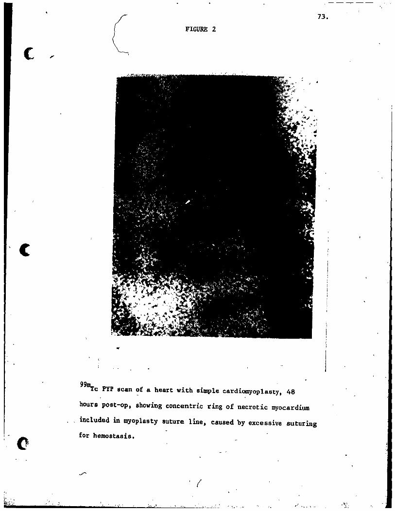
73.
FIGURE 2
(
(
..
99mtc PYP scan of a heatt with sLmple cardio~oplasty, 48
houra post-op, showing concentric ring of necrotic myocardium
included in myoplasty suture line, caused by excessive aututing
o for hemostasis.
. ( "
f" \-, , ':> _

o
•
74. ~IGURE 3
(
'.
Ph~tograph of a formalin fixed myocàrdium and simple cardiomyoplaaty
84 days- post-op. Th~ \eart is oriented with the apex at the bottom
of the picture and the 1eft ventricular free "'a11 shown. The pedicle'
graft had been dissected to illustrate the thinning, fibrosia and
aneurysm formation of the muscle included in the repair. Note a1ao
the large mural thrombus and the concentric ring of acar tissue in
the myocardium.

c-
o '.
FIGURE 4
Four ehamber eehocardiogram of the heart (LA - 1eft atriUm, -
LV - left ventricle. IVS - inter-ventricû1ar s~ptum). Echo
cardiogram of a simple cardiomyoplasty showing mural thrombus
at apex of le ft ventricle indicated by arrow 14 days post-op.
7SV
i
Ji 1
1 1
If

o
o
o J,
76. · FIGURE 5
\ Photomicrograph of latissimus dorsi muscle included in ~ardiomyo-
piast y with pericardial patch neo-endocardium technique, 65 day.
post-op. Note normal muscle fibers without atrophy, necrosis and - /
only minimal arteriolar hyperpl~&ia. /
" .. ~
, \
1
1
- 1
_'.1

c
't. 1.. \ \,
, .
77. FIGURE 6
,
..
,.-
Photomicrograph of blood-pericardia1 interface in cardiomyoplasty
with pericardial neo-endocardium, 51 days post-op. Dark 8taintng
layer demonstrating positivity to anti-factor VIII stain proves
endothelializ~tion of perieard!al surface.
) "'.,
/'-~Jl~ •. ,j1~t"M;:;J;\I'E t _~ ~ ~1I" 1 1,..., /', - ~ .. ' ,." ~,~...rl~"'l .. i, ",~~" .l'... .. r ...
'r

78.
o TABLE 1:
CLASSIFICATION OF CARDIOMYOPLASTY
1. Anatomical Segment of Cardiac Repaira
A. Atrial
B. Ventricular
1. Full thickness.
2. Partial thicknesB
II. Anatomical Status of Myoplasty: ,
A. Pedicle graft
o 1. Innervated
2. Denervated
B. Free graft
III. Functional Status of Myoplasty:
A. Adynamie (non-stimulated)
B. Dynamic
1. Single pulse stimulatio~
2. Pulse train (burst) stimulation
.'
o
.. . .'~" __ J ....... ' ._

c
(
)
79.
REFERENCES:
1. Walsh, G., Chiu, R.C.-J.I Skeletal muscle for cardiac repair and
assiste In Biornechanical Cardiac Assist: Cardiom 0 l and
Muscle Powered Deviees, R.C.-J. Chiu, ed., Chapter l, p.1, Futura
Publishing Co., Mount Kisco, New York, 1986.
2. Carpentier, A., Chachques, J.C.: Myocardial substitution with a
stimulated skeletal muscle: First successfu1 olinical case.
Lancet VI: 1267, June 1, 1985.
3. Magovern, G.J., Park, S.B., Magovern, Jr., G.J. et,al: Latissimus
dorsi as a functioning synchronously paced muscle component in the
repair of a left ventricular aneurysm. Ann. Thorac. Surg. 41: 116,
1986.
t>-4. Macovialt, J., Electrical conditioning of in situ skeletal muscle
for: replacement of myocardium. J. Surg. Res."~2: 429, 1982.
" 5. Drinkwater, O., Ch~u, R.C.-J., Modry, D. et al: Ca~diac assist and.
myocardial repair with synchronously stimulated skeletal muscle • • Surg. Forum 31: 271, 1980.
6. Petrovsky, B. V. 1 Burg!cal treatment of cudiae aneurysms.
7.
J. Cardiovasc. Burg. 7t 87; 1966.
..
Papp, M.D.. Expedll8ntal use of intercostal musc~e flaps for
repair of induced cardiac defects. J. Thorae. & Cardiovaso. sqrg.
90, 261, 1985. ,
~Î •• ! .'

o
o
•• • 0
-, ~~~ ..
Î 80.
8. Beek, C.S.: The development of a new b~ood &upply to the heart
by operation. Ann. Surg. 102: BOl, 1935.
,9. O'Shaughnessy, L.: Experimental method of providing a collateral
circulation to the heart. Br. J. Surg. 23: 665, 1936.
10. Kantrowitz, A.: The experimental use of th~ diaphragm as an
auxiliary myoeardium. Surg. Forum 9: 266, 1959.
Il. Termet, H., Chalencon, J.L., Estour, E. et al: Transplantation
sur le myocarde d'un muscle stie excite par pace maker. Ann. Chir.
Thor. Car. 5: 568, 1966.
12. Shepard, M.P.: Diaphragrnatie muscle and cardiac surgery. Ann.
Roy. Coll. Surg. Engl. 45: 212, 1969.
13. Nakamura, K., Glenn, W.L.: Graft of diaphraqm as a functioning
substitute for myocardium. J. Surg. Res. 4: 435, 1964.
14. Macoviak, J., Stephenson, L.W., Spielman, S.r Replacement of
ventricular myocardium with diaphraqmatic skeletal muscle.
J. Thorae. & Cardiovasc. Surg. 81: 519, 1981.
. 15. Sola, O~M., Dillard, D.H., Ivey, T.D. et, al: Autotransp1aptation
of akeletal œuscle into myocardium. Circ. 71r 341, 1985 •
, ,
'.'
o
•

o
o
o
, '
... 1 - \
1 •
8l.
/ t-
16. Dewar, M.L., Drinkwater, D.C., Wittnich, C. et all aynchronoualy
st~ulated skeletal muscle graft for myocardial repaire An
Experimental Study. J. Thorac. & Cardiovasc •. Surq. 871 325', 1984.
17. Dewar, M. L., Chiu, R.C .-J .1 Skeletal muscle graft for myocardial
repaire (Rep1y to J.A. Macoviak's Latter to the Editor). J. "'
'J7horac. & Cardiovasc. Surq. 88: 794, 1984. ". "
18. Leriche, R.: Essai experimentale de traitement de certains
infarctus du myocarde et de l'aneurisme du coeur par une greffe de
muscle strie. Bull. Soc. Nat. Chi,r. 59: 229,1933.
r 19. Gaines, W. E., Goldberg, N. H., McLauqhlin, J. S. 1 Reconstruction of
~,
the right vehtricular outflow tract with a vasculariZéd free flap
of striated muscle. Surg. Forum 36: 250, 1985.
20. Yee, E.S., Ebert, P.A.: The PQtential use of myofascial flaps
for congenital cardiovascular malformations. In Biomechanical
Cardiac Assist: Cardiomyoplasty and Muscle Powered Deviees,
R.C.-J. Chiu, ed., Chapter 8, p. 115, Futura Publishinq Co.,
Mount Kisco, New York, 1986.
21. Asinger, R.W., Mikell, F.L., Elsperger, J. et ab Incidence ot
left ventricular thrombosis after acute transmural myocardial .
infarction. New Engl. J. Med. 305: 297, 1981.

82.
22. Salmons, S.I The response of skeletal muscle to different vatterns
of use. In Plasticity of Muscle, pp.387-399, Walter de Gruyter &
Co., New York, 1980.
'23. Brister, S., Fradet, G., Dewar, M. et ~ll Transforming skeletal
muscle for myoeardial assiste Feasibility study. Cano J. Surg.
28: 341; 1985.
:)
24. walsh, G.L., Dewar, M.L., Khalafalla, A.S., Neilson, I.R., ~
De4tmon, J.H., Chiu, R.C.-J.: Characteristies of transformed
fatigue resistant skeletal muscle .for long-term cardiac assist
by extra-aortie balloon counterpulsatiog. Surg. Forum 37: 205,
1986.
(
c - \'(
1 ~,
ffj,\l;~-~- ~ u~ _ ,~\.~ •• ! ._ ...... _.~._ .;~~n; .. _~~~~; ",.'<.'."...})..::L.:~':~ .. " ~: .. JJ,,~.~: !,_{ .... _ ... , , '

o
o
o
" ,
C!tAPTER IV
-
w
FUTURE PERSPECTIVES
/
'A' '.~

,
-c
o
,/ 1
with the advent of improved Medical management an~ surgieal 0-
techniques for pediatrie and adult cardiac patien~s, predictably more
peoplé are surviving with limited card~ac reserve. While the mortality '---
may be lese, the morbidity'of this subpopulation who are f~nctioning at ... -
a New York Heart Class IV with li~ited exercise tolerance is higher.
The best option at present is fo,.cardiac transplantation with a close
to 90\ one year survival in this group of patients with a life
expectancy of less than 6 months withoùt intervention. Despite
requirements for immunosuppressivQ drugs with potential renél and j
infectious side .,.effects, this offers the best "palliation" for a limited "?
few. If our North American organ retrieval and transplantation services
\ were able to function optimally, at best l, 000 donor hearts would become' \
~vailable per year, which represents less than 3\ of the estimated
'35,~0 patients annually who bec~e end-stage cardiac cripples~ . l 0
Despite over JO years of research into artific~al hearts, there
remains, the omnipresent problem of bio-incompatibility of thè synthetic
materials and the potentially lethal thrombo-embolic side effects. ( -
There is a continued deJpendency bn cumbersome external power sources. ,
Our extra-aortie ballon concept diagrammed on page 42 is subject to Many
of the above mentioned complications and is limited by its blind-end -' . . ,
bud design. This {onfi9uration vas used mere1y to p~ove hemodYh'ami~ , ,-assistance WAS possible and was not inten~d for long-term use.
" '1 Il';
U1 timate thrombosis of the balloPn was Inevitable. This has led to. the'" ..,-'
developmenbof a second generation of'extra-aortic balloon
counterpülsatora (aee Figures 1 .. 2, ~) • In this design, the submuseular .
balloon la ~ in contin~ity with. the vascular system and ls instead
filled with a hydraulic fluid which when compre~sed transmit~ the 'volume -. ~
. . . . ~~,~'~ ............ __ .. ____ t_-______________________ ~~~~i. Jl ___ -
-
\.
. .
•
.... ;.

o •
o
o , ,
o -~'
84. ,.
chanqe to a' flexible dlap~CJ1I' which "oounterpul •• '" blood flowfn9 .
, '" , . throuqh a qraft which 1& anastomosed t~ th. aort.. Th~. allov. blood 'ta
flow throuqh the conduit rather than stagnate in a blin~-8nd bud balloan
if the system ahould fail. Oiotting may still be a problem at
connection ~ites ~~ ~have witnessed ~n valves and oouplera in \
artificial~rts. ,The requirements of constant movement and
flexibility of a diaphragm ln the,vascular system will require intense
research in materials and rheology. The requirement for movement of
li skeletal muscle 8gainst a prosthetic balloon ma.y over time result ln
G,
.t •
slippage in" position of the prosthesis and the ultimate fibrosis et the
interface between biologie tissues and prosthetic materials. A :" .
prosthetic_balloon which permits tissue ingrowth would be Most suitable.
Our pulse train stimulator use,d to eharaeterize the skeletal"
musclels el~ctrIcal mechanieal parameterB weB powered by a 6 volt
transformer and waB not implantable. In conjunction with Medtronic ..
Pacemaker Division, a pacemaker has been developed which now combines '-- ,
'the R-wave- sensinq cc:pabili ties of a Symbios pacemaker (;: standard.
pacemaker clinically used today in patients with.electric~ disturbanbeS . , j." ,
an" ,hear;t blocks) with a pacemaker which is capable "of generating a
programmable traiJ) of pulses such as the Itrel pacemaker which we have
used to transform our skeletal museles. This hybrid pacemaker is now ~
completely implantable device (sée Figure 4). In long-term cardiac,
assistance, however, battery technology IMY Hmi t the total amount of . ~ner9Y'whtch i~ avail8ble to stimulate s~eletal muscle. More research
18 neces8ary in elect~e design to elucidate the optimum mode of pacin9 ,
a motorneuron'to avoid fibrosis and increased resistance at the 1
, electrode nerve interface. "Fatigue" witnesaed in even traniformed
"
'4

"
o
<'
6~, ~ ~; " .. ~~T.'t .~t_ J. : . .:'
:J' 85 •
• muscle may be related ~re to nerve stimulation ra~h~r than aetual_ L.
muscle contractile pr?teln fatigue. \
• <i
V" Cardiomyoplasty techniques will always be troubled by differences' ,
... -in orientation of skéletal muscle fibers which are linear compared to
"---.. cardiac muscle fibers ~hleh are in a syneytium •. Although a skeletal '
muscle layed onto or into the heart ls capable of vlgorous contraction
duriQg stimulatio~, much of its energy is wasted as it tends ~o pull the
heart rather than squeeze with the ventriele in a more pumping motion.
There nevertheless appears to be potential for use às inlay patches to
ènlarge outflow tracts and with autogenous tissue, there remains the /
possibility for growth with the child. A severely deformed, hypoplastie~
_heart, however, would be bett~r removed ~d transplanted (as we now • 0 •
(
witness the dawn of the era of pediatrie eardiae transplantation), ~. .' . . rather than attempts to alter cardiae chambers with ~kele~al muscle.
Limitatio~s, however, ~ill continue to exist with.scarcity of donor
hearts, an even more eritical shortage in the pediatrie population. o " ,
" '
,
'"
J'
Gene manipulation may one d~y be important in assist1ng and int~n8ifying -.J .'
musele~transformation and may help,with intigration of skeletal musdle
with cardiae muscle., l,
, \
: 1 .,. . ,
In the 1700' s,i if one looked into thè sky and saw something flying, . , , .~
one w~uld he 1~0' correct in assuming that it was a bird, as that' 'fas ,-' { . .' the only thing with this càpability at that time. If a doctor in the
'" , 1
21st century ex~es a patient wit~ ~ known history pf congestive . /
heart failure or congenital,heart 4i~~ase, and feels a goodÂuality • ,;>. ! 1 1 . / C~fotid pulsation, will,he ~r she ~e correct in assuming that the heart 'Ii 1 ! / '
1 • ~ "
18 the on1y organ )Il{ich 18 ~uncti0l1i~g wall? / '# •
/ ' .' .
" " t~
.J • \ l , .J

~.
) -
o
_.
, <
,0
'<If
o
.. J
+,
Ao~.
FICURE l
,
SYSTOLE
'. ,
, 1
Pulaé.Traln Slimulaior
Second Generation,Extra-aortic Balloon Design
86.
During cardiac systole, the hydraulic pouch in its
sub-muscular position is filIed •
•
" , .'
"
"
.'
t
l"
\
;~
,ù
..
" . ..
,,-
1 .
.>
, .
"
"

" " '
, .
.0
,.t :
"
"
-'- ..
'" \
. '
\ ' \
FIGURE 2
1 <:1
DIASTO~E
\
Aorl. /
/
. .
87.
Skln
PulS8,Traln SUrnuiaior
/
Second 'Generat,iQn Extra-aortie B'alloon Design , . Dudng cardiac diastole, there i8, counterpulsat,ion
i9 '
of the colulnn 'ol:blood within the, graft .thr~ugh. , < ,
{.'
a flexible membrane.
l "
' . ./'
{ , 4'\
'1
.' 1 , • , ., ~
"
"
. ... "
..

o~
, ,
o . ,
{
"
o , , r
-, , ~1i;tï~----":-;-:-----------~---~-______ " •• II!II!!.', ..... III! . ' ,) .' , ,.
. '
FIGURE 3
.. OFF
Aorta
. "
\
\
88 •
,1
r
Skln
Pulse Tlllin Slhnulltor
• Second Generation Extra-aortie Bal100n Design
When the machine i8 off, there i8 a free laminar
, 'flow of blood through the conduit. The pigtail 1
~ addition to the hydra~lic pouch allows for
periodic adju8t~nts of the reating fluid
tension.
,
" ..
'.
5

o
.Ô
c
, ,
..
l'
.,
. , 1
.'
FIGURE 4
Implantable cardiomyostimulator designed by • 1 1
M8dtronic in Holland. This pacemaker is
capable of sensing the R-wave of ~ardi4c
excitation and can generate -a :xain of' pulses
'appropriately timed.
..
.. ., ..... ,
89 .
1
~':'- _ F.. _ ",_~ l ~~ ___ I~~: .11i. H~,-~~~i~1Jl~:Â_ ',~.iti .i=t~ A.~K~} ~ .. ;:_ -.,.\~ _~._(·.L .L.! ". '; .. -. f' : u ~ .. _ !;':' . ..- 2~~ ",") .. .!~ ~ , ;_ ~~ ,\~~lM.-!1~.~~. ,)-. itt:.. .. ~k~ .. :UJ ld ~ ~~~:';',---~_:C .. -i •. ~... .~~ ,-~',.1' .~,,'t -,' _.~ ... ,..~.:A:-_ •
, 1 , ,
1 -1
1
r