Embed Size (px)
Citation preview

Introduction
Systemic Lupus Erythematosus (SLE) is a chronic in-flammatory rheumatic disease which affects the connectivetissue. Its etiology is as yet unknown, while its patho-genesis involves the immune system (1). The organs mostaffected are the skin and the musculoskeletal, hemato-poietic, digestive and cardiopulmonary systems, hencethe nomenclature “systemic” (2).
LSE has been associated with changes in the gene-tic factors involved in modulating the immune tolerance
SUMMARY: Ischemic necrosis with sigma perforation in a patient withSystemic Lupus Erythematosus (SLE): case report.
I. IANNELLA, S. CANDELA, L. DI LIBERO, F. ARGANO, E. TARTAGLIA
G. CANDELA
Systemic Lupus Erythematosus (SLE) is a chronic inflammatoryrheumatic disease which affects the connective tissue. Its etiology is asyet unknown, while its pathogenesis involves the immune system.
Both genetic and environmental and hormonal factors play a keyrole in the impaired immune regulation. A correlation with estrogens isdemonstrated by the fact that the greatest incidence is found in youngwomen, when estrogen secretion is at its highest. The disease is also re-ported to worsen in women taking oral contraceptives. It is therefore be-lieved that the components of oral contraceptives, estrogens (ethinylestradiol) and progestins, can affect the immune profile.
Of the various complications attributed to systemic lupus erythe-matosus, gastrointestinal disorders are less common but potentially byfar the most serious.
We report a case of ischemic necrosis with sigma perforation in apatient with SLE. Signs and symptoms of acute abdomen in patientswith SLE are rare (0.2%), but serious. Most patients require an explo-ratory laparotomy, as the causes are often linked with vasculitis.
RIASSUNTO: Necrosi ischemica da perforazione del sigma in pazientecon panvasculite da lupu eritematoso sistemico: caso clinico.
I. IANNELLA, S. CANDELA, L. DI LIBERO, F. ARGANO, E. TARTAGLIA
G. CANDELA
Il Lupus Eritematoso Sistemico (LES) è una malattia reumaticainfiammatoria cronica ad eziologia ancora sconosciuta e a patogenesiimmunitaria. Colpisce i tessuti connettivi di tutto l'organismo.
Sono state identificate alterazioni in fattori genetici ma anche fat-tori ambientali ed ormonali giocano un ruolo chiave nel determinareun’alterata immunoregolazione. La correlazione con gli estrogeni vieneancor più evidenziata dal fatto che la maggiore incidenza della patolo-gia si riscontra nelle donne giovani, quando è massima la loro secrezio-ne. Inoltre in donne con LES è stato riportato un peggioramento dellamalattia durante l'assunzione dei contraccettivi orali. Queste osserva-zioni inducono a ritenere che i componenti dei contraccettivi orali, co-stituiti da estrogeni (etinilestradiolo) e composti ad attività progestini-ca, siano in grado di modificare l'assetto del sistema immunitario.
Tra le varie complicanze attribuite al lupus eritematoso sistemico,quelle a carico dell’apparato gastrointestinale sono meno comuni mapotenzialemente molto più gravi.
Riportiamo il caso di una necrosi ischemica del sigma con perfora-zione in paziente con LES. Sintomi e segni di un addome acuto sonorari in pazienti con LES (0.2%) ma gravi. Comunque la maggior par-te dei pazienti con tali sintomi richiede una laparotomia esplorativa inquanto le cause sono spesso legate a fenomeni vasculitici.
KEY WORDS: Systemic lupus erythematosus - Intestinal infarction - Vasculitis - Colon perforation.Lupus eritematoso sistemico - Infarto intestinale - Vasculite - Perforazione del colon.
Ischemic necrosis with sigma perforation in a patient with Systemic Lupus Erythematosus (SLE): case report
I. IANNELLA1, S. CANDELA1, L. DI LIBERO2, F. ARGANO3, E. TARTAGLIA4, G. CANDELA5
G Chir Vol. 33 - n. 3 - pp. 77-80March 2012
77
1 S.M.d.p “.Incurabili” Hospital, Naples, ItalySurgery and Laparoscopic Surgery (Director: C. Romano) 2 “Sanatrix” Clinic, Naples, ItalySurgery Division 3 S.M.d.p.”Incurabili” Hospital, Naples, ItalyDiagnostic Radiology 4 SUN Medicine and Surgery Faculty, Naples, Italy Student5 SUN Medicine and Surgery Faculty, Naples, ItalyGeneral Surgery VII
© Copyright 2012, CIC Edizioni Internazionali, Roma
0094 7 Ischemic_Iannella:- 19-03-2012 14:29 Pagina 77
78
I. Iannella et al.
of autoreactive cell clones. However, environmental andhormonal factors are also important (3). One of the mostimportant reasons for believing that estrogens have a keyrole in the etiopathogenesis of SLE is that women aremore vulnerable to autoimmune diseases, as they havea higher immune response than men (4). It has been cal-culated that the incidence of autoimmune diseases in wo-men is nine times higher than in men. Further eviden-ce for the correlation with estrogens comes from the factthat the greatest incidence is found in young women,when estrogen secretion is at its highest (5-11).
The use of oral contraceptives has been found to fa-vor the onset of SLE in women without clinical or la-boratory signs of the disease, and in known sufferers, aworsening of the disease while taking oral contracepti-ves has been reported (12-15). It is therefore believed thatthe components of oral contraceptives, estrogens (ethinylestradiol) and progestins, are capable of changing the im-mune profile. Despite this, it should be remembered thatthird generation contraceptive pills contain very low do-ses of estrogens, the component responsible for an in-creased risk of thromboembolic disease (16).
Of the various complications attributed to systemiclupus erythematosus, gastrointestinal disorders are lesscommon but potentially much more serious (17).We re-port a case of ischemic necrosis with sigma perforationin a patient with SLE.
Case report
A 40-year-old woman with SLE was admitted to the Unit of Sur-gery and Laparoscopy Surgery at “Incurabili” Hospital, Naples, Italywith diagnosis of abdominal obstrucion. The patient, who had inthe past had a left adnexectomy for ovarian cysts, had around 20 daysearlier undergone a total right laparohysterectomy for uterine fi-bromatosis. She had suspended her SLE cortisone therapy (predni-sone 25 mg/day) a few days before surgery.
On admission to our unit the patient was suffering from abdo-minal pain, nausea and vomiting; these signs and symptoms had al-ready begun during her convalescence after laparohysterectomy. Aphysical examination on admission revealed her abdomen to be ten-se, while some blood tests were abnormal: CPK 309 (normal value[n.v.] <145), fibrinogen 552 (n.v. 150-450), RBC 3.71, WBC 8.9,lymphocytes 26.4 (n.v. 25-50), and proteinuria. Abdominal X-rayrevealed air-fluid levels; pelvic ultrasound revealed corpuscolated filmin the right abdomen and conglomerated fluid-filled intestinal loo-ps.
Intestinal obstruction was diagnosed and the patient underwentlaparotomy with adhesiolysis and debridement of the terminal ileum,as the terminal ileum was found to be anchored to the right poste-rior peritoneum through an omega-shaped bridle.
After 36 hours the patient presented abdominal obstruction si-gns and fever. Cortisone therapy was re-begun with Methylpredni-solone 20 mg i.v. followed by Deltacortene Prednisone 10 mg/dayi.v. Further tests were carried out: blood Albumin 3.1 (n.v. 3.5-5.2);serum nephelometry (IgG 558 [n.v. 751-1560]); C reactive protein212 (n.v. 1-8); RA-test 52.1 (n.v. 0-25); ANA neg., AMA neg., ASMAneg., anti-LKM neg., anti-native DNA neg., ENA neg., LAC-sen-sitive APTT =1.19.
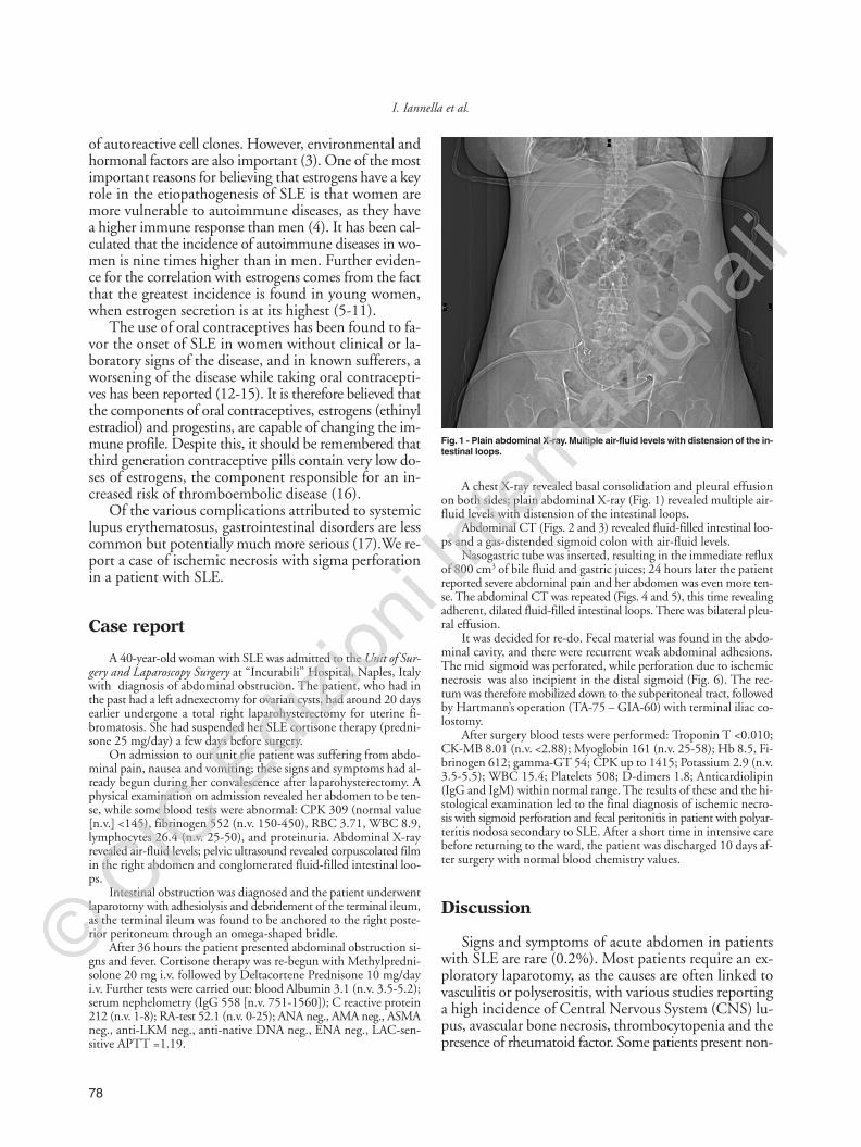
A chest X-ray revealed basal consolidation and pleural effusionon both sides; plain abdominal X-ray (Fig. 1) revealed multiple air-fluid levels with distension of the intestinal loops.
Abdominal CT (Figs. 2 and 3) revealed fluid-filled intestinal loo-ps and a gas-distended sigmoid colon with air-fluid levels.
Nasogastric tube was inserted, resulting in the immediate refluxof 800 cm3 of bile fluid and gastric juices; 24 hours later the patientreported severe abdominal pain and her abdomen was even more ten-se. The abdominal CT was repeated (Figs. 4 and 5), this time revealingadherent, dilated fluid-filled intestinal loops. There was bilateral pleu-ral effusion.
It was decided for re-do. Fecal material was found in the abdo-minal cavity, and there were recurrent weak abdominal adhesions.The mid sigmoid was perforated, while perforation due to ischemicnecrosis was also incipient in the distal sigmoid (Fig. 6). The rec-tum was therefore mobilized down to the subperitoneal tract, followedby Hartmann’s operation (TA-75 – GIA-60) with terminal iliac co-lostomy.
After surgery blood tests were performed: Troponin T <0.010;CK-MB 8.01 (n.v. <2.88); Myoglobin 161 (n.v. 25-58); Hb 8.5, Fi-brinogen 612; gamma-GT 54; CPK up to 1415; Potassium 2.9 (n.v.3.5-5.5); WBC 15.4; Platelets 508; D-dimers 1.8; Anticardiolipin(IgG and IgM) within normal range. The results of these and the hi-stological examination led to the final diagnosis of ischemic necro-sis with sigmoid perforation and fecal peritonitis in patient with polyar-teritis nodosa secondary to SLE. After a short time in intensive carebefore returning to the ward, the patient was discharged 10 days af-ter surgery with normal blood chemistry values.
Discussion
Signs and symptoms of acute abdomen in patientswith SLE are rare (0.2%). Most patients require an ex-ploratory laparotomy, as the causes are often linked tovasculitis or polyserositis, with various studies reportinga high incidence of Central Nervous System (CNS) lu-pus, avascular bone necrosis, thrombocytopenia and thepresence of rheumatoid factor. Some patients present non-
Fig. 1 - Plain abdominal X-ray. Multiple air-fluid levels with distension of the in-testinal loops.
0094 7 Ischemic_Iannella:- 19-03-2012 14:29 Pagina 78

specific signs and symptoms of dyspepsia associated withactive SLE, without any clear evidence of gastrointesti-nal involvement. This could in any case be linked withmesenteric vascular disease, peritoneal inflammation orcertain medications. Gastrointestinal signs may take va-rious forms: mesenteric vasculitis, esophageal disease, in-flammatory intestinal disease, liver disorders or pancreaticdisorders. Treatment with cortisone or immunosup-pressants may also be a predisposing factor for acute ab-domen.
All surgical procedures are more risky in patients withSLE than in healthy subjects, due to both the greater like-lihood of complications in long-term users of cortico-steroids and immunosuppressants and physical and psy-chological trauma related to the procedure and its pos-sible consequences for the immune system and thus pro-gression of the disease. When surgery is indispensable,these risks must be anticipated.
As regards our case, numerous studies have indica-ted that, in the presence of uterine fibromatosis, treat-
79
Ischemic necrosis with sigma perforation in a patient with Systemic Lupus Erythematous (SLE): case report
Figs. 2 and 3 - Abdominal CT scan. Fluid-filled intestinal loops and gas-distended sigmoid colon with air-fluid levels.
Figs. 4 and 5 - Abdominal CT scan after 24 hours. Adherent, dilated and fluid-filled intestinal loops.
0094 7 Ischemic_Iannella:- 19-03-2012 14:29 Pagina 79

ment with GH-SH analogs, which can induce phar-macological menopause and thus block the menome-trorrhagia usually associated with fibroma, does not haverepercussions for SLE.
Conclusions
This rare case suggests that greater attention is ne-cessary when evaluating SLE patients with abdominalpain and abnormal blood chemical parameters who aretaking or have recently suspended treatment with cor-tisone. The decision to perform surgery should be me-ticulously evaluated, bearing in mind the procedure’srisks and consequences for an underlying immune di-sease.
80
I. Iannella et al.
1. Papa MZ, Shiloni E, McDonald HD. Total colonic necrosis acatastrophic complication of systemic lupus erythematosus. DisColon Rectum 1986;29(9):576-8.
2. Lee JR, Paik CN, Kim JD, Chung WC, Lee KM, Yang JM. Ische-mic colitis associated with intestinal vasculitis: histologicalproof in systemic lupus erythematosus. World J Gastroenterol2008;14(22):3591-3.
3. Versaci A, Macri A, Scuderi G, Bartolone S, Familiari L, LupattelliT, Famulari C. Ischemic colitis following colonoscopy in a sy-stemic lupus erythematosus patient: report of a case. Dis ColonRectum 2005;48:866-9.
4. Gore RM, Marn CS, Ujiki GT, Craig RM, Marquardt J. Ische-mic colitis associated with systemic lupus erythematosus. Dis Co-lon Rectum 1983;26:449-51.
5. Reissman P, Weiss EG, Teoh TA, Lucas FV, Wexner SD. Gan-grenous ischemic colitis of the rectum: a rare complication of sy-stemic lupus erythematosus. Am J Gastroenterol 1994;89:2234-6.
6. Miyahara S, Ito S, Soeda A, Chino Y, Hayashi T, Takahashi R,Goto D, Matsumoto I, Tsutsumi A, Sumida T. Two cases of sy-stemic lupus erythematosus complicated with colonic ulcers. In-tern Med 2005;44:1298-306.
7. Lee CK, Ahn MS, Lee EY, Shin JH, Cho YS, Ha HK, Yoo B,Moon HB. Acute abdominal pain in systemic lupus erythema-tosus: focus on lupus enteritis (gastrointestinal vasculitis). AnnRheum Dis 2002;61:547-50.
8. Boumpas DT, Austin HA III, Fessler BJ, et al. Systemic lupus
erythematosus: emerging concepts. Part 1: renal, neuropsychiatric,cardiovascular, pulmonary, and hematologic disease. Ann InternMed 1995;122(12):940-50. Review.
9. Boumpas DT, Fessler BJ, Austin HA III, et al. Systemic lupuserythematosus. Emerging concepts Part 2: Dermatologic and jointdisease, the antiphospholipid antibody syndrome, pregnancy andhormonal therapy, morbidity and mortality and pathogenesis.Ann Intern Med 1995;123(1):42-53. Review.
10. Mills JA. Systemic lupus erythematosus. N Engl J Med1994;330(26):1871-9.
11. Gladman DD. Prognosis and treatment of systemic lupuserythematosus. Curr Opin Rheumatol 1995;7(5):402-8. Review.
12. Hochberg MC. Updating the American College of Rheumato-logy. Revised criteria for the classification of systemic lupus erythe-matosus. Arthritis Rheum 1997; 40(9):1725.
13. Aladjem H, Schur P. In Search of the Sun: A Woman's Coura-geous Victory over Lupus. Scribner Publishing 1988.
14. Wallace DJ. The Lupus Book: A Guide for Patients and TheirFamilies. Oxford University Press, Oxford, UK 2000.
15. Lahita RG. Systemic lupus erythematosus. Ed. R.G Lahita. Thirded. 1999. Academic Press
16. Triolo G, Ferrante A, Triolo G. ImmunoReumatologia. Ed. Edi-SES, 2001 pp. 1-198
17. Cervera R, Khamashta MA, Font J, et al. Morbidity and mor-tality in systemic lupus erythematosus during a 10-year period.A comparison of early and late manifestations in a cohort of 1,000patients. Medicine (Baltimore) 2003; 82:299.
References
Fig. 6 - Intraoperative findings. Perforated mid sigmoid colon with incipient ne-crotic perforation in the distal segment.
0094 7 Ischemic_Iannella:- 19-03-2012 14:29 Pagina 80