Embed Size (px)
Citation preview

Scaffold Degradation Product Effect on
Myoblastic Stem CellsIshan Chatterjee
Grade 9 – Fox Chapel Area High School
1

Tissue Engineering
Hottest job for 21st Century
2
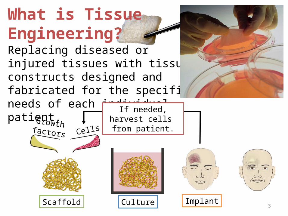
Replacing diseased or injured tissues with tissue constructs designed and fabricated for the specific needs of each individual patient.
What is Tissue Engineering?
CellsGrowth factors
Scaffold Culture Implant
If needed, harvest cells from patient.
3
Introduction: Stem Cells• Stem Cell -- An unspecialized cell characterized by
the capacity to give rise to various differentiated cell types
• C2C12 mus musculus (mouse) myoblast cell line – human stem cell surrogate
• C2C12 cells - model cell differentiation through myotube structure formation
• Serum Starvation (10% 1%) provides an inexpensive, easy means of inducing myogenesis
4
Introduction: Scaffolds
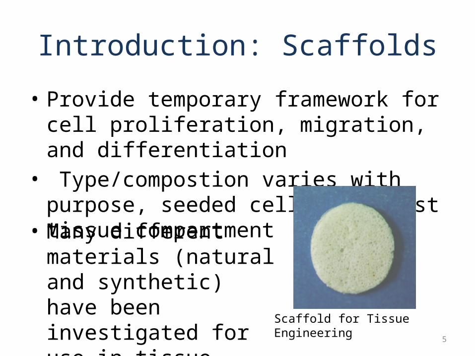
• Provide temporary framework for cell proliferation, migration, and differentiation
• Type/compostion varies with purpose, seeded cells, and host tissue compartment
• Many different materials (natural and synthetic) have been investigated for use in tissue regeneration
Scaffold for Tissue Engineering
5
Introduction: Scaffolds• Collagen Gel Scaffold
– Structurally similar to natural extracellular matrix– Can often be processed under relatively mild conditions– May be delivered in a minimally invasive manner– Cheaper than other scaffold materials
• Gelatin is a hydrolyzed form of collagen• Properties
– Strength and elasticity– Biocompatibility– Biodegradability– Non-toxic breakdown materials
6
Objective • Investigate the effect of scaffold degradation
products (gelatin) on murine (mouse) myoblastic stem cell line (C2C12). Specifically, to assess the degradation product effects on cell proliferation, differentiation, and toxicity.– Toxicity & Proliferation: Effect measured by counting
number of surviving stem cells after exposure to different concentrations of scaffold degradation products
– Differentiation: assessed by visually evaluating and estimating myotube formation (photomicrographs)
7
Hypothesis• Null hypothesis: Scaffold degradation products
will have no significant effects on proliferation, survivorship, and differentiation of C2C12 cells in vitro
• Alternate hypothesis: Scaffold degradation products will have significant effects on proliferation, survivorship, and differentiation of C2C12 cells in vitro
• Single factor Analysis of Variance (ANOVA) + Dunnett’s test for comparing multiple treatment groups against control– Null hypothesis rejected if p-value < 0.01 8
Experimental Design• Two Experiments (Toxicity/Proliferation and
Differentiation) each with four groups, two control plus two treatment:1. Group D (Control): no scaffold degradation
product solution, contains deionized water2. Group C (Control): no scaffold degradation
product solution, contains neutralized pH solution3. Group A: 1.0 mL (20% by volume) scaffold
degradation product solution4. Group B: 0.1 mL (2% by volume) scaffold
degradation product solution9
Materials and Apparatus• 20 g gelatin (Knox “Gelatine” Packets)• C2C12 murine myoblastic stem cells • Deionized sterile water• 100 mL graduated cylinder• Test tube rack• Incubator (37.0°C)• 100 - 1000 µL pipette• 0.1 – 1 mL pipette• 1 – 10 mL pipette• 70% Ethanol (for sterilization)• Felt-tip marker• 15 mL sterile conical tubes• DMEM media (10% calf serum & 1% calf
serum) contains salts, amino acids, vitamins, & glucose
• Sterile pipette tips• 0.22 micron syringe filters + 10 mL syringe• 150 mL HCl acid (0.1 molarity)• 15 mL NaOH base (1.0 molarity)• 200 g scales• Electric kettle
• 75 mL culture flasks• 25 cm2 culture flasks• 50 mL Trypsin-EDTA• 1 mL Trypan Blue• 32 mL PBS saline• 32 mL 100% ice-cold ethanol• 16 mL Toluidine Blue• 2 Hemocytometers• Light microscope• Inverted microscope (with imaging
capabilities)• Class II Biosafety hood• Labcoats, Eye Protection, Disposible Gloves• Razor• pH testing strips• 1 mL tubes• Delicate task wipes• Counter
10
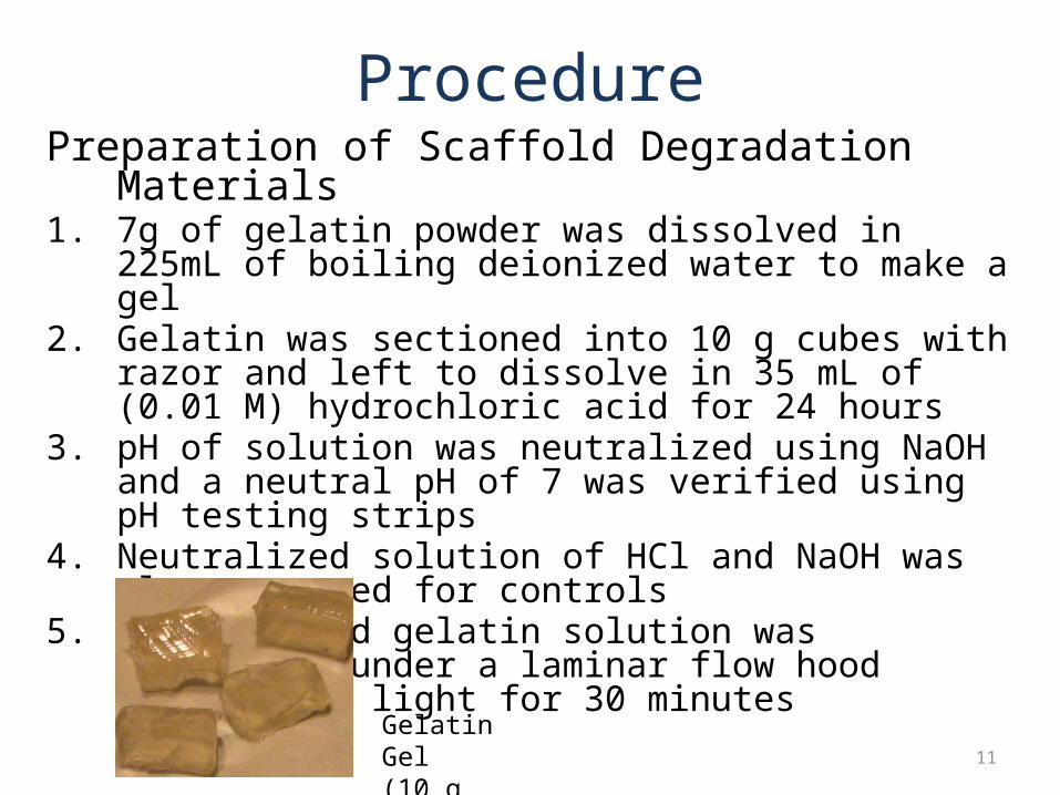
ProcedurePreparation of Scaffold Degradation Materials1. 7g of gelatin powder was dissolved in 225mL of boiling
deionized water to make a gel2. Gelatin was sectioned into 10 g cubes with razor and left to
dissolve in 35 mL of (0.01 M) hydrochloric acid for 24 hours 3. pH of solution was neutralized using NaOH and a neutral pH of
7 was verified using pH testing strips4. Neutralized solution of HCl and NaOH was also prepared for
controls5. The degraded gelatin solution was sterilized under a laminar
flow hood ultraviolet light for 30 minutes
11
Gelatin Gel(10 g cubes)
Procedure (contd.)Stem Cell Line Culture1. 1 mL aliquot of C2C12 cells from a cryotank was used to
inoculate 30 mL of 10% serum DMEM media in a 75mm2 culture flask
2. Media changed with 15 mL fresh media to remove cryo-freezing fluid and incubated (37° C, 5% CO2) for 2 days until 30-50% confluent (4*106 to 5*106 cells)
3. The culture was passed into 3 fresh 75mm2 flasks in preparation for experiment
12
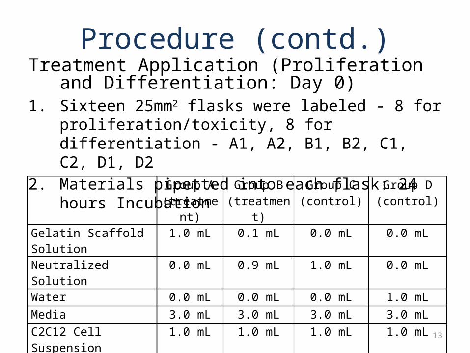
Procedure (contd.)Treatment Application (Proliferation and
Differentiation: Day 0)1. Sixteen 25mm2 flasks were labeled - 8 for
proliferation/toxicity, 8 for differentiation - A1, A2, B1, B2, C1, C2, D1, D2
2. Materials pipetted into each flask: 24 hours IncubationGroup A
(treatment)Group B
(treatment)Group C (control)
Group D(control)
Gelatin Scaffold Solution 1.0 mL 0.1 mL 0.0 mL 0.0 mLNeutralized Solution 0.0 mL 0.9 mL 1.0 mL 0.0 mLWater 0.0 mL 0.0 mL 0.0 mL 1.0 mLMedia 3.0 mL 3.0 mL 3.0 mL 3.0 mLC2C12 Cell Suspension 1.0 mL 1.0 mL 1.0 mL 1.0 mLTotal Volume 5.0 mL 5.0 mL 5.0 mL 5.0mLGelatin Concentration 0.1% 0.01% 0.0% 0.0 %
13
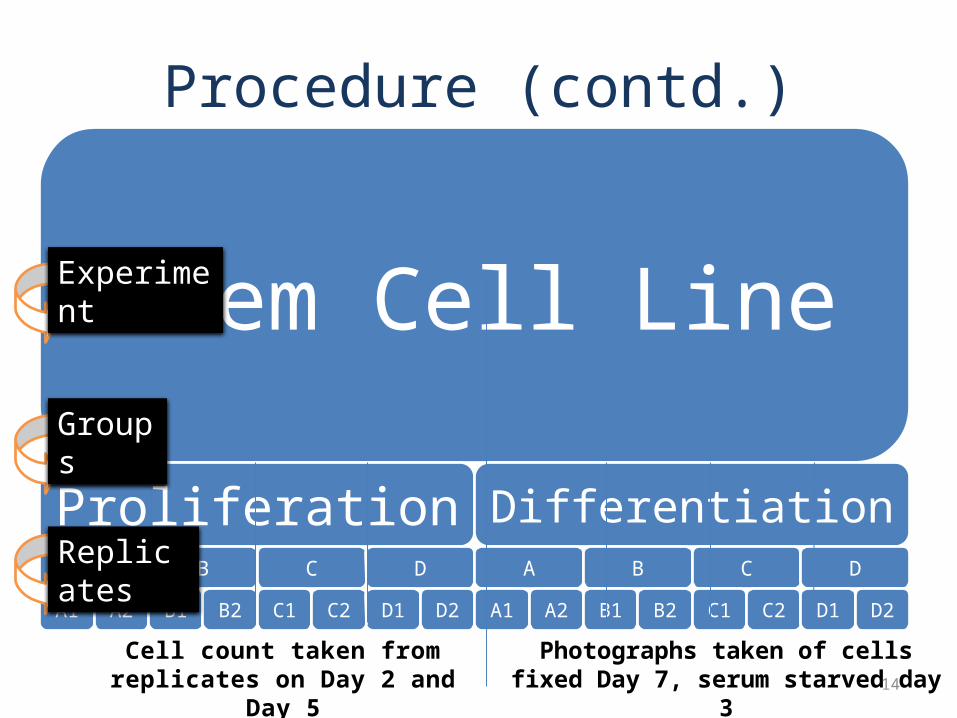
Procedure (contd.)
Stem Cell Line
ProliferationA
A1 A2
B
B1 B2
C
C1 C2
D
D1 D2
DifferentiationA
A1 A2
B
B1 B2
C
C1 C2
D
D1 D2
Cell count taken from replicates on Day 2 and Day 5
Photographs taken of cells fixed Day 7, serum starved day 3
Experiment
Replicates
Groups
14
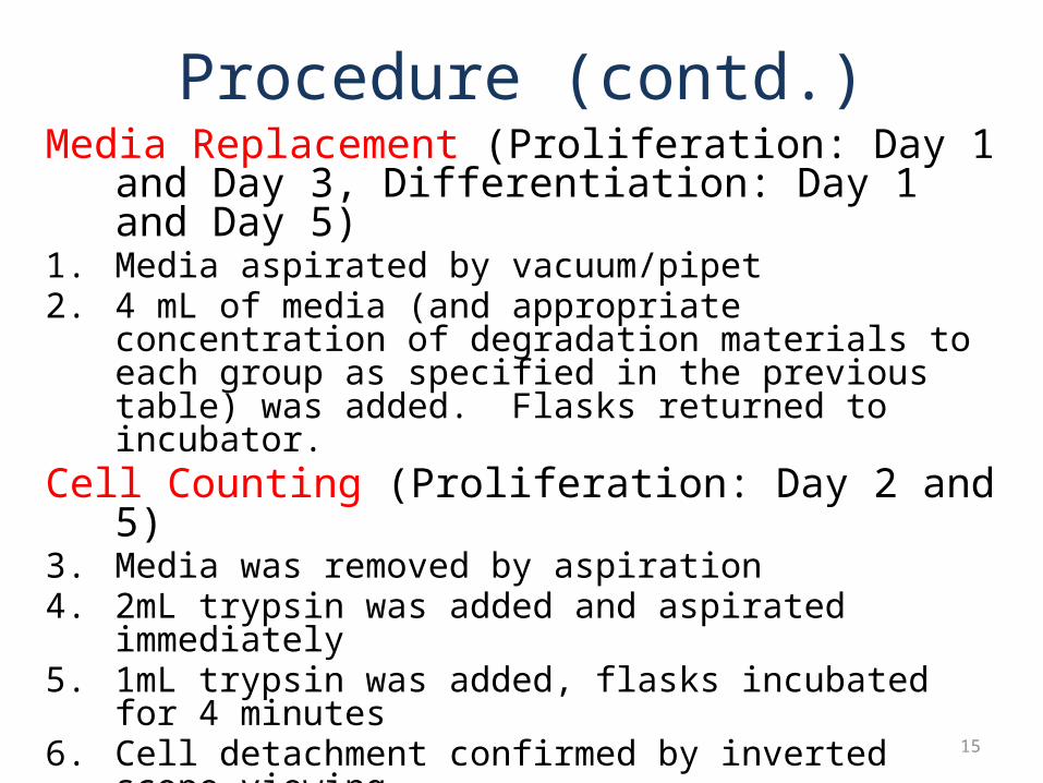
Procedure (contd.)Media Replacement (Proliferation: Day 1 and Day 3,
Differentiation: Day 1 and Day 5)1. Media aspirated by vacuum/pipet2. 4 mL of media (and appropriate concentration of degradation
materials to each group as specified in the previous table) was added. Flasks returned to incubator.
Cell Counting (Proliferation: Day 2 and 5)3. Media was removed by aspiration4. 2mL trypsin was added and aspirated immediately5. 1mL trypsin was added, flasks incubated for 4 minutes6. Cell detachment confirmed by inverted scope viewing7. Trypsinization quenched by addition of 4 mL media8. 1 mL of cell suspension added to sterile 1.5 mL microtubes
15
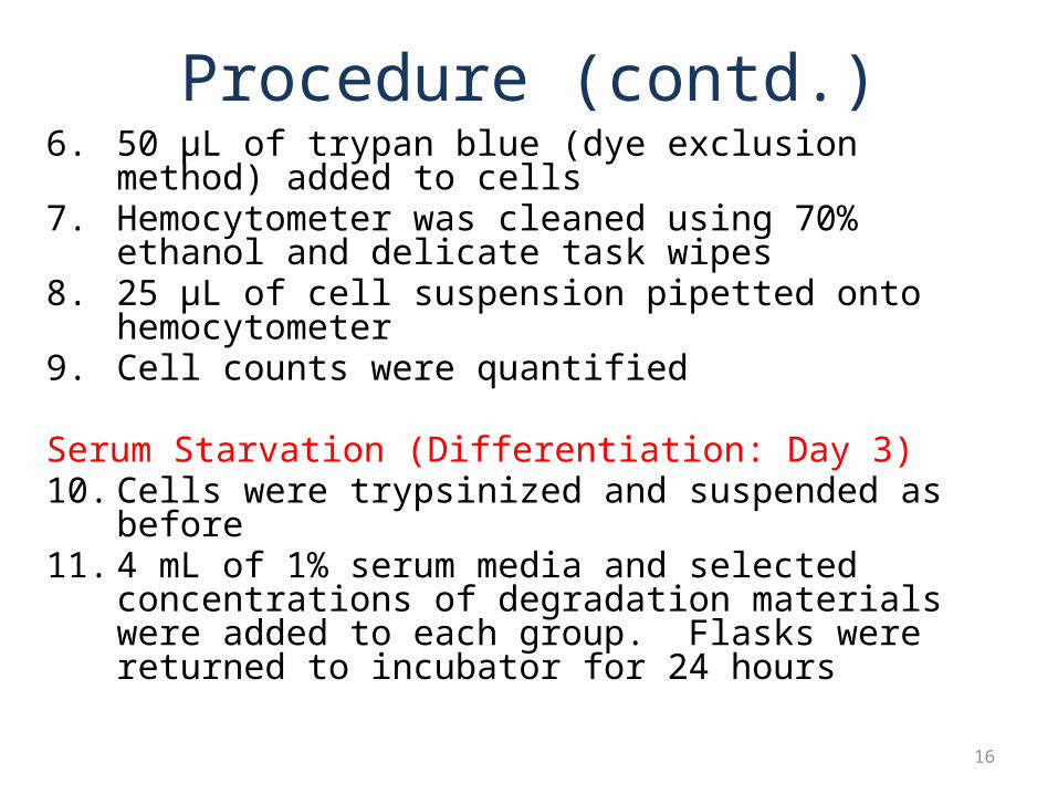
Procedure (contd.)6. 50 µL of trypan blue (dye exclusion method) added to cells7. Hemocytometer was cleaned using 70% ethanol and delicate
task wipes8. 25 µL of cell suspension pipetted onto hemocytometer9. Cell counts were quantified
Serum Starvation (Differentiation: Day 3)10. Cells were trypsinized and suspended as before 11. 4 mL of 1% serum media and selected concentrations of
degradation materials were added to each group. Flasks were returned to incubator for 24 hours
16
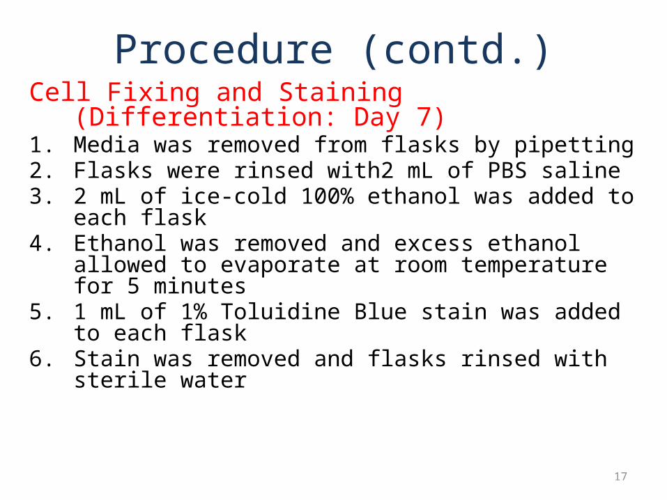
Procedure (contd.)Cell Fixing and Staining (Differentiation: Day 7)1. Media was removed from flasks by pipetting2. Flasks were rinsed with2 mL of PBS saline 3. 2 mL of ice-cold 100% ethanol was added to each flask4. Ethanol was removed and excess ethanol allowed to
evaporate at room temperature for 5 minutes5. 1 mL of 1% Toluidine Blue stain was added to each flask6. Stain was removed and flasks rinsed with sterile water
17

Procedure contd.Cell Photomicrography (Differentiation: Day 8)1. A Zeiss inverted microscope optical imaging system and
connected computer was prepared for photomicrography2. 5 pictures of each flask were recorded: top right, bottom
right, top left, bottom left, and center
Proliferation: Day 3 Proliferation: Day 6 Differentiation: Day 7
Examples of Cell Photomicrographs:
18
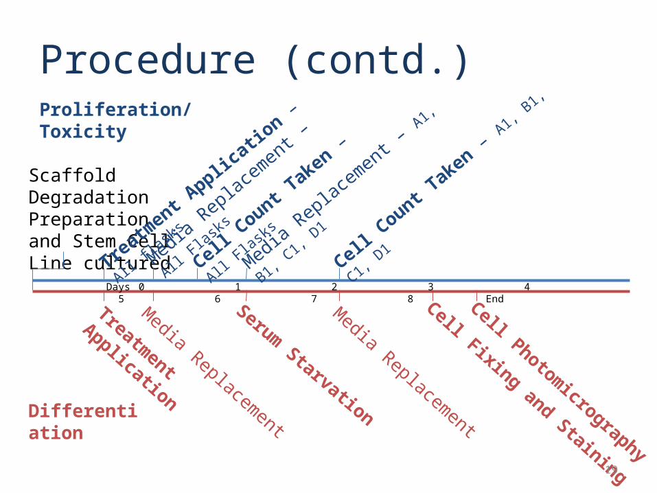
Procedure (contd.)Proliferation/Toxicity
Differentiation
Days 0 1 2 3 4 5 6 7 8 End
Scaffold Degradation Preparation and Stem Cell Line cultured
Treatment Application
Treatment A
pplication – All fl
asks
Media Replacement – All F
lasks
Media Replacement – A1, B
1, C1, D
1
Media Replacement
Media Replacement
Serum Starvation
Cell Fixing and Staining
Cell Photomicrography
Cell Count T
aken – All Flasks
Cell Count T
aken – A1, B1, C
1, D1
19
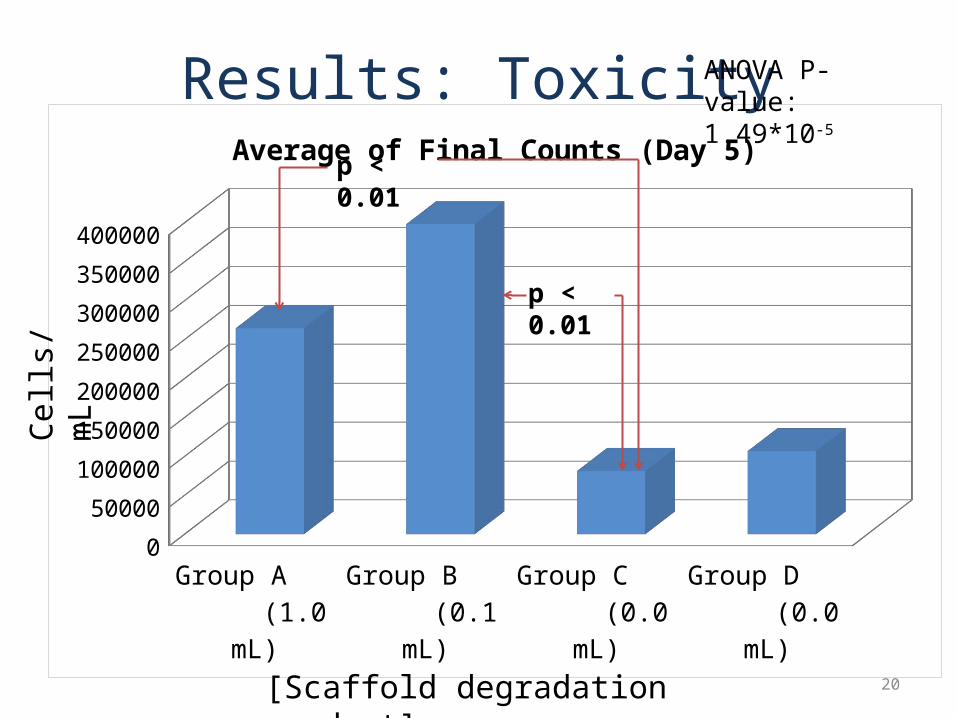
Group A (1.0 mL)
Group B (0.1 mL)
Group C (0.0 mL)
Group D (0.0 mL)
0
50000
100000
150000
200000
250000
300000
350000
400000
Average of Final Counts (Day 5)
Results: Toxicityp < 0.01
[Scaffold degradation product]
p < 0.01
Cells
/mL
ANOVA P-value: 1.49*10-5
20
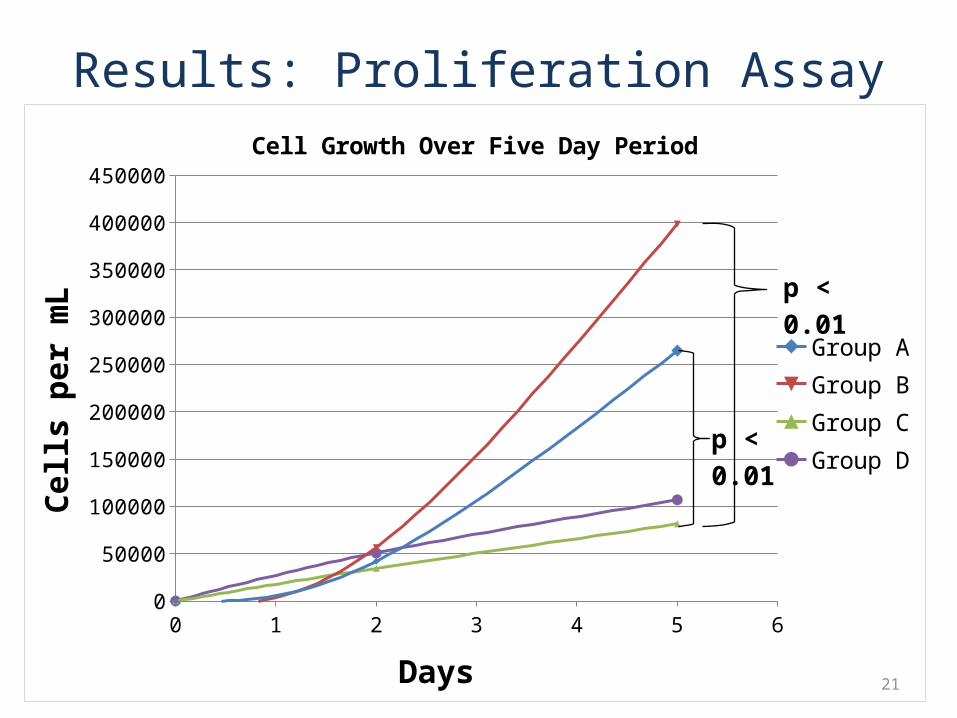
Results: Proliferation Assay
0 1 2 3 4 5 60
50000
100000
150000
200000
250000
300000
350000
400000
450000
Cell Growth Over Five Day Period
Group AGroup BGroup CGroup D
Days
Cells
per
mL
p < 0.01
p < 0.01
21
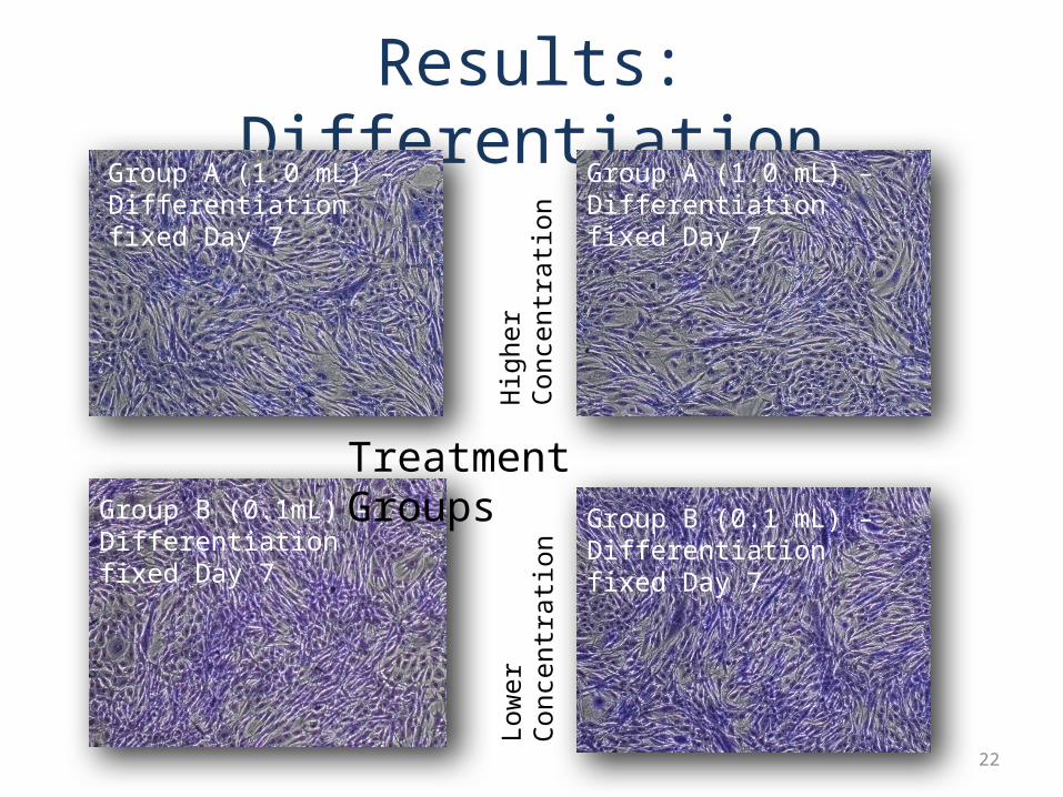
Results: DifferentiationGroup A (1.0 mL) – Differentiation fixed Day 7
Group A (1.0 mL) – Differentiation fixed Day 7
Group B (0.1mL) – Differentiation fixed Day 7
Group B (0.1 mL) – Differentiation fixed Day 7
Treatment GroupsH
ighe
r C
once
ntra
tion
Low
er C
once
ntra
tion
22
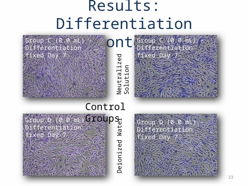
Results: Differentiation (contd.)Group C (0.0 mL) – Differentiation fixed Day 7
Group C (0.0 mL) – Differentiation fixed Day 7
Group D (0.0 mL) – Differentiation fixed Day 7
Group D (0.0 mL) – Differentiation fixed Day 7
Control GroupsN
eutr
aliz
ed S
olut
ion
Dei
oniz
ed W
ater
23
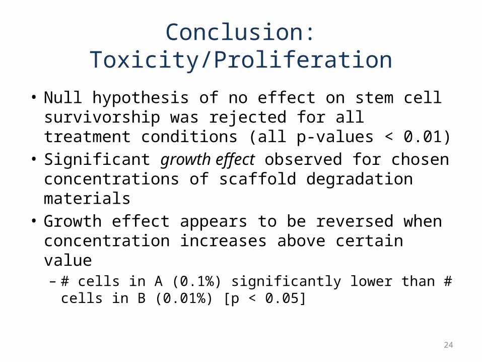
Conclusion: Toxicity/Proliferation
• Null hypothesis of no effect on stem cell survivorship was rejected for all treatment conditions (all p-values < 0.01)
• Significant growth effect observed for chosen concentrations of scaffold degradation materials
• Growth effect appears to be reversed when concentration increases above certain value– # cells in A (0.1%) significantly lower than # cells in B
(0.01%) [p < 0.05]
24
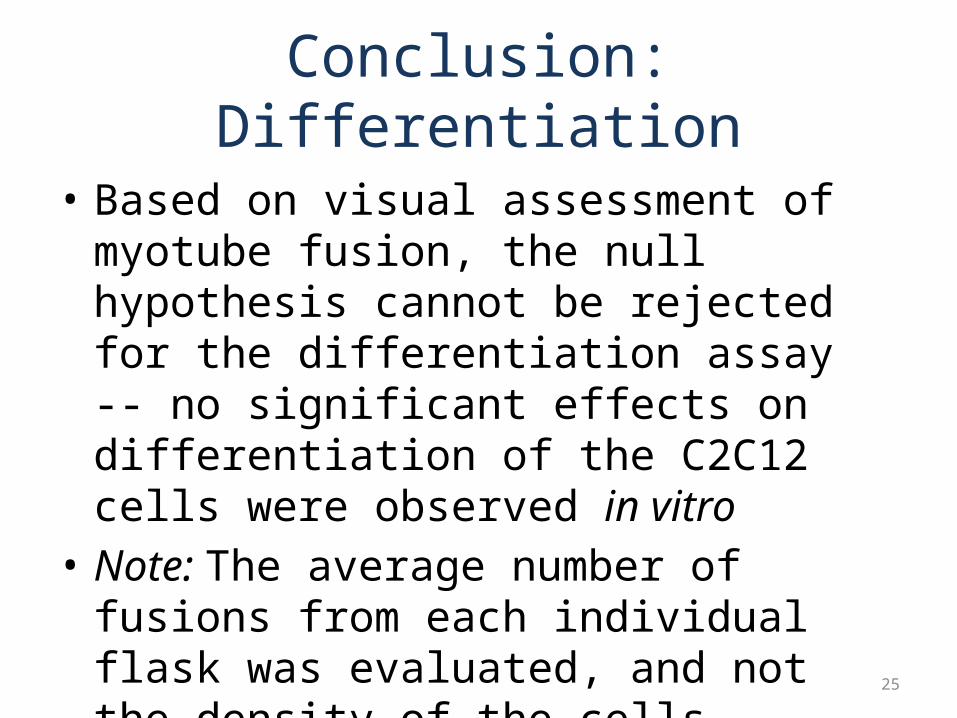
Conclusion: Differentiation
• Based on visual assessment of myotube fusion, the null hypothesis cannot be rejected for the differentiation assay -- no significant effects on differentiation of the C2C12 cells were observed in vitro
• Note: The average number of fusions from each individual flask was evaluated, and not the density of the cells.
25
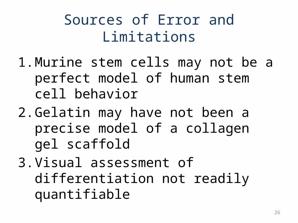
Sources of Error and Limitations
1. Murine stem cells may not be a perfect model of human stem cell behavior
2. Gelatin may have not been a precise model of a collagen gel scaffold
3. Visual assessment of differentiation not readily quantifiable
26
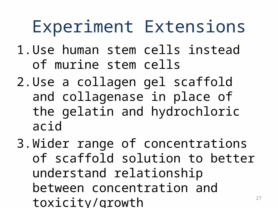
Experiment Extensions1. Use human stem cells instead of murine stem
cells2. Use a collagen gel scaffold and collagenase in
place of the gelatin and hydrochloric acid3. Wider range of concentrations of scaffold
solution to better understand relationship between concentration and toxicity/growth
4. For differentiation assay, explore the use of quantitative cell differentiation markers and techniques (anti-Myo D antibodies, RT-PCR, etc.)
27
ReferencesAn Outreach Education Manual in Tissue Engineering, Pittsburgh Tissue
Engineering Initiative "Gelatin." Wikipedia, the free encyclopedia.
<http://en.wikipedia.org/wiki/Gelatin>. "JoslinResearch.org: Home to the Research Division of the Joslin Diabetes Center in
Boston." Diabetes Research at the Joslin Diabetes Center in Boston. <http://www.joslinresearch.org/medianet/Reagent_Contents_Detail.asp?Item_ID=82>.
"Pittsburgh Tissue Engineering Initiative | Scaffold-Guided." Pittsburgh Tissue Engineering Initiative | Advancing Regenerative Medicine. <http://www.ptei.org/interior.php?pageID=84>.
"Review of "Scaffolding in Tissue Engineering", by Peter X. Ma and Jennifer Elisseeff (Editors)." BioMedical Engineering OnLine. <http://www.biomedical-engineering-online.com/content/5/1/52>.
"Stem cell - definition from Biology-Online.org." Life Science Reference - Biology Online. <http://www.biology-online.org/dictionary/Stem_cells>.
"Tissue engineering." Wikipedia, the free encyclopedia. <http://en.wikipedia.org/wiki/Tissue_engineering>. 28

Thank You!
Acknowledgements
• Pittsburgh Tissue Engineering Initiative• Mr. Mark Krotec• Dr. Phil Cambell• Dr. Conrad Zapanta• Mr. Gregory Schubert• My parents
29