Embed Size (px)
Citation preview
Illinois Wesleyan University
Digital Commons @ IWU Digital Commons @ IWU
John Wesley Powell Student Research Conference 2015, 26th Annual JWP Conference
Apr 18th, 2:00 PM - 3:00 PM
Isolation and Characterization of an A4 Mycobacteriophage from Isolation and Characterization of an A4 Mycobacteriophage from
Central Illinois Central Illinois
Aida `Cheung Illinois Wesleyan University
Jadeah Spindler Illinois Wesleyan
Ruchen Tian Illinois Wesleyan University
Jordan Miller Illinois Wesleyan University
Audrey Smith Illinois Wesleyan University
See next page for additional authors Follow this and additional works at: https://digitalcommons.iwu.edu/jwprc
Part of the Biology Commons
`Cheung, Aida; Spindler, Jadeah; Tian, Ruchen; Miller, Jordan; Smith, Audrey; Linder, Meghan; Remijas, Tiffany; and Alvey, Faculty Advisor, Richard, "Isolation and Characterization of an A4 Mycobacteriophage from Central Illinois" (2015). John Wesley Powell Student Research Conference. 7. https://digitalcommons.iwu.edu/jwprc/2015/posters2/7
This Event is protected by copyright and/or related rights. It has been brought to you by Digital Commons @ IWU with permission from the rights-holder(s). You are free to use this material in any way that is permitted by the copyright and related rights legislation that applies to your use. For other uses you need to obtain permission from the rights-holder(s) directly, unless additional rights are indicated by a Creative Commons license in the record and/ or on the work itself. This material has been accepted for inclusion by faculty at Illinois Wesleyan University. For more information, please contact [email protected]. ©Copyright is owned by the author of this document.

Presenter Information Presenter Information Aida `Cheung; Jadeah Spindler; Ruchen Tian; Jordan Miller; Audrey Smith; Meghan Linder; Tiffany Remijas; and Richard Alvey, Faculty Advisor
This event is available at Digital Commons @ IWU: https://digitalcommons.iwu.edu/jwprc/2015/posters2/7
Isolation and Characterization of an A4 Mycobacteriophage from Central Illinois Aida Cheung, Meghan Linder, Jordan Miller, Tiffany Remijas, Audrey Smith, Jadeah Spindler, Ruchen Tian, Richard Alvey*
Illinois Wesleyan University, Biology Department, Bloomington, Illinois, 61701
INTRODUCTION
Bacteriophages are easy to use in research because of their easily culturable make-up; this makes bacteriophages, viruses that infect and replicate inside bacteria, an ideal model organism to study for a variety of purposes including food products, counteracting biotoxins, and studying principles of ecology and evolution. Illinois Wesleyan University's current students of 2014-2015 General Biology class took part in the Howard Hughes Medical Institute SEA-PHAGES program to research bacteriophage population diversity. In the fall semester, students collected samples from various environments and then isolated their individual phage in the lab through a soil enrichment technique. The phages were then purified and characterized. Finally, the phage Morrow was chosen to be sequenced.This spring semester, students analyzed Morrow's genome through annotation and bioinformatics using programs such as DNA Master and Phamerator. Through this, genes with protein sequences can be identified. These results are significant in contributing to the understanding of mycobacteriophage diversity.
MATERIALS AND METHODS 1. Phage Isolation and Characterization
Each student collected a soil sample from the Midwest region and utilized Mycobacterium smegmatis mc2155 cultures as the host bacteria for phage infection. Morrow was collected from just outside the Morrow Plots at the University of Illinois at Urbana-Champaign shown in Figure 1. Using provided procedures from the manual published by Science Education Alliance such as direct plating or soil enrichment, phages were isolated and identified. Once a pure population of a single phage was isolated, its plaques were characterized and its DNA was isolated and analyzed using enzyme digests.
2. Restriction Digest
Restriction digest patterns help determine how each phage is unique. Restriction enzymes were mixed with Morrow’s DNA sample to cut restriction sites. Then a gel electrophoresis was performed to separate the cut fragments.
Only the sample mixed with the HaeIII enzyme was cut while all others looked identical to the uncut sample. The cause could be that Morrow’s DNA only contained HaeIII restriction sites, or the enzymes used were defective. As this restriction pattern is seen in other A4 phages, it is likely Morrow only contains HaeIII restriction sites. The restriction digests for Morrow and BellusTerra, a closely related phage, are shown for comparison, in Figure 3 and Figure 4, respectively.
3. Immunity Testing
Immunity testing helps determine relationships between the newly discovered phages. Bacterial colonies that were immune to Morrow were selected for and tested for immunity to seven other phages in the class. Three colonies produced enough bacterial growth to be plated and spotted with other phages. The results are shown in Table 1. From these results, it can be inferred Morrow is related to MickyD, Colesidwell96, and possibly Audrizzle82 and Benedict11, because the bacteria selected for its immunity to Morrow, was also at least partially immune to these phages.
These results may be considered abnormal and indecisive, as the bacteria selected for its immunity to Morrow was only partially immune.
Figure 1: Collection site
Phage
Resistance
A B C
Morrow Partial Partial Partial
MickeyD Total Total Total
Colesidwell96 Total Total Total
Ovington32 None None None
Minky None None None
Audrizzle82 Partial None Partial
Pavarotti None None None Benedict11 Partial Total None
Table 1: Immunity Testing Results for Morrow
Size (bp) GC% Genes tRNAs
BellusTerra 51,236 63.9 89 0
Morrow 51,411 63.8 91 0
Table 3: Comparison between Morrow and BellusTerra
6. Comparisons to a Closely Related Completed Phage Morrow and BellusTerra have similar characteristics within their
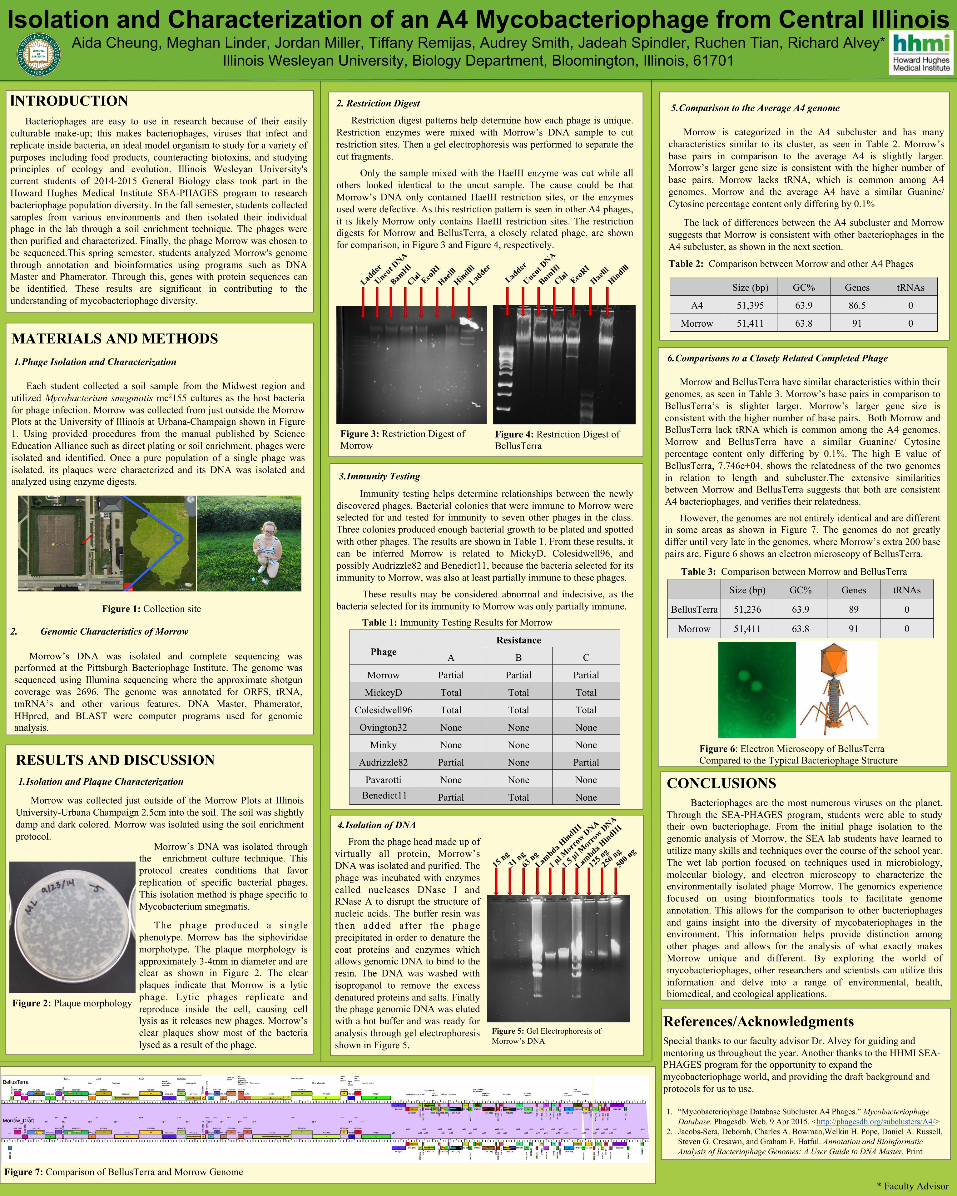
genomes, as seen in Table 3. Morrow’s base pairs in comparison to BellusTerra’s is slighter larger. Morrow’s larger gene size is consistent with the higher number of base pairs. Both Morrow and BellusTerra lack tRNA which is common among the A4 genomes. Morrow and BellusTerra have a similar Guanine/ Cytosine percentage content only differing by 0.1%. The high E value of BellusTerra, 7.746e+04, shows the relatedness of the two genomes in relation to length and subcluster.The extensive similarities between Morrow and BellusTerra suggests that both are consistent A4 bacteriophages, and verifies their relatedness.
However, the genomes are not entirely identical and are different in some areas as shown in Figure 7. The genomes do not greatly differ until very late in the genomes, where Morrow’s extra 200 base pairs are. Figure 6 shows an electron microscopy of BellusTerra.
Figure 2: Plaque morphology
RESULTS AND DISCUSSION
1. Isolation and Plaque Characterization
Morrow was collected just outside of the Morrow Plots at Illinois University-Urbana Champaign 2.5cm into the soil. The soil was slightly damp and dark colored. Morrow was isolated using the soil enrichment protocol.
* Faculty Advisor
Morrow’s DNA was isolated and complete sequencing was performed at the Pittsburgh Bacteriophage Institute. The genome was sequenced using Illumina sequencing where the approximate shotgun coverage was 2696. The genome was annotated for ORFS, tRNA, tmRNA’s and other various features. DNA Master, Phamerator, HHpred, and BLAST were computer programs used for genomic analysis.
Morrow’s DNA was isolated through the enrichment culture technique. This protocol creates conditions that favor replication of specific bacterial phages. This isolation method is phage specific to Mycobacterium smegmatis.
The phage produced a single phenotype. Morrow has the siphoviridae morphotype. The plaque morphology is approximately 3-4mm in diameter and are clear as shown in Figure 2. The clear plaques indicate that Morrow is a lytic phage. Lytic phages replicate and reproduce inside the cell, causing cell lysis as it releases new phages. Morrow’s clear plaques show most of the bacteria lysed as a result of the phage.
2. Genomic Characteristics of Morrow
4. Isolation of DNA
From the phage head made up of virtually all protein, Morrow’s DNA was isolated and purified. The phage was incubated with enzymes called nucleases DNase I and RNase A to disrupt the structure of nucleic acids. The buffer resin was then added after the phage precipitated in order to denature the coat proteins and enzymes which allows genomic DNA to bind to the resin. The DNA was washed with isopropanol to remove the excess denatured proteins and salts. Finally the phage genomic DNA was eluted with a hot buffer and was ready for analysis through gel electrophoresis shown in Figure 5.
Figure 5: Gel Electrophoresis of Morrow’s DNA
CONCLUSIONS
Bacteriophages are the most numerous viruses on the planet. Through the SEA-PHAGES program, students were able to study their own bacteriophage. From the initial phage isolation to the genomic analysis of Morrow, the SEA lab students have learned to utilize many skills and techniques over the course of the school year. The wet lab portion focused on techniques used in microbiology, molecular biology, and electron microscopy to characterize the environmentally isolated phage Morrow. The genomics experience focused on using bioinformatics tools to facilitate genome annotation. This allows for the comparison to other bacteriophages and gains insight into the diversity of mycobateriophages in the environment. This information helps provide distinction among other phages and allows for the analysis of what exactly makes Morrow unique and different. By exploring the world of mycobacteriophages, other researchers and scientists can utilize this information and delve into a range of environmental, health, biomedical, and ecological applications.
Figure 6: Electron Microscopy of BellusTerra Compared to the Typical Bacteriophage Structure
Figure 3: Restriction Digest of Morrow
Figure 4: Restriction Digest of BellusTerra
5. Comparison to the Average A4 genome
Morrow is categorized in the A4 subcluster and has many
characteristics similar to its cluster, as seen in Table 2. Morrow’s base pairs in comparison to the average A4 is slightly larger. Morrow’s larger gene size is consistent with the higher number of base pairs. Morrow lacks tRNA, which is common among A4 genomes. Morrow and the average A4 have a similar Guanine/ Cytosine percentage content only differing by 0.1%
The lack of differences between the A4 subcluster and Morrow suggests that Morrow is consistent with other bacteriophages in the A4 subcluster, as shown in the next section. Table 2: Comparison between Morrow and other A4 Phages
Size (bp) GC% Genes tRNAs
A4 51,395 63.9 86.5 0
Morrow 51,411 63.8 91 0
References/Acknowledgments
Special thanks to our faculty advisor Dr. Alvey for guiding and mentoring us throughout the year. Another thanks to the HHMI SEA-PHAGES program for the opportunity to expand the mycobacteriophage world, and providing the draft background and protocols for us to use. 1. “Mycobacteriophage Database Subcluster A4 Phages.” Mycobacteriophage
Database. Phagesdb. Web. 9 Apr 2015. <http://phagesdb.org/subclusters/A4/> 2. Jacobs-Sera, Deborah, Charles A. Bowman,Welkin H. Pope, Daniel A. Russell,
Steven G. Cresawn, and Graham F. Hatful. Annotation and Bioinformatic Analysis of Bacteriophage Genomes: A User Guide to DNA Master. Print
Figure 7: Comparison of BellusTerra and Morrow Genome