Embed Size (px)
Citation preview
International Journal of Innovations in Engineering and Technology (IJIET) http://dx.doi.org/10.21172/ijiet.83.028
Volume 8 Issue 3 June 2017 208 ISSN: 2319-1058
Isolation and Characterization of Endophytic Bacteria in Soybean (Glycine max L. (Merrill) Cultivated on Alluvial Soil of Can Tho city,
Vietnam Van Thi Phuong Nhu
Dept. of Biology Science, Phu Yen University, Vietnam
Cao Ngoc Diep
Dept. Microbiology Biotechnology, Biotechnology R&D Institute, Can Tho University, Can Tho City, Vietnam
Abstract. Eighty-six endophytic bacterial isolates were isolated from 70 soybean plants (including whole plant as stem, root and nodule) which collected at 5 districts of Can Tho city, Mekong Delta, Vietnam; they developed on three kinds of medium (PDA, TSA and G6) after 2 or 3 days incubation and they made the pellicles on semi-solid media. The bacterial isolates were tested in-vitro for plant growth promoting properties including nitrogen fixation, phosphate solubilization and IAA production together with producing siderophores. All of them had the ability of ammonium synthesis, phosphate solubilization and IAA biosynthesis but there were 38.37% bacterial isolates producing siderophores. The sequences from selected nitrogen-fixing and phosphate-solubilizing bacteria (26 isolates) showed high degrees of similarity to those of the GenBank reference strains (between 97% and 100%). From 26 isolates, 7 belonged to Bacilli and 17 were Gamma-Proteobacteria. Based on Pi value (nucleotide diversity), Bacilli group had the highest Theta value and Thete values (per sequence) from S of SNP for DNA polymorphism were calculated from each group and Bacilli group had the highest values in comparison with gammaproteobacteria. From these results showed that there are five strains as Enterobacter cloacae TSR1A, Enterobacter cloacae CPR1A, Bacillus sp. OSR12, Bacillus subtilis TST10c and Acinetobacter sp. TGN1 revealed promising candidates with multiple beneficial characteristics and they have the potential for application as inoculants adapted to unfertile soil and local crops because they are not only best strains but also combined with rhizobia strains for improvement of better grain yield and quality seed of soybean cultivation on alluvilal soil in the future.
Keywords: 16S rRNA Gene Sequence, Alluvial soil, Endophytic Bacteria, Stem, Root, Root-nodule of soybean
I. INTRODUCTION
Soybean (Glycine max) is one of the most important oil seed crop in the world. It contains 18 to 22% oil, highly desirable in diet and have 40 to 42% of good quality protein [1]; Soybean protein is rich in valuable amino acid lycine (5%) in which most of the cereals are deficient [2]. Soybean (Glycine max (L.) Merrill) is an Asiatic leguminous plant, occupying large acreages of land worldwide for its oil and protein [3]. Rhizobia are perhaps the best known beneficial plant-associated bacteria because of the importance of the nitrogen fixation that occurs during the Rhizobium-legume symbiosis [4]. In recent years, interest in endophytic micro-organisms has increased, as they play a key role in agricultural environment and are promising because of their potential use in sustainable agriculture [5]. Endophytes have been found in almost every plant studied [6]; endophytes are sheltered from environmental stresses and microbial competition by the host plant, and they seem to be ibiquitous in plant tissues, having been isolated from flowers, fruits, leaves, stems, roots, and seeds of various plant species [7]. Endophyte-plant associations have been found to improve plant health and may help host plant to rescue from various biotic and abiotic stresses [8][9]. Endophytic bacteria have been isolated from legume plants such as alfalfa [10], clover [11], pea [12] and soybean [13]. Besides that, Sturz et al. [11] reported the

International Journal of Innovations in Engineering and Technology (IJIET) http://dx.doi.org/10.21172/ijiet.83.028
Volume 8 Issue 3 June 2017 209 ISSN: 2319-1058
isolation of 15 non-rhizobial species from clover root nodules, eight of which were found only in root nodule tissues. Can Tho city locates in the center of Mekong Delta, Vietnam (10o01’57” N and 105o47’03” E) (Figure 1), composes of 5 districts and 4 towns with 1409 km2 (over 80% agriculatural land) (GENERAL STATISTICS OFFICE of VIET NAM, 2016). Together with corn, mung bean, sesame seeds and other crops, soybean has been cultivated routinely on alluvial soil in dry season each year. This study was aimed to isolate the non-nodulating endophytic bacteria from the root nodules and soybean plants (stem and root). Using 16S rRNA gene sequence analysis, we also studied the taxonomic position of these non-nodulating endophytic bacteria and compared endophytic bacterial isolates which planted in alluvilal soil in the Mekong Delta
II. MATERIALS AND METHODS
A. Plant sample and Isolation endophytic bacteria in soybean nodules and plants Soybean plants used in the experiment were local cultivars (Glycine max L. Merr), were cultivated at 5 districts as Vinh Thanh, Co Do, Thoi Lai, Thot Not, O Mon in Can Tho city (Figure 1). Thot Not, and O Mon are districts which locate along the Hau river.
Figure 1. Can Tho city map Plant samples were collected at the flowering-stage (35-40 days after sowing), five samples were carefully removed, washed under tap water to remove soil, and separated into roots and nodules. Nodules were put in beaker, soaked in distilled water, and drained. They were rinsed in 70% ethanol for 30 s and then sterilized with 0.1% HgCl2 for 3 min [4]. After that, nodules were washed ten times with sterile water [10]. Surface-disinfected tissue was aseptically macerated with homogenizers and tissues were diluted with 1 mL sterile water. One hundred microliters from appropriate dilutions were palted on two different media, viz potato dextrose agar (PDA) and tryptic soy agar (TSA) [14] together with G6 medium [glycerol instead of mannitol] [15]. Samples were obtained whole plant after that soil rhizosphere was separated for described experiments above, soybean roots and stems were washed with tap water to remove attached clay; stems and roots were cut separately. Subsequently, the stems and roots were immersed in 70% ethanol in 3 min, washed with fresh sodium hypochlorite solution (2.5% available Cl-) for 5 min, rinsed with hydrogen peroxide (3%) for 30s and finally
International Journal of Innovations in Engineering and Technology (IJIET) http://dx.doi.org/10.21172/ijiet.83.028
Volume 8 Issue 3 June 2017 210 ISSN: 2319-1058
washed five times with sterile distilled water. To confirm that the sterilization process was successful, the aliquots of the sterile distilled water used in the final rinse were set on tryptone - yeast extract – glucose agar medium plates. The plates were examined for bacterial growth after incubation at 28oC for 3 days. Soybean stem and root samples that were not contaminated as detected by culture-dependent sterility test were used for further analysis. Samples (stems or roots) were cut to 1-2 cm pieces and macerated with a sterile mortar and pestle; tissue extracts were then serially diluted 10-fold in sterile water, 200 µl-aliquot samples were used to inoculate in (in triplicate) Nitrogen-free semisolid LGI in 5 ml tubes. After 48-72 h incubation, bacteria growing in tubes as a white or yellow pellicle at a depth of 1 to 4 mm were streaked on LGI agar plates, the cultures were streaked on media to obtain single colonies.
Bacterial colonies were differentiated from the basis of colony morphology and pigmentation. The colonies were subculture on the agar-based subculture medium plates by striking technique and re-incubated at 300C for 4 days. This isolation process carries out in shifts of the agar-based culture medium to the agar-based subculture medium until monocultures were obtained. Monocultures were culture on the agar-based culture medium slant in the test-tube (12 ml) and incubated at 300C for 4 days following by stored 40C in refrigerator. Morphological characterization of the isolates was carried by Gram staining. For motility, each isolate was spot-inoculated on the center of semi-soloid nutrient agar plates (0.2% agar) and incubated at 30oC [4]. Cell shape was observed under light microscope, colony characterization as size, color, shape were recorded at 2 – 3 days after plating into petri-dishes. B. Characterization of endophytic bacteria for plant growth promoting attributes
Bacterial isolates were also studied in vitro for plant growth promoting properties including indole acetic acid (IAA) production, nitrogen fixation, solubilization of phosphate. For indoleacetic acid production, 5 µl for log phase culture was inoculated in 5 ml of LB (Luria-Bertani; Bacto-Tryptone 10.0 g/l, yeast extract 5.0 g/l, NaCl 5.0 g/l) broth with L-tryptophane and incubated on shaker for 24 h. Auxin quantification was carried out following the method of Gordon and Weber [16]. For nitrogen fixation ability and phosphate solubilization: the ability to fix N2 was tested on Burk’N free liquid medium incubation at 30oC and the ammonium concentration in medium was measured by Phenol Nitroprusside method after 2,4,6 and 8 day inoculation (DAI) and inorganic phosphate solubilization ability was tested on NBRIP liquid medium and they incubated at 30oC and the P2O5 concentration was measured by ammonium molypdate method after 5, 10, 15 and 20 DAI [17]. Furthermore, siderophore production was assayed by the bacterial isolates according to Schwyn and Neilands [18] using NBRIP medium without tryptophan which was diluted fivefold. The isolates were inoculated spot onto Chrome azurol S agar plates divided into equal sectors, and the plates were incubated at 28oC for 48 h. Development of a yellow, orange or violet halo around the bacterial colony was considered to be positive for siderophore production. C. 16S rDNA gene amplification and sequencing
Bacterial DNA isolated was conducted by published protocols [19] and the following primers were used for PCR amplification of 16S ribosomal DNA: p515FPL [20] and p13B [21] [22]. The 50 µL reaction mixture consisted of 2.5 U Taq Polymerase (Fermentas), 0.1 mM of each desoxynecleotide triphosphate, 1.5 mM magnesium chloride, 0.4 mM spermidine (Sigma), 10 pM of each primer (Fermentas) and 10 ng DNA, 10% (vol/vol) dimethyl disulfide (Fermentas). The thermocycling profide was carried out with an initial denaturation at 94oC (3 min) followed by 30 cycles of denaturation at 94oC (60 s), annealing at 57oC (60 s), extension at 72oC (120 s) and a final extension at 72oC (4 min) in C1000 Thermal Cycler (Bio-Rad). Aliquots (10 µl) of PCR products were electrophoresed and visualized in 1% agarose gels using standard electrophoresis procedures. Partial 16S rRNA gene of selectived isolates in each site was sequenced by MACROGEN, Republic of Korea (dna.macrogen.com). Finally, 16S rRNA sequence of the isolate was compared with that of other microorganisms by way BLAST (http://www.ncbi.nlm.nih.gov/BLAST/Blast.cgi); In the best isolate(s)(high ability of nitrogen fixation, phosphate solubilization and IAA synthesis) and 16 isolates of 3 sites were chosen to sequence and the results were compared to sequences of GenBank based on partial 16S rRNA sequences to show relationships between endophytic strains [23] and phylogenetic tree were constructed by the neighbor-joining method using the MEGA software version 6.06 based on 1000 bootstraps.
International Journal of Innovations in Engineering and Technology (IJIET) http://dx.doi.org/10.21172/ijiet.83.028
Volume 8 Issue 3 June 2017 211 ISSN: 2319-1058
D. SNPs Discovery
The sequence date from 17 root-associated bacterial isolates were analysed with SeqScape@Software (Applied Biosystem, Foster City, CA, USA). SeqScape is a sequence comparison tool for variant identification, SNP discovery and validation. It considers alignment depth, the base calls in each of the sequnces and the associated base quality values. Putative SNPs were accepted as true sequence variants if the quality value exceeded 20. It means a 1% chance basecall is incorrect. E.Nucleotide Diversity (Ө)
Nucleotide diversity (Ө) was calculated by the method described by Halushka et al. [24]
n
Ө = K/aL a = ∑ l/(i - l)
i=2 where K is the number of SNPs identified in an alignment length, n is alleles and L is the total length of sequence (bp). F.Data analyses
Data from ammonium, orthophosphate and IAA concentrations in media were analysed in completely randomized design with three replicates. Yield component and grain yield together with pH and soil characteristics were analysed with five replications. Duncan test at P=0.01 or P=0.05 were used to differentiate between statistically different means using SPSS version 16.
III. RESULSTS AND DISCUSSION
A.Plant sample and Isolation endophytic bacteria in soybean plant samples
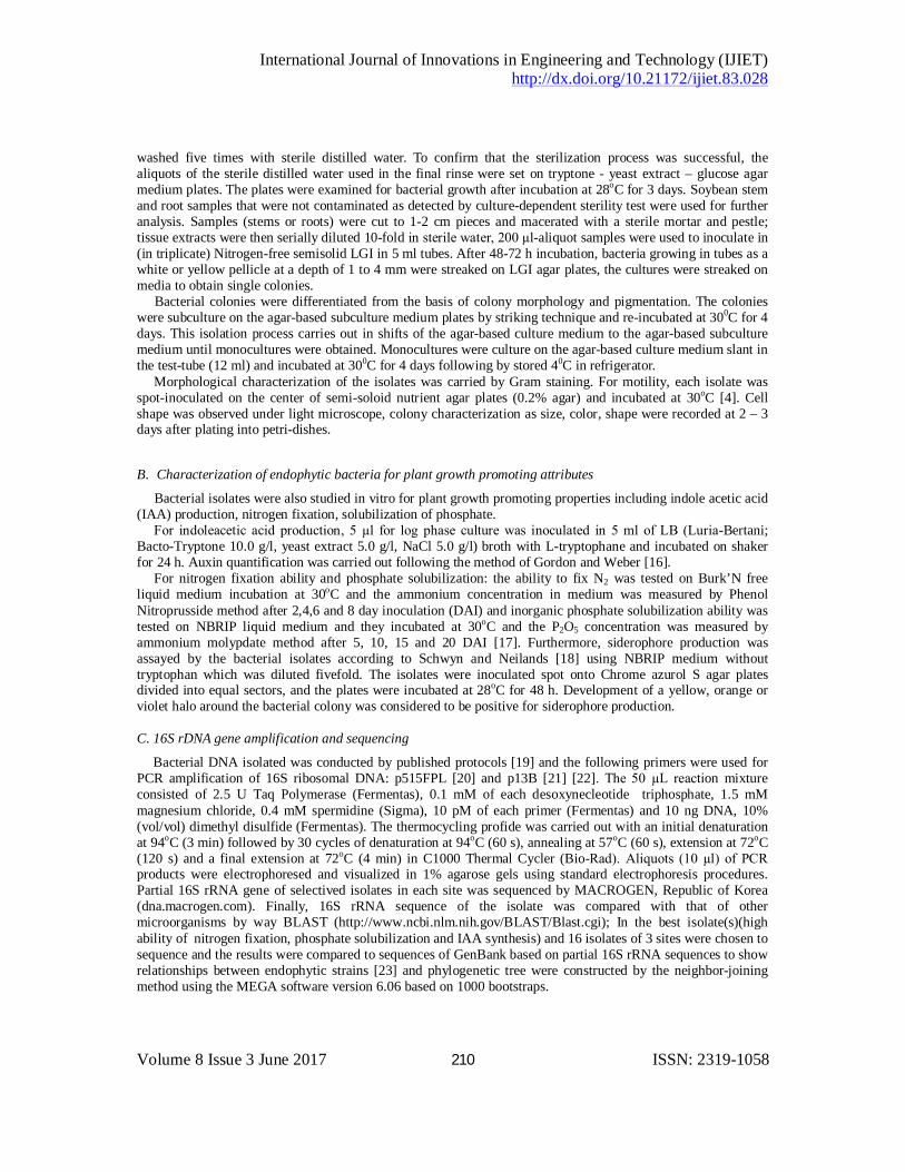
From colonies were plated and develop on TSA and PDA media after incubation at 30oC; we wished to isolate non-rhizobia from within soybean plant samples. Total of 86 endophytic bacterial isolates (consisted of 41 isolates from Thot Not, 19 isolates from O Mon, 6 from Co Do, 11 from Vinh Thanh and 9 from Thoi Lai); 32 isolates isolated on PDA medium, 42 isolates on TSA medium and 12 isolates on G6 medium. The endophytic bacteria developed in the pellicles of semi solid (in two kinds of medium) after 36 h incubation in semi-solid (Figure 2) as the previous results of Thu Ha et al. [25], Diep et al [26].
Figure - 2 Endophytic bacteria made pellicles in semi-solid (TSA and PDA media) after 36 h incubation at 30oC
Pellicles appeared on surface of semi- solid medium

International Journal of Innovations in Engineering and Technology (IJIET) http://dx.doi.org/10.21172/ijiet.83.028
Volume 8 Issue 3 June 2017 212 ISSN: 2319-1058
Almost their colonies have round-shaped; milky, white clear (on PDA’s medium and TSA medium); entire or loabate margin (Figure 3); diameter size of these colonies varied from 0.2 to 3.0 mm and all of them are Gram-positve or Gram-negative by Gram stain.
Figure - 3 Characteristics of colonies of bacterial isolates after grown on two kinds of medium
B.Characterization of endophytic bacteria for plant growth promoting attributes
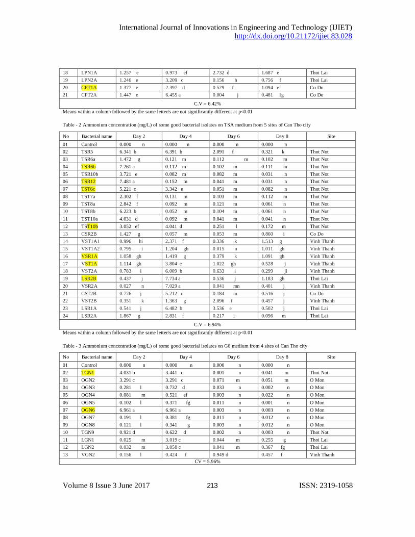
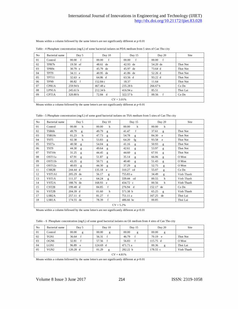
The result from Table 1 showed that there were some isolates having the highest ammonium concentration as isolates TPR6a, TPR7a, TPR7b, TPT7, TPT8, CPT1A and CPT2A on PDA medium, TSR5, TSR6b, TSR6b, TSR12, TST6c, TST10b, TSR2B, VSR2A on TSA medium (Table 2) and TGN1, OGN6 on G6 medium (Table 3) however there were a lot of isolates having the high ability of phosphate solubilization such as isolates CPR1A, TPN1A, CPT1A on PDA medium (Table 4), CSR2B, VST1A1 on PDA medium (Table 5) and the isolate VGN2 on G6 medium (Table 6).
Table - 1 Ammonium concentration (mg/L) of some good bacterial isolates on PDA medium from 5 sites of Can Tho city
No Bacterial name Day 2 Day 4 Day 6 Day 8 Site 01 Control 0.000 jj 0.000 j 0.004 j 0.001 j 03 TPR6a 3.111 c 0.131 h 0.005 j 0.001 j Thot Not 04 TPR6b 1.382 f 0.081 i 0.021 i 0.031 i Thot Not 05 TPR7a 6.001 a 4.012 b 0.003 j 0.031 i Thot Not 06 TPR7b 4.221 b 0.051 i 0.002 j 0.051 i Thot Not 07 TPR9b 3.372 c 2.981 c 0.001 j 0.141 h Thot Not 08 TPR10a 2.201 d 0.181 g 0.002 j 0.131 h Thot Not 09 TPT6a 4.021 b 0.722 f 0.004 j 0.121 h Thot Not 10 TPT7 4.001 b 3.861 bc 0.003 j 0.052 i Thot Not 11 TPT8 4.152 b 0.091 i 0.001 j 0.051 i Thot Not 12 TPT10 3.151 c 0.092 i 0.001 j 0.031 i Thot Not 14 CPR1A 0.493 fg 0.008 j 0.434 fg 0.966 f Co Do 15 CPR2A 0.775 f 0.707 f 0.345 g 1.049 ef Co Do 16 VPR2B 1.066 e 0.008 j 0.209 h 1.254 ef Vinh Thanh 17 VPR1B 1.266 e 3.973 b 0.936 ef 1.391 e Vinh Thanh
PR5 PT6a
SR5 ST12b
International Journal of Innovations in Engineering and Technology (IJIET) http://dx.doi.org/10.21172/ijiet.83.028
Volume 8 Issue 3 June 2017 213 ISSN: 2319-1058
18 LPN1A 1.257 e 0.973 ef 2.732 d 1.687 e Thoi Lai 19 LPN2A 1.246 e 3.209 c 0.156 h 0.756 f Thoi Lai 20 CPT1A 1.377 e 2.397 d 0.529 f 1.094 ef Co Do 21 CPT2A 1.447 e 6.455 a 0.004 j 0.481 fg Co Do
C.V = 6.42% Means within a column followed by the same letter/s are not significantly different at p<0.01
Table - 2 Ammonium concentration (mg/L) of some good bacterial isolates on TSA medium from 5 sites of Can Tho city
No Bacterial name Day 2 Day 4 Day 6 Day 8 Site 01 Control 0.000 n 0.000 n 0.000 n 0.000 n 02 TSR5 6.341 b 6.391 b 2.091 f 0.321 k Thot Not 03 TSR6a 1.472 g 0.121 m 0.112 m 0.102 m Thot Not 04 TSR6b 7.261 a 0.112 m 0.102 m 0.111 m Thot Not 05 TSR10b 3.721 e 0.082 m 0.082 m 0.031 n Thot Not 06 TSR12 7.481 a 0.152 m 0.041 m 0.031 n Thot Not 07 TST6c 5.221 c 3.342 e 0.051 m 0.082 n Thot Not 08 TST7a 2.302 f 0.131 m 0.103 m 0.112 m Thot Not 09 TST8a 2.842 f 0.092 m 0.121 m 0.061 n Thot Not 10 TST8b 6.223 b 0.052 m 0.104 m 0.061 n Thot Not 11 TST10a 4.031 d 0.092 m 0.041 m 0.041 n Thot Not 12 TST10b 3.052 ef 4.041 d 0.251 l 0.172 m Thot Not 13 CSR2B 1.427 g 0.057 m 0.053 m 0.860 i Co Do 14 VST1A1 0.996 hi 2.371 f 0.336 k 1.513 g Vinh Thanh 15 VST1A2 0.795 i 1.204 gh 0.015 n 1.011 gh Vinh Thanh 16 VSR1A 1.058 gh 1.419 g 0.379 k 1.091 gh Vinh Thanh 17 VST1A 1.114 gh 3.804 e 1.022 gh 0.528 j Vinh Thanh 18 VST2A 0.783 i 6.009 b 0.633 i 0.299 jl Vinh Thanh 19 LSR2B 0.437 j 7.734 a 0.536 j 1.183 gh Thoi Lai 20 VSR2A 0.027 n 7.029 a 0.041 mn 0.401 j Vinh Thanh 21 CST2B 0.776 j 5.212 c 0.184 m 0.516 j Co Do 22 VST2B 0.351 k 1.363 g 2.096 f 0.457 j Vinh Thanh 23 LSR1A 0.541 j 6.482 b 3.536 e 0.502 j Thoi Lai 24 LSR2A 1.867 g 2.831 f 0.217 i 0.096 m Thoi Lai
C.V = 6.94%
Means within a column followed by the same letter/s are not significantly different at p<0.01
Table - 3 Ammonium concentration (mg/L) of some good bacterial isolates on G6 medium from 4 sites of Can Tho city
No Bacterial name Day 2 Day 4 Day 6 Day 8 Site 01 Control 0.000 n 0.000 n 0.000 n 0.000 n 02 TGN1 4.031 b 3.441 c 0.001 n 0.041 m Thot Not 03 OGN2 3.291 c 3.291 c 0.071 m 0.051 m O Mon 04 OGN3 0.281 l 0.732 d 0.033 n 0.002 n O Mon 05 OGN4 0.081 m 0.521 ef 0.003 n 0.022 n O Mon 06 OGN5 0.102 l 0.371 fg 0.011 n 0.001 n O Mon 07 OGN6 6.961 a 6.961 a 0.003 n 0.003 n O Mon 08 OGN7 0.191 l 0.381 fg 0.011 n 0.012 n O Mon 09 OGN8 0.121 l 0.341 g 0.003 n 0.012 n O Mon 10 TGN9 0.921 d 0.622 d 0.002 n 0.003 n Thot Not 11 LGN1 0.025 m 3.019 c 0.044 m 0.255 g Thoi Lai 12 LGN2 0.032 m 3.058 c 0.041 m 0.367 fg Thoi Lai 13 VGN2 0.156 l 0.424 f 0.949 d 0.457 f Vinh Thanh
CV = 5.96%
International Journal of Innovations in Engineering and Technology (IJIET) http://dx.doi.org/10.21172/ijiet.83.028
Volume 8 Issue 3 June 2017 214 ISSN: 2319-1058
Means within a column followed by the same letter/s are not significantly different at p<0.01
Table - 4 Phosphate concentration (mg/L) of some bacterial isolates on PDA medium from 5 sites of Can Tho city
No Bacterial name Day 5 Day 10 Day 15 Day 20 Site 01 Control 00.00 f 00.00 f 00.00 f 00.00 f
02 TPR7b 19.50 ef 48.61 de 42.93 de 54.20 de Thot Not 03 TPR9c 30.79 e 45.79 de 45.97 de 73.64 d Thot Not 04 TPT9 34.11 e 40.95 de 41.86 de 52.26 d Thot Not 05 TPT11 32.63 e 64.86 d 63.56 d 93.22 d Thot Not 06 TPN9 00.82 f 152.84 c 18.37 11.64 Thot Not 07 CPR1A 259.94 b 467.48 a 235.28 b 266.67 b Co Do 08 LPN1A 245.61 b 212.34 b 416.94 a 85.51 Thoi Lai 09 CPT1A 320.80 b 72.84 d 322.57 b 00.56 f Co Do
CV = 3.01%
Means within a column followed by the same letter/s are not significantly different at p<0.01 Table - 5 Phosphate concentration (mg/L) of some good bacterial isolates on TSA medium from 5 sites of Can Tho city
No Bacterial name Day 5 Day 10 Day 15 Day 20 Site 01 Control 00.00 h 00.00 h 00.00 h 00.00 h
02 TSR6b 49.79 g 49.79 g 41.47 f 37.61 g Thot Not 03 TSR10c 01.23 h 47.73 g 54.78 g 84.30 e Thot Not 04 TST5 02.38 h 53.83 g 64.20 fg 93.58 e Thot Not 05 TST7a 40.58 g 54.04 g 41.16 g 50.93 g Thot Not 06 TST9 44.30 g 49.64 g 42.61 g 53.97 g Thot Not 07 TST10c 31.25 g 44.45 g 44.60 g 67.91 g Thot Not 08 OST11a 67.91 g 51.87 g 35.14 g 66.06 g O Mon 09 OST11b 43.35 g 50.71 g 40.40 g 51.43 g O Mon 10 OST12a 40.03 g 44.30 g 37.29 g 52.75 g O Mon 11 CSR2B 244.44 d 135.18 e 310.27 cd 55.07 g Co Do 12 VST1A1 205.29 de 56.17 g 755.83 a 34.49 g Vinh Thanh 13 VST1A 111.37 e 64.24 g 339.44 cd 00.55 h Vinh Thanh 14 VST2A 188.76 de 168.93 e 434.72 c 00.56 h Vinh Thanh 15 CST2B 199.48 d 84.85 f 276.94 d 152.17 de Co Do 16 VST2B 204.39 d 01.60 h 571.38 b 65.25 g Vinh Thanh 17 LSR2A 257.11 d 81.27 f 751.11 a 167.25 de Thoi Lai 18 LSR1A 174.55 de 78.39 f 486.66 bc 00.85 Thoi Lai
CV = 5.2%
Means within a column followed by the same letter/s are not significantly different at p<0.01
Table – 6 Phosphate concentration (mg/L) of some good bacterial isolates on G6 medium from 4 sites of Can Tho city
No Bacterial name Day 5 Day 10 Day 15 Day 20 Site 01 Control 00.00 g 00.00 g 00.00 g 00.00 g
02 TGN1 36.64 f 56.31 f 46.79 f 70.19 e Thot Not 03 OGN6 32.81 f 57.56 f 56.83 f 115.75 d O Mon 04 LGN1 96.89 e 124.69 d 471.71 a 00.36 g Thoi Lai 05 VGN2 120.28 d 01.29 g 282.22 b 178.55 c Vinh Thanh
CV = 4.81%
Means within a column followed by the same letter/s are not significantly different at p<0.01
International Journal of Innovations in Engineering and Technology (IJIET) http://dx.doi.org/10.21172/ijiet.83.028
Volume 8 Issue 3 June 2017 215 ISSN: 2319-1058
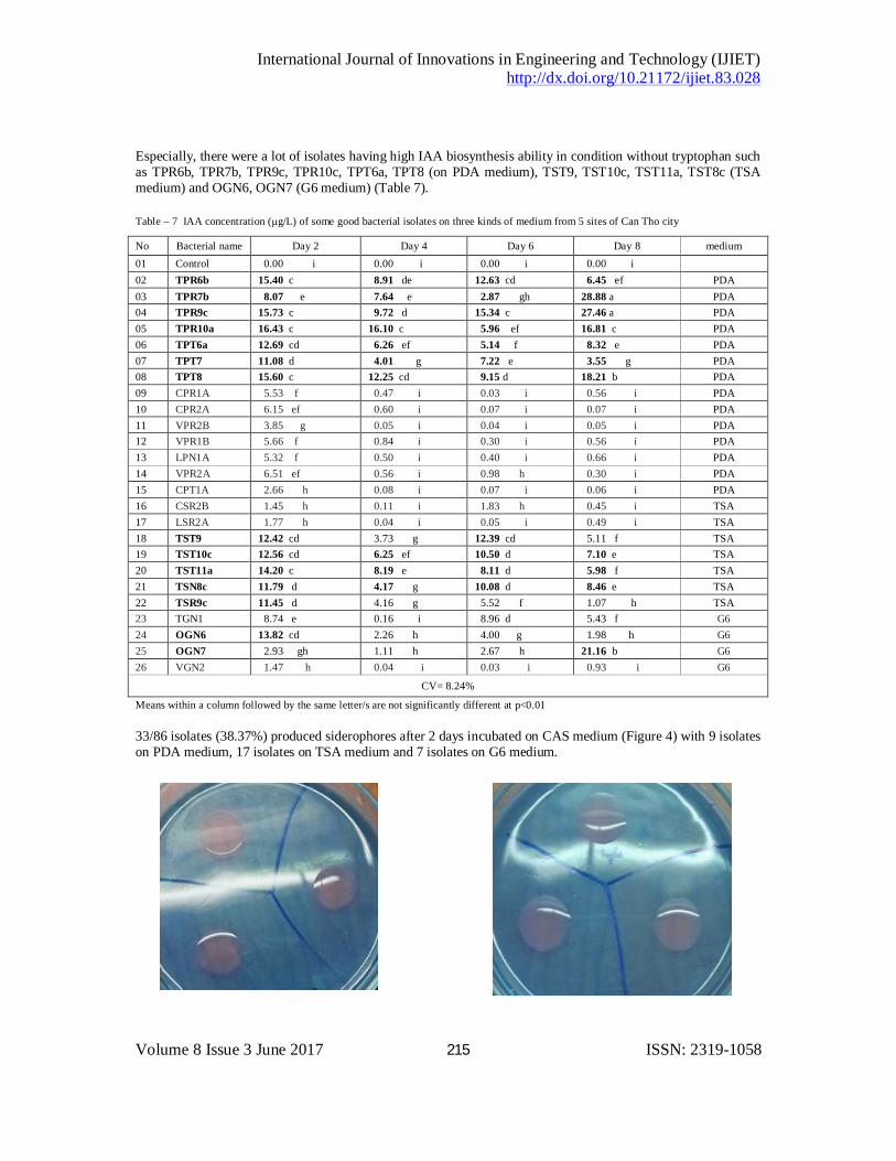
Especially, there were a lot of isolates having high IAA biosynthesis ability in condition without tryptophan such as TPR6b, TPR7b, TPR9c, TPR10c, TPT6a, TPT8 (on PDA medium), TST9, TST10c, TST11a, TST8c (TSA medium) and OGN6, OGN7 (G6 medium) (Table 7). Table – 7 IAA concentration (µg/L) of some good bacterial isolates on three kinds of medium from 5 sites of Can Tho city
No Bacterial name Day 2 Day 4 Day 6 Day 8 medium 01 Control 0.00 i 0.00 i 0.00 i 0.00 i
02 TPR6b 15.40 c 8.91 de 12.63 cd 6.45 ef PDA 03 TPR7b 8.07 e 7.64 e 2.87 gh 28.88 a PDA
04 TPR9c 15.73 c 9.72 d 15.34 c 27.46 a PDA 05 TPR10a 16.43 c 16.10 c 5.96 ef 16.81 c PDA
06 TPT6a 12.69 cd 6.26 ef 5.14 f 8.32 e PDA 07 TPT7 11.08 d 4.01 g 7.22 e 3.55 g PDA
08 TPT8 15.60 c 12.25 cd 9.15 d 18.21 b PDA 09 CPR1A 5.53 f 0.47 i 0.03 i 0.56 i PDA 10 CPR2A 6.15 ef 0.60 i 0.07 i 0.07 i PDA 11 VPR2B 3.85 g 0.05 i 0.04 i 0.05 i PDA 12 VPR1B 5.66 f 0.84 i 0.30 i 0.56 i PDA 13 LPN1A 5.32 f 0.50 i 0.40 i 0.66 i PDA 14 VPR2A 6.51 ef 0.56 i 0.98 h 0.30 i PDA 15 CPT1A 2.66 h 0.08 i 0.07 i 0.06 i PDA 16 CSR2B 1.45 h 0.11 i 1.83 h 0.45 i TSA 17 LSR2A 1.77 h 0.04 i 0.05 i 0.49 i TSA 18 TST9 12.42 cd 3.73 g 12.39 cd 5.11 f TSA 19 TST10c 12.56 cd 6.25 ef 10.50 d 7.10 e TSA 20 TST11a 14.20 c 8.19 e 8.11 d 5.98 f TSA 21 TSN8c 11.79 d 4.17 g 10.08 d 8.46 e TSA 22 TSR9c 11.45 d 4.16 g 5.52 f 1.07 h TSA 23 TGN1 8.74 e 0.16 i 8.96 d 5.43 f G6 24 OGN6 13.82 cd 2.26 h 4.00 g 1.98 h G6 25 OGN7 2.93 gh 1.11 h 2.67 h 21.16 b G6 26 VGN2 1.47 h 0.04 i 0.03 i 0.93 i G6
CV= 8.24%
Means within a column followed by the same letter/s are not significantly different at p<0.01 33/86 isolates (38.37%) produced siderophores after 2 days incubated on CAS medium (Figure 4) with 9 isolates on PDA medium, 17 isolates on TSA medium and 7 isolates on G6 medium.
International Journal of Innovations in Engineering and Technology (IJIET) http://dx.doi.org/10.21172/ijiet.83.028
Volume 8 Issue 3 June 2017 216 ISSN: 2319-1058
Figure 4. Bacterial isolates made a range of halo round well contaning bacterial liquid on CAS agar after 48 h incubation
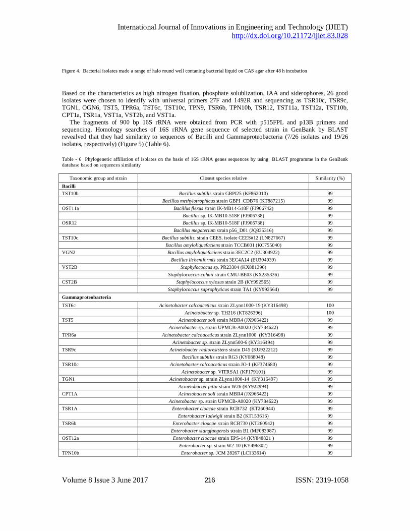
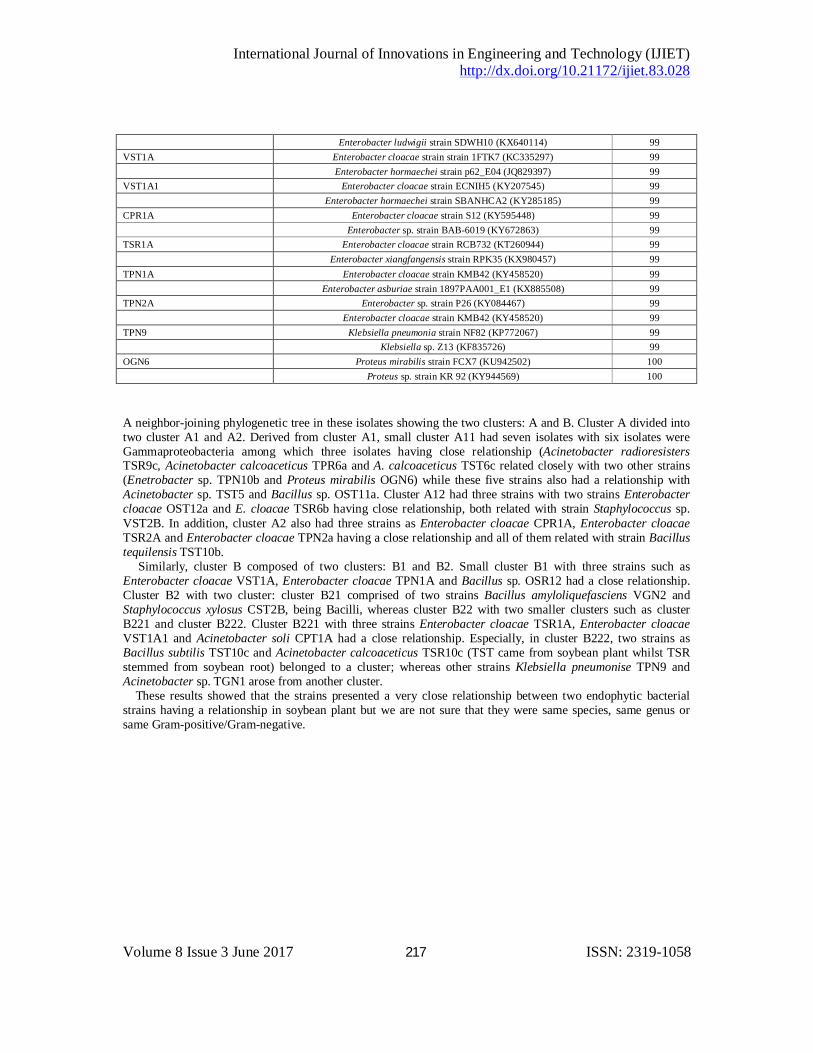
Based on the characteristics as high nitrogen fixation, phosphate solublization, IAA and siderophores, 26 good isolates were chosen to identify with universal primers 27F and 1492R and sequencing as TSR10c, TSR9c, TGN1, OGN6, TST5, TPR6a, TST6c, TST10c, TPN9, TSR6b, TPN10b, TSR12, TST11a, TST12a, TST10b, CPT1a, TSR1a, VST1a, VST2b, and VST1a. The fragments of 900 bp 16S rRNA were obtained from PCR with p515FPL and p13B primers and sequencing. Homology searches of 16S rRNA gene sequence of selected strain in GenBank by BLAST revealved that they had similarity to sequences of Bacilli and Gammaproteobacteria (7/26 isolates and 19/26 isolates, respectively) (Figure 5) (Table 6). Table - 6 Phylogenetic affiliation of isolates on the basis of 16S rRNA genes sequences by using BLAST programme in the GenBank database based on sequences similarity
Taxonomic group and strain Closest species relative Similarity (%) Bacilli TST10b Bacillus subtilis strain GBPI25 (KF862010) 99 Bacillus methylotrophicus strain GBPI_CDB76 (KT887215) 99 OST11a Bacillus flexus strain IK-MB14-518F (FJ906742) 99 Bacillus sp. IK-MB10-518F (FJ906738) 99 OSR12 Bacillus sp. IK-MB10-518F (FJ906738) 99 Bacillus megaterium strain p56_D01 (JQ835316) 99 TST10c Bacillus subtilis, strain CEES, isolate CEES#12 (LN827667) 99 Bacillus amyloliquefaciens strain TCCB001 (KC755040) 99 VGN2 Bacillus amyloliquefaciens strain 3EC2C2 (EU304922) 99 Bacillus licheniformis strain 3EC4A14 (EU304939) 99 VST2B Staphylococcus sp. PR23304 (KX881396) 99 Staphylococcus cohnii strain CMU-BE03 (KX235336) 99 CST2B Staphylococcus xylosus strain 2B (KY992565) 99 Staphylococcus saprophyticus strain TA1 (KY992564) 99 Gammaproteobacteria TST6c Acinetobacter calcoaceticus strain ZLynn1000-19 (KY316498) 100 Acinetobacter sp. TH216 (KT826396) 100 TST5 Acinetobacter soli strain MBR4 (JX966422) 99 Acinetobacter sp. strain UPMCB-A0020 (KY784622) 99 TPR6a Acinetobacter calcoaceticus strain ZLynn1000 (KY316498) 99 Acinetobacter sp. strain ZLynn500-6 (KY316494) 99 TSR9c Acinetobacter radioresistens strain D45 (KU922212) 99 Bacillus subtilis strain RG3 (KY088048) 99 TSR10c Acinetobacter calcoaceticus strain JO-1 (KF374680) 99 Acinetobacter sp. VITRSA1 (KF179101) 99 TGN1 Acinetobacter sp. strain ZLynn1000-14 (KY316497) 99 Acinetobacter pittii strain W26 (KY922994) 99 CPT1A Acinetobacter soli strain MBR4 (JX966422) 99 Acinetobacter sp. strain UPMCB-A0020 (KY784622) 99 TSR1A Enterobacter cloacae strain RCB732 (KT260944) 99 Enterobacter ludwigii strain B2 (KT153616) 99 TSR6b Enterobacter cloacae strain RCB730 (KT260942) 99 Enterobacter xiangfangensis strain B1 (MF083087) 99 OST12a Enterobacter cloacae strain EPS-14 (KY848821 ) 99 Enterobacter sp. strain W2-10 (KY496302) 99 TPN10b Enterobacter sp. JCM 28267 (LC133614) 99
International Journal of Innovations in Engineering and Technology (IJIET) http://dx.doi.org/10.21172/ijiet.83.028
Volume 8 Issue 3 June 2017 217 ISSN: 2319-1058
Enterobacter ludwigii strain SDWH10 (KX640114) 99 VST1A Enterobacter cloacae strain strain 1FTK7 (KC335297) 99 Enterobacter hormaechei strain p62_E04 (JQ829397) 99 VST1A1 Enterobacter cloacae strain ECNIH5 (KY207545) 99 Enterobacter hormaechei strain SBANHCA2 (KY285185) 99 CPR1A Enterobacter cloacae strain S12 (KY595448) 99 Enterobacter sp. strain BAB-6019 (KY672863) 99 TSR1A Enterobacter cloacae strain RCB732 (KT260944) 99 Enterobacter xiangfangensis strain RPK35 (KX980457) 99 TPN1A Enterobacter cloacae strain KMB42 (KY458520) 99 Enterobacter asburiae strain 1897PAA001_E1 (KX885508) 99 TPN2A Enterobacter sp. strain P26 (KY084467) 99 Enterobacter cloacae strain KMB42 (KY458520) 99 TPN9 Klebsiella pneumonia strain NF82 (KP772067) 99 Klebsiella sp. Z13 (KF835726) 99 OGN6 Proteus mirabilis strain FCX7 (KU942502) 100 Proteus sp. strain KR 92 (KY944569) 100
A neighbor-joining phylogenetic tree in these isolates showing the two clusters: A and B. Cluster A divided into two cluster A1 and A2. Derived from cluster A1, small cluster A11 had seven isolates with six isolates were Gammaproteobacteria among which three isolates having close relationship (Acinetobacter radioresisters TSR9c, Acinetobacter calcoaceticus TPR6a and A. calcoaceticus TST6c related closely with two other strains (Enetrobacter sp. TPN10b and Proteus mirabilis OGN6) while these five strains also had a relationship with Acinetobacter sp. TST5 and Bacillus sp. OST11a. Cluster A12 had three strains with two strains Enterobacter cloacae OST12a and E. cloacae TSR6b having close relationship, both related with strain Staphylococcus sp. VST2B. In addition, cluster A2 also had three strains as Enterobacter cloacae CPR1A, Enterobacter cloacae TSR2A and Enterobacter cloacae TPN2a having a close relationship and all of them related with strain Bacillus tequilensis TST10b. Similarly, cluster B composed of two clusters: B1 and B2. Small cluster B1 with three strains such as Enterobacter cloacae VST1A, Enterobacter cloacae TPN1A and Bacillus sp. OSR12 had a close relationship. Cluster B2 with two cluster: cluster B21 comprised of two strains Bacillus amyloliquefasciens VGN2 and Staphylococcus xylosus CST2B, being Bacilli, whereas cluster B22 with two smaller clusters such as cluster B221 and cluster B222. Cluster B221 with three strains Enterobacter cloacae TSR1A, Enterobacter cloacae VST1A1 and Acinetobacter soli CPT1A had a close relationship. Especially, in cluster B222, two strains as Bacillus subtilis TST10c and Acinetobacter calcoaceticus TSR10c (TST came from soybean plant whilst TSR stemmed from soybean root) belonged to a cluster; whereas other strains Klebsiella pneumonise TPN9 and Acinetobacter sp. TGN1 arose from another cluster. These results showed that the strains presented a very close relationship between two endophytic bacterial strains having a relationship in soybean plant but we are not sure that they were same species, same genus or same Gram-positive/Gram-negative.

International Journal of Innovations in Engineering and Technology (IJIET) http://dx.doi.org/10.21172/ijiet.83.028
Volume 8 Issue 3 June 2017 218 ISSN: 2319-1058
Figure 3. Phylogenetic tree showing the relative position of endophytic bacterial isolates by the neighbor-joining method of complete 16S
rRNA sequence (p515FPL primer)
Bootstrap values of 1000 replicates are shown at the nodes of the trees.
Among 26 strains, there were 21 strains having length nucleotide (over 600) and Theta values (per sequence) from S of SNP for DNA polymorphism were calculated for Each group, and Gammaproteobacteria group had the highest values as comparison with Bacilli (Table 7). Nucleotide polymorphism can be measured by many parameters, such as halotypes (genes) diversity, nucleotide diversity, (Pi), Theta ( )(per group) etc .In this study, nucleotide diversity was estimated by Theta ( ), the number of segregating sites [27], and its standard deviation
Acinetobacter_radioresistens_TSR9c
Acinetobacter_calcoaceticus_TPR6a
Acinetobacter_calcoaceticus_TST6c
Enterobacter_sp._TPN10b
Proteus_mirabilis_OGN6
Acinetobacter_sp._TST5
Bacillus_sp._OST11a
Staphylococcus_sp._VST2B
Enterobacter_cloacae_OST12a
Enterobacter_cloacae_TSR6b
Bacillus_tequilensis_TST10b
Enterobacter_cloacae_CPR1A
Enterobacter_cloacae_TSR2A
Enterobacter_cloacae_TPN2A
Bacillus_sp._OSR12
Enterobacter_cloacae_VST1A
Enterobacter_cloacae_TPN1A
Bacillus_amyloliquefaciens_VGN2
Staphylococcus_xylosus_CST2B
Enterobacter_cloacae_TSR1A
Enterobacter_cloacae_VST1A1
Acinetobacter_soli_CPT1A
Bacillus_subtilis_TST10c
Acinetobacter_calcoaceticus_TSR10c
Klebsiella_pneumoniae_TPN9
Acinetobacter_sp._TGN1
5799
99
9599
99
99
60
6994
32
99
81
36
42
32
25
31
27
19
4
3
0
0.5
Cluster A11
Cluster A12
Cluster A1
Cluster B21
Cluster B221
Cluster B222
Cluster
B2 Cluster
B
Cluster
A
Cluster B1
Cluster A2
Cluster B22
International Journal of Innovations in Engineering and Technology (IJIET) http://dx.doi.org/10.21172/ijiet.83.028
Volume 8 Issue 3 June 2017 219 ISSN: 2319-1058
(S ). These parameters were estimated by DNA Sequence Polymorphism software version 4.0 [28]. Pi values explained nucleotide diversity of sequences for each gene, the highest values, more diversity among groups.
Table - 7 Genetic diversity of 21 strains
Nucleotie diversity Theta (per site) from Eta Theta (per site) from S (�)
21 strains 0.69990 0.79357 ± 0.092 0.27795 ± 0.0085
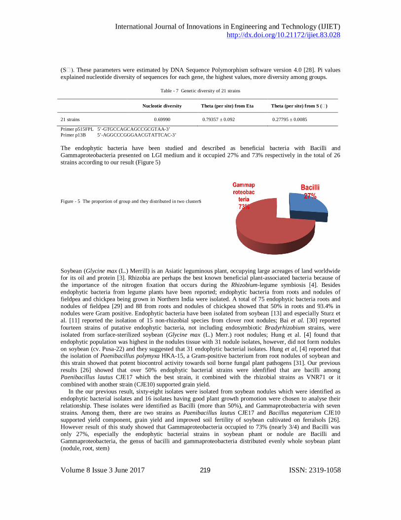
Primer p515FPL 5’-GTGCCAGCAGCCGCGTAA-3’ Primer p13B 5’-AGGCCCGGGAACGTATTCAC-3’ The endophytic bacteria have been studied and described as beneficial bacteria with Bacilli and Gammaproteobacteria presented on LGI medium and it occupied 27% and 73% respectively in the total of 26 strains according to our result (Figure 5)
Figure - 5 The proportion of group and they distributed in two clusters
Soybean (Glycine max (L.) Merrill) is an Asiatic leguminous plant, occupying large acreages of land worldwide for its oil and protein [3]. Rhizobia are perhaps the best known beneficial plant-associated bacteria because of the importance of the nitrogen fixation that occurs during the Rhizobium-legume symbiosis [4]. Besides endophytic bacteria from legume plants have been reported; endophytic bacteria from roots and nodules of fieldpea and chickpea being grown in Northern India were isolated. A total of 75 endophytic bacteria roots and nodules of fieldpea [29] and 88 from roots and nodules of chickpea showed that 50% in roots and 93.4% in nodules were Gram positive. Endophytic bacteria have been isolated from soybean [13] and especially Sturz et al. [11] reported the isolation of 15 non-rhizobial species from clover root nodules; Bai et al. [30] reported fourteen strains of putative endophytic bacteria, not including endosymbiotic Bradyrhizobium strains, were isolated from surface-sterilized soybean (Glycine max (L.) Merr.) root nodules; Hung et al. [4] found that endophytic population was highest in the nodules tissue with 31 nodule isolates, however, did not form nodules on soybean (cv. Pusa-22) and they suggested that 31 endophytic bacterial isolates. Hung et al, [4] reported that the isolation of Paenibacillus polymyxa HKA-15, a Gram-positive bacterium from root nodules of soybean and this strain showed that potent biocontrol activity towards soil borne fungal plant pathogens [31]. Our previous results [26] showed that over 50% endophytic bacterial strains were idenfified that are bacilli among Paenibacillus lautus CJE17 which the best strain, it combined with the rhizobial strains as VNR71 or it combined with another strain (CJE10) supported grain yield. In the our previous result, sixty-eight isolates were isolated from soybean nodules which were identified as endophytic bacterial isolates and 16 isolates having good plant growth promotion were chosen to analyse their relationship. These isolates were identified as Bacilli (more than 50%), and Gammaproteobacteria with seven strains. Among them, there are two strains as Paenibacillus lautus CJE17 and Bacillus megaterium CJE10 supported yield component, grain yield and improved soil fertility of soybean cultivated on ferralsols [26]. However result of this study showed that Gammaproteobacteria occupied to 73% (nearly 3/4) and Bacilli was only 27%, especially the endophytic bacterial strains in soybean phant or nodule are Bacilli and Gammaproteobacteria, the genus of bacilli and gammaproteobacteria distributed evenly whole soybean plant (nodule, root, stem)

International Journal of Innovations in Engineering and Technology (IJIET) http://dx.doi.org/10.21172/ijiet.83.028
Volume 8 Issue 3 June 2017 220 ISSN: 2319-1058
Based on biosafety and good characteristics as nitrogen fixation, phosphate solubilization, IAA synthesis, and siderophores production, Bacillus sp. OSR12, Bacillus subtilis TST10c, Acinetobacter sp. TGN1, Enterobacter cloacae TSR1A, Enterobacter cloacae CPR1A, were selected to evaluate their effects on pot-experiment and field trials; together with rhizobia into inoculant for soybean production in alluvial soil. Compared to Gram-negative bacteria, Gram positive bacteria strains have the advantages as its ability to form endospores and produce different antibiotics. On the otherhand, Bacilli can survive for a long time in carrier in comparison with other bacteria in inoculant production commercially and especially endophytic bacterial Bacilli strains will be selected with characteristic of biological safety.
IV. CONCLUSION Sixty-eight entophytic bacterial isolates were isolated from 70 soybean plant samples which collected at 5 districts: O Mon, Thot Not, Vinh Thanh, Co Do and Thoi Lai, Can Tho city, they developed on two kinds of medium (PDA and TSA) after 2 or 3 days incubation and they made the pellicles on semi-solid media. They were identified as endophytic bacterila isolates and 26 isolates having good plant growth promotion were chosen to analyse their relationship. These isolates were identified as Bacilli (27%), and Gammaproteobacteria (73%). Among them, there are five good strains (together with rhizobial strains), they will be suggested to produce as inoculant for soybean cultivation on aluvial soil in the future.
V. ACKNOWLEDGEMENTS The authors thank the helpfulness of Microbiology BSc. Students and technicians in the Environment Microbiology Laboratory, Biotechnology R&D Institute, Can Tho University, Vietnam; especially Associate Professor Dr. TRUONG TRONG NGON, Head of Molecular Biotechnology Department, Biotechnology R&D Institute, Can Tho University, Vietnam for analysing molecular data, Ms. NGUYEN THI XUAN MY in the laboratory expriments and Ms. DAO THI MINH CHAU, Environment Microbiology Laboratory, Biotechnology R&D Institute, Can Tho University, Vietnam for grammartical english.
REFERENCE
[01] Z. Fatima, M. Zia, and M.F. Chauhary, “Effect of Rhizobium and phosphorus on growth of soybean (Glycine max) and survival of Rhizobium and P solubilizing bacteria,” Pak. J. Bul., vol. 38(2), pp: 459-464, 2006.
[02] M. Rana, P. Pathania and S.L. Khaswan, “Effect of biofertilizers and phosphorus on productivity and nutrient uptake of soybean (Glycine max L.),” Ann. Agric. Res. New Series vol. 34 (3), pp : 245-247, 2013.
[03] Fayzalla, E.A., El-Barougy, E; and El-Rayes, M.M., “Control of Soil-Borne Pathogenic Fungi of Soybean by Biofumigation with Mustard Seed Meal. J. Appl. Sci.; vol. 9,pp:2272-2279, 2009.
[04] P.Q. Hung, S.M. Kumar, V. Govindsamy, and A. Annapurna, “Isolation and characterization of endophytic bacteria from wild and cultivated soybean varieties” Biol. Fertil. Soils.; vol. 44,pp:155-162, 2007.
[05] S.S. Dudeja, and Giri, R, “Beneficial properties, colonization, establishment and molecular diversity of endohytic bacteria in legumes and non legumes,” African Journal of Microbiology Research, vol. 8(15), pp:1562-1572, 2014.
[06] R.P. Ryan, K. Germaine, A. Franks, D. J. Ryan and D. N. Dowling, “Bacterial endophytes: recent developments and applications.”FEMS Microbiol Lett. vol. 278(1), pp:1-9, 2008.
[07] D.Y. Kobayashi, and Palumbo, J.D, “Bacterial endophytes and their effects on plants and uses in agriculture”, pp. 199-233, 2000. In: C.W. Bacon and J.F. White (ed.), Microbial Endophytes. Marcel Dekker, Inc, New York, N.Y.
[08] S. Hasegawa, A. Meguro, M. Shimizu, T. Nishimura, and H. Kunoch, “Endophytic actinomycetes and their interactions with host plants’” Actinomycetologica, vol. 20, pp:72-81, 2006.
[09] Z. Sapak, S. Meon, Z.A.M. Ahmad , “Effect of endophytic bacteria on growth and suppression of Ganoderma infection in oil palm,” Int. J. Agri. Biol., vol.10, pp:127-32, 2008.
[10] S. Gagne, C. Richard, H. Roussem, and H. Antoun, “Xylem-residing bacteria in alfalfa roots” Can. J. Microbiol., vol.33,pp: 996-1000, 1987.
[11] A.V. Sturz, and J. Nowak, “Endophytic communities of rhizobacteria and the strategies required to create yield enhancing associations with crops,” Appl. Soil. Ecol., vol.15, pp.183-190, 2000.
International Journal of Innovations in Engineering and Technology (IJIET) http://dx.doi.org/10.21172/ijiet.83.028
Volume 8 Issue 3 June 2017 221 ISSN: 2319-1058
[12] L. Elvira-Rucuenco, and J.W.L. van Vuurde, “Natural incidence of endophytic bacteria in pea cultivars under field condition. Can. J. Microbiol., vol. 46., pp: 1036-1041, 2000.
[13] N.W. Oehrle, D.B. Kart, R.J. Kremer, and D.W. Emerich, “Enhanced attachment of Bradyrhizobium japonicum to soybean through reduced root colonization of internally seedborne microorganisms” Can. J. Microbiol.; vol.46, pp.600-606, 2000.
[14] C.H. Collins, C.H., and P.M. Lyne, “Microbiological methods” 1984; Butterworths, London. [15] J.M. Vincent, A manual for practical study of root nodule bacteria. Blackwell Scientific Publication, Oxford, pp:169-170, 1970. [16] S.A. Gordon and R.P. Weber., “Colometric estimation of indolacetic acid,” Plant Physiol., vol 26, pp.192-195, 1951. [17) H.M. Tam and C.N. Diep, “Isolation, Characterization and Identification of Endophytic Bacteria in Sugarcane (Saccharum spp. L.)
Cultivated on Soils of the Dong Nai province, Southeast of Vietnam,” American J. Life Science, vol.2, No.2 2014, pp. 361-368. doi: 10.11648/j.ails.2014206.16.
[18] B., Schwyn, and J.B. Neilands. “Universal chemical assay for the detection and determination of siderophores”. Analytical Biochemistry, vol. 60(1), pp. 47–56, 1987.
[19] B. Neumann, A. Pospiech, and H.U. Schairrer, “Rapid isolation of genomic DNA from Gram-negative,” Trends Gent., vol. 8, pp. 332-333, 1992.
[20] D.A. Relman, T.M. Schmidt, E.P. MacDermontt, and S. Falkow, “Identification of the uncultured bacillus of Whipple’s disease,” N. Engl. J. Med., vol. 327, pp.293-301, 1992.
[21] D.A. Relman, J.S. Loutit, T.M. Schmidt, S. Falkow, and L.S. Tompkin, “The agent of bacillary angiomatosis. An approach to the identification of uncultuted pathogens,” N. Engl. J. Med., vol. 323, pp.1573-1580, 1990.
[22] D.K. Zinniel, , P. Lambrecht, N. B. Harris, Z. Feng, D. Daniel Kuczmarski, P. Higley, C. A. Ishimaru, A. Arunakumari, R. G. Barletta and A. K. Vidaver, “Isolation and characterization of endophytic colonizing bacteria from agronomic crops and prairie plants,” Appl. Environ. Microbiol. 68(5): 2198–2208, 2002.
[23] K. Tamura, D. Peterson, N. Peterson, G. Stecher, M. Nei, and S. Kumar, “MEGA5: Molecular Evolutionary Genetics Analysis using Maximum Likehood, Evolutionary Distance and Maximum Parsimony Methods,” Mol. Biol. Evol., vol. 28, pp: 2731-2739, 2011.
[24] M.K. Halushka, J.B. Fan, K. Bentley, L. Hsie, N. Shen, A. Weder, R. Cooper, R. Lipshutz, and A. Charavarti, “P atterns of single-nucleotide polymorphisms in candidate genes for blood-pressure homestasis,” Nat. Genet., vol.22(3), pp:239-247, 1999.
[25] N.T.Thu Ha, H.T. Toan, C.N.Diep, “Isolation anf characrterization of endophytic bacteria in several forage grass cultivars,” J. Biotechnology., vol. 7(2), pp:241-250, 2009.
[26] C. N. Diep, N. T. X. My, and V. T. P. Nhu, “Isolation and charcaterization of endophytic bacteria in soybean root nodules,” World Journal of Pharmacy and Pharmaceutical Science. vol.5(6), pp;222-241, 2016.
[27] G.A. Watterson, “On the number of segregation sites in general models without recobination,” Theor. Pop. Biol., vol. 7, pp. 256-276, 1975.
[28] J. Rozas, and R. Rozas, “DnaSP version 4.1: an integrated program for molecular population genetics and molecular evolution analysis,” Bioinformatics, vol. 15, pp: 174-175, 2005.
[29] S. Narula, R.C. Anand, and S.S. Dudeja, “Beneficial traits of endophytic bacteria from field pea nodules and plant growth promotion of field pea,” J. Food Legume, vol. 26(3-4), pp:73-79, 2013.
[30] Y.M.Bai , F. D’-Aoust , D.L.Smith and B.T. Driscoll, “Isolation of plant-growth-promoting Bacillus strains from soybean root nodules,” Can J Microbiol, vol. 48, pp:230–238, 2002.
[31] M. Senthikumar, V. Govindasamy, and K. Annapurna, “Biocontrol potential of soybean bacterial endophytes againstchar-coal rot fungus Rhizoctonia bataticola,” Curr Microbiol., vol. 58, pp:288–293, 2008.

![ISOLATION OF ENDOPHYTIC BACTERIA FROM …2].pdf · volume: 2: issue-3: july-sept -2011 issn 0976-4550 isolation of endophytic bacteria from green gram and study on](https://img.pdfslide.net/doc/110x75/5abcfea97f8b9af27d8ea50b/isolation-of-endophytic-bacteria-from-2pdfvolume-2-issue-3-july-sept-2011.jpg)