Embed Size (px)
Citation preview

JOURNAL OF VIROLOGY, Aug. 1972, p. 193-201Copyright © 1972 American Society for Microbiology
Vol. 10, No. 2Printed in U.S.A.
Isolation and Characterization of Simian Virus 40Ribonucleic Acid
R. A. WEINBERG,' S. 0. WARNAAR,' AND E. WINOCOUR
Department of Genietics, Weizmann Institute of Science, Relhovot, Israel
Received for publication 4 April 1972
Deoxyribonucleic acid-ribonucleic acid (RNA) hybridization in formamide was
used to isolate simian virus 40-specific RNA. Early in the lytic cycle, a 19S viral RNAspecies was observed. Late in the lytic cycle, 16S and 19S viral species were found.The 16S and 19S species of viral RNA were localized in the cytoplasm. High-molecu-lar-weight heterogeneous RNA, containing viral sequences, was isolated from thenuclear fraction of infected cells late in the lytic cycle. This RNA may contain non-
viral sequences linked to viral sequences. The formamide hybridization techniquecan be used to isolate intact late lytic viral RNA which is at least 99 %c pure.
During simian virus 40 (SV40) lytic infection ofmonkey kidney cells, host-cell ribonucleic acid(RNA) metabolism continues at a normal oreven an increased rate (16). Early in the lyticcycle, viral RNA constitutes only 0.01 to 0.1 %of the total RNA made in the cell. Later in infec-tion, 2 to 3% of the total RNA synthesized is hy-bridizable to viral deoxyribonucleic acid (DNA;2, 17, 19). Analysis of viral RNA metabolism isthus complicated by continued host synthesis.Since various attempts to suppress selectively hostRNA metabolism have proven unsuccessful,DNA-RNA hybridization is at present the onlytechnique for the resolution and analysis of SV40RNA metabolism.We have utilized the technique of DNA-RNA
hybridization in formamide (13) to produce unde-graded and relatively pure SV40 homologousRNA for analysis of viral RNA metabolism.Labeled RNA from infected cells is hybridized in50% formamide (13) to DNA immobilized oncellulose nitrate filters. The filters are washed freefrom nonhybridized material; the hybridizedRNA is eluted from the filters with a high con-centration of formamide and is subsequentlyanalyzed on sucrose gradients or polyacrylamidegels. The RNA is undegraded after such treat-ment, and RNA of a molecular weight up toseveral million can be produced.
In BS-C-1 monkey cells infected at a multiplic-ity of 50 to 100 plaque-forming units per cell,viral DNA synthesis begins about 18 hours post-infection, and cytopathic effects appear about 24
1 Present address: Department of Biology, MassachusettsInstitute of Technology, Cambridge, Mass. 02139.
2 Present address: Laboratory for Physiological Chemistry,State University of Leiden, Leiden, The Netherlands.
hr later (2). The RNA produced during the lyticcycle has been divided into "early" RNA, whichis made before the onset of viral DNA synthesis,and "late" RNA, made after DNA synthesis hasbegun. The amount of viral RNA made late in theinfectious cycle is about 40 times greater than theamount of early RNA (2). The RNA made latein the infection contains sequences homologousto 75 to 100% of the viral genome; the early RNAcontains only about one-third as many sequences(2, 14, 17, 19, 20).This report presents some of the properties of
the different classes of viral RNA isolated by theformamide hybridization technique. First, theproperties of the homogeneously sedimentingviral RNA species present in large amount in thecytoplasm late in the infection are described.Second, we examined early RNA, both by labelinginfected cells early in the lytic cycle and by label-ing cells later in the cycle in the presence ofinhibitors of DNA synthesis. Finally, we investi-gated properties of the high-molecular-weightheterogeneous nuclear RNA produced by thevirus late in the lytic cycle.
MATERIALS AND METHODSRNA preparation. Cells were washed three times in
phosphate-buffered saline and then lysed with lysisbuffer [1%T (w/v) sodium dodecyl sulfate (SDS),0.001 M ethylenediaminetetraacetate (EDTA), 0.1 MNaCl, 0.1%o polyvinyl sulfate, 0.01 M tris(hydroxy-methyl)aminomethane (Tris), pH 7.4], which isidentical to SDS buffer (18) but with the addition ofpolyvinyl sulfate and the doubling of the SDS con-centration. The lysate was extracted at 25 C by use ofthe phenol-chloroform-isoamyl alcohol procedure(18). This "cold" phenol procedure extracted 95% of
193
on April 2, 2018 by guest
http://jvi.asm.org/
Dow
nloaded from
WEINBERG, WARNAAR, AND WINOCOUR
the virus-specific RNA. Only a further 5% wasextractable from the cold phenol interface by subse-quent hot phenol (65 C) extraction. After two cyclesof phenol-chloroform-isoamyl alcohol extraction, thenucleic acids in the aqueous phase were precipitatedat -20 C with 2 volumes of ethanol. The precipitate,collected by centrifugation (15 min, 15,000 rev/minin a Sorvall RC2 centrifuge), was resuspended in 2ml of SDS buffer. A 400-,ug amount of nonradioactivemonkey liver RNA was added as carrier, and theRNA was precipitated by adding LiCl to 2 M (inlater experiments, the carrier RNA was omitted).The RNA precipitate was collected as above, and theLiCl precipitation was repeated. By this procedure,99% of the DNA was removed. The RNA was finallyprecipitated by 2 volumes of ethanol. When the RNAwas to be analyzed by acrylamide gel electrophoresis,the extract was treated with Worthington electro-phoretically purified deoxyribonuclease (10 .g/ml,37 C, 5 min, in 0.05 M NaCl, 0.01 M MgCl2, and 0.01 MTris, pH 7.4). Acrylamide gel electrophoresis wasperformed as previously described (26); the 2.6%acrylamide gels of 15 cm length were run for 9 hr at75 v. Cell fractionation was performed by the hypo-tonic swelling-Dounce homogenization technique(18; the nuclei were washed with 1% Nonidet P40or Tween 40). Sucrose gradient sedimentation ofisolated RNA was performed in 15 to 30% (w/w)sucrose gradients made with SDS buffer (18). Di-methyl sulfoxide (DMSO) centrifugation (22) wasperformed in a gradient of 5 to 20% (w/v) sucrose ina solvent of 99% DMSO.
Formamide hybridization. Precipitated RNA wascentrifuged and resuspended in 0.6 ml of hybridiza-tion buffer [0.75 M NaCI, 0.5% (w/v) SDS, 50%(v/v) formamide (Fluka), 0.01 M Tris, pH 7.9].The salt and formamide concentrations were takenfrom the work of McConaughy et al. (13). A similarhybridization procedure has been reported previouslyby others (6). The solution of RNA was incubated at37 C for 18 hr with 5 to 10 ,ug of DNA immobilizedon a membrane filter (7). The SV40 DNA used forhybridization was prepared as before (2) from CsClband purified virus (3). After incubation, the filterwas withdrawn from the solution, washed under suc-tion with 50 ml of SSC (0.15 M NaCl, 0.015 M SO-dium citrate) containing 0.5% (w/v) SDS. The filterwas then incubated with 10 ml of hybridization bufferfor 2 hr at 37 C. Finally, each side of the filter waswashed under suction with 20 ml of SSC and 10 ml ofhybridization buffer. To recover hybridized RNAfrom the filter, the filter was incubated at 37 C for 1hr in 1 ml of elution buffer (90 volumes of formamide,9 volumes of distilled water, 1 volume of SDS buffer).The eluate was collected, and the eluted RNA wasprecipitated by the addition of 100 ,ug of yeast trans-fer RNA, NaCl to 0.2 M, and 2 volumes of ethanol.
RESULTSLate lytic homogeneous RNA species. The
sedimentation profile of virus-specific RNA fromBS-C-1 cells labeled 30 to 40 hr postinfection isshown in Fig. 1. Total cellular RNA was isolated
2 ~ I .14s
fn=X
250 1 20I0
0~~~~~~~ MI~~~~~~~~
*\ 0
10 20 30TUBE NO.
FIG. 1. Sedimentation profile of the virus-specificRNA isolatedfrom BS-C-I cells late in the lytic cycle.BS-C-I cells were infected with SV40 (100 plaque-forming units per cell) or mock-infected with mediumalone, and labeled with 3H-uridine (25 ,uCi/ml; 28 Cilmmole) between the 30th and 40th hr postinfection. Thelabeled RNA species were isolated from the infectedcells, hybridized in formamide to SV40 DNA, and elutedas described in Materials and Methods. The elutedRNAwas layered over a gradient ofSDS-sucrose (15 to 30%)and centrifugedfor 16 hr at 25,000 rev/min in a SpincoSW25.1 rotor at 25 C. Fractions were counted directlyin Bray's scintillation fluid. Symbols: *, 3H-RNAfrom infected cells; A, 3H-RNA from mock-infectedcells; 0, 32P-ribosomal RNA marker (contains also4S RNA).
and then hybridized in formamide to SV40 DNAimmobilized on a cellulose nitrate filter. Thehybridized RNA was eluted from the filter andanalyzed by sucrose gradient band sedimentation.Two apparently homogeneous species of RNA,which sediment at 16S and 19S, were consistentlyfound. The small amount of material sedimentingat less than 16S suggests that only a minimalamount of degradation occurred during thehybridization and elution steps. Depending uponlabeling and extraction procedures, some hetero-geneous material sedimenting at greater than 20Swas also observed. No such species of RNA weredetected in mock-infected cultures (Fig. 1).When the eluate from the formamide hybridiza-
tion step was analyzed by electrophoresis throughpolyacrylamide gels (Fig. 2a), the two mainspecies of viral RNA were resolved to a greaterdegree. However, the RNA species which sedi-mented at 19S relative to the ribosomal markersmigrated in the gel like RNA with an apparentsedimentation coefficient of about 22S. Polyoma
194 J. VIROL.
on April 2, 2018 by guest
http://jvi.asm.org/
Dow
nloaded from

SIMIAN VIRUS 40 RNA
250
200
2 1500-I
100
50
a.cmit)
C-
a-rl)
a-C-)
Ire)
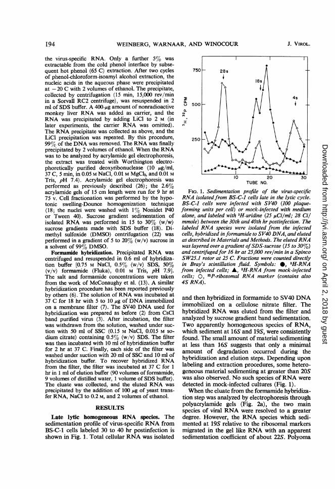
10 20 30 40 10 20 30SLICE NO. SLICE NO.
FIG. 2. Acrylamide gel analysis of virus-specific RNA. 3H-RNA was isolatedfrom cells infected and labeled asin Fig. 1. (a) A sample was hybridized to SV40 DNA in formamide, eluted, and applied to an acrylamide gel forelectrophoresis (Materials and Methods). (b) Another sample of the 3H-RNA was fractionated on an acrylamidegel without prior hybridization to SV40 DNA. After gel electrophoresis, each gel fraction was incubated in 2 XSSCfor 18 hr at 65 C with a filter containing 1 ,ug ofSV40 DNA. The filters were treatedfor I hr with 20 jxg ofpancreatic ribonuclease, dried, and counted in toluene scintillation fluid. Symbols: 0, 3H-viral RNA; 0, "2P-ribo-somal RNA marker.
virus RNA showed an electrophoretic profilealmost identical to that shown in Fig. 2a (R.Weinberg, unpublished data). To determine thepossible effects of the formamide hybridizationprocedure on the size and relative amounts of thetwo species, we applied whole cellular RNA,isolated from cells late in the infectious cycle,directly to an acrylamide gel without prior hy-bridization. The RNA from each fraction of thegel was hybridized independently to filter-boundSV40 DNA (Fig. 2b). Although the data inFig. 2b clearly confirm the existence of two RNAspecies, they have slightly different electrophoreticmobilities, perhaps owing to the exposure of thefirst sample (Fig. 2a) to the denaturing effects offormamide before gel electrophoresis.A subsequent report examines in detail two
properties of the 16S and 19S RNA species whichaffect their relative proportion and homogeneityas seen in the figures here. First, the 19S latelytic species is metabolically much more labilethan the 16S. Second, the size of each of thesespecies decreases with time, a phenomenon con-tributing to their band width in some of thegradient and gel profiles shown here. This latterphenomenon may derive from a decrease in thesize of the polyadenylic acid tail of these RNAspecies as the messenger RNA molecules becomeolder (R. A. Weinberg, in preparation). These twophenomena will not be examined further in thisreport.The sedimentation rates in SDS-sucrose gra-
dients of the two species shown in Fig. 1 are stableafter recentrifugation, reextraction with phenol,and rehybridization of the RNA. The RNA inFig. 3a was prepared as in Fig. 1. Peak fractionsfrom each of the two species were pooled andresedimented (Fig. 3b and 3c). Each RNA speciesresedimented at the same rate as originally ob-served. The peak tubes from these gradients (Fig.3b and 3c) were pooled, precipitated, resuspendedin lysis buffer, and then reextracted with phenol-chloroform-isoamyl alcohol in the presence ofunlabeled, infected BS-C-1 cells. After reextrac-tion, each RNA was rehybridized in formamideto SV40 DNA, eluted, and resedimented. EachRNA species continued to sediment at the rateinitially observed (Fig. 3d and 3e). Thus, the twoRNA species are not two conformations of thesame RNA species in equilibrium with oneanother. Their sedimentation rate is unaffected bythe second cycle of sedimentation, extraction, andhybridization. Other experiments (not shown)indicated that these viral RNA species retaintheir sedimentation rates if they are denaturedwith 90% DMSO (9) before being sedimentedthrough a sucrose density gradient. Also, thetwo RNA species co-sedimented approximatelywith 18S ribosomal RNA when centrifuged in99% DMSO (22). They cannot be separatedfrom each other, however, because of the lowresolution afforded by DMSO gradients, but bothappear to have molecular weights of at least650,000.
VOL. 10, 1972 195
on April 2, 2018 by guest
http://jvi.asm.org/
Dow
nloaded from

196 WEINBERG, WARNAAR, AND WINOCOUR J. VIROL.
30 a) 8
10, ~ 1
F_ '~~t-4b) 18S °' ~~-I ) 2016- .0tT
' 8
n
18 S 18S
16 2 *O.-sol
L.\J _P_
1 2
4~~ ~ ~ ~ ~TB \JO.10 20 10 20
TUBE No.FIG. 3. Sedimentation of 16S anid 19S viral RNA after additional processintg. Ten cultures of BS-C-1 cells were
inifected with SV40 and labeled with 3H-uridine (500 ,uCi/ml, 15 Clmmole) from 33 to 44 hr postinfection. (a) Theinfected cells were lysed in RSB (18) conttaining 1% Nonidet P40, and the cytoplasmic virus-specific RNA specieswere extracted, purified by hybridizationz to S V40 DNA in formamide, anid characterized by centrifugation through15 to 30% SDS-sucrose gradientts (Materials and Methods). The sedimentation conditions used here were 34,000rev/min, 15 hr, in a Spinco S W41 rotor. (b) Fractions 6, 7, and 8 of pantel a were pooled and the RNA was re-cenitrifuged as above. (c) Fractionis 11, 12, and 13 of panel a were pooled, and centrifuged as above. (d and e)Fractions 6, 7, and 8 ofpanel b were pooled as the 19S species; fractions 11 and 12 ofpanel c were pooled as the16S species. The RNA from these two pools was precipitated with ethanol, and redissolved in 3 ml of lysis buffer.To each of two cultures of iunilabeled SV40-infected BS-C-1 cells, at 44 hr postinfection, was added 1.5 ml of the19S RNA in lysis buffer; the 16S RNA in lysis buffer was similarly added to two othaer unlabeled, SV40-inifectedcultures. The RNA was extracted with phenol-chloroform-isoamyl alcohol, hybridized to SV40 DNA, informamide,recovered from the hybrid complex, and centrifuged in 15 to 30% SDS-sucrose as above. (d) 19S RNA. (e) 16SRNA. Symbols: *, 3H-viral RNA; 0, '2P-ribosomal RNA marker.
The formamide hybridization-elution technique 1% host RNA contamination. Another measureyields viral RNA virtually free from host con- of the specificity of the formamide procedure istamination. Cytoplasmic RNA from infected provided by Table 1. RNA from the nuclear andcells produces 120 times more radioactivity cytoplasmic fractions of SV40-infected cellseluted from the viral DNA filters after hybridiza- isolated late in infection was first hybridized intion than does cytoplasmic RNA from mock- formamide to SV40 DNA filters. The hybridizedinfected cells. This eluted RNA thus has less than RNA was then eluted, divided into portions, and
on April 2, 2018 by guest
http://jvi.asm.org/
Dow
nloaded from

SIMIAN VIRUS 40 RNA
TABLE 1. Specificity of the formamide hybridizationt procedurea
Formamide h 0rid)zation to Rehybridization of eluate in 6 X SSC at 65 C
Origin of 3H-RNA 3H-RNA 3H-RNA3H-RNA bound to and 3H-RNA bound to Inputincubated eluted from incubated DNA on filter filter hybridized
(counts/min) filter (counts/min) (counts/ (%)(counts/min) min)
SV40-infected cellsNuclear fraction......... 9.2 X 10⁶ 5.6 X 10⁴ 1.3 X 10⁴ SV40 (10 jlg) 3,000 23.0
1.3 X 10⁴ BS-C-1 (10 ,Ug) 400 3.11.3 X 104 Blank 76 0.6
Cytoplasmic fraction..... 13.1 X 10⁶ 1.6 X 10⁵ 4.5 X 10⁴ SV40 (l0,ug) 19,000 41.54.5 X 104 BS-C-1 (l0,ug) 470 1.04.5 X 104 Blank 120 0.3
a The infection, labeling, and cell fractionation are described in the legend to Fig. 6. The eluates ofthe first cycle of formamide hybridization were rehybridized by the Gillespie and Spiegelman (7) pro-cedure: 20 hr, 65 C in 6 X SSC, followed by ribonuclease treatment of 20 ,ug/ml, 1 hr at room temperature.
rehybridized by the standard Gillespie-Spiegel-man technique (7) to SV40 DNA, BS-C-1 DNA,or blank filters containing no DNA. The elutedcytoplasmic RNA hybridized with high efficiencyback to SV40 DNA. The nuclear RNA, signifi-cantly, hybridized less efficiently back to SV40DNA (see below).
Early lytic RNA synthesis. Early RNA is de-fined as the class of viral RNA species which issynthesized from the beginning of the infectiouscycle to the onset of DNA synthesis. Previousworkers have shown that, in the presence ofinhibitors of DNA synthesis, late RNA synthesisand late functions are suppressed (4, 10, 20),whereas early RNA continues to be synthesized(20). Thus, late RNA synthesis occurs at a timelater than viral DNA synthesis, and is also de-pendent upon it. Besides inhibitors of DNA syn-thesis, actinomycin D at certain concentrationsappears to allow the accumulation of relativelylarge amounts of early RNA with concomitantsuppression of late RNA synthesis (5). We havestudied the sedimentation properties of viral RNAsynthesized early in the infectious cycle, and ofviral RNA made in the presence of cytosinearabinoside (an inhibitor of DNA synthesis) orin the presence of actinomycin (an inhibitor ofRNA synthesis).
Viral DNA synthesis is undetectable before the16th hr postinfection (2). In Fig. 4, we comparethe sedimentation of late RNA (labeled from 40to 46 hr postinfection, Fig. 4a) with early RNA(labeled from 2 to 16 hr postinfection, Fig. 4b).Although the 16S peak predominated later ininfection, the 19S RNA appeared as the mainspecies early in infection. Experiments withmetabolic inhibitors produced similar results.
The effect of cytosine arabinoside on viral RNAis shown in Fig. 5. This drug almost completelysuppressed the 16S species, but allowed accumula-tion of the 19S species. Other experiments, withthe use of different hybridization techniques,demonstrated that under certain conditions 19SRNA is the sole viral RNA made in the presenceof cytosine arabinoside (R. Weinberg, Z. Ben-Ishai, R. Dulbecco, in preparation). ActinomycinD, which allows accumulation of early RNA (5),enhanced the 19S RNA relative to the 165 RNAspecies. None of these experiments, however,demonstrated that the early 19S RNA was identi-cal to the 19S RNA species observed late in thelytic cycle.The formamide hybridization technique can be
used to prepare relatively pure early RNA in spiteof its low concentration in the cell. Table 2 showsthat almost 3% of the radioactively labeled RNApresent 40 to 46 hr postinfection hybridized toSV40 DNA, about 100 times more than theamount hybridizing in the mock-infected control.Early RNA, labeled from 2 to 16 hr postinfection,hybridized to a value only about four times thebackground amount. Early RNA could be furtherpurified, however, by eluting this RNA andrehybridizing it in a second cycle of hybridizationto SV40 DNA. Under these conditions, the earlyRNA hybridized to SV40 DNA to a value 30times higher than to the blank filter, and the lateRNA hybridized 300 times more to SV40 DNAthan to a blank filter.
Heterogeneous nuclear RNA. The sedimentationanalysis of extracts of unfractionated, infectedcells indicated that SV40 RNA contains bothhomogeneous RNA species, and high-molecular-weight heterogeneous RNA (Fig. 1). Figure 6
197VOL. 10, 1972
on April 2, 2018 by guest
http://jvi.asm.org/
Dow
nloaded from

WEINBERG, WARNAAR, AND WINOCOUR
TUBE No20
FIG. 4. Sedimentation of viral RNA labeled early inthe lytic cycle. Thirty-ninie BS-C-J cultures were in-fected with plaque-purified virus at 500 plaque-formingunits/cell. A similar group of 39 cultures was mock-infected. Both groups were labeled with 3H-uridine(250 ,Ci/ml, 30 Ci/mmole) from 2 to 16 hr postinfec-tion. Control "late" RNA was extracted from twoplates infected as above but labeled from 40 to 46 hrpostinfection. 3H-RNA was extractedfrom the infectedcells, and the viral RNA was selected by formamidehybridization to plaque-purified SV40 DNA (Table 2,first cycle hybridization). The viral RNA was thenanalyzed on an SDS-sucrose gradient (30,000 rev/min,16 hr, SW41 rotor). (a) Late RNA: 40 to 46 hr post-infection. (b) Early RNA: 2 to 16 hr postinfection.Symbols: *, 3H-viral RNA; 0, 32P-ribosomal RNAmarker.
shows that, late in infection, the high-molecular-weight heterogeneous viral RNA is largely nuclearwhereas the homogeneous species are largelycytoplasmic (21, 24). The gradients were cen-trifuged for a short time to demonstrate the largesize of the heterogeneous material, and thus donot adequately resolve the two cytoplasmicspecies (Fig. 6b).
Figure 6 suggests that the more rapidly sedi-menting nuclear RNA does not represent aggrega-tion of low-molecular-weight RNA, because suchmaterial is present only in the nuclear fraction.However, a more rigorous proof of the covalentintegrity of the high-molecular-weight RNA isprovided by the DMSO gradient analysis of Fig.7. Whole-cell RNA (nuclear plus cytoplasmic)from cells late in the lytic cycle was centrifuged ona DMSO gradient stabilized with sucrose. Al-
though the DMSO gradient has low resolvingpower, and does not separate the two homogene-ous cytoplasmic species, it demonstrates thatmuch of the heterogeneous viral RNA sediments
u})18s
I
I1_ ~ ~~~~~ _ 4
\'SiS'r D--I ~~~~~-S
~' 18s
2 -2
01 10 20 30TUBE No.
FIG. 5. Effect of cytosine arabinoside on viral RNA.Three cultures of BS-C-J cells were infected at a multi-plicity of 100 plaque-forming units/cell. After virusadsorption, two plates were refed with medium contain-ing cytosine arabinoside (AraC) at 5 ,ug/ml; the thirdwas refed with normal medium. Cells were labeled inthe presence of the drug from 22 to 33 hr postinfectionin the case of the first two plates. The third was labeledat the same time without the drug. The RNA from thedrug-treated and control cultures were hybridized informamide to SV40 DNA, eluted, andanalyzed on SDS-sucrose gradients. (a) Viral RNA from untreated con-irol culture; (b) viral RNA from cytosine arabinoside-treated culture; (c) recentrifugation offractions 15 to20, panel b. Panels a and b: SDS-sucrose gradient,25,000 rev/min, 16 hr, in an SW36 rotor. Panel c:SDS-sucrose gradient at 32,000 rev/min, 16 hr, in anSW36 rotor. Symbols: 0, 3H-viral RNA; 0, 32p_ribosomal RNA marker.
0x
C)0-
c6sI
10
30
J20
110
N 15
II
30 (C\J 5
8
I2
198 J. VIROL.
r )
on April 2, 2018 by guest
http://jvi.asm.org/
Dow
nloaded from

SIMIAN VIRUS 40 RNA
TABLE 2. Selection of virus-specific RNA labeledearly in the lytic cyclea
Percentage of inputhybridized and
Counts/min elutedOrigin of 3H-RNA of input to
hybridization SV40DNA Blank(10 Mg)
First cycle of form-amide hybridi-zation
Infected cells 2-16hr pib........... 1.5 X 10⁹ 0.120 0.028
Mock-infectedcells ............ 1.4 X l09 0.035 0.025
Infected cells 40-46 hr pi......... 7.0 X 10⁷ 2.85 0.030
Second cycle of form-amide hybridi-zation
Infected cells 2-16hr pi............ 9.0 X 10⁵ 4.40 0.150
Infected cells 40-46 hr pi ......... 1.6 X 10⁶ 12.50 0.040
a The preparation of the RNA is described inthe legend to Fig. 4. The eluates of the 1st cycle ofhybridization in formamide were rehybridized informamide in a second cycle of hybridization.
b Postinfection.
much further than even 285 ribosomal RNA.Since a complete transcript of the viral genomeshould sediment as 27S RNA, we can confirmthe reports of others that there are RNA mole-cules of double and triple the length of the genomewhich contain viral sequences and which do notappear to be aggregates (1, 24).The large viral homologous RNA molecules
might derive from the transit of the RNA polym-erase molecule several times around the circularviral DNA template before the nascent RNAchain is terminated. This possibility is unlikely inview of the data presented in Table 3. High-molecular-weight viral RNA was prepared bythe formamide hybridization-elution procedureand sedimented on a DMSO gradient. The ma-terial sedimenting faster than 28S ribosomal RNA(Fig. 7a) hybridized back to SV40 DNA at amuch lower efficiency than does viral homologousRNA smaller than 28S (Table 3). This lowerefficiency of hybridization back to viral DNA mayarise because high-molecular-weight viral RNAmight hybridize with a lower efficiency thanlower-molecular-weight viral RNA containingthe same sequences. However, when the high-molecular-weight viral RNA was degraded bylimited alkali treatment, the hybridization effi-ciency of the resulting fragments (Fig. 7b) did
not increase (Table 3). This experiment indicatesthat the high-molecular-weight viral homologousRNA molecules prepared by the formamidehybridization-elution procedure contain a farsmaller proportion of SV40 sequences than theirlower-molecular-weight counterparts (see also8, 24).
Additional control experiments (S. 0. Warnaar,unpublished data) indicated that the high-molecu-lar-weight viral RNA is not an artifact of the typeof virus used to infect the cells, nor of the type ofviral DNA used for the hybridization-elutionstep. Thus, this viral RNA can be isolated fromcells infected with plaque-purified SV40 virus,by hybridization to DNA derived from plaque-purified virus. The latter control precludes thepossibility that the nonviral sequences found inthe heterogeneous RNA arise exclusively fromhost DNA sequences incorporated into the viralDNA of SV40 virus passaged at high titer (11).Recent experiments, with the formamide hy-bridization-elution technique described in this
cq
a.
a-
IO
In
10 20TIJBE No.
FIG. 6. Nuclear and cytoplasmic virus-specific RNA.One infected BS-C-1 culture was labeled as in Fig. 1.After washing with phosphate-buffered saline, the cellswere swollen in RSB buffer (18). The cells were ho-mogenized with a Dounce-type homogenizer and thenuclei were sedimented by centrifugation. The super-natant fluid was termed the cytoplasmic fraction; thepellet was termed the nuclear fraction. RNA from eachfraction was extracted and hybridized in formamide toSV40 DNA. The eluted RNA was sedimented as in Fig.I but for 14 hr. (a) Viral nuclear RNA; (b) viral cyto-plasmic RNA. Symbols: 0, 3H-viral RNA; 0, 2P-ribosomal RNA marker.
VOL. 10, 1972 199
on April 2, 2018 by guest
http://jvi.asm.org/
Dow
nloaded from

WEINBERG, WARNAAR, AND WINOCOUR
S ioof b) L
10
50
b
10 -~20 30
TUBE No
FIG. 7. DMSO gradienit anialysis of high-molecular-weight viral RNA. Five cultures of SV40-infected BS-C-i cells were labeled with 3H-uridine (200 1Ci/ml, 30Ci/mmole) from 24 to 47 hr postinifection. (a) 3H-RNAwas extracted from whole cells, hybridized to S V40DNA in formamide, eluted, and cenztrifuged through a5 to 20%7o sucrose gradienit in 99% DMSO. The inputto the formamide hybridizationt was 3.5 X 10⁸ countslmin; 5.5 X 10⁶ counts/miln were elutedfrom the SV40filter, and 3.3 X 10³ counts/mili were eluted from a
blank filter. The eluted RNA was cenitrifuged at 45,000rev/mitl for 20 hr at 26 C in ani SW5O.1 rotor. (b)Fractions 2 to 10 of patlel a were pooled; onie portionof the pooled RNA was treated withl 0.05 mr Na2CO3(pH 11.0) for 3 min at 80 C, cooled, nieutralized, andrecentrifuged as above. Symbols: 0, 'H-viral RNA;0, "P-ribosomal RNA marker (the arrows denote thepositions ofthe 18S and 28S species ofribosomal RNA).
paper, have demonstrated that high-molecular-weight viral RNA molecules from lytically in-fected cells contain covalently linked sequenceswhich are complementary to both the viral andhost-cell genomes (S. Rozenblatt and E. Wino-cour, in preparation).
DISCUSSIONThe application of the formamide hybridiza-
tion technique to SV40 RNA metabolism hasindicated the existence of discrete viral RNAspecies. A 19S species is made early in infection,whereas 16S and 19S species are made late in theinfectious cycle. The existence of one or both of
these species was suggested by previous reports(15, 24). These homogeneous species all appear tobe localized in the cytoplasm, and all containpolyadenylic acid sequences (R. Weinberg, Z.Ben-Ishai, J. E. Newbold, and R. Dulbecco, inpreparation). They are probably messenger RNAspecies, but this can be shown only by a demon-stration of their functional association with poly-ribosomes.
It would be tempting to suggest that the early19S species and the late 19S species are identicalin base sequence and represent the early portionof the viral genome, whereas the 16S RNA is 3transcribed from the late portion of the genome.The molecular weights of the 16S and 19S species(ca. 650,000 and 900,000) together add up toapproximately the molecular weight expectedfor a complete transcript of the viral genome(ca. 1.5 X 10⁶). However, preliminary experi-ments show that the late 16S and 19S speciessaturate similar proportions of the viral ge-nome, and appear to compete strongly witheach other for the same DNA sequences (S. 0.Warnaar and E. Winocour, unpublished data).The late 16S and 19S species may thus be derivedfrom overlapping parts of the viral genome ratherthan mutually exclusive portions, and the 19Sand 16S may have a precursor-product relation-ship. The heterogeneous high-molecular-weight
TABLE 3. Rehybridizationi of large virus-specificRNA to SV40 DNA before and after alkali
degradationi to smaller size
Percentage ofinput bound
3H input to filtersOrigin of 3H-RNA' (counts/
min) SV40DNA Blank(10 Mg)
>28S RNAh 33,000 7.7 1.00>28S RNA after alkali 60,600 8.9 0.25degradation to 10-18S 30,300 8.8 0.30RNAc
<28S RNAd 13,000 30.0 1.20
a The preparation and fractionation of thisRNA on a DMSO-sucrose gradient is describedin the legend to Fig. 7.
bPooled fractions 2-10 of Fig. 7a after deoxy-ribonuclease treatment.
c A sample of pooled fractions 2-10 of Fig. 7awas treated with 0.05 M Na2CO3 (pH 11.0) for 3min at 80 C. After cooling and neutralization, theRNA was centrifuged on a DMSO gradient asshown in Fig. 7b. Fractions 16-22 of this secondgradient were pooled and used, after deoxyribo-nuclease treatment.
d Fractions 11-21 in Fig. 7a after deoxyribo-nuclease treatment.
200 J. VIROL.
on April 2, 2018 by guest
http://jvi.asm.org/
Dow
nloaded from

SIMIAN VIRUS 40 RNA
nuclear RNA of the virus may, in turn, be pre-
cursor to the cytoplasmic homogeneous species.SV40-transformed cells contain a virus-related
high-molecular-weight nuclear RNA (12, 24)which contains sequences complementary to boththe viral and cellular genomes (25). Such mole-cules arise, presumably, by the co-transcriptionof integrated viral DNA and adjacent cellularDNA. It has recently been demonstrated -that,under certain conditions of lytic infection, closedcircular SV40 DNA molecules containing a
covalently linked segment of host DNA are pro-
duced (11, 23). These molecules probably arisefrom virus DNA which integrated into hostchromosomal DNA and which was subsequentlyexcised t6gether with a segment of host DNA.Recent experiments have also demonstrated thatSV40-induced high-molecular-weight nuclearRNA, from lytically infected cells, hybridizesboth to ,plaque-purified viral DNA and to host-cell DNA (S. Rozenblatt and E. Winocour, inpreparation). We would therefore suggest thatthe high molecular weight of some of the SV40nuclear RNA is due to the co-transcription of hostand viral DNA sequences during lytic infection.The formamide hybridization technique pre-
sented here can be used to make viral RNA foranalytical and preparative purposes. The tech-nique may prove useful for the isolation of otherRNA species transcribed from episomal or viralgenomes. We are presently attempting in vitrotranslation of the viral RNA prepared by thisprocedure.
ACKNOWLEDGMENTS
R.A.W. was the recipient of a fellowship from the Helen HayWhitney Foundation. S.O.W. was the recipient of fellowshipsfrom the Netherlands Organization for the Advancement of PureResearch (Z.W.O.) and the European Molecular Biology Organi-zation. This work was supported by grant DRG-1061-A from theDamon Runyon Memorial Fund for Cancer Research.We thank B. Danovitch for devoted and expert technical assist-
ance.
LITERATURE CITED
1. Acheson, N. H., E. Buetti, K. Scherrer, and R. Weil. 1971.Transcription of the polyoma virus genome: synthesis -andcleavage of giant late polyoma-specific RNA. Proc. Nat.Acad. Sci. U.S.A. 68:2231-2235.
2. Aloni, Y., E. Winocour, and L. Sachs. 1968. Characterizationof the simian virus 40-specific RNA in virus yielding andtransformed cells. J. Mol. Biol. 31:415-429.
3. Black, P. H., E. M. Crawford, and L. V. Crawford. 1964. Thepurification of simian virus 40. Virology 24:381-387.
4. Butel, J. S., and F. Rapp. 1965. The effect of arabino-furano-sylcytosine on the growth cycle of simian virus 40. Virology27:490-495.
5. Carp, R. I., G. Sauer, and F. Sokol. 1969. The effect of ac-
tinomycin D on the transcription and replication of simianvirus 40 deoxyribonucleic acid. Virology 37:214-226.
6. Fry, M., and M. Artman. 1969. Deoxyribonucleic acid-ribo-nucleic acid hybridization. Biochem. J. 115:287-294.
7. Gillespie, D., and S. Spiegelman. 1965. A quantitative assayfor DNA-RNA hybrids with DNA immobilized as a mem-brane. J. Mol. Biol. 12:829-842.
8. Jaenisch, R. 1972. Evidence for SV40 specific RNA contain-ing virus and host specific sequences. Nature N. Biol. 235:46-47.
9. Katz, L., and S. Penman. 1966. The solvent denaturation ofdouble-stranded RNA from poliovirus infected HeLa cells.Biochem. Biophys. Res. Commun. 23:557-560.
10. Kit, S., D. R. Dubbs, P. M. Frearson, and J. L. Melnick.1966. Enzyme induction in SV40 infected green monkeykidney cultures. Virology 29:69-83.
11. Lavi, S., and E. Winocour. 1972. Acquisition of sequenceshomologous to host deoxyribonucleic acid by closed circu-lar simian virus 40 deoxyribonucleic acid. J. Virol. 9:309-316.
12. Lindberg, U., and J. E. Darnell. 1970. SV40-specific RNA inthe nucleus and polyribosomes of transformed cells. Proc.Nat. Acad. Sci. U.S.A. 65:1089-1096.
13. McConaughy, B. L., C. D. Laird, and B. J. McCarthy. 1969.Nucleic acid reassociation in formamide. Biochemistry8:3289-3295.
14. Martin, M. A., and D. Axelrod. 1969. SV40 gene activityduring lytic infection and in a series of SV40 transformedmouse cells. Proc. Nat. Acad. Sci. U.S.A. 64:1203-1210.
15. Martin, M. A., and J. C. Byrne. 1970. Sedimentation proper-ties of simian virus 40-specific ribonucleic acid present ingreen monkey cells during productive infection and inmouse cells undergoing abortive infection. J. Virol. 6:463-469.
16. Oda, K., and R. Dulbecco. 1968. Induction of cellular RNAsynthesis in BS-C-1 cells infected by SV40. Virology 35:439-444.
17. Oda, K., and R. Dulbecco. 1968. Regulation of transcriptionof the SV40 DNA in productively infected and. in trans-formed cells. Proc. Nat. Acad. Sci. U.S.A. 60:525-532.
18. Penman, S. 1966. RNA metabolism in the HeLa cell nucleus.J. Mol. Biol. 17:117-130.
19. Sauer, G., and J. R. Kidwai. 1968. The transcription of theSV40 genome in productively infected and transformedcells. Proc. Nat. Acad. Sci. U.S.A. 61:1256-1263.
20. Sauer, G. 1971. Apparent differences in transcriptional con-trol in cells productively infected and transformed bySV40. Nature N. Biol. 231:135-138.
21. Sokol, F., and R. I. Carp. 1971. Molecular size of simianvirus 40-specific RNA synthesized in productively infectedcells. J. Gen. Virol. 11:177-188.
22. Strauss, J. H., Jr., R. B. Kelly, and R. L. Sinsheimer. 1968.Denaturation of RNA with dimethylsulfoxide. Biopoly-mers 6:793-807.
23. Tai, H. T., C. A. Smith, P. A. Sharp, and J. Vinograd. 1)72.Sequence heterogeneity in closed simian virus 40 deoxy-ribonucleic acid. J. Virol. 9:317-325.
24. Tonegawa, S., G. Walter, A. Bernardini, and R. Dulbecco.1970. Transcription of the SV40 genome in transformedcells and during lytic infection. Cold Spring Harbor Symp.Quant. Biol. 35:823-831.
25. Wall, R., and J. E. Darnell. 1971. Presence of cell and virusspecific sequences in the same molecules of nuclear RNAfrom virus transformed cells. Nature N. Biol. 232:73-76.
26. Weinberg, R., M. Willems, U. Loening, and S. Penman. 1967.Acrylamide gel electrophoresis of HeLa cell nucleolarRNA. Proc. Nat. Acad. Sci. U.S.A. 58:1088-1095.
VOL. IO, 1972 201
on April 2, 2018 by guest
http://jvi.asm.org/
Dow
nloaded from