Embed Size (px)
Citation preview
Phyton (Austria)Special issue:
"Root-soil interactions"Vol. 40 Fasc. 4 (65)-(70) 25.7.2000
Isolation and Identification of MycorrhizationHelper Bacteria in Norway spruce, Picea abies (L.)
Karst.
By
BARBARA GERIC1)2), MAJA RUPNIK0 & HOJKA KRAIGHER2)
K e y w o r d s : Mycorrhization Helper Bacteria (MHB), rhizosphere, fluorescent pseu-domonads, Bacillus, ectomycorrhiza.
S u m m a r y
GERlC B., RUPNIK M. & KRAIGHER H. 2000. Isolation and identification of mycorrhiza-tion helper bacteria in Norway spruce, Picea abies (L.) Karst. - Phyton (Horn, Austria) 40 (4): (65)- (70).
We have isolated bacteria from bulk soil, the mycorrhizosphere and the hyphosphere ofNorway spruce {Picea abies (L.) Karst) seedlings in mycorrhizal association with the fungus Am-phinema byssoides (Pers.) J. Erikss.. Out of 157 bacterial strains, ten (6.4 %) were fluorescent pseu-domonads and 12 (7.6 %) were from the genus Bacillus. The selected strain P. fluorescens 53a,tested for its effect on growth and mycorrhization of Norway spruce seedlings, grown in vitro in thepresence of the ectomycorrhizal fungus Paxillus involutus (Batsch. ex Fr.) Fr., showed a positiveeffect on growth (fresh and dry weight after one and two months of growth) and mycorrhization(number of mycorrhizal seedlings after three months) and a negative effect on the total root length,root surface area and total root volume after 3 months.
I n t r o d u c t i o n
The rhizosphere is defined as the plant-root surface and the surroundingsoil which is influenced by the living root (DANDURAND & KNUDSEN 1996). Theoperational definition of the rhizosphere, in other studies as well as in ours, is thethin layer of soil adhering to the root system after the loose soil particles have beenremoved by shaking (DANDURAND & KNUDSEN 1996). In the rhizosphere, fungi
1} Biotechnical Faculty, Biology Department, Vecna pot 111, 1000 Ljubljana, Slovenia, e-mail: [email protected]
2) Slovenian Forestry Institute, Vecna pot 2, 1000 Ljubljana, Slovenia, e-mail:[email protected]
©Verlag Ferdinand Berger & Söhne Ges.m.b.H., Horn, Austria, download unter www.biologiezentrum.at
(66)
and bacteria can be found, living on organic compounds released by the root. Theycan have a positive or a negative effect on plant growth. They also affect the sym-biotic establishment of mycorrhizal fungi on plant roots in various ways (GARBAYE
1994). MHBs (Mycorrhization Helper Bacteria) are bacteria associated with my-corrhizal roots and mycorrhizal fungi. They selectively promote the establishmentof mycorrhizal symbiosis (GARBAYE 1994). MHBs have a potential use in foresttree seedling production for promoting growth and as protection against pathogenicmicroorganisms. The two most studied groups of MHBs are fluorescent pseudo-monads and the genus Bacillus, which are also known to have a PGPR effect (PlantGrowth-Promoting Rhizobacteria) (HÖFLICH & al. 1994, PEROTTO & BONFANTE
1997). The mycorrhization helper effect was observed in various symbiotic part-nerships (GARBAYE & BOWEN 1989, GARBAYE & al. 1992, GARBAYE 1994, DUN-
STAN & al. 1998). The ectomycorrhizal combination of Douglas fir {Pseudotsugamenzeisii Mirb. Franco) and Laccaria laccata Scop, ex Fr. has been studied mostextensively (DUPONNOIS & GARBAYE 1991, DUPONNOIS & GARBAYE 1991a, Du-PONNOIS & al. 1993, GARBAYE 1994). For this study, Norway spruce seedlingswere chosen. We studied the diversity of bacteria in the bulk soil, the rhizosphereand the hyphosphere of the nursery, the effects of bacteria on the growth of seed-lings in vitro, and the effects of bacteria on mycorrhization.
M a t e r i a l s a n d M e t h o d s
Isolation and identification of bacterial strains. We isolated bacteria from bulk soil, themycorrhizosphere and the hyphosphere (VAN ELSAS & SMALLA 1996) of 4- to 6-year-old Norwayspruce seedlings ectomycorrhizal with the fungus Amphinema byssoides (AGERER 1987-1998),grown in the nursery of the Slovenian Forestry Institute. Samples of bulk soil (~ 0.1 g) were sus-pended in 1 ml of 0.9 % NaCl mixed by vortexing for 30 seconds. Mycorrhizal roots without a cen-tral cylinder (length up to 0.5 cm), representing the mycorrhizosphere, and rhizomorphs (length upto 1 cm) representing the hyphosphere were macerated, suspended in 1 ml of 0.9 % NaCl and vor-texed for 30 seconds. Each sample was diluted (lOx, lOOx and lOOOx) and 0.1 ml was inoculated onthree different solid media: tripton soy agar (TSA) (GERHARDT & al. 1994), King B (BACON &HlNTON 1996) and 0.2 % brain heart infusion (BHI) supplemented with 1 % peptone, 0.2 % yeastextract, 0.2 % NaCl, and 0.2 % D-glucose (pH 7.2) and incubated at room temperature for two days.Colonies with different morphology were isolated in pure cultures. Each sample (0.1 ml) was alsoinoculated in two liquid enrichment media for pseudomonads: in 30 ml of tryptophan medium(GERHARDT & al. 1994) and in 3 ml of succinate-salts medium (GERHARDT & al. 1994). The trypto-phan medium was incubated with shaking (100 rpm) at room temperature for four days, then 0.1 mlof culture was inoculated in fresh medium and incubated with shaking (100 rpm) at room tempera-ture for two days. The culture was then diluted (lOx, lOOx and lOOOx) and inoculated on plates ofsolidified tryptophan medium. Pure cultures of colonies with different morphology were isolated.The liquid succinate-salts medium was incubated for four days at 30°C, then 0.1 ml of diluted cul-ture (lOx, lOOx and lOOOx) was inoculated on plates of solidified succinate-salts medium and incu-bated for two days at 30°C. Morphologically different colonies were isolated in pure cultures.
Pure cultures obtained from primary plates were characterised by colony morphology,Gram staining, oxidase and catalase reaction, and type of metabolism on OF-glucose (oxidation-fermentation test). All Gram-negative, glucose-nonfermenting, fluorescent pigment-producingstrains were further identified with the following tests: growth at 4°C, 37°C and 42°C, growth onMcConkey agar, growth on 6.5 % NaCl, indole production, production of arginine dihydrolase andlysine decarboxylase, production of lecithinase and lipase, nitrate reduction, production of gas from
©Verlag Ferdinand Berger & Söhne Ges.m.b.H., Horn, Austria, download unter www.biologiezentrum.at
(67)
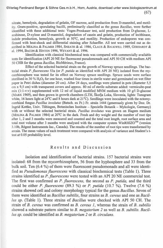
nitrate, hemolysis, degradation of gelatin, OF sucrose, acid production from D-mannitol, and motil-ity. Gram-positive, sporulating bacilli, preliminarily classified as the genus Bacillus, were furtheridentified with these additional tests: Voges-Proskauer test, acid production from D-glucose, L-arabinose, D-xylose and D-mannitol, degradation of casein and gelatin, production of lecithinase,indole production, hemolysis, growth at 50°C, and motility. Production of endospores was con-firmed with heat-shock and differential staining after Schaeffer. All test were carried out as de-scribed in MOLINA & PALMER 1984, SNEATH & al. 1986, CLAUS & BALKWILL 1989, GERHARDT &
al. 1994, BACON & HINTON 1996, WEYANT& al. 1996.
Identification with classical biochemical tests was compared with commercially availabletests for identification (API 20 NE for fluorescent pseudomonads and API 50 CH with medium API50 CHB for the genus Bacillus, BioMerieux, France).
Effect of the selected bacterial strain on the growth of Norway spruce seedlings. The bac-terial strain P. fluorescens 53a (or P. putida as identified with API 20 NE) isolated from the my-corrhizosphere was tested for its effect on Norway spruce seedlings. Spruce seeds were surfacesterilised in 30 % H2O2 for one hour, washed four times in sterile water and germinated on wet filterpaper in Petri dishes (diameter 20 cm). After 24 days, seedlings were planted in pots (diameter 5,5cm x 9,5 cm) with transparent covers and approx. 80 ml of sterile substrate added: vermiculte-peatmix (l:l-v:v) supplemented with 12 ml of liquid modified MNM medium with 10 g/1 D-glucose(MARX 1969), and then grown in growth chambers (LTH, Skofja Loka, Slovenia, lights: Tunsgramwhite, 16 hours light at 24°C and 8 hours dark at 21°C). Seedlings were inoculated with the ectomy-corrhizal fungus Paxillus involutus (Batsch. ex Fr.) Fr. strain 1444 (generously given by Doz. Dr.Ingrid Kottke, Univ. Tübingen, Botanisches Institute - Spezielle Botanik - Mykologie, Germany)with or without the selected bacterial strain. Paxillus involutus was grown on Hagem medium(MOLINA & PALMER 1984) at 20°C in the dark. Fresh and dry weight and the number of root tipsafter 1, 2 and 3 months were measured and counted and the total root length, root surface area andtotal root volume after 3 months were analysed with the computer system WinRHIZO™ (version3.10b, Regent Instruments Inc., Canada). The results of the number of root tips were transformed byarcsin. The mean values of each treatment were compared with analysis of variance and Student's t-test at 0.05 probability level.
R e s u l t s a n d D i s c u s s i o n
Isolation and identification of bacterial strains. 157 bacterial strains wereisolated: 68 from the mycorrhizosphere, 56 from the hyphosphere and 33 from thebulk soil. Ten (6.4 %) of them were fluorescent pseudomonads and all were identi-fied as Pseudomonas fluorescens with classical biochemical tests (Table 1). Threestrains identified as P. fluorescens were tested with an API 20 NE commercial test.The first was confirmed as P. fluorescens, the second as P. putida, and the thirdcould be either P. fluorescens (89.3 %) or P. putida (10.7 %). Twelve (7.6 %)strains showed cell and colony morphology typical for the genus Bacillus. Seven ofthem were identified as Bacillus subtilis, four strains as B. cereus and one as Bacil-lus sp. (Table 1). Three strains of Bacillus, were checked with API 50 CH. Thestrain of B. cereus was confirmed as B. cereus 1, whereas the strain of B. subtilisshowed a substrate pattern similar to B. megaterium 2 as well as B. subtilis. Bacil-lus sp. could be identified as B. megaterium 2 or B. circulans.
©Verlag Ferdinand Berger & Söhne Ges.m.b.H., Horn, Austria, download unter www.biologiezentrum.at

(68)
Table 1. Identification of fluorescent pseudomonads and the genus Bacillus with classicalbiochemical and commercial tests.
No. of identification with classical identification with commercial testsstrains biochemical tests API 20 NE
1 Pseudomonas fluorescens P. fluorescens (99.8%)1 Pseudomonas fluorescens P. putida (99.5 %)1 Pseudomonas fluorescens P. fluorescens (89.3) or P. putida (10.7 %)7 Pseudomonas fluorescens /
No. of identification with classical identification with commercial testsstrains biochemical tests API 50 CH
1 Bacillus cereus B. cereus 11 Bacillus subtilis B. megaterium 2 or B. subtilis1 Bacillus sp. B. megaterium 2 or B.circulans3 Bacillus cereus I6 Bacillus subtilis /
Symbol: / not done
Cultivation of bacterial isolates have variously been reported to recoveronly about 0.01 to 10 % of bacteria enumerated by direct counts (DANDURAND &KNUDSEN 1996). Recovery of populations from the rhizosphere depends on themethod used (washing, vortexing, sonification, and blending or maceration) (DAN-DURAND & KNUDSEN 1996). Therefore the number of strains in the tested materialmight be much higher than 157. We identified 6.4 % out of 157 isolates as fluores-cent pseudomonads and 7.6 % as the genus Bacillus, which agrees with the previ-ously known data for percentages of different bacteria species isolated from soiland the rhizosphere (GARBAYE & BOWEN 1989, GROOMBRIDGE 1992). Variation incertain characteristics was observed among strains within the species P. fluorescens(growth at 4°C and 37°C, growth at 6.5 % NaCl, production of arginine dihy-drolase, production of lipase, nitrate reduction, degradation of gelatin) and B. sub-tilis (acid production from L-arabinose, D-xylose and D-mannitol, degradation ofgelatin and casein), which indicate that not just a single clone of the identifiedstrains colonized the tested material. Instead, it seems that growth conditions in thenursery selectively promote various strains of the identified bacterial species.
Effect of the bacterial strain Pseudomonas fluorescens 53a on the growthof Norway spruce seedlings. Three months after the inoculation of spruce seed-lings, we observed structures similar to Hartig net under the microscope on twoseedlings inoculated with the fungus, and seven seedlings inoculated with the fun-gus and the bacterial strain (Table 2). A positive effect of the bacterial strain onfresh weight after one month, and on dry weight after one and two months afterinoculation, was found, whereas there was no significant effect on seedling bio-mass after three months (Table 2). After three months of incubation we observed asignificantly smaller number of root tips in the group inoculated with the fungusand bacteria, but there were no differences after one and two months (data notshown). The total root length, root surface area and total root volume were meas-
©Verlag Ferdinand Berger & Söhne Ges.m.b.H., Horn, Austria, download unter www.biologiezentrum.at
(69)
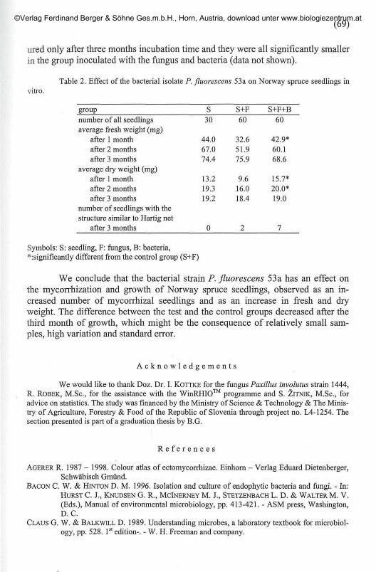
ured only after three months incubation time and they were all significantly smallerin the group inoculated with the fungus and bacteria (data not shown).
Table 2. Effect of the bacterial isolate P.fluorescens 53a on Norway spruce seedlings invitro.
group
number of all seedlingsaverage fresh weight (mg)
after 1 monthafter 2 monthsafter 3 months
average dry weight (mg)after 1 monthafter 2 monthsafter 3 months
number of seedlings with thestructure similar to Hartig net
after 3 months
S30
44.067.074.4
13.219.319.2
0
S+F
60
32.651.975.9
9.616.018.4
2
S+F+B
60
42.9*60.168.6
15.7*20.0*19.0
7
Symbols: S: seedling, F: fungus, B: bacteria,*:significantly different from the control group (S+F)
We conclude that the bacterial strain P. fluorescens 53a has an effect onthe mycorrhization and growth of Norway spruce seedlings, observed as an in-creased number of mycorrhizal seedlings and as an increase in fresh and dryweight. The difference between the test and the control groups decreased after thethird month of growth, which might be the consequence of relatively small sam-ples, high variation and standard error.
A c k n o w l e d g e m e n t s
We would like to thank Doz. Dr. I. KOTTKE for the fungus Paxillus involutus strain 1444,R. ROBEK, M.Sc, for the assistance with the WinRHIO™ programme and S. ZlTNlK, M.Sc, foradvice on statistics. The study was financed by the Ministry of Science & Technology & The Minis-try of Agriculture, Forestry & Food of the Republic of Slovenia through project no. L4-1254. Thesection presented is part of a graduation thesis by B.G.
R e f e r e n c e s
AGERER R. 1987 - 1998. Colour atlas of ectomycorrhizae. Einhorn - Verlag Eduard Dietenberger,Schwäbisch Gmünd.
BACON C. W. & HINTON D. M. 1996. Isolation and culture of endophytic bacteria and fungi. - In:HURST C. J., KNUDSEN G. R., MCINERNEY M. J., STETZENBACH L. D. & WALTER M. V.
(Eds.), Manual of environmental microbiology, pp. 413-421. - ASM press, Washington,D. C.
CLAUS G. W. & BALKWILL D. 1989. Understanding microbes, a laboratory textbook for microbiol-ogy, pp. 528. 1st edition-. - W. H. Freeman and company.
©Verlag Ferdinand Berger & Söhne Ges.m.b.H., Horn, Austria, download unter www.biologiezentrum.at

(70)
DANDURAND L. M. C. & KNUDSEN G. R. 1996. Sampling microbes from the rhizosphere and phy-losphere. - In: HURST C. J., KNUDSEN G. R., MCINERNEY M. J., STRTZENBACH L. D. &
WALTER M. V. (Eds.), Manual of environmental microbiology, pp. 383-390. - ASMpress, Washington, D. C.
DUNSTAN W. A., MALAJCZUKN. & DELL B. 1998. Effects of bacteria on mycorrhizal developmentand growth of container grown Eucalyptus diversicolor F. Muell. seedlings. - Plant Soil201:241-249.
DUPONNOIS R. & GARBAYE J. 1991. Mycorrhization helper bacteria associated with the Douglas fir-Laccaria laccata symbiosis: Effect in aseptic and glasshouse conditions. - Ann. Sei. For.48:239-251.
— & — 1991a. Effect of dual inoculation on Douglas fir with the ectomycorrhizal fungusLaccaria laccata and mycorrhization helper bacteria (MHB) in two bare-root forestnurseries. - Plant Soil 138: 169-176.
— , — , BOUCHARD D. & CHURIN J. L. 1993. The fungus-specificity of mycorrhizationhelper bacteria (MHBs) used as an alternative to soil fumigation for ectomycorrhizalinoculation of bare-root Douglas-fir planting stocks with Laccaria laccata. - Plant Soil157: 257-262.
GARBAYE J. 1994. Transley review No. 76, Helper bacteria: A new dimension to the micorrhizalsymbiosis. - New Phytol. 128: 197-210.
— & BOWEN G. D. 1989. Stimulation of mycorrhizal infection of Pinus radiata by some mi-croorganisms associated with the mantle of ectomycorrhizas. - New Phytol. 112: 383-388.
— , CHURIN J. L. & DUPONNOIS R. 1992. Effects of substrate disinfection, fungicide treatmentsand mycorrhization helper bacteria (MHB) on ectomycorrhizal formation of pedunculateoak inoculated with Laccaria laccata in two bare-root nurseries. - Biol. Fertil. Soils 13:35-47.
GERHARDT P., MURRAY R. G. E., WOOD W. A. & KRIEG N. R. (Eds.) 1994. Methods for generaland molecular bacteriology, pp. 193-210, p. 644. - American Society for Microbiology,Washington, D.C.
GROOMBRIDGE B. 1992. Global biodiversity, status of the earth's living resources. A report com-piled by the world conservation monitoring centre, pp. 575. - Chapman & Hall, London.
HÖFLICH G., WIEHE W. & KÜHN G. 1994. Plant growth stimulation by inoculation with symbioticand associative rhizosphere microorganisms. - Experientia 50: 897-905.
MARX D. H. 1969. The influence of ectotrophic mycorrhizal fungi on the resistence of pine roots topathogenic infection. I. Antagonism of mycorrhizal fungi to root pathogenic fungi andsoil bacteria. - Phytopathology 59: 153-163.
MOLINA R. & PALMER J. G. 1984. Isolation, maintenance, and pure culture manipulation of ecto-mycorrhizal fungi. - In: SCHENCKN. C. (Ed.), Methods and principles of mycorrhizal re-search, pp. 115-129. - The American Phytopathological Society, Minnesota.
PEROTTO S. & BONFANTE P. 1997. Bacterial associations with mycorrhizal fungi: Close and distantfriends in the rhizosphere. - Trends Microbiol. 5: 496-501.
SNEATHP. H. A., M A I R N . S., SHARPEM. E. & HOLT J. G. (Eds.) 1986. Bergey's manual of system-atic bacteriology, Volume 2, pp. 1104 - 1139. - Williams & Wilkins, Baltimore.
VAN ELSAS J. D. & SMALLA K. 1996. Methods for sampling soil microbes. - In: HURST C. J.,KNUDSEN G. R., MCINERNEY M. J., STRTZENBACH L. D. & WALTER M. V. (Eds.), Man-
ual of environmental microbiology, pp. 383-390. - ASM press Washington, D.C.WEYANT R. S., MOSS C. W., WEAVER R. E., HOLLIS D. G., JORDAN J. G., COOK E. C. &
DANESHVARM. J. 1996. Identification of unusual pathogenic gram-negative aerobic andfacultatively anaerobic bacteria, p. 11, pp. 46-49. - Williams & Wilkins, Maryland.
©Verlag Ferdinand Berger & Söhne Ges.m.b.H., Horn, Austria, download unter www.biologiezentrum.at