Embed Size (px)
Citation preview

~ 50 ~
International Journal of Mosquito Research 2017; 4(1): 50-57
ISSN: 2348-5906 CODEN: IJMRK2 IJMR 2017; 4(1): 50-57 © 2017 IJMR Received: 22-11-2016 Accepted: 24-12-2016 Matthew Lukenge (A) Mosquito Research Programme, Uganda Virus Research Institute (UVRI), P. O. Box 49, Entebbe, Uganda (B) Department of Zoology Entomology and Fisheries Sciences, College of Natural Sciences, Makerere University Kampala, P. O. Box 7062, Kampala, Uganda Josephine Birungi Mosquito Research Programme, Uganda Virus Research Institute (UVRI), P. O. Box 49, Entebbe, Uganda Jonathan Kayondo Mosquito Research Programme, Uganda Virus Research Institute (UVRI), P. O. Box 49, Entebbe, Uganda Charles Masembe Department of Zoology Entomology and Fisheries Sciences, College of Natural Sciences, Makerere University Kampala, P. O. Box 7062, Kampala, Uganda Louis G Mukwaya Mosquito Research Programme, Uganda Virus Research Institute (UVRI), P. O. Box 49, Entebbe, Uganda Correspondence Matthew Lukenge (A) Mosquito Research Programme, Uganda Virus Research Institute (UVRI), P. O. Box 49, Entebbe, Uganda (B) Department of Zoology Entomology and Fisheries Sciences, College of Natural Sciences, Makerere University Kampala, P. O. Box 7062, Kampala, Uganda
Isolation and molecular characterization of Gram positive entomopathogenic bacteria against the
major malaria vector Anopheles gambiae in Uganda
Matthew Lukenge, Josephine Birungi, Jonathan Kayondo, Charles Masembe and Louis G Mukwaya
Abstract The challenges of the cardinal vector control approaches have left a dire urgency for supplementary interventions. The study screened and isolated entomopathogenic bacteria from tree hole soil debris. Gram positive endospore forming bacteria were selected for, using the pasteurization technique against Anopheles gambiae (target species) and potent isolates were identified with molecular techniques using the 16s rRNA region. From a total of 616 samples examined, only 4.69% (21/448) were potent. Six of these were concurrently evaluated against Aedes (test) and Anopheles (target) species and were all equally potent by 24 hrs, however, Anopheles gambiae species were more susceptible within 3hrs (p-value = 0.006˂ 0.05 = α). Identification revealed 100% homology with Bacillus group. In this study two isolates that typed as Bacillus anthracis str. Shikan-NIID have not been known to have entomopathogenic activity.Propositions for further studies to evaluate reproducibility under field conditions, efficacy and safety studies are underway. Keywords: Anopheles gambiae, Bacillus anthracis, Bio-control, 16s rRNA, Entomopathogens 1. Introduction Mosquito-borne diseases cause some of the most life threatening, debilitating viral and parasitic infections, imparting a huge socio-economic burden on human lives particularly in developing countries [43]. Malaria parasites, transmitted by female Anopheles mosquitoes causes the most important vector borne disease claiming closely half a million lives per annum [29, 43]. Despite the several control interventions in place, malaria remains a major global public health threat [43]. The World Health Organisation (WHO) recommends an integrated vector control (IVC) approach including the use of bio-rationals such as entomo-pathogenic bacteria but very few of such agents are available for practical application [33]. Bacterial entomo-pathogens naturally exist and are by far the most advanced mosquitocidal bio-rational agents as viable options to chemical control [17]. Unfortunately, much as the vectors have developed resistance to chemical insecticides [34, 31, 26] and some with detrimental health and environmental effects like Dichlorodiphenyltrichloroethane (DDT) [39], bacteria entomopathogens have also been compromised by the development of resistance. For instance, studies have demonstrated resistance to Bacillus sphaericus toxins in Culex quinquefasciatus population from 35 to 150,000 and from 10 to 10,000 fold in laboratory and field conditions respectively [13, 45]. A high level of cross resistance to variants of B. sphaericus C3-41 and 2362 were also demonstrated under laboratory conditions [13]. In Uganda, very little is known about the presence and potency of entomopathogenic bacteria. This study therefore, explores the potential of using indigenous bacterial entomopathogens to supplement the limited arsenal of alternative malaria vector control interventions.
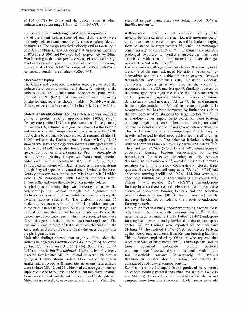
2. Methods 2.1 Study area Sampling was done from the mainland whose vegetation is heavily encroached by human activities and the islands which have a more protected forest cover. Tree hole soil debris was collected from A) Wakiso mainland (Entebbe Botanical forestation (0O03’42.13’’N, 32O28’40.92’’E), and Zika Forest (0O07’15.96‘‘N, 32O31’34.25‘‘E), along the shores of Lake
51
International Journal of Mosquito Research
Victoria, B). Mityana District, (Naama Eucalyptus forest (0O25‘10.50‘‘N, 31O58 ‘58.80‘‘E) and C) Kalangala island-lutoboka forestation (0O18’44.88’’S, 32O17’50.22’’E) (Fig.1). 2.2 Sample collection and processing Tree hole soil debris was collected into sterile zipper bags and preserved at 4 °C until processing. Approximately 1g of soil was suspended in 10ml distilled water and the suspension heat-shocked at 80 °C for 20 minutes to eliminate non-spore formers and simultaneously activate the growth of spores [11]. Serial dilutions of up to 1:106 were made and plated onto nutrient agar followed by incubation for 24 hours at 37 °C. Partial microscopic identification of the isolates was performed on colonies presenting with Gray-white, opaque, flat/ low convex, granular with entire, wavy margins) using Gram and endospore reactions. This was used to categorise the bacterial isolates according to spore shape and position [32,
25]. 2.3 Screening for potent isolates An established colony of Aedes aegypti at the Uganda Virus Research Institute (UVRI) and Anopheles gambiae s.s donated from the International Centre for Insect Physiology and Ecology (ICIPE)-Mbita Kenya were used under laboratory conditions at 25 ± 1 °C, 60 ± 5% RH, 12D:12L photoperiod for evaluation of the potency of bacteria isolates. A colony forming unit (CFU) was cultured in 50 ml of nutrient broth and incubated at 200 rpm for 72hrs at 37 OC in an orbital shaker incubator for spore production. To every assay dish, 45 ml of each isolate culture was added and exposed to 20 late 3rd instar larvae of Aedes aegypti (test species) alongside controls. Aedes aegypti species are major test organisms in evaluation of bacteria entomopathogens in mosquitoes [42] and thus employed in this study before actual evaluation against An. gambiae, the target vector, which is harder to colonise. Isolates showing more than 50% mortality were considered potent. Mortality was observed up to 24 hrs. Screened isolates showing more than 50% mortality were re-evaluated twice for reproducibility. Thereafter, isolates potent against Aedes aegypti were concurrently, evaluated against Anopheles gambiae s.s the target species. 2.4 Evaluation of potency against An. Gambiae Each isolate potent against Ae. aegypti (test species) was sub-cultured in 100ml of nutrient broth and incubated in an orbital shaker at 200 rpm for 72hrs at 37 °C. From this, 45 ml of each bacteria-broth suspension was exposed to both Anopheles gambiae s.s and Aedes aegypti and mortality was then observed after 3hrs, 12hrs and 24hrs in case of viable controls. The exposed concentrations were determined by making serial dilutions and estimating the CFU/ml. Bacterial suspensions were serially diluted 100 fold in 3 different sterile tubes by transferring 10µl stock into 990µl sterile distilled water. All dilutions were plated using the spread method onto nutrient agar and incubated for 24hrs at 37 °C. Colony forming units were counted on the most feasible dilution containing 30-300 CFU. The Exposed concentration was expressed as (CFU/ml) = number of CFU* Dilution factor/ volume exposed. The bio-assay were repeated where control mortality was greater than 20% or corrected using Abbot’s formula where it was less than 20% [42, 1].
2.5 Characterization of potent bacteria Potent isolates (≥ 50% mortality) against target and test species were categorised using microscopy and motility and identified using molecular techniques (16s rRNA). a). Testing for motility Culture plates were prepared using 1% (w/v) tryptone, 0.5% (w/v) NaCl and 0.3% (w/v) agar. Isolates were then inoculated onto the middle of the plate from top to bottom by streaking two closely drawn parallel lines with 2 mm width along the diameter of the plates. The plates were then incubated at 30 °C overnight. Motile isolates were observed to spread out from the inoculation lines [21]. b). 16s rRNA molecular analysis A distinct bacterial CFU was emulsified into100µl sterile distilled water, boiled at 100°C for 10min, and centrifuged at 14,000g for 10min. A 50µl PCR reaction was set up as follows (PCR master-mix (PCR premix 2x (22.5µl), 0.25µM of 27F (5’-GAGTTTGATC(M)TGGCTCAG-3’) and 1525R (5’-AGGAGGTG(W)TCCA(R) CC-3’) primers [9], Taq polymerase enzyme 1.25U and 1µl template) [9]. Amplification proceeded with the following cycling conditions: Initial denaturation at 94 ºC for 5 min; followed by 30 cycles of denaturation (94 ºC for 30 s), annealing (59 ºC for 30 s), extension (72 ºC for 1 min, 30 seconds) and final extension at 72 ºC for 5 min. (Chan et al., 2007). The PCR products alongside the 100bp marker were electrophoresed on 1.5% (w/v) agarose gel and viewed using a transluminator. The PCR products were then purified using the fastgene PCR purification kit. Purified samples (25ul of 50-70ng/µl) were then sent to an outsourced lab (Macrogen-Korea) for sequencing both strands using the same primers above. The sequences were cleaned and edited using Bioedit [15] and Codon code Aligner (Codon Code Aligner v.2.0.6, Codon Code Corporation) bioinformatics software to generate a consensus sequence using the forward and reverse strands. The individual sequences were then blasted using the Megablast application in the NCBI public database (http://blast.ncbi.nlm.nih.gov/Blast.cgi). The sequences were aligned and the phylogenetic and molecular evolutionary genetic relationships were analysed with construction of a phytogenic tree in MEGA6 bioinformatics software (38) using default settings. 3. Results A total of 594 bacterial colonies were assessed, and 75.4% (448/594) were Gram positive endospore forming bacilli (Fig.3). Of these, 24.6% (145/594) were non endospore formers of which 21.4% (31/145) were thermophiles. Regionally, Kalangala had more endospore formers 48.7% (218/448) followed by Wakiso 30.1% (135/448) and lastly Mityana 21.2% (95/448). 3.1 Evaluation of larvicidity against Aedes aegypti Endospore-forming bacteria were exposed to Aedes aegypti (test ssp) and 4.69% (21/448) showed more than 50% potency within 24hrs. The Kalangala island isolates were more potent at 7.62% (16/218) followed by Wakiso isolates at 2.96% (4/135) and Mityana 1.05% (1/95) respectively. Majority of the isolates were within percentage mortality ranges of 81-90 (57.1%), followed 71-80 (23.8%), 91-95 (14.3%) and lastly
52
International Journal of Mosquito Research
96-100 (4.8%) by 24hrs and the concentration at which isolates were potent ranged from 1.1- 1.4x10⁸ CFU/ml. 3.2 Evaluation of isolates against Anopheles gambiae Six of the potent isolates screened against Ae. aegypti were randomly selected and concurrently assessed alongside An. gambiae s.s. The assays revealed a closely similar mortality in both An. gambiae s.s and Ae. aegypti at an average mortality of 98.3% (95-100) and 96% (90-100) respectively by 24hrs. Worth noting is that, An. gambiae s.s species showed a high level of susceptibility within 3hrs of exposure at an average mortality of 71.7% (60-85%) compared to 35% (5-60%) in Ae. aegypti population (p-value = 0.006; 0.05). Microscopic typing The Grams and endospore reactions were used to type the isolates for endospore position and shape. A majority of the isolates 71.4% (15/21) had central and spherical spores, while the rest 28.6% (6/21) had para central and spherically positioned endospores as shown in table 1. Notably, was that all isolates were motile except for isolate MR-12 and MR-21. Molecular identification: The 16s rRNA gene was amplified giving a product size of approximately 1500bp (Fig4). Twenty one purified DNA samples were sent for sequencing but sixteen (16) returned with good sequences for the forward and reverse strands. Comparison with sequences in the NCBI public data base using a Megablast search returned all hits 94-100% similar to the Bacillus genus. Isolates MR-01, 04, 05 showed 99-100% homology with Bacillus thuringiensis YBT-1518 while MR-07 was also homologous with the similar species but a rather different strain i.e. Bacillus thuringiensis strain Lr7/2 though they all typed with Para central, spherical endospores (Table 1). Isolates MR-09, 10, 12, 13, 14, 15, 16, 19, showed homology with Bacillus species of various strains though they all typed with central and spherical endospores. Notably however, were the isolates MR-12 and MR-21 which were 100% homologous with Bacillus anthracis strain Shikan-NIID and were the only two non-motile isolates. A phylogenetic relationship was investigated using the Neighbour-joining method through the alignment and cladistics analysis of the nucleotide sequences among the bacteria isolates (figure 5). The analysis involving 16 nucleotide sequences with a total of 1414 positions analysed in the final dataset using MEGA6 using default settings. The optimal tree had the sum of branch length =0.047 and the percentage of replicate trees in which the associated taxa were clustered together in the bootstrap test (1000 replicates). The tree was drawn to scale of 0.002 with branch lengths in the same units as those of the evolutionary distances used to infer the phylogenetic tree. Molecular findings showed that majority of the identified isolates belonged to Bacillus cereus 43.75% (7/16), followed by Bacillus thuringiensis 31.25% (5/16), Bacillus sp. 12.5% (2/16) and lastly Bacillus anthracis 12.5% (2/16). Phylogeny revealed that isolates MR-14, 15 and 16 were 41% similar typing as B. cereus strains. Isolates MR-1, 4 and 5 were 56% similar and all typed as B. thuringiensis strains. Interestingly were isolates MR-12 and 21 which had the strongest bootstrap support value of 68%, despite the fact that they were obtained from two different and distant forestations of Kalangala and Mityana respectively (please see map in figure1). When blast
searched in gene bank, these two isolates typed 100% as Bacillus anthracis. 4. Discussion The use of chemical or synthetic insecticides as a cardinal approach towards mosquito vector control has been observed to have several limitations ranging from resistance in target vectors [26], effect on non-target organisms and the environment [39, 32]. In humans and animals, prolonged exposure to synthetic insecticides has been associated with cancer, immune-toxicity, liver damage, reproductive and birth defects [39]. Bacterial entomopathogens particularly Bacillus thuringiensis are some of the most advanced bio-rational vector control alternatives and thus a viable option to explore. Bacillus thuringiensis var isrealensis (Bti) registered moderate commercial success as it was used in the control of mosquitoes in the USA and Europe [6]. Similarly, success of the same agent was registered in the WHO Onchocerciasis control program targeting blackfly vectors (Simulium damnosum complex) in western Africa [12]. The rapid progress in the implementation of Bti and its related organisms in mosquito control, has been hampered by limitations such as the development of resistance in the target vectors [41, 8, 13]. It is therefore, rather imperative to search for more bacteria entomopathogens that can supplement the existing ones and emphasize isolation and use of indigenous entomopathogens. This is because bacteria entomopathogens’ efficiency is heavily influenced by their geographical regions of origin as well as application [33]. The selective isolation techniques utilised herein was also employed by Martin and Aileen (22, 3). They isolated 87.74% (773/881) and 70% Gram positive endospore forming bacteria respectively. A related investigation for selective screening of only Bacillus thuringiensis by Keshavarzi [16], revealed a 24.71% (127/514) isolation yield. In this study comparatively, closely three quarters of the collected soil samples i.e. 75.4% (448/594) had endospore forming bacilli and 19.2% (114/594) were non-endospore forming bacilli. These findings also concur with Aileen [3] who isolated 12.3% (108/881) non-endospore forming bacteria therefore, soil debris is indeed a productive source of endospore forming bacteria and the selective pasteurization technique (80 OC for 20 minutes) greatly increases the chances of isolating Gram positive endospore forming bacteria. Despite the fact that many endospore forming bacteria exist, only a few of these are actually entomopathogenic [27]. To this note, the study revealed that only 4.69% (21/448) endospore forming bacilli were actually larvicidal to the test mosquito vector. Similar findings were reported by Asimeng and Mutinga [5] who isolated 4.17% (5/120) pathogenic bacteria against Anopheles arabiensis from Kenyan breeding habitats. This is further emphasised by Ohba [27] who reported that more than 90% of encountered Bacillus thuringiensis isolates (most advanced endospore forming bacterial entomopathogens) are actually non-insecticidal with only a few insecticidal variants. Consequently, all Bacillus thuringiensis isolates should therefore, not entirely be considered as obligate entomopathogens. Samples from the Kalangala Island produced more potent endospore forming bacteria than mainland samples (Wakiso and Mityana). This could be attributed to the fact that island samples were from forest reserves which have a relatively

53
International Journal of Mosquito Research
thicker leaf canopy (Fig.2) and minimal interference from human activities. Tree holes within scattered vegetation have direct exposure to U.V light and increased temperature both of which are bacteria toxin deactivators thus a reduction in the virulence and larvicidity [24, 19, 20]. Unlike Kalangala forest reserves (Island), Botanical, and Zika forest have been having continuous interference from human activities [7, 14] affecting the leafy canopy thickness and thus increasing penetration of U.V into the tree holes. Similarly, Mityana Eucalyptus forests’ reduced number could be due to the fact that chemical pesticides and fertilizers are occasionally applied for the proper maturation of this commercial forestation. Indeed, research has proved that chemical pesticides and fertilisers reduce the virulence of entomopathogens in general with highest impact seen in entomopathogenic nematodes [4, 30]. The pathogenicity of the isolates has been shown to vary among vector species. Poopathi and Abidah [37] reported that Anopheles stephensi was more susceptible than Aedes aegypti though Culex quinquefasciatus was more susceptible than An. stephensi to bacteria pathogens. Notably, this study shows consistency with Poopathi and Abidah, [37] in regards to susceptibility levels. Although by 24hrs all the exposed isolates showed equal virulence in both Anopheles gambiae and Aedes species (p-value= 0.367 ˃ 0.05 = α), the Anopheles species were more susceptible than Aedes species by 3hrs (p-value = 0.006˂ 0.05 = α). To avoid predisposing humans and other mammals to potential infections, proper identification of entomopathogens is of utmost importance to draw certainty that actually the organisms to be used in field application are not pathogenic to humans and other mammals. In this study, identification followed microscopy and molecular characterization. Microscopy revealed that all the 21 potent isolates were Gram positive and endospore forming bacilli which concurred with the molecular characterization which revealed a 99-100% homology to the general Bacillus group. It also further confirms that the selective pasteurization technique utilised herein is an effective isolation technique for endospore forming Bacillus bacteria. Phylogenetic analysis revealed that isolates MR-01, 04 and 05 had a strong evolutionary relationship at 56% (Fig.4) and the Blast hits showed that these isolates related to Bacillus thuringiensis serovar kurstaki str. YBT-1520 by 100%. Bacillus thuringiensis serovar kurstaki str. YBT-1520 has been documented as one of the insecticidal bacterial agents [10] which have similarly been reflected in this study. On a related note, isolates MR-06 and 07 which revealed a 99% homology to Bacillus thuringiensis YBT-1518 and Bacillus thuringiensis strain Lr7/2 respectively which have also been documented to have nematocidal activity [40, 44, 28] though in this study they showed mosquitocidal activity for both species. Noteworthy, are isolates MR-12 and MR-21 which were 99% homologous with Bacillus anthracis strain Shikan-NIID with an evolutionary relationship of 65% though isolated from
different localities i.e. Kalangala and Mityana respectively. The study reports a potential novel finding of the isolated B. anthracis a commonly known human pathogen currently seen to have insecticidal activity. In this study however, MR-12 and MR-21 were potent to both Anopheles gambiae and Aedes aegypti mosquito species. Bacillus anthracis belongs to the B. cereus group consisting of the strains: B. cereus, B. anthracis, B. thuringiensis, B. weihenstephanensis, B. mycoides, and B. pseudomycoides. The B. cereus, B. anthracis, B. thuringiensis have been reported either pathogenic or opportunistic to either mammals or insects while the B. weihenstephanensis, B. mycoides, and B. pseudomycoides are considered non-pathogenic to neither insects nor mammals [2]. 5. Recommendation All the strains in the Bacillus cereus group are genetically and phenotypically heterogeneous overall, but some of the strains are more closely related and phylogenetically intermixed at the chromosome level [36, 18]. Further evaluation of a wider genetic region is therefore needed to confirm that the B. anthracis isolated herein are exclusively insect pathogens or the known human pathogen has variants which are insecticidal as well. An antibiotic susceptibility profile should be carried out to establish the sensitivity patterns of each of the isolates. Efficacy, safety and effect on non-target organism’s trials should be performed before field trials. 6. Conclusion The 21 entomopathogenic bacteria isolated in this study confirm the presence and potential of utilisation of native entomopathogenic bacteria as a supplementary intervention in malaria vector control in Uganda. Molecular characterisation however, seems to be a precise approach in identification as it fully complied and concurred with the microscopic findings that all the isolates got were indeed bacilli and endospore forming bacteria. 7. Acknowledgement This work was funded by a grant to Dr. Louis Mukwaya (Malaria Research and Control Project) from the Presidential Support to Scientists-Uganda, administered by the Uganda National Council of Science and Technology (UNCST). This work was also supported by Training Health Researchers into Vocational Excellence in East Africa (THRiVE), grant number 087540 funded by the Wellcome Trust which facilitated training at the International Centre for Insect Physiology and Ecology (ICIPE). The contents of the study are solely the responsibility of the authors and do not necessarily represent the official views of this supporting office. Special thanks to Makerere University Kampala and the Uganda Virus Research Institute (UVRI) where the study was carried out.
54
International Journal of Mosquito Research
Fig 1: Map of the Uganda (Left) and Geo-referenced sampling sites: Kalangala Island, Wakiso (Botanical and Zika forest) and Mityana mainland.
Fig 2: Impression of forest covers of Mityana and Kalangala forestation sampling sites.
55
International Journal of Mosquito Research
Fig 3: Grams Micrograph, Observe the Para central endospores (E), Bacilli (R) & free spores (S)
Fig 4: Bacterial isolates electrophoretic micrograph of the 16s rRNA 1,500bp products
Fig 4: Phylogenetic tree showing the relationships of the sequences of the bacterial isolates and their blast-searched similarities. The MR-xx on the branch end represents the laboratory identification of the isolate, while to the right of the square bracket is indicated the related organism
blasted in gene bank.
Table 1: Isolates spore position and shape (Gram and endospore reaction)
Isolate Code Spore Shape
Spore Position
MR-01, MR-04, MR-05, MR-06, MR-07, MR-08, MR-15, MR-20
Spherical Para
central MR-02, MR-03, MR-09, MR-10, MR-11, MR-12, MR-13, MR-14, MR-16,
MR-17, MR-18, MR-19, MR-21 Spherical Central
8. References 1. Abbott WS, A method of computing the effectiveness of
an insecticide. J Econ. Entomol, 1925; 18:265-267. 2. Agaisse H, Gominet M, Okstad OA, Kolsto AB, Lereclus
D. PlcR is a pleiotropic regulator of extracellular virulence factor gene expression in Bacillus thuringiensis. Mol Microbiol, 1999; 32(5):1043-53.
3. Aileen G, Rodríguez RY, Bruzón M, Ariamys C, Zulema M, René G. Isolation and Characterization of Entomopathogenic Bacteria from Soil Samples from the Western Region of Cuba. J Vector Ecol. 2013; 38(1):46-52.
4. Amutha M, Gulsar BJ, Surulivelu T, Gopalakrishnan N Effect of commonly used insecticides on the growth of white Muscardine fungus, Beauveria bassiana under laboratory conditions. J Biopes 2010; 3:143-146.
5. Asimeng EJ, Mutinga MJ. Isolation of mosquito-toxic bacteria from mosquito-breeding sites in Kenya. J Am Mosq Control Assoc., 1992; 8(1):86-88.
6. Brian AF, Hyun-woo P, Dennis KB, Margaret CW, Jeffrey JJ, Yuko S et al. Developing recombinant bacteria for control of mosquito larvae. J Am Mosq Control Assoc., 2007; 23(2):164-175.
7. Buxton AP. Observations on the diurnal behaviour of the red tail monkey, Cercopithecus ascanius schmidti, (Matschie) in a small forest in Uganda. J Anim Ecol. 1952; 21:25-58.
8. Camilla CN. Management of Resistance to Bacillus thuringiensis Toxins. The Environmental Studies Program -University of Chicago, PDP publishers, New york. 2000, 94-95.
9. Chan GK, Tiew SZ, Ching, CN. Rapid isolation method of soil bacilli and screening of their quorum quenching activity. Asia Pac. J Mol. Biol. Biotechnol. 2007; 15
MR-14
MR-15Bacillus cereus strain Lr4/2
Bacillus cereus strain SBTBC-007 MR-16
Bacillus cereus gene MR-19
Bacillus cereus strain 37POZ31 MR-03
MR-12
MR-21Bacillus anthracis str. Shikan-NIID
Bacillus thuringiensis strain Lr7/2 MR-07
Bacillus thuringiensis YBT-1518 MR-06
Bacillus cereus strain Lr4/2 MR-10
Bacillus cereus strain 37POZ31 MR-02
MR-01
MR-04
MR-05
Bacillus thuringiensis serovar kurstaki str. YBT-1520
Bacillus sp. AHBR2 MR-13
63
56
56
68
43
12
39 57
36
36
41
0.002
56
International Journal of Mosquito Research
(3):153-156. 10. Dandan WH, Yuhua G, Deju C, Xiaoli Z, Ning Q, Ming
S et al. Proteomic analysis reveals the strategies of Bacillus thuringiensis YBT-1520 for survival under long-term heat stress. Proteomics, 2011; 11:1-12.
11. El-kersh TA, Al-sheikh YA, Al-akeel RA, Alsayed AA. Isolation and characterization of native Bacillus thuringiensis isolates from Saudi Arabia. African Journal of Biotechnology. 2012; 11(8):1924-1938.
12. Guillet P, Kurtak DC, Philippon B, Meyer R. (Use of Bacillus thuringiensis for onchocerciasis control in West Africa. In: de Barjac H, Sutherland D, eds. Bacterial Control of Mosquitoes and Black Flies: Biochemistry, Genetics, and Applications of Bacillus thuringiensis israelensis and Bacillus sphaericus. New Brunswick, NJ: Rutgers University Press. 1990, 187-201.
13. Guofeng P, Cláudia MF, Oliveira ZY, Christina NL, Maria HSF, Jianpin Y et al. A Strain of Bacillus sphaericus Causes Slower Development of Resistance in Culex quinquefasciatus. Appl. Environ. Microbiol. 2002; 68(6):3003-09.
14. Haddow AJ, Williams MC, Woodall JP, Simpson DIH, Goma LKH. Twelve isolations of Zika virus from Aedes (Stegomyia) africanus (Theobald) taken in and above a Uganda forest. Bull World Health Organ, 1964; 31(1):57-69.
15. Hall TA. BioEdit: a user-friendly biological sequence alignment editor and analysis program for Windows 95/98/NT. Nucl. Acids. Symp. Ser. 1999; 41:95-98.
16. Keshavarzi M. Isolation, Identification and Differentiation of Local B. thuringiensis Strains. J Agric. Sci. Technol. 2008; 10:493-499.
17. Kleespies RG, Huger AM, Stephan D. Diagnosis of insect pathogens from locusts and other orthopterans. IOBC WPRS Bulletin. 2000; 23(2):311-334.
18. Kolsto AB, Tourasse NJ, Okstad OA. What sets Bacillus anthracis apart from other Bacillus species? Annu Rev Microbiol. 2009; 63(1):451-476.
19. Lacey AL. Manual of techniques in insect pathology. ACADEMIC Press, London, 1997, 124-125.
20. Lacey LA, Lacey CM. The medical importance or rice land mosquitoes and their control using alternatives to chemical insecticides. J Am. Mos. Control Assoc. 1990; 6:1-93.
21. Maheswaran G, Sreeramanan S, Reena CM, Marimuthu K, Xavier R. Occurrence of Bacillus thuringiensis in faeces of herbivorous farm animals. Afr. J Biotechnol. 2010; 9:8013-8019.
22. Martin PAW, Travers RS. Worldwide abundance and distribution of Bacillus thuringiensis isolates. Appl Environ Microbiol. 1989; 55:2437-42.
23. Ministry of Health, Uganda (MOH), “National policy on malaria treatment. 2005, 2015 [internet]. National policy on malaria treatment 2005. http://www.health.go.ug/mcp/NationalPolicyonMalariaTreatment. June 12, 2015.
24. Mittal PK. Biolarvicides in vector control: challenges and prospects. J Vect Borne Dis 2003; 40:20-32.
25. Monica C. District laboratory practice in developing countries. 2nd Edn, Cambridge University Press, Newyork. 2006, 39.
26. Morgan JC, Irving H, Okedi LM, Steven A, Wondj CS.
Pyrethroid Resistance in an Anopheles funestus Population from Uganda. PLoS ONE. 2010; 5(7):1-8.
27. Ohba M, Mizuki E, Uemori A. Parasporin, a new anticancer Protein Group from Bacillus thuringiensis. Anticancer Res. 2009; 29:427-434.
28. Pengxia W, Chunyi Z, Mengmeng G, Suxia G, Yiguang Z, Jinshui Z et al. Complete genome sequence of Bacillus thuringiensis YBT-1518, a typical strain with high toxicity to nematodes. J Biotechnol. 2014; 171:1-2.
29. Presidential Malaria Initiative-Uganda (PMI), Malaria operations plan for FY 2012. [internet]. Malaria operations plan. http://www.pmi.gov/docs/default-source/default-document-library/country-profiles/uganda_profile.pdf?sfvrsn=16, 03-Oct-2015.
30. Radová S. Effect of selected pesticides on the vitality and virulence of the entomopathogenic nematode Steinernema feltiae (Nematoda: Steinernematidae). Plant Protect. Sci. 2010; 46:83-88.
31. Ramphul U, Boase T, Bass C, Okedi LM, Donnelly MJ, Müller P. Insecticide resistance and its association with target-site mutations in natural populations of Anopheles gambiae from eastern Uganda. Trans. R. Soc. Trop. Med. Hyg. 2009; 103(11):1121-6.
32. Rogan WJ, Chen A. Health risks and benefits of bis (4-chlorophenyl)-1,1,1-trichloroethane (DDT). Lancet, 2005; 366(9487):763-73.
33. Romi R, Ravoniharimelina B, Ramiakajato M, Majori G. Field trails of Bacillus thuringiensis H-14 and Bacillus sphaericus (Strain 2362) formulations against Anopheles arabiensis in the central highlands of Madagascar. J. Am. Mos. Control Assoc. 1993; 9:325-329.
34. Rubaihayo J, Tukesiga E, Abaasa A. Reduced susceptibility to pyrethroid insecticide treated nets by the malaria vector Anopheles gambiae s.l. in western Uganda. Malar J. 2008; 7(92):2875-7.
35. Schaeffer AB, Fulton M. A simplified method of staining endospores. Science, 1993; 77:194.
36. Slamti L, Perchat S, Gominet M, Vilas BG, Fouet A, Mock M et al. Distinct mutations in PlcR explain why some strains of the Bacillus cereus group are nonhemolytic. J Bacteriol. 2004; 186(11):3531-8.
37. Poopathi S, Abidha S. A Medium for the Production of Biopesticides (Bacillus sphaericus and Bacillus thuringiensis subsp. israelensis) in Mosquito Control. J Econ Entomol. 2009; 102(4):1423-1430.
38. Tamura K, Stecher G, Peterson D, Filipski A, Kumar S. MEGA6: Molecular Evolutionary Genetics Analysis version 6.0. Mol Biol Evol. 2013; 30:2725-2729.
39. Turusov V, Rakitsky V, Tomatis L. Dichlorodiphenyltrichloroethane (DDT): ubiquity, persistence, and risks. Environ Health Perspect. 2002; 110:125-128.
40. Weixing Y, Lei Z, Yingying Liu, Neil C, Donghai P, Lifang R. Mining new crystal protein genes from Bacillus thuringiensis on the basis of mixed plasmid-enriched genome sequencing and a computational pipeline. Appl. Environ. Microbiol. 2012L; 78(14):4795-4801.
41. Whalon ME, McGaughey WH. Bacillus thuringiensis: Use and Resistance management. In: Insecticides with novel modes of action: mechanism and application, Ishaaya, I. and Degheele, D., Eds., Springer, Berlin, 1998, 106-137.
57
International Journal of Mosquito Research
42. World Health Organisation (WHO). 2005. Guidelines for Laboratory and Field testing of Mosquito larvicides. WHO/CDS/WHOPES/GCDPP/2005.13.
43. World Health Organisation (WHO). World Malaria Report WHO Pres, Geneva: Switzerland, 2015.
44. Xiaoxia L, Ling C, Qiong H, Jinshui Z, Wei Zhou D, Peng L et al. Factor Bmp1 Functions as a Nematicidal Virulence Bacillus thuringiensis Metalloproteinase Appl. Environ. Microbiol. 2013; 79(2):460.
45. Yuan ZMC, Zhang YM, Liu EY. High-level field resistance to Bacillus sphaericus C3–41 in Culex quinquefasciatus from Southern China. Biocontrol Sci. Technol. 2000; 10:43-51.