Embed Size (px)
Citation preview
Isolation and Structure Elucidation of Anticancer and Antimalarial Natural Products
Yixi Liu
Dissertation submitted to the faculty of the Virginia Polytechnic Institute and State University in partial fulfillment of the requirements for the degree of
Doctor of Philosophy
In Chemistry
David G. I. Kingston, chair
James M. Tanko
Harry C. Dorn
Webster L. Santos
March 26th 2015 Blacksburg, Virginia
Keywords: Natural Products, Antiproliferative, Antimalarial, -pyrone, Lignan,
Sesquiterpene lactone, Naphthoquinone, Stilbenenoid, Alkaloid, Butanolid, Diterpene.

Isolation, Synthesis and StructureActivity Relationship Study of Anticancer and Antimalarial Agents from Natural Products
Yixi Liu
ABSTRACT
As part of an International Cooperative Biodiversity Group (ICBG) program and a
continuing search for antiproliferative natural products from the Madagascar rainforest,
and a collaborative research project established between Virginia Tech and the Institute
for Hepatitis and Virus Research (IHVR) focusing on searching for bioactive natural
products from tropical forests in South Africa, 20 extracts were selected for investigation
based on their antiproliferative activities against A2780 human ovarian cancer cell line or
antimalarial activities against the Dd2 strain of Plasmodium falciparum. Bioassay-guided
fractionation of seven of the extracts yielded twenty new compounds and twenty-four
known compounds, and their structures were elucidated by using a combination of 1D
(1H and 13C) and 2D NMR spectroscopy including COSY, HASQC, HMQC, HMBC, and
NOESY sequences, mass spectrometry, UV, IR, ECD, optical rotation, and chemical
conversions. In addition, ten known compounds were isolated from another five of the
extracts, while studies on the remaining extracts were suspended due to loss of activity,
unworkable small amounts of material, or low structural interest.
The plants and their metabolites are discussed in the following order: five new
antimalarial 5,6-dihydro--pyrones and six bicyclic tetrahydro--pyrone derivatives
from Cryptocarya rigidifolia (Lauraceae); two new and five known antiproliferative
lignans from Cleistanthus boivinianus (Phyllanthaceae); two new and two known
antiproliferative sesquiterpenes lactones from Piptocoma antillana (Asteraceae); one new
iii
antiproliferative 1,4-naphthoquinone, one known antiproliferative isoflovonoid, and five
known antiproliferative stilbenoids from Stuhlmannia moavi (Leguminosae); four known
antiproliferative bisbenzylisoquinoline alkaloids from Anisocycla grandidieri
(Menispermaceae); one new and two known antiproliferative butanolides, and two new
antiproliferative secobutanolides from Ocotea macrocarpa (Lauraceae); one new
antiproliferative and five known antiproliferative diterpenoids from Malleastrum
rakotozafyi (Meliceae); and 10 known compounds from Monoporus sp. (Myrsinaceae),
Premna corymbosa (Verbenaceae), Premna perplexanes (Verbenaceae), Epallage
longipes (Asteraceae), and Cinnamosma fragrans (Canellaceae).
iv
Table of Contents
ABSTRACT ........................................................................................................................ ii
Table of Contents ............................................................................................................... iv
List of Tables .................................................................................................................... xii
List of Schemes ................................................................................................................ xiii
List of Figures .................................................................................................................. xiv
1 Introduction .................................................................................................................. 1
1.1 Natural Products...................................................................................... 1
1.2 Cancer and Anticancer Agents from Natural Products ........................... 2
1.3 Malaria and Antimalarial Agents from Natural Products ....................... 5
1.4 Discovery of New Natural Product Drugs .............................................. 7
1.4.1 Bioactivity directed fractionation ........................................................... 7
1.4.2 Dereplication approach ........................................................................... 8
1.4.2.1 Dereplication ............................................................................. 8
1.4.2.2 UV spectroscopy ....................................................................... 9
1.4.2.3 LC-MS and LC-MS/MS ........................................................... 9
1.4.2.4 LC-NMR spectroscopy ........................................................... 11
1.5 The Madagascar ICBG project ............................................................. 13
1.6 References ............................................................................................. 16
2 Antimalarial 5,6-Dihydro--pyrones from Cryptocarya rigidifolia: Related Bicyclic
Tetrahydro--Pyrones are Artifacts .................................................................................. 21
2.1 Introduction ........................................................................................... 21
2.2 Results and Discussion ......................................................................... 23
v
2.2.1 Isolation of active compounds .............................................................. 23
2.2.2 Structure elucidation of compound 2.1 ................................................. 24
2.2.1 Structure elucidation of compound 2.2 ................................................. 26
2.2.2 Structure elucidation of compound 2.3 ................................................. 28
2.2.3 Structure elucidation of compound 2.4 ................................................. 29
2.2.4 Structure elucidation of compound 2.5 ................................................. 30
2.2.5 Structure elucidation of compound 2.6 ................................................. 31
2.2.6 Structure elucidation of compound 2.7 and 2.8 .................................... 35
2.2.7 Structure elucidation of compound 2.92.11 ........................................ 36
2.2.8 Evidence that bicyclic tetrahydropyrones 2.6 – 2.11 are artifacts ........ 36
2.2.9 Biological activities .............................................................................. 38
2.3 Conclusion ............................................................................................ 39
2.4 Experimental Section ............................................................................ 40
2.4.1 General experimental procedures ......................................................... 40
2.4.2 Plant material ........................................................................................ 40
2.4.3 Extraction and isolation ........................................................................ 41
2.4.4 Cryptorigidifoliol A (2.1)...................................................................... 43
2.4.5 Cryptorigidifoliol B (2.2) ...................................................................... 43
2.4.6 Cryptorigidifoliol C (2.3) ...................................................................... 43
2.4.7 Cryptorigidifoliol D (2.4)...................................................................... 43
2.4.8 Cryptorigidifoliol E (2.5) ...................................................................... 44
2.4.9 Cryptorigidifoliol F (2.6) ...................................................................... 44
2.4.10 Cryptorigidifoliol G (2.7)...................................................................... 44
vi
2.4.11 Cryptorigidifoliol H (2.8)...................................................................... 44
2.4.12 Cryptorigidifoliol I (2.9) ....................................................................... 45
2.4.13 Cryptorigidifoliol J (2.10) ..................................................................... 45
2.4.14 Cryptorigidifoliol K (2.11).................................................................... 45
2.4.15 Dimethyldisulfide derivatization. ......................................................... 45
2.4.16 Preparation of the R and S-MPA ester derivatives of 5,6-dihydro--
pyrones .............................................................................................................. 46
2.4.17 Preparation of bicyclic tetrahydropyrones ............................................ 46
2.4.18 Antiproliferative bioassay ..................................................................... 46
2.4.19 Antimalarial bioassay............................................................................ 47
2.5 References ............................................................................................. 48
3 Antiproliferative Compounds from Cleistanthus boivinianus from the Madagascar
Dry Forest ......................................................................................................................... 53
3.1 Introduction ........................................................................................... 54
3.2 Results And Discussion ........................................................................ 55
3.2.1 Isolation of active compounds .............................................................. 55
3.2.2 Structure elucidation of compound 3.1 ................................................. 57
3.2.3 Structure elucidation of compound 3.2 ................................................. 61
3.2.4 Biological activities .............................................................................. 62
3.3 Experimental Section ............................................................................ 65
3.3.1 General experimental procedures ......................................................... 65
3.3.2 Plant material ........................................................................................ 65
3.3.3 Extraction and isolation ........................................................................ 66
vii
3.3.4 Cleisthanolide A (3.1) ........................................................................... 67
3.3.5 4-O-(-D-Glucopyranosyl)-dehydropodophyllotoxin (3.2) ................. 68
3.3.6 (±)--Apopicropodophyllin (3.4) .......................................................... 68
3.3.7 Antiproliferative bioassay ..................................................................... 68
3.3.8 Antimalarial bioassay............................................................................ 68
3.3.9 Acid hydrolysis of compounds 3.1 and 3.2........................................... 68
3.4 References ............................................................................................. 70
4 Antiproliferative and Antimalarial Sesquiterpene Lactones from Piptocoma antillana
from Puerto Rico ............................................................................................................... 76
4.1 Introduction ........................................................................................... 76
4.2 Results and discussion .......................................................................... 77
4.2.1 Isolation of active compounds .............................................................. 77
4.2.2 Structure elucidation of compounds 4.1 and 4.2................................... 78
4.2.3 Structure elucidation of compound 4.3 ................................................. 79
4.2.4 Structure elucidation of compound 4.4 ................................................. 81
4.2.5 Bioactivities .......................................................................................... 83
4.3 Experimental Section ............................................................................ 83
4.3.1 General experimental procedures ......................................................... 83
4.3.2 Plant material ........................................................................................ 84
4.3.3 Extraction and isolation ........................................................................ 84
4.3.4 5-O-Methyl-5-epiisogoyazensolide (4.1) .............................................. 85
4.3.5 5-O-Methylgoyazensolide (4.2) ............................................................ 85
4.3.6 Antiproliferative bioassay ..................................................................... 86
viii
4.3.7 Antimalarial bioassay............................................................................ 86
4.4 References ............................................................................................. 87
5 Bioactive Compounds from Stuhlmannia moavi from the Madagascar Dry Forest .. 90
5.1 Introduction ........................................................................................... 90
5.2 Results and Discussion ......................................................................... 92
5.2.1 Isolation of active compounds .............................................................. 92
5.2.2 Structure elucidation of compound 5.1 ................................................. 94
5.2.3 Biological activities .............................................................................. 96
5.3 Experimental ......................................................................................... 97
5.3.1 General experimental procedures ......................................................... 97
5.3.2 Plant material ........................................................................................ 98
5.3.3 Extraction and isolation ........................................................................ 98
5.3.4 Stuhlmoavin (5.1) ................................................................................. 99
5.3.5 X-ray crystallography of 5.1 ............................................................... 100
5.3.6 Antiproliferative bioassay ................................................................... 100
5.3.7 Antimalarial bioassay.......................................................................... 101
5.4 References ........................................................................................... 102
6 Structure Elucidation of Antiproliferative Bisbenzylisoquinoline Alkaloids from
Anisocycla grandidieri from the Madagascar Dry Forest............................................... 107
6.1 Introduction ......................................................................................... 107
6.2 Results and Discussion ....................................................................... 108
6.2.1 Isolation of bioactive compounds ....................................................... 108
6.2.2 Structure elucidation of compound 6.1 ............................................... 110
ix
6.2.3 Structure elucidation of compound 6.5 ............................................... 114
6.2.4 Structure elucidation of compound 6.2 ............................................... 115
6.2.5 Structure elucidation of compound 6.6 ............................................... 116
6.2.6 Antiproliferative bioactivities ............................................................. 116
6.3 Experimental ....................................................................................... 117
6.3.1 General experimental procedures ....................................................... 117
6.3.2 Plant material ...................................................................................... 117
6.3.3 Extraction and isolation ...................................................................... 118
6.3.4 NMR spectroscopy.............................................................................. 118
6.3.5 Stebisimine (6.1) ................................................................................. 120
6.3.6 (+)-1, 2-Dehydrotelobine (6.5) ........................................................... 121
6.3.7 (+)-2'-Norcocsuline (6.2) .................................................................... 121
6.3.8 Puetogaline B (6.6) ............................................................................. 121
6.3.9 X-ray Crystallography ........................................................................ 121
6.3.9.1 Stebisimine (6.1) ................................................................... 121
6.3.9.2 (+)-2'-Norcocsuline (6.2) ...................................................... 122
6.3.10 Antiproliferative bioassays ................................................................. 123
6.4 References ........................................................................................... 124
7 Antiproliferative Compounds from Ocotea macrocarpa from the Madagascar Dry
Forest............................................................................................................................... 128
7.1 Introduction ......................................................................................... 128
7.2 Results and Discussion ....................................................................... 129
7.2.1 Isolation of active compounds ............................................................ 129
x
7.2.2 Structure elucidation of compound 7.1 ............................................... 130
7.2.3 Antiproliferative Activities ................................................................. 135
7.3 Experimental section ........................................................................... 135
7.3.1 General experimental procedures ....................................................... 135
7.3.2 Plant material ...................................................................................... 136
7.3.3 Extraction and isolation ...................................................................... 136
7.3.4 Macrocarpolide A (7.1) ....................................................................... 137
7.3.5 Macrocarpolide B (7.2) ....................................................................... 138
7.3.6 Macrocarpolide C (7.3) ....................................................................... 138
7.3.7 Antiproliferative bioassay ................................................................... 138
7.4 References ........................................................................................... 139
8 Antiproliferative Compounds from Malleastrum sp. from the Madagascar Dry Forest
143
8.1 Introduction ......................................................................................... 143
8.2 Results and Discussion ....................................................................... 145
8.2.1 Isolation of active compounds ............................................................ 145
8.2.2 Structure elucidation of compound 8.1 ............................................... 146
8.2.3 Antiproliferative activities .................................................................. 149
8.3 Experimental section ........................................................................... 149
8.3.1 General experimental procedures ....................................................... 149
8.3.2 Plant material ...................................................................................... 150
8.3.3 Extraction and isolation ...................................................................... 150
8.3.4 18-Oxo-cleroda-3,13-dien-16,15-olide (8.1) ...................................... 151
xi
8.3.5 Antiproliferative bioassay ................................................................... 151
8.4 References ........................................................................................... 152
9 Miscellaneous Natural Products Studied.................................................................. 155
9.1 Introduction ......................................................................................... 155
9.2 Anticancer Extracts ............................................................................. 155
9.2.1 Monoporus sp. (Myrsinaceae) MG 0596 (LF) .................................... 155
9.2.2 Premna corymbosa (Verbenaceae) MG 4508 (RT) ............................ 156
9.2.3 Premna perplexans (Verbenaceae) MG 2860 (RT) + MG 2862 (WD) +
MG 2863 (LF+FR) .......................................................................................... 156
9.2.4 Epallage longipes (Asteraceae), MG 4849 (RT) ................................ 157
9.2.5 Cinnamosma fragrans (Canellaceae), MG 2360 (RT) ....................... 158
10 Summary and General Conclusions ...................................................................... 159
10.1 Isolation of Bioactive Natural Products .............................................. 159
10.2 Antiproliferative Extracts.................................................................... 159
10.3 Antimalarial extract ............................................................................ 162
10.4 Conclusions ......................................................................................... 162
11 Appendix ............................................................................................................... 164
xii
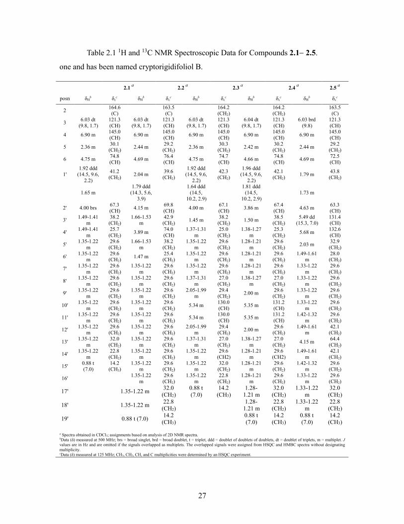
List of Tables Table 2.1 1H and 13C NMR Spectroscopic Data for Compounds 2.12.5. ...................... 27
Table 2.2 1H and 13C NMR Spectroscopic Data for Compounds 2.62.8, and 1H NMR
Spectroscopic Data for Compounds 2.92.11 .......................................................... 34
Table 2.3 Bioactivities of 5,6-Dihydro--pyrones 2.12.5 .............................................. 39
Table 2.4 Bioactivities of Bicyclic Tetrahydro--pyrone Derivatives 2.62.11 .............. 39
Table 3.1 1H NMR (500 MHz, CD3OD) and 13C NMR (125 MHz, CD3OD) Chemical
Shift Data (δ, ppm) for Compounds 3.1 and 3.2 ....................................................... 60
Table 3.2 Antiproliferative and Antimalarial Activities (IC50 Values, M) ..................... 64
Table 4.1 1H and 13C NMR Data (δ, ppm) for Compounds 4.3 and 4.4 ........................... 82
Table 5.1 1H and 13C NMR Data (δ, ppm) for Compound 5.1 (500 and 125 MHz) ......... 96
Table 6.1 1H NMR (500 MHz) Chemical Shift Data (δ, ppm) for Compounds 6.16.4 113
Table 6.2 13C-NMR (125 MHz) Chemical Shift Data (, ppm) for Compounds 6.16.4
................................................................................................................................. 113
Table 6.3 Antiproliferative Activities (M) of Compounds 6.1, 6.2, 6.5, and 6.6. ........ 117
Table 7.1 1H and 13C NMR Data for Compound 7.1 ...................................................... 132
Table 7.2 1H and 13C NMR Data for Compounds 7.2 and 7.3. ....................................... 134
Table 8.1 1H and 13C NMR Data for Compound 8.1 ...................................................... 149
xiii
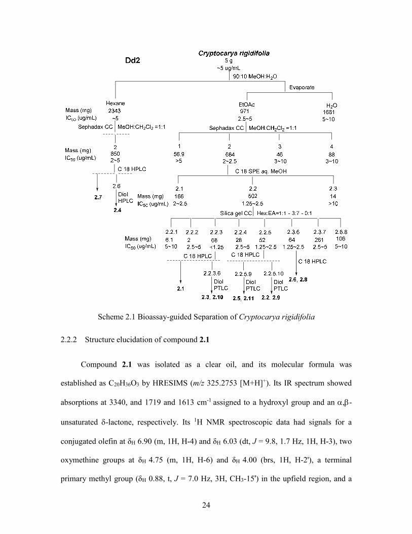
List of Schemes Scheme 2.1 Bioassay-guided Separation of Cryptocarya rigidifolia ............................... 24
Scheme 2.2 Proposed Mechanism of Formation of Tetrahydro--pyrone Derivatives ... 37
Scheme 3.1 Bioassay-guided Separation of Cleistanthus boivinianus ............................. 56
Scheme 4.1 Bioassay-guided Separation of Piptocoma antillana .................................... 78
Scheme 5.1 Bioassay-guided Separation of Stuhlmannia moavi ...................................... 93
Scheme 6.1 Bioassay-guided Separation of Anisocycla grandidieri .............................. 110
Scheme 7.1 Bioassay-guided Separation of Ocotea macrocarpa ................................... 130
Scheme 8.1 Bioassay-guided Separation of Malleastrum sp. ......................................... 146
xiv
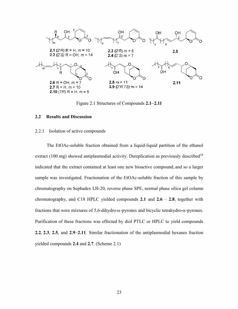
List of Figures Figure 2.1 Structures of Compounds 2.12.11 ................................................................. 23
Figure 2.2 Key HMBC Correlations of 2.1....................................................................... 26
Figure 2.3 H) = δSδR Data of R and S-MPA Derivatives of Compound 2.1 ............. 26
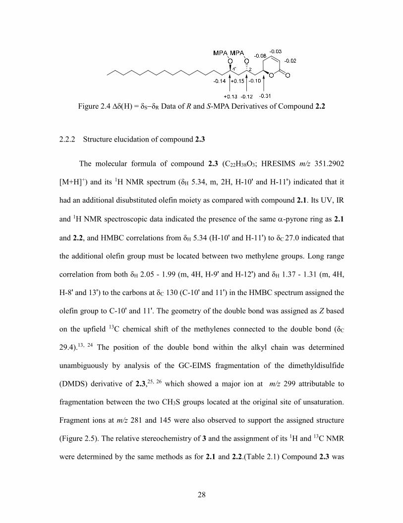
Figure 2.4 H) = δSδR Data of R and S-MPA Derivatives of Compound 2.2 ............. 28
Figure 2.5 H) = δSδR Data of R and S-MPA Derivative and EI-MS Fragmentations of
the DMDS Adduct of Compound 2.3 ....................................................................... 29
Figure 2.6 H) = δSδR Data of R and S-MPA Derivative and EI-MS Fragmentations of
the DMDS Adduct of Compound 2.4 ....................................................................... 30
Figure 2.7 ESI-MS Fragmentations of Compound 2.5 ..................................................... 31
Figure 2.8 Key HMBC Correlations of 2.6....................................................................... 32
Figure 2.9 EI-MS Fragmentations of the DMDS Adduct of Compound 2.6 .................... 33
Figure 2.10 EI-MS Fragmentations of the DMDS Adduct of Compound 2.7 .................. 35
Figure 3.1 Structures of Compounds 3.13.7 ................................................................... 55
Figure 3.2 Key HMBC and NOESY Correlations of 3.1 ................................................. 58
Figure 3.3 Structure of Compound 3.4a ........................................................................... 59
Figure 3.4 Key HMBC and NOESY Correlations of 3.2 ................................................. 62
Figure 3.5 Structure of Compound 3.2a ........................................................................... 62
Figure 4.1 Structures of Compounds 4.14.4 ................................................................... 77
Figure 4.2 Key HMBC and NOESY Correlations of 4.3 ................................................. 81
Figure 4.3 Key HMBC and NOESY Correlations of 4.4 ................................................. 83
Figure 5.1 Structures of Compounds from Stuhlmannia moav......................................... 92
Figure 5.2 Key HMBC correlations of 5.1 ....................................................................... 95
xv
Figure 5.3 Anisotropic Displacement Ellipsoid Drawing of Compound 5.1. ................... 96
Figure 6.1 Structures of Compounds 6.16.6 ................................................................. 109
Figure 6.2 Key HMBC (Single Headed Arrows) and NOESY (Double Headed Arrows)
Correlations of 6.1 .................................................................................................. 112
Figure 6.3 Anisotropic Displacement Ellipsoid Drawing of Compound 6.1 .................. 112
Figure 6.4 Key HMBC (Single Headed Arrows) and NOESY (Double Headed Arrows)
Correlations of 6.5 .................................................................................................. 115
Figure 6.5 Anisotropic Displacement Ellipsoid Drawing of Compound 6.2. ................. 116
Figure 7.1 Structures of Compounds 7.17.5. ................................................................ 130
Figure 7.2 Key HMBC Correlations of 7.1..................................................................... 131
Figure 7.3 Key HMBC Correlations of 7.2..................................................................... 133
Figure 8.1 Structures of Compounds 8.18.5. ................................................................ 145
Figure 8.2 Key HMBC and NOESY Correlations of 8.1. .............................................. 148
1 Introduction
1.1 Natural Products
In the broadest sense, natural products include any substance produced by life, but
the term is commonly used in reference to chemical compounds or substances produced
by a living organism found in nature. Natural products often have pharmacological or
biological activities. Therefore, humans have relied on nature for their basic needs
throughout the ages, especially for the treatment of a wide spectrum of diseases. Plants,
in particular, have formed the basis of traditional medicine systems, and records dating
from around 2600 BCE document the uses of approximately 1000 plant-derived
substances in Mesopotamia.1
It is worth discussing the reasons for the important roles played by natural
products, especially natural products from plants. Natural products can be characterized
as secondary metabolites from natural resources, like plants, marine organisms and
microbes. So the first reason can be from a biological point of view. Plants are fixed in a
place, thus it is almost impossible for them to kill predators such as insects and animals
or to defend themselves by movement. As time passes, they change, recombine and
evolve their genes to develop a very effective chemical defensive system, which they do
in part by producing numerous compounds with diverse chemical structures.2 It has been
reported that these secondary metabolites are related to predator deterrence.3 And some
of those secondary metabolites produced by the evolved genes can be used utilized by
humans as bioactive natural products.
Furthermore, natural products are irreplaceable as the source of lead compounds
for future drug design. Natural compounds are friendlier ligands to biological receptors

2
than pure synthetic compounds, since they come from natural organisms. However, they
may not be very effective drugs directly after they are isolated, and chemists may need to
use medicinal chemistry to modify their structures to improve their potency or their
pharmacokinetic properties.
1.2 Cancer and Anticancer Agents from Natural Products
Cancer is one of the leading causes of death in the world. According to the
statistical data from the World Cancer Report, about 8.2 million people died because of
cancer in 2012, which means 22,500 deaths per day, among them about 62% are from
developing countries and 38% are from developed countries. If this rate continues, 17.5
million cancer deaths per year from cancer can be predicted by 2050.4, 5 Therefore the
cure of cancer has been a worldwide concern for a long time. Plants have proven to be the
most reliable source and successful strategy in modern cancer treatment. An impressive
number of modern drugs derived from plants have been used to treat many types of
cancer, like lymphomas, leukemias and solid tumors.6 From Newman and Cragg’s
research, 63% of a total of 155 anticancer drugs marketed from 1981 to 2006 are natural
products or analogues of natural products, and most of them come from North America,
Europe, and Japan.7 Butler also lists 79 new anticancer agents that entered clinical trial as
natural products or their derivatives in recent years.8
The earliest attempt at searching for anticancer agents from plants was in the
1950s. It started with the discovery of the vinca alkaloids. The vinca alkaloids are a series
of compounds composed of indole and indoline subunits. They are the first natural
products entering into clinical use as anticancer agents and represent one of the most
3
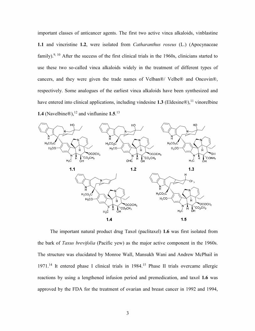
important classes of anticancer agents. The first two active vinca alkaloids, vinblastine
1.1 and vincristine 1.2, were isolated from Catharanthus roseus (L.) (Apocynaceae
family).9, 10 After the success of the first clinical trials in the 1960s, clinicians started to
use these two so-called vinca alkaloids widely in the treatment of different types of
cancers, and they were given the trade names of Velban®/ Velbe® and Oncovin®,
respectively. Some analogues of the earliest vinca alkaloids have been synthesized and
have entered into clinical applications, including vindesine 1.3 (Eldesine®),11 vinorelbine
1.4 (Navelbine®),12 and vinflunine 1.5.13
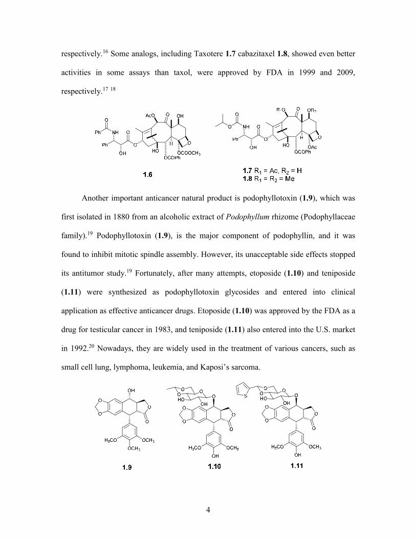
The important natural product drug Taxol (paclitaxel) 1.6 was first isolated from
the bark of Taxus brevifolia (Pacific yew) as the major active component in the 1960s.
The structure was elucidated by Monroe Wall, Mansukh Wani and Andrew McPhail in
1971.14 It entered phase I clinical trials in 1984.15 Phase II trials overcame allergic
reactions by using a lengthened infusion period and premedication, and taxol 1.6 was
approved by the FDA for the treatment of ovarian and breast cancer in 1992 and 1994,
4
respectively.16 Some analogs, including Taxotere 1.7 cabazitaxel 1.8, showed even better
activities in some assays than taxol, were approved by FDA in 1999 and 2009,
respectively.17 18
Another important anticancer natural product is podophyllotoxin (1.9), which was
first isolated in 1880 from an alcoholic extract of Podophyllum rhizome (Podophyllaceae
family).19 Podophyllotoxin (1.9), is the major component of podophyllin, and it was
found to inhibit mitotic spindle assembly. However, its unacceptable side effects stopped
its antitumor study.19 Fortunately, after many attempts, etoposide (1.10) and teniposide
(1.11) were synthesized as podophyllotoxin glycosides and entered into clinical
application as effective anticancer drugs. Etoposide (1.10) was approved by the FDA as a
drug for testicular cancer in 1983, and teniposide (1.11) also entered into the U.S. market
in 1992.20 Nowadays, they are widely used in the treatment of various cancers, such as
small cell lung, lymphoma, leukemia, and Kaposi’s sarcoma.

5
1.3 Malaria and Antimalarial Agents from Natural Products
Malaria is a mosquito-borne infectious disease caused by eukaryotic parasitic
protozoans of the genus Plasmodium. Malaria typically causes symptoms including fever,
fatigue, vomiting and headaches, and severely yellow skin, which can lead to seizures,
coma or death. The disease is widespread throughout tropical and subtropical regions
including much of sub-Saharan Africa, Asia, the Pacific Islands, and South America.
Malaria remains one of the major infectious diseases that threaten human lives.
According to the world malaria report by the World Health Organization (WHO) global
malaria programme in 2014, about 219 million cases of malaria and an estimated 660,000
deaths occur from malaria every year, and it is responsible for over 430000 child deaths
in Africa every year. Emerging drug- and insecticide-resistance continues to pose a major
threat, and if left unaddressed, could trigger an upsurge in deaths.
In the 17th century, quinine (1.12) and quinidine (1.13) were isolated from the bark
of the cinchona tree.21 They were the first effective antimalarial medicines and were used
for centuries until chloroquine (1.14) was discovered in the 1940s. Chloroquine is more
effective for all types of malaria with fewer side effects.22 However, resistant strains
started to emerge in the 1960s. Other antimalarial agents with similar heterocyclic ring
structures were discovered afterwards, including mefloquinone (1.15) and mepacrine
(1.16).
6
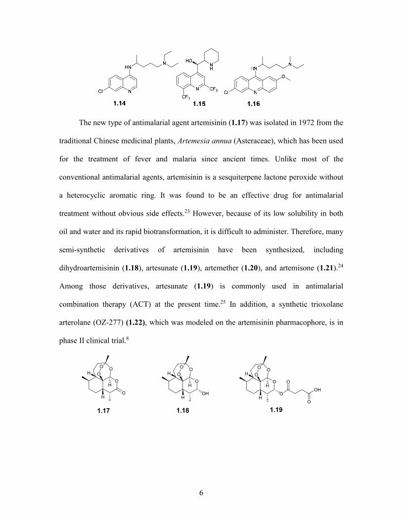
The new type of antimalarial agent artemisinin (1.17) was isolated in 1972 from the
traditional Chinese medicinal plants, Artemesia annua (Asteraceae), which has been used
for the treatment of fever and malaria since ancient times. Unlike most of the
conventional antimalarial agents, artemisinin is a sesquiterpene lactone peroxide without
a heterocyclic aromatic ring. It was found to be an effective drug for antimalarial
treatment without obvious side effects.23 However, because of its low solubility in both
oil and water and its rapid biotransformation, it is difficult to administer. Therefore, many
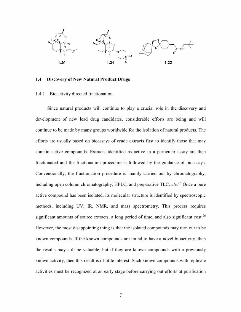
semi-synthetic derivatives of artemisinin have been synthesized, including
dihydroartemisinin (1.18), artesunate (1.19), artemether (1.20), and artemisone (1.21).24
Among those derivatives, artesunate (1.19) is commonly used in antimalarial
combination therapy (ACT) at the present time.25 In addition, a synthetic trioxolane
arterolane (OZ-277) (1.22), which was modeled on the artemisinin pharmacophore, is in
phase II clinical trial.8
O
OH
HO
O
O
H
1.17
O
OH
H
O
O
H
O
O
OH
O
1.19
O
OH
HOH
O
O
H
1.18
7
1.4 Discovery of New Natural Product Drugs
1.4.1 Bioactivity directed fractionation
Since natural products will continue to play a crucial role in the discovery and
development of new lead drug candidates, considerable efforts are being and will
continue to be made by many groups worldwide for the isolation of natural products. The
efforts are usually based on bioassays of crude extracts first to identify those that may
contain active compounds. Extracts identified as active in a particular assay are then
fractionated and the fractionation procedure is followed by the guidance of bioassays.
Conventionally, the fractionation procedure is mainly carried out by chromatography,
including open column chromatography, HPLC, and preparative TLC, etc.26 Once a pure
active compound has been isolated, its molecular structure is identified by spectroscopic
methods, including UV, IR, NMR, and mass spectrometry. This process requires
significant amounts of source extracts, a long period of time, and also significant cost.26
However, the most disappointing thing is that the isolated compounds may turn out to be
known compounds. If the known compounds are found to have a novel bioactivity, then
the results may still be valuable, but if they are known compounds with a previously
known activity, then this result is of little interest. Such known compounds with replicate
activities must be recognized at an early stage before carrying out efforts at purification

8
and structural identification to avoid duplicated compounds. The increased costs of the
classical bioactivity-directed fractionation approach and the needs for high throughput
bioactive novel compound discovery stimulated a dereplication approach which can
differentiate natural product extracts that contain useless or known compounds from
those that contain novel compounds which are of interest to us, as early as possible.
1.4.2 Dereplication approach
1.4.2.1 Dereplication
Dereplication is the process of screening samples of mixtures with desired
bioactivities, so as to recognize and determine if the activity is due to a previously known
substance. Dereplication ensures that the research focus is always on novel chemistry. If
a compound has been isolated previously, it should be possible to use the published
information to identify the compound when it appears again, avoiding the need to repeat
the entire isolation and structure-determination process.
Numerous dereplication approaches have been developed based on the coupling of
HPLC with different detection techniques, including LC-UV, LC-UV-DAD (diode-array
detector), LC-MS, LC-MS/MS, and LC-NMR, etc.26 The different LC detectors each
have their own advantages and disadvantages in resolution, sensitivity, and scale
requirement. In recent years, the use of capillary and cryo NMR probes and SPE-NMR is
increasing, in order to increase sensitivity and accuracy. Since the dereplication process
mainly starts from crude plant extracts which are complex mixtures that may contain
hundreds of constituents, the combined application of LC coupled techniques such as LC-
UV-MS-NMR is the most effective approach for dereplication. The combination of the
9
high separation efficiency of HPLC with different detectors can acquire on-line
spectroscopic data for the HPLC peaks of interest, even when treating with a very
complicated mixture.27
1.4.2.2 UV spectroscopy
UV spectroscopy was the first widely used detection method for HPLC. Sensitivity
is not a problem to give good data and a diode-array detector allows a complete UV
spectrum of each compound eluted to be recorded “on the fly”. However, UV spectra are
not compound specific, and this approach only works for the compounds that contain
distinctive enough chromophore to be recognized, and a database is needed for searching.
For example, it is always easy to recognize mycophenolic acid because of its distinctive
chromophores. However, problems arise for compounds with less distinctive
chromophores, such as peptides.28 Diode-array UV detectors have advantages over
single-wavelength detectors, because the spectra of eluted compounds can be stored in
digital form and can be handled by different algorithms, especially for peptides
containing aromatic amino acids such as tryptophan, tyrosine, and phenylalanine.
However, there are still many compounds that cannot be detected by a UV detector.
1.4.2.3 LC-MS and LC-MS/MS
One of the major advances in recent years has been the coupling of HPLC with
MS. LC-MS and LC-MS/MS are very sensitive and facile approaches and can analyze
complex mixtures and provide structural information independent of source, sample
preparation, or conditions of measurement. The information includes molecular mass and
molecular formula, which are highly desired information for dereplication and are easy to

10
search in most of the commercial databases, such as AntiBase, Dictionary of Natural
Products (DNP), MarinLit, and also in SciFinder Scholar or STN International
(CAPlus).26
MS/MS is a collision induced technique and is used to fragment a precursor ion
into several product ions, which are selected and subjected to MS/MS/MS analysis, and
so on.29 This process can identify the fragmentation pathway of a precursor from its first
generation fragments (MS/MS) to the nth generation products (MSn). During the whole
process, some neutral products released can also be identified. The molecular formula
analysis includes C, H, N, O, and S, which are the basic elements for most natural
products. High resolution mass separation and precise mass measurements can be used to
identify the molecular formulas of small molecules or ions. Adding up the molecular
formulas of small products can lead to the unique determination of the molecular formula
of the precursor compounds or ions, and the resulting molecular formula of the precursors
can conversely be used to confirm the molecular formula of the small products.29
As a successful example of the application of the LC-MS approach, Cordell and
Shin30 studied the fruit extract of S. anacardium L. (Anacardiaceae) and found the ion
chromatograms at m/z 315 and 317 corresponded to the active compounds. Through
comparison with the NAPRALERT database and in the DNP, the compounds 1,2-
dihydroxy-3-pentadeca-7',10'-dienylbenzene and 2-dihydroxy-3-pentadec-8'-enyl-
benzene were identified and were later confirmed. They also studied the root extract of B.
parviflora L. (Begoniaceae) for its strong cytotoxicity against various human cancer cell
lines. Cucurbitacin D (M = 516), hexanorcucurbitacin D (M = 402), cucurbitacin B (M =
11
558) and dihydrocucurbitacin B (M = 560) were also matched in the NAPRALERT
database, and they were confirmed later.
The LC-MS technique enables a rapid and efficient characterization of a natural
product, but its main disadvantage is that it is easy to make false identifications. This is
caused by the uncertainty regarding the presence of pseudomolecular ions (MH+, MNa+,
MK+, MNH4+, MCH4CN+, etc.), and also by the presence of ions from minor components
that ionize more readily than the major ones.31
1.4.2.4 LC-NMR spectroscopy
LC-NMR spectroscopy is the most powerful tool for compound identifications, and
it can also be used for dereplication, based on the technique of pattern recognition. Unlike
UV and MS, NMR data allows ready recognition of a wide variety of functional groups
and the environment in which they exist in a compound of interest. By comparing NMR
spectra, the identification of known compounds can be rapid, especially with the addition
of clues given by UV and MS data. The major limitation of this approach is the high cost
of the instrumentation required. In one LC-NMR approach, structural types and even
unique compounds can be identified by the presence and chemical shift values of
distinctive peaks, such as methyl groups,-protons in peptides, acetal protons, and
aromatic substitution patterns. This approach is a powerful one when it is coupled to a
comprehensive database, such as the DNP database.
A modern LC-NMR system consists of a high resolution NMR instrument (400-
800 MHz) with an LC-NMR flow probe which is connected to an HPLC system. The
control unit of the HPLC is connected to the data acquisition system of the NMR
spectrometer, in order to get synchronous data from the two different operating systems.
12
An obvious concern of the LC-NMR technique is how to recognize signals of compounds
in low concentration, in the presence of large signals of HPLC solvent.32 Most of the
HPLC separations are performed by large amounts of binary or ternary solvent mixtures,
as it is too expensive to use deuterated solvent for the analysis. The development of
solvent suppression, as a fast and reliable technique, eliminates this major concern. By
using solvent suppression techniques, even non-deuterated solvents like methanol and
acetonitrile can be widely used for reverse phase HPLC analysis to obtain high quality
spectra. However, water is usually replaced by D2O, due to its relatively low price and for
the purpose of giving higher quality spectra.27
With the development of efficient flow probe design and the use of high magnetic
fields, the sensitivity and accuracy of LC-NMR techniques have been highly improved.
We can also increase the sample volume, magnetic field strength, quality of the NMR
coil, and lower the temperature to obtain more and more mature LC-NMR techniques for
dereplication. It is a potentially complementary technique to LC-UV-MS in natural
product dereplication, by providing detailed on-line structural analysis of natural
products. The access to NMR data at the initial steps of the dereplication of crude extracts
significantly accelerates the whole dereplication.27
Wolfender and Ndjoko33 performed LC-UV-MS and LC-NMR for the isolation of
compounds from the roots of Cordia linnaei (Boraginaceae), which was interesting for its
antifungal and larvicidal properties. During the dereplication process, they identified two
naphthoquinones as previously known compounds. They also investigated Lisianthius
seemannii. The LC-MS-NMR analysis indicated the presences of a high molecular
weight secoiridoid, and the main constituent was found to be lisianthioside.33
13
1.5 The Madagascar ICBG project
In 1992, three federal institutions of the United States, the National Institutes of
Health (NIH), the National Science Foundation (NSF), and the U.S. Agency for
International Development (AID) initiated out a program in order to help with the
development of natural products drug discovery. This program was a way to promote the
conservation of biological diversity and economic development through the formation of
International Cooperative Biodiversity Groups (ICBG). These multi-institutional
partnerships combine the resources of technologically advanced institutions in the United
States with biologically diversity rich countries.34
In 1993, a group led by Dr. David Kingston from VPISU (Virginia Polytechnic
Institute and State University) was funded by NIH to work in Suriname. The group
included the Missouri Botanical Garden, Conservation International, Bristol-Myers
Squibb, and Bedrijf Geneesmiddelen Voorziening Suriname. The Suriname project was
successful in collecting and analyzing plant samples, and in isolating bioactive
compounds. However, the project was discontinued due to the relatively limited
biodiversity in Suriname, with an estimated 4000 plant species. A parallel ICBG program
was initiated in Madagascar in 1998, and the project moved completely to Madagascar in
2003.
Madagascar is the world’s third largest island and is the home of an estimated
13,000 plant species.35 More than 90% of the species cannot be found anywhere else in
the world. Deforestation since the last century or so has reduced the forest cover to less
than 10% of the original vegetations, and much of the remaining forest has been seriously
degraded.36 From 1998 to 2003, the collection of plant samples was carried out near the
14
Zahamena National Park in the wet forests to the south west of the capital of
Antananarivo. After 2003, the collection moved to the endangered dry forest in northern
Madagascar.34 Northern Madagascar is the center of distribution of various species within
the country due to the special geology, soil types, and climate in the region. Except for
the existing protected areas, the remaining forests in northern Madagascar are being
significantly reduced. This is mainly caused by the rapid increase of the human
population. The increasing population and natural disasters like fire and landslides are
both important factors in the loss of the remaining forests.34
Work in Madagascar from 1998 to 2008 was focused on the collection of plant
samples. Since 2008, the focus shifted to the collection of microbial and marine samples.
The current program goals include several aspects.34 First, to collect microbial and
marine samples. The microbial samples are processed in Madagascar to isolate pure
cultures, and these are grown and extracted to yield crude microbial extracts. Marine
samples are also extracted in Madagascar, while botanical inventory continues to guide
analysis of species distribution. This analysis can contribute to the determination of the
priority of the areas that need protection and conservation in northern Madagascar;
Furthermore, based on the results of the new conservation analysis, some guidance can be
obtained for the status of the existing protected areas in northern Madagascar;
Additionally, this process will promote the development of a phytochemical research
community in Madagascar, including the improvement of institutional facilities and the
professional level of Madagascar scientists.
This dissertation reports results from searching for novel antiproliferative and
antiplasmodial agents from the extracts from Madagascar rain forests and South Africa.
15
The research focuses on the isolation and structure elucidation of bioactive compounds
from various plant extracts. The research involves bioassay-guided fractionation and a
series of analytical techniques, including 1D and 2D-NMR, LC-MS/GC-MS/MS, UV, IR,
optical rotation, and electronic circular dichroism (ECD). In addition, chemical
conversions were also involved for some structure determinations. The A2780 human
ovarian cancer cell line was the most frequently used antiproliferative bioassay, in
addition to some other anticancer cell lines. The Dd2 drug-resistant strain of Plasmodium
falciparum was used for the antimalarial assay. The research work is discussed in detail
in the following chapters.
16
1.6 References
1. Cragg, G. M.; Newman, D. J. Natural products: a continuing source of novel drug
leads. Biochim. Biophys. Acta. 2013, 1830, 3670-3695.
2. Cragg, G. M.; Kingston, D. G. I.; Newman, D. J. Anticancer agents from natural
products. CRC press. 2012.
3. Kingston, D. G.; Newman, D. J. Mother nature's combinatorial libraries; their
influence on the synthesis of drugs. Curr. Opin. Drug Discov. Devel. 2002, 5, 304-316.
4. Stewart, B. W., Wild, C. P. World cancer report 2014. IARC Nonserial 2014.
5. Global cancer facts and figures. Am. Cancer Soc. Atlanta 2007.
6. Habner, B. A. The pharmacological basis of therapeutics. 11th ed.; New York, p
1315-1403.
7. Newman, D. J.; Cragg, G. M. Natural products as sources of new drugs over the
30 years from 1981 to 2010. J. Nat. Prod. 2012, 75, 311-335.
8. Butler, M. S. Natural products to drugs: natural product-derived compounds in
clinical trials. Nat. Prod. Rep. 2008, 25, 475-516.
9. Noble, R. L.; Beer, C. T.; Cutts, J. H. Role of chance observations in
chemotherapy: Vinca rosea. Ann. N. Y. Acad. Sci. 1958, 76, 882-894.
10. Svoboda, G. H.; Johnson, I. S.; Gorman, M.; Neuss, N. Current status of research
on the alkaloids of Vinca rosea Linn. (Catharanthus roseus G. Don). J. Pharm. Sci. 1962,
51, 707-720.
11. Barnett, C. J.; Cullinan, G. J.; Gerzon, K.; Hoying, R. C.; Jones, W. E.; Newlon,
W. M.; Poore, G. A.; Robison, R. L.; Sweeney, M. J. Structure-activity relationships of
dimeric Catharanthus alkaloids. 1. Deacetyl vinblastine amide (vindesine) sulfate. J.
17
Med. Chem. 1978, 21, 88-96.
12. Mangeney, P.; Andriamialisoa, R. Z.; Lallemand, J. Y.; Langlois, N.; Langlois, Y.;
Potier, P. 5'-Nor anhydrovinblastine-prototype of a new class of vinblastine derivatives.
Tetrahedron 1979, 35, 2175-2179.
13. Fahy, J.; Duflos, A.; Ribet, J.-P.; Jacquesy, J.-C.; Berrier, C.; Jouannetaud, M.-P.;
Zunino, F. Vinca alkaloids in superacidic media: A method for creating a new family of
antitumor derivatives. J. Am. Chem. Soc. 1997, 119, 8576-8577.
14. Wani, M. C.; Taylor, H. L.; Wall, M. E.; Coggon, P.; McPhail, A. T. Plant
antitumor agents. VI. Isolation and structure of taxol, a novel antileukemic and antitumor
agent from Taxus brevifolia. J. Am. Chem. Soc. 1971, 93, 2325-2327.
15. Schiff, P. B.; Fant, J.; Horwitz, S. B. Promotion of microtubule assembly in vitro
by taxol. Nature 1979, 277, 665-667.
16. Holmes, F. A.; Walters, R. S.; Theriault, R. L.; Forman, A. D.; Newton, L. K.;
Raber, M. N.; Buzdar, A. U.; Frye, D. K.; Hortobagyi, G. N. Phase II trial of taxol, an
active drug in the treatment of metastatic breast cancer. J. Natl. Cancer Inst. 1991, 83,
1797-1805.
17. Bissery, M.-C.; Nohynek, G.; Sanderink, G.-J.; Lavelie, F. Docetaxel
(Taxotere(R)) a review of preclinical and clinical experience. Part I: preclinical
experience. Anti-Cancer Drugs 1995, 6, 339-355.
18. Galsky, M. D.; Dritselis, A.; Kirkpatrick, P.; Oh, W. K. Cabazitaxel. Nat. Rev.
Drug Discov. 2010, 9, 677-678.
19. Canel, C.; Moraes, R. M.; Dayan, F. E.; Ferreira, D. Podophyllotoxin.
Phytochemistry 2000, 54, 115-120.
18
20. Stähelin, H., von Wartburg, A. The chemical and biological route from
podophyllotoxin. J. Am. Chem. Soc. 1962, 84, 1748-1749.
21. Schlitzer, M. Malaria chemotherapeutics part I: History of antimalarial drug
development, currently used therapeutics, and drugs in clinical development. Chem. Med.
Chem. 2007, 2, 944-986.
22. Watt, G.; Long, G. W.; Padre, L. P.; Alban, P.; Sangalang, R.; Ranoa, C. P.
Chloroquine and quinine: A randomized, double-blind comparison of efficacy and side
effects in the treatment of Plasmodium falciparum malaria in the Philippines. Trans. R.
Soc. Trop. Med. Hyg. 1988, 82, 205-208.
23. Chaturvedi, D.; Goswami, A.; Pratim Saikia, P.; Barua, N. C.; Rao, P. G.
Artemisinin and its derivatives: A novel class of anti-malarial and anti-cancer agents.
Chem. Soc. Rev. 2010, 39, 435-454.
24. Haynes, R. K. From artemisinin to new artemisinin antimalarials: biosynthesis,
extraction, old and new derivatives, stereochemistry and medicinal chemistry
requirements. Curr. Top. Med. Chem. 2006, 6, 509-537.
25. Crespo-Ortiz, M. P.; Wei, M. Q. Antitumor activity of artemisinin and its
derivatives: From a well-known antimalarial agent to a potential anticancer drug. J.
Biomed. Biotechnol. 2012, 2012, 247597.
26. Lang, G.; Mayhudin, N. A.; Mitova, M. I.; Sun, L.; van der Sar, S.; Blunt, J. W.;
Cole, A. L.; Ellis, G.; Laatsch, H.; Munro, M. H. Evolving trends in the dereplication of
natural product extracts: New methodology for rapid, small-scale investigation of natural
product extracts. J. Nat. Prod. 2008, 71, 1595-1599.
27. Wolfender, J. L.; Ndjoko, K.; Hostettmann, K. The potential of LC-NMR in
19
phytochemical analysis. Phytochem. Anal. 2001, 12, 2-22.
28. Aguilar, M. I. HPLC of peptides and proteins. In HPLC of Peptides and Proteins,
Springer New York: 2004; Vol. 251, pp 3-8.
29. Konishi, Y.; Kiyota, T.; Draghici, C.; Gao, J.-M.; Yeboah, F.; Acoca, S.;
Jarussophon, S.; Purisima, E. Molecular formula analysis by an MS/MS/MS technique to
expedite dereplication of natural products. Analyt. Chem. 2007, 79, 1187-1197.
30. Cordell, G. A.; Shin, Y. G. Finding the needle in the haystack. The dereplication
of natural product extracts. Pure Appl. Chem. 1999, 71, 1089-1094.
31. Lang, G.; Mayhudin, N. A.; Mitova, M. I.; Sun, L.; van der Sar, S.; Blunt, J. W.;
Cole, A. L. J.; Ellis, G.; Laatsch, H.; Munro, M. H. G. Evolving trends in the
dereplication of natural product extracts: new methodology for rapid, small-scale
investigation of natural product extracts. J. Nat. Prod. 2008, 71, 1595-1599.
32. Spraul, M.; Hofmann, M.; Lindon, J. C.; Nicholson, J. K.; Wilson, I. D. Liquid
chromatography coupled with high-field proton nuclear magnetic resonance
spectroscopy: Current status and future prospects. Anal. Proc. 1993, 30, 390-392.
33. Wolfender, J. L.; Queiroz, E. F.; Hostettmann, K. Phytochemistry in the
microgram domain - a LC-NMR perspective. Magn. Reson. Chem. 2005, 43, 697-709.
34. Miller, J. S.; Birkinshaw, C.; Callmander, M. The Madagascar International
Cooperative Biodiversity Group (ICBG): Using natural products research to build science
capacity. http://hdl.handle.net/10125/181
35. Goodman, S. M.; Benstead, J. P. Updated estimates of biotic diversity and
endemism for Madagascar. Oryx 2005, 39, 73-77.
36. Myers, N.; Mittermeier, R. A.; Mittermeier, C. G.; Da Fonseca, G. A. B.; Kent, J.

20
Biodiversity hotspots for conservation priorities. Nature 2000, 403, 853-858.
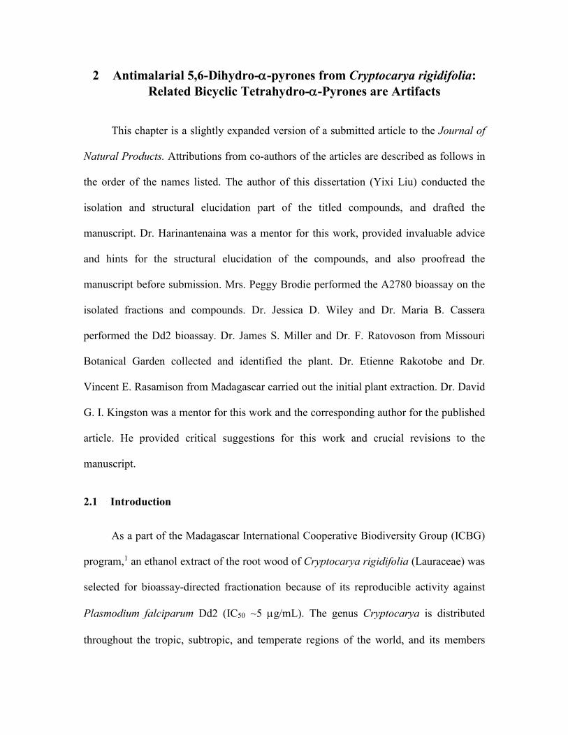
2 Antimalarial 5,6-Dihydro--pyrones from Cryptocarya rigidifolia: Related Bicyclic Tetrahydro--Pyrones are Artifacts
This chapter is a slightly expanded version of a submitted article to the Journal of
Natural Products. Attributions from co-authors of the articles are described as follows in
the order of the names listed. The author of this dissertation (Yixi Liu) conducted the
isolation and structural elucidation part of the titled compounds, and drafted the
manuscript. Dr. Harinantenaina was a mentor for this work, provided invaluable advice
and hints for the structural elucidation of the compounds, and also proofread the
manuscript before submission. Mrs. Peggy Brodie performed the A2780 bioassay on the
isolated fractions and compounds. Dr. Jessica D. Wiley and Dr. Maria B. Cassera
performed the Dd2 bioassay. Dr. James S. Miller and Dr. F. Ratovoson from Missouri
Botanical Garden collected and identified the plant. Dr. Etienne Rakotobe and Dr.
Vincent E. Rasamison from Madagascar carried out the initial plant extraction. Dr. David
G. I. Kingston was a mentor for this work and the corresponding author for the published
article. He provided critical suggestions for this work and crucial revisions to the
manuscript.
2.1 Introduction
As a part of the Madagascar International Cooperative Biodiversity Group (ICBG)
program,1 an ethanol extract of the root wood of Cryptocarya rigidifolia (Lauraceae) was
selected for bioassay-directed fractionation because of its reproducible activity against
Plasmodium falciparum Dd2 (IC50 ~5 g/mL). The genus Cryptocarya is distributed
throughout the tropic, subtropic, and temperate regions of the world, and its members
22
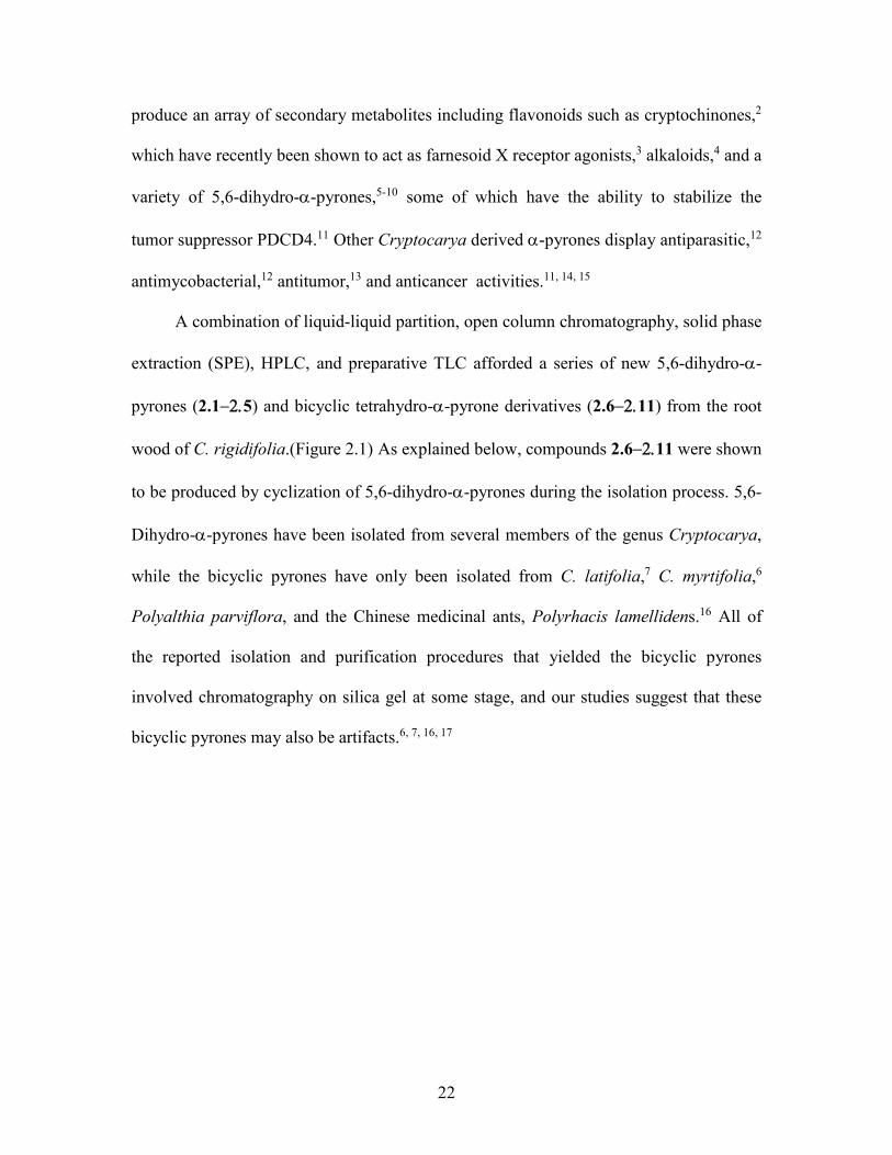
produce an array of secondary metabolites including flavonoids such as cryptochinones,2
which have recently been shown to act as farnesoid X receptor agonists,3 alkaloids,4 and a
variety of 5,6-dihydro--pyrones,5-10 some of which have the ability to stabilize the
tumor suppressor PDCD4.11 Other Cryptocarya derived -pyrones display antiparasitic,12
antimycobacterial,12 antitumor,13 and anticancer activities.11, 14, 15
A combination of liquid-liquid partition, open column chromatography, solid phase
extraction (SPE), HPLC, and preparative TLC afforded a series of new 5,6-dihydro--
pyrones (2.15) and bicyclic tetrahydro--pyrone derivatives (2.611) from the root
wood of C. rigidifolia.(Figure 2.1) As explained below, compounds 2.611 were shown
to be produced by cyclization of 5,6-dihydro--pyrones during the isolation process. 5,6-
Dihydro--pyrones have been isolated from several members of the genus Cryptocarya,
while the bicyclic pyrones have only been isolated from C. latifolia,7 C. myrtifolia,6
Polyalthia parviflora, and the Chinese medicinal ants, Polyrhacis lamellidens.16 All of
the reported isolation and purification procedures that yielded the bicyclic pyrones
involved chromatography on silica gel at some stage, and our studies suggest that these
bicyclic pyrones may also be artifacts.6, 7, 16, 17
23
Figure 2.1 Structures of Compounds 2.12.11
2.2 Results and Discussion
2.2.1 Isolation of active compounds
The EtOAc-soluble fraction obtained from a liquid-liquid partition of the ethanol
extract (100 mg) showed antiplasmodial activity. Dereplication as previously described18
indicated that the extract contained at least one new bioactive compound, and so a larger
sample was investigated. Fractionation of the EtOAc-soluble fraction of this sample by
chromatography on Sephadex LH-20, reverse phase SPE, normal phase silica gel column
chromatography, and C18 HPLC yielded compounds 2.1 and 2.6 – 2.8, together with
fractions that were mixtures of 5,6-dihydro--pyrones and bicyclic tetrahydro--pyrones.
Purification of these fractions was effected by diol PTLC or HPLC to yield compounds
2.22.3, 2.5, and 2.92.11. Similar fractionation of the antiplasmodial hexanes fraction
yielded compounds 2.4 and 2.7. (Scheme 2.1)
24
Scheme 2.1 Bioassay-guided Separation of Cryptocarya rigidifolia
2.2.2 Structure elucidation of compound 2.1
Compound 2.1 was isolated as a clear oil, and its molecular formula was
established as C20H36O3 by HRESIMS (m/z 325.2753 [M+H]+). Its IR spectrum showed
absorptions at 3340, and 1719 and 1613 cm-1 assigned to a hydroxyl group and an ,-
unsaturated -lactone, respectively. Its 1H NMR spectroscopic data had signals for a
conjugated olefin at δH 6.90 (m, 1H, H-4) and δH 6.03 (dt, J = 9.8, 1.7 Hz, 1H, H-3), two
oxymethine groups at δH 4.75 (m, 1H, H-6) and δH 4.00 (brs, 1H, H-2'), a terminal
primary methyl group (δH 0.88, t, J = 7.0 Hz, 3H, CH3-15') in the upfield region, and a

25
multiplet at δH 2.36 (2H, H-5), representing a deshielded methylene group. In the HMBC
spectrum the methylene protons at δH 2.36 (H-5) showed correlations both to an olefin
group (δC 121.3, C-3 and δC 145.0, C-4) and to the oxymethine resonance at δH 4.75 and
δC 74.8 (C-6). In addition, both the olefinic protons (δH 6.90, H-4 and δH 6.03, H-3) and
the oxymethine at δH 4.75 (H-6) correlated with a carbonyl carbon at δC 164.6 (C-2)
(Figure 1). These data indicated a 6-substituted-5,6-dihydro--pyrone, a common ring
system of secondary metabolites found in Cryptocarya species.19 The remaining
oxymethine group at δH 4.00 (1H, H-2') was assigned to C-2', which was flanked by two
methylenes (C-1' and C-3') as indicated by HMBC correlations (Figure 2.2) from the
methylene protons at δH 1.92 (ddd, J = 14.5, 9.6, 2.2 Hz, H-1') and δH 1.65 (m, H-1') to
δC 30.1 (C-5); from δH 4.00 (H-2') to the oxymethine carbon at δC 74.8 (C-6); and from H-
2' to the two neighboring methylene carbons at δC 41.2 (C-1') and δC 38.2 (C-3'). The
absolute configuration at C-6 was determined to be R by the positive Cotton effects at
256 nm observed in its ECD spectrum in MeOH, arising from the n→* transitions of the
lactone ring.13, 20-22 The 1H NMR spectra of the (R) and (S) Mosher ester derivatives of
2.123 revealed slight differences in the 1H chemical shifts of C-6 and adjacent protons that
allowed the assignment of the R configuration to C-2' of 2.1 (Figure 2.3). The complete
assignment of all protons and carbons of 2.1 (Table 2.1) was accomplished by further
interpretation of its HMBC and HSQC spectra. Compound 2.1 was thus assigned as 6R-
(2'R-hydroxypentadecyl)-5,6-dihydro-2H-pyran-2-one, and named cryptorigidifoliol A.21,
22

26
Figure 2.2 Key HMBC Correlations of 2.1
Figure 2.3 H) = δSδR Data of R and S-MPA Derivatives of Compound 2.1
2.2.1 Structure elucidation of compound 2.2
Compound 2.2 was obtained as an oil with the molecular formula C24H44O4 based
on its HRESIMS spectrum (m/z 397.3317 [M+H]+). The UV, IR and 1H NMR
spectroscopic data of 2.2 were comparable to those of 2.1, suggesting that 2.2 is also a 6-
substituted-5,6-dihydro--pyrone. The major difference between the 1H NMR
spectroscopic data of 2.1 and 2.2 was the presence of an additional signal for an
oxymethine proton at δH 3.89 (m, 1H, H-4') in 2.2. The HMBC correlation between the
oxymethine proton and the carbon signal at δC 69.8 (C-2') assigned the additional
hydroxyl group to C-4', and this assignment is supported by the HMBC cross peaks
between the two adjacent methylene protons (δH 1.61, m, 2H, H-3'; δH 1.47, m, 2H, H-5')
and the oxymethine carbon at δC 74.0. Similarly to 2.1, the complete assignments of all
protons and carbons of 2.2 (Table 2.1) were accomplished by interpretation of its HMBC
and HSQC spectra. ECD and the Mosher ester method were used to assign the
configurations of C-6, C-2', and C-4' as R, S, R, respectively. (Figure 2.4) Compound 2.2
awas thus assigned as 6R-(2'S,4'R-dihydroxynonadecyl)-5,6-dihydro-2H-pyran-2-
27
Table 2.1 1H and 13C NMR Spectroscopic Data for Compounds 2.12.5.
one and has been named cryptorigidifoliol B.
2.1
a 2.2
a 2.3
a 2.4
a 2.5
a
posn δHb δc
c δHb δc
c δHb δc
c δHb δc
c δHb δc
c
2 164.6
(C)
163.5 (C)
164.2 (CH2)
164.2 (CH2)
163.5
(C)
3 6.03 dt
(9.8, 1.7) 121.3 (CH)
6.03 dt (9.8, 1.7)
121.3 (CH)
6.03 dt (9.8, 1.7)
121.3 (CH)
6.04 dt (9.8, 1.7)
121.3 (CH)
6.03 brd (9.8)
121.3 (CH)
4 6.90 m 145.0 (CH)
6.90 m 145.0 (CH)
6.90 m 145.0 (CH)
6.90 m 145.0 (CH)
6.90 m 145.0 (CH)
5 2.36 m 30.1
(CH2) 2.44 m
29.2 (CH2)
2.36 m 30.3
(CH2) 2.42 m
30.2 (CH2)
2.44 m 29.2
(CH2)
6 4.75 m 74.8 (CH)
4.69 m 76.4 (CH)
4.75 m 74.7 (CH)
4.66 m 74.8 (CH)
4.69 m 72.5 (CH)
1' 1.92 ddd
(14.5, 9.6, 2.2)
41.2 (CH2)
2.04 m 39.6
(CH2)
1.92 ddd (14.5, 9.6,
2.2)
42.3 (CH2)
1.96 ddd (14.5, 9.6,
2.2)
42.1 (CH2)
1.79 m 43.8
(CH2)
1.65 m 1.79 ddd
(14.3, 5.6, 3.9)
1.64 ddd
(14.5, 10.2, 2.9)
1.81 ddd
(14.5, 10.2, 2.9)
1.73 m
2' 4.00 brs 67.3 (CH)
4.15 m 69.8 (CH)
4.00 m 67.1 (CH)
3.86 m 67.4 (CH)
4.63 m 63.3 (CH)
3' 1.49-1.41
m 38.2
(CH2) 1.66-1.53
m 42.9
(CH2) 1.45 m
38.2 (CH2)
1.50 m 38.5
(CH2) 5.49 dd
(15.3, 7.0) 131.4 (CH)
4' 1.49-1.41
m 25.7
(CH2) 3.89 m
74.0 (CH)
1.37-1.31 m
25.0 (CH2)
1.38-1.27 m
25.3 (CH2)
5.68 m 132.6 (CH)
5' 1.35-1.22
m 29.6
(CH2) 1.66-1.53
m 38.2
(CH2) 1.35-1.22
m 29.6
(CH2) 1.28-1.21
m 29.6
(CH2) 2.03 m
32.9 (CH2)
6' 1.35-1.22
m 29.6
(CH2) 1.47 m
25.4 (CH2)
1.35-1.22 m
29.6 (CH2)
1.28-1.21 m
29.6 (CH2)
1.49-1.61 m
28.0 (CH2)
7' 1.35-1.22
m 29.6
(CH2) 1.35-1.22
m 29.6
(CH2) 1.35-1.22
m 29.6
(CH2) 1.28-1.21
m 29.6
(CH2) 1.33-1.22
m 29.6
(CH2)
8' 1.35-1.22
m 29.6
(CH2) 1.35-1.22
m 29.6
(CH2) 1.37-1.31
m 27.0
(CH2) 1.38-1.27
m 27.0
(CH2) 1.33-1.22
m 29.6
(CH2)
9' 1.35-1.22
m 29.6
(CH2) 1.35-1.22
m 29.6
(CH2) 2.05-1.99
m 29.4
(CH2) 2.00 m
29.6 (CH2)
1.33-1.22 m
29.6 (CH2)
10' 1.35-1.22
m 29.6
(CH2) 1.35-1.22
m 29.6
(CH2) 5.34 m
130.0 (CH)
5.35 m 131.2 (CH)
1.33-1.22 m
29.6 (CH2)
11' 1.35-1.22
m 29.6
(CH2) 1.35-1.22
m 29.6
(CH2) 5.34 m
130.0 (CH)
5.35 m 131.2 (CH)
1.42-1.32 m
29.6 (CH2)
12' 1.35-1.22
m 29.6
(CH2) 1.35-1.22
m 29.6
(CH2) 2.05-1.99
m 29.4
(CH2) 2.00 m
29.6 (CH2)
1.49-1.61 m
42.1 (CH2)
13' 1.35-1.22
m 32.0
(CH2) 1.35-1.22
m 29.6
(CH2) 1.37-1.31
m 27.0
(CH2) 1.38-1.27
m 27.0
(CH2) 4.15 m
64.4 (CH2)
14' 1.35-1.22
m 22.8
(CH2) 1.35-1.22
m 29.6
(CH2) 1.35-1.22
m 29.6
(CH2) 1.28-1.21
m 29.6
(CH2) 1.49-1.61
m 42.1
(CH2)
15' 0.88 t (7.0)
14.2 (CH3)
1.35-1.22 m
29.6 (CH2)
1.35-1.22 m
32.0 (CH2)
1.28-1.21 m
29.6 (CH2)
1.42-1.32 m
29.6 (CH2)
16' 1.35-1.22
m 29.6
(CH2) 1.35-1.22
m 22.8
(CH2) 1.28-1.21
m 29.6
(CH2) 1.33-1.22
m 29.6
(CH2)
17' 1.35-1.22 m 32.0
(CH2) 0.88 t (7.0)
14.2 (CH3)
1.28-1.21 m
32.0 (CH2)
1.33-1.22 m
32.0 (CH2)
18' 1.35-1.22 m 22.8
(CH2)
1.28-1.21 m
22.8 (CH2)
1.33-1.22 m
22.8 (CH2)
19' 0.88 t (7.0) 14.2
(CH3)
0.88 t (7.0)
14.2 (CH3)
0.88 t (7.0)
14.2 (CH3)
a Spectra obtained in CDCl3; assignments based on analysis of 2D NMR spectra. bData (δ) measured at 500 MHz; brs = broad singlet, brd = broad doublet, t = triplet, ddd = doublet of doublets of doublets, dt = doublet of triplets, m = multiplet. J values are in Hz and are omitted if the signals overlapped as multiplets. The overlapped signals were assigned from HSQC and HMBC spectra without designating multiplicity. cData (δ) measured at 125 MHz; CH3, CH2, CH, and C multiplicities were determined by an HSQC experiment.
28
Figure 2.4 H) = δSδR Data of R and S-MPA Derivatives of Compound 2.2
2.2.2 Structure elucidation of compound 2.3
The molecular formula of compound 2.3 (C22H38O3; HRESIMS m/z 351.2902
[M+H]+) and its 1H NMR spectrum (δH 5.34, m, 2H, H-10' and H-11') indicated that it
had an additional disubstituted olefin moiety as compared with compound 2.1. Its UV, IR
and 1H NMR spectroscopic data indicated the presence of the same -pyrone ring as 2.1
and 2.2, and HMBC correlations from δH 5.34 (H-10' and H-11') to δC 27.0 indicated that
the additional olefin group must be located between two methylene groups. Long range
correlation from both δH 2.05 - 1.99 (m, 4H, H-9' and H-12') and δH 1.37 - 1.31 (m, 4H,
H-8' and 13') to the carbons at δC 130 (C-10' and 11') in the HMBC spectrum assigned the
olefin group to C-10' and 11'. The geometry of the double bond was assigned as Z based
on the upfield 13C chemical shift of the methylenes connected to the double bond (δC
29.4).13, 24 The position of the double bond within the alkyl chain was determined
unambiguously by analysis of the GC-EIMS fragmentation of the dimethyldisulfide
(DMDS) derivative of 2.3,25, 26 which showed a major ion at m/z 299 attributable to
fragmentation between the two CH3S groups located at the original site of unsaturation.
Fragment ions at m/z 281 and 145 were also observed to support the assigned structure
(Figure 2.5). The relative stereochemistry of 3 and the assignment of its 1H and 13C NMR
were determined by the same methods as for 2.1 and 2.2.(Table 2.1) Compound 2.3 was
29
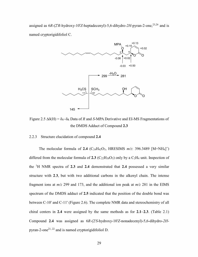
assigned as 6R-(2'R-hydroxy-10'Z-heptadecenyl)-5,6-dihydro-2H-pyran-2-one,23,26 and is
named cryptorigidifoliol C.
O O
OHH3CS SCH3
299-H2O
281
145
O O
O
MPA +0.13
+0.02+0.15
+0.50
+0.02
-0.03
-0.08
2' 6
10'11'
Figure 2.5 H) = δSδR Data of R and S-MPA Derivative and EI-MS Fragmentations of
the DMDS Adduct of Compound 2.3
2.2.3 Structure elucidation of compound 2.4
The molecular formula of 2.4 (C24H42O3, HRESIMS m/z: 396.3489 [M+NH4]+)
differed from the molecular formula of 2.3 (C22H38O3) only by a C2H4 unit. Inspection of
the 1H NMR spectra of 2.3 and 2.4 demonstrated that 2.4 possessed a very similar
structure with 2.3, but with two additional carbons in the alkenyl chain. The intense
fragment ions at m/z 299 and 173, and the additional ion peak at m/z 281 in the EIMS
spectrum of the DMDS adduct of 2.5 indicated that the position of the double bond was
between C-10' and C-11' (Figure 2.6). The complete NMR data and stereochemistry of all
chiral centers in 2.4 were assigned by the same methods as for 2.12.3. (Table 2.1)
Compound 2.4 was assigned as 6R-(2'S-hydroxy-10'Z-nonadecenyl)-5,6-dihydro-2H-
pyran-2-one21, 22 and is named cryptorigidifoliol D.

30
Figure 2.6 H) = δSδR Data of R and S-MPA Derivative and EI-MS Fragmentations of the DMDS Adduct of Compound 2.4
2.2.4 Structure elucidation of compound 2.5
The 1H NMR spectrum of compound 2.5 (C24H42O4, HRESIMS m/z: 377.3060 [M-
OH]+) showed the presence of two oxymethine groups (δH 4.63, m, 1H, H-2'; δH 4.15, m,
1H, H-12') and a double bond (δH 5.68, m, 1H, H-4'; δH 5.49, dd, J = 15.3, 7.0, 1H, H-3')
on the alkyl chain, besides the -pyrone ring signals at δH 6.9, 6.03, 4.69, and 2.44. The
large coupling constant (15.3 Hz) observed for H-3' indicated the E geometry of the
double bond. In the HMBC spectrum, correlations were observed between the protons at
δH 1.79 and 1.73 (each m, 1H, H-1') and δC 131.4 (C-3'), and between the proton at δH
4.63 (m, 1H, H-2') and δC 132.6 (C-4'). These observations suggested the connection of
the olefin group with the oxymethine at C-2'. The ESIMS of 2.5 showed significant ions
at m/z 265 and 247, together with a less intense ion at m/z 111, consistent with
assignment of the second hydroxyl group to C-12' (Figure 2.7). The configurations at C-6
and C-2' were assigned to be R and S, respectively by interpretation of the ECD
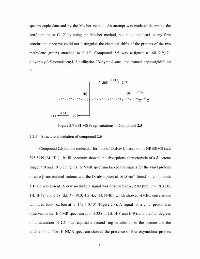
31
spectroscopic data and by the Mosher method. An attempt was made to determine the
configuration at C-12' by using the Mosher method, but it did not lead to any firm
conclusion, since we could not distinguish the chemical shifts of the protons of the two
methylene groups attached to C-12'. Compound 2.5 was assigned as 6R-(2'R,12'-
dihydroxy-3'E-nonadecenyl)-5,6-dihydro-2H-pyran-2-one, and named cryptorigidifoliol
E.
O O
OHHO
265
129
-H2O247
-H2O111
12'
Figure 2.7 ESI-MS Fragmentations of Compound 2.5
2.2.5 Structure elucidation of compound 2.6
Compound 2.6 had the molecular formula of C24H42O4 based on its HRESIMS (m/z
395.3149 [M+H]+). Its IR spectrum showed the absorptions characteristic of a -lactone
ring (1719 and 1073 cm-1). Its 1H NMR spectrum lacked the signals for the vinyl protons
of an ,-unsaturated lactone, and the IR absorption at 1615 cm-1 found in compounds
2.12.5 was absent. A new methylene signal was observed at δH 2.89 (brd, J = 19.3 Hz,
1H, H-4a) and 2.78 (dd, J = 19.3, 4.5 Hz, 1H, H-4b), which showed HMBC correlations
with a carbonyl carbon at δC 169.7 (C-3) (Figure 2.8). A signal for a vinyl proton was
observed in the 1H NMR spectrum at δH 5.33 (m, 2H, H-8' and H-9'), and the four degrees
of unsaturation of 2.6 thus required a second ring in addition to the lactone and the
double bond. The 1H NMR spectrum showed the presence of four oxymethine protons
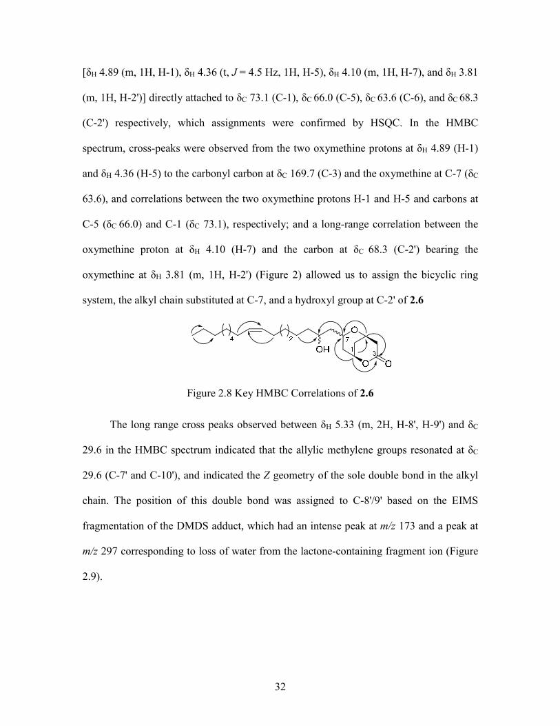
32
[δH 4.89 (m, 1H, H-1), δH 4.36 (t, J = 4.5 Hz, 1H, H-5), δH 4.10 (m, 1H, H-7), and δH 3.81
(m, 1H, H-2')] directly attached to δC 73.1 (C-1), δC 66.0 (C-5), δC 63.6 (C-6), and δC 68.3
(C-2') respectively, which assignments were confirmed by HSQC. In the HMBC
spectrum, cross-peaks were observed from the two oxymethine protons at δH 4.89 (H-1)
and δH 4.36 (H-5) to the carbonyl carbon at δC 169.7 (C-3) and the oxymethine at C-7 (δC
63.6), and correlations between the two oxymethine protons H-1 and H-5 and carbons at
C-5 (δC 66.0) and C-1 (δC 73.1), respectively; and a long-range correlation between the
oxymethine proton at δH 4.10 (H-7) and the carbon at δC 68.3 (C-2') bearing the
oxymethine at δH 3.81 (m, 1H, H-2') (Figure 2) allowed us to assign the bicyclic ring
system, the alkyl chain substituted at C-7, and a hydroxyl group at C-2' of 2.6
Figure 2.8 Key HMBC Correlations of 2.6
The long range cross peaks observed between δH 5.33 (m, 2H, H-8', H-9') and δC
29.6 in the HMBC spectrum indicated that the allylic methylene groups resonated at δC
29.6 (C-7' and C-10'), and indicated the Z geometry of the sole double bond in the alkyl
chain. The position of this double bond was assigned to C-8'/9' based on the EIMS
fragmentation of the DMDS adduct, which had an intense peak at m/z 173 and a peak at
m/z 297 corresponding to loss of water from the lactone-containing fragment ion (Figure
2.9).
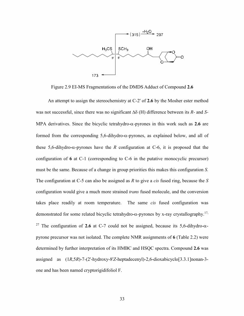
33
Figure 2.9 EI-MS Fragmentations of the DMDS Adduct of Compound 2.6
An attempt to assign the stereochemistry at C-2' of 2.6 by the Mosher ester method
was not successful, since there was no significant (H) difference between its R- and S-
MPA derivatives. Since the bicyclic tetrahydro--pyrones in this work such as 2.6 are
formed from the corresponding 5,6-dihydro--pyrones, as explained below, and all of
these 5,6-dihydro--pyrones have the R configuration at C-6, it is proposed that the
configuration of 6 at C-1 (corresponding to C-6 in the putative monocyclic precursor)
must be the same. Because of a change in group priorities this makes this configuration S.
The configuration at C-5 can also be assigned as R to give a cis fused ring, because the S
configuration would give a much more strained trans fused molecule, and the conversion
takes place readily at room temperature. The same cis fused configuration was
demonstrated for some related bicyclic tetrahydro--pyrones by x-ray crystallography.17,
27 The configuration of 2.6 at C-7 could not be assigned, because its 5,6-dihydro--
pyrone precursor was not isolated. The complete NMR assignments of 6 (Table 2.2) were
determined by further interpretation of its HMBC and HSQC spectra. Compound 2.6 was
assigned as (1R,5R)-7-(2'-hydroxy-8'Z-heptadecenyl)-2,6-dioxabicyclo[3.3.1]nonan-3-
one and has been named cryptorigidifoliol F.
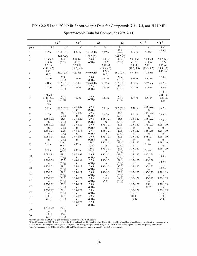
34
Table 2.2 1H and 13C NMR Spectroscopic Data for Compounds 2.62.8, and 1H NMR
Spectroscopic Data for Compounds 2.92.11
2.6
a 2.7
a 2.8
2.9
2.10
a 2.11
a
posn δHb δc
c δHb δc
c δHb δc
c δHb δH
b δHb
1 4.89 m 73.1 (CH) 4.89 m 73.1 (CH) 4.89 m 73.1
((CH) 4.89 m 4.90 m
4.89 m
3 169.7 (C) 169.7 (C) 169.7 (C)
4 2.89 brd (19.3)
36.4 (CH2)
2.89 brd (19.3)
36.4 (CH2)
2.89 brd (19.3)
36.4 (CH2)
2.91 brd (19.3)
2.85 brd (19.3)
2.87 brd (19.3)
2.78 dd
(19.3, 4.5)
2.78 dd (19.3, 4.5)
2.78 dd
(19.3, 4.5)
2.81 dd (19.3, 5.3)
2.78 dd (19.3, 4.5)
2.79 dd (19.3, 5.2)
5 4.36 t (4.5)
66.0 (CH) 4.35 brs 66.0 (CH) 4.36 t (4.5)
66.0 (CH) 4.41 brs 4.34 brs 4.40 brs
6 1.41 m 29.4
(CH2) 1.51 m
29.4 (CH2)
1.41 m 29.4
(CH2) 1.38 m 1.51 m
1.30 m
7 4.10 m 63.6 (CH) 3.73 brs 73.6 (CH) 4.12 m 63.6 (CH) 4.02 m 3.73 brs 4.27 m
8 1.92 m 37.8
(CH2) 1.95 m
37.6 (CH2)
1.98 m 37.8
(CH2) 2.04 m 1.96 m 1.94 m
1.78 m
1' 1.58 ddd
(14.5, 8.7, 3.4)
42.2 (CH2)
1.57 m 35.6
(CH2) 1.63 m
42.2 (CH2)
1.64 m 1.57 m 5.41 ddt
(15.3, 7.0, 1.4)
1.62 m
2' 3.81 m 68.3 (CH) 1.35-1.22
m 29.6
(CH2) 3.81 m 68.3 (CH) 3.79 m
1.35-1.22 m
5.67 m
3' 1.67 m 36.8
(CH2) 1.35-1.22
m 29.6
(CH2) 1.67 m
36.8 (CH2)
1.64 m 1.35-1.22
m 2.03 m
4' 1.35-1.22
m 25.8
(CH2) 1.35-1.22
m 29.6
(CH2) 1.35-1.22
m 25.8
(CH2) 1.35-1.22
m 1.35-1.22
m 1.36 m
5' 1.35-1.22
m 29.6
(CH2) 1.35-1.22
m 29.6
(CH2) 1.35-1.22
m 29.6
(CH2) 1.35-1.22
m 1.35-1.22
m 1.29-1.19
m
6' 1.38-1.28
m 27.3
(CH2) 1.46-1.38
m 27.3
(CH2) 1.35-1.22
m 29.6
(CH2) 1.35-1.22
m 1.45-1.38
m 1.29-1.19
m
7' 2.03-1.98
m 29.6
(CH2) 2.07-1.97
m 29.6
(CH2) 1.35-1.22
m 29.6
(CH2) 1.35-1.22
m 2.08-1.97
m 1.29-1.19
m
8' 5.33 m 130.2 (CH)
5.34 m 130.2 (CH)
1.35-1.22 m
29.6 (CH2)
1.35-1.22 m
5.36 m 1.29-1.19
m
9' 5.33 m 130.2 (CH)
5.34 m 130.2 (CH)
1.35-1.22 m
29.6 (CH2)
1.35-1.22 m
5.36 m 1.29-1.19
m
10' 2.03-1.98
m 29.6
(CH2) 2.07-1.97
m 29.6
(CH2) 1.35-1.22
m 29.6
(CH2) 1.35-1.22
m 2.07-1.98
m 1.63 m
11' 1.38-1.28
m 27.3
(CH2) 1.46-1.38
m 27.3
(CH2) 1.35-1.22
m 29.6
(CH2) 1.35-1.22
m 1.46-1.38
m 3.89 m
12' 1.35-1.22
m 29.6
(CH2) 1.35-1.22
m 29.6
(CH2) 1.35-1.22
m 32.0
(CH2) 1.35-1.22
m 1.35-1.22
m 1.63 m
13' 1.35-1.22
m 29.6
(CH2) 1.35-1.22
m 29.6
(CH2) 1.35-1.22
m 22.8
(CH2) 1.35-1.22
m 1.35-1.22
m 1.29-1.19
m
14' 1.35-1.22
m 29.6
(CH2) 1.35-1.22
m 29.6
(CH2) 0.88 t (7.0)
14.2 (CH3)
1.35-1.22 m
1.35-1.22 m
1.29-1.19 m
15' 1.35-1.22
m 32.0
(CH2) 1.35-1.22
m 29.6
(CH2)
1.35-1.22 m
0.88 t (7.0)
1.29-1.19 m
16' 1.35-1.22
m 22.8
(CH2) 1.35-1.22
m 29.6
(CH2)
1.35-1.22 m
1.29-1.19
m
17' 0.88 t (7.0)
14.2 (CH3)
1.35-1.22 m
29.6 (CH2)
0.88 t
(7.0)
0.88 t (7.0)
18' 1.35-1.22
m 32.0
(CH2)
19' 1.35-1.22
m 22.8
(CH2)
20' 0.88 t (7.0)
14.2 (CH3)
a Spectra obtained in CDCl3; assignments based on analysis of 2D NMR spectra. bData (δ) measured at 500 MHz; s = singlet, br d = broad doublet, dd = doublet of doublets, ddd = doublet of doublets of doublets, m = multiplet. J values are in Hz and are omitted if the signals overlapped as multiplets. The overlapped signals were assigned from HSQC and HMBC spectra without designating multiplicity. cData (δ) measured at 125 MHz; CH3, CH2, CH, and C multiplicities were determined by an HSQC experiment.
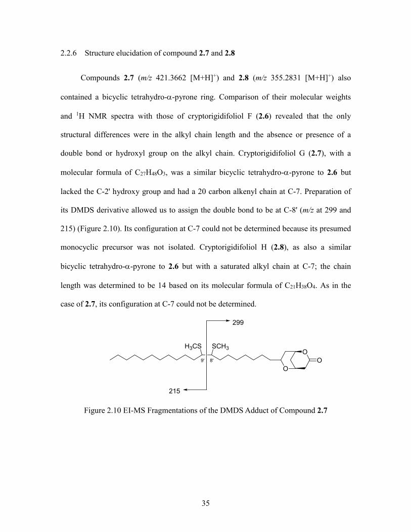
35
2.2.6 Structure elucidation of compound 2.7 and 2.8
Compounds 2.7 (m/z 421.3662 [M+H]+) and 2.8 (m/z 355.2831 [M+H]+) also
contained a bicyclic tetrahydro--pyrone ring. Comparison of their molecular weights
and 1H NMR spectra with those of cryptorigidifoliol F (2.6) revealed that the only
structural differences were in the alkyl chain length and the absence or presence of a
double bond or hydroxyl group on the alkyl chain. Cryptorigidifoliol G (2.7), with a
molecular formula of C27H48O3, was a similar bicyclic tetrahydro--pyrone to 2.6 but
lacked the C-2' hydroxy group and had a 20 carbon alkenyl chain at C-7. Preparation of
its DMDS derivative allowed us to assign the double bond to be at C-8' (m/z at 299 and
215) (Figure 2.10). Its configuration at C-7 could not be determined because its presumed
monocyclic precursor was not isolated. Cryptorigidifoliol H (2.8), as also a similar
bicyclic tetrahydro--pyrone to 2.6 but with a saturated alkyl chain at C-7; the chain
length was determined to be 14 based on its molecular formula of C21H38O4. As in the
case of 2.7, its configuration at C-7 could not be determined.
O
O
O
299
215
SCH3H3CS
8'9'
Figure 2.10 EI-MS Fragmentations of the DMDS Adduct of Compound 2.7
36
2.2.7 Structure elucidation of compound 2.92.11
Compounds 2.92.11 (cryptorigidifoliols IK) are the bicyclic derivatives of
2.2and 2.5, as determined by the conversions described below and by comparison of
their IR, 1H NMR and HRESIMS data with those of their 5,6-dihydro--pyrone
precursors and those of compounds 2.62.8. Since the configurations of the 2'-hydroxyl
group of precursors 2.2and 2.5 have been established, the corresponding
configurations at C-7 of 2.9, 2.10, and 2.11 were assigned as (S), (R), and (R),
respectively. Surprisingly, compounds 2.7 and 2.10 showed no detectable optical activity.
Since it is unlikely that they are racemic, given their formation from non-racemic
precursors, the lack of measurable optical activity may be due to the nearly symmetrical
structures of these compounds. Compounds 2.6, 2.8, 2.9, and 2.11 had no significant
(H) difference between their R- and S-MPA derivatives, so their configurations at these
positions could not be determined.
2.2.8 Evidence that bicyclic tetrahydropyrones 2.6 – 2.11 are artifacts
The initial purification of the bioactive EtOAc fraction involved Sephadex LH-20
column chromatography, reverse phase SPE, normal phase silica gel column
chromatography, and C18 HPLC. This procedure yielded fractions that in most cases were
mixtures of 5,6-dihydro--pyrones and bicyclic tetrahydropyrones. In addition, it was
noted that the early eluting fractions from the open silica gel column contained either
pure 5,6-dihydro--pyrone 2.1, or mixtures of 5,6-dihydro--pyrones 2.2, 2.3, and 2.5
and bicyclic tetrahydropyrones 2.92.11. The later-eluting fractions, however, yielded
only the bicyclic tetrahydropyrones 2.6, 2.7, and 2.8. This suggested that the 5,6-dihydro-
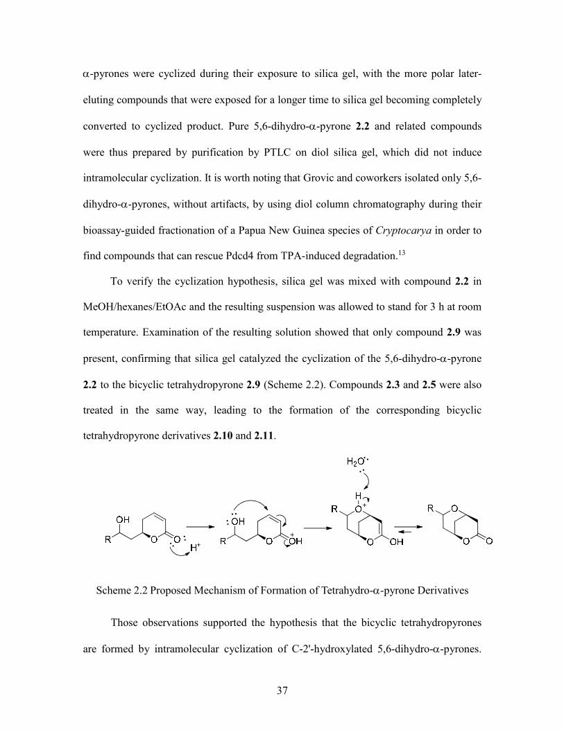
37
-pyrones were cyclized during their exposure to silica gel, with the more polar later-
eluting compounds that were exposed for a longer time to silica gel becoming completely
converted to cyclized product. Pure 5,6-dihydro--pyrone 2.2 and related compounds
were thus prepared by purification by PTLC on diol silica gel, which did not induce
intramolecular cyclization. It is worth noting that Grovic and coworkers isolated only 5,6-
dihydro--pyrones, without artifacts, by using diol column chromatography during their
bioassay-guided fractionation of a Papua New Guinea species of Cryptocarya in order to
find compounds that can rescue Pdcd4 from TPA-induced degradation.13
To verify the cyclization hypothesis, silica gel was mixed with compound 2.2 in
MeOH/hexanes/EtOAc and the resulting suspension was allowed to stand for 3 h at room
temperature. Examination of the resulting solution showed that only compound 2.9 was
present, confirming that silica gel catalyzed the cyclization of the 5,6-dihydro--pyrone
2.2 to the bicyclic tetrahydropyrone 2.9 (Scheme 2.2). Compounds 2.3 and 2.5 were also
treated in the same way, leading to the formation of the corresponding bicyclic
tetrahydropyrone derivatives 2.10 and 2.11.
Scheme 2.2 Proposed Mechanism of Formation of Tetrahydro--pyrone Derivatives
Those observations supported the hypothesis that the bicyclic tetrahydropyrones
are formed by intramolecular cyclization of C-2'-hydroxylated 5,6-dihydro--pyrones.

38
This does not however rule out the possibility that at least some bicyclic
tetrahydropyrones are formed in the plant, and examination of the crude extract by 1H
NMR spectroscopy indicated that it did contain signals consistent with the presence of
bicyclic tetrahydropyrones. This result could either be due to the presence of the cyclized
compounds in the plant or to intramolecular cyclization during the extraction of the plant
and the processing of the extract in Madagascar prior to analysis. Regrettably we did not
have access to fresh plant material to test these alternate hypotheses, but the fact that
chromatography over silica gel was used in every case where the cyclized compounds are
reported strongly suggests that these compounds are indeed formed during the
purification process.
2.2.9 Biological activities
Compounds 2.12.11 were evaluated for their antiparasitic activity against the
chloroquine/mefloquine-resistant Dd2 strain of Plasmodium falciparum. Compounds
2.12.7 exhibited moderate antimalarial activity, with IC50 values of 9.2 ± 0.9, 5.8 ± 1.4,
5.5 ± 0.7, 9.0 ± 3.0, 4 ± 2, 7.4 ± 0.6, and 6.0 ± 0.5 M, respectively. Compounds
2.82.11 were all less active, with IC50 values > 10 M (Tables 2.3 and 2.4). These data
indicate that the presence of the ,-unsaturated ketone is not essential for antimalarial
activity, since the bicyclic compounds 2.6 and 2.7 have comparable activity to the ,-
unsaturated compounds 2.1–2.5.
All of the compounds were also evaluated for their antiproliferative activity against
A2780 human ovarian cancer cells, but only compound 2.3 had an IC50 value less than 10
M (Tables 2.3 and 2.4). Since these compounds have only moderate antiparasitic
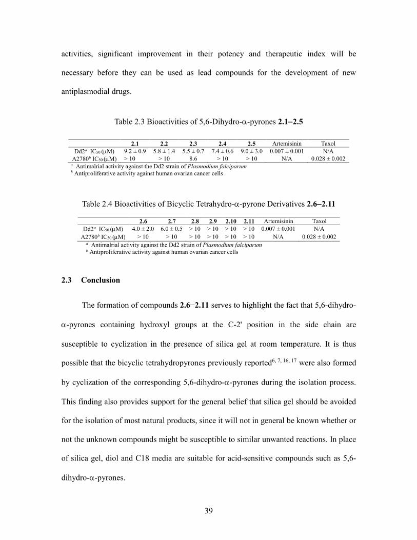
39
activities, significant improvement in their potency and therapeutic index will be
necessary before they can be used as lead compounds for the development of new
antiplasmodial drugs.
Table 2.3 Bioactivities of 5,6-Dihydro--pyrones 2.12.5
a Antimalrial activity against the Dd2 strain of Plasmodium falciparum b Antiproliferative activity against human ovarian cancer cells
Table 2.4 Bioactivities of Bicyclic Tetrahydro--pyrone Derivatives 2.62.11
a Antimalrial activity against the Dd2 strain of Plasmodium falciparum b Antiproliferative activity against human ovarian cancer cells
2.3 Conclusion
The formation of compounds 2.6−2.11 serves to highlight the fact that 5,6-dihydro-
-pyrones containing hydroxyl groups at the C-2' position in the side chain are
susceptible to cyclization in the presence of silica gel at room temperature. It is thus
possible that the bicyclic tetrahydropyrones previously reported6, 7, 16, 17 were also formed
by cyclization of the corresponding 5,6-dihydro--pyrones during the isolation process.
This finding also provides support for the general belief that silica gel should be avoided
for the isolation of most natural products, since it will not in general be known whether or
not the unknown compounds might be susceptible to similar unwanted reactions. In place
of silica gel, diol and C18 media are suitable for acid-sensitive compounds such as 5,6-
dihydro--pyrones.
2.1 2.2 2.3 2.4 2.5 Artemisinin Taxol
Dd2a IC50 (M) 9.2 ± 0.9 5.8 ± 1.4 5.5 ± 0.7 7.4 ± 0.6 9.0 ± 3.0 0.007 ± 0.001 N/A
A2780b IC50 (M) > 10 > 10 8.6 > 10 > 10 N/A 0.028 ± 0.002
2.6 2.7 2.8 2.9 2.10 2.11 Artemisinin Taxol
Dd2a IC50 (M) 4.0 ± 2.0 6.0 ± 0.5 > 10 > 10 > 10 > 10 0.007 ± 0.001 N/A
A2780b IC50 (M) > 10 > 10 > 10 > 10 > 10 > 10 N/A 0.028 ± 0.002
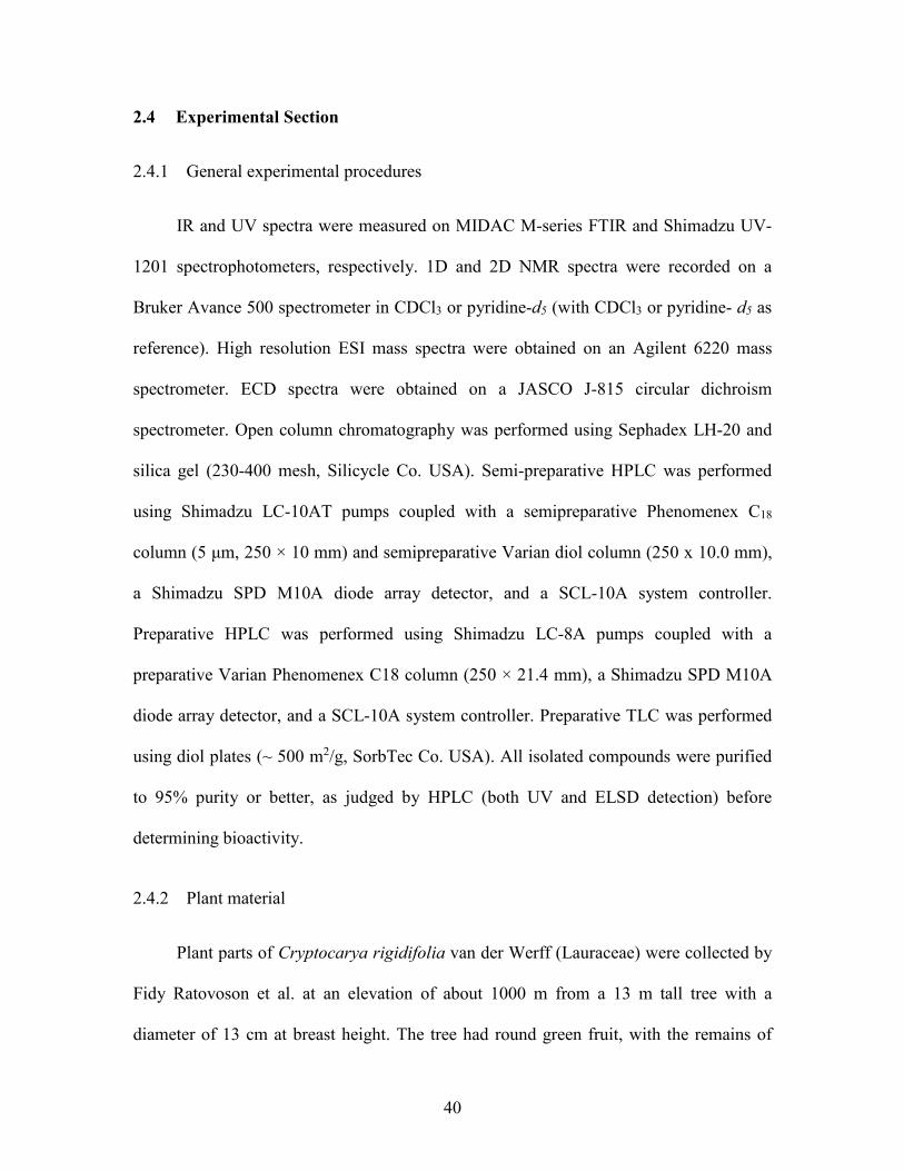
40
2.4 Experimental Section
2.4.1 General experimental procedures
IR and UV spectra were measured on MIDAC M-series FTIR and Shimadzu UV-
1201 spectrophotometers, respectively. 1D and 2D NMR spectra were recorded on a
Bruker Avance 500 spectrometer in CDCl3 or pyridine-d5 (with CDCl3 or pyridine- d5 as
reference). High resolution ESI mass spectra were obtained on an Agilent 6220 mass
spectrometer. ECD spectra were obtained on a JASCO J-815 circular dichroism
spectrometer. Open column chromatography was performed using Sephadex LH-20 and
silica gel (230-400 mesh, Silicycle Co. USA). Semi-preparative HPLC was performed
using Shimadzu LC-10AT pumps coupled with a semipreparative Phenomenex C18
column (5 μm, 250 × 10 mm) and semipreparative Varian diol column (250 x 10.0 mm),
a Shimadzu SPD M10A diode array detector, and a SCL-10A system controller.
Preparative HPLC was performed using Shimadzu LC-8A pumps coupled with a
preparative Varian Phenomenex C18 column (250 × 21.4 mm), a Shimadzu SPD M10A
diode array detector, and a SCL-10A system controller. Preparative TLC was performed
using diol plates (~ 500 m2/g, SorbTec Co. USA). All isolated compounds were purified
to 95% purity or better, as judged by HPLC (both UV and ELSD detection) before
determining bioactivity.
2.4.2 Plant material
Plant parts of Cryptocarya rigidifolia van der Werff (Lauraceae) were collected by
Fidy Ratovoson et al. at an elevation of about 1000 m from a 13 m tall tree with a
diameter of 13 cm at breast height. The tree had round green fruit, with the remains of
41
brown calyx above. Collection was made near the towns of Imerimandroso and
Antanandava in the district of Ambatondrazaka, north of the Zahamena National Park, at
coordinates 17°28'45"S 048°44'10"E. The tree was growing in medium altitude
rainforest, on the edge of an old area of slash and burn agriculture. Duplicate voucher
specimens (Ratovoson 250) were deposited at the Centre National d'Application des
Recherches Pharmaceutiques (CNARP), the Herbarium of the Department of Forestry
and Fishery Research (TEF), and the Missouri Botanical Garden, St. Louis, Missouri
(MO).
2.4.3 Extraction and isolation
A ground sample of C. rigidifolia root wood (310 g) was extracted with EtOH at
room temperature to yield 16.3 g of crude EtOH extract designated MG 0441, and a
portion of this extract was made available to Virginia Tech for bioassay-guided isolation.
The crude EtOH extract (5.0 g) of the wood root of C. rigidifolia was dissolved in 90%
aq. MeOH and extracted with hexanes. The 90% aq. MeOH layer was then evaporated,
suspended in H2O, and extracted with EtOAc. The antimalarial activities were
concentrated in the EtOAc (IC50 2.5~5 g/mL) and hexanes fractions (IC50 ~5 g/mL).
The EtOAc fraction (971 mg) was subjected to Sephadex LH-20 size chromatography to
give four fractions, and the most active fraction Fr-2 (IC50 2.5~5 g/mL) was fractionated
by C18 SPE using 70% aq. MeOH, 100% MeOH, and CH2Cl2 separately. The 100%
MeOH fraction (IC50 1.25~2.5 g/mL) was then divided into 8 sub-fractions by silica gel
column chromatography (hexanes:EtOAc from 1:1 to 3:7 to 1:9, and a final elution with
MeOH). The third sub-fraction (IC50 <1.25 g/mL) was subjected to C18 HPLC with a
42
solvent gradient from H2O:CH3CN, 20:80 to 10:90 from 0.01 to10 min, to 0:100 from 10
to 15 min, and ending with 100% CH3CN to 30 min, furnishing compound 2.1 (tR 20.2
min, 1.1 mg, IC50 2.98 g/mL) and an active fraction (tR 20.8 min). Further purification
of the active fraction by diol PTLC yielded 2.3 (1.4 mg, IC50 1.93 g/mL) and 2.10 (0.4
mg, IC50 5.74 g/mL). The fourth sub-fraction from silica gel column chromatography
was subjected to C18 HPLC with a solvent gradient from H2O:CH3CN, 30:70 to 20:80
from 0.01 to15 min, to 10:90 from 15 to 35 min, to 0:100 from 35 to 50 min, and ending
with 100% CH3CN to 60 min. Purification by diol PTLC of the mixtures with tR 37.5 min
and tR 40.2 min obtained from this process gave compounds 2.5 (0.9 mg, IC50 3.55
g/mL), 2.11 (3.1 mg, IC50 >20 g/mL), 2.2 (1.3 mg, IC50 2.30 g/mL) and 2.9 (0.5 mg,
IC50 6.57 g/mL). The fifth sub-fraction from silica gel column chromatography was
further purified by C 18 HPLC by using the same profile as the fourth sub-fraction, to
give pure compounds 2.8 (4.2 mg, tR 38 min, IC50 13.6 g/mL) and 2.6 (9.6 mg, tR 40.8
min, IC50 1.58 g/mL).
The hexanes fraction was subjected to Sephadex LH-20 size chromatography to
give three fractions, and the most active fraction Fr-2 (IC50 2.5~5 g/mL) was
fractionated by preparative C18 HPLC with a solvent gradient from H2O:CH3CN, 30:70
to 0:100 from 0.01 to 30 min, and ending with 100% CH3CN to 45 min. This process
yielded compound 2.7 (1.6 mg, tR 39 min, IC50 2.50 g/mL) and an active fraction (tR
32.5 min). Further purification of the fraction by diol HPLC with a solvent gradient from
hexanes:EtOAc, 90:10 to 80:20 from 0.01 to 10 min, to 70:30 from 10 to 15 min, to
60:40 from 15 to 20 min, to 0:100 from 20 to 25 min, and ending with 100% EtOAc wash
to 35 min, furnished compound 2.4 (0.9 mg, IC50 2.76 g/mL).

43
2.4.4 Cryptorigidifoliol A (2.1)
Colorless oil; []21D +5.6 (c 0.54, MeOH); UV (MeOH) λmax (log ε): 204 (3.63)
nm; IR νmax 3340, 2922, 2849, 1719, 1613, 1089 cm-1; ECD (c 0.031 mM, MeOH) λmax
(Δε) 203 (+8.5), 235 (+2.4), 256 (+3.8); 1H and 13C NMR data, see Table 2.1; HRESIMS
m/z [M+H]+ 325.2753 (calcd for C20H37O3+, 325.2737).
2.4.5 Cryptorigidifoliol B (2.2)
Colorless oil; []21D +9.1 (c 0.11, MeOH); UV (MeOH) λmax (log ε): 204 (3.83)
nm; IR νmax 3343, 2922, 2850, 1714, 1615, 1092 cm-1; ECD (c 0.031 mM, MeOH) λmax
(Δε) 209 (+9.7), 235 (+1.9), 256 (+3.5); 1H and 13C NMR data, see Table 2.1; HRESIMS
m/z [M+H]+: 397.3317 (calcd for C24H45O4+, 397.3312).
2.4.6 Cryptorigidifoliol C (2.3)
Colorless oil; []21D +18.8 (c 0.96, MeOH); UV (MeOH) λmax (log ε): 204 (3.91)
nm; IR νmax 3439, 2924, 2855, 1728, 1623, 1044 cm-1; ECD (c 0.031 mM, MeOH) λmax
(Δε) 207 (+9.3), 235 (+1.2), 255 (+3.1); 1H and 13C NMR data, see Table 2.1; HRESIMS
m/z [M+H]+ 351.2902 (calcd for C22H39O3+, 351.2894).
2.4.7 Cryptorigidifoliol D (2.4)
Colorless oil; []21D 0 (c 1.98, MeOH); UV (MeOH) λmax (log ε): 204 (3.50) nm;
IR νmax 3444, 2922, 2855, 1724, 1625, 1034 cm-1; ECD (c 0.031 mM, MeOH) λmax (Δε)
208 (+10.6), 235 (+1.6), 256 (+3.0); 1H and 13C NMR data, see Table 2.1; HRESIMS m/z
[M+NH4]+ 396.3489 (calcd for C24H46NO3+, 396.3472).
44
2.4.8 Cryptorigidifoliol E (2.5)
Colorless oil; []21D -25 (c 0.4, MeOH); UV (MeOH) λmax (log ε): 204 (3.24) nm;
IR νmax: 3382, 2922, 2845, 1729, 1625, 1077 cm-1; ECD (c 0.031 mM, MeOH) λmax (Δε)
207 (+11.0), 234 (+2.3), 256 (+3.2); 1H and 13C NMR data, see Table 2.1; HRESIMS m/z
[M-OH]+ 377.3060 (calcd for C24H41O3+, 377.3050).
2.4.9 Cryptorigidifoliol F (2.6)
Colorless oil; []21D -14.3 (c 4.25, MeOH); UV (MeOH) λmax (log ε): 204 (3.14)
nm; IR νmax 3450, 2932, 2855, 1719, 1073 cm-1; 1H and 13C NMR data, see Table 2.2;
HRESIMS m/z [M+H]+ 395.3149 (calcd for C24H43O4+, 396.3156).
2.4.10 Cryptorigidifoliol G (2.7)
Colorless oil; []21D 0 (c 1.57, MeOH); UV (MeOH) λmax (log ε): 204 (3.14) nm;
IR νmax 3450, 2932, 2855, 1719, 1073 cm-1; 1H and 13C NMR data, see Table 2.2;
HRESIMS m/z [M+H]+ 421.3662 (calcd for C27H49O3+, 421.3676).
2.4.11 Cryptorigidifoliol H (2.8)
Colorless oil; []21D -10.6 (c 0.66, MeOH); UV (MeOH) λmax (log ε): 204 (2.66)
nm; IR νmax 3540, 2917, 2845, 1719, 1070 cm-1; 1H and 13C NMR data, see Table 2.2;
HRESIMS m/z [M+H]+355.2831 (calcd for C21H39O4+, 355.2843).

45
2.4.12 Cryptorigidifoliol I (2.9)
Colorless oil; []21D -7.5 (c 0.4, MeOH); UV (MeOH) λmax (log ε): 204 (3.43) nm;
IR νmax 3340, 2943, 2846, 1727, 1068 cm-1; 1H and 13C NMR data, see Table 2.2;
HRESIMS m/z [M+H]+ 397.3317 (calcd for C24H45O4+, 397.3312).
2.4.13 Cryptorigidifoliol J (2.10)
Colorless oil; []21D 0 (c 1.21, MeOH); UV (MeOH) λmax (log ε): 204 (3.62) nm;
IR νmax 3345, 2939, 2844, 1739, 1056 cm-1; 1H and 13C NMR data, see Table 2.2;
HRESIMS m/z [M+H]+351.2902 (calcd for C22H39O3+, 351.2894).
2.4.14 Cryptorigidifoliol K (2.11)
Colorless oil; []21D -9.5 (c 3.06, MeOH); UV (MeOH) λmax (log ε): 204 (3.10) nm;
IR νmax 3431, 2956, 2867, 1732, 1036 cm-1; 1H and 13C NMR data, see Table 2.2;
HRESIMS m/z [M+H]+ 377.3060 (calcd for C24H41O3+, 377.3050).
2.4.15 Dimethyldisulfide derivatization.
Pure samples (~200 g) dissolved in 200 L of hexanes were treated with
dimethyldisulfide (400 L) and 12 drops of iodine solution (6% w/v in ether) in a 6
dram vial at 40°C for 30 h. The mixture was then cooled and diluted with hexanes (1 mL),
and iodine was removed by extracting with thiosulphate (1 mL, 5% w/v). The organic
layer was removed, and the water layer was re-extracted with hexanes:CH2Cl2 (1:1). The
combined organic layers were dried and subjected to analysis by GC-EIMS.25, 26
46
2.4.16 Preparation of the R and S-MPA ester derivatives of 5,6-dihydro--pyrones
The in-NMR-tube reaction28 was carried out to prepare the MPA ester derivatives.
The selected 5,6-dihydro--pyrone (0.2 mg) was dissolved in pyridine-d5 in a clean NMR
tube, and R-(−)-α-methoxyphenylacetyl (MPA) chloride (0.5 mg) was added to the NMR
tube under an N2 gas flow, and the NMR tube was gently shaken. The reaction NMR tube
was kept at room temperature overnight and was then analyzed by 1H NMR spectroscopy.
The S-MPA esters of the 5,6-dihydro--pyrones were prepared from S-MPA chloride by
the same method.
2.4.17 Preparation of bicyclic tetrahydropyrones
Compound 2.2 (0.2 mg) was dissolved in a mixture of MeOH (2 mL), EtOAc (0.5
mL) and hexanes (0.5 mL). Enough silica gel powder was added to the solution to absorb
it, and the mixture was kept at room temperature for 3 h. The resulting damp powder was
eluted by MeOH, and the eluate concentrated on a rotavapor. The product was identified
as 2.9 by 1H NMR spectroscopy. Compounds 2.3 and 2.5 (each 0.2 mg) were also treated
separately and worked up under the same conditions. The products were identified as
2.10 and 2.11, respectively, by 1H NMR spectroscopy.
2.4.18 Antiproliferative bioassay
The A2780 ovarian cancer cell line antiproliferative bioassay was performed at
Virginia Tech as previously reported.29, 30 The A2780 cell line is a drug-sensitive ovarian
cancer cell line.31 Paclitaxel was used as the positive control; it had an IC50 value of 73 ±
15 nM.

47
2.4.19 Antimalarial bioassay
The effect of each compound on parasite growth of the Dd2 strain of P. falciparum
was measured in a 72 h treatment using the malaria SYBR green I-based fluorescence
assay as described previously.32, 33 Artemisinin was used as the positive control; it had an
IC50 value of 7 ± 1 nM.

48
2.5 References
1. Kingston, D. G. I. A natural love of natural products. J. Org. Chem. 2008, 73,
3975-3984.
2. Chou, T. H.; Chen, J. J.; Lee, S. J.; Chiang, M. Y.; Yang, C. W.; Chen, I. S.
Cytotoxic flavonoids from the leaves of Cryptocarya chinensis. J. Nat. Prod. 2010, 73,
1470-1475.
3. Lin, H. R.; Chou, T. H.; Huang, D. W.; Chen, I. S. Cryptochinones from
Cryptocarya chinensis act as farnesoid X receptor agonists. Bioorg. Med. Chem. Lett.
2014, 24, 4181-4186.
4. Wu, T. S.; Lin, F. W. Alkaloids of the wood of Cryptocarya chinensis. J. Nat.
Prod. 2001, 64, 1404-1407.
5. Fu, X.; Sevenet, T.; Hamid, A.; Hadi, A.; Remy, F.; Pais, M. Kurzilactone from
Cryptocarya kurzii. Phytochemistry 1993, 33, 1272-1274.
6. Drewes, S. E.; Horn, M. M.; Shaw, R. S. α-Pyrones and their derivatives from two
Cryptocarya species. Phytochemistry 1995, 40, 321-323.
7. Drewes, S. E.; Sehlapelo, B. M.; Horn, M. M.; Scott-Shaw, R.; Sandor, P. 5,6-
Dihydro-α-pyrones and two bicyclic tetrahydro-α-pyrone derivatives from Cryptocarya
latifolia. Phytochemistry 1995, 38, 1427-1430.
8. Toneto Novaes, L. F.; Drekener, R. L.; Avila, C. M.; Pilli, R. A. Total synthesis of
cryptomoscatones D1 and D2: Stereochemical assignment of cryptomoscatone D1.
Tetrahedron 2014, 70, 6467-6473.
9. Deschamps, J. R.; George, C.; Flippen-Anderson, J. L.; Spencer, G. A new
Cryptocarya lactone. Acta Crystallogr., Sect. E: Struct. Rep. Online 2001, 57, o648-o649.
49
10. Rali, T.; Wossa, S. W.; Leach, D. N. Comparative chemical analysis of the
essential oil constituents in the bark, heartwood and fruits of Cryptocarya massoy (Oken)
Kosterm. (Lauraceae) from Papua New Guinea. Molecules 2007, 12, 149-154.
11. Cuccarese, M. F.; Wang, Y.; Beuning, P. J.; O'Doherty, G. A. Cryptocaryol
structure-activity relationship study of cancer cell cytotoxicity and ability to stabilize
PDCD4. ACS Med. Chem. Lett. 2014, 5, 522-526.
12. Cardona, W.; Quinones, W.; Robledo, S.; Velez, I. D.; Murga, J.; Garcia-Fortanet,
J.; Carda, M.; Cardona, D.; Echeverri, F. Antiparasite and antimycobacterial activity of
passifloricin analogs. Tetrahedron 2006, 62, 4086-4092.
13. Grkovic, T.; Blees, J. S.; Colburn, N. H.; Schmid, T.; Thomas, C. L.; Henrich, C.
J.; McMahon, J. B.; Gustafson, K. R. Cryptocaryols A-H, α-pyrone-containing 1,3-
polyols from Cryptocarya sp. Implicated in stabilizing the tumor suppressor Pdcd4. J.
Nat. Prod. 2011, 74, 1015-1020.
14. Meragelman, T. L.; Scudiero, D. A.; Davis, R. E.; Staudt, L. M.; McCloud, T. G.;
Cardellina, J. H.; Shoemaker, R. H. Inhibitors of the NF-κB Activation Pathway from
Cryptocarya rugulosa. J. Nat. Prod. 2009, 72, 336-339.
15. Sturgeon, C. M.; Cinel, B.; Diaz-Marrero, A. R.; McHardy, L. M.; Ngo, M.;
Andersen, R. J.; Roberge, M. Abrogation of ionizing radiation-induced G2 checkpoint
and inhibition of nuclear export by Cryptocarya pyrones. Cancer Chemother. Pharmacol.
2008, 61, 407-413.
16. Jiang, Z. H.; Yang, Q. X.; Tanaka, T.; Kouno, I. Bicyclic polyketide lactones from
Chinese medicinal ats, Polyrhacis lamellidens. J. Nat. Prod. 2008, 71, 724-727.
17. Liou, J. R.; Wu, T. Y.; Thang, T. D.; Hwang, T. L.; Wu, C. C.; Cheng, Y. B.;
50
Chiang, M. Y.; Lan, Y. H.; El-Shazly, M.; Wu, S. L.; Beerhues, L.; Yuan, S. S.; Hou, M.
F.; Chen, S. L.; Chang, F. R.; Wu, Y. C. Bioactive 6S-styryllactone constituents of
Polyalthia parviflora. J. Nat. Prod. 2014, 77, 2626-2632.
18. Liu, Y.; Rakotondraibe, H.; Brodie, P. J.; Wiley, J. D.; Cassera, M. B.; Goetz, M.;
Kingston, D. G. I. Antiproliferative and antimalarial sesquiterpene lactones from
Piptocoma antillana from Puerto Rico. Nat. Prod. Commun. 2014, 9, 1403-1406.
19. Davies-Coleman, M. T.; Rivett, D. E. A. Naturally occurring 6-substituted 5,6-
dihydro-α-pyrones. Prog. Chem. Org. Nat. Prod. 1989, 55, 1-35.
20. Snatzke, G. Circular dichroism and optical rotatory dispersion. Principles and
application to the investigation of the stereochemistry of natural products. Angew. Chem.,
Int. Ed. Engl. 1968, 7, 14-25.
21. Raoelison, G. E.; Terreaux, C.; Queiroz, E. F.; Zsila, F.; Simonyi, M.; Antus, S.;
Randriantsoa, A.; Hostettmann, K. Absolute configuration of two new 6-alkylated α-
pyrones (=2H-pyran-2-ones) from Ravensara crassifolia. Helv. Chim. Acta 2001, 84,
3470-3476.
22. Costa, V. C. d. O.; Tavares, J. F.; Silva, A. B.; Duarte, M. C.; Agra, M. d. F.;
Barbosa-Filho, J. M.; de Souza, I. L. L.; da Silva, B. A.; Silva, M. S. Hyptenolide, a new
α-pyrone with spasmolytic activity from Hyptis macrostachys. Phytochem. Lett. 2014, 8,
32-37.
23. Dale, J. A.; Mosher, H. S. Nuclear magnetic resonance enantiomer regents.
Configurational correlations via nuclear magnetic resonance chemical shifts of
diastereomeric mandelate, O-methylmandelate, and α-methoxy-α-
trifluoromethylphenylacetate (MTPA) esters. J. Am. Chem. Soc. 1973, 95, 512-519.
51
24. Jie, M. S. F. L. K.; Pasha, M. K.; Alam, M. S. Synthesis and nuclear magnetic
resonance properties of all geometrical isomers of conjugated linoleic acids. Lipids 1997,
32, 1041-1044.
25. Nichols, P. D.; Guckert, J. B.; White, D. C. Determination of monounsaturated
fatty acid double-bond position and geometry for microbial monocultures and complex
consortia by capillary GC-MS of their dimethyl disulfide adducts. J. Microbiol. Methods
1986, 5, 49-55.
26. Mansour, M. P.; Holdsworth, D. G.; Forbes, S. E.; Macleod, C. K.; Volkman, J. K.
High contents of 24:6(n-3) and 20:1(n-13) fatty acids in the brittle star Amphiura
elandiformis from Tasmanian coastal sediments. Biochem. Syst. Ecol. 2005, 33, 659-674.
27. Drewes, S. E.; Horn, M. M.; Ramesar, N. S.; Ferreira, D.; Nel, R. J. J.; Hutchings,
A. Absolute configurations of all four stereoisomers of cryptocaryalactone and deacetyl
cryptocaryalactone. Phytochemistry 1998, 49, 1683-1687.
28. Su, B. N.; Park, E. J.; Mbwambo, Z. H.; Santarsiero, B. D.; Mesecar, A. D.; Fong,
H. H. S.; Pezzuto, J. M.; Kinghorn, A. D. New chemical constituents of Euphorbia
quinquecostata and absolute configuration assignment by a convenient Mosher ester
procedure carried out in NMR tubes. J. Nat. Prod. 2002, 65, 1278-1282.
29. Cao, S.; Brodie, P. J.; Miller, J. S.; Randrianaivo, R.; Ratovoson, F.; Birkinshaw,
C.; Andriantsiferana, R.; Rasamison, V. E.; Kingston, D. G. I. Antiproliferative xanthones
of terminalia calcicola from the Madagascar rain forest. J. Nat. Prod. 2007, 70, 679-681.
30. Pan, E.; Harinantenaina, L.; Brodie, P., J.; Miller, J. S.; Callmander, M. W.;
Rakotonandrasana, S.; Rakotobe, E.; Rasamison, V. E.; Kingston, D. G. I. Four
Diphenylpropanes and a Cycloheptadibenzofuran from Bussea sakalava from the
52
Madagascar Dry Forest. J. Nat. Prod. 2010, 73, 1792-1795.
31. Louie, K. G.; Behrens, B. C.; Kinsella, T. J.; Hamilton, T. C.; Grotzinger, K. R.;
McKoy, W. M.; Winker, M. A.; Ozols, R. F. Radiation survival parameters of
antineoplastic drug-sensitive and -resistant human ovarian cancer cell lines and their
modification by buthionine sulfoximine. Cancer Res. 1985, 45, 2110-2115.
32. Bennett, T. N.; Paguio, M.; Gligorijevic, B.; Seudieu, C.; Kosar, A. D.; Davidson,
E.; Roepe, P. D. Novel, rapid, and inexpensive cell-based quantification of antimalarial
drug efficacy. Antimicrob. Agents Chemother. 2004, 48, 1807-1810.
33. Smilkstein, M.; Sriwilaijaroen, N.; Kelly, J. X.; Wilairat, P.; Riscoe, M. Simple
and inexpensive fluorescence-based technique for high-throughput antimalarial drug
screening. Antimicrob. Agents Chemother. 2004, 48, 1803-1806.

3 Antiproliferative Compounds from Cleistanthus boivinianus from the Madagascar Dry Forest
This chapter is a slightly expanded version of a submitted article to the Journal of
Natural Product, which is waiting for the final decision after revision. Attributions from
co-authors of the articles are described as follows in the order of the names listed. The
author of this dissertation (Yixi Liu) mentored the undergraduate student (Kelly Young)
with the initial isolation with two compounds and finished up with isolating five more
compounds, and elucidated the structures for all compounds, and drafted the manuscript.
Dr. Harinantenaina was a mentor for this work, provided invaluable advice and hints for
the structural elucidation of the compounds, and also proofread the manuscript before
submission. Mrs. Peggy Brodie performed the A2780 bioassay on the isolated fractions
and compounds. Dr. Jessica D. Wiley and Dr. Maria B. Cassera performed the Dd2
bioassay. Dr. Martin W. Callmander, and Dr. Richard Randrianaivo from Missouri
Botanical Garden collected and identified the plant. Dr. Etienne Rakotobe and Dr.
Vincent E. Rasamison from Madagascar carried out the initial plant extraction. Dr. Karen
TenDyke and Dr. Yongchun Shen from Eisai Inc. performed the A2058, HCT-116 and
MES-SA bioassays on the compounds isolated. Dr. David G. I. Kingston was a mentor
for this work and the corresponding author for the published article. He provided critical
suggestions for this work and crucial revisions to the manuscript.
54
3.1 Introduction
As a part of the Madagascar International Cooperative Biodiversity Group (ICBG)
program,1, 2 an ethanol extract of the stems of Cleistanthus boivinianus (Baill.) Müll. Arg.
(Phyllanthaceae) was found to have moderate antiproliferative activity against the A2780
ovarian cancer cell line (IC50 4.5 g/ml), and it was thus selected for evaluation for the
presence of novel antiproliferative agents. The genus Cleistanthus is native to Africa,
India, and across to Australia,3 and comprises approximately 140 species, but only five of
these have been investigated chemically: C. collinus, C. patulus, C. schlechteri var.
schlechteri, C. gracilis,3 and C. indochinensis.4 Arylnaphthalide lignans are major
constituents of the genus, while arytetralin, furofuranoid, and dibenzylbutane lignans and
terpenoids have also been isolated from it and are known for their toxic properties.3-11
Bioassay-guided isolation of the stems of Cleistanthus boivinianus furnished the
new bioactive aryltetralin lignan 3.1 as the most potent antiproliferative (A2780)
component with IC50 values at nanomolar level. It is an analogue of the cytotoxic natural
product podophyllotoxin, with a substitution at C-3 instead of the C-4 position. This is
the first report on this compound with this unique structure. Its strong activity suggested
that analogues of podophyllotoxin-type compounds with substitution at C-3 instead of C-
4 display more potent activity than those substituted C-4 with no substitution at C-3. In
addition, the new arylnaphthalide lignan 3.2 together with the five known aryltetralin
lignans deoxypicropodophyllotoxin (3.3),12 (±)--apopicropodophyllin (3.4),13-15 (-)-
desoxypodophyllotoxin (3.5),16 (-)-yatein (3.6),17 and -peltatin-5-O--D-
glucopyranoside (3.7)18 were also isolated. (Figure 3.1) The structures of the known

55
compounds 3.33.7 were determined by comparison of their 1H NMR, mass spectra, and
optical rotation values with the data reported in the literature, except for the case of 3.4
which had no optical rotation. The isolation of racemic -apopicropodophyllin is
noteworthy, since all previous isolates have been of the dextrorotatory isomer.
Figure 3.1 Structures of Compounds 3.13.7
3.2 Results And Discussion
3.2.1 Isolation of active compounds
Dereplication of an active EtOAc-soluble fraction obtained from liquid-liquid
partition of the extract (100 mg) as previously described19 indicated that it contained at
least one new bioactive compound, and so a larger sample was investigated. Fractionation
of this extract yielded an antiproliferative EtOAc fraction which was further subjected to
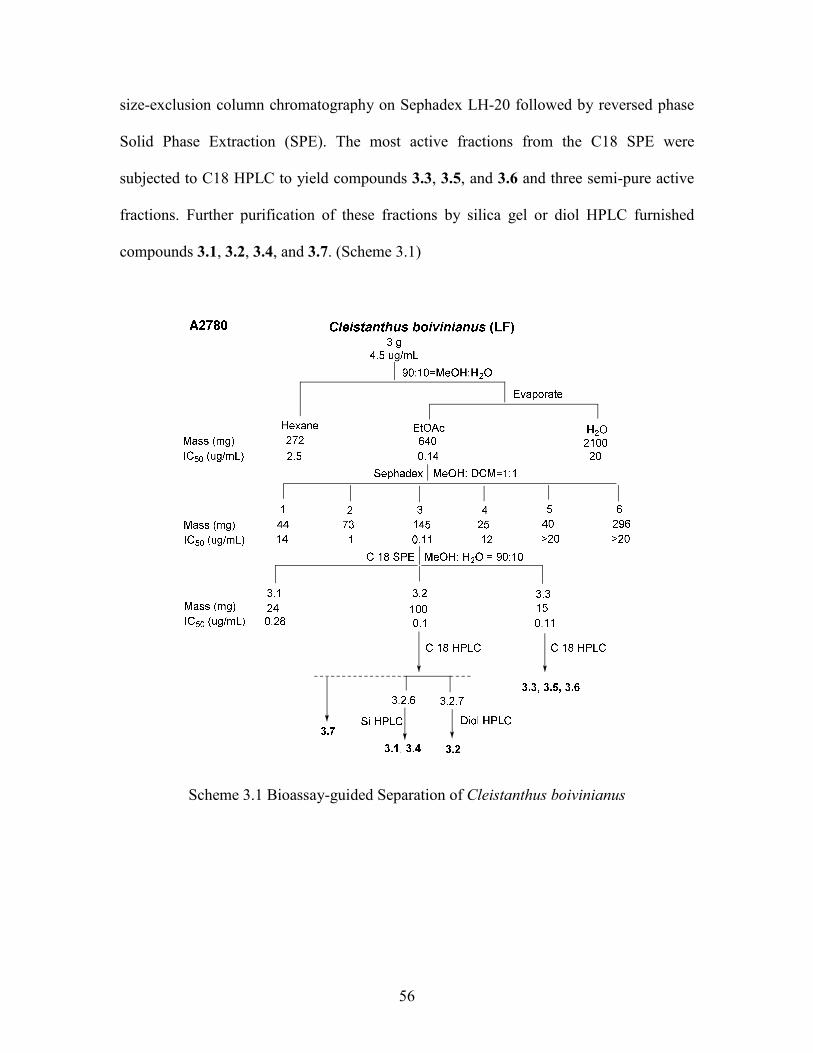
56
size-exclusion column chromatography on Sephadex LH-20 followed by reversed phase
Solid Phase Extraction (SPE). The most active fractions from the C18 SPE were
subjected to C18 HPLC to yield compounds 3.3, 3.5, and 3.6 and three semi-pure active
fractions. Further purification of these fractions by silica gel or diol HPLC furnished
compounds 3.1, 3.2, 3.4, and 3.7. (Scheme 3.1)
Scheme 3.1 Bioassay-guided Separation of Cleistanthus boivinianus
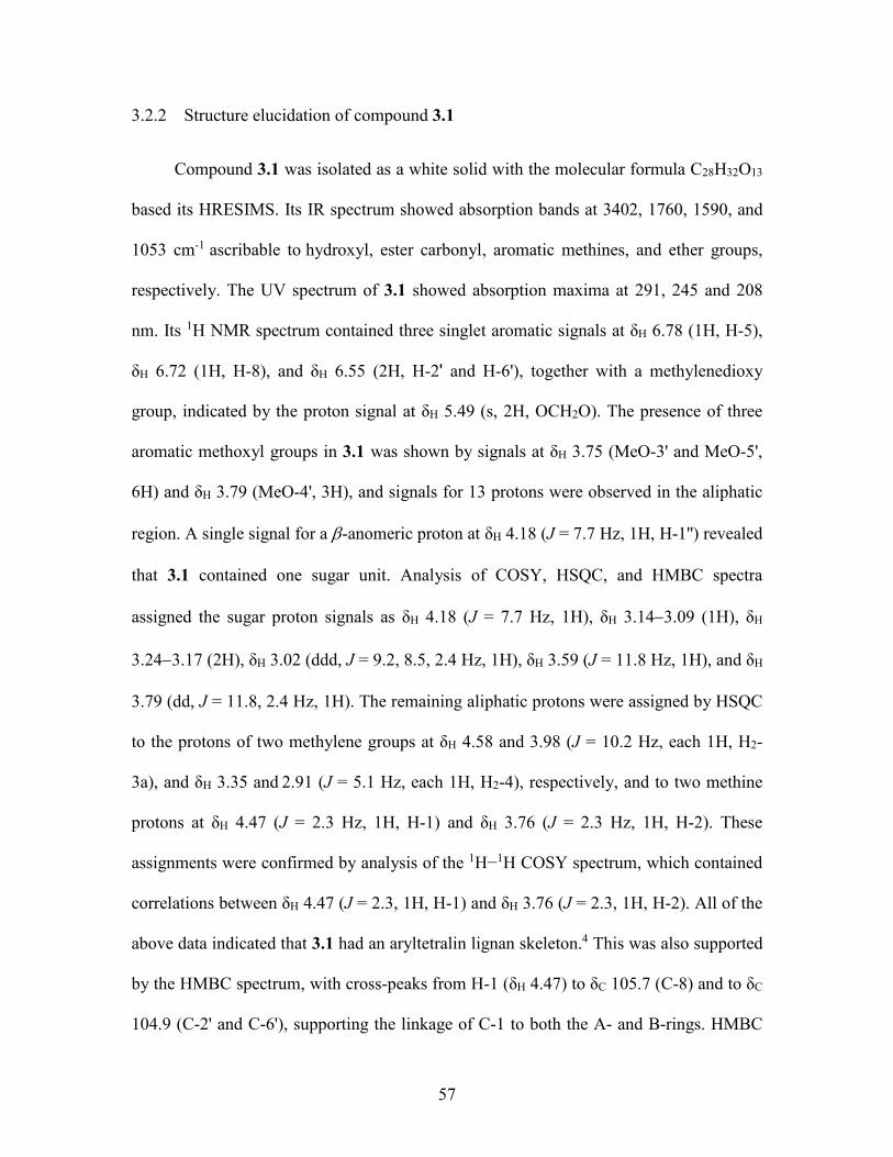
57
3.2.2 Structure elucidation of compound 3.1
Compound 3.1 was isolated as a white solid with the molecular formula C28H32O13
based its HRESIMS. Its IR spectrum showed absorption bands at 3402, 1760, 1590, and
1053 cm-1 ascribable to hydroxyl, ester carbonyl, aromatic methines, and ether groups,
respectively. The UV spectrum of 3.1 showed absorption maxima at 291, 245 and 208
nm. Its 1H NMR spectrum contained three singlet aromatic signals at δH 6.78 (1H, H-5),
δH 6.72 (1H, H-8), and δH 6.55 (2H, H-2' and H-6'), together with a methylenedioxy
group, indicated by the proton signal at δH 5.49 (s, 2H, OCH2O). The presence of three
aromatic methoxyl groups in 3.1 was shown by signals at δH 3.75 (MeO-3' and MeO-5',
6H) and δH 3.79 (MeO-4', 3H), and signals for 13 protons were observed in the aliphatic
region. A single signal for a -anomeric proton at δH 4.18 (J = 7.7 Hz, 1H, H-1'') revealed
that 3.1 contained one sugar unit. Analysis of COSY, HSQC, and HMBC spectra
assigned the sugar proton signals as δH 4.18 (J = 7.7 Hz, 1H), δH 3.143.09 (1H), δH
3.243.17 (2H), δH 3.02 (ddd, J = 9.2, 8.5, 2.4 Hz, 1H), δH 3.59 (J = 11.8 Hz, 1H), and δH
3.79 (dd, J = 11.8, 2.4 Hz, 1H). The remaining aliphatic protons were assigned by HSQC
to the protons of two methylene groups at δH 4.58 and 3.98 (J = 10.2 Hz, each 1H, H2-
3a), and δH 3.35 and 2.91 (J = 5.1 Hz, each 1H, H2-4), respectively, and to two methine
protons at δH 4.47 (J = 2.3 Hz, 1H, H-1) and δH 3.76 (J = 2.3 Hz, 1H, H-2). These
assignments were confirmed by analysis of the 1H−1H COSY spectrum, which contained
correlations between δH 4.47 (J = 2.3, 1H, H-1) and δH 3.76 (J = 2.3, 1H, H-2). All of the
above data indicated that 3.1 had an aryltetralin lignan skeleton.4 This was also supported
by the HMBC spectrum, with cross-peaks from H-1 (δH 4.47) to δC 105.7 (C-8) and to δC
104.9 (C-2' and C-6'), supporting the linkage of C-1 to both the A- and B-rings. HMBC
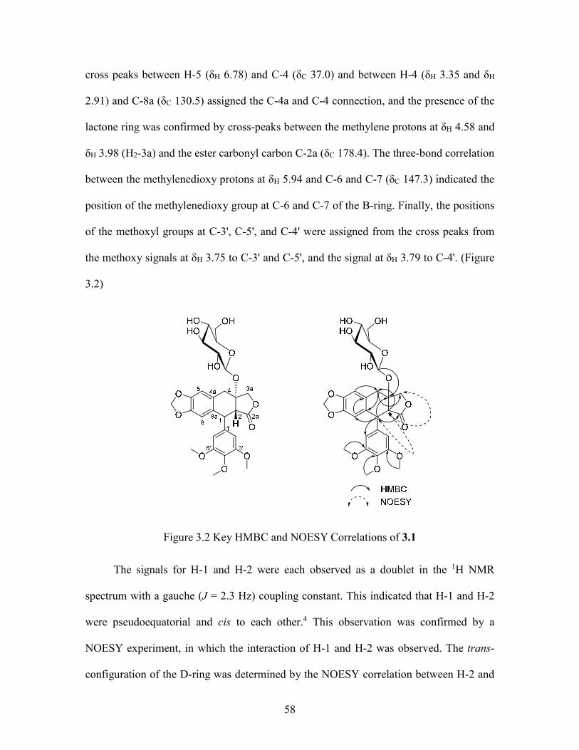
58
cross peaks between H-5 (δH 6.78) and C-4 (δC 37.0) and between H-4 (δH 3.35 and δH
2.91) and C-8a (δC 130.5) assigned the C-4a and C-4 connection, and the presence of the
lactone ring was confirmed by cross-peaks between the methylene protons at δH 4.58 and
δH 3.98 (H2-3a) and the ester carbonyl carbon C-2a (δC 178.4). The three-bond correlation
between the methylenedioxy protons at δH 5.94 and C-6 and C-7 (δC 147.3) indicated the
position of the methylenedioxy group at C-6 and C-7 of the B-ring. Finally, the positions
of the methoxyl groups at C-3', C-5', and C-4' were assigned from the cross peaks from
the methoxy signals at δH 3.75 to C-3' and C-5', and the signal at δH 3.79 to C-4'. (Figure
3.2)
Figure 3.2 Key HMBC and NOESY Correlations of 3.1
The signals for H-1 and H-2 were each observed as a doublet in the 1H NMR
spectrum with a gauche (J = 2.3 Hz) coupling constant. This indicated that H-1 and H-2
were pseudoequatorial and cis to each other.4 This observation was confirmed by a
NOESY experiment, in which the interaction of H-1 and H-2 was observed. The trans-
configuration of the D-ring was determined by the NOESY correlation between H-2 and

59
H-3a at δH 4.58 d (10.2). The 1R absolute configuration was established by the electronic
circular dichroism (ECD) spectrum of 3.1, which gave a positive Cotton effect at 288 nm
(Δε +1.02), consistent with the positive effect for other 1R lignans.4, 20-25 These data
combined with the relative configuration determined above allowed the R and S
configurations to be assigned for the C-2 and C-3 chiral centers, respectively.
Mild acid hydrolysis of 3.1 gave (+)--apopicropodophyllin (3.4a) and a sugar that
was identified as D-glucose by TLC and optical rotation comparison with a standard
sample. (Figure 3.3) The placement of glucose at C-3 was evident from the HMBC
correlation between the anomeric proton at δH 4.18 and the quaternary carbon at δC 83.6
(C-3). The configuration of the glucose unit was assigned as on the basis of the
coupling constant (J = 7.7 Hz) of the anomeric proton. The complete assignment of all
protons and carbons of 3.1 (Table 3.1) was accomplished by analysis of the COSY,
HSQC, HMBC, and NOESY spectra. Thus, compound 3.1 was assigned as (1R,2R,3S)-3-
O-(-D-glucopyranosyl)-3',4',5'-trimethoxy-6,7-methylenedioxy-1,9-cyclolignan-11,12-
olide, and named cleistanolide A.
Figure 3.3 Structure of Compound 3.4a
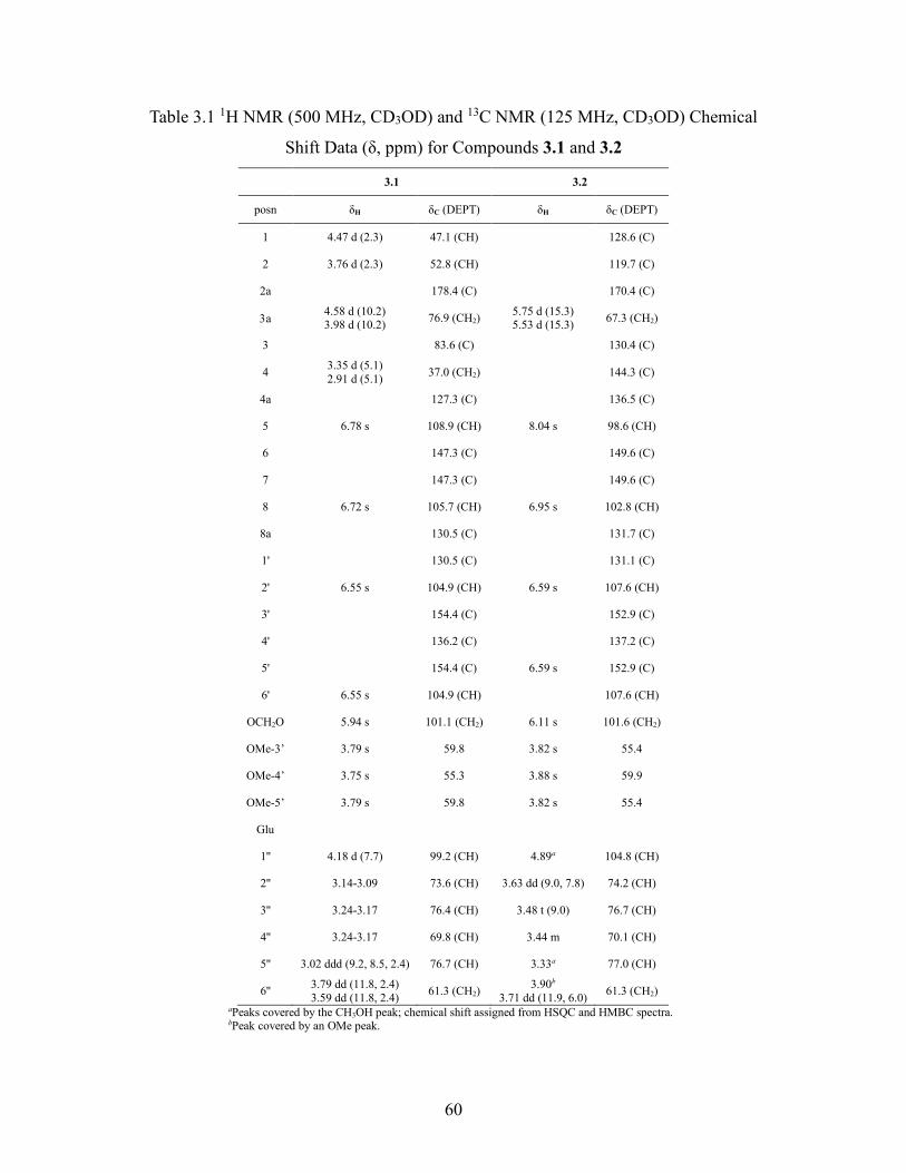
60
Table 3.1 1H NMR (500 MHz, CD3OD) and 13C NMR (125 MHz, CD3OD) Chemical
Shift Data (δ, ppm) for Compounds 3.1 and 3.2
3.1 3.2
posn δH δC (DEPT) δH δC (DEPT)
1 4.47 d (2.3) 47.1 (CH) 128.6 (C)
2 3.76 d (2.3) 52.8 (CH) 119.7 (C)
2a 178.4 (C) 170.4 (C)
a 4.58 d (10.2) 3.98 d (10.2)
76.9 (CH2) 5.75 d (15.3) 5.53 d (15.3)
67.3 (CH2)
3 83.6 (C) 130.4 (C)
4 3.35 d (5.1) 2.91 d (5.1)
37.0 (CH2) 144.3 (C)
4a 127.3 (C) 136.5 (C)
5 6.78 s 108.9 (CH) 8.04 s 98.6 (CH)
6 147.3 (C) 149.6 (C)
7 147.3 (C) 149.6 (C)
8 6.72 s 105.7 (CH) 6.95 s 102.8 (CH)
8a 130.5 (C) 131.7 (C)
1' 130.5 (C) 131.1 (C)
2' 6.55 s 104.9 (CH) 6.59 s 107.6 (CH)
3' 154.4 (C) 152.9 (C)
4' 136.2 (C) 137.2 (C)
5' 154.4 (C) 6.59 s 152.9 (C)
6' 6.55 s 104.9 (CH) 107.6 (CH)
OCH2O 5.94 s 101.1 (CH2) 6.11 s 101.6 (CH2)
OMe-3’ 3.79 s 59.8 3.82 s 55.4
OMe-4’ 3.75 s 55.3 3.88 s 59.9
OMe-5’ 3.79 s 59.8 3.82 s 55.4
Glu
1'' 4.18 d (7.7) 99.2 (CH) 4.89a 104.8 (CH)
2'' 3.14-3.09 73.6 (CH) 3.63 dd (9.0, 7.8) 74.2 (CH)
3'' 3.24-3.17 76.4 (CH) 3.48 t (9.0) 76.7 (CH)
4'' 3.24-3.17 69.8 (CH) 3.44 m 70.1 (CH)
5'' 3.02 ddd (9.2, 8.5, 2.4) 76.7 (CH) 3.33a 77.0 (CH)
6'' 3.79 dd (11.8, 2.4) 3.59 dd (11.8, 2.4)
61.3 (CH2) 3.90b
3.71 dd (11.9, 6.0) 61.3 (CH2)
aPeaks covered by the CH3OH peak; chemical shift assigned from HSQC and HMBC spectra. bPeak covered by an OMe peak.
61
As indicated by the experiment described above, cleistanolide A is sensitive to
conversion to its ,-unsaturated derivative 3.4 a, and a sample of 3.1 left in
MeOH/CHCl3 for a few days underwent decomposition, most likely caused by traces of
HCl in the CHCl3. Attempted enzymatic hydrolysis of 3.1 did not proceed under normal
conditions, so the aglycone could not be isolated.
3.2.3 Structure elucidation of compound 3.2
Compound 3.2 had the molecular formula of C28H29O13, based on its HRESIMS
data. Its IR spectrum showed absorptions for OH and ester carbonyl groups at 3400 and
1734 cm-1, respectively. Its 1H NMR spectrum was similar to that obtained for 3.1, and its
UV absorption maxima at 260, 315, and 351 nm indicated the presence of a naphthalene
nucleus,13 suggesting that 3.2 was an arylnaphthalide lignan. The 1H NMR spectrum of
3.2 exhibited signals for two aromatic protons at δH 8.04 and 6.95 (s, each 1H, H-5 and
H-8) and for an oxymethylene group at δH 5.75 and 5.53 (J = 15.4 Hz, each 1H, H2-3a),
but lacked the signals for the two methine protons at C-1 and C-2 and the methylene
protons at C-4 observed in 3.1. These facts confirmed the presence of a naphthalene unit
in 3.2. In the HMBC spectrum, the correlation from H-5 (δH 8.04) and H2-3a (δH 5.75 and
5.53) to δC 144.3 (C-4) confirmed the linkage of C-4 to both the B- and D-rings, while
the correlations of δH 6.59 (s, 1H, H-2' or/and H-6') and δH 6.95 (H-8) to δC 128.6 (C-1)
connected the A- and B-rings to C-1. A comparison of the NMR spectra of 3.2 with those
of dehydropodophyllotoxin (3.2a) suggested that compound 3.2 is a glycosylated
derivative of 3.2a.26 The HMBC correlation between the anomeric proton signal at δH
4.89 (H-1'') and δC 144.3 (C-4) confirmed that the sugar was located at C-4 of the
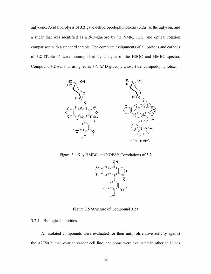
62
aglycone. Acid hydrolysis of 3.2 gave dehydropodophyllotoxin (3.2a) as the aglycon, and
a sugar that was identified as a -D-glucose by 1H NMR, TLC, and optical rotation
comparison with a standard sample. The complete assignments of all protons and carbons
of 3.2 (Table 1) were accomplished by analysis of the HSQC and HMBC spectra.
Compound 3.2 was thus assigned as 4-O-(-D-glucopyranosyl)-dehydropodophyllotoxin.
Figure 3.4 Key HMBC and NOESY Correlations of 3.2
Figure 3.5 Structure of Compound 3.2a
3.2.4 Biological activities
All isolated compounds were evaluated for their antiproliferative activity against
the A2780 human ovarian cancer cell line, and some were evaluated in other cell lines
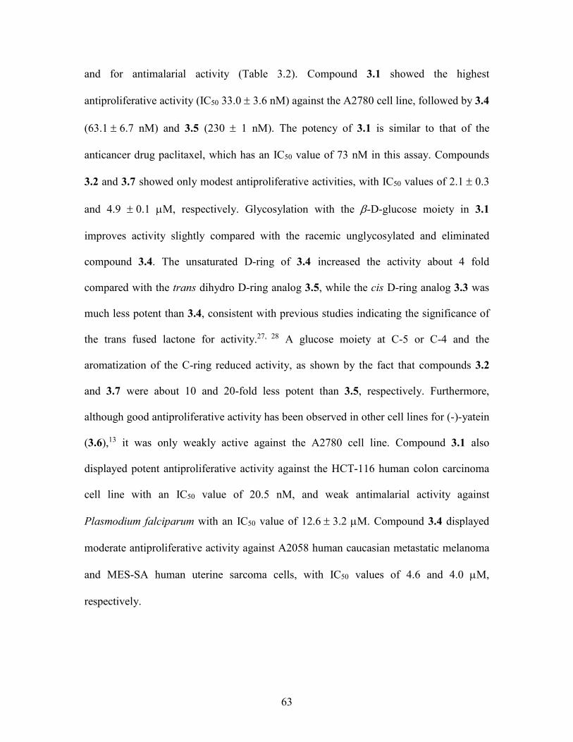
63
and for antimalarial activity (Table 3.2). Compound 3.1 showed the highest
antiproliferative activity (IC50 33.0 3.6 nM) against the A2780 cell line, followed by 3.4
(63.16.7 nM) and 3.5 (230 1 nM). The potency of 3.1 is similar to that of the
anticancer drug paclitaxel, which has an IC50 value of 73 nM in this assay. Compounds
3.2 and 3.7 showed only modest antiproliferative activities, with IC50 values of 2.10.3
and 4.9 0.1 M, respectively. Glycosylation with the -D-glucose moiety in 3.1
improves activity slightly compared with the racemic unglycosylated and eliminated
compound 3.4. The unsaturated D-ring of 3.4 increased the activity about 4 fold
compared with the trans dihydro D-ring analog 3.5, while the cis D-ring analog 3.3 was
much less potent than 3.4, consistent with previous studies indicating the significance of
the trans fused lactone for activity.27, 28 A glucose moiety at C-5 or C-4 and the
aromatization of the C-ring reduced activity, as shown by the fact that compounds 3.2
and 3.7 were about 10 and 20-fold less potent than 3.5, respectively. Furthermore,
although good antiproliferative activity has been observed in other cell lines for (-)-yatein
(3.6),13 it was only weakly active against the A2780 cell line. Compound 3.1 also
displayed potent antiproliferative activity against the HCT-116 human colon carcinoma
cell line with an IC50 value of 20.5 nM, and weak antimalarial activity against
Plasmodium falciparum with an IC50 value of 12.6 3.2 M. Compound 3.4 displayed
moderate antiproliferative activity against A2058 human caucasian metastatic melanoma
and MES-SA human uterine sarcoma cells, with IC50 values of 4.6 and 4.0 M,
respectively.
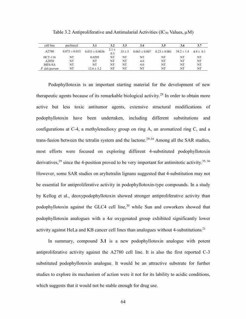
64
Table 3.2 Antiproliferative and Antimalarial Activities (IC50 Values, M)
cell line paclitaxel 3.1 3.2 3.3 3.4 3.5 3.6 3.7
A2780 0.073 ± 0.015 0.033 0.0036 2.1
03 35 5 0.0630.007 0.23 0.001 34.2 ± 1.4 4.9 0.1
HCT-116 NT 0.0205 NT NT NT NT NT NT A2058 NT NT NT NT 4.6 NT NT NT
MES-SA NT NT NT NT 4.0 NT NT NT P. falciparum NT 12.6 3.2 NT NT NT NT NT NT
Podophyllotoxin is an important starting material for the development of new
therapeutic agents because of its remarkable biological activity.29 In order to obtain more
active but less toxic antitumor agents, extensive structural modifications of
podophyllotoxin have been undertaken, including different substitutions and
configurations at C-4, a methylenedioxy group on ring A, an aromatized ring C, and a
trans-fusion between the tetralin system and the lactone.29-34 Among all the SAR studies,
most efforts were focused on exploring different 4-substituted podophyllotoxin
derivatives,29 since the 4-position proved to be very important for antimitotic activity.35, 36
However, some SAR studies on aryltetralin lignans suggested that 4-substitution may not
be essential for antiproliferative activity in podophyllotoxin-type compounds. In a study
by Kellog et al., deoxypodophyllotoxin showed stronger antiproliferative activity than
podophyllotoxin against the GLC4 cell line,30 while Sun and coworkers showed that
podophyllotoxin analogues with a 4 oxygenated group exhibited significantly lower
activity against HeLa and KB cancer cell lines than analogues without 4-substitutions.21
In summary, compound 3.1 is a new podophyllotoxin analogue with potent
antiproliferative activity against the A2780 cell line. It is also the first reported C-3
substituted podophyllotoxin analogue. It would be an attractive substrate for further
studies to explore its mechanism of action were it not for its lability to acidic conditions,
which suggests that it would not be stable enough for drug use.
65
3.3 Experimental Section
3.3.1 General experimental procedures
IR and UV spectra were measured on MIDAC M-series FTIR and Shimadzu UV-
1201 spectrophotometers, respectively. 1H and 13C NMR spectra were recorded on a
Bruker Avance 500 spectrometer in CD3OD (with CD3OD as reference) and CDCl3 (with
CDCl3 as reference). Mass spectra were obtained on an Agilent 6220 mass spectrometer.
Open column chromatography was performed using Sephadex LH-20, and solid phase
extraction was performed using C18 cartridges. Semi-preparative HPLC was performed
using Shimadzu LC-10AT pumps coupled with a semi-preparative Phenomenex C18
column (5 μm, 250 × 10 mm), a Shimadzu SPD M10A diode array detector, and a SCL-
10A system controller. All isolated compounds were purified to 95% purity or better, as
judged by HPLC (both UV and ELSD detection) before determining bioactivity.
3.3.2 Plant material
Leaves of Cleistanthus boivinianus (Baill.) Müll. Arg. (collection: Stéphan
Rakotonandrasana et al. 1036) were collected at an elevation of 51 m from a 3 m tall tree
with yellow flowers. Collection was made 5 km north-east of the village of
Marivorahona, PK 123, Andohanantsohihy, in a mosaic of dry forest and savanna trees;
coordinates 13°06'37"S 049°09'39"E. Collection was made by Stéphan Rakotonandrasana
with assistance from R. Randrianaivo, R. Rakotonandrasana, C. Claude, V. Benjara, and
M. Modeste. Duplicate voucher specimens are deposited at the Centre National
d'Application des Recherches Pharmaceutiques (CNARP), the Herbarium of the Parc
Botanique et Zoologique de Tsimbazaza, Antananarivo, Madagascar (TAN), the Missouri

66
Botanical Garden, St. Louis, Missouri (MO), and the Museum National d'Histoire
Naturelle in Paris, France (P).
3.3.3 Extraction and isolation
A ground sample of Cleistanthus boivinianus leaves (250 g) was extracted with
EtOH at room temperature to yield 33.4 g of crude EtOH extract designated MG4031. A
total of 5.88 g of this extract was made available to Virginia Tech. An active EtOAc-
soluble fraction obtained from liquid-liquid partition of the extract (100 mg) was
subjected to dereplication studies using size-exclusion chromatography, reverse phase
HPLC coupled with bioassay, high-resolution ESIMS, 1H NMR, and a database search
using the online Dictionary of Natural Product (DNP). The results indicated that the
extract contained at least one new bioactive compound, and so a 3.0 g sample was
investigated. The crude EtOH extract was dissolved in 90% aq. MeOH (300 mL), and
extracted with hexanes (3 × 200 mL)., and extracted with hexanes (3 × 200 mL).
Evaporation of the hexane-soluble fraction afforded 271 mg of residue. The 90% aq.
MeOH layer was then evaporated, suspended in H2O (300 mL), and extracted with
EtOAc (3 × 200 mL) to yield 640 mg of an EtOAc-soluble fraction with an IC50 value of
0.19 µg/mL. The EtOAc fraction was subjected to Sephadex LH-20 open column
chromatography (CH2Cl2:MeOH, 1:1) to give 6 fractions. The most active fraction Fr 3
(IC50 0.11 µg/mL) was then divided into 3 sub-fractions by C18 solid phase extraction by
using 40% aq. MeOH (Fr 3-1), 70% Aq. MeOH (Fr 3-2), and 100% MeOH (Fr 3-3).
Further purification of the most active sub-fraction Fr 3-3 (IC50 0.11 µg/mL) by HPLC on
a C18 column with a solvent gradient from H2O:CH3CN, 60:40 to 50:50 from 0 to 17
min, to 40:60 from 17 to 22 min, to 40:60 from 22 to 27 min and ending with 100%
67
CH3CN from 27 to 35 min yielded compounds 3.3 (1.2 mg, tR 23.5 min), 3.5 (1.5 mg, tR
24.5 min), and 3.6 (2.1 mg, tR 26.5 min). Purification of the sub-fraction Fr 3-2 (IC50 0.1
µg/mL) by C18 HPLC furnished 8 fractions (solvent gradient from H2O:CH3CN, 70:30 to
62:38 from 0 to 15 min, and ending with 100% CH3CN from 15 to 24 min), among which
the 3rd (tR 12 min), 6th (tR 14.5 min), and 7th (tR 15.5 min) fractions were active with
IC50 value of 1.5, 0.1, and 1.2 µg/mL, respectively. Further purification of the 3rd fraction
by HPLC on a silica gel column with a solvent gradient from CHCl3:MeOH, 95:5 to
90:10 from 0 to 5 min, to 88:12 from 5 to 12 min, and ending with a 100% MeOH wash
from 13 to 20 min, yielded compound 3.7 (2.8 mg, tR 13.5 min). Further purification of
the 6th fraction by HPLC on a silica gel column with a solvent gradient from
CHCl3:MeOH, 95:5 to 90:10 from 0 to 5 min, to 80:20 from 5 to 10 min, to 70:30 from
10 to 15 min, and ending with a 100% MeOH wash from 16 to 25 min, yielded
compound 3.4 (5 mg, tR 6 min) and 3.1 (19 mg, tR 14 min). Further purification of the 7th
fraction by HPLC on a diol column with a solvent gradient from CHCl3:MeOH, 100:0 to
95:5 from 0 to 5 min, to 90:10 from 5 to 15 min, to 87.5:12.5 from 15 to 20 min, and
ending with a 100% MeOH wash from 20 to 25 min, yielded compound 3.2 (3 mg, tR
22.5 min).
3.3.4 Cleisthanolide A (3.1)
Amorphous powder; []21D +120 (c 0.1, MeOH); CD (c 0.031, MeOH) λmax ():
210 (+22.59), 234 (+5.98), 248 (-1.33), 288 (+1.02) nm; UV (MeOH) λmax(log ε): 291
(3.19), 245 (3.7), 208 (4.4) nm; IR (film) νmax 3402, 2912, 1760, 1590, 1053 cm-1; 1H and
13C NMR data, see Table 3.1; HRESIMS m/z [M+Na]+ 599.1741 (calcd for
C28H32NaO13+
, 599.1735)
68
3.3.5 4-O-(-D-Glucopyranosyl)-dehydropodophyllotoxin (3.2)
Amorphous powder, []21D -14 (c 0.22, MeOH); UV (MeOH) λmax (log ε): 351
(3.69), 315 (4.0), 260 (4.7) nm; IR (film) νmax: 3400, 2920, 1734, 1645, 1070 cm-1; 1H
and 13C NMR data, see Table 3.1; HRESIMS m/z [M+H]+ 573.1635 (calcd for
C28H29O13+
, 599.1603)
3.3.6 (±)--Apopicropodophyllin (3.4)
Amorphous powder, []21D 0 (c 0.5, CHCl3). Its 1H and 13C NMR spectra were
identical with literature data.13-15
3.3.7 Antiproliferative bioassay
The A2780 ovarian cancer cell line antiproliferative bioassay was performed at
Virginia Tech as previously reported.37, 38 The A2780 cell line is a drug-sensitive ovarian
cancer cell line.39 Paclitaxel was used as the positive control; it had an IC50 value of 0.073
± 0.015 M.
3.3.8 Antimalarial bioassay
The effect of each compound on parasite growth of the Dd2 strain of P. falciparum
was measured in a 72 h growth assay in the presence of compound as described
previously.40-42
3.3.9 Acid hydrolysis of compounds 3.1 and 3.2
Compound 3.1 (2 mg) was dissolved in dilute citrate/phosphate buffer (pH 7.0, 4
mL) and -D-glucosidase (4 mg) was added. The mixture was incubated for 2 weeks at

69
37 °C; no reaction was observed. The reaction mixture was then adjusted to pH 5.0 with
dilute HCl and stirred at 37°C overnight. The mixture was then briefly heated to boiling,
extracted with EtOAc (3x10 mL) and both the organic and the water layers were
evaporated to dryness under reduced pressure. The structure of the white powder (1.2
mg) derived from the organic layer was determined to be 3.4a by its 1H NMR spectrum
and optical rotation ([]21D: +86, c 1.2, CHCl3; lit. +96.813). The semi-solid carbohydrate
from the water layer (0.5 mg) was dissolved in 2 mL of water and kept overnight before
determination of its optical rotation. Compound 3.2 (1 mg) was dissolved in dilute HCl
(pH 5.0, 2 mL) and stirred at 37 °C overnight. The reaction mixture was extracted with
EtOAc (3x10 mL) and both the organic and the water layers were evaporated to dryness
under reduced pressure. The structure of the dried powder from the organic layer was
determined to be 3.2a by its 1H NMR spectrum. TLC of the sugar from both 3.1 and 3.2
on a silica gel plate with CHCl3:MeOH:H2O = 15:6:1 showed that it had an identical Rf
value (0.23) to glucose. The isolated glucose had []21D (c 0.5, H2O) values of +41.9 and
+43.2, respectively.
70
3.4 References
1. Rakotondraibe, H. L.; Graupner, P. R.; Xiong, Q.; Olson, M.; Wiley, J. D.; Krai,
P.; Brodie, P., J.; Callmander, M. W.; Rakotobe, E.; Ratovoson, F.; Rasamison, V. E.;
Cassera, M. B.; Hahn, D. R.; Kingston, D. G. I.; Fotso, S. Neolignans and Other
Metabolites from Ocotea cymosa from the Madagascar Rain Forest and their Biological
Activities. J. Nat. Prod. 2015, 78, doi: 10.1021/np5008153.
2. Kingston, D. G. I. A natural love of natural products. J. Org. Chem. 2008, 73,
3975-3984.
3. Pinho, P. M. M.; Kijjoa, A. Chemical constitutents of the plants of the genus
Cleistanthus and their biological activity. Phytochem. Rev. 2007, 6, 175-182.
4. Thanh, V. T. T.; Pham, V. C.; Mai, H. D. T.; Litaudon, M.; Gueritte, F.; Retailleau,
P.; Nguyen, V. H.; Chau, V. M. Cytotoxic lignans from fruits of Cleistanthus
indochinensis: Synthesis of cleistantoxin derivatives. J. Nat. Prod. 2012, 75, 1578-1583.
5. Trinh, T. T. V.; Pham, V. C.; Doan, T. M. H.; Nguyen, V. H.; Chau, V. M.;
Litaudon, M.; Gueritte, F. Cytotoxic aryltetralin lignans from fruits of Cleistanthus
indochinensis. Planta Med. 2014, 80, 695-702.
6. Thanh, V. T. T.; Pham, V. C.; Nguyen, H. H.; Mai, H. D. T.; Minh, H. N. T.;
Nguyen, V. H.; Litaudon, M.; Gueritte, F.; Chau, V. M. Cleistanone: A triterpenoid from
Cleistanthus indochinensis with a new carbon skeleton. Eur. J. Org. Chem. 2011, 2011,
4108-4111.
7. Parasuraman, S.; Raveendran, R. Effect of cleistanthin A and B on adrenergic and
cholinergic receptors. Pharmacogn. Mag. 2011, 7, 243-247.
8. Pinho, P. M.; Naengchomnong, W.; Kijjoa, A.; Nazareth, N.; Silva, A. M. S.;

71
Eaton, G.; Herz, W. An unusual glucoside from Cleistanthus gracilis. Phytochemistry
2006, 67, 1789-1792.
9. Ramesh, C.; Ravindranath, N.; Ram, T. S.; Das, B. Studies on phytochemicals -
42. Arylnaphthalide lignans from Cleistanthus collinus. Chem. Pharm. Bull. 2003, 51,
1299-1300.
10. Sastry, K. V.; Rao, E. V.; Buchanan, J. G.; Sturgeon, R. J. Cleistanthoside B, a
diphyllin glycoside from Cleistanthus patulus heartwood. Phytochemistry 1987, 26,
1153-1154.
11. Pradheepkumar, C. P.; Shanmugam, G. Anticancer potential of cleistanthin A
isolated from the tropical plant Cleistanthus collinus. Oncol. Res. 1999, 11, 225-232.
12. Broomhead, A. J.; Dewick, P. M. Aryltetralin lignans from Linum flavum and
Linum capitatum. Phytochemistry 1990, 29, 3839-3844.
13. Novelo, M.; Cruz, J. G.; Hernandez, L.; Pereda-Miranda, R.; Chai, H.; Mar, W.;
Pezzuto, J. M. Chemical studies on Mexican Hyptis species. VI. Biologically active
natural products from Mexican medicinal plants. II. Cytotoxic constituents from Hyptis
verticillata. J. Nat. Prod. 1993, 56, 1728-1736.
14. Pelter, A.; Ward, R. S.; Li, Q.; Pis, J. An asymmetric synthesis of
isopodophyllotoxin. Tetrahedron: Asymmetry 1994, 5, 909-910.
15. Andrews, R. C.; Teague, S. J.; Meyers, A. I. Asymmetric Total Synthesis of (-)-
Podophyllotoxin+. J. Am. Chem. Soc. 1988, 110, 7854-7858.
16. San Feliciano, A.; Medarde, M.; Lopez, J. L.; Puebla, P.; Miguel del Corral, J. M.;
Barrero, A. F. Lignans from Juniperus thurifera. Phytochemistry 1989, 28, 2863-2866.
17. Carpenter, C. D.; O'Neill, T.; Picot, N.; Johnson, J. A.; Robichaud, G. A.; Webster,

72
D.; Gray, C. A. Anti-mycobacterial natural products from the Canadian medicinal plant
Juniperus communis. J. Ethnopharmacol. 2012, 143, 695-700.
18. Tanoguchi, M.; Arimoto, M.; Saika, H.; Yamaguchi, H. Studies on the constituents
of the seeds of Hernandia ovigera L. VI. Isolation and structural determination of three
lignans. Chem. Pharm. Bull. 1987, 35, 4162-4168.
19. Liu, Y.; Rakotondraibe, H.; Brodie, P. J.; Wiley, J. D.; Cassera, M. B.; Goetz, M.;
Kingston, D. G. I. Antiproliferative and antimalarial sesquiterpene lactones from
Piptocoma antillana from Puerto Rico. Nat. Prod. Commun. 2014, 9, 1403-1406.
20. Yamaguchi, H.; Arimoto, M.; Tanoguchi, M.; Ishida, T.; Inoue, M. Studies on the
constituents of the seeds of Hernandia ovigera L. III. Structures of two new lignans.
Chem. Pharm. Bull. 1982, 30, 3212-3218.
21. Sun, Y. J.; Li, Z. L.; Chen, H.; Liu, X. Q.; Zhou, W.; Hua, H. M. Three new
cytotoxic aryltetralin lignans from Sinopodophyllum emodi. Bioorg. Med. Chem. Lett.
2011, 21, 3794-3797.
22. Zhao, C.; Nagatsu, A.; Hatano, K.; Shirai, N.; Kato, S.; Ogihara, Y. New lignan
glycosides from Chinese medicinal plant, Sinopodophillum emodi. Chem. Pharm. Bull.
2003, 51, 255-261.
23. Gu, J. Q.; Park, E. J.; Totura, S.; Riswan, S.; Fong, H. H. S.; Pezzuto, J. M.;
Kinghorn, A. D. Constituents of the twigs of Hernandia ovigera that inhibit the
transformation of JB6 murine epidermal cells. J. Nat. Prod. 2002, 65, 1065-1068.
24. Klyne, W.; Stevenson, R.; Swan, R. J. Optical rotatory dispersion. XXVIII.
Absolute configuration of otobain and derivatives. J. Chem. Soc. C 1966, 893-896.
25. Swan, R. J.; Klyne, W.; MacLean, H. Optical rotatory dispersion studies. XLI.
73
The absolute configuration of plicatic acid. Can. J. Chem. 1967, 45, 319-324.
26. Atta-ur-Rahman.; Ashraf, M.; Choudhary, M. I.; Habib-ur-Rehman.; Kazmi, M.
H. Antifungal aryltetralin lignans from leaves of Podophyllum hexandrum.
Phytochemistry 1995, 40, 427-431.
27. Rai, K. M. L.; Basavaraju, Y. B.; Sadashivamurthy, B. Biological assay and
antimitotic activity of novel analogues of β-apopicropodophyllin. Indian J. Pharm. Sci.
2007, 69, 116-118.
28. Gensler, W. J.; Gatsonis, C. D. The podophyllotoxin—picropodophyllin
equilibrium. J. Org. Chem. 1966, 31, 3224-3227.
29. Liu, Y. Q.; Yang, L.; Tian, X. Podophyllotoxin: current perspectives. Curr. Bioact.
Compd. 2007, 3, 37-66.
30. Middel, O.; Woerdenbag, H. J.; van Uden, W.; van Oeveren, A.; Jansen, J. F. G.
A.; Feringa, B. L.; Konings, A. W. T.; Pras, N.; Kellogg, R. M. Synthesis and cytotoxicity
of novel lignans. J. Med. Chem. 1995, 38, 2112-2118.
31. Lee, K. H.; Beers, S. A.; Mori, M.; Wang, Z. Q.; Kuo, Y. H.; Li, L.; Liu, S. Y.;
Chang, J. Y.; Han, F. S.; Cheng, Y. C. Antitumor agents. 111. New 4-hydroxylated and 4-
halogenated anilino derivatives of 4'-demethylepipodophyllotoxin as potent inhibitors of
human DNA topoisomerase II. J. Med. Chem. 1990, 33, 1364-1368.
32. Dantzig, A.; LaLonde, R. T.; Ramdayal, F.; Shepard, R. L.; Yanai, K.; Zhang, M.
Cytotoxic responses to aromatic ring and configurational variations in α-conidendrin,
podophyllotoxin, and sikkimotoxin derivatives. J. Med. Chem. 2001, 44, 180-185.
33. Lee, K. H. Current developments in the discovery and deign of nw dug cndidates
from pant natural product leads. J. Nat. Prod. 2004, 67, 273-283.
74
34. Srivastava, V.; Negi, A. S.; Kumar, J. K.; Gupta, M. M.; Khanuja, S. P. S. Plant-
based anticancer molecules: A chemical and biological profile of some important leads.
Bioorg. Med. Chem. 2005, 13, 5892-5908.
35. Liu, Y. Q.; Tian, J.; Qian, K.; Zhao, X. B.; Morris-Natschke, S. L.; Yang, L.; Nan,
X.; Tian, X.; Lee, K. H. Recent progress on C-4-modified podophyllotoxin analogs as
potent antitumor agents. Med. Res. Rev. 2015, 35, 1-62.
36. Gordaliza, M.; Castro, M. A.; Miguel Del Corral, J. M.; San Feliciano, A.
Antitumor properties of podophyllotoxin and related compounds. Curr. Pharm. Des.
2000, 6, 1811-1839.
37. Cao, S.; Brodie, P. J.; Miller, J. S.; Randrianaivo, R.; Ratovoson, F.; Birkinshaw,
C.; Andriantsiferana, R.; Rasamison, V. E.; Kingston, D. G. I. Antiproliferative xanthones
of terminalia calcicola from the Madagascar rain forest. J. Nat. Prod. 2007, 70, 679-681.
38. Pan, E.; Harinantenaina, L.; Brodie, P., J.; Miller, J. S.; Callmander, M. W.;
Rakotonandrasana, S.; Rakotobe, E.; Rasamison, V. E.; Kingston, D. G. I. Four
Diphenylpropanes and a Cycloheptadibenzofuran from Bussea sakalava from the
Madagascar Dry Forest. J. Nat. Prod. 2010, 73, 1792-1795.
39. Louie, K. G.; Behrens, B. C.; Kinsella, T. J.; Hamilton, T. C.; Grotzinger, K. R.;
McKoy, W. M.; Winker, M. A.; Ozols, R. F. Radiation survival parameters of
antineoplastic drug-sensitive and -resistant human ovarian cancer cell lines and their
modification by buthionine sulfoximine. Cancer Res. 1985, 45, 2110-2115.
40. Bennett, T. N.; Paguio, M.; Gligorijevic, B.; Seudieu, C.; Kosar, A. D.; Davidson,
E.; Roepe, P. D. Novel, rapid, and inexpensive cell-based quantification of antimalarial
drug efficacy. Antimicrob. Agents Chemother. 2004, 48, 1807-1810.
75
41. Smilkstein, M.; Sriwilaijaroen, N.; Kelly, J. X.; Wilairat, P.; Riscoe, M. Simple
and inexpensive fluorescence-based technique for high-throughput antimalarial drug
screening. Antimicrob. Agents Chemother. 2004, 48, 1803-1806.
42. Rakotondraibe, L. H.; Graupner, P. R.; Xiong, Q.; Olson, M.; Wiley, J. D.; Krai,
P.; Brodie, P. J.; Callmander, M. W.; Rakotobe, E.; Ratovoson, F.; Rasamison, V. E.;
Cassera, M. B.; Hahn, D. R.; Kingston, D. G. I.; Fotso, S. Neolignans and other
metabolites from Ocotea cymosa from the Madagascar rain forest and their biological
activities. J. Nat. Prod. 2015, Ahead of Print.
4 Antiproliferative and Antimalarial Sesquiterpene Lactones from Piptocoma antillana from Puerto Rico
This chapter is a slightly expanded version of a published article.1 Attributions
from co-authors of the articles are described as follows in the order of the names listed.
The author of this disseration (Yixi Liu) conducted the isolation and structural elucidation
part of the titled compounds, and drafted the manuscript. Dr. Harinantenaina was a
mentor for this work, provided invaluable advice and hints for the structural elucidation
of the compounds, and also proofread the manuscript before submission. Mrs. Peggy
Brodie performed the A2780 bioassay on the isolated fractions and compounds. Dr.
Jessica D. Wiley and Dr. Maria B. Cassera performed the Dd2 bioassay. Dr. Michael
Goetz from Natural Products Discovery Institute provided the plant extract. Dr. David G.
I. Kingston was a mentor for this work and the corresponding author for the published
article. He provided critical suggestions for this work and crucial revisions to the
manuscript.
4.1 Introduction
The genus Piptocoma, a member of the Asteraceae family, was first discovered in
the West Indies, and consisted initially of only three species. The genus was expanded in
2000, when Pruski described the Central and South American plant species from the
genus Pollalesta as cogeneric with Piptocoma, and this raised the number of Piptocoma
species to eighteen.2 A literature search demonstrated that the only reports on the
chemical constituents of the genus Piptocoma were in the last two years, describing the
isolation of cytotoxic sesquiterpene lactones, phenylpropanol coumarates, and flavonoids
from P. rufescens.3, 4
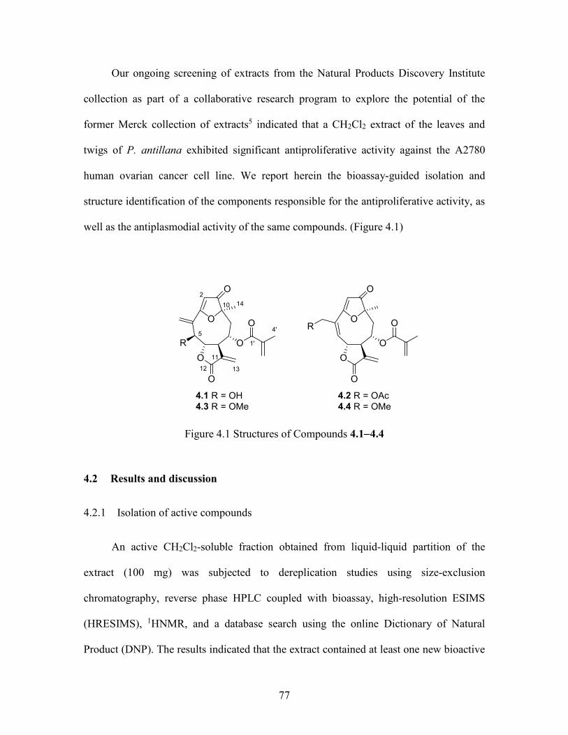
77
Our ongoing screening of extracts from the Natural Products Discovery Institute
collection as part of a collaborative research program to explore the potential of the
former Merck collection of extracts5 indicated that a CH2Cl2 extract of the leaves and
twigs of P. antillana exhibited significant antiproliferative activity against the A2780
human ovarian cancer cell line. We report herein the bioassay-guided isolation and
structure identification of the components responsible for the antiproliferative activity, as
well as the antiplasmodial activity of the same compounds. (Figure 4.1)
4.2 R = OAc4.4 R = OMe
4.1 R = OH4.3 R = OMe
O
R
O
O
O
O
O
2
5
10
13
11
14
1'
4'
O
O
O
O
O
OR
12
Figure 4.1 Structures of Compounds 4.14.4
4.2 Results and discussion
4.2.1 Isolation of active compounds
An active CH2Cl2-soluble fraction obtained from liquid-liquid partition of the
extract (100 mg) was subjected to dereplication studies using size-exclusion
chromatography, reverse phase HPLC coupled with bioassay, high-resolution ESIMS
(HRESIMS), 1HNMR, and a database search using the online Dictionary of Natural
Product (DNP). The results indicated that the extract contained at least one new bioactive
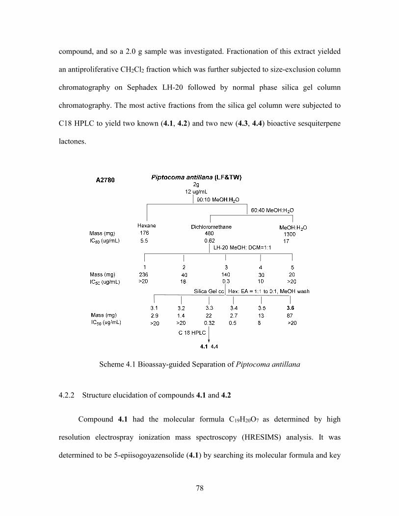
78
compound, and so a 2.0 g sample was investigated. Fractionation of this extract yielded
an antiproliferative CH2Cl2 fraction which was further subjected to size-exclusion column
chromatography on Sephadex LH-20 followed by normal phase silica gel column
chromatography. The most active fractions from the silica gel column were subjected to
C18 HPLC to yield two known (4.1, 4.2) and two new (4.3, 4.4) bioactive sesquiterpene
lactones.
Scheme 4.1 Bioassay-guided Separation of Piptocoma antillana
4.2.2 Structure elucidation of compounds 4.1 and 4.2
Compound 4.1 had the molecular formula C19H20O7 as determined by high
resolution electrospray ionization mass spectroscopy (HRESIMS) analysis. It was
determined to be 5-epiisogoyazensolide (4.1) by searching its molecular formula and key
79
1H NMR spectroscopic features in the DNP database and comparison of its full 1H NMR
spectrum with reported literature data.6-8 Compound 4.2 was identified as 1-oxo-3,10-
epoxy-8-(2-methylacryloxy)-15-acetoxy-germacra-2,4,11(13)-trien-6(12)-olide (15-
acetylgoyazensolide) by comparison of its spectroscopic data with values reported in the
literature.9-11
4.2.3 Structure elucidation of compound 4.3
Compound 4.3 was obtained as an amorphous white powder and had the molecular
formula C20H22O7 as indicated by HRESIMS analysis (m/z: 375.1461 [M+H]+, calcd. for
C20H23O7+
, 375.1438). Its IR spectrum showed absorptions characteristic of an ,-
unsaturated -lactone (1765 and 1640 cm-1), -lactones typically have IR absorptions at
about 1765 cm-1 due to the carbonyl resonance, and at 1640 cm-1 from the C=C stretch;
an ,-unsaturated ester (1710 and 1645 cm-1), the appearance of the ester carbonyl
stretch at 1710 cm-1 is due to the normal ester carbonyl resonance at 1740 cm-1 reduced to
1710 cm-1 by conjugation with the double bond; as well as a dihydrofuran-3-one ring
(1709 and 1582 cm-1), the appearance of the carbonyl stretch at 1709 cm-1 is due to the
normal cyclopentanone carbonyl stretch at 1740 cm-1 reduced to 1709 by conjugation
with the double bond.3, 8, 9, 12 The similarity of the IR spectroscopic data of 4.3 with those
of 4.1, in conjunction with the UV absorption characteristics at λmax 208 and 270 nm,
suggested that 4.3 was also a furan ring-containing germacranolide similar to
goyazensolide or furanoheliangolide.8, 9, 12 Its 1H NMR spectrum displayed signals for a
methyl group at δH1.53 (s, 3H, CH3-14), two exocyclic methylene groups as two doublets
at δH 6.21 (J= 3.3 Hz, 1H) and 5.46 (J=2.9 Hz, 1H) and two singlets at δH6.00 and 5.78
80
(each 1H), representing (H2-13 and H2-15), and an olefinic proton at δH5.82 (s, 1H, H-2).
These signals are all very similar to those in the goyazensolide skeleton of 4.1. In the 13C
NMR spectrum, the presence of signals attributable to a conjugated ketone carbonyl at δC
204.4 ppm (C-1), a lactone carbonyl at δC 169.1(C-12), the carbons of an oxygen bearing
alkene at δC 106.8 (C-2) and 185.9 (C-3) and of two exocyclic alkenes at δC 133.9 (C-11),
123.8 (C-13), 134.5 (C-4), and 127.1 (C-15), and three oxygen bearing methines at δC
84.5 (C-5), 85.1 (C-6), and 72.3 (C-8), supported the preliminary structural assignment.11
In the HMBC experiment, protons at δH 6.00 and 5.78 correlated with both C-3 (δC 185.9)
and C-5 (δC 84.5), which allowed us to assign the two olefinic protons to be those of H2-
15 (Fig. 2). In the same manner, the other pair of the exocyclic methylene protons at δH
6.21 and 5.46 were determined to be those of H2-13 due to their correlations with δC
169.1(C-12) and δC 44.8(C-7). The 1H and 13C NMR spectra of 4.1 also revealed the
expected signals from the goyazensolide methacrylate side chain, namely for a methyl
group at δH1.82 (s, 3H, H-4'), two olefinic proton signals at δH 5.95 and 5.53 (m, 1H for
each, H-3'), and four carbon resonances at δC 166.9 (C-1'), 135.4 (C-2'), 126.4 (C-3'), and
18.0 (C-4').11 This assignment was confirmed by the HMBC correlations (Figure 4.2)
between the proton signals of H-4' with both C-1' and C-3'. The acyl group was located at
C-8 by the long range HMBC cross peak observed between the signals at δH 4.31 (ddd, J
=11.8, 1.4, 1.3 Hz, 1H, H-8) and δC 166.9 (C-1'). The1H spectrum of 4.3 contained an
additional proton signal at δH3.73 (s, 3H) as compared with the spectrum of 4.1. A carbon
signal at δC 57.2 and a deshielded proton signal at δH 4.23 (H-5) confirmed the presence
of a methoxyl group at C-5 in 4.3 as compared with the hydroxyl group of 4.1. The
HMBC correlation between δH3.73 (s, 3H) and δC 84.5 (C-5) substantiated the placement
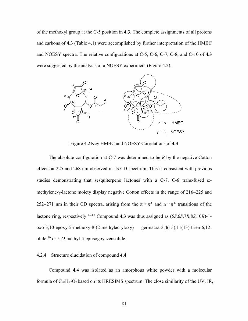
81
of the methoxyl group at the C-5 position in 4.3. The complete assignments of all protons
and carbons of 4.3 (Table 4.1) were accomplished by further interpretation of the HMBC
and NOESY spectra. The relative configurations at C-5, C-6, C-7, C-8, and C-10 of 4.3
were suggested by the analysis of a NOESY experiment (Figure 4.2).
Figure 4.2 Key HMBC and NOESY Correlations of 4.3
The absolute configuration at C-7 was determined to be R by the negative Cotton
effects at 225 and 268 nm observed in its CD spectrum. This is consistent with previous
studies demonstrating that sesquiterpene lactones with a C-7, C-6 trans-fused -
methylene--lactone moiety display negative Cotton effects in the range of 216225 and
252271 nm in their CD spectra, arising from the →* and n→* transitions of the
lactone ring, respectively.13-15 Compound 4.3 was thus assigned as (5S,6S,7R,8S,10R)-1-
oxo-3,10-epoxy-5-methoxy-8-(2-methylacryloxy) germacra-2,4(15),11(13)-trien-6,12-
olide,16 or 5-O-methyl-5-epiisogoyazensolide.
4.2.4 Structure elucidation of compound 4.4
Compound 4.4 was isolated as an amorphous white powder with a molecular
formula of C20H22O7 based on its HRESIMS spectrum. The close similarity of the UV, IR,
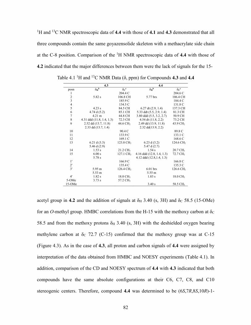
82
1H and 13C NMR spectroscopic data of 4.4 with those of 4.1 and 4.3 demonstrated that all
three compounds contain the same goyazensolide skeleton with a methacrylate side chain
at the C-8 position. Comparison of the 1H NMR spectroscopic data of 4.4 with those of
4.2 indicated that the major differences between them were the lack of signals for the 15-
Table 4.1 1H and 13C NMR Data (δ, ppm) for Compounds 4.3 and 4.4
4.3 4.4 posn δH
b δCc δH
b δCc
1 204.4 C 204.6 C 2 5.82 s 106.8 CH 5.77 brs 106.4 CH 3 185.9 C 184.4 C 4 134.5 C 131.8 C 5 4.23 s 84.5 CH 6.27 dt (2.9, 1.4) 137.3 CH 6 4.74 d (5.2) 85.1 CH 5.33 ddt (5.5, 2.9, 1.4) 81.3 CH 7 4.21 m 44.8 CH 3.80 ddd (5.5, 3.2, 2.7) 50.9 CH 8 4.31 ddd (11.8, 1.4, 1.3) 72.3 CH 4.54 dt (11.8, 2.2) 73.2 CH 2.52 dd (13.7, 11.8) 44.6 CH2 2.49 dd (13.9, 11.8) 43.9 CH2 2.33 dd (13.7, 1.4) 2.32 dd(13.9, 2.2)
10 90.4 C 89.8 C 11 133.9 C 133.1 C 12 169.1 C 168.6 C 13 6.21 d (3.3) 123.8 CH2 6.23 d (3.2) 124.6 CH2
5.46 d (2.9) 5.47 d (2.7) 14 1.53 s 21.2 CH3 1.54 s 20.7 CH3 15 6.00 s 127.1 CH2 4.16 ddd (12.8, 1.4, 1.3) 72.7 CH2
5.78 s 4.12 ddd (12.8,1.4, 1.3) 1' 166.9 C 166.8 C 2' 135.4 C 135.3 C 3' 5.95 m 126.4 CH2 6.01 brs 126.6 CH2 5.53 m 5.55 m
4' 1.82 s 18.0 CH3 1.83 s 18.0 CH3 5-OMe 3.73 s 57.2 CH3 15-OMe 3.40 s 58.5 CH3
acetyl group in 4.2 and the addition of signals at δH 3.40 (s, 3H) and δC 58.5 (15-OMe)
for an O-methyl group. HMBC correlations from the H-15 with the methoxy carbon at δC
58.5 and from the methoxy protons δH 3.40 (s, 3H) with the deshielded oxygen bearing
methylene carbon at δC 72.7 (C-15) confirmed that the methoxy group was at C-15
(Figure 4.3). As in the case of 4.3, all proton and carbon signals of 4.4 were assigned by
interpretation of the data obtained from HMBC and NOESY experiments (Table 4.1). In
addition, comparison of the CD and NOESY spectrum of 4.4 with 4.3 indicated that both
compounds have the same absolute configurations at their C6, C7, C8, and C10
stereogenic centers. Therefore, compound 4.4 was determined to be (6S,7R,8S,10R)-1-

83
oxo-3,10-epoxy-15-methoxy-8-(2-methylacryloxy)germacra-2,4(5),11(13)-trien-6,12-
olide,9 or 15-O-methylgoyazensolide.
Figure 4.3 Key HMBC and NOESY Correlations of 4.4
4.2.5 Bioactivities
Compounds 4.14.4 were evaluated for their antiproliferative activity against the
A2780 human ovarian cancer cell line. They showed micromolar activities with half-
maximum inhibitory concentration (IC50) values of 1.56 0.04, 1.62 0.05, 1.50.5,
and 0.6 0.3M, respectively. All the compounds were further evaluated for their
antimalarial activity against P. falciparum Dd2 (a chloroquine/mefloquine-resistant
strain), and showed IC50 values of 9.0 ± 0.6, 8.0 ± 0.4, 6.2 ± 0.5, and 2.2 ± 0.5 M,
respectively.
4.3 Experimental Section
4.3.1 General experimental procedures
IR and UV spectra were measured on MIDAC M-series FTIR and Shimadzu UV-
1201 spectrophotometers, respectively. 1H and 13C NMR spectra were recorded on a
Bruker Avance 500 spectrometer in CDCl3, with CDCl3 as reference. Mass spectra were

84
obtained on an Agilent 6220 mass spectrometer. Open column chromatography was
performed using Sephadex LH-20 and silica gel (230-400 mesh, Silicycle Co. USA).
Semi-preparative HPLC was performed using Shimadzu LC-10AT pumps coupled with a
semipreparative Phenomenex C18 column (5 μm, 250 × 10 mm), a Shimadzu SPD M10A
diode array detector, and a SCL-10A system controller. All isolated compounds were
purified to 95% purity or better, as judged by HPLC (both UV and ELSD detection)
before determining bioactivity.
4.3.2 Plant material
Leaves and twigs of P. antillana Urb were collected by Hannah Stevens of the
New York Botanical Garden near Manatí, Puerto Rico (18o4.385 N, 66o54.382 W). A
voucher specimen is on deposit at the NYBG under the accession number HS 00246a.
4.3.3 Extraction and isolation
The dried and powdered leaves and twigs of P. antillana were exhaustively
extracted with ethanol in two 24-hour percolation steps; successive partition of the
concentrated extract with hexane and methylene chloride gave an active methylene
chloride extract. For purposes of fractionation and purification, 2.0 g of the original
ethanol extract designated 1000892-7G (IC50 12g/mL) was suspended in 90% aq.
MeOH (300 mL), and extracted with hexanes (3 × 200 mL). Evaporation of the hexane-
soluble fraction afforded 176 mg of residue. The 90% aq. MeOH layer was then diluted
to 60% and extracted with CH2Cl2 (3 × 200 mL) to yield 479 mg of CH2Cl2-soluble
fraction. The aqueous MeOH layer was concentrated to give 1.3 g of brown residue. The
CH2Cl2 fraction was found to have antiproliferative activity with an IC50 value of 0.62
85
µg/mL, and was subjected to Sephadex LH-20 open column chromatography
(CH2Cl2:MeOH, 1:1) to give 5 fractions. The most active fraction Fr 3 (IC50 0.5 µg/mL)
was then divided into six sub-fractions by silica gel column chromatography
(hexanes:EtOAc, 1:1). Further purification of the most active sub-fraction Fr 3-4 (IC500.3
µg/mL) was done by using HPLC on a C-18 column with a solvent gradient from
H2O:CH3CN, 70:30 to 68:32 from 0 to 30 min, to 58:42 from 30 to 60 min, and ending
with 100% CH3CNfrom 60 to 80 min. This process yielded compounds 4.1 (2.5 mg, tR31
min), 4.4 (1.5 mg, tR60 min), 4.3 (1.1 mg, tR 64.5 min), and 4.2 (1.2 mg, tR66 min).
4.3.4 5-O-Methyl-5-epiisogoyazensolide (4.1)
Amorphous powder; []21D -95.6 (c 0.27, MeOH); UV (MeOH) λmax (log ε): 270
(2.20), 208 (3.89) nm; IR (film) νmax 1765, 1710, 1709, 1645, 1640, 1582, 1454 cm-1;
ECD (c 0.031, MeOH) λmax () 206 (+10), 225 (-4.0), 268 (-4.2), 320 (+2.5); 1H and 13C
NMR data, see Table 4.1; HRESIMS m/z [M+H]+ 375.1461 (calcd for C20H23O7+
375.1438)
4.3.5 5-O-Methylgoyazensolide (4.2)
Amorphous powder; []21D -121.8 (c 0.27, MeOH); UV (MeOH) λmax (log ε) : 267
(2.67), 207 (4.01) nm; IR (film) νmax 1769, 1710, 1709, 1653, 1587, 1456 cm-1; ECD (c
0.031, MeOH) λmax () 203 (+5.7), 216 (-1.8), 276 (-2.3), 318 (+2.9); 1H and 13C NMR
data, see Table 4.1. HRESIMS m/z [M+H]+ 374.1459 (calcd for C20H23O7+, 375.1438)
86
4.3.6 Antiproliferative bioassay
The A2780 ovarian cancer cell line antiproliferative bioassay was performed at
Virginia Tech as previously reported.17 The A2780 cell line is a drug-sensitive ovarian
cancer cell line.18 Paclitaxel was used as the positive control; it had an IC50 value of 0.024
M.
4.3.7 Antimalarial bioassay
The effect of each compound on parasite growth of the Dd2 strain of P. falciparum
was measured in a 72 h growth assay in the presence of compound as described
previously with minor modifications.19, 20 Briefly, ring stage parasite cultures (1%
hematocrit and 1% parasitemia) were grown for 72 h in the presence of increasing
concentrations of the drug in a 5.05% CO2, 4.93% O2, and 90.2% N2 gas mixture at 37 °C.
After 72 h in culture, parasite growth was determined by DNA quantitation using SYBR
Green I.31 The half-maximal inhibitory concentration (IC50) calculation was performed
with GraFit software using a nonlinear regression curve fitting. IC50 values are the
average of three independent determinations with each determination in duplicate and are
expressed ± SD. Artemisinin was used as the positive control; it had an IC50 value of 7
nM.

87
4.4 References
1. Liu, Y.; Rakotondraibe, H.; Brodie, P. J.; Wiley, J. D.; Cassera, M. B.; Goetz, M.;
Kingston, D. G. I. Antiproliferative and antimalarial sesquiterpene lactones from
Piptocoma antillana from Puerto Rico. Nat. Prod. Commun. 2014, 9, 1403-1406.
2. Pruski, J. F. Compositae of the Guayana highland—X. Reduction of Pollalesta to
Piptocoma (Vernonieae: Piptocarphinae) and consequent nomenclatral adjustments Noron
1996, 6, 96-102.
3. Ren, Y.; Acuna, U. M.; Jimenez, F.; Garcia, R.; Mejia, M.; Chai, H.; Gallucci, J.
C.; Farnsworth, N. R.; Soejarto, D. D.; Carcache, d. B. E. J.; Kinghorn, A. D. Cytotoxic
and NF-κB inhibitory sesquiterpene lactones from Piptocoma rufescens. Tetrahedron
2012, 68, 2671-2678.
4. Ren, Y.; Jimenez, F.; Garcia, R.; Mejia, M.; Chai, H.; Farnsworth, N. R.; Soejarto,
D. D.; Kinghorn, A. D. A cytotoxic dimeric furanoheliangolide from Piptocoma
rufescens. Tetrahedron Lett. 2013, 54, 5457-5460.
5. Trinh, T. T. V.; Pham, V. C.; Doan, T. M. H.; Nguyen, V. H.; Chau, V. M.;
Litaudon, M.; Gueritte, F. Cytotoxic aryltetralin lignans from fruits of Cleistanthus
indochinensis. Planta Med. 2014, 80, 695-702.
6. Devi, J. C.; Pai, K. V. K. Chemical examination of fruits of Cleistanthus collinus.
Pharma Chem. 2011, 3, 160-164.
7. Wolfender, J. L.; Ndjoko, K.; Hostettmann, K. The potential of LC-NMR in
phytochemical analysis. Phytochem. Anal. 2001, 12, 2-22.
8. Delgado, G.; Romo, d. V. A.; Herz, W. Sesquiterpene lactones from Viguiera
species. Phytochemistry 1982, 21, 1305-1308.
88
9. Batistada, C. F.; Dias, D. A.; Lopes, J. L. C.; Vichnewski, W. Flavonoids and
heliangolides from Lychnophora diamantinana. Phytochemistry 1993, 34, 261-263.
10. Soares, A. C. F.; Silva, A. N.; Matos, P. M.; Henrique, d. s. E.; Heleno, V. C. G.;
Lopes, N. P.; Lopes, J. L. C.; Sass, D. C. Complete 1H and 13C NMR structural
assignments for a group of four goyazensolide-type furanoheliangolides. Quim. Nova
2012, 35, 2205-2209.
11. Lunardello, M. A.; Tomaz, J. C.; Vichnewski, W.; Lopes, J. L. C. Sesquiterpene
lactones and flavonoids from Eremanthus mattogrossensis and Eremanthus eriopus. J.
Braz. Chem. Soc. 1995, 6, 307-3011.
12. Herz, W.; Kumar, N. Sesquiterpene lactones of Calea zacatechichi and C.
urticifolia. Phytochemistry 1980, 19, 593-597.
13. Youssef, D.; Frahm, A. W. Circular dichroism of C-7, C-6 trans-fused
guaianolides of Centaurea scoparia. Phytochemistry 1996, 41, 1107-1111.
14. Stoecklin, W.; Waddell, T. G.; Geissman, T. A. Circular dichroism and optical
rotatory dispersion of sesquiterpene lactones. Tetrahedron 1970, 26, 2397-2409.
15. Waddell, T. G.; Stoecklin, W.; Geissman, T. A. Circular dichroism of
sesquiterpene lactones. Tetrahedron Lett. 1969, 1313-1316.
16. Valdes, D. A.; Bardon, A.; Catalan, C. A. N.; Gedris, T. E.; Herz, W.
Goyazensolides and isogoyazensolides from Argentine Centratherum punctatum ssp.
punctatum. Biochem. Syst. Ecol. 1998, 26, 805-808.
17. Parasuraman, S.; Raveendran, R. Diuretic effects of cleistanthin A and cleistanthin
B from the leaves of Cleistanthus collinus in wistar rats. J. Young Pharm. 2012, 4, 73-77.
18. Kovendan, K.; Arivoli, S.; Maheshwaran, R.; Baskar, K.; Vincent, S. Larvicidal
89
efficacy of Sphaeranthus indicus, Cleistanthus collinus and Murraya koenigii leaf
extracts against filarial vector, Culex quinquefasciatus Say (Diptera: Culicidae). Parasitol
Res 2012, 111, 1025-1035.
19. Thanh, V. T. T.; Pham, V. C.; Nguyen, H. H.; Mai, H. D. T.; Minh, H. N. T.;
Nguyen, V. H.; Litaudon, M.; Gueritte, F.; Chau, V. M. Cleistanone: A triterpenoid from
Cleistanthus indochinensis with a new carbon skeleton. Eur. J. Org. Chem. 2011, 2011,
4108-4111.
20. Parasuraman, S.; Raveendran, R. Effect of cleistanthin A and B on adrenergic and
cholinergic receptors. Pharmacogn. Mag. 2011, 7, 243-247.
5 Bioactive Compounds from Stuhlmannia moavi from the Madagascar Dry Forest
This chapter is a slightly expanded version of a s a published article.1 Attributions
from co-authors of the articles are described as follows in the order of the names listed.
The author of this dissertation (Yixi Liu) conducted the isolation and structural
elucidation part of the titled compounds, and drafted the manuscript. Dr. Harinantenaina
was a mentor for this work, provided invaluable advice and hints for the structural
elucidation of the compounds, and also proofread the manuscript before submission. Mrs.
Peggy Brodie performed the A2780 bioassay on the isolated fractions and compounds.
Dr. Jessica D. Wiley and Dr. Maria B. Cassera performed the Dd2 bioassay. Dr. Carla
Slebodnick screened the X-ray crystal sample. Dr. Martin W. Callmander and Dr.
Richard Randrianaivo from Missouri Botanical Garden collected the plant, and Dr. Chris
Birkinshawand and Dr. Wendy Applequist provided clarification of a confused taxonomy.
Dr. Etienne Rakotobe and Dr. Vincent E. Rasamison from Madagascar carried out the
initial plant extraction. Dr. David G. I. Kingston was a mentor for this work and the
corresponding author for the published article. He provided critical suggestions for this
work and crucial revisions to the manuscript.
5.1 Introduction
As a part of the Madagascar International Cooperative Biodiversity Group (ICBG)
program,2, 3 an ethanol extract of the leaves of a plant initially identified as an
Eligmocarpus sp. was selected for evaluation as a possible source of new antiproliferative
agents based on its activity against the A2780 ovarian cancer cell line. The final
identification of the plant proved to be a botanical challenge. Although it was initially
91
identified in the field as a species of Eligmocarpus, it was then reidentified as Bussea
sakalava Du Puy & R. Rabev., and was finally classified as Stuhlmannia moavi Taub.
(synonym: Caesalpinia insolita (Harms) Brenan & J.B. Gillett) (Leguminosae).4
The largely tropical genus Caesalpinia, a member of the informal Caesalpinia
group5 in the Leguminoseae family, traditionally comprises up to 150 species, although
Caesalpinia sensu stricto is now considered to comprise only about a dozen species.6
Seventeen species of the genus Caesalpinia, as circumscribed in its broadest sense, have
been found in China, and some of them have been used for the treatment of rheumatism
and inflammatory diseases in Chinese traditional medicine.7 Triterpenes, flavonoids and
other compounds have been isolated from plants of the genus Caesalpinia sensu lato,8, 9
and 5-hydroxy-1,4-naphthoquinone has been isolated from the Asian species C. sappan.10
The genus is also a rich source of cassane-type diterpenes,11, 12 which have been reported
to have significant antimalarial,13 antibacterial,14 antihelmintic,15 antiproliferative and
antineoplasmic activities.16, 17 Caesalpinia is one of 21 genera included in the Caesalpinia
group of legumes,5 a group of genera that includes the East African and Madagascan
monospecifc genus Stuhlmannia.5, 18 The only previous chemical studies on the genus
Stuhlmannia led to the isolation of some antimicrobial furanoid diterpenes.19
We describe herein the isolation and structure elucidation of the new bioactive 1,4-
naphthoquinone stuhlmoavin (5.1) and several other bioactive compounds (Fig. 5.1).
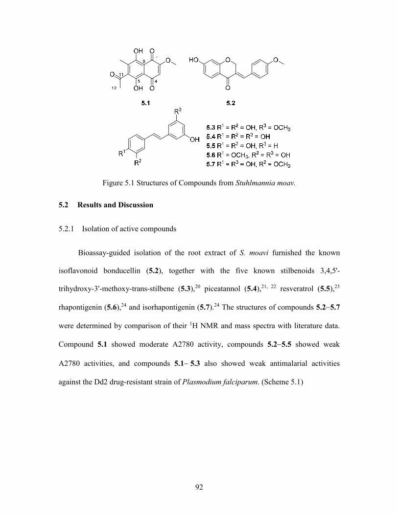
92
Figure 5.1 Structures of Compounds from Stuhlmannia moav.
5.2 Results and Discussion
5.2.1 Isolation of active compounds
Bioassay-guided isolation of the root extract of S. moavi furnished the known
isoflavonoid bonducellin (5.2), together with the five known stilbenoids 3,4,5'-
trihydroxy-3'-methoxy-trans-stilbene (5.3),20 piceatannol (5.4),21, 22 resveratrol (5.5),23
rhapontigenin (5.6),24 and isorhapontigenin (5.7).24 The structures of compounds 5.25.7
were determined by comparison of their 1H NMR and mass spectra with literature data.
Compound 5.1 showed moderate A2780 activity, compounds 5.25.5 showed weak
A2780 activities, and compounds 5.15.3 also showed weak antimalarial activities
against the Dd2 drug-resistant strain of Plasmodium falciparum. (Scheme 5.1)
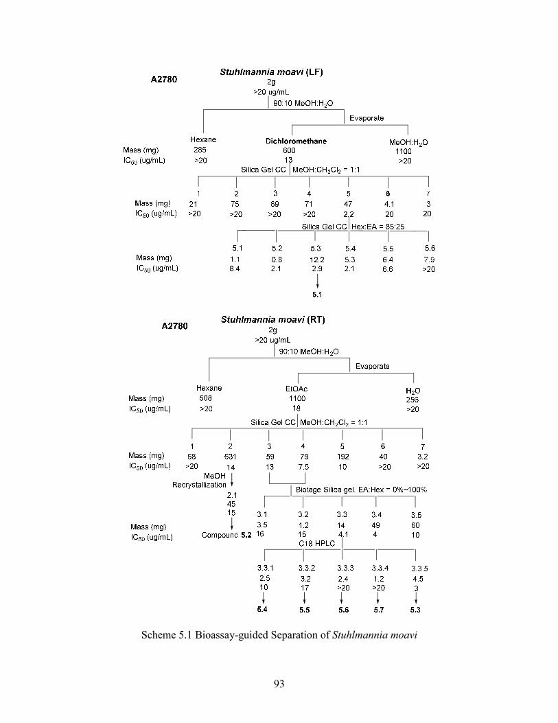
93
Scheme 5.1 Bioassay-guided Separation of Stuhlmannia moavi
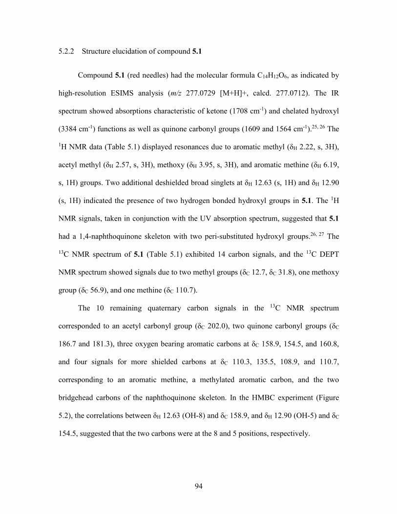
94
5.2.2 Structure elucidation of compound 5.1
Compound 5.1 (red needles) had the molecular formula C14H12O6, as indicated by
high-resolution ESIMS analysis (m/z 277.0729 [M+H]+, calcd. 277.0712). The IR
spectrum showed absorptions characteristic of ketone (1708 cm-1) and chelated hydroxyl
(3384 cm-1) functions as well as quinone carbonyl groups (1609 and 1564 cm-1).25, 26 The
1H NMR data (Table 5.1) displayed resonances due to aromatic methyl (δH 2.22, s, 3H),
acetyl methyl (δH 2.57, s, 3H), methoxy (δH 3.95, s, 3H), and aromatic methine (δH 6.19,
s, 1H) groups. Two additional deshielded broad singlets at δH 12.63 (s, 1H) and δH 12.90
(s, 1H) indicated the presence of two hydrogen bonded hydroxyl groups in 5.1. The 1H
NMR signals, taken in conjunction with the UV absorption spectrum, suggested that 5.1
had a 1,4-naphthoquinone skeleton with two peri-substituted hydroxyl groups.26, 27 The
13C NMR spectrum of 5.1 (Table 5.1) exhibited 14 carbon signals, and the 13C DEPT
NMR spectrum showed signals due to two methyl groups (δC 12.7, δC 31.8), one methoxy
group (δC 56.9), and one methine (δC 110.7).
The 10 remaining quaternary carbon signals in the 13C NMR spectrum
corresponded to an acetyl carbonyl group (δC 202.0), two quinone carbonyl groups (δC
186.7 and 181.3), three oxygen bearing aromatic carbons at δC 158.9, 154.5, and 160.8,
and four signals for more shielded carbons at δC 110.3, 135.5, 108.9, and 110.7,
corresponding to an aromatic methine, a methylated aromatic carbon, and the two
bridgehead carbons of the naphthoquinone skeleton. In the HMBC experiment (Figure
5.2), the correlations between δH 12.63 (OH-8) and δC 158.9, and δH 12.90 (OH-5) and δC
154.5, suggested that the two carbons were at the 8 and 5 positions, respectively.
95
Figure 5.2 Key HMBC correlations of 5.1
The HMBC correlations between the proton signals of the methyl group at δH 2.57
(CH3-12) and both the carbonyl carbon at δC 202.0 (C-11) and the aromatic carbon at δC
142.1 (C-6), together with the long range HMBC correlation between δH 12.90 (OH-5)
and δC 142.1 (C-6), indicated the attachment of an acetyl group at the C-6 position. In
addition, the methyl group was assigned to C-7 based on the long-range correlations (Fig.
5.2) observed between the signal at δH 2.22 and δC 142.1 (C-6), δC 135.5 (C-7), and δC
158.9 (C-8). Furthermore, the signals for the deshielded hydroxyl groups OH-1 and OH-4
also correlated with carbon signals at δC 110.7 and δC 108.9, which were assigned to the
two bridgehead carbons at C-9 and C-10, respectively. In the same manner, the methine
group at the C-3 position was confirmed by observation of the HMBC long range
correlation between δH 6.19 (H-3) and C-10 (δC 108.9). Moreover, the signal at H-3
correlated with both of the two quinone carbonyl groups at δC 181.3 and δC 186.7, which
were assigned to C-4 and C-1. The methoxy group was assigned to C-2 (δC 160.8), which
agrees with the correlation between δH 3.95 and δC 160.8 in the HMBC experiment.
Compound 5.1 was thus deduced to be 6-acetyl-2-methoxy-7-methyl-1,4-naphthoquinone
and has been named stuhlmoavin.

96
Table 5.1 1H and 13C NMR Data (δ, ppm) for Compound 5.1 (500 and 125 MHz)
position δHa δC (DEPT) a
1 186.7 (C)b
2 160.8 (C)
3 6.19 s 110.3 (CH)
4 181.3 (C)b
5 154.5 (C)
6 142.1 (C)
7 135.5 (C)
8 158.9 (C)
9 110.7 (C)
10 108.9 (C)
11 202.0 (C)
12 2.57 s 31.8 (CH3)
2-OCH3 3.95 s 56.9 (CH3)
7-CH3 2.22 s 12.7 (CH3)
5-OH 12.90 s
8-OH 12.63 s aIn CDCl3. Assignments based on HMBC
bThese signals may be exchanged
The proposed structure of 5.1 was confirmed by single-crystal X-ray diffraction
analysis (Fig. 5.3).
Figure 5.3 Anisotropic Displacement Ellipsoid Drawing of Compound 5.1.
5.2.3 Biological activities
Compounds 5.15.5 showed antiproliferative activity against the A2780 human
ovarian cancer cell line. Among the seven compounds isolated, 5.1 showed moderate
activity with an IC50 value of 8.1 ± 0.4 M. Compounds 5.25.5 showed weaker
97
activities with IC50 values of 10.6 ± 0.4 M, 54 ± 1M, 41 ± 1 M, and 74 ± 1 M,
respectively. Compounds 5.15.5 were also evaluated for antimalarial activity against
Plasmodium falciparum Dd2 (a chloroquine/mefloquine-resistant strain), and compounds
5.15.3 showed moderate activities with IC50 values of 24 ± 7, 26 ± 3, and 27 ± 2 M,
respectively.
5.3 Experimental
5.3.1 General experimental procedures
IR and UV spectra were measured on MIDAC M-series FTIR and Shimadzu UV-
1201 spectrophotometers, respectively. 1H and 13C NMR spectra were recorded on a
Bruker Avance 500 spectrometer in CDCl3 with TMS as internal standard. Mass spectra
were obtained on an Agilent 6220 mass spectrometer. Open column chromatography was
performed using Sephadex LH-20 and silica gel (230-400 mesh, Silicycle Co. USA).
Flash chromatography was performed on a Biotage Horizon flash chromatograph using a
C18 column (Phenomenex, 150 x 12 mm, 11 g). Semi-preparative HPLC was performed
using Shimadzu LC-10AT pumps coupled with a semipreparative Phenomenex C18
column (5 μm, 250 × 10 mm), a Shimadzu SPD M10A diode array detector, and a SCL-
10A system controller. All isolated compounds were purified to 95% purity or better, as
judged by HPLC and by NMR (both UV and ELSD detection) before determining
bioactivity. The HPLC chromatograms and NMR spectra can be found in the
Supplementary Data.
98
5.3.2 Plant material
Leaves and roots of Stuhlmannia moavi Taub.4 Brenan and Gillett3 collected in dry
forest 3 km north of Tsaratanana in the Sava locality, Daraina, in northern Madagascar
The sample was from a tree 12 m high, 30 cm DBH, with yellow flowers and orange
anthers. The tree is common in this location. It was identified as Stuhlmannia moavi
Taub. (synonyms: C. insolita (Harms) Brenan & J.B. Gillett, from Madagascar and
Caesalpinia dalei Brenan & J.B. Gillett, from East Africa) by comparison with the
literature,3,4 and by matching photographs taken in the field with herbarium specimens
housed in the collections of the Royal Botanic Gardens, Kew.
5.3.3 Extraction and isolation
A ground sample of Stuhlmannia moavi leaves (309 g) was extracted with EtOH at
room temperature to yield 27.9 g of crude EtOH extract designated MG 4425. A total of
5.64 g of this extract was made available to Virginia Tech. The crude EtOH extract (2.0
g) was dissolved in 90% aq. MeOH (300 mL), and extracted with hexanes (3 × 200 mL).
Evaporation of the hexane-soluble fraction afforded 285 mg of residue. The 90% aq.
MeOH layer was then evaporated, suspended in H2O (300 mL), and extracted with
EtOAc (3 × 200 mL) to yield 600 mg of EtOAc-soluble fraction. The aqueous layer was
concentrated to give 1.1 g of brown residue. The hexanes fraction was found to be
cytotoxic with an IC50 value of 13 µg/mL and was subjected to Sephadex LH-20 open
column chromatography (CH2Cl2:MeOH, 1:1) to give 7 fractions. Further purification of
the most active fraction (IC50 3.2 µg/mL) furnished the major and most active compound
5.1 (5.8 mg) with an IC50 of 2.6 µg/mL.

99
A ground sample of S. moavi roots (335 g) was extracted with EtOH at room
temperature to yield 19.6 g of crude EtOH extract designated MG 4423. A total of 4.36 g
of this extract was made available to Virginia Tech. A part of the crude EtOH extract (2.0
g) was dissolved in 90% aq. MeOH (300 mL) and extracted with hexanes (3 × 200 mL).
Evaporation of the hexanes-soluble fraction yielded 508 mg of residue. The 90% aq.
MeOH layer was then evaporated, suspended in H2O (300 mL), and extracted with
EtOAc (3 × 200 mL) to yield 1.106 mg of EtOAc-soluble fraction. The aqueous layer
was concentrated to give 256 mg of brown residue. The EtOAc fraction was found to be
weakly cytotoxic with an IC50 value of 18 µg/mL and was subjected to Sephadex LH-20
open column chromatography (CH2Cl2: MeOH = 1:1) to give 7 fractions. Compound 5.2
(10.1 mg) was crystallized from MeOH from the second fraction. The third fraction from
the Sephadex column (IC50 13 µg/mL) was further fractionated using a C18 flash
chromatograph column to yield 5 fractions. The third subfraction was subjected to HPLC
on a C18 column with a solvent gradient from H2O:MeOH, 55:45 (for 30 min), to 40:60
from 30 to 35 min, to 0:100 from 35 to 40 min, and ending with 100% MeOH to 55 min.
This process yielded compounds 5.4 (2.5 mg, tR 14 min), 5.5 (3.2 mg, tR 23.8 min), 5.6
(2.4 mg, tR 26.5 min), 5.7 (1.2 mg, tR 32 min) and 5.3 (4.5 mg, tR 41.5 min).
5.3.4 Stuhlmoavin (5.1)
Red crystals; mp 185 °C; UV λmax (MeOH) (log ε): 217 (4.11), 296 (3.54), 492
(3.27) nm; IR νmax 3384, 1708, 1609 and 1564 cm-1; 1H and 13C NMR data, see Table 5.1;
HRESIMS m/z [M+H]+ 277.0729 (calcd for C14H14O6+, 277.0712).

100
5.3.5 X-ray crystallography of 5.1
A red needle (0.01 x 0.04 x 0.62 mm3) was centered on the goniometer of an
Agilent SuperNova A diffractometer operating with CuK radiation. The data collection
routine, unit cell refinement, and data processing were carried out with the program
CrysAlisPro.28 The Laue symmetry and systematic absences were consistent with the
monoclinic space group P21/n. The structure was solved using SHELXS-9729 and refined
using SHELXL-97 via OLEX2.30 The final refinement model involved anisotropic
displacement parameters for non-hydrogen atoms and a riding model for all hydrogen
atoms. A 2-position disorder model was used for the ketone oxygen with relative
occupancies that refined to 0.490(13) and 0.510(13). The relatively poor refinement
statistics and elongated displacement parameters are attributed to the small size and high
mosaicity of the crystal. Crystal data: C14H12O6, Mr =276.24, monoclinic, P21/n, a =
4.9896(3) Å, b = 23.446(3) Å, c = 10.5394(7) Å, = 90°, = 100.893(6)°, = 90°, V =
1210.73(18) Å3, 9071 reflections, 196 parameters; crystal size 0.62 x 0.04 x 0.01 mm3.
The final indices were R1 = 0.0803, wR2 = 0.2080 [I>2σ(I)]. Crystallographic data for
compound 1 have been deposited as Supporting Information at the Cambridge
Crystallographic Data Centre (deposition no. CCDC 919140).31
5.3.6 Antiproliferative bioassay
A2780 ovarian cancer cell line antiproliferative bioassay was performed at Virginia
Tech as previously reported.32 The A2780 cell line is a drug-sensitive ovarian cancer cell
line.33 Paclitaxel was used as the positive control; it had an IC50 value of 0.024 M.
101
5.3.7 Antimalarial bioassay
The effect of each compound on parasite growth of the Dd2 strain of P. falciparum
was measured in a 72 h growth assay in the presence of compound as described
previously with minor modifications.34, 35 Briefly, ring stage parasite cultures (200 μL per
well, with 1% hematocrit and 1% parasitemia) were grown for 72 h in the presence of
increasing concentrations of the drug in a 5.05% CO2, 4.93% O2, and 90.2% N2 gas
mixture at 37 °C. After 72 h in culture, parasite viability was determined by DNA
quantitation using SYBR Green I (50 μL of SYBR Green I in lysis buffer at 0.4 μL of
SYBR Green I/mL of lysis buffer).31 The half-maximum inhibitory concentration (IC50)
calculation was performed with GraFit software using a nonlinear regression curve
fitting. IC50 values are the average of three independent determinations with each
determination in duplicate and are expressed ± SEM. Artemisinin was used as the
positive control; it had an IC50 value of 7 nM.
102
5.4 References
1. Liu, Y.; Harinantenaina, L.; Brodie, P. J.; Bowman, J. D.; Cassera, M. B.;
Slebodnick, C.; Callmander, M. W.; Randrianaivo, R.; Rakotobe, E.; Rasamison, V. E.;
Applequist, W.; Birkinshaw, C.; Lewis, G. P.; Kingston, D. G. I. Bioactive compounds
from Stuhlmannia moavi from the Madagascar dry forest. Bioorg. Med. Chem. 2013, 21,
7591-7594.
2. Cao, S.; Kingston, D. G. I. Biodiversity conservation and drug discovery: Can
they be combined? The Suriname and Madagascar experiences. Pharm. Biol. 2009, 47,
809 – 823.
3. Kingston, D. G. I. Modern natural products drug discovery and its relevance to
biodiversity conservation. J. Nat. Prod. 2011, 74, 496-511.
4. Brenan, J. P. M. Notes on African Caesalpinioïdeae. Kew Bull. 1963, 17, 197-218.
5. Lewis, G. In Legumes of the World, Lewis, G.; Schrire, B.; Mackinder , B.; Lock,
M., Eds. Royal Botanic Gardens: Kew, UK, 2005; pp 127-161.
6. Gagnon, E.; Lewis, G. P.; Solange Sotuyo, J.; Hughes, C. E.; Bruneau, A. A
molecular phylogeny of Caesalpinia sensu lato: Increased sampling reveals new insights
and more genera than expected. S. Afr. J. Bot. 2013, in press.
7. Wu, M.; Wang, Y. F.; Zhang, M. L.; Huo, C. H.; Dong, M.; Shi, Q. W.; Kiyota, H.
Chemical constituents of plants from the genus Caesalpinia. Chem. Biodivers 2011, 8,
1370-1399.
8. Woldemichael, G. M.; Singh, M. P.; Maiese, W. M.; Timmermann, B. N.
Constituents of antibacterial extract of Caesalpinia paraguariensis Burk. Z. Naturforsch.
(C) 2003, 58, 70-75.

103
9. Srinivas, K. V. N. S.; Koteswara, R. Y.; Mahender, I.; Das, B.; Rama, K. K. V. S.;
Hara, K. K.; Murty, U. S. N. Flavanoids from Caesalpinia pulcherrima. Phytochemistry
2003, 63, 789-793.
10. Lim, M. Y.; Jeon, J. H.; Jeong, E. Y.; Lee, C. H.; Lee, H. S. Antimicrobial activity
of 5-hydroxy-1,4-naphthoquinone isolated from Caesalpinia sappan toward intestinal
bacteria. Food Chem. 2006, 100, 1254-1258.
11. Cheng, J.; Roach, J. S.; McLean, S.; Reynolds, W. F.; Tinto, W. F. Three new
cassane diterpenes from Caesalpinia pulcherrima. Nat. Prod. Commun. 2008, 3, 1751-
1754.
12. Pranithanchai, W.; Karalai, C.; Ponglimanont, C.; Subhadhirasakul, S.;
Chantrapromma, K. Cassane diterpenoids from the stem of Caesalpinia pulcherrima.
Phytochemistry 2009, 70, 300-304.
13. Kalauni, S. K.; Awale, S.; Tezuka, Y.; Banskota, A. H.; Linn, T. Z.; Asih, P. B. S.;
Syafruddin, D.; Kadota, S. Antimalarial activity of cassane- and norcassane-type
diterpenes from Caesalpinia crista and their structure-activity relationship. Biol. Pharm.
Bull. 2006, 29, 1050-1052.
14. Dickson, R. A.; Houghton, P. J.; Hylands, P. J. Antibacterial and antioxidant
cassane diterpenoids from Caesalpinia benthamiana. Phytochemistry 2007, 68, 1436-
1441.
15. Jabbar, A.; Zaman, M. A.; Iqbal, Z.; Yaseen, M.; Shamim, A. Anthelmintic
activity of Chenopodium album (L.) and Caesalpinia crista (L.) against trichostrongylid
nematodes of sheep. J. Ethnopharmacol. 2007, 114, 86-91.
16. Hou, Y.; Cao, S.; Brodie, P.; Miller, J. S.; Birkinshaw, C.; Ratovoson, F.;

104
Rakotondrajaona, R.; Andriantsiferana, R.; Rasamison, V. E.; Kingston, D. G. I.
Antiproliferative cassane diterpenoids of Cordyla madagascariensis ssp.
madagascariensis from the Madagascar rainforest. J. Nat. Prod. 2008, 71, 150-152.
17. Yadav, P. P.; Maurya, R.; Sarkar, J.; Arora, A.; Kanojiya, S.; Sinha, S.; Srivastava,
M. N.; Raghubir, R. Cassane Diterpenes from Caesalpinia bonduc. Phytochemistry 2009,
70, 256-261.
18. Lewis, G. P. Notes on Stuhlmannia Taub. and the correct placement of
Caesalpinia insolita (Harms) Brenan & Gillett (Leguminosae: Caesalpinioideae:
Casalpinieae). . Kew Bull. 1996, 51, 377-379.
19. Odalo, J. O.; Joseph, C. C.; Nkunya, M. H. H.; Sattler, I.; Lange, C.; Dahse, H.-
M.; Moellman, U. Cytotoxic, anti-proliferative and antimicrobial furanoditerpenoids from
Stuhlmannia moavi. Phytochemistry 2009, 70, 2047-2052.
20. Prakash, S.; Khan, M. A.; Khan, K. Z.; Zaman, A. Stilbenes of Gnetum-Ula.
Phytochemistry 1985, 24, 622-624.
21. Hart, J. H. Role of phytostilbenes in decay and disease resistance. Ann. Rev.
Phytopathol. 1981, 19, 437-58.
22. Cardona, M. L.; Fernandez, M. I.; Garcia, M. B.; Pedro, J. R. Synthesis of natural
polyhydroxystilbenes. Tetrahedron 1986, 42, 2725-30.
23. Nakajima, K.; Taguchi, H.; Endo, T.; Yosioka, I. The constituents of Scirpus
fluviatilis (Torr.) A. Gray. I. The structures of two new hydroxystilbene dimers, scirpusin
A and B. Chem. Pharm. Bull. 1978, 26, 3050-7.
24. Silayo, A.; Ngadjui, B. T.; Abegaz, B. M. Homoisoflavonoids and stilbenes from
the bulbs of Scilla nervosa subsp. rigidifolia. Phytochemistry 1999, 52, 947-955.

105
25. Gerber, N. N.; Wieclawek, B. The structures of two naphthoquinone pigments
from an actinomycete. J. Org. Chem. 1966, 5, 1496-8.
26. Hanumaiah, T.; Rao, B. K.; Rao, C. P.; Rao, G. S. R.; Rao, J. U. M.; Rao, K. V. J.;
Marshall, D. S.; Thomson, R. H. Naphthalenes and naphthoquinones from Ventilago
species. Phytochemistry 1985, 24, 1811-1815.
27. Natori, S.; Kumada, Y.; Nishikawa, H. The structure of mompain, a
naphthoquinone from Helicobasidium mompa Tanaka, and its relation to spinochrome A
(M). Chem. Pharm. Bull. 1965, 13, 633-5.
28. Kingston, D. G. I.; Newman, D. J. Mother nature's combinatorial libraries; Their
influence on the synthesis of drugs. Curr. Opin. Drug Disc. 2002, 5, 304-316.
29. Sheldrick, G. M. A short history of SHELX. Acta Crystallogr., Sect. A: Found.
Crystallogr. 2008, A64, 112-122.
30. Dolomanov, O. V.; Bourhis, L. J.; Gildea, R. J.; Howard, J. A. K.; Puschmann, H.
OLEX2: a complete structure solution, refinement and analysis program. J. Appl.
Crystallogr. 2009, 42, 339-341.
31. Fahy, J.; Duflos, A.; Ribet, J. P.; Jacquesy, J. C.; Berrier, C.; Jouannetaud, M. P.;
Zunino, F. Vinca alkaloids in superacidic media: A method for creating a new family of
antitumor derivatives. J. Am. Chem. Soc. 1997, 119, 8576-8577.
32. Cao, S.; Brodie, P. J.; Miller, J. S.; Randrianaivo, R.; Ratovoson, F.; Birkinshaw,
C.; Andriantsiferana, R.; Rasamison, V. E.; Kingston, D. G. I. Antiproliferative xanthones
of terminalia calcicola from the Madagascar rain forest. J. Nat. Prod. 2007, 70, 679-681.
33. Louie, K. G.; Behrens, B. C.; Kinsella, T. J.; Hamilton, T. C.; Grotzinger, K. R.;
McKoy, W. M.; Winker, M. A.; Ozols, R. F. Radiation survival parameters of
106
antineoplastic drug-sensitive and -resistant human ovarian cancer cell lines and their
modification by buthionine sulfoximine. Cancer Res. 1985, 45, 2110-2115.
34. Bennett, T. N.; Paguio, M.; Gligorijevic, B.; Seudieu, C.; Kosar, A. D.; Davidson,
E.; Roepe, P. D. Novel, rapid, and inexpensive cell-based quantification of antimalarial
drug efficacy. Antimicrob. Agents Chemother. 2004, 48, 1807-1810.
35. Smilkstein, M.; Sriwilaijaroen, N.; Kelly, J. X.; Wilairat, P.; Riscoe, M. Simple
and inexpensive fluorescence-based technique for high-throughput antimalarial drug
screening. Antimicrob. Agents Chemother. 2004, 48, 1803-1806.
6 Structure Elucidation of Antiproliferative Bisbenzylisoquinoline Alkaloids from Anisocycla grandidieri from the Madagascar Dry
Forest
This chapter is a slightly expanded version of a published article.1 Attributions
from co-authors of the articles are described as follows in the order of the names listed.
The author of this disseration (Yixi Liu) conducted the isolation and structural elucidation
part of the titled compounds, and drafted the manuscript. Dr. Harinantenaina was a
mentor for this work, provided invaluable advice and hints for the structural elucidation
of the compounds, and also proofread the manuscript before submission. Mrs. Peggy
Brodie performed the A2780 bioassay on the isolated fractions and compounds. Dr. Carla
Slebodnick screened the single crystal samples. Dr. Martin W. Callmander, and Dr.
Richard Randrianaivo from Missouri Botanical Garden collected and identified the plant.
Dr. Etienne Rakotobe and Dr. Vincent E. Rasamison from Madagascar carried out the
initial plant extraction. Dr. Karen TenDyke and Dr. Yongchun Shen from Eisai Inc.
performed the H460, MCF-7, and UACC-257 bioassays on the compounds isolated. Dr.
David G. I. Kingston was a mentor for this work and the corresponding author for the
published article. He provided critical suggestions for this work and crucial revisions to
the manuscript.
6.1 Introduction
As a part of the Madagascar International Cooperative Biodiversity Group (ICBG)
program, an ethanol extract of the stems of Anisocycla grandidieri Baill.
(Menispermaceae) was found to have moderate activity against the A2780 ovarian cancer
cell line (IC50 9.4 g/mL). It was thus selected for evaluation for the presence of novel

108
anticancer agents. Plant species belonging to the genus Anisocycla are rich sources of
unique bisbenzylisoquinoline (BBIQ) alkaloids,2-5 which are known to have
antibacterial,6 cytotoxic,7-9 antimalarial,7, 8 anticancer,10 hypotensive,10 and
antiplasmodial11, 12 activities. In 1972, Schlittler and coworkers13 reported the isolation
and structure characterization of the four BBIQ alkaloids stebisimine (6.1), (+)-2'-
norcocsuline (6.2), trilobine (6.3) and (-)-epistephanine (6.4) from A. grandidieri, while
(+)-1,2-dehydrotelobine (6.5) was first isolated from Daphnandra apatela in 1978.14
Because of the lack of full NMR spectroscopic data in previous reports, we describe
herein the NMR assignments of all carbons and protons of stebisimine (6.1),13, 15 (+)-1,2-
dehydrotelobine (6.5),14 and (+)-2'-norcocsuline (6.2).13, 16 The structures and
stereochemistries of 6.1 and 6.2 were confirmed by single crystal X-ray crystallography.
The results of antiproliferative evaluation of all isolated compounds against the A2780
(human ovarian cancer), H460 (large cell lung cancer), MCF-7 (breast ductal carcinoma),
and UACC-257 (melanoma) cell lines are also reported in this study. (Figure 6.1)
6.2 Results and Discussion
6.2.1 Isolation of bioactive compounds
Bioassay-guided separation including liquid-liquid partition, LH-20 size-exclusive and
silica gel column chromatography, was used to obtain the four pure compounds
stebisimine (6.1), (+)-2'-norcocsuline (6.2), (+)-1,2-dehydrotelobine (6.5), and
puetogaline B (6.6). Herein, we report the structural elucidation and antiproliferative
properties of the four compounds. (Scheme 6.1)
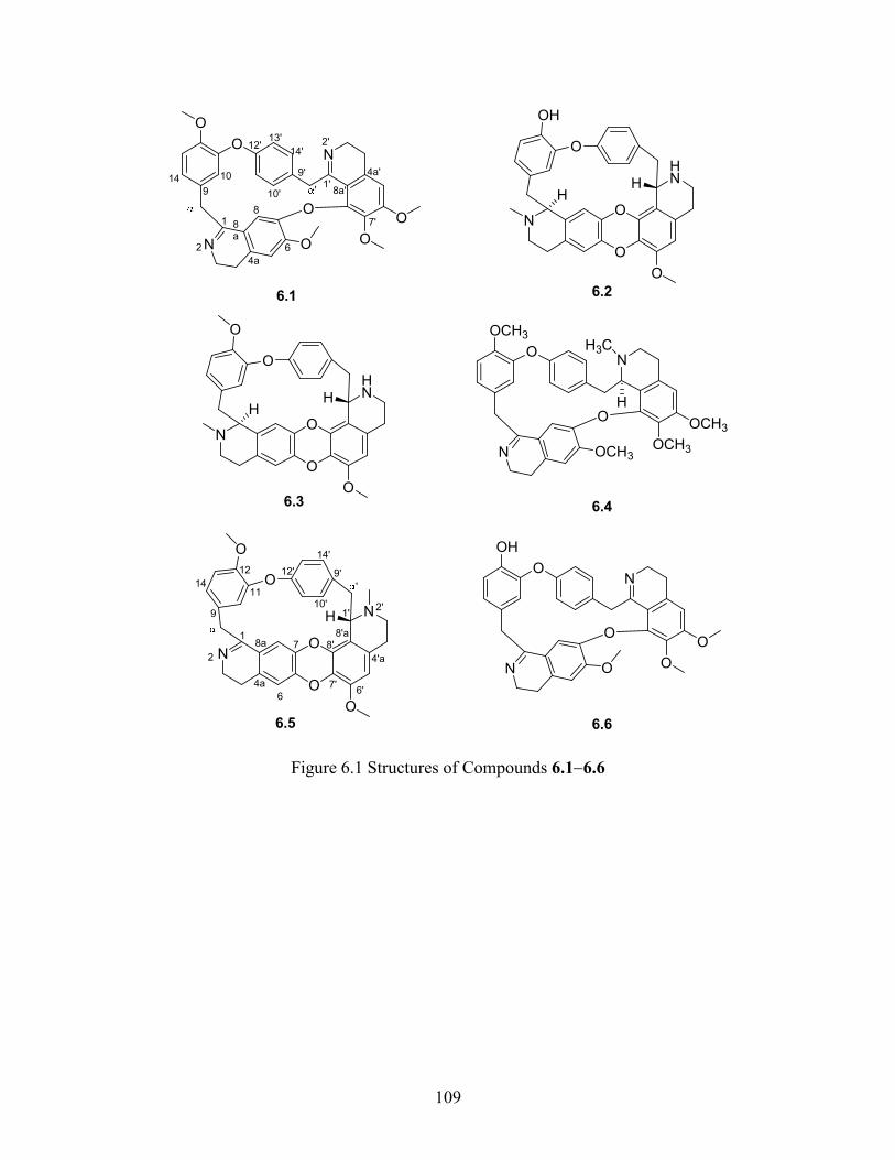
109
O
OCH3
N
N
O
OCH3OCH3
OCH3
H3C
H
O
O
N
HN
O
O
H
OH
H
6.5
6.2
O
O
N
N
O
O O
O
6.1
1
2
4a
6
78a
9
1411
12 12'
10'
9'
14'
'
1'
8'a
2'
4'a
6'7'
8'
1
2
4a
6
8
8a
9
1014
12'
10'
9'
14'
13'
'1'
2'
4a'
8a'
7'
O
O
N
N
O
O
H
O
O
OH
N
N
O
O O
O
6.6
O
O
N
HN
O
O
H
O
H
6.3 6.4
Figure 6.1 Structures of Compounds 6.16.6
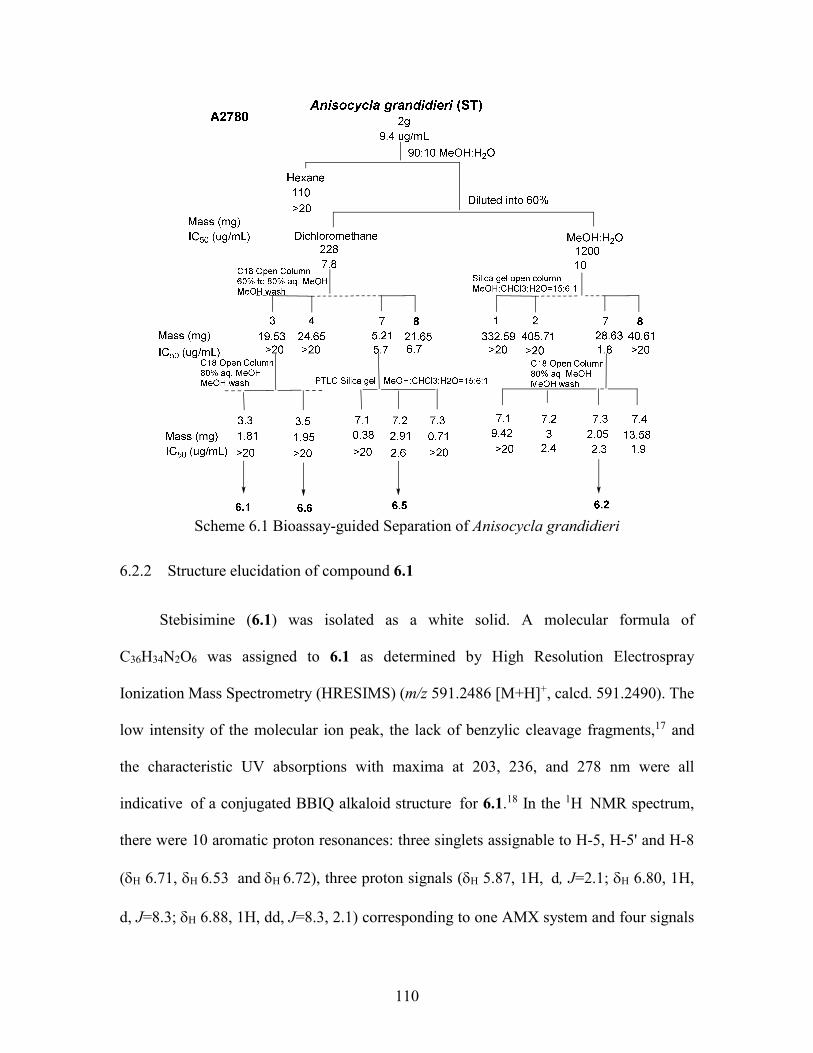
110
Scheme 6.1 Bioassay-guided Separation of Anisocycla grandidieri
6.2.2 Structure elucidation of compound 6.1
Stebisimine (6.1) was isolated as a white solid. A molecular formula of
C36H34N2O6 was assigned to 6.1 as determined by High Resolution Electrospray
Ionization Mass Spectrometry (HRESIMS) (m/z 591.2486 [M+H]+, calcd. 591.2490). The
low intensity of the molecular ion peak, the lack of benzylic cleavage fragments,17 and
the characteristic UV absorptions with maxima at 203, 236, and 278 nm were all
indicative of a conjugated BBIQ alkaloid structure for 6.1.18 In the 1H NMR spectrum,
there were 10 aromatic proton resonances: three singlets assignable to H-5, H-5' and H-8
(H 6.71, H andH 6.72), three proton signals (H 5.87, 1H, d, J=2.1; H 6.80, 1H,
d, J=8.3; H 6.88, 1H, dd, J=8.3, 2.1) corresponding to one AMX system and four signals
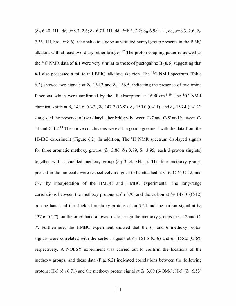
111
(H 6.40, 1H, dd, J=8.3, 2.6; H 6.79, 1H, dd, J=8.3, 2.2; H 6.98, 1H, dd, J=8.3, 2.6; H
7.35, 1H, brd, J=8.6) ascribable to a para-substituted benzyl group presents in the BBIQ
alkaloid with at least two diaryl ether bridges.17 The proton coupling patterns as well as
the 13C NMR data of 6.1 were very similar to those of puetogaline B (6.6) suggesting that
6.1 also possessed a tail-to-tail BBIQ alkaloid skeleton. The 13C NMR spectrum (Table
6.2) showed two signals at C 164.2 and C 166.5, indicating the presence of two imine
functions which were confirmed by the IR absorption at 1600 cm-1.19 The 13C NMR
chemical shifts at C 143.6 (C-7), C 147.2 (C-8’), C 150.0 (C-11), and C 153.4 (C-12’)
suggested the presence of two diaryl ether bridges between C-7 and C-8' and between C-
11 and C-12'.19 The above conclusions were all in good agreement with the data from the
HMBC experiment (Figure 6.2). In addition, The 1H NMR spectrum displayed signals
for three aromatic methoxy groups (H 3.86, H 3.89, H 3.95, each 3-proton singlets)
together with a shielded methoxy group (H 3.24, 3H, s). The four methoxy groups
present in the molecule were respectively assigned to be attached at C-6, C-6', C-12, and
C-7' by interpretation of the HMQC and HMBC experiments. The long-range
correlations between the methoxy protons at H 3.95 and the carbon at C 147.0 (C-12)
on one hand and the shielded methoxy protons at H 3.24 and the carbon signal at C
137.6 (C-7') on the other hand allowed us to assign the methoxy groups to C-12 and C-
7'. Furthermore, the HMBC experiment showed that the 6- and 6'-methoxy proton
signals were correlated with the carbon signals at C 151.6 (C-6) and C 155.2 (C-6'),
respectively. A NOESY experiment was carried out to confirm the locations of the
methoxy groups, and these data (Fig. 6.2) indicated correlations between the following
protons: H-5 (H 6.71) and the methoxy proton signal at H 3.89 (6-OMe); H-5' (H 6.53)
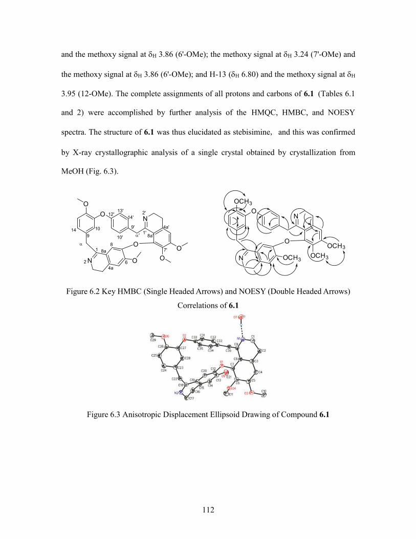
112
and the methoxy signal at H 3.86 (6'-OMe); the methoxy signal at H 3.24 (7'-OMe) and
the methoxy signal at H 3.86 (6'-OMe); and H-13 (H 6.80) and the methoxy signal at H
3.95 (12-OMe). The complete assignments of all protons and carbons of 6.1 (Tables 6.1
and 2) were accomplished by further analysis of the HMQC, HMBC, and NOESY
spectra. The structure of 6.1 was thus elucidated as stebisimine, and this was confirmed
by X-ray crystallographic analysis of a single crystal obtained by crystallization from
MeOH (Fig. 6.3).
O
OCH3
N
N
O
OCH3OCH3
OCH3
O
O
N
N
O
O O
O1
2
4a6
8
8a
9
1014
12'
10'
9'
14'
13'
'1'
2'
4a'
8a'
7'
Figure 6.2 Key HMBC (Single Headed Arrows) and NOESY (Double Headed Arrows)
Correlations of 6.1
Figure 6.3 Anisotropic Displacement Ellipsoid Drawing of Compound 6.1
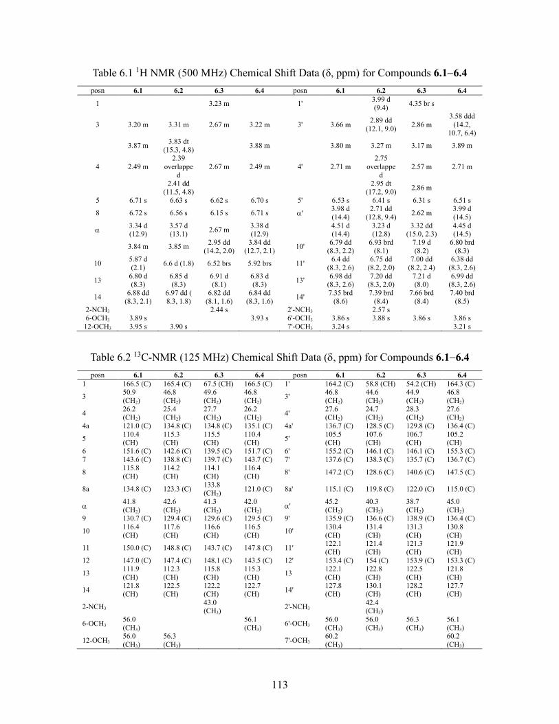
113
Table 6.1 1H NMR (500 MHz) Chemical Shift Data (δ, ppm) for Compounds 6.16.4
posn 6.1 6.2 6.3 6.4 posn 6.1 6.2 6.3 6.4
1 3.23 m 1' 3.99 d (9.4)
4.35 br s
3 3.20 m 3.31 m 2.67 m 3.22 m 3' 3.66 m 2.89 dd
(12.1, 9.0) 2.86 m
3.58 ddd (14.2,
10.7, 6.4)
3.87 m 3.83 dt
(15.3, 4.8) 3.88 m 3.80 m 3.27 m 3.17 m 3.89 m
4 2.49 m 2.39
overlapped
2.67 m 2.49 m 4' 2.71 m 2.75
overlapped
2.57 m 2.71 m
2.41 dd
(11.5, 4.8)
2.95 dt (17.2, 9.0)
2.86 m
5 6.71 s 6.63 s 6.62 s 6.70 s 5' 6.53 s 6.41 s 6.31 s 6.51 s
8 6.72 s 6.56 s 6.15 s 6.71 s ' 3.98 d (14.4)
2.71 dd (12.8, 9.4)
2.62 m 3.99 d (14.5)
3.34 d (12.9)
3.57 d (13.1)
2.67 m 3.38 d (12.9)
4.51 d (14.4)
3.23 d (12.8)
3.32 dd (15.0, 2.3)
4.45 d (14.5)
3.84 m 3.85 m 2.95 dd
(14.2, 2.0) 3.84 dd
(12.7, 2.1) 10'
6.79 dd (8.3, 2.2)
6.93 brd (8.1)
7.19 d (8.2)
6.80 brd (8.3)
10 5.87 d (2.1)
6.6 d (1.8) 6.52 brs 5.92 brs 11' 6.4 dd
(8.3, 2.6) 6.75 dd
(8.2, 2.0) 7.00 dd
(8.2, 2.4) 6.38 dd
(8.3, 2.6)
13 6.80 d (8.3)
6.85 d (8.3)
6.91 d (8.1)
6.83 d (8.3)
13' 6.98 dd
(8.3, 2.6) 7.20 dd
(8.3, 2.0) 7.21 d (8.0)
6.99 dd (8.3, 2.6)
14 6.88 dd
(8.3, 2.1) 6.97 dd ( 8.3, 1.8)
6.82 dd (8.1, 1.6)
6.84 dd (8.3, 1.6)
14' 7.35 brd
(8.6) 7.39 brd
(8.4) 7.66 brd
(8.4) 7.40 brd
(8.5) 2-NCH3 2.44 s 2'-NCH3 2.57 s 6-OCH3 3.89 s 3.93 s 6'-OCH3 3.86 s 3.88 s 3.86 s 3.86 s
12-OCH3 3.95 s 3.90 s 7'-OCH3 3.24 s 3.21 s
Table 6.2 13C-NMR (125 MHz) Chemical Shift Data (, ppm) for Compounds 6.16.4
posn 6.1 6.2 6.3 6.4 posn 6.1 6.2 6.3 6.4 1 166.5 (C) 165.4 (C) 67.5 (CH) 166.5 (C) 1' 164.2 (C) 58.8 (CH) 54.2 (CH) 164.3 (C)
3 50.9 (CH2)
46.8 (CH2)
49.6 (CH2)
46.8 (CH2)
3' 46.8 (CH2)
44.6 (CH2)
44.9 (CH2)
46.8 (CH2)
4 26.2 (CH2)
25.4 (CH2)
27.7 (CH2)
26.2 (CH2)
4' 27.6 (CH2)
24.7 (CH2)
28.3 (CH2)
27.6 (CH2)
4a 121.0 (C) 134.8 (C) 134.8 (C) 135.1 (C) 4a' 136.7 (C) 128.5 (C) 129.8 (C) 136.4 (C)
5 110.4 (CH)
115.3 (CH)
115.5 (CH)
110.4 (CH)
5' 105.5 (CH)
107.6 (CH)
106.7 (CH)
105.2 (CH)
6 151.6 (C) 142.6 (C) 139.5 (C) 151.7 (C) 6' 155.2 (C) 146.1 (C) 146.1 (C) 155.3 (C) 7 143.6 (C) 138.8 (C) 139.7 (C) 143.7 (C) 7' 137.6 (C) 138.3 (C) 135.7 (C) 136.7 (C)
8 115.8 (CH)
114.2 (CH)
114.1 (CH)
116.4 (CH)
8' 147.2 (C) 128.6 (C) 140.6 (C) 147.5 (C)
8a 134.8 (C) 123.3 (C) 133.8 (CH2)
121.0 (C) 8a' 115.1 (C) 119.8 (C) 122.0 (C) 115.0 (C)
41.8 (CH2)
42.6 (CH2)
41.3 (CH2)
42.0 (CH2)
' 45.2 (CH2)
40.3 (CH2)
38.7 (CH2)
45.0 (CH2)
9 130.7 (C) 129.4 (C) 129.6 (C) 129.5 (C) 9' 135.9 (C) 136.6 (C) 138.9 (C) 136.4 (C)
10 116.4 (CH)
117.6 (CH)
116.6 (CH)
116.5 (CH)
10' 130.4 (CH)
131.4 (CH)
131.3 (CH)
130.8 (CH)
11 150.0 (C) 148.8 (C) 143.7 (C) 147.8 (C) 11' 122.1 (CH)
121.4 (CH)
121.3 (CH)
121.9 (CH)
12 147.0 (C) 147.4 (C) 148.1 (C) 143.5 (C) 12' 153.4 (C) 154 (C) 153.9 (C) 153.3 (C)
13 111.9 (CH)
112.3 (CH)
115.8 (CH)
115.3 (CH)
13 122.1 (CH)
122.8 (CH)
122.5 (CH)
121.8 (CH)
14 121.8 (CH)
122.5 (CH)
122.2 (CH)
122.7 (CH)
14' 127.8 (CH)
130.1 (CH)
128.2 (CH)
127.7 (CH)
2-NCH3 43.0 (CH3)
2'-NCH3 42.4 (CH3)
6-OCH3 56.0 (CH3)
56.1 (CH3)
6'-OCH3 56.0 (CH3)
56.0 (CH3)
56.3 (CH3)
56.1 (CH3)
12-OCH3 56.0 (CH3)
56.3 (CH3)
7'-OCH3 60.2 (CH3)
60.2 (CH3)

114
6.2.3 Structure elucidation of compound 6.5
(+)-1,2-Dehydrotelobine (6.5) showed UV absorptions at 203, 235, 263, and 336
nm that also agreed with the reported data.14 Its molecular formula was assigned as
C35H32N2O5 based on its HRESIMS (m/z 561.2381 [M+H]+, calcd. 561.2389). The 1H
NMR spectrum displayed ten aromatic signals with same coupling patterns to those of
compound 6.1, suggesting that compound 6.5 possessed the same aromatic substitution
patterns as 6.1. Furthermore, the 13C NMR spectrum displayed only one carbon signal at
C 165.4 (C-1), with an additional typical N-methyl proton signal at H 2.57 (3H, s) and a
deshielded signal for a methine (H 3.99, d, J = 9.4;C 58.8), suggesting that one of the
imine groups which were present in 6.1 was replaced with a tertiary amine bearing a
methyl group. The long range HMBC correlations from the methyl at H 2.57 (2'-NMe) to
the methine at C-1' (C 58.8), and to the methylene at C-3' (C 44.6) corroborated the
location of the methyl group at the 2’ position. By comparison with 6.1, the absence of
the shielded methoxy proton signal at H 3.24 (7'-OMe 3H, s) and of one of the three
aromatic methoxy proton signals in 6.5, together with the number of the degrees of
unsaturation (Ω=21), suggested the presence of a new diaryl ether bridge between C-6
and C-7' instead of an imine function in 6.5. The above conclusion was supported by the
results of the HMBC experiment (Figure 6.4). All proton and carbon signals of 6.5 were
assigned by interpretation of the data obtained from HMQC and HMBC experiments.
The stereochemistry at C-1' was assigned as S based on the comparison of its optical
rotation value with the reported data in the literature.20 The structure of 6.5 was thus
assigned as (+)-1,2-dehydrotelobine.
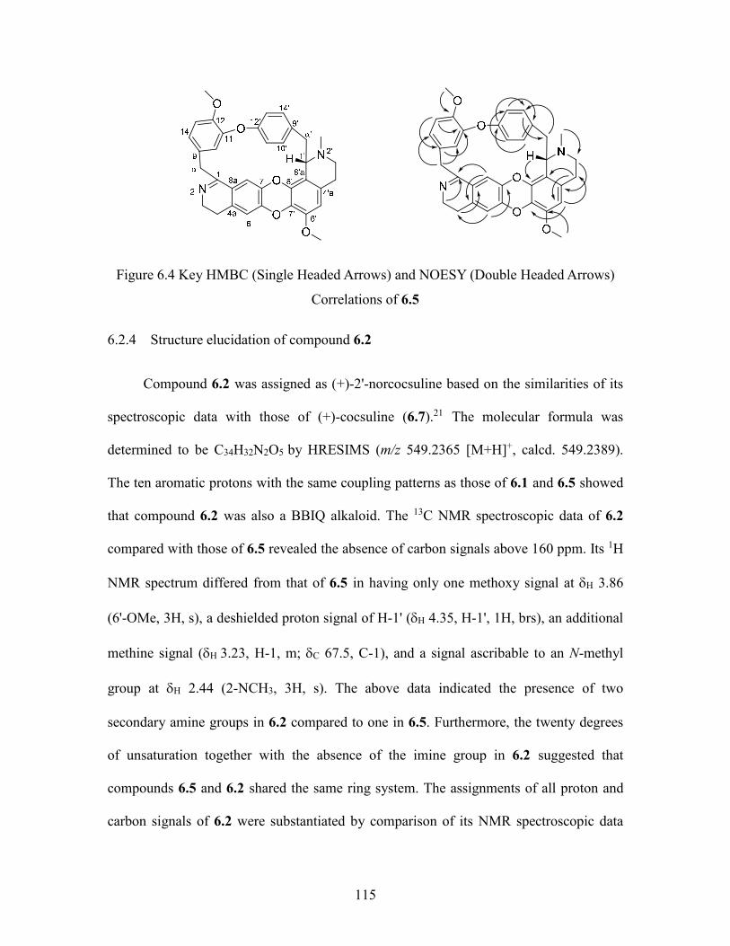
115
Figure 6.4 Key HMBC (Single Headed Arrows) and NOESY (Double Headed Arrows)
Correlations of 6.5
6.2.4 Structure elucidation of compound 6.2
Compound 6.2 was assigned as (+)-2'-norcocsuline based on the similarities of its
spectroscopic data with those of (+)-cocsuline (6.7).21 The molecular formula was
determined to be C34H32N2O5 by HRESIMS (m/z 549.2365 [M+H]+, calcd. 549.2389).
The ten aromatic protons with the same coupling patterns as those of 6.1 and 6.5 showed
that compound 6.2 was also a BBIQ alkaloid. The 13C NMR spectroscopic data of 6.2
compared with those of 6.5 revealed the absence of carbon signals above 160 ppm. Its 1H
NMR spectrum differed from that of 6.5 in having only one methoxy signal at H 3.86
(6'-OMe, 3H, s), a deshielded proton signal of H-1' (H 4.35, H-1', 1H, brs), an additional
methine signal (H 3.23, H-1, m; C 67.5, C-1), and a signal ascribable to an N-methyl
group at H 2.44 (2-NCH3, 3H, s). The above data indicated the presence of two
secondary amine groups in 6.2 compared to one in 6.5. Furthermore, the twenty degrees
of unsaturation together with the absence of the imine group in 6.2 suggested that
compounds 6.5 and 6.2 shared the same ring system. The assignments of all proton and
carbon signals of 6.2 were substantiated by comparison of its NMR spectroscopic data
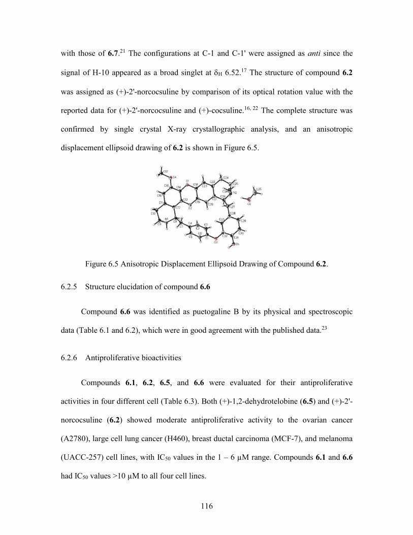
116
with those of 6.7.21 The configurations at C-1 and C-1' were assigned as anti since the
signal of H-10 appeared as a broad singlet at H 6.52.17 The structure of compound 6.2
was assigned as (+)-2'-norcocsuline by comparison of its optical rotation value with the
reported data for (+)-2'-norcocsuline and (+)-cocsuline.16, 22 The complete structure was
confirmed by single crystal X-ray crystallographic analysis, and an anisotropic
displacement ellipsoid drawing of 6.2 is shown in Figure 6.5.
Figure 6.5 Anisotropic Displacement Ellipsoid Drawing of Compound 6.2.
6.2.5 Structure elucidation of compound 6.6
Compound 6.6 was identified as puetogaline B by its physical and spectroscopic
data (Table 6.1 and 6.2), which were in good agreement with the published data.23
6.2.6 Antiproliferative bioactivities
Compounds 6.1, 6.2, 6.5, and 6.6 were evaluated for their antiproliferative
activities in four different cell (Table 6.3). Both (+)-1,2-dehydrotelobine (6.5) and (+)-2'-
norcocsuline (6.2) showed moderate antiproliferative activity to the ovarian cancer
(A2780), large cell lung cancer (H460), breast ductal carcinoma (MCF-7), and melanoma
(UACC-257) cell lines, with IC50 values in the 1 – 6 µM range. Compounds 6.1 and 6.6
had IC50 values >10 µM to all four cell lines.
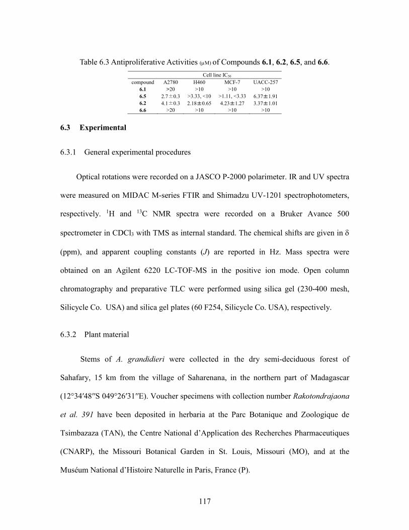
117
Table 6.3 Antiproliferative Activities (M) of Compounds 6.1, 6.2, 6.5, and 6.6.
Cell line IC50
compound A2780 H460 MCF-7 UACC-257
6.1 >20 >10 >10 >10
6.5 2.7±0.3 >3.33, <10 >1.11, <3.33 6.37±1.91
6.2 4.1±0.3 2.18±0.65 4.23±1.27 3.37±1.01
6.6 >20 >10 >10 >10
6.3 Experimental
6.3.1 General experimental procedures
Optical rotations were recorded on a JASCO P-2000 polarimeter. IR and UV spectra
were measured on MIDAC M-series FTIR and Shimadzu UV-1201 spectrophotometers,
respectively. 1H and 13C NMR spectra were recorded on a Bruker Avance 500
spectrometer in CDCl3 with TMS as internal standard. The chemical shifts are given in
(ppm), and apparent coupling constants (J) are reported in Hz. Mass spectra were
obtained on an Agilent 6220 LC-TOF-MS in the positive ion mode. Open column
chromatography and preparative TLC were performed using silica gel (230-400 mesh,
Silicycle Co. USA) and silica gel plates (60 F254, Silicycle Co. USA), respectively.
6.3.2 Plant material
Stems of A. grandidieri were collected in the dry semi-deciduous forest of
Sahafary, 15 km from the village of Saharenana, in the northern part of Madagascar
(12°34′48′′S 049°26′31′′E). Voucher specimens with collection number Rakotondrajaona
et al. 391 have been deposited in herbaria at the Parc Botanique and Zoologique de
Tsimbazaza (TAN), the Centre National d’Application des Recherches Pharmaceutiques
(CNARP), the Missouri Botanical Garden in St. Louis, Missouri (MO), and at the
Muséum National d’Histoire Naturelle in Paris, France (P).
118
6.3.3 Extraction and isolation
A ground sample of A. grandidieri stems (250 g) was extracted with EtOH at room
temperature to yield 12.2 g of crude EtOH extract designated MG 4002. A total of 4.84 g
of this extract was made available to Virginia Tech. The crude EtOH extract (2.0 g) was
dissolved in 90% aq. MeOH (300 mL) and extracted with hexanes (3 × 200 mL) to afford
110 mg of residue after evaporation of the hexanes-soluble fraction. The 90% aq. MeOH
layer was then diluted to 60% and extracted with CH2Cl2 (3×300 mL) to yield 228 mg of
CH2Cl2-soluble fraction. The 60% aq. MeOH layer was concentrated to give 1190 mg of
brown residue. The CH2Cl2 and 60% aq. MeOH fractions were found to have
antiproliferative activity with IC50 values of 6.2 µg/mL and 11 µg/mL, respectively. The
CH2Cl2 fraction was then divided into 11 fractions by an open silica gel column
(CHCl3:MeOH:H2O=15:6:1 as eluent). Preparative TLC on silica gel of the most active
fraction (Fr-7, 5.7 µg/mL) yielded compound 6.5 (2.9 mg). The third fraction (Fr-3) was
subjected to an open C-18 column to furnish compounds 6.1 (1.8 mg) and 6.6 (2.0 mg) in
the CH2Cl2 fraction. The 60% aq. MeOH fraction was applied to an open silica gel
column (solvent system: CHCl3:MeOH:H2O=15:6:1) to give 8 fractions. Further
purification of the most active fraction (Fr-7, 3.7 µg/mL) by an open C18 column
(solvent systems: 80% aq. MeOH and MeOH) gave compound 6.2 (2.1 mg).
6.3.4 NMR spectroscopy
The NMR spectra were recorded on a Bruker Advance II 500-MHz spectrometer
(Bruker Biospin Corporation, Billerica, Massachusetts, USA). All samples were
dissolved in CDCl3, and NMR experiments were recorded at 298 K. Chemical shifts ()
119
in ppm are referenced to TMS at 0.00 ppm for 1H and 13C. Coupling constants (J) are
given in Hertz. The pulse conditions of 1D NMR were as follows for 6.1: for 1H,
spectrometer frequency (SF)=500.16 MHz, acquisition time (AQ) = 3.6372 s, relaxation
delay (RD) = 1.000 s, spectral width (SW) = 9009.0 Hz, FT data size = 32 K, digital
resolution = 0.137 Hz, line broadening (LB) = 1.0 Hz, number of dummy scans (DS) = 2;
for 13C, SF = 125.77 MHz, AQ = 1.0420 s, RD=1.000 s, SW = 31446.5 Hz, FT data size
= 32K, digital resolution = 0.48 Hz, LB = 1.0 Hz,. The 2D spectra used 1024-256
(HMQC), 1024-189 (HMBC), 1024-128 (NOESY) data point matrices, which were zero
filled to 1024-1024 (HMQC), 1024-378 (HMBC), 1024-1024 (NOESY), after sinebell
weighting applied to both dimensions. For HMQC, SF = 500.16 MHz (1H), 125.77 MHz
(13C), AQ=0.1364 s, RD = 1.5000 s, NS = 86, DS = 16, pre-scan delay (DE) = 6.50 ms,
dwell time (DW) = 97.000 ms, receiver gain (RG) = 30, SW = 7507.5 Hz (1H), 31446.5
Hz (13C); for HMBC, AQ = 0.1364 s, RD = 1.5000 s, NS = 80, DS = 16, DE = 6.50 ms,
DW = 76.800 ms, RG = 30, SW = 7500.2 Hz (1H), 31466.5 Hz (13C); for NEOSY, AQ =
0.1600 s, RD = 1.000s, NS = 32, DS = 32, DE = 6.50ms, DW = 100 ms, RG = 20, SW =
6399.5 Hz (1H), 6399.5 Hz (13C). The pulse conditions were as follows for 6.5: for 1H, SF
= 500.16 MHz, AQ = 3.6372s, RD = 1.0000 s, SW = 9009.0Hz, FT data size = 32K,
digital resolution = 0.137 Hz, LB = 1.0 Hz; for 13C, SF = 125.77 MHz, AQ = 1.0420 s,
RD = 1.0000 s, SW = 31446.5 Hz, FT data size = 32 K, digital resolution = 0.48 Hz, LB
= 1.0 Hz. The 2D spectra used 1024-84 (HMQC), 1024-239 (HMBC) data point matrices,
which were zero filled to 102-512 (HMQC), 1024-1024 (HMBC), after sinebell
weighting applied to both dimensions. For HMQC, SF = 500.16 MHz (1H), 125.77 MHz
(13C), AQ = 0.1364 s, RD = 1.5000 s, NS = 160, DS = 16, DE = 6.00 ms, DW = 97.000

120
ms, RG = 23, SW = 7507.5 Hz (1H), 31446.5 Hz (13C); for HMBC, SF = 500.16 MHz
(1H), 125.770MHz (13C), AQ = 0.1364 s, RD = 1.5000 s, NS = 120, DS = 16, DE = 6.50
ms, DW = 76.800 ms, RG = 30, SW = 7507.5 Hz (1H), 31446.5 Hz (13C); The pulse
conditions were as follows for 6.2: for 1H, SF = 500.16 MHz, AQ = 3.6372 s, RD =
1.0000 s, SW = 9009.0 Hz, FT data size = 32 K, digital resolution = 0.147 Hz, LB = 1.0
Hz; for 13C, SF = 125.77 MHz, AQ = 1.0420 s, RD = 1.0000 s, SW = 31446.5 Hz, FT
data size = 32K, digital resolution = 0.48 Hz, LB = 1.00 Hz. The pulse conditions were as
follows for 6.6: for 1H, SF = 500.16 MHz, AQ = 3.6372 s, RD = 1.0000 s, SW = 9009.0
Hz, FT data size = 32K, digital resolution = 0.147 Hz, LB = 1.0 Hz; for 13C, SF = 125.77
MHz, AQ = 1.0420 s, RD = 1.0000 s, SW = 31446.5 Hz, FT data size = 32 K, digital
resolution = 0.48 Hz, LB = 1.00 Hz. The 2D spectra use 1024-256 (HMQC), 1024-256
(HMBC) data point matrices, which were zero filled to 102-1024(HMQC), 1024-516
(HMBC), after sinebell weighting applied to both dimensions. For HMQC, SF = 500.16
MHz (1H), 125.77 MHz (13C), AQ = 0.1364 s, RD = 1.5000 s, NS = 85, DS = 16, DE =
6.50 ms, DW = 97.000 ms, RG = 30, SW = 7507.5 Hz (1H), 31446.5 Hz (13C); for
HMBC, SF = 500.16 MHz (1H), 125.770 MHz (13C), AQ = 0.2047 s, RD = 1.5000 s, NS
= 120, DS = 32, DE = 6.50 ms, DW = 100 ms, RG = 30, SW = 4997.6 Hz (1H), 31446.5
Hz (13C)
6.3.5 Stebisimine (6.1)
White crystals from MeOH; UV λmax (MeOH) (log ε): 203 (2.42), 236 (1.77), 278
(0.84), 315 nm; IR νmax 3000, 1600, 1502, 1278, 1098, 1013 cm-1; 1H and 13C NMR data,
see Tables 6.1 and 6.2. HRESIMS m/z [M+H]+ 591.2486 (calcd for C36H35N2O6+,
591.2490).
121
6.3.6 (+)-1, 2-Dehydrotelobine (6.5)
Amorphous powder, [α]21D +125.2°(c 0.7, MeOH); UV λmax (MeOH) (log ε):
203(2.11), 235 (0.6), 263 (0.35), 336 (0.17) nm; IR νmax 2925, 1599, 1501, 1279, 1098
cm-1; 1H and 13C NMR data, see Tables 6.1 and 6.2; HRESIMS m/z: 561.2381 [M+H]+
(calcd C35H33N2O6+, 561.2389)
6.3.7 (+)-2'-Norcocsuline (6.2)
White crystals from MeOH, [α]21D +250°(c 0.1, MeOH); UV λmax (MeOH) (log
ε): 203 (1.25), 240 (0.75), 293 (0.16) nm; IR νmax 3420, 2925, 1501, 1279, 1098 cm-1; 1H
and 13C NMR data, see Tables 6.1 and 6.2; HRESIMS m/z: 549.2365 [M+H]+ (calcd for
C35H33N2O6+, 549.2389).
6.3.8 Puetogaline B (6.6)
Amorphous powder; UV λmax (MeOH) (log ε): 205 (1.41), 234 (0.95), 278 (0.48)
nm; IR νmax 2925, 1599, 1505, 1279, 1098, 1013 cm-1; 1H and 13C NMR data, see Tables
6.1 and 6.2; HRESIMS m/z: 577.2338 [M+H]+ [calcd for C35H33N2O6+, 577.2389
6.3.9 X-ray Crystallography
6.3.9.1 Stebisimine (6.1)
A colorless plate (0.007 x 0.147 x 0.149 mm3) was centered on the goniometer of
an Oxford Diffraction Nova diffractometer. The data collection routine, unit cell
refinement, and data processing were carried out with the program CrysAlisPro.24 The
Laue symmetry was consistent with the triclinic space groups P1 and P-1. As the
122
molecule is not chiral, the centric space group, P-1, was chosen. The structure was solved
by direct methods and refined using SHELXTL NT.25 The final refinement model
involved anisotropic displacement parameters for non-hydrogen atoms. A riding model
was used for the aromatic and alkyl hydrogens. Hydrogen atom positions of the water
molecule were located from the residual electron density map and refined independently;
the displacement parameters were fixed at Uiso(H) = 1.5 Ueq(O). Cryatal data:
C36H34N2O6•H2O, Mr =608.67, triclinic, P-1, a= 11.4221(12) Å, b= 11.4745(10) Å, c=
13.2216(14) Å, = 102.650(8)°, = 105.377(9)°, = 107.547(9)°, V=1506.5(3) Å3, 9585
reflections, 416 parameters; crystal size 0.149 x 0.147 x 0.007 mm3. The final indices
were R1 = 0.0464, wR2 = 0.1085 [I>2σ(I)]. Crystallographic data for compound 1 have
been deposited as Supporting Information at the Cambridge Crystallographic Data Centre
(deposition no. CCDC 919139).26
6.3.9.2 (+)-2'-Norcocsuline (6.2)
A colorless rod (0.8 x 0.12 x 0.30 mm3) was centered on the goniometer of an
Oxford Diffraction SuperNova diffractometer operating with CuK radiation. The data
collection routine, unit cell refinement, and data processing were carried out with the
program CrysAlisPro.24 The Laue symmetry and systematic absences were consistent
with the orthorhombic space group P212121. The structure was solved using SHELXS-
9726 and refined using SHELXL-9726 via the OLEX2 Program System.27 The final
refinement model involved anisotropic displacement parameters for non-hydrogen atoms.
A riding model was used for the aromatic and alkyl hydrogens. Hydrogen atom positions
for the hydroxyl and amine groups were located from the residual electron density peaks
and refined independently; the displacement parameters were fixed at Uiso(H) = 1.5
123
Ueq(O). The absolute configuration was established from anomalous dispersion effects
(99% Friedel mates measured; Flack x = 0.00(15);28 Hooft P2(true) = 1.000, P3(true) =
1.000, P3(rac-twin) = 0.7x10-37; P3(false) = 0.2x10-139, y = 0.-0.03(4)).29 Cryatal data:
C34H32N2O5•CH3OH, Mr =580.66, orthorhombic, P212121, a = 7.25361(9) Å, b =
13.36293(14) Å, c = 30.1627(3) Å, =90°, V=2923.65(6) Å3, 31248 reflections, 401
parameters; crystal size 0.30 x 0.12 x 0.08 mm3. The final indices were R1 = 0.0327, wR2
= 0.0854 [I>2σ(I)]. Crystallographic data for compound 6.3 have been deposited as
Supporting Information at the Cambridge Crystallographic Data Centre (deposition no.
CCDC 919138).26
6.3.10 Antiproliferative bioassays
Antiproliferative Bioassay. The A2780 ovarian cancer cell line assay was
performed at Virginia Tech as previously reported.30 The A2780 cell line is a drug-
sensitive ovarian cancer cell line.31
124
6.4 References
1. Liu, Y.; Harinantenaina, L.; Brodie, P. J.; Slebodnick, C.; Callmander, M. W.;
Rakotondrajaona, R.; Rakotobe, E.; Rasamison, V. E.; TenDyke, K.; Shen, Y.; Kingston,
D. G. I. Structure elucidation of antiproliferative bisbenzylisoquinoline alkaloids from
Anisocycla grandidieri from the Madagascar dry forest. Magn. Reson. Chem. 2013, 51,
574-579.
2. Kanyinda, B.; Vanhaelen-Fastre, R.; Vanhaelen, M. Benzylisoquinoline alkaloids
from Anisocycla jollyana leaves. J. Nat. Prod. 1995, 58, 1587-1589.
3. Kanyinda, B.; Vanhaelen-Fastre, R.; Vanhaelen, M.; Ottinger, R.
Bisbenzylisoquinoline alkaloids from Anisocycla cymosa roots. J. Nat. Prod. 1993, 56,
957-960.
4. Kanyinda, B.; Vanhaelen-Fastre, R.; Vanhaelen, M.; Ottinger, R. Identification by
two-dimensional NMR spectroscopy of two new benzylisoquinoline alkaloids from
leaves of Anisocycla cymosa. J. Nat. Prod. 1992, 55, 607-612.
5. Kanyinda, B.; Vanhaelen-Fastre, R.; Vanhaelen, M. A new bisbenzylisoquinoline-
N-oxide alkaloid from seeds of Anisocycla cymosa. J. Nat. Prod. 1993, 56, 618-620.
6. Sedmera, P.; Nguyen, T. N.; Valka, I.; Cave, A.; Cortes, D.; Simanek, V. A new
bisbenzylisoquinoline alkaloid from Phaeanthus vietnamensis and its antibacterial
activity. Heterocycles 1990, 30, 205-209.
7. Lin, L. Z.; Shieh, H. L.; Angerhofer, C. K.; Pezzuto, J. M.; Cordell, G. A.; Xue,
L.; Johnson, M. E.; Ruangrungsi, N. Traditional medicinal plants of Thailand. XIX.
Cytotoxic and antimalarial bisbenzylisoquinoline alkaloids from Cyclea barbata. J. Nat.
Prod. 1993, 56, 22-29.
125
8. Likhitwitayawuid, K.; Angerhofer, C. K.; Cordell, G. A.; Pezzuto, J. M.
Traditional medicinal plants of Thailand. XX. Cytotoxic and antimalarial
bisbenzylisoquinoline alkaloids from Stephania erecta. J. Nat. Prod. 1993, 56, 30-38.
9. Klausmeyer, P.; McCloud, T. G.; Scudiero, D. A.; Currens, M. J.; Cardellina, J.
H., II; Shoemaker, R. H. Discovery and preliminary SAR of bisbenzylisoquinoline
alkaloids as inducers of C/EBPα. Bioorg. Med. Chem. 2012, 20, 4646-4652.
10. Hussain, S. F.; Khan, L.; Guinaudeau, H.; Leet, J. E.; Freyer, A. J.; Shamma, M.
The alkaloidal profile of Cocculus pendulus. Tetrahedron 1984, 40, 2513-2517.
11. Steele, J. C. P.; Simmonds, M. S. J.; Veitch, N. C.; Warhurst, D. C. Evaluation of
the anti-plasmodial activity of bisbenzylisoquinoline alkaloids from Abuta grandifolia.
Planta Med. 1999, 65, 413-416.
12. Mambu, L.; Martin, M.-T.; Razafimahefa, D.; Ramanitrahasimbola, D.;
Rasoanaivo, P.; Frappier, F. Spectral characterization and antiplasmodial activity of
bisbenzylisoquinolines from Isolona ghesquiereina. Planta Med. 2000, 66, 537-540.
13. Schlittler, E.; Weber, N. Alkaloids of Anisocyclea grandidieri (Menispermaceae).
Helv. Chim. Acta 1972, 55, 2061-2064.
14. Bick, I. R. C.; Sotheeswaran, S. Alkaloids of Daphnandra apatela. Aust. J. Chem.
1978, 31, 2077-2083.
15. Barton, D. H. R.; Kirby, G. W.; Wiechers, A. Phenol oxidation and biosynthesis.
XI. The structure of stebisimine and the biosynthesis of epistephanine. J. Chem. Soc. C
1966, 2313-2319.
16. Lavault, M.; Bruneton, J.; Cave, A.; Chan, K. C.; Deverre, J. R.; Sevenet, T.;
Guinaudeau, H. Bis(benzylisoquinoline) alkaloids from Albertisia cf. A. papuana. Can. J.
126
Chem. 1987, 65, 343-347.
17. Guinaudeau, H.; Freyer, A. J.; Shamma, M. Spectral characteristics of the
bisbenzylisoquinoline alkaloids. Nat. Prod. Rep. 1986, 3, 477-488.
18. Guha, K. P.; Mukherjee, B.; Mukherjee, R. Bis(benzylisoquinoline) alkaloids - a
review. J. Nat. Prod. 1979, 42, 1-84.
19. Zhang, H.; Yue, J.-M. New bisbenzylisoquinoline alkaloids from Cocculus
laurifolius. Nat. Prod. Res. 2006, 20, 553-557.
20. Likhitwitayawuid, K.; Angerhofer, C. K.; Cordell, G. A.; Pezzuto, J. M.
Traditional medicinal plants of Thailand. XX. Cytotoxic and antimalarial
bisbenzylisoquinoline alkaloids from Stephania erecta. J. Nat. Prod. 1993, 56, 30-8.
21. Atta-ur-Rahman; Atia-Tul-Wahab; Nawaz, S. A.; Choudhary, M. I. New
cholinesterase inhibiting bisbenzylisoquinoline alkaloids from Cocculus pendulus. Chem.
Pharm. Bull. 2004, 52, 802-806.
22. Bhakuni, D. S.; Joshi, P. P. Alkaloids of Cocculus pendulus. Tetrahedron 1975,
31, 2575-2579.
23. Mahiou, V.; Roblot, F.; Fournet, A.; Hocquemiller, R. Bisbenzylisoquinoline
alkaloids from Guatteria boliviana (Annonaceae). Phytochemistry 2000, 54, 709-716.
24. Gordaliza, M.; Castro, M. A.; del Corral, J. M. M.; San Feliciano, A. Antitumor
properties of podophyllotoxin and related compounds. Curr. Pharm. Design 2000, 6,
1811-1839.
25. Sheldrick, G. M. A short history of SHELX. Acta Crystallogr., Sect. A: Found.
Crystallogr. 2008, A64, 112-122.
26. Kingston, D. G. I.; Newman, D. J. Mother nature's combinatorial libraries; Their
127
influence on the synthesis of drugs. Curr. Opin. Drug Disc. 2002, 5, 304-316.
27. Dolomanov, O. V.; Bourhis, L. J.; Gildea, R. J.; Howard, J. A. K.; Puschmann, H.
OLEX2: a complete structure solution, refinement and analysis program. J. Appl.
Crystallogr. 2009, 42, 339-341.
28. Flack, H. D. On enantiomorph-polarity estimation. Acta Crystallogr., Sect. A:
Found. Crystallogr. 1983, A39, 876-881.
29. Hooft, R. W. W.; Straver, L. H.; Spek, A. L. Determination of absolute structure
using Bayesian statistics on Bijvoet differences. J. Appl. Crystallogr. 2008, 41, 96-103.
30. Cao, S.; Brodie, P. J.; Miller, J. S.; Randrianaivo, R.; Ratovoson, F.; Birkinshaw,
C.; Andriantsiferana, R.; Rasamison, V. E.; Kingston, D. G. I. Antiproliferative xanthones
of terminalia calcicola from the Madagascar rain forest. J. Nat. Prod. 2007, 70, 679-681.
31. Louie, K. G.; Behrens, B. C.; Kinsella, T. J.; Hamilton, T. C.; Grotzinger, K. R.;
McKoy, W. M.; Winker, M. A.; Ozols, R. F. Radiation survival parameters of
antineoplastic drug-sensitive and -resistant human ovarian cancer cell lines and their
modification by buthionine sulfoximine. Cancer Res. 1985, 45, 2110-2115.
7 Antiproliferative Compounds from Ocotea macrocarpa from the Madagascar Dry Forest
This chapter is a slightly expanded version of a published article.1 Attributions
from co-authors of the articles are described as follows in the order of the names listed.
The author of this dissertation (Yixi Liu) mentored the undergraduate student (Emily
Cheng) with the initial isolation of two compounds and isolated three more compounds,
elucidated the structures of all compounds, and drafted the manuscript. Dr.
Harinantenaina was a mentor for this work, provided invaluable advice and hints for the
structural elucidation of the compounds, and he also proofread the manuscript before
submission. Mrs. Peggy Brodie performed the A2780 bioassay on the isolated fractions
and compounds. Dr. Jessica D. Wiley and Dr. Maria B. Cassera performed the Dd2
bioassay. Dr. Wendy Applequist from Missouri Botanical Garden supervised the
collection of the plant and Dr. Andriamalala Rakotondrafara made the actual plant
collection. Dr. Michel Ratsimbason and Vincent E. Rasamison from Madagascar carried
out the initial plant extraction. Dr. David G. I. Kingston was a mentor for this work and
the corresponding author for the published article. He provided critical suggestions for
this work and crucial revisions to the manuscript.
7.1 Introduction
As a part of the Madagascar International Cooperative Biodiversity Group (ICBG)
program,2, 3 an ethanol extract of the roots of Ocotea macrocarpa was found to have
moderate activity against the A2780 ovarian cancer cell line (IC50 3.9 g/ml). This
extract was thus selected for further evaluation for the presence of novel anticancer
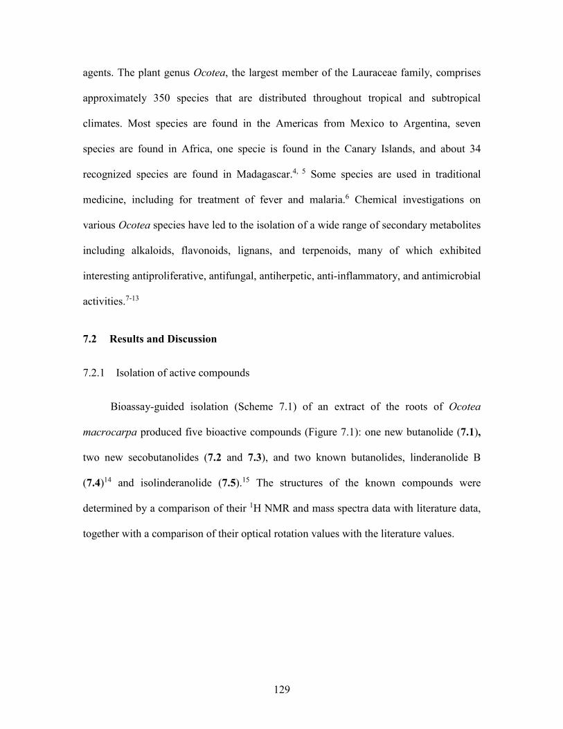
129
agents. The plant genus Ocotea, the largest member of the Lauraceae family, comprises
approximately 350 species that are distributed throughout tropical and subtropical
climates. Most species are found in the Americas from Mexico to Argentina, seven
species are found in Africa, one specie is found in the Canary Islands, and about 34
recognized species are found in Madagascar.4, 5 Some species are used in traditional
medicine, including for treatment of fever and malaria.6 Chemical investigations on
various Ocotea species have led to the isolation of a wide range of secondary metabolites
including alkaloids, flavonoids, lignans, and terpenoids, many of which exhibited
interesting antiproliferative, antifungal, antiherpetic, anti-inflammatory, and antimicrobial
activities.7-13
7.2 Results and Discussion
7.2.1 Isolation of active compounds
Bioassay-guided isolation (Scheme 7.1) of an extract of the roots of Ocotea
macrocarpa produced five bioactive compounds (Figure 7.1): one new butanolide (7.1),
two new secobutanolides (7.2 and 7.3), and two known butanolides, linderanolide B
(7.4)14 and isolinderanolide (7.5).15 The structures of the known compounds were
determined by a comparison of their 1H NMR and mass spectra data with literature data,
together with a comparison of their optical rotation values with the literature values.
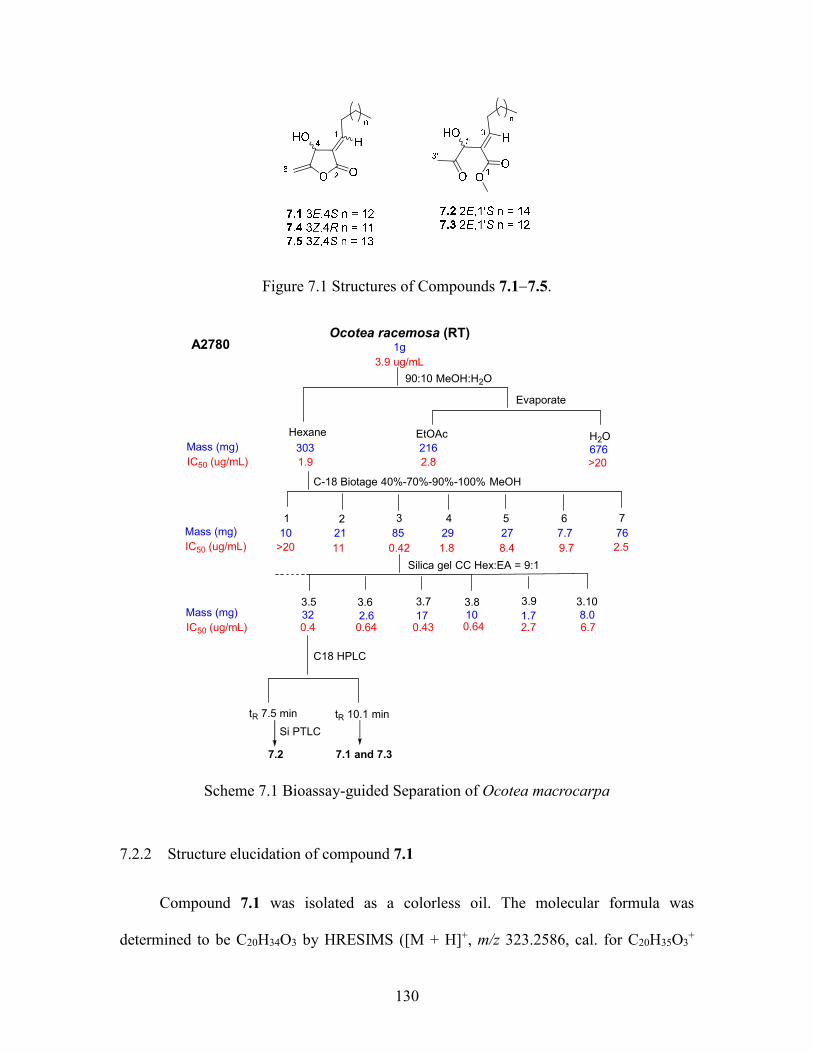
130
Figure 7.1 Structures of Compounds 7.17.5.
Ocotea racemosa (RT)
1g
3.9 ug/mL
Hexane
303
1.9
90:10 MeOH:H2O
216
2.8
Evaporate
676>20
C-18 Biotage 40%-70%-90%-100% MeOH
1
10
>20
2
21
3
85
0.42
4
29
1.8
5
27
8.4
6
7.7
9.7
7
76
2.511
320.4
2.60.64
3.7
170.43
100.64
1.72.7
3.108.06.7
3.5 3.6 3.93.8
Mass (mg)
IC50 (ug/mL)
Mass (mg)
IC50 (ug/mL)
Mass (mg)
IC50 (ug/mL)
H2OEtOAc
Silica gel CC Hex:EA = 9:1
A2780
C18 HPLC
tR 7.5 min tR 10.1 min
Si PTLC
7.2 7.1 and 7.3
Scheme 7.1 Bioassay-guided Separation of Ocotea macrocarpa
7.2.2 Structure elucidation of compound 7.1
Compound 7.1 was isolated as a colorless oil. The molecular formula was
determined to be C20H34O3 by HRESIMS ([M + H]+, m/z 323.2586, cal. for C20H35O3+
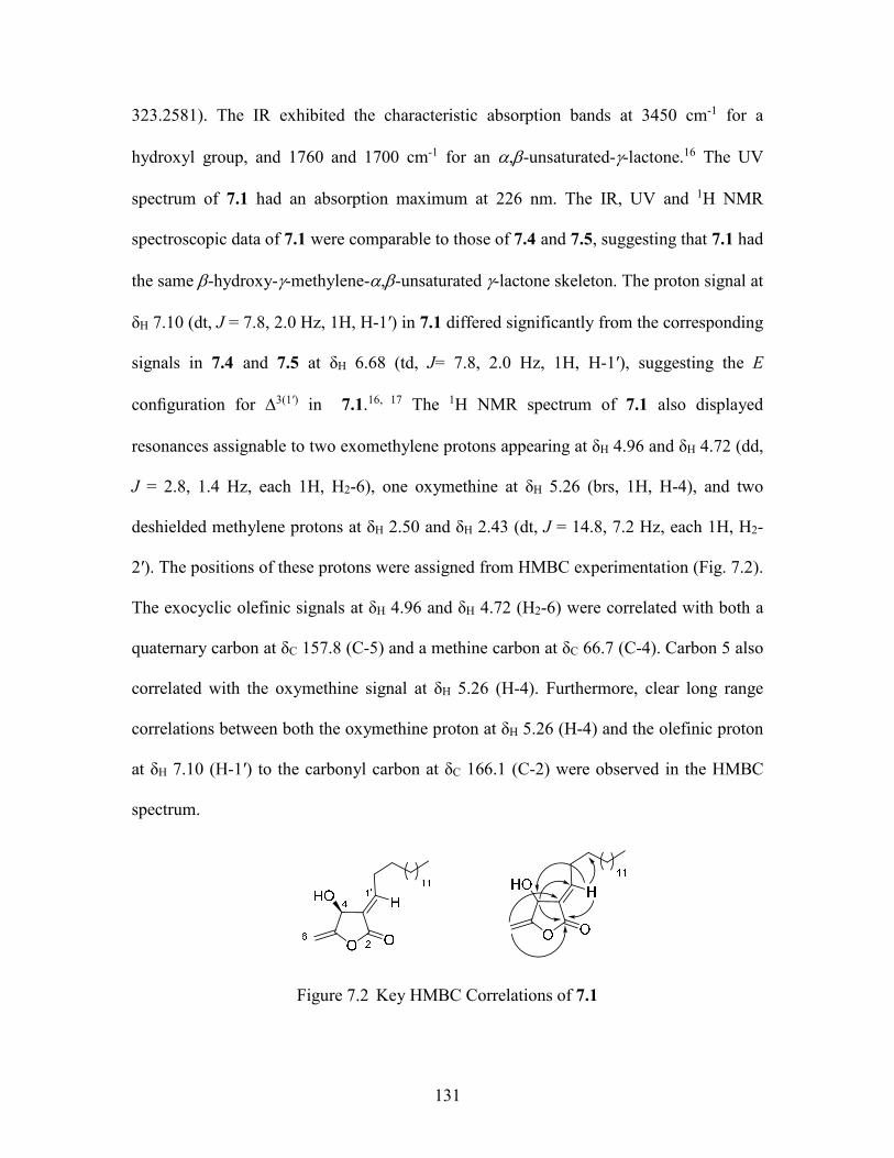
131
323.2581). The IR exhibited the characteristic absorption bands at 3450 cm-1 for a
hydroxyl group, and 1760 and 1700 cm-1 for an ,-unsaturated--lactone.16 The UV
spectrum of 7.1 had an absorption maximum at 226 nm. The IR, UV and 1H NMR
spectroscopic data of 7.1 were comparable to those of 7.4 and 7.5, suggesting that 7.1 had
the same -hydroxy--methylene-,-unsaturated -lactone skeleton. The proton signal at
δH 7.10 (dt, J = 7.8, 2.0 Hz, 1H, H-1′) in 7.1 differed significantly from the corresponding
signals in 7.4 and 7.5 at δH 6.68 (td, J= 7.8, 2.0 Hz, 1H, H-1′), suggesting the E
configuration for 3(1′) in 7.1.16, 17 The 1H NMR spectrum of 7.1 also displayed
resonances assignable to two exomethylene protons appearing at δH 4.96 and δH 4.72 (dd,
J = 2.8, 1.4 Hz, each 1H, H2-6), one oxymethine at δH 5.26 (brs, 1H, H-4), and two
deshielded methylene protons at δH 2.50 and δH 2.43 (dt, J = 14.8, 7.2 Hz, each 1H, H2-
2′). The positions of these protons were assigned from HMBC experimentation (Fig. 7.2).
The exocyclic olefinic signals at δH 4.96 and δH 4.72 (H2-6) were correlated with both a
quaternary carbon at δC 157.8 (C-5) and a methine carbon at δC 66.7 (C-4). Carbon 5 also
correlated with the oxymethine signal at δH 5.26 (H-4). Furthermore, clear long range
correlations between both the oxymethine proton at δH 5.26 (H-4) and the olefinic proton
at δH 7.10 (H-1′) to the carbonyl carbon at δC 166.1 (C-2) were observed in the HMBC
spectrum.
Figure 7.2 Key HMBC Correlations of 7.1
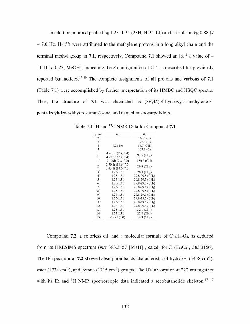
132
In addition, a broad peak at δH 1.251.31 (28H, H-3′14′) and a triplet at δH 0.88 (J
= 7.0 Hz, H-15′) were attributed to the methylene protons in a long alkyl chain and the
terminal methyl group in 7.1, respectively. Compound 7.1 showed an []21D value of –
11.11 (c 0.27, MeOH), indicating the S configuration at C-4 as described for previously
reported butanolides.17-19 The complete assignments of all protons and carbons of 7.1
(Table 7.1) were accomplished by further interpretation of its HMBC and HSQC spectra.
Thus, the structure of 7.1 was elucidated as (3E,4S)-4-hydroxy-5-methylene-3-
pentadecylidene-dihydro-furan-2-one, and named macrocarpolide A.
Table 7.1 1H and 13C NMR Data for Compound 7.1
posn δH δc 2 166.1 (C) 3 127.4 (C) 4 5.26 brs 66.7 (CH) 5 157.8 (C)
6 4.96 dd (2.8, 1.4) 4.72 dd (2.8, 1.4)
91.5 (CH2)
1′ 7.10 dt (7.8, 2.0) 150.3 (CH)
2′ 2.50 dt (14.6, 7.7) 2.43 dt (14.6, 7.7)
29.8 (CH2)
3′ 1.25-1.31 28.3 (CH2) 4′ 1.25-1.31 29.8-29.5 (CH2) 5′ 1.25-1.31 29.8-29.5 (CH2) 6′ 1.25-1.31 29.8-29.5 (CH2) 7′ 1.25-1.31 29.8-29.5 (CH2) 8′ 1.25-1.31 29.8-29.5 (CH2) 9′ 1.25-1.31 29.8-29.5 (CH2)
10′ 1.25-1.31 29.8-29.5 (CH2) 11’ 1.25-1.31 29.8-29.5 (CH2) 12′ 1.25-1.31 29.8-29.5 (CH2) 13′ 1.25-1.31 32.1 (CH2) 14′ 1.25-1.31 22.8 (CH2) 15′ 0.88 t (7.0) 14.3 (CH3)
Compound 7.2, a colorless oil, had a molecular formula of C23H42O4, as deduced
from its HRESIMS spectrum (m/z 383.3157 [M+H]+, calcd. for C23H43O4+, 383.3156).
The IR spectrum of 7.2 showed absorption bands characteristic of hydroxyl (3458 cm-1),
ester (1734 cm-1), and ketone (1715 cm-1) groups. The UV absorption at 222 nm together
with its IR and 1H NMR spectroscopic data indicated a secobutanolide skeleton.17, 19
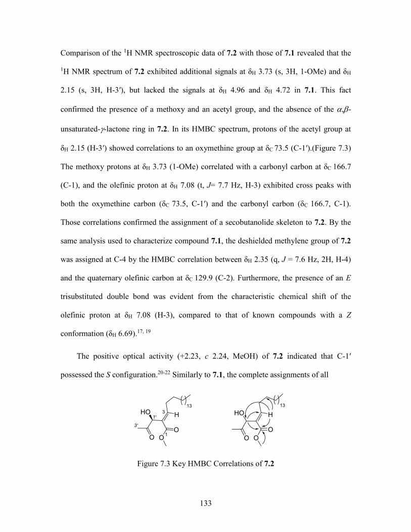
133
Comparison of the 1H NMR spectroscopic data of 7.2 with those of 7.1 revealed that the
1H NMR spectrum of 7.2 exhibited additional signals at δH 3.73 (s, 3H, 1-OMe) and δH
2.15 (s, 3H, H-3′), but lacked the signals at δH 4.96 and δH 4.72 in 7.1. This fact
confirmed the presence of a methoxy and an acetyl group, and the absence of the ,-
unsaturated--lactone ring in 7.2. In its HMBC spectrum, protons of the acetyl group at
δH 2.15 (H-3′) showed correlations to an oxymethine group at δC 73.5 (C-1′).(Figure 7.3)
The methoxy protons at δH 3.73 (1-OMe) correlated with a carbonyl carbon at δC 166.7
(C-1), and the olefinic proton at δH 7.08 (t, J= 7.7 Hz, H-3) exhibited cross peaks with
both the oxymethine carbon (δC 73.5, C-1′) and the carbonyl carbon (δC 166.7, C-1).
Those correlations confirmed the assignment of a secobutanolide skeleton to 7.2. By the
same analysis used to characterize compound 7.1, the deshielded methylene group of 7.2
was assigned at C-4 by the HMBC correlation between δH 2.35 (q, J = 7.6 Hz, 2H, H-4)
and the quaternary olefinic carbon at δC 129.9 (C-2). Furthermore, the presence of an E
trisubstituted double bond was evident from the characteristic chemical shift of the
olefinic proton at δH 7.08 (H-3), compared to that of known compounds with a Z
conformation (δH 6.69).17, 19
The positive optical activity (+2.23, c 2.24, MeOH) of 7.2 indicated that C-1′
possessed the S configuration.20-22 Similarly to 7.1, the complete assignments of all
HO
O
O
H
O
13
HO
OO
H
O
3'
1'3
1
13
Figure 7.3 Key HMBC Correlations of 7.2
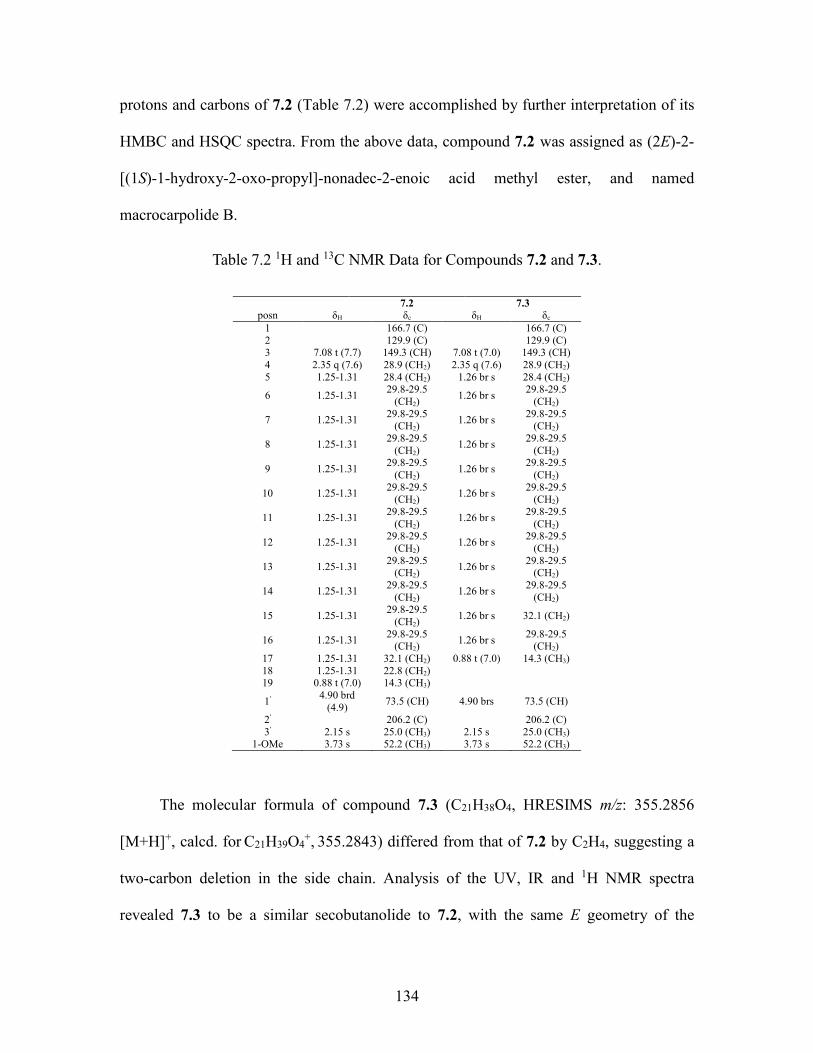
134
protons and carbons of 7.2 (Table 7.2) were accomplished by further interpretation of its
HMBC and HSQC spectra. From the above data, compound 7.2 was assigned as (2E)-2-
[(1S)-1-hydroxy-2-oxo-propyl]-nonadec-2-enoic acid methyl ester, and named
macrocarpolide B.
Table 7.2 1H and 13C NMR Data for Compounds 7.2 and 7.3.
7.2 7.3
posn δH δc δH δc 1 166.7 (C) 166.7 (C) 2 129.9 (C) 129.9 (C) 3 7.08 t (7.7) 149.3 (CH) 7.08 t (7.0) 149.3 (CH) 4 2.35 q (7.6) 28.9 (CH2) 2.35 q (7.6) 28.9 (CH2) 5 1.25-1.31 28.4 (CH2) 1.26 br s 28.4 (CH2)
6 1.25-1.31 29.8-29.5
(CH2) 1.26 br s
29.8-29.5 (CH2)
7 1.25-1.31 29.8-29.5
(CH2) 1.26 br s
29.8-29.5 (CH2)
8 1.25-1.31 29.8-29.5
(CH2) 1.26 br s
29.8-29.5 (CH2)
9 1.25-1.31 29.8-29.5
(CH2) 1.26 br s
29.8-29.5 (CH2)
10 1.25-1.31 29.8-29.5
(CH2) 1.26 br s
29.8-29.5 (CH2)
11 1.25-1.31 29.8-29.5
(CH2) 1.26 br s
29.8-29.5 (CH2)
12 1.25-1.31 29.8-29.5
(CH2) 1.26 br s
29.8-29.5 (CH2)
13 1.25-1.31 29.8-29.5
(CH2) 1.26 br s
29.8-29.5 (CH2)
14 1.25-1.31 29.8-29.5
(CH2) 1.26 br s
29.8-29.5 (CH2)
15 1.25-1.31 29.8-29.5
(CH2) 1.26 br s 32.1 (CH2)
16 1.25-1.31 29.8-29.5
(CH2) 1.26 br s
29.8-29.5 (CH2)
17 1.25-1.31 32.1 (CH2) 0.88 t (7.0) 14.3 (CH3) 18 1.25-1.31 22.8 (CH2) 19 0.88 t (7.0) 14.3 (CH3)
1′ 4.90 brd
(4.9) 73.5 (CH) 4.90 brs 73.5 (CH)
2′ 206.2 (C) 206.2 (C) 3′ 2.15 s 25.0 (CH3) 2.15 s 25.0 (CH3)
1-OMe 3.73 s 52.2 (CH3) 3.73 s 52.2 (CH3)
The molecular formula of compound 7.3 (C21H38O4, HRESIMS m/z: 355.2856
[M+H]+, calcd. for C21H39O4+, 355.2843) differed from that of 7.2 by C2H4, suggesting a
two-carbon deletion in the side chain. Analysis of the UV, IR and 1H NMR spectra
revealed 7.3 to be a similar secobutanolide to 7.2, with the same E geometry of the

135
trisubstituted double bond [δH 7.08 (t, J = 7.0 Hz, 1H, H-3)], but with two carbons less in
the alkyl chain. Similarly to 7.2, the S configuration at C-1′ was deduced by the positive
optical rotation value of +2.27 (c 0.88, MeOH).20-22 The complete assignments of all
protons and carbons of 7.3 (Table 7.2) were accomplished by interpretation of its HMBC
and HSQC spectra. Therefore, compound 7.3 was assigned as (2E)-2-[(1S)-1-hydroxy-2-
oxo-propyl]-heptadec-2-enoic acid methyl ester, and named macrocarpolide C.
7.2.3 Antiproliferative Activities
Compounds 7.17.5 showed good antiproliferative activities against the drug-
sensitive A2780 ovarian cell line23 as previously described24 using paclitaxel (IC50 0.073
± 0.015 M) as the positive control. Their IC50 values were 2.57 ± 0.12 (7.1), 1.98 ±
0.23 (7.2), 1.67 ± 0.05 (7.3), 2.43 ± 0.41 (7.4), and 1.65 ± 0.44 M (7.5). The similar
IC50 values for the five compounds suggests that they have a similar mechanism of
action, possibly as Michael acceptors.
7.3 Experimental section
7.3.1 General experimental procedures
IR and UV spectra were measured on MIDAC M-series FTIR and Shimadzu UV-
1201 spectrophotometers, respectively. 1H and 13C NMR spectra were recorded on a
Bruker Avance 500 spectrometer in CDCl3 (with CDCl3 as reference). Mass spectra were
obtained on an Agilent 6220 mass spectrometer. Open column chromatography and
preparative TLC was performed using silica gel (230–400 mesh, Silicycle Co. USA) and
silica gel plates (60 F254, Silicycle Co. USA), respectively. Flash chromatography was
136
performed on a Biotage Horizon flash chromatograph using a C18 column (Phenomenex,
150 × 12 mm, 11 g). Semi-preparative HPLC was performed using Shimadzu LC-10AT
pumps coupled with a semipreparative Phenomenex C18 column (5 μm, 250 × 10 mm), a
Shimadzu SPD M10A diode array detector, and a SCL-10A system controller.
7.3.2 Plant material
Plant parts of Ocotea macrocarpa Kosterm. (Lauraceae) were collected by
Andriamalala Rakotondrafara et al. on 23 March 2011. The collection was made in the
district of Ankazobe in the Analamanga region of Madagascar, in the Analamaitso forest
near the village of Tsaramandroso. The coordinates were 17°57'32"S 047°07'33"E, and
the elevation was 1478 m. The tree was 14 m tall with a diameter at breast height of 25
cm. It had cream-yellow petals and yellow inflorescences. Duplicate voucher specimens
(Rakotondrafara 989) have been deposited at the Centre National d'Application des
Recherches Pharmaceutiques (CNARP), Parc Botanique and Zoologique de Tsimbazaza
(TAN), the Missouri Botanical Garden, St. Louis, Missouri (MO), and the herbarium of
the National Museum of Natural History, Paris (P).
7.3.3 Extraction and isolation
A ground sample of Ocotea macrocarpa roots (250 g) was extracted with EtOH at
room temperature to yield 8.3 g of crude EtOH extract designated MG4725. A total of
1.85 g of this extract was made available to Virginia Tech. The crude EtOH extract (1.0
g) was dissolved in 90% aq. MeOH (300 mL), and extracted with hexanes (3 × 200 mL).
Evaporation of the hexanes-soluble fraction afforded 302.7 mg of residue. The 90% aq.
MeOH layer was then evaporated, suspended in H2O (300 mL), and extracted with
137
EtOAc (3 × 200 mL) to yield 215.5 mg of EtOAc-soluble fraction. The aqueous layer
was concentrated to give 476.6 mg of brown residue. The hexanes fraction was found to
be cytotoxic with an IC50 value of 1.9 µg/mL and was subjected to C 18 flash column
chromatography (aq. MeOH, 40% to 70% to 90% to 100%) to give 7 new fractions. The
most active fraction Fr 3 (85.4 mg, IC50 0.42 µg/mL) was then divided into 10 sub-
fractions by silica gel open column chromatography (Hex:EtOAc = 9:1). Further
purification of one the most active sub-fractions Fr 3-5 (IC50 0.4 µg/mL) was done by
using HPLC on a C18 column with a solvent gradient from H2O:MeOH, 10:90 to 0:100
from 0 to 10 min and ending with 100% MeOH from 10 to 30 min. This process yielded
two active fractions with retention time of 7.5 min and 10.1 min respectively. Preparative
silica gel TLC (Hex:EtOAc= 9:1) purification of the two fractions yielded compounds 7.2
(0.76 µg/mL), and compounds 7.1 (0.91 µg/mL) and 7.3 (0.83 µg/mL), respectively.
Further purification of the other active sub-fraction from silica gel open column Fr 3-6 by
C18 HPLC, with a solvent gradient from H2O:MeOH, 30:70 to 20:80 from 0 to 15 min,
to 10:90 from 15 to 25 min, to 0:100 from 25 to 35 min, and ending with 100% MeOH
furnished compounds 7.4 (tR 23.1 min, 0.75 µg/mL) and 7.5 (tR 27.6 min, 0.55 µg/mL).
7.3.4 Macrocarpolide A (7.1)
Colorless oil, []21D -11.11 (c 0.27, MeOH); UV (MeOH) λmax (log ε) : 226 (3.35)
nm; IR (film) νmax 3450, 1760, 1700, 1049 cm-1; 1H and 13C NMR data, see Table 7.1;
HRESIMS m/z [M+H]+ 323.2586 (calcd for C20H35O3+
, 323.2581)
138
7.3.5 Macrocarpolide B (7.2)
Colorless oil, []21D +2.23 (c 2.24, MeOH); UV (MeOH) λmax (log ε) : 222 (3.26)
nm; IR (film) νmax 3458, 1734, 1715, 1040 cm-1; 1H and 13C NMR data, see Table 7.1;
HRESIMS m/z [M+H]+ 383.3157 (calcd for C23H43O4+
, 383.3156)
7.3.6 Macrocarpolide C (7.3)
Colorless oil, []21D +2.27 (c 0.88, MeOH); UV (MeOH) λmax (log ε) : 222 (3.43)
nm; IR (film) νmax 3455, 1736, 1710, 1060 cm-1; 1H and 13C NMR data, see Table 7.1;
HRESIMS m/z [M+H]+ 355.2856 (calcd for C21H39O4+
, 355.2843)
7.3.7 Antiproliferative bioassay
The A2780 ovarian cancer cell line antiproliferative bioassay was performed at
Virginia Tech as previously reported.24 The A2780 cell line is a drug-sensitive ovarian
cancer cell line.23 Paclitaxel was used as the positive control; it had an IC50 value of 0.073
± 0.015 M.
139
7.4 References
1. Liu, Y.; Cheng, E.; Rakotondraibe, L. H.; Brodie, P. J.; Applequist, W.;
Randrianaivo, R.; Rakotondrafara, A.; Ratsimbason, M.; Rasamison, V. E.; Kingston, D.
G. I. Antiproliferative compounds from Ocotea macrocarpa from the Madagascar dry
forest. Tetrahedron Lett. 2015, Ahead of Print.
2. Harinantenaina, L.; Brodie, P. J.; Maharavo, J.; Bakary, G.; TenDyke, K.; Shen,
Y.; Kingston, D. G. I. Antiproliferative homoscalarane sesterterpenes from two
Madagascan sponges. Bioorg. Med. Chem. 2013, 21, 2912-2917.
3. Murphy, B. T.; Brodie, P.; Slebodnick, C.; Miller, J. S.; Birkinshaw, C.;
Randrianjanaka, L. M.; Andriantsiferana, R.; Rasamison, V. E.; TenDyke, K.; Suh, E. M.;
Kingston, D. G. I. Antiproliferative limonoids of a Malleastrum sp. from the Madagascar
rainforest. J. Nat. Prod. 2008, 71, 325-329.
4. Rohwer, J. G. Toward a phylogenetic classification of the lauraceae: Evidence
from matK sequences. Syst. Bot. 2000, 25, 60-71.
5. van der Werff, H. A revision of the genus Ocotea Aubl. (Lauraceae) in
Madagascar and the Comoro Islands. Adansonia, sér. 3, 2013, 35, 235-279.
6. Coelho de Souza, G.; Haas, A. P. S.; von Poser, G. L.; Schapoval, E. E. S.;
Elisabetsky, E. Ethnopharmacological studies of antimicrobial remedies in the south of
Brazil. J. Ethnopharmacol. 2004, 90, 135-143.
7. Destryana, R. A.; Young, D. G.; Woolley, C. L.; Huang, T. C.; Wu, H. Y.; Shih, W.
L. Antioxidant and anti-inflammation activities of Ocotea, Copaiba and blue cypress
essential oils in vitro and in vivo. J. Am. Oil Chem. Soc. 2014, 91, 1531-1542.
8. Jose de Camargo, M.; Miranda, M. L. D.; Kagamida, C. M.; Rodrigues, E. D.;
140
Garcez, F. R.; Garcez, W. S. Sesquiterpenes of Ocotea lancifolia (Lauraceae). Quim.
Nova 2013, 36, 1008-1013.
9. Garett, R.; Romanos, M. T. V.; Borges, R. M.; Santos, M. G.; Rocha, L.; da Silva,
A. J. R. Antiherpetic activity of a flavonoid fraction from Ocotea notata leaves. Rev.
Bras. Farmacogn. 2012, 22, 306-313.
10. Yamaguchi, M. U.; Garcia, F. P.; Cortez, D. A. G.; Ueda-Nakamura, T.; Filho, B.
P. D.; Nakamura, C. V. Antifungal effects of Ellagitannin isolated from leaves of Ocotea
odorifera (Lauraceae). Antonie Van Leeuwenhoek 2011, 99, 507-514.
11. Garcez, F. R.; da Silva, A. F. G.; Garcez, W. S.; Linck, G.; Matos, M. d. F. C.;
Santos, E. C. S.; Queiroz, L. M. M. Cytotoxic aporphine alkaloids from Ocotea
acutifolia. Planta Med. 2011, 77, 383-387.
12. Castro, R. D.; Lima, E. O. Antifungal activity of Brazilian sassafras (Ocotea
odorifera Vell.) and rosemary (Rosmarinus officinalis L.) essential oils against genus
Candida. Rev. Bras. Plant. Med. 2011, 13, 203-208.
13. Cuca, L. E.; Leon, P.; Coy, E. D. A bicyclo[3.2.1]octanoid neolignan and toxicity
of the ethanol extract from the fruit of Ocotea heterochroma. Chem. Nat. Compd. 2009,
45, 179-181.
14. Seki, K.; Sasaki, T.; Wano, S.; Haga, K.; Kaneko, R. Linderanolides and
isolinderanolides, ten butanolides from Lindera glauca. Phytochemistry 1995, 40, 1175-
1181.
15. Anderson, J. E.; Ma, W.; Smith, D. L.; Chang, C. J.; McLaughlin, J. L.
Biologically active γ-lactones and methylketoalkenes from Lindera benzoin. J. Nat. Prod.
1992, 55, 71-83.
141
16. Cheng, H. I.; Lin, W. Y.; Duh, C. Y.; Lee, K. H.; Tsai, I. L.; Chen, I. S. New
cytotoxic butanolides from Litsea acutivena. J. Nat. Prod. 2001, 64, 1502-1505.
17. Cheng, M. J.; Tsai, I. L.; Lee, S. J.; Jayaprakasam, B.; Chen, I. S. Steryl epoxide,
secobutanolide and butanolides from the stem wood of Machilus zuihoensis.
Phytochemistry (Elsevier) 2005, 66, 1180-1185.
18. Martinez V, J. C.; Yoshida, M.; Gottlieb, O. R. The chemistry of Brazilian
lauraceae. Part LXI. ω-Ethyl, ω-ethenyl and ω-ethynyl-α-alkylidene-γ-lactones from
Clinostemon mahuba. Phytochemistry 1981, 20, 459-464.
19. Tsai, I. L.; Hung, C. H.; Duh, C. Y.; Chen, I. S. Cytotoxic butanolides and
secobutanolides from the stem wood of formosan Lindera communis. Planta Med. 2002,
68, 142-145.
20. Chen, C. Y.; Chen, C. H.; Wong, C. H.; Liu, Y. W.; Lin, Y. S.; Wang, Y. D.; Hsui,
Y. R. Cytotoxic constituents of the stems of Cinnamomum subavenium. J. Nat. Prod.
2007, 70, 103-106.
21. Kuo, S. Y.; Hsieh, T. J.; Wang, Y. D.; Lo, W. L.; Hsui, Y. R.; Chen, C. Y. Cytotoxic
constituents from the leaves of Cinnamomum subavenium. Chem. Pharm. Bull. 2008, 56,
97-101.
22. Tanaka, H.; Nakamura, T.; Ichino, K.; Ito, K. Secoisolancifolide and
secoisoobtusilactone in Actinodaphne longifolia. Phytochemistry 1989, 28, 1905-1907.
23. Louie, K. G.; Behrens, B. C.; Kinsella, T. J.; Hamilton, T. C.; Grotzinger, K. R.;
McKoy, W. M.; Winker, M. A.; Ozols, R. F. Radiation survival parameters of
antineoplastic drug-sensitive and -resistant human ovarian cancer cell lines and their
modification by buthionine sulfoximine. Cancer Res. 1985, 45, 2110-2115.
142
24. Cao, S.; Brodie, P. J.; Miller, J. S.; Randrianaivo, R.; Ratovoson, F.; Birkinshaw,
C.; Andriantsiferana, R.; Rasamison, V. E.; Kingston, D. G. I. Antiproliferative xanthones
of terminalia calcicola from the Madagascar rain forest. J. Nat. Prod. 2007, 70, 679-681.
8 Antiproliferative Compounds from Malleastrum sp. from the Madagascar Dry Forest
This chapter is a slightly expanded version of an article submitted to the journal
Natural Product Communications. Attributions from co-authors of the articles are
described as follows in the order of the names listed. The author of this dissertation (Yixi
Liu) mentored the undergraduate student (Houston Wiedle Jr.) with the compounds
isolation, and elucidated the structures of all compounds, and drafted the manuscript.
Mrs. Peggy Brodie performed the A2780 bioassay on the isolated fractions and
compounds. Dr. Martin Callmander and Dr. Robert Rakotondrajaona from Missouri
Botanical Garden collected and identified the plant. Dr. Etienne Rakotobe and Dr.
Vincent E. Rasamison from Madagascar carried out the initial plant extraction. Dr. David
G. I. Kingston was a mentor for this work and the corresponding author for the published
article. He provided critical suggestions for this work and crucial revisions to the
manuscript.
8.1 Introduction
As a part of the Madagascar International Cooperative Biodiversity Group (ICBG)
program,1, 2 an ethanol extract of the leaves of a Malleastrum sp. (Meliceae) was selected
for evaluation as a possible source of new antiproliferative agents based on its activity
against the A2780 ovarian cancer cell line. The Meliceae family has its origins in the
western Gondwanan region of Pangea and played an important part in the dispersal of the
mahogany biota. The dispersal was most likely an “out-of-Africa” scenario with dispersal
across Eurasia and North America due to a land bridge. With multiple fossil findings, it
has been confirmed that the entry of Meliceae into southern continents from Oligocene to

144
Pliocene must be considered as important in establishing a pantropical distribution for the
family.3 This family has been known to produce structurally unique, highly oxygenated,
and biologically active liminoids.4 Liminoids have been shown to inhibit the growth of
estrogen receptor negative and positive human breast cancer cells in culture. There has
also been success in inhibiting cancer cell lines of leukemia, ovary, cervix, stomach, and
liver. Prevalent in many known antioxidant foods, rings in the limonoid nucleus play a
critical role in antineoplastic activity.5 Therefore, plants in the Meliceae family may hold
compounds that can be utilized in conjunction with radiation therapies of certain
cancers.6
As a member of the Meliceae family, the genus Malleastrum was first segregated
from the genus Cipadessa and has been found to contain eleven species. Recent chemical
work on the genus Malleastrum has afforded new limonoids with tetranortriterpenoid
skeletons.7 These compounds have also been found in other plants such as Carapa
guianensis and have been used in popular medicine to treat inflammation and infection.
Compounds in the plant material such as 6α-acetoxygedunin, 7-deacetoxy-7-oxogedunin,
andirobin, gedunin and methyl-angolensate impair the production of inflammatory
mediators that trigger leukocyte infiltration into the inflammatory site as well as the
inflammatory cytokines tumor necrosis factor (TNF)-α and IL-1β.8
A bioassay-guided isolation of compounds from the leaves of the plant,
Malleastrum rakotozafyi, afforded a new clerodane diterpene, 18-oxo-cleroda-3,13-dien-
16,15-olide (8.1), together with three known clerodane diterpenes: 16,18-
dihydroxykolavenic acid lactone (8.2),9 solidagolactone (8.3),10 and (-)-kolavenol (8.4),11
and one known labdane diterpene: 3-oxo-ent-Iabda-8(17),13-dien-15,16-olide (8.5).12

145
(Figure 8.1)
Figure 8.1 Structures of Compounds 8.18.5.
8.2 Results and Discussion
8.2.1 Isolation of active compounds
An active CH2Cl2-soluble fraction obtained from liquid-liquid partition of the
extract (100 mg) was subjected to dereplication studies using size-exclusion
chromatography, reverse phase HPLC coupled with bioassay, high-resolution ESIMS
(HRESIMS), 1HNMR, and a database search using the online Dictionary of Natural
Product (DNP). The results indicated that the extract contained a possible new bioactive
compound, therefore a larger scale sample (1.0 g) was investigated. Fractionation of this
extract yielded an antiproliferative CH2Cl2 fraction which was further subjected to
Sephadex LH-20 column chromatography followed by normal phase solid phase extract
(SPE). The most active fractions from the silica gel SPE were subjected to C18 HPLC to
yield one new (8.1) and four known (8.28.5) bioactive diterpenes. (Scheme 8.1)
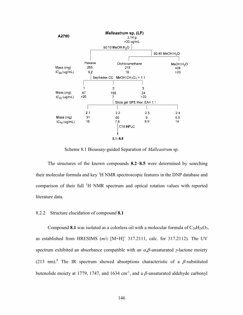
146
Scheme 8.1 Bioassay-guided Separation of Malleastrum sp.
The structures of the known compounds 8.28.5 were determined by searching
their molecular formula and key 1H NMR spectroscopic features in the DNP database and
comparison of their full 1H NMR spectrum and optical rotation values with reported
literature data.
8.2.2 Structure elucidation of compound 8.1
Compound 8.1 was isolated as a colorless oil with a molecular formula of C20H28O3,
as established from HRESIMS (m/z [M+H]+ 317.2111, calc. for 317.2112). The UV
spectrum exhibited an absorbance compatible with an ,-unsaturated -lactone moiety
(213 nm).9 The IR spectrum showed absorptions characteristic of a -substituted
butenolide moiety at 1779, 1747, and 1634 cm-1, and a -unsaturated aldehyde carbonyl
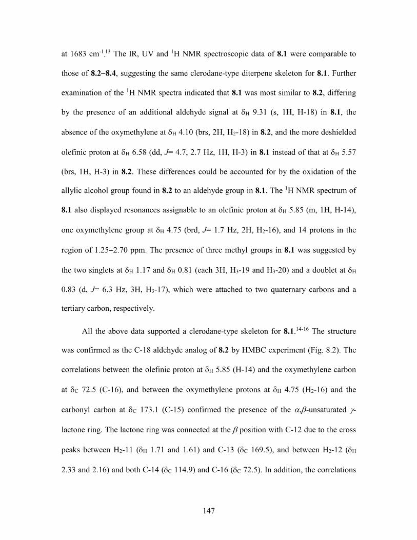
147
at 1683 cm-1.13 The IR, UV and 1H NMR spectroscopic data of 8.1 were comparable to
those of 8.28.4, suggesting the same clerodane-type diterpene skeleton for 8.1. Further
examination of the 1H NMR spectra indicated that 8.1 was most similar to 8.2, differing
by the presence of an additional aldehyde signal at H 9.31 (s, 1H, H-18) in 8.1, the
absence of the oxymethylene at H 4.10 (brs, 2H, H2-18) in 8.2, and the more deshielded
olefinic proton at H 6.58 (dd, J= 4.7, 2.7 Hz, 1H, H-3) in 8.1 instead of that at H 5.57
(brs, 1H, H-3) in 8.2. These differences could be accounted for by the oxidation of the
allylic alcohol group found in 8.2 to an aldehyde group in 8.1. The 1H NMR spectrum of
8.1 also displayed resonances assignable to an olefinic proton at H 5.85 (m, 1H, H-14),
one oxymethylene group at H 4.75 (brd, J= 1.7 Hz, 2H, H2-16), and 14 protons in the
region of 1.252.70 ppm. The presence of three methyl groups in 8.1 was suggested by
the two singlets at H 1.17 and H 0.81 (each 3H, H3-19 and H3-20) and a doublet at H
0.83 (d, J= 6.3 Hz, 3H, H3-17), which were attached to two quaternary carbons and a
tertiary carbon, respectively.
All the above data supported a clerodane-type skeleton for 8.1.14-16 The structure
was confirmed as the C-18 aldehyde analog of 8.2 by HMBC experiment (Fig. 8.2). The
correlations between the olefinic proton at H 5.85 (H-14) and the oxymethylene carbon
at C 72.5 (C-16), and between the oxymethylene protons at H 4.75 (H2-16) and the
carbonyl carbon at C 173.1 (C-15) confirmed the presence of the ,-unsaturated -
lactone ring. The lactone ring was connected at the position with C-12 due to the cross
peaks between H2-11 (H 1.71 and 1.61) and C-13 (C 169.5), and between H2-12 (H
2.33 and 2.16) and both C-14 (C 114.9) and C-16 (C 72.5). In addition, the correlations
148
between the aldehyde proton at H 9.31 (H-18) and both the olefinic carbon C-3 (C 153.1)
and the quaternary carbon C-5 (C 37.6) allowed us to place the aldehyde group at C-4. In
the same manner, the three methyl groups were located at C-5, C-8, and C-9 based on
long range HMBC correlations between H 1.17 (H3-19) and both C 151.5 (C-4) and C
34.6 (C-6), between H 0.83 (H3-17) and both C-7 (C 27.7) and C-9 (C 37.5), and
between H3-20 and both C-8 (C 35.9) and C-10 (C 46.4).
O
O
OH
HMBC
NOESY
HO
O
1
35
7
9
1113
15
16
17
18 19
20
O
Figure 8.2 Key HMBC and NOESY Correlations of 8.1.
Furthermore, the relative configuration of 8.1 was determined by a NOESY
experiment (Fig. 8.2). Observation of clear NOE correlations between H 0.81 (H3-20)
and both H 0.83 (H3-17) and H 1.17 (H3-19), indicated the cis relationships between the
three methyl groups. At the same time, the NOE correlation between H 1.27 (H-10) and
H 1.61 (H-11a) suggested the cis relationships between C-10 and the C-11C-12 side
chain bearing the lactone ring, and a trans A/B ring junction of 8.1. The complete
assignments of all protons and carbons of 8.1 (Table 8.1) were accomplished by further
interpretation of its HMBC and HSQC spectra. Thus, the structure of compound 8.1 was
elucidated as 18-oxo-cleroda-3,13-dien-16,15-olide.
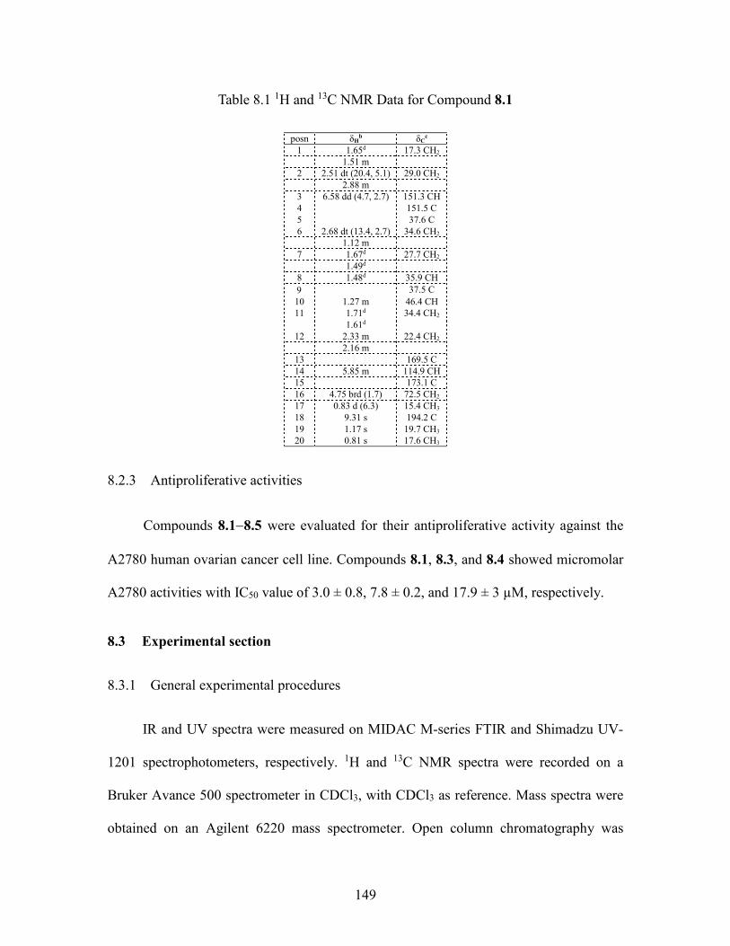
149
Table 8.1 1H and 13C NMR Data for Compound 8.1
posn δH
b δCc
1 1.65d 17.3 CH2 1.51 m
2 2.51 dt (20.4, 5.1) 29.0 CH2 2.88 m
3 6.58 dd (4.7, 2.7) 151.3 CH 4 151.5 C 5 37.6 C 6 2.68 dt (13.4, 2.7) 34.6 CH2 1.12 m
7 1.67d 27.7 CH2 1.49d
8 1.48d 35.9 CH
37.5 C
10 1.27 m 46.4 CH 11 1.71d 34.4 CH2
1.61d 12 2.33 m 22.4 CH2
2.16 m 13 169.5 C 14 5.85 m 114.9 CH 15 173.1 C 16 4.75 brd (1.7) 72.5 CH2 17 0.83 d (6.3) 15.4 CH3 18 9.31 s 194.2 C 19 1.17 s 19.7 CH3 20 0.81 s 17.6 CH3
8.2.3 Antiproliferative activities
Compounds 8.18.5 were evaluated for their antiproliferative activity against the
A2780 human ovarian cancer cell line. Compounds 8.1, 8.3, and 8.4 showed micromolar
A2780 activities with IC50 value of 3.0 ± 0.8, 7.8 ± 0.2, and 17.9 ± 3 µM, respectively.
8.3 Experimental section
8.3.1 General experimental procedures
IR and UV spectra were measured on MIDAC M-series FTIR and Shimadzu UV-
1201 spectrophotometers, respectively. 1H and 13C NMR spectra were recorded on a
Bruker Avance 500 spectrometer in CDCl3, with CDCl3 as reference. Mass spectra were
obtained on an Agilent 6220 mass spectrometer. Open column chromatography was
150
performed using Sephadex LH-20. Solid phase extract was performed using silica gel
(230-400 mesh, Silicycle Co. USA). Semi-preparative HPLC was performed using
Shimadzu LC-10AT pumps coupled with a semipreparative Phenomenex C-18 column (5
μm, 250 × 10 mm), a Shimadzu SPD M10A diode array detector, and a SCL-10A system
controller.
8.3.2 Plant material
8.3.3 Extraction and isolation
A ground sample of M. sp. leaves g was extracted with EtOH at room temperature
to yield g of crude EtOH extract designated MG 3652. A total of 3.14 g of this extract
was made available to Virginia Tech. A part of the crude EtOH extract (1.0 g) was
dissolved in 90% aq. MeOH (300 mL) and extracted with hexanes (3 × 200 mL).
Evaporation of the hexanes-soluble fraction yielded 265.1 mg of residue. The 90% aq.
MeOH layer was then evaporated, suspended in H2O (300 mL), and extracted with
dichloromethane (3 × 200 mL) to yield 218.3 mg of CH2Cl2-soluble fraction. The
aqueous layer was concentrated to give 428 mg of green residue. The hexanes fraction
was found to be weakly cytotoxic with an IC50 value of 9.2 µg/mL and was subjected to
Sephadex LH-20 open column chromatography (CH2Cl2: MeOH = 1:1) to give 3
fractions. The most active fraction Fr-2 (IC50 7 g/mL) was fractionated by silica gel SPE
using hexanes/EtOAc (70:30 to 50:50 to 30:70 to 0:100, MeOH wash) to give 4 sub-
fractions. The second sub-fraction Fr-2-2 (IC50 6.8 g/mL) was then subjected to C18
HPLC with a solvent gradient from H2O:CH3CN, 30:70 to 20:80 from 0.01 to10 min, to
10:90 from 10 to 15 min, to 0:100 from 15 to 20 min, and ending with 100% CH3CN to
151
35 min, furnishing compounds 8.1 (tR 13.5 min, IC50 0.97 g/mL), 8.2 (tR 9.5 min,
IC50 >20 g/mL), 8.3 (tR 23.8 min, IC50 2.37 g/mL), 8.4 (tR 28.6 min, IC50 5.2 g/mL),
and 8.5 (tR 12 min, IC50 >20 g/mL).
8.3.4 18-Oxo-cleroda-3,13-dien-16,15-olide (8.1)
Amorphous powder, []21D -60.2 (c 0.68, MeOH); UV (MeOH) λmax (log ε): 213
(3.96) nm; IR (film) νmax 2922, 2864, 1779, 1747, 1715, 1683, 1559 cm-1; 1H and 13C
NMR data, see Table 8.1; HRESIMS: m/z [M+H]+ 317.2112 (calcd. for C20H29O3+:
317.2111)
8.3.5 Antiproliferative bioassay
The A2780 ovarian cancer cell line antiproliferative bioassay was performed at
Virginia Tech as previously reported.17 The A2780 cell line is a drug-sensitive ovarian
cancer cell line.6 Paclitaxel was used as the positive control; it had an IC50 value of 0.073
± 0.015 M.
152
8.4 References
1. Kingston, D. G. I. Modern natural products drug discovery and its relevance to
biodiversity conservation. J. Nat. Prod. 2011, 74, 496-511.
2. Murphy, B. T.; Brodie, P.; Slebodnick, C.; Miller, J. S.; Birkinshaw, C.;
Randrianjanaka, L. M.; Andriantsiferana, R.; Rasamison, V. E.; TenDyke, K.; Suh, E. M.;
Kingston, D. G. I. Antiproliferative limonoids of a Malleastrum sp. from the Madagascar
rainforest. J. Nat. Prod. 2008, 71, 325-329.
3. Muellner, A. N.; Savolainen, V.; Samuel, R.; Chase, M. W. The mahogany family
"out-of-Africa": Divergence time estimation, global biogeographic patterns inferred from
plastid rbcL DNA sequences, extant, and fossil distribution of diversity. Mol. Phylogenet.
Evol. 2006, 40, 236-250.
4. Coombes, P. H.; Mulholland, D. A.; Randrianarivelojosia, M. Vilasinin limonoids
from Malleastrum antsingyense J.F. Leroy (Meliodeae: Meliaceae). Biochem. Syst. Ecol.
2008, 36, 74-76.
5. Miller, E. G.; Porter, J. L.; Binnie, W. H.; Guo, I. Y.; Hasegawa, S. Further studies
on the anticancer activity of citrus limonoids. J. Agric. Food Chem. 2004, 52, 4908-4912.
6. Louie, K. G.; Behrens, B. C.; Kinsella, T. J.; Hamilton, T. C.; Grotzinger, K. R.;
McKoy, W. M.; Winker, M. A.; Ozols, R. F. Radiation survival parameters of
antineoplastic drug-sensitive and -resistant human ovarian cancer cell lines and their
modification by buthionine sulfoximine. Cancer Res. 1985, 45, 2110-2115.
7. Murphy, B. T.; Brodie, P.; Slebodnick, C.; Miller, J. S.; Birkinshaw, C.;
Randrianjanaka, L. M.; Andriantsiferana, R.; Rasamison, V. E.; TenDyke, K.; Suh, E. M.;
Kingston, D. G. I. Antiproliferative limonoids of a Malleastrum sp. from the Madagascar
153
rain forest. J. Nat. Prod. 2008, 71, 325-329.
8. das Gracas Henriques, M.; Penido, C. The therapeutic properties of Carapa
guianensis. Curr. Pharm. Des. 2014, 20, 850-856.
9. Cao, S.; Hou, Y.; Brodie, P.; Miller, J. S.; Randrianaivo, R.; Rakotobe, E.;
Rasamison, V. E.; Kingston, D. G. I. Antiproliferative compounds of Cyphostemma
greveana from a Madagascar dry forest. Chem. Biodiversity 2011, 8, 643-650.
10. Hara, N.; Asaki, H.; Fujimoto, Y.; Gupta, Y. K.; Singh, A. K.; Sahai, M. Clerodane
and ent-halimane diterpenes from Polyalthia longifolia. Phytochemistry 1995, 38, 189-
194.
11. Monti, H.; Tiliacos, N.; Faure, R. Copaiba oil: isolation and characterization of a
new diterpenoid with the dinorlabdane skeleton. Phytochemistry 1999, 51, 1013-1015.
12. Zdero, C.; Bohlmann, F.; Mungai, G. M. Carvotacetone derivatives and other
constituents from representatives of the Sphaeranthus group. Phytochemistry 1991, 30,
3297-303.
13. Gao, F.; Mabry, T. J. Ten cis-clerodane-type diterpene lactones from Gutierrezia
texana. Phytochemistry 1986, 26, 209-216.
14. Bohlmann, F.; Zdero, C. Naturally occurring terpene derivatives. Part 132.
Diterpenes with a new carbon skeleton from Printzia laxa. Phytochemistry 1978, 17, 487-
489.
15. Henderson, M. S.; McCrindle, R.; McMaster, D. Constituents of Solidago species.
V. Nonacidic diterpenoids from Solidago gigantea var serotina. Can. J. Chem. 1973, 51,
1346-1358.
16. Wagner, H.; Seitz, R.; Lotter, H.; Herz, W. New furanoid ent-clerodanes from
154
Baccharis tricuneata. J. Org. Chem. 1978, 43, 3339-3345.
17. Cao, S.; Brodie, P. J.; Miller, J. S.; Randrianaivo, R.; Ratovoson, F.; Birkinshaw,
C.; Andriantsiferana, R.; Rasamison, V. E.; Kingston, D. G. I. Antiproliferative xanthones
of terminalia calcicola from the Madagascar rain forest. J. Nat. Prod. 2007, 70, 679-681.
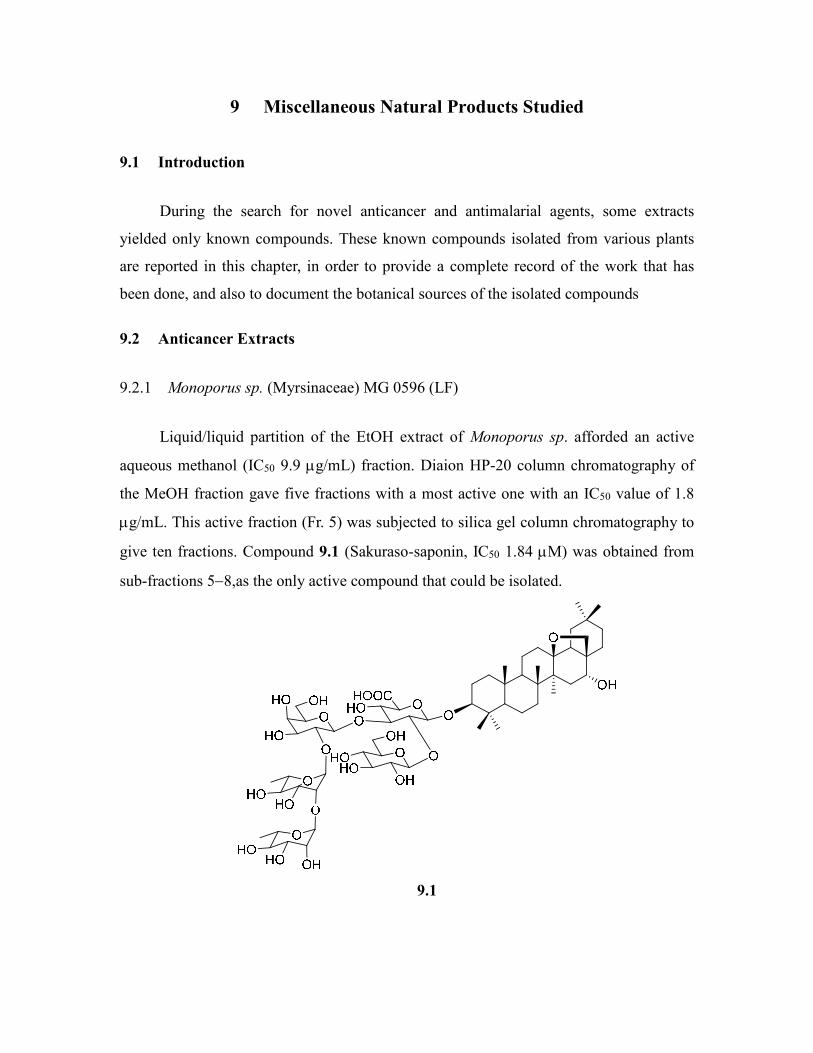
9 Miscellaneous Natural Products Studied
9.1 Introduction
During the search for novel anticancer and antimalarial agents, some extracts
yielded only known compounds. These known compounds isolated from various plants
are reported in this chapter, in order to provide a complete record of the work that has
been done, and also to document the botanical sources of the isolated compounds
9.2 Anticancer Extracts
9.2.1 Monoporus sp. (Myrsinaceae) MG 0596 (LF)
Liquid/liquid partition of the EtOH extract of Monoporus sp. afforded an active
aqueous methanol (IC50 9.9 g/mL) fraction. Diaion HP-20 column chromatography of
the MeOH fraction gave five fractions with a most active one with an IC50 value of 1.8
g/mL. This active fraction (Fr. 5) was subjected to silica gel column chromatography to
give ten fractions. Compound 9.1 (Sakuraso-saponin, IC50 1.84 M) was obtained from
sub-fractions 58,as the only active compound that could be isolated.
9.1
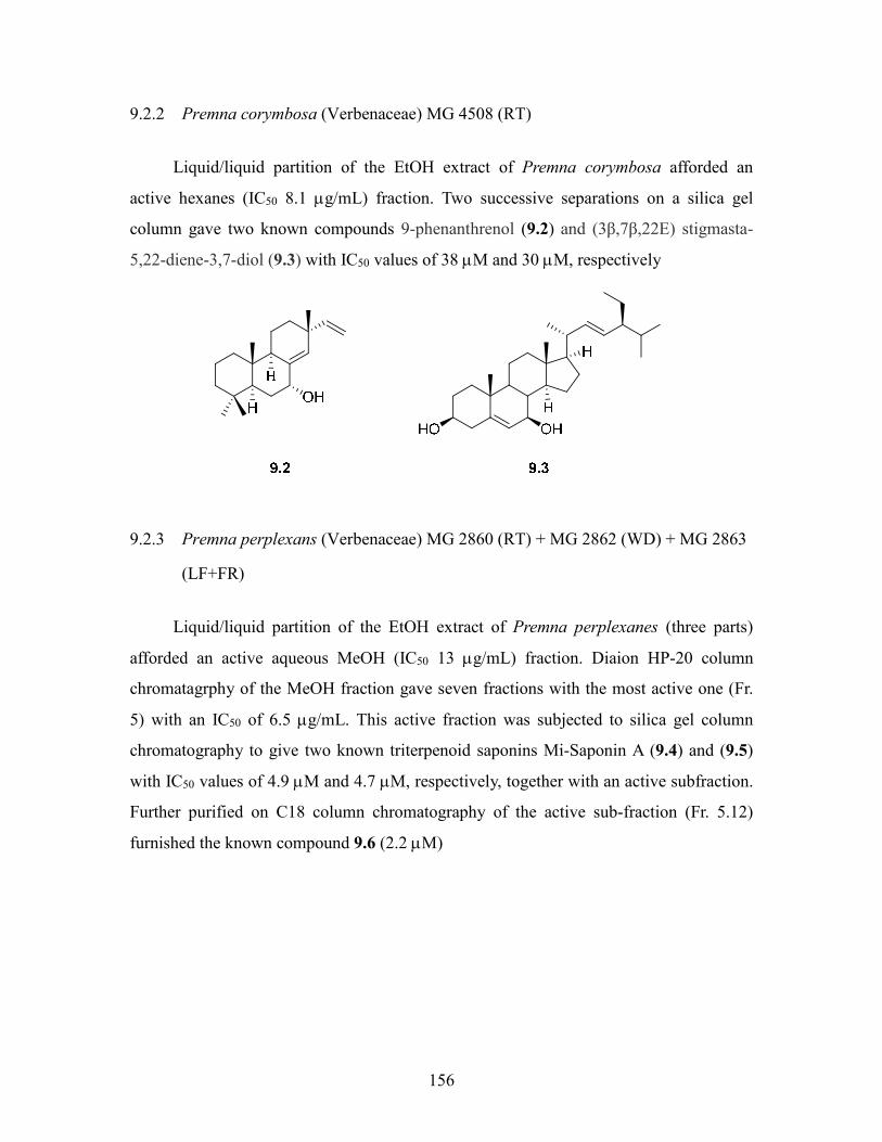
156
9.2.2 Premna corymbosa (Verbenaceae) MG 4508 (RT)
Liquid/liquid partition of the EtOH extract of Premna corymbosa afforded an
active hexanes (IC50 8.1 g/mL) fraction. Two successive separations on a silica gel
column gave two known compounds 9-phenanthrenol (9.2) and (3β,7β,22E) stigmasta-
5,22-diene-3,7-diol (9.3) with IC50 values of 38 M and 30 M, respectively
9.2.3 Premna perplexans (Verbenaceae) MG 2860 (RT) + MG 2862 (WD) + MG 2863
(LF+FR)
Liquid/liquid partition of the EtOH extract of Premna perplexanes (three parts)
afforded an active aqueous MeOH (IC50 13 g/mL) fraction. Diaion HP-20 column
chromatagrphy of the MeOH fraction gave seven fractions with the most active one (Fr.
5) with an IC50 of 6.5 g/mL. This active fraction was subjected to silica gel column
chromatography to give two known triterpenoid saponins Mi-Saponin A (9.4) and (9.5)
with IC50 values of 4.9 M and 4.7 M, respectively, together with an active subfraction.
Further purified on C18 column chromatography of the active sub-fraction (Fr. 5.12)
furnished the known compound 9.6 (2.2 M)
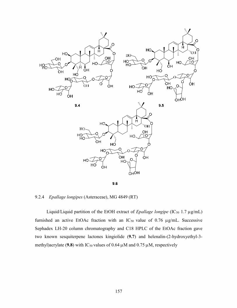
157
9.2.4 Epallage longipes (Asteraceae), MG 4849 (RT)
Liquid/Liquid partition of the EtOH extract of Epallage longipe (IC50 1.7 g/mL)
furnished an active EtOAc fraction with an IC50 value of 0.76 g/mL. Successive
Sephadex LH-20 column chromatography and C18 HPLC of the EtOAc fraction gave
two known sesquiterpene lactones kingiolide (9.7) and helenalin-(2-hydroxyethyl-3-
methyl)acrylate (9.8) with IC50 values of 0.64 M and 0.75 M, respectively
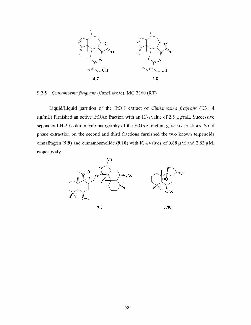
158
9.2.5 Cinnamosma fragrans (Canellaceae), MG 2360 (RT)
Liquid/Liquid partition of the EtOH extract of Cinnamosma fragrans (IC50 4
g/mL) furnished an active EtOAc fraction with an IC50 value of 2.5 g/mL. Successive
sephadex LH-20 column chromatography of the EtOAc fraction gave six fractions. Solid
phase extraction on the second and third fractions furnished the two known terpenoids
cinnafragrin (9.9) and cinnamosmolide (9.10) with IC50 values of 0.68 M and 2.82 M,
respectively.
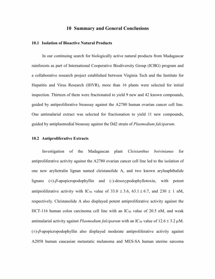
10 Summary and General Conclusions
10.1 Isolation of Bioactive Natural Products
In our continuing search for biologically active natural products from Madagascar
rainforests as part of International Cooperative Biodiversity Group (ICBG) program and
a collaborative research project established between Virginia Tech and the Institute for
Hepatitis and Virus Research (IHVR), more than 16 plants were selected for initial
inspection. Thirteen of them were fractionated to yield 9 new and 42 known compounds,
guided by antiproliferative bioassay against the A2780 human ovarian cancer cell line.
One antimalarial extract was selected for fractionation to yield 11 new compounds,
guided by antiplasmodial bioassay against the Dd2 strain of Plasmodium falciparum.
10.2 Antiproliferative Extracts
Investigation of the Madagascan plant Cleistanthus boivinianus for
antiproliferative activity against the A2780 ovarian cancer cell line led to the isolation of
one new aryltetralin lignan named cleistanolide A, and two known arylnaphthalide
lignans (±)--apopicropodophyllin and (-)-desoxypodophyllotoxin, with potent
antiproliferative activity with IC50 value of 33.0 3.6, 63.16.7, and 230 1 nM,
respectively. Cleistanolide A also displayed potent antiproliferative activity against the
HCT-116 human colon carcinoma cell line with an IC50 value of 20.5 nM, and weak
antimalarial activity against Plasmodium falciparum with an IC50 value of 12.6 3.2 M.
(±)--apopicropodophyllin also displayed moderate antiproliferative activity against
A2058 human caucasian metastatic melanoma and MES-SA human uterine sarcoma
160
cells, with IC50 values of 4.6 and 4.0 M, respectively. A new arylnaphthalide lignan
named 4-O-(-D-glucopyranosyl)-dehydropodophyllotoxin and a known arylnaphthalide
lignan with moderate antiproliferative activity with IC50 values of 2.103 μM and 4.9
0.1 μM, respectively. In addition, two known arylnaphthalide lignans
deoxypicropodophyllotoxin and (-)-yatein were isolated with IC50 values more than 10
M.
Investigation of the Puerto Rico plant Piptocoma antillana (Asteraceae) for
antiproliferative activity against the A2780 ovarian cancer cell line led to the isolation of
two new antiproliferative goyazensolide type sesquiterpenes lactones named 5-O-
methyl-5-epiisogoyazensolide and 15-O-methylgoyazensolide, together with the two
known sesquiterpene lactones 5-epiisogoyazensolide and 15-acetylgoyazensolide. The
isolated compounds showed micromolar activities with half-maximum inhibitory
concentration (IC50) values of 1.56 0.04, 1.62 0.05, 1.50.5, and 0.6 0.3M,
respectively.
Investigation of the Madagascan plant Stuhlmannia moavi (Leguminosae) for
antiproliferative activity against the A2780 ovarian cancer cell line led to the isolation of
one new 1,4-naphthoquinone named stuhlmoavin with moderate activity with an IC50
value of 8.1 ± 0.4 M, together with the known isoflavonoid bonducellin and the five
known stilbenoids 3,4,5'-trihydroxy-3'-methoxy-trans-stilbene, piceatannol, resveratrol,
rhapontigenin, and isorhapontigenin, with weak activities with IC50 values of 10.6 ± 0.4
M, 54 ± 1M, 41 ± 1 M, and 74 ± 1 M, respectively. Stuhlmoavin, bonducellin, and
3,4,5'-trihydroxy-3'-methoxy-trans-stilbene also showed moderate antimalarial activity
against Plasmodium falciparum Dd2 with IC50 values of 24 ± 7, 26 ± 3, and 27 ± 2 M,
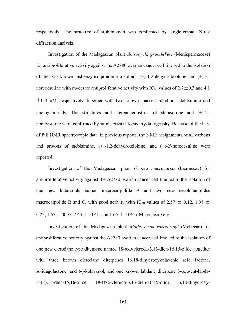
161
respectively. The structure of stuhlmoavin was confirmed by single-crystal X-ray
diffraction analysis.
Investigation of the Madagascan plant Anisocycla grandidieri (Menispermaceae)
for antiproliferative activity against the A2780 ovarian cancer cell line led to the isolation
of the two known bisbenzylisoquinoline alkaloids (+)-1,2-dehydrotelobine and (+)-2'-
norcocsuline with moderate antiproliferative activity with IC50 values of 2.7±0.3 and 4.1
±0.3 µM, respectively, together with two known inactive alkaloids stebisimine and
puetogaline B. The structures and stereochemistries of stebisimine and (+)-2'-
norcocsuline were confirmed by single crystal X-ray crystallography. Because of the lack
of full NMR spectroscopic data in previous reports, the NMR assignments of all carbons
and protons of stebisimine, (+)-1,2-dehydrotelobine, and (+)-2'-norcocsuline were
reported.
Investigation of the Madagascan plant Ocotea macrocarpa (Lauraceae) for
antiproliferative activity against the A2780 ovarian cancer cell line led to the isolation of
one new butanolide named macrocarpolide A and two new secobutanolides
macrocarpolide B and C, with good activity with IC50 values of 2.57 ± 0.12, 1.98 ±
0.23, 1.67 ± 0.05, 2.43 ± 0.41, and 1.65 ± 0.44 M, respectively.
Investigation of the Madagascan plant Malleastrum rakotozafyi (Meliceae) for
antiproliferative activity against the A2780 ovarian cancer cell line led to the isolation of
one new clerodane type diterpene named 18-oxo-cleroda-3,13-dien-16,15-olide, together
with three known clerodane diterpenes 16,18-dihydroxykolavenic acid lactone,
solidagolactone, and (-)-kolavenol, and one known labdane diterpene 3-oxo-ent-labda-
8(17),13-dien-15,16-olide. 18-Oxo-cleroda-3,13-dien-16,15-olide, 6,18-dihydroxy-

162
kolavenic acid lactone, and solidagolactone showed micromolar A2780 activities with
IC50 value of 3.01 ± 0.8, 7.84 ± 0.2, and 17.9 ± 3 µM, respectively.
10.3 Antimalarial extract
Bioassay-guided separation of the Madagascar plant Cryptocarya rigidifolia for
antiplasmodial activity led to the isolation of five new 5,6-dihydro--pyrones named
cryptorigidifoliol AE and six bicyclic tetrahydro--pyrone derivatives named
cryptorigidifoliol FK. Cryptorigidifoliol AG exhibited moderate antimalarial activity,
with IC50 values of 9.2 ± 0.9, 5.8 ± 1.4, 5.5 ± 0.7, 9 ± 3, 4.0 ± 2, 7.4 ± 0.6, and 6.0 ± 0.5
M, respectively. Cryptorigidifoliol HK were all less active, with IC50 values > 10 M.
The bicyclic tetrahydro--pyrones were shown to be produced by cyclization of 5,6-
dihydro--pyrones during the isolation process involved by silica gel.
10.4 Conclusions
The complex structures and biological importance of the substances found in
nature are very fascinating. In the six-year research period, more than twenty crude
extracts were studied, leading to the isolation of twenty new compounds and thirty-four
known compounds. Among these compounds, (1R,2R,3S)-3-O-(-D-glucopyranosyl)-
3',4',5'-trimethoxy-6,7-methylenedioxy-1,9-cyclolignan-11,12-olide (cleistanolide A, 3.1)
isolated from the stems of Cleistanthus boivinianus, showed the most potent
antiproliferative activity against the A2780 cell line with an IC50 value of 33.0 3.6 nM.
This compound is the first example of a new class of podophyllotoxin analogs with
substitution at C-3. The potent activity of this compound suggests that C-3 substituted
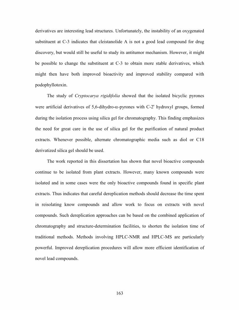
163
derivatives are interesting lead structures. Unfortunately, the instability of an oxygenated
substituent at C-3 indicates that cleistanolide A is not a good lead compound for drug
discovery, but would still be useful to study its antitumor mechanism. However, it might
be possible to change the substituent at C-3 to obtain more stable derivatives, which
might then have both improved bioactivity and improved stability compared with
podophyllotoxin.
The study of Cryptocarya rigidifolia showed that the isolated bicyclic pyrones
were artificial derivatives of 5,6-dihydro--pyrones with C-2' hydroxyl groups, formed
during the isolation process using silica gel for chromatography. This finding emphasizes
the need for great care in the use of silica gel for the purification of natural product
extracts. Whenever possible, alternate chromatographic media such as diol or C18
derivatized silica gel should be used.
The work reported in this dissertation has shown that novel bioactive compounds
continue to be isolated from plant extracts. However, many known compounds were
isolated and in some cases were the only bioactive compounds found in specific plant
extracts. Thus indicates that careful dereplication methods should decrease the time spent
in reisolating know compounds and allow work to focus on extracts with novel
compounds. Such dereplication approaches can be based on the combined application of
chromatography and structure-determination facilities, to shorten the isolation time of
traditional methods. Methods involving HPLC-NMR and HPLC-MS are particularly
powerful. Improved dereplication procedures will allow more efficient identification of
novel lead compounds.

11 Appendix 1H NMR spectrum of 2.1 in CDCl3 (500 Hz)
1H NMR spectrum of 2.2 in CDCl3 (500 Hz)
O O
OH OH
14
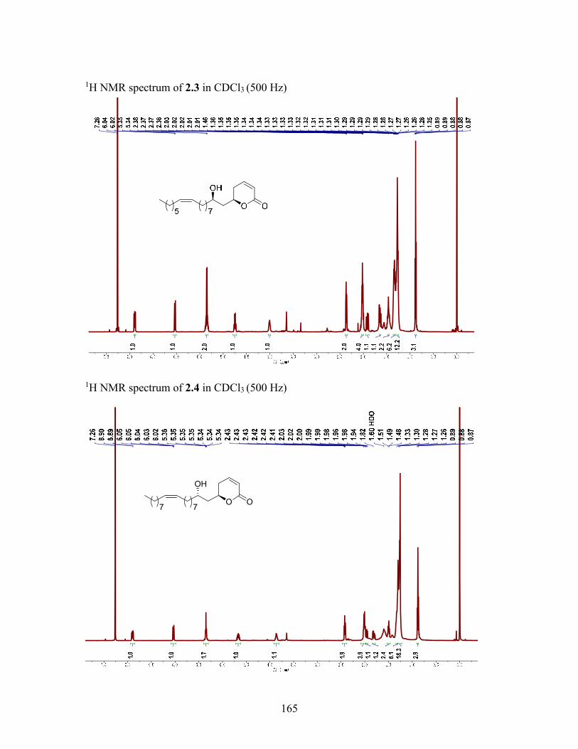
165
1H NMR spectrum of 2.3 in CDCl3 (500 Hz)
1H NMR spectrum of 2.4 in CDCl3 (500 Hz)
O O
OH
77

166
1H NMR spectrum of 2.5 in CDCl3 (500 Hz)
1H NMR spectrum of 2.6 in CDCl3 (500 Hz)
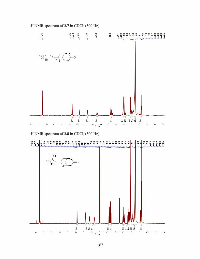
167
1H NMR spectrum of 2.7 in CDCl3 (500 Hz)
1H NMR spectrum of 2.8 in CDCl3 (500 Hz)
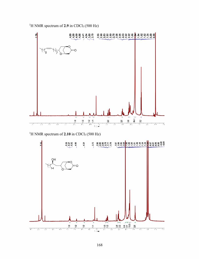
168
1H NMR spectrum of 2.9 in CDCl3 (500 Hz)
1H NMR spectrum of 2.10 in CDCl3 (500 Hz)
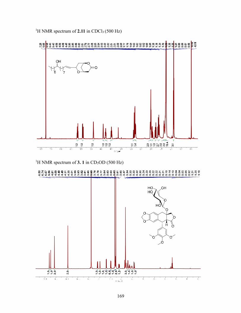
169
1H NMR spectrum of 2.11 in CDCl3 (500 Hz)
1H NMR spectrum of 3. 1 in CD3OD (500 Hz)
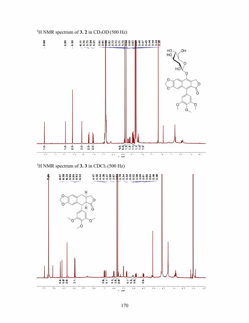
170
1H NMR spectrum of 3. 2 in CD3OD (500 Hz)
1H NMR spectrum of 3. 3 in CDCl3 (500 Hz)
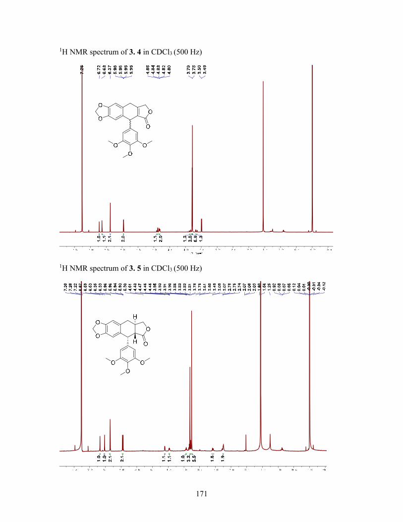
171
1H NMR spectrum of 3. 4 in CDCl3 (500 Hz)
1H NMR spectrum of 3. 5 in CDCl3 (500 Hz)
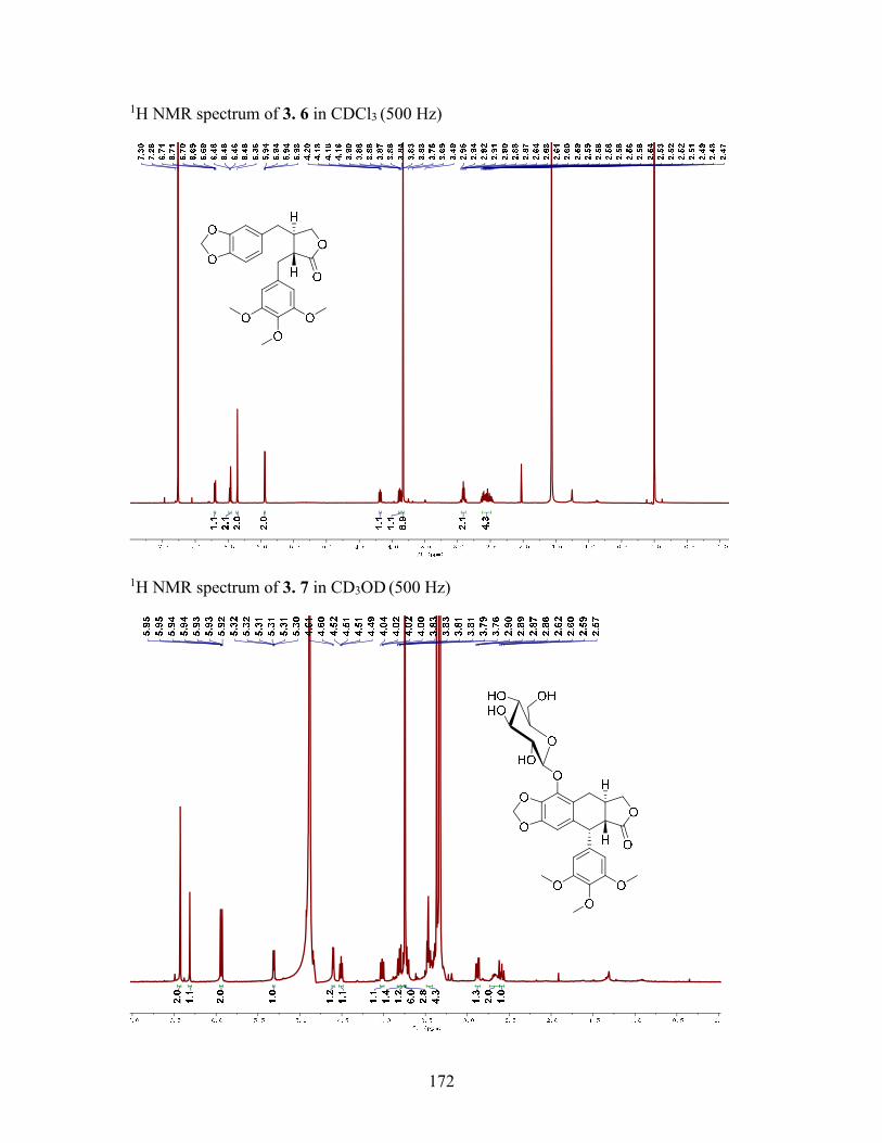
172
1H NMR spectrum of 3. 6 in CDCl3 (500 Hz)
1H NMR spectrum of 3. 7 in CD3OD (500 Hz)

173
1H NMR spectrum of 4. 1 in CDCl3 (500 Hz)
1H NMR spectrum of 4. 2 in CDCl3 (500 Hz)
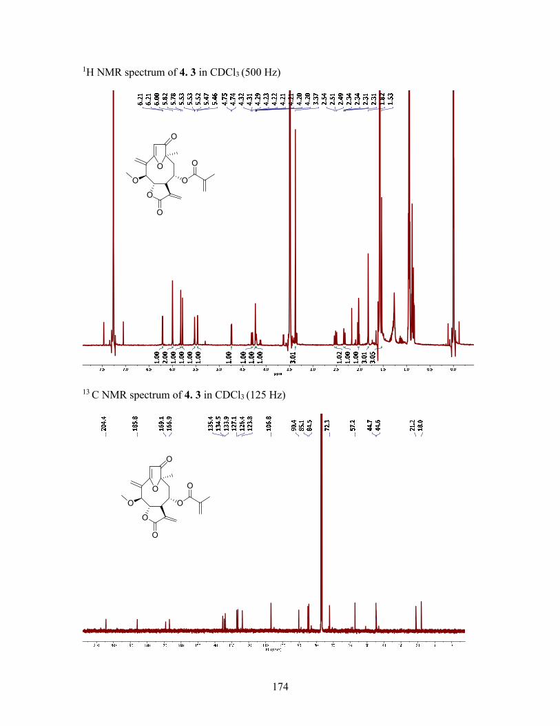
174
1H NMR spectrum of 4. 3 in CDCl3 (500 Hz)
13 C NMR spectrum of 4. 3 in CDCl3 (125 Hz)
O
O
O
O
O
O
O
O
O
O
O
O
O
O

175
1H NMR spectrum of 4. 4 in CDCl3 (500 Hz)
13 C NMR spectrum of 4. 4 in CDCl3 (125 Hz)
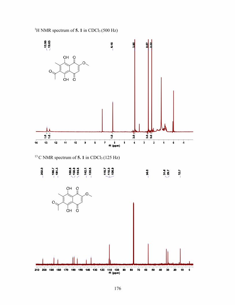
176
1H NMR spectrum of 5. 1 in CDCl3 (500 Hz)
13 C NMR spectrum of 5. 1 in CDCl3 (125 Hz)
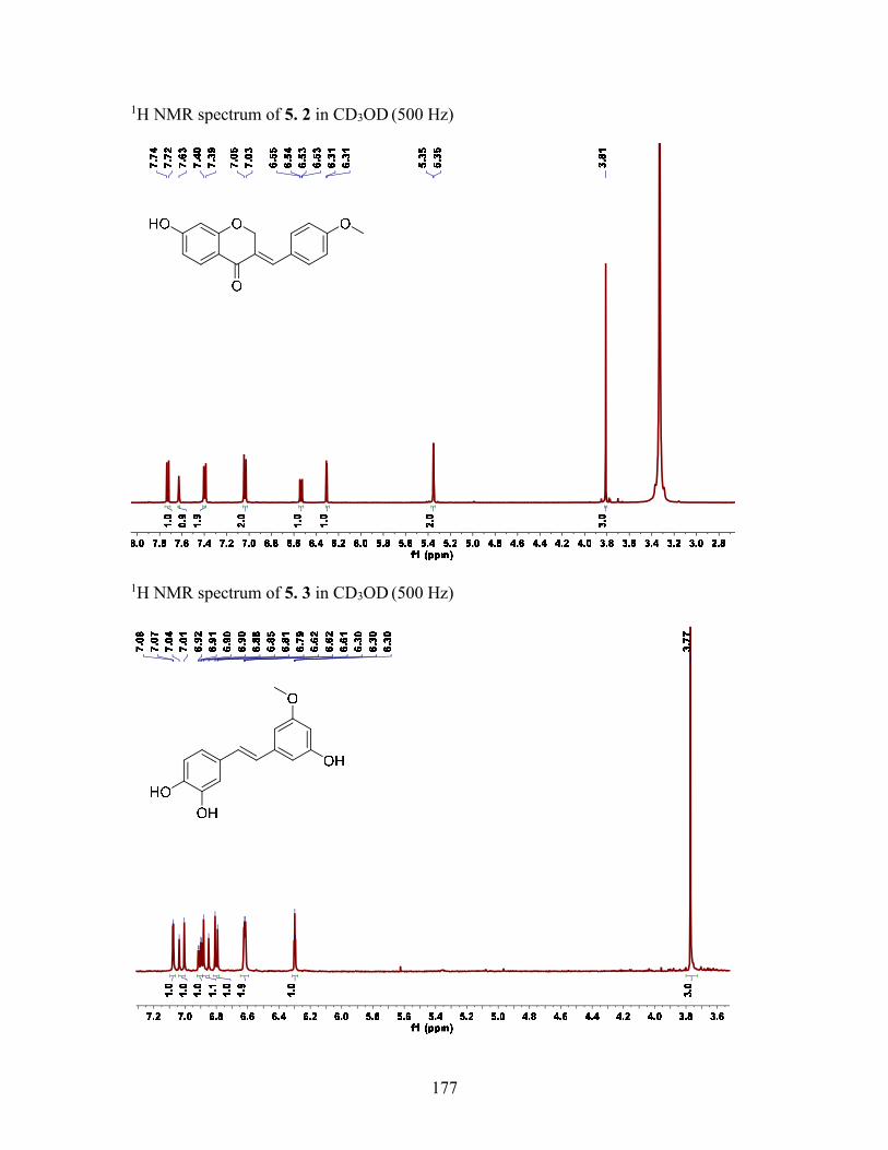
177
1H NMR spectrum of 5. 2 in CD3OD (500 Hz)
1H NMR spectrum of 5. 3 in CD3OD (500 Hz)

178
1H NMR spectrum of 5. 4 in CD3OD (500 Hz)
1H NMR spectrum of 5. 5 in CD3OD (500 Hz)
OH
OH
OH
HO
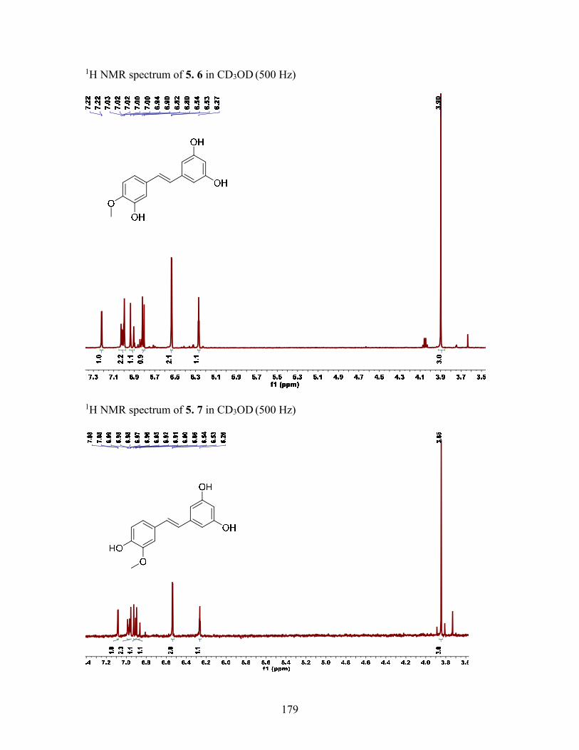
179
1H NMR spectrum of 5. 6 in CD3OD (500 Hz)
1H NMR spectrum of 5. 7 in CD3OD (500 Hz)

180
1H NMR spectrum of 6.1 in CDCl3 (500 Hz)
13C NMR spectrum of 6.1 in CDCl3 (125 Hz)
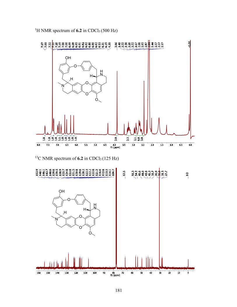
181
1H NMR spectrum of 6.2 in CDCl3 (500 Hz)
13C NMR spectrum of 6.2 in CDCl3 (125 Hz)
O
O
N
HN
O
O
H
OH
H
O
O
N
HN
O
O
H
OH
H
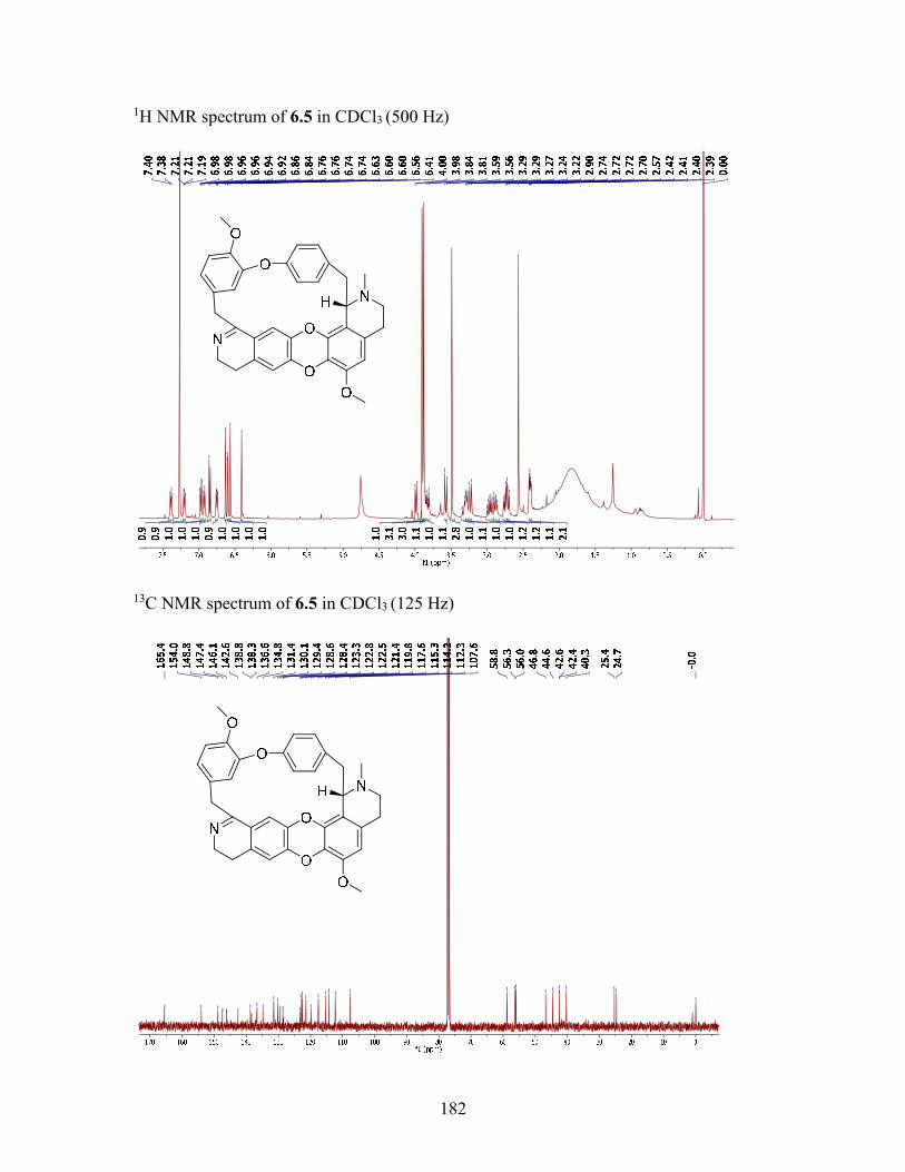
182
1H NMR spectrum of 6.5 in CDCl3 (500 Hz)
13C NMR spectrum of 6.5 in CDCl3 (125 Hz)

183
1H NMR spectrum of 6.6 in CDCl3 (500 Hz)
13C NMR spectrum of 6.6 in CDCl3 (125 Hz)
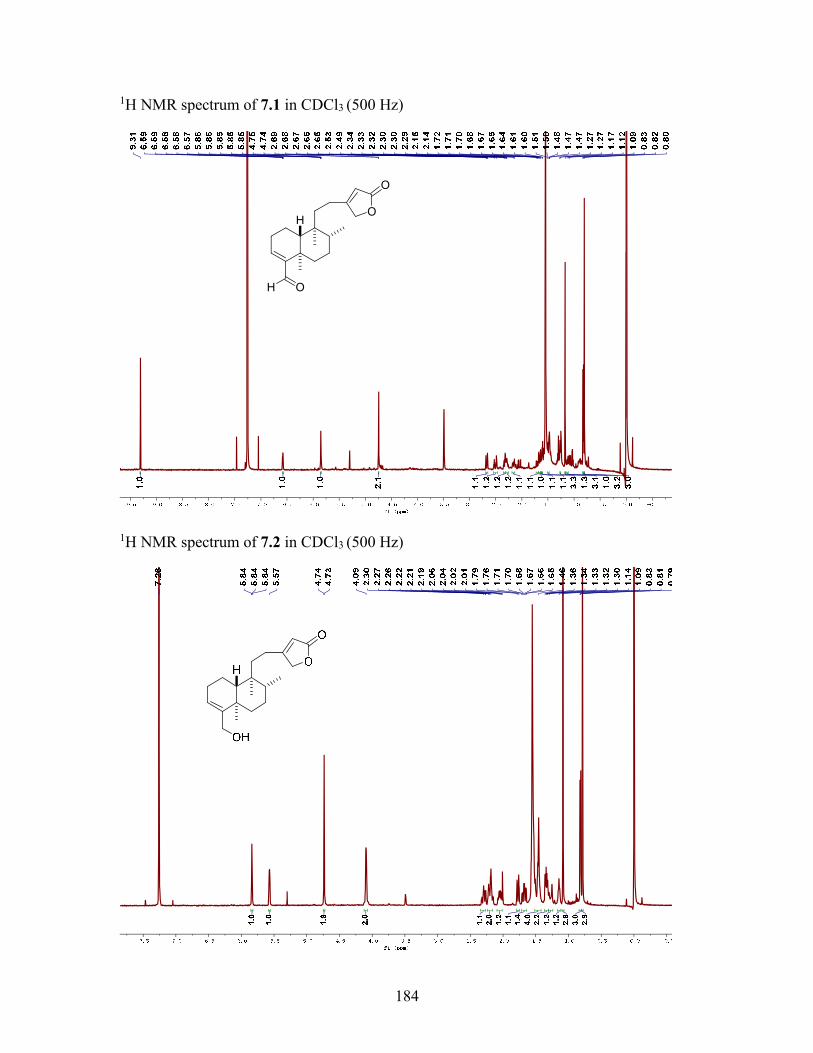
184
1H NMR spectrum of 7.1 in CDCl3 (500 Hz)
1H NMR spectrum of 7.2 in CDCl3 (500 Hz)
HO
O
OH
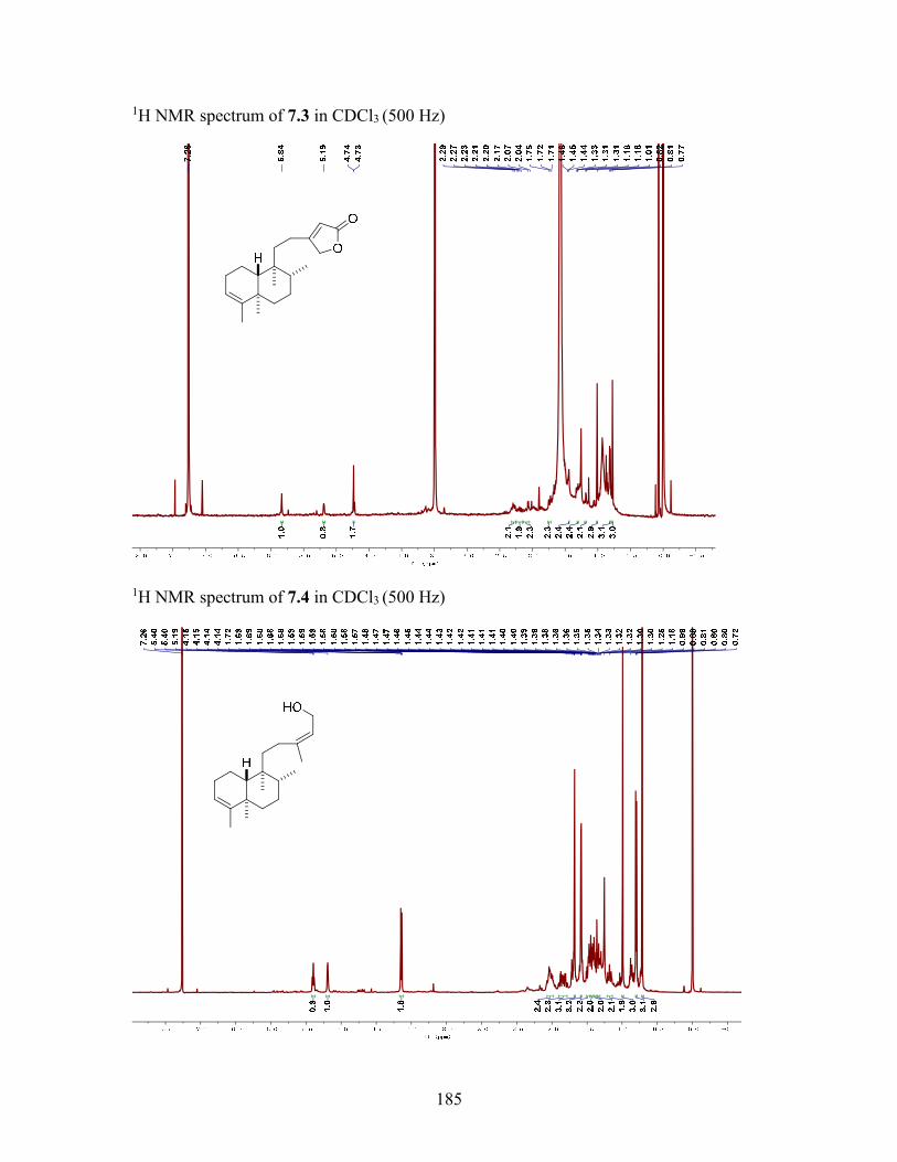
185
1H NMR spectrum of 7.3 in CDCl3 (500 Hz)
1H NMR spectrum of 7.4 in CDCl3 (500 Hz)
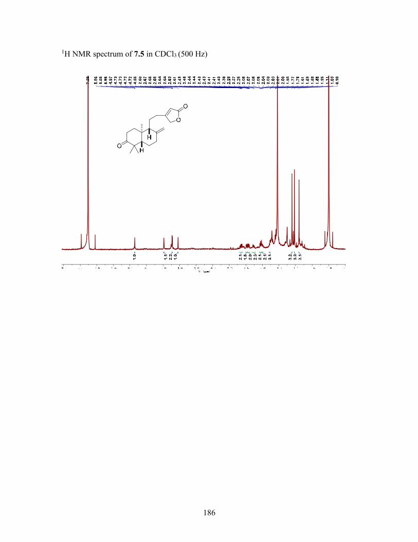
186
1H NMR spectrum of 7.5 in CDCl3 (500 Hz)