Embed Size (px)
Citation preview
International Journal of Scientific & Engineering Research Volume 10, Issue 11, November-2019 433 ISSN 2229-5518
IJSER © 2019
http://www.ijser.org
ISOLATION OF DIFFERENT BACTERIA FROM CHICKEN FEACES IN DISTRICT
MANSEHRA, REGION, KPK Wahidullah1, Samiyah Tasleem2,Kashif Haleem1, Zeeshan Niaz1
Microbiology, Hazara University, Mansehra, KPK1 and Department of Microbiology
University of Karachi and Federal Urdu University of Arts, Science & Technology, Karachi,
Pakistan2
ABSTRACT:
The economy of Pakistan is generally dependent on the poultry sector after agriculture. Poultry
waste litter is mainly added to the soil as a fertilizer. The last step in the poultry management
policy poses a great risk to the environment due to nutrients and microorganisms contained in
high concentrations in these waste materials. To find out the prevalence of microbes in poultry
industry research were perform in district Mansehra of Pakistan. This study was conducted in
district Mansehra region during the month of July to November 2018 to evaluate the frequency
of different bacteria in the fresh chicken faeces at different poultry farm of Dhodyal, Buffa and
Shinkyari region of district Mansehra. In this study total of 204 fresh faeces sample were
collected from poultry farm of district Mansehra region and then isolation and identification of
the pathogenic bacteria were done by culturing and by different biochemical test. 204 fresh faces
samples of chicken were collected out of which 134 from Dhodyal, 40 from Baffa, and 30 from
Shinkyari, the percentage of overall isolated bacteria in these three region was 70,6%, 70%, and
80%. The number of bacteria was isolated and identified as E. coli, Salmonella, Shigella,
Klebsiella, Proteus, Pseudomonas, Staphylococcus and Streptococcus. The frequency of isolated
bacteria in these three region was E. coli (31.6%, 30%, 40%), Salmonella (10%,17%,13%),
Shigella (5%,10%), Klebsiella (5%), Proteus (3.37%), Pseudomonas (5%,5%,10%),
Staphylococcus (7.46%,7.50% 20%), Streptococcus (5. 96%). The current study endorsed
appropriate information spreading to farmer and poultry feed producers about the public health
significance of proper poultry litter disposal.
IJSER
International Journal of Scientific & Engineering Research Volume 10, Issue 11, November-2019 434 ISSN 2229-5518
IJSER © 2019
http://www.ijser.org
Key words: Microorganism isolation from poultry industry of Hazara Division, Pathogenic
Microorganisms.
Introduction:
Poultry farms are used for raising chickens, turkeys, ducks, and other birds for egg or meat
production. Poultry sector play very essential role and fulfill the requirement of protein
deficiency. In Pakistan the commercial poultry sector was started in 1960. The government
positive policy and the poultry farming community persistence was the result of initial growth of
poultry industry (Sadiq, 2004). Although poultry are affected by enteric illnesses, this illness is
one of the most important groups of diseases and is continuing to cause high economic losses in
many areas around the globe due to increased death rates, decreased weight gain, increased
medication costs and increased feed change rates. Several pathogens (Viruses, bacteria and
parasites) are involved as possible causes of enteric disorders either alone (mono-causal), in
synergy with different other microorganisms.
Before 1963 in Pakistan native chicken provided the source of egg and meat and these birds at
the age of four-month yield an average 30 eggs and 0.769 kg of meat per annum (Sahota and
Bhatti, 2003).These birds have been raised as backyard activities to fulfill the individual
domestic desire.
The consumption of chicken meat is gradually increasing globally, the previous history and
available information show that it reached to 14.2 kilogram per person, per annum (Rouger et
al., 2017). In 2010 the market share of mutton and beef had reduced to 20% to 55% while the
poultry meat had increased to 25% respectively (Hussain et al., 2015).
MICROBIOLOGY OF POULRTY/ CHICKEN:
Different microorganisms such as protozoan, fungi, algae, protests, archaea, and bacteria are the
dynamic part of microbial diversity and these are usually briefly mentioned or not at all which
requires consideration. Microorganisms are distributed throughout the biosphere. Bacteria like to
live in an energy rich environment (Ganesan and Muthuchelian, 2009). The caeca of the
gastrointestinal tract of poultry is one of the favorite places for bacteria which presented
favorable environment for more than 200 bacterial strains. The species of Lactobacillus,
Enterococcus and Enterobacteriaceae are the major microflora of chick cecum at the first day of
IJSER
International Journal of Scientific & Engineering Research Volume 10, Issue 11, November-2019 435 ISSN 2229-5518
IJSER © 2019
http://www.ijser.org
age. After second week of the chicks age, the species of Eubacteria and bacteroides species got
established (van der Wielen et al., 2001). Amongst the three regions of gastrointestinal tract of
chicken such as duodenum, cecum and ileum, duodenum has the lowest bacterial population
while cecum has highest population of bacteria. The account of lactobacillus average is about
1x10⁹ in the ceca of chicken, along with lactobacillus huge amount of Enterococcus and
Enterobacteriaceae were found. At the age of 12 day of chicks the account of facultative
anaerobic and obligate anaerobic bacteria is 10 to 15 time more than aerobic bacteria.(Zhu et al.,
2002).
The enteric bacteria in the Enterobacteriaceae family are the major pathogen and the chicken
may also be asymptomatic carrier and shaded of these pathogenic bacteria in their faeces,
thereby contaminating the environment.(Kilonzo-Nthenge et al., 2008; Obi et al., 2008) The rich
nutrient contented of poultry dropping provide energy for potentially harmful organism (Chang
et al., 2004).
The family Enterobacteriaceae, including Klebsiella, Escherichia coli and Salmonella species are
a major or secondary pathogens of poultry production.(Kilonzo-Nthenge et al., 2008; Obi et al.,
2008).
In chicken farm administration, one of the most tough phase is the administration of the newly
flock introduced. For the process to be money-making, a good disease prevention program
should be offered for the newly submitted chicks to evade any future losses. Disease can be
spread by human, other birds, newly introduced chicks or contaminated gear. Disease
controlling from the beginning is the significant for the success of the operation (Mobley and
Kahan).
STUDY AREA
District Mansehra has a large number of poultry farms and plays very important role in meat
production. The peoples of this area mostly depend on poultry. On the other hand, the effect
disease in chickens before of this has never been studied in that area. The environmental and
management circumstances are the main causative points which can greatly increase the
incidence of poultry in District Mansehra and probable area would be focused. Correspondingly,
in recent past, antimicrobial susceptibility test was mostly suggested for the investigation and
IJSER
International Journal of Scientific & Engineering Research Volume 10, Issue 11, November-2019 436 ISSN 2229-5518
IJSER © 2019
http://www.ijser.org
observing of antimicrobial resistance in different areas continuously. Hence, this study is
designed to find the prevalence of different bacteria in chicken faeces in this area and to found
the antimicrobial sensitivity profile against the pathogens related with poultry.
AIMS AND OBJECTIVES
To isolation and identification of bacterial consortium in chicken faeces samples from collected
in Mansehra Region.
MATHODOLOGY:
SAMPLE COLLECTION:
A total 204 fresh chicken faeces samples were collected from different poultry farms of District
Mansehra region. The samples of fresh chicken fecal matter were taken randomly from chickens
of different health status by using sterilized cotton swabs and transported to the laboratory.
Transport Media
Different transport media are used for delivering fecal samples to laboratories such as Campy-
thioglycolate, Carry Blaire, Semi-solid motility test medium and Alkaline peptone water. In the
current research, sterilized cotton swab was used for transporting chicken fecal samples (without
contamination and disruption of samples taken with culture swabs) to the laboratory 0f
microbiology Department Hazara University within two hours for isolation and identification of
different bacteria.
TO STERALISE PETRI PLATES AND AS WELL AS TEST TUBES:
In drying method, we sterilized the petri plates as well as the test tubes with tap water and also
kept in hot air oven. Then autoclaved the petri plates and the test tubes for 15 minutes at 121 ºc
under 15 psi pressure. For further process the petri plates and the test tubes were kept in Luminar
flow hood.
Culture media preparation:
The preparation of media was done according to the labeled requirement given on the bottle. For
the ingredient of each media separate flask were used and then mix the ingredient by heating and
IJSER
International Journal of Scientific & Engineering Research Volume 10, Issue 11, November-2019 437 ISSN 2229-5518
IJSER © 2019
http://www.ijser.org
shaking. Then the flasks were covered by aluminum foil and autoclaved. After autoclave the
media flask were kept to cooled. In the blood agar flask blood were added and gently shake to
mix the blood in agar (hours and sheep blood). Then the Luminar flow hood was washed with
ethanol and on the ultra violet light for 10 minutes before the media was transferred.
INCUBATION PERIOD FOR PETRI PLATES:
The incubation period up to twenty-four hours (24 hos) for petri plates given to eliminate the
microbe’s contamination because for further process no microbial contamination occurs on the
petri plates.
Sample processing:
Sterilize cotton swab which is taken from chicken faeces were directly inoculated on blood agar,
MeConkey agar, and Salmonella, Shigella agar (SS agar), for the detection of different bacterial
colony. The petri plates were kept in in reversed position at 37°c for one day and after the
incubation period the colony were appeared on some plates.
Purification:
The colony on MeConkey agar, S.S agar and blood agar were obtained and identified on the
basis of morphology and biochemical properties. For obtaining of pure colonies again I cultured
of already isolated colonies and kept in incubator at 37°c for 24 hours.
Morphological identification:
Colonial Identification:
After 24 hours’ incubation bacterial growth were occur in colonial form. On the basis of colony,
surface texture, color, size, elevations, shape and edges different bacterial colonies were
identified. Two types of colonies were formed on Mac-Conk agar (a) Grayish (b) yellowish.
Those colonies which are yellowish were identified as Lactose fermenter and those colonies
which are grayish in color identified as the Non-lactose fermenter. On blood agar white grayish
colony of different bacteria were appeared which have different shape, size and some bacteria
such is streptococcus species showed hemolysis on blood agar which may be alpha, beta and
gamma hemolysis, while two different types of colonies were appeared on SS agar. Opaque
IJSER
International Journal of Scientific & Engineering Research Volume 10, Issue 11, November-2019 438 ISSN 2229-5518
IJSER © 2019
http://www.ijser.org
colorless colonies with black center were identified as Salmonella and Smooth round transparent
colorless with no black center colonies were identified as Shigella. Further morphological
identification of different bacterial colonies was done through gram-staining.
GRAMSTAINIG AND MICROSCOPY:
For gram staining process, a clean slide was taken and then up to one drop of normal saline was
dropped on the slide. A well sterilized wire loop was used to make a smear on the slide and the
slide was dried and fixed through heat. The crystal violet was poured on the slide and the
incubation period for 1 minute and 30 seconds and washed the slide properly with tap water.
After it we used the gram iodine for 1 minute and 30 seconds and then washed again with tap
water. After the gram iodine we used alcohol up to 95 % for 15 to 20 seconds and then washed
with tap water. Then we used the safranin for 1 minute and then again washed with water as
well. Now we used to dry the slides in normal air and then we examined the slide under a
microscope. A thick layer of the peptidoglycan-90% present in the cell wall of gram positive
bacteria and the cell wall-stain with purple color and while a thin layer of peptidoglycan-10%
present in the cell wall of gram negative bacteria and the cell wall were stained with red or pink
color. Further, identification of bacteria was done on the basis of biochemical test such as
catalase, oxidase and amvis test.
Biochemical identification
MATHOD FOR CATALASE TEST:
There are two methods are used for catalase test which are given below
A: TUBE MATHOD:
1: Poured 1-2 ml of Hydrogen peroxide solution mix in a test tube.
2: pick of several colonies from the 20 to 24 hours’ incubation period of culture medium with the
help of sterile wire loop and dip in the solution of Hydrogen peroxide.
3: Examine the abrupt producing of oxygen bubble occurs inside a test tube.
B: SLIDE MATHOD:
1: Pick the small colonies from the growth by using the sterile wire loop and placed the
colonies on a well cleaned and dry glass slide.
IJSER
International Journal of Scientific & Engineering Research Volume 10, Issue 11, November-2019 439 ISSN 2229-5518
IJSER © 2019
http://www.ijser.org
2: Mix 3% of Hydrogen peroxide with colonies on the slide.
3: Examine the bubbles of oxygen produced.
PROCEDURES OF OXIDASE TEST:
This test was used for those bacteria which have an enzyme cytochrome oxidase.
1: This test was also used for the differentiation of bacteria like Campylobacter, Moraxella,
Neisseria, Pasturella and as well as Moraxella.
2: It is mainly used to differentiate pseudomonas species from the co related species.
IMVIC TEST:
REAGENT FOR IMVIC TEST:
Methyl Red:
Distilled water 500ml, Ethyl alcohol.
Method for preparation:
in 300 ml ethyl alcohol, Methyl red was dissolved then distilled water was added. For each test
Five drops of above reagent were used (Distinct red color is positive test. Yellow color is
negative reaction).
Vogues -Proskour Reagents:
KOH (40%) (0.2 ml) Naphthol solution (0.6 Alpha – Naphthol)
Method of preparation: (VP) broth tubes were Inoculated by suspected colonies, incubated for
48 ± 2 hr. at 35 C°, then 0.6 ml L- Naphthol solution and 0.2 ml 40% KOH (potassium
hydroxide) solutions were added then mixed well. Eosin pink color is considered positive.
Kovacs reagent, (ICMSF,1978): Para dimethyl amino benzaldehyde 5.0 g, Isoamyl or (normal
amyl) alcohol 75.0 ml Hydrochloric acid (cone.) 25.0 ml.
Method of preparation: Benzaldehyde was dissolved in isoamyl alcohol then HCL conc. was
added slowly, 0.5 ml of above mixture was used for each indole test.
BIOCHEMICAL TESTS:
IJSER
International Journal of Scientific & Engineering Research Volume 10, Issue 11, November-2019 440 ISSN 2229-5518
IJSER © 2019
http://www.ijser.org
Methyl Red test:
Five ml of culture in MR-VP both was incubated at 37C° for 24-48h. Five drops of methyl red
solution were added to each tube. Distinct red color is a positive test while a yellow color shown
as a negative test.
Vogues- Proskour test:
MR-VP broth was incubated at 37C° for 24-48h and then a Few drops of reagent alpha-Naphthol
solution and 0.2ml 40% KOH were added and mixed, pink color development was considered
positive.
Indole test:
Five ml of culture in peptone water was incubated at 37C° for 24-48h. Indole production was
tested by adding 0.5 ml of Kovacs, reagent. Appearance of distinct rosy colour in upper layer is
appositive test.
RESULTS
In the present study, was conducted to investigate different pathogenic bacteria from chicken
faeces in three different regions of Mansehra district. Total samples of chicken faeces were 204,
that were collected from different poultry form. Out of these, 134 sample were collected from
dhodyal region,40 sample from Baffa region and 30 samples were collected from shinkyari
region. All the samples were conformed primarily with the help of its growth characteristics on
three different media such as Blood agar, Meconkey agar and Salmonella Shigella agar and then
isolate were identified with the help of biochemical tests.
Prevalence of pathogenic bacteria in three different regions of District Mansehra:
204 fresh faeces sample were collected from three different regions of Mansehra which include
Dhodyal, Buffa, and Shinkyari for the detection of different pathogenic bacteria.134 samples
were collected from Dhodyal region, 30 samples from Shinkyari and 40 samples were collected
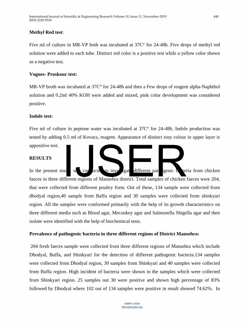
from Buffa region. High incident of bacteria were shown in the samples which were collected
from Shinkyari region. 25 samples out 30 were positive and shown high percentage of 83%
followed by Dhodyal where 102 out of 134 samples were positive in result showed 74.62%. In
IJSER
International Journal of Scientific & Engineering Research Volume 10, Issue 11, November-2019 441 ISSN 2229-5518
IJSER © 2019
http://www.ijser.org
Buffa region its percentage was quite low where 28 out of 40 samples were positive and having a
percentage of 70%. From three different regions the percentage of positive bacteria were isolated
as shown (fig 3.1)
I S O L A T I O N A N D I D E N T I F I C A T IO N O F P A T H O G E N I C B A C T E R I A F R O M C H IC K E N F A C E S I N D IS T R I C T M A N S E H R A
R E G IO N K P K .
% P
re
va
len
ce
Dhodia
l R
e gio
n
Ba ff
a
Shin
kiy
a r i0
20
40
60
80
100
7 4 .6 2 %
7 0 %
8 3 %
Fig.3.1: Percentage distribution of various pathogenic bacteria screened out in chicken
feces from Dhodyal, Buffa and Shinkyari region.
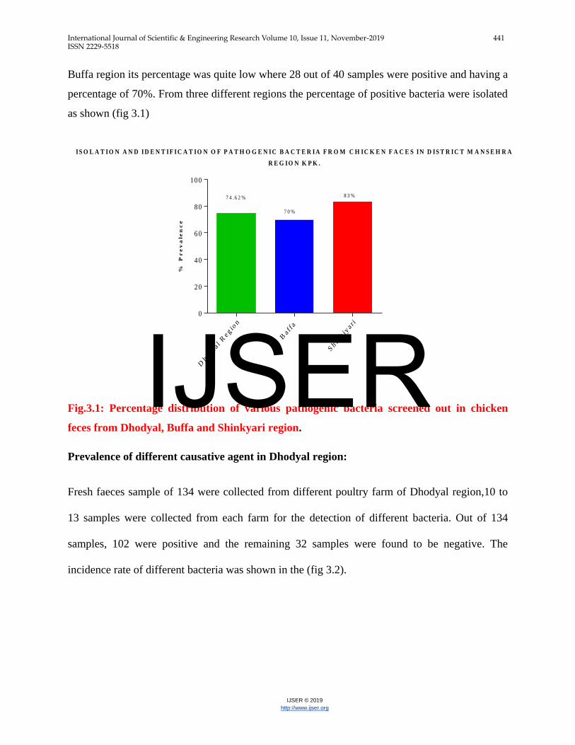
Prevalence of different causative agent in Dhodyal region:
Fresh faeces sample of 134 were collected from different poultry farm of Dhodyal region,10 to
13 samples were collected from each farm for the detection of different bacteria. Out of 134
samples, 102 were positive and the remaining 32 samples were found to be negative. The
incidence rate of different bacteria was shown in the (fig 3.2).
IJSER
International Journal of Scientific & Engineering Research Volume 10, Issue 11, November-2019 442 ISSN 2229-5518
IJSER © 2019
http://www.ijser.org
D if f e r e n t C a u s it iv e a g e n ts in c h ic k e n s fe c e s in D h o d ia l%
Pr
ev
ale
nc
e
E. c o
li
Pro
teus
spp
.
Sta
phy lo
c oc c u
s s p
p
Str
e pto
c oc c u
s s p
p
Salm
on
e lla
Kle
bs i
e lla
pse
ud
om
on
as
Sh
ige ll
a
0
10
20
30
40
7 .4 7 %5 .9 7 % 5 .9 7 %
1 0 .4 4 %
3 .7 3 %5 .9 7 %
3 .7 3 %
3 1 .3 4 %
Fig 3.2: Percentage distribution of various pathogenic bacteria screened out in chicken
feces from Dhodyal.
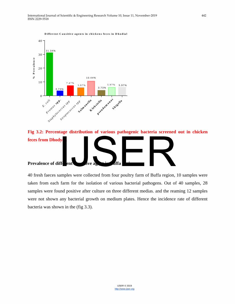
Prevalence of different causative agent in Buffa region:
40 fresh faeces samples were collected from four poultry farm of Buffa region, 10 samples were
taken from each farm for the isolation of various bacterial pathogens. Out of 40 samples, 28
samples were found positive after culture on three different medias. and the reaming 12 samples
were not shown any bacterial growth on medium plates. Hence the incidence rate of different
bacteria was shown in the (fig 3.3).
IJSER
International Journal of Scientific & Engineering Research Volume 10, Issue 11, November-2019 443 ISSN 2229-5518
IJSER © 2019
http://www.ijser.org
D if f e r e n t C a u s it iv e a g e n t s in C h ic k e n s B a f f a r e g io n s%
Pr
ev
ale
nc
e
E. c o
li
Salm
on
e lla
Sh
ige ll
a
Sta
phy lo
c oc c u
s
Psu
dom
on
as
0
10
20
30
40
3 0 %
1 7 .5 %
1 0 %
5 %7 .5 %
Fig 3.3 Percentage distribution of various pathogenic bacteria screened out in chicken feces
from Buffa.
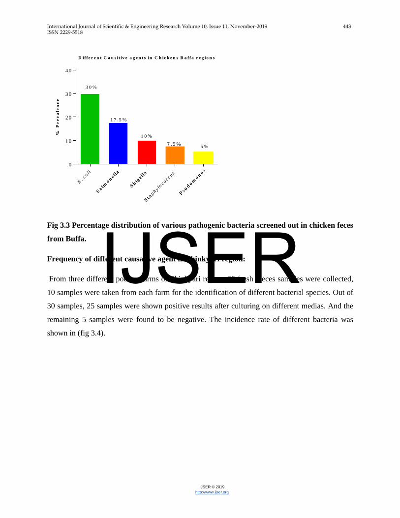
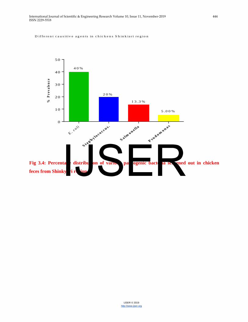
Frequency of different causative agent in Shinkyari region:
From three different poultry farms of Shinkyari region, 30 fresh faeces samples were collected,
10 samples were taken from each farm for the identification of different bacterial species. Out of
30 samples, 25 samples were shown positive results after culturing on different medias. And the
remaining 5 samples were found to be negative. The incidence rate of different bacteria was
shown in (fig 3.4).
IJSER
International Journal of Scientific & Engineering Research Volume 10, Issue 11, November-2019 444 ISSN 2229-5518
IJSER © 2019
http://www.ijser.org
% P
re
va
len
ce
E. c
o li
Sta
ph
y loc o c cu
s .
Sa lm
o ne ll
a
Psu
do m
o na s
0
10
20
30
40
50
4 0 %
2 0 %
1 3 .3 %
5 .0 0 %
D if fe r e n t c a u s i t iv e a g e n ts in c h ic k e n s S h in k ia r i r e g io n
Fig 3.4: Percentage distribution of various pathogenic bacteria screened out in chicken
feces from Shinkyari region.
IJSER
International Journal of Scientific & Engineering Research Volume 10, Issue 11, November-2019 445 ISSN 2229-5518
IJSER © 2019
http://www.ijser.org
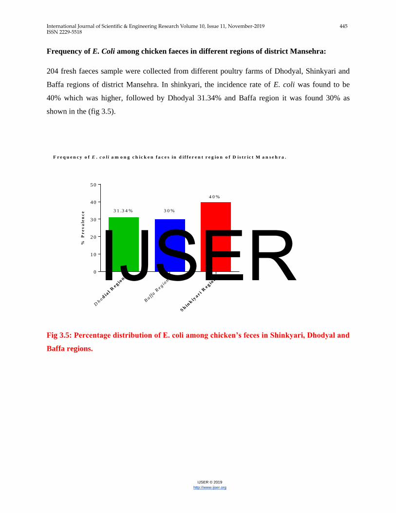
Frequency of E. Coli among chicken faeces in different regions of district Mansehra:
204 fresh faeces sample were collected from different poultry farms of Dhodyal, Shinkyari and
Baffa regions of district Mansehra. In shinkyari, the incidence rate of E. coli was found to be
40% which was higher, followed by Dhodyal 31.34% and Baffa region it was found 30% as
shown in the (fig 3.5).
F r e q u e n c y o f E . c o l i a m o n g c h ic k e n f a c e s in d i f f e r e n t r e g io n o f D is t r ic t M a n s e h r a .
% P
re
va
len
ce
Dhod
ial
Re g
ion
Baff
a R
e gio
n
Sh
ink
iyar i
Re g
ion
0
10
20
30
40
50
3 1 .3 4 % 3 0 %
4 0 %
Fig 3.5: Percentage distribution of E. coli among chicken’s feces in Shinkyari, Dhodyal and
Baffa regions.
IJSER
International Journal of Scientific & Engineering Research Volume 10, Issue 11, November-2019 446 ISSN 2229-5518
IJSER © 2019
http://www.ijser.org
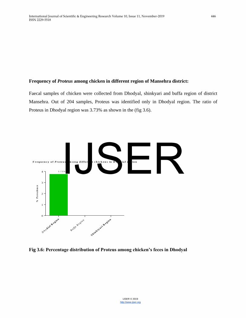
Frequency of Proteus among chicken in different region of Mansehra district:
Faecal samples of chicken were collected from Dhodyal, shinkyari and buffa region of district
Mansehra. Out of 204 samples, Proteus was identified only in Dhodyal region. The ratio of
Proteus in Dhodyal region was 3.73% as shown in the (fig 3.6).
F r e q u e n c y o f P r o te u s a m o n g d i f f e r e n t c h ic k e n s in D h o d ia l r e g io n
% P
re
va
len
ce
Dh o d
ial
Re g io
n
Ba ff
a Re g io
n
Sh
ink
iya r i
Re g io
n
0
1
2
3
4 3 .7 3 %
Fig 3.6: Percentage distribution of Proteus among chicken’s feces in Dhodyal
IJSER
International Journal of Scientific & Engineering Research Volume 10, Issue 11, November-2019 447 ISSN 2229-5518
IJSER © 2019
http://www.ijser.org
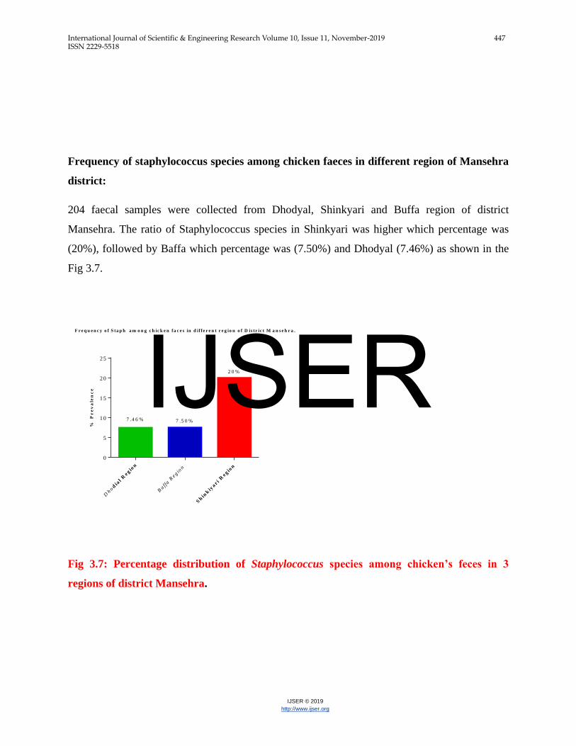
Frequency of staphylococcus species among chicken faeces in different region of Mansehra
district:
204 faecal samples were collected from Dhodyal, Shinkyari and Buffa region of district
Mansehra. The ratio of Staphylococcus species in Shinkyari was higher which percentage was
(20%), followed by Baffa which percentage was (7.50%) and Dhodyal (7.46%) as shown in the
Fig 3.7.
F r e q u e n c y o f S ta p h a m o n g c h ic k e n fa c e s in d if fe r e n t r e g io n o f D is tr ic t M a n s e h r a .
% P
re
va
len
ce
Dhod
ial R
e gio
n
Baff
a R
e gio
n
Sh
ink
iyar i
Re g
ion
0
5
10
15
20
25
7 .4 6 % 7 .5 0 %
2 0 %
Fig 3.7: Percentage distribution of Staphylococcus species among chicken’s feces in 3
regions of district Mansehra.
IJSER
International Journal of Scientific & Engineering Research Volume 10, Issue 11, November-2019 448 ISSN 2229-5518
IJSER © 2019
http://www.ijser.org
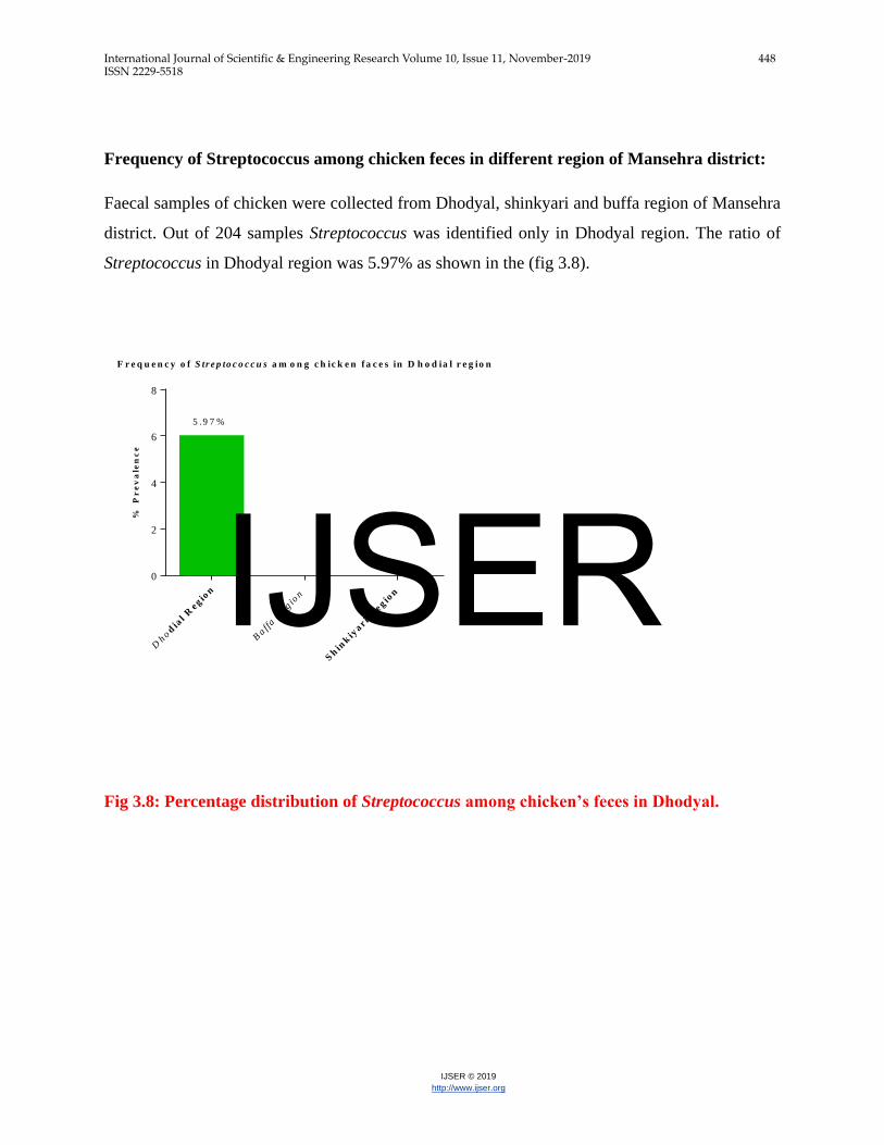
Frequency of Streptococcus among chicken feces in different region of Mansehra district:
Faecal samples of chicken were collected from Dhodyal, shinkyari and buffa region of Mansehra
district. Out of 204 samples Streptococcus was identified only in Dhodyal region. The ratio of
Streptococcus in Dhodyal region was 5.97% as shown in the (fig 3.8).
F r e q u e n c y o f S tr e p to c o c c u s a m o n g c h ic k e n f a c e s in D h o d ia l r e g io n
% P
re
va
len
ce
Dhod
ial R
e gio
n
Baff
a R
e gio
n
Sh
ink
iyar i
Re g
ion
0
2
4
6
8
5 .9 7 %
Fig 3.8: Percentage distribution of Streptococcus among chicken’s feces in Dhodyal.
IJSER
International Journal of Scientific & Engineering Research Volume 10, Issue 11, November-2019 449 ISSN 2229-5518
IJSER © 2019
http://www.ijser.org
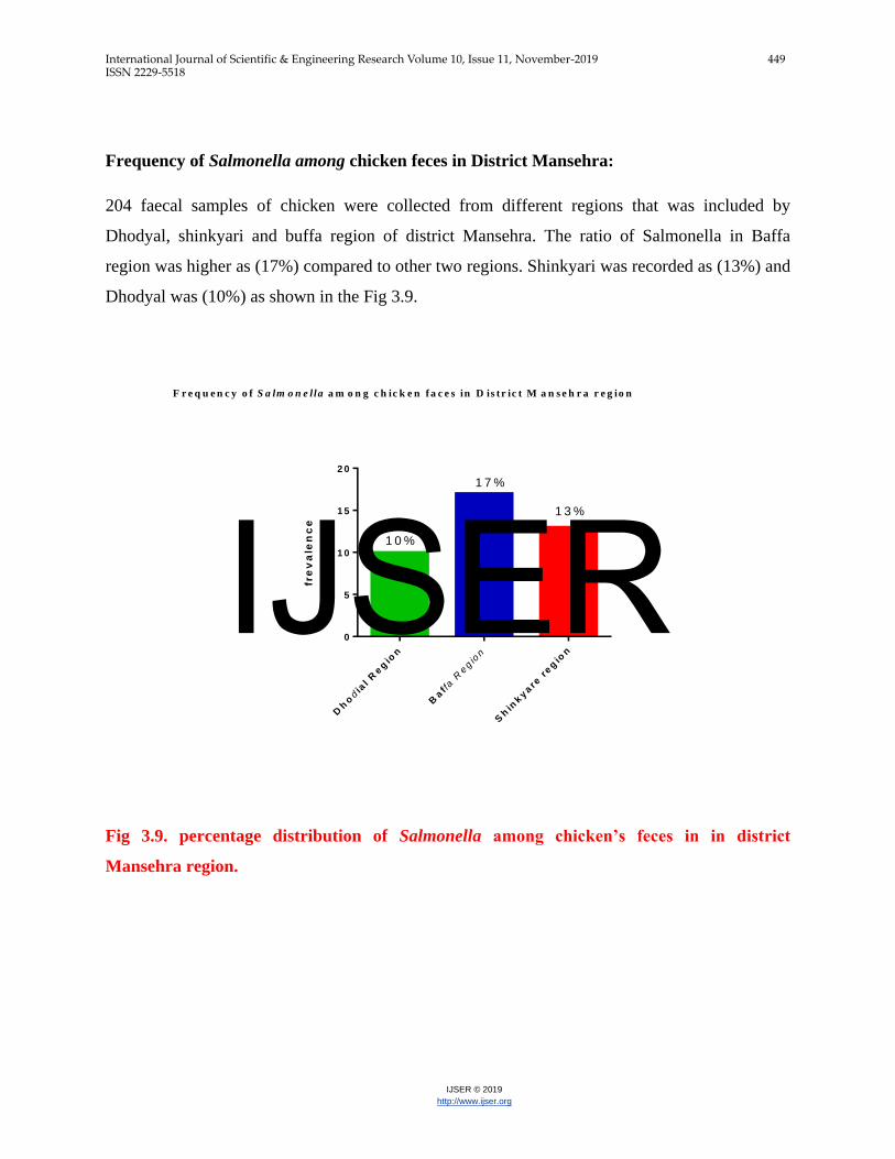
Frequency of Salmonella among chicken feces in District Mansehra:
204 faecal samples of chicken were collected from different regions that was included by
Dhodyal, shinkyari and buffa region of district Mansehra. The ratio of Salmonella in Baffa
region was higher as (17%) compared to other two regions. Shinkyari was recorded as (13%) and
Dhodyal was (10%) as shown in the Fig 3.9.
Fig 3.9. percentage distribution of Salmonella among chicken’s feces in in district
Mansehra region.
fre
va
len
ce
Dh
odia
l R
eg
ion
Baf f
a R
egio
n
Sh
inkyare
reg
ion
0
5
1 0
1 5
2 0
F r e q u e n c y o f S a lm o n e lla a m o n g c h ic k e n f a c e s in D is tr ic t M a n s e h r a r e g io n
1 0 %
1 7 %
1 3 %
IJSER
International Journal of Scientific & Engineering Research Volume 10, Issue 11, November-2019 450 ISSN 2229-5518
IJSER © 2019
http://www.ijser.org
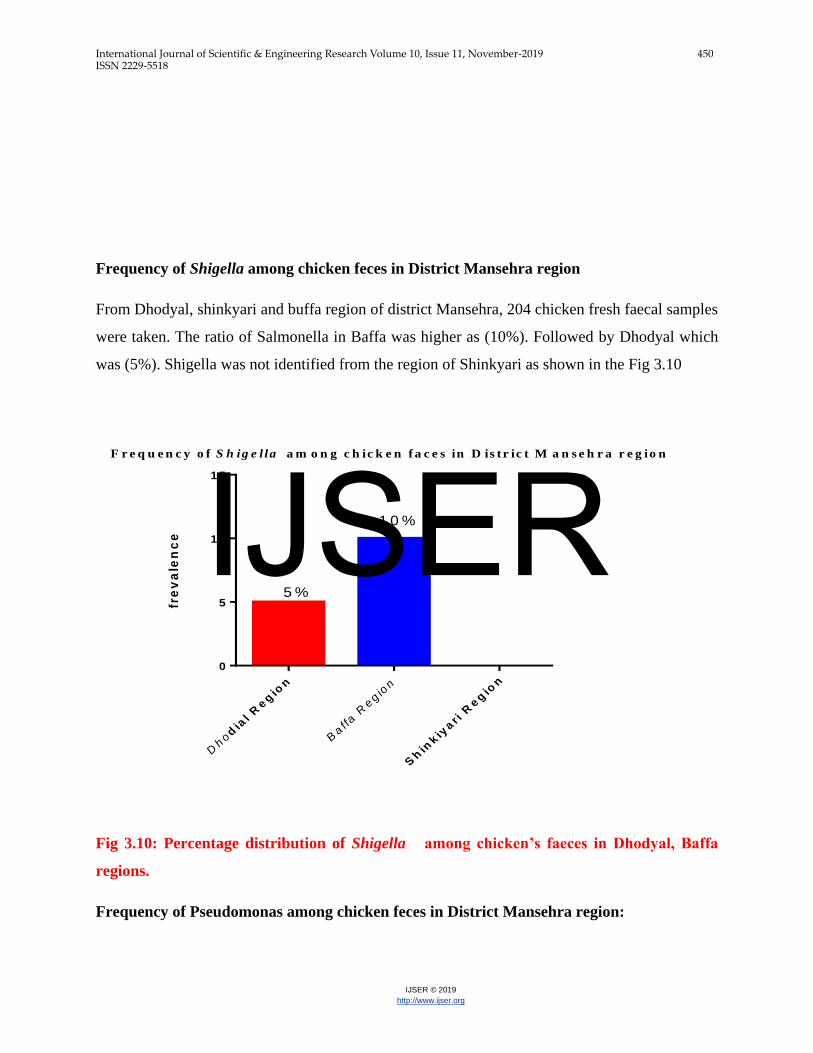
Frequency of Shigella among chicken feces in District Mansehra region
From Dhodyal, shinkyari and buffa region of district Mansehra, 204 chicken fresh faecal samples
were taken. The ratio of Salmonella in Baffa was higher as (10%). Followed by Dhodyal which
was (5%). Shigella was not identified from the region of Shinkyari as shown in the Fig 3.10
fre
va
len
ce
Dhod
ial R
eg
ion
Baffa R
egio
n
Sh
inkiy
ar i
Reg
ion
0
5
1 0
1 5
F r e q u e n c y o f S h ig e l la a m o n g c h ic k e n f a c e s in D is tr ic t M a n s e h r a r e g io n
5 %
1 0 %
Fig 3.10: Percentage distribution of Shigella among chicken’s faeces in Dhodyal, Baffa
regions.
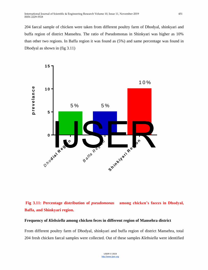
Frequency of Pseudomonas among chicken feces in District Mansehra region:
IJSER
International Journal of Scientific & Engineering Research Volume 10, Issue 11, November-2019 451 ISSN 2229-5518
IJSER © 2019
http://www.ijser.org
204 faecal sample of chicken were taken from different poultry farm of Dhodyal, shinkyari and
buffa region of district Mansehra. The ratio of Pseudomonas in Shinkyari was higher as 10%
than other two regions. In Baffa region it was found as (5%) and same percentage was found in
Dhodyal as shown in (fig 3.11)
pre
ve
lan
ce
Dhod
ial R
eg
ion
Baffa R
egio
n
Sh
inkiy
ar i
Reg
ion
0
5
1 0
1 5
5 % 5 %
1 0 %
Fig 3.11: Percentage distribution of pseudomonas among chicken’s faeces in Dhodyal,
Baffa, and Shinkyari region.
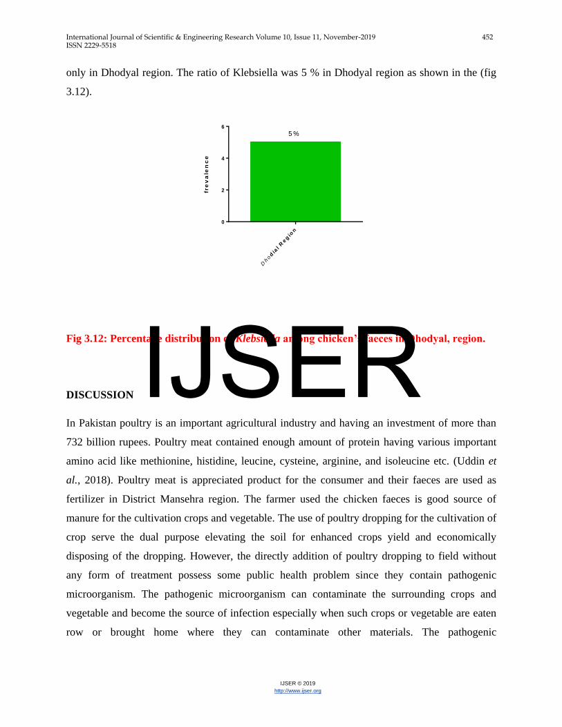
Frequency of Klebsiella among chicken feces in different region of Mansehra district
From different poultry farm of Dhodyal, shinkyari and buffa region of district Mansehra, total
204 fresh chicken faecal samples were collected. Out of these samples Klebsiella were identified
IJSER
International Journal of Scientific & Engineering Research Volume 10, Issue 11, November-2019 452 ISSN 2229-5518
IJSER © 2019
http://www.ijser.org
only in Dhodyal region. The ratio of Klebsiella was 5 % in Dhodyal region as shown in the (fig
3.12).
fre
va
len
ce
Dhod
ial R
eg
ion
0
2
4
6
5 %
Fig 3.12: Percentage distribution of Klebsiella among chicken’s faeces in Dhodyal, region.
DISCUSSION
In Pakistan poultry is an important agricultural industry and having an investment of more than
732 billion rupees. Poultry meat contained enough amount of protein having various important
amino acid like methionine, histidine, leucine, cysteine, arginine, and isoleucine etc. (Uddin et
al., 2018). Poultry meat is appreciated product for the consumer and their faeces are used as
fertilizer in District Mansehra region. The farmer used the chicken faeces is good source of
manure for the cultivation crops and vegetable. The use of poultry dropping for the cultivation of
crop serve the dual purpose elevating the soil for enhanced crops yield and economically
disposing of the dropping. However, the directly addition of poultry dropping to field without
any form of treatment possess some public health problem since they contain pathogenic
microorganism. The pathogenic microorganism can contaminate the surrounding crops and
vegetable and become the source of infection especially when such crops or vegetable are eaten
row or brought home where they can contaminate other materials. The pathogenic
IJSER
International Journal of Scientific & Engineering Research Volume 10, Issue 11, November-2019 453 ISSN 2229-5518
IJSER © 2019
http://www.ijser.org
microorganism can also be discharge onto surface that are used for drinking after run-off during
rain fall.(Orji et al., 2005).
The present study was conducted to investigate the prevalence of pathogenic bacteria from
chicken faeces in three different regions (Shinkyari, Dhodyal, and Baffa) of district Mansehra.
However, there are limited report were available from Pakistan. However, no such work has
ever been done in Hazara division, especially in District Mansehra region. Out of total 204
samples analyzed, 155 samples were positive showing a high prevalence of pathogenic bacteria
in poultry i.e. 75.98%.
Among positive cases in three different regions, highest incidence of bacteria was shown in
Shinkyari region and least incidence in buffa region with percentage of 83% and 70%
respectively. Whereas, the prevalence of bacteria in Dhodyal region was appeared 74.62%.
Pathogenic bacteria isolated in the present study included E. coli, Salmonella, Shigella, Proteus,
Staphylococci, Pseudomonas, Klebsiella, and Streptococci. The E. coli, Staphylococci,
Salmonella and Pseudomonas species were isolated from all three regions with highest
prevalence in Shinkyari region compared to Dhodyal and Baffa. Contrarily, Proteus,
Streptococci and Klebsiella species were only found in Dhodyal region and Shigella in Dhodyal
and Baffa region of district Mansehra.
E. coli was found most prevalent among all the isolated bacteria in selected regions probably
because of its commonness in the normal microflora of intestine. Its prevalence in Dhodyal,
Shinkyari and Baffa regions was 31.34%, 40% and 30%. In contrast to present study, 55.50% E.
coli was isolated from chicken dropping in Nigeria in 2017 (Ajayi and Omoya, 2017).and
Similar study was done in Kenya and isolated E. coli from poultry dropping with the prevalence
rate of 57% (Langata et al., 2019). while in 2015 42% E.coli was isolated from cloacal and liver
of broiler chicken(Khatun et al., 2015).In 2016 E.coli isolated from commercial broiler and
backyard poultry in Lahore Pakistan with prevalence rate of 64.2 and 54.5.(Akhtar et al., 2016)
The Salmonella species, after E. coli, were found more prevalent in Shinkyari (13.3%) and Baffa
(17.5%) regions compared to Dhodyal where its prevalence was only 10.44%. A study was
steered in Faisal Abad Pakistan and isolated Salmonella from poultry dropping with percentage
IJSER
International Journal of Scientific & Engineering Research Volume 10, Issue 11, November-2019 454 ISSN 2229-5518
IJSER © 2019
http://www.ijser.org
of 55% (Akhtar et al., 2010) while 5%was shown by (Mohammed and Ibrahim, 2012).and
21.4% 32% was shown by the report,(Bhuvaneswari et al., 2015).
Similarly, the prevalence of Staphylococci was highest in Shinkyari region i.e. 20%, followed by
Baffa and Dhodyal region where its prevalence was 7.50% and 7.46%. The percentage S. aureus
isolated from this study was less than 24.4% Occurance reported by (Bala et al., 2016) and more
than (Pesewu et al., 2018) isolated coagulase negative S. aureus from chicken meat with the
percentage of 9.2%.
The Shigella was only isolated from Baffa and Dhodyal regions of district Mansehra. The ratio
of Shigella in Baffa region was 10% followed by 5% in Dhodyal region. Whereas, it was not
identified in chicken fecal samples from Shinkyari region. the previous study reported in Saudi
Arabia Taif and isolated Shigella from Cloacal area of poultry with the percentage of
18%%.(Abo-Amer and Shobrak, 2015) Which were higher from my study.
The Proteus, Streptococci and Klebsiella species were only found in Dhodyal region. Their
prevalence was reported as 3.73%, 5.97%, and 5% respectively. In addition to their isolation
only from Dhodyal region, the Proteus and Klebsiella were the least prevalent species among all
the isolated bacteria from this region. Contrast to my study (Jambalang et al., 2017) isolated
Klebsiella and proteus from the chicken egg in South Africa, and (Nahar et al., 2014) isolated
proteus from chicken dropping with the percentage of 39%. While (Siddiqui et al., 2008),
isolated Streptococcus from heart blood, lung, gallbladder intestine and faeces of birds with
percentage of 6.796%. the difference from my study is due to diagnosis, different environment
and geographical variation.
Pseudomonas species were isolated from all three regions of district Mansehra. Their prevalence
in Shinkyari region was highest (10%), followed by Baffa and Dhodyal region where
Pseudomonas was equally prevalent i.e. 5%. Moreover, the prevalence of Pseudomonas in Baffa
and Dhodyal region was lowest among all the isolated bacteria from these two regions. In
contrast to my research 5% pseudomonas species isolated from meat in Iraq (Noori and Alwan,
2016)
CONCLUSION:
IJSER
International Journal of Scientific & Engineering Research Volume 10, Issue 11, November-2019 455 ISSN 2229-5518
IJSER © 2019
http://www.ijser.org
The current result provided indication that poultry waste can serve as an environmental reservoir
for multiple antibiotic resistance bacteria and hence can serve as possible routes for the entrance
of multidrug resistance zoonotic pathogen in the human being. This has very significance
inference for human health, as multidrug resistance infections were problematic to treated and
frequently required costly antibiotic and long term therapy. This can considerably rise the cost of
treatment and even death. The study consequently endorsed appropriate information
dissemination to agriculturalist and poultry feed maker about the public health prominence of
proper poultry waste disposal.
Reference:
Abo-Amer, A. E. & Shobrak, M. Y. (2015). Isolation and Molecular Characterization of
Multidrug-Resistant Salmonella, Shigella and Proteus from Domestic Birds. The Thai
Journal of Veterinary Medicine, 45, 23.
Ajayi, K. O. & Omoya, F. O. (2017). Antibiotic Usage Pattern in Poultry and Resistance Pattern
of Human Pathogenic Bacteria Isolated from Poultry Droppings in Akure, Nigeria.
International Journal of Biomedical Science and Engineering, 5, 35.
Akhtar, F., Hussain, I., Khan, A. & Rahman, S. (2010). Prevalence and antibiogram studies of
Salmonella enteritidis isolated from human and poultry sources. Pak. Vet. J, 30, 25-28.
Akhtar, F., Rabbani, M., Muhammad, K., Younus, M., Ghafoor, A., Sheikh, A., Ahmad, A.,
Muhammad, J., Rasool, A. & Shaheen, A. (2016). Comparative antibiotic resistance
profile of the multidrug resistant E. coli isolated from commercial and backyard poultry.
JAPS: Journal of Animal & Plant Sciences, 26.
Bala, H., Igwe, J., Olayinka, B., Olonitola, O., Onaolapo, J. & Cordelia, N. O. (2016). Antibiotic
susceptibility profile of Staphylococcus aureus isolated from healthy chickens in poultry
farms in Kano state, Nigeria. Sky Journal of Microbiology Research, 4, 42-46.
Bhuvaneswari, M., Shanmughapriya, S. & Natarajaseenivasan, K. (2015). Prevalence of
Multidrug-Resistant (MDR) Salmonella enteritidis in Poultry and Backyard Chicken
from Tiruchirappalli, India. Microbiology Journal, 5: 28, 35.
Chang, W.-N., Huang, C.-R., Lei, C.-B., Lee, P.-Y., Chien, C.-C., Chang, H.-W., Chang, C.-S. &
Lu, C.-H. (2004). Serotypes of clinical cerebrospinal fluid Cryptococcus neoformans
isolates from southern Taiwan and their in vitro susceptibilities to amphotericin B,
fluconazole, and voriconazole. Japanese journal of infectious diseases, 57, 113-115.
Ganesan, R. & Muthuchelian, K. (2009). Molecular identification of bacterial species in Gundaru
river basin of Thirumangalam, Madurai District, South India. JOURNAL OF PURE AND
APPLIED MICROBIOLOGY, 3, 289-294.
Hussain, J., Rabbani, I., Aslam, S. & Ahmad, H. (2015). An overview of poultry industry in
Pakistan. World's poultry science journal, 71, 689-700.
Jambalang, A. R., Buys, E. M. & Botha, F. S. (2017). Bacterial species from retailed poultry
eggs in Tshwane, South Africa: Implication for consumers. South African Journal of
Science, 113, 1-7.
IJSER
International Journal of Scientific & Engineering Research Volume 10, Issue 11, November-2019 456 ISSN 2229-5518
IJSER © 2019
http://www.ijser.org
Khatun, M. N., Atmme, E., Ahmed, S., Parvej, M. S., Akhter, S., Ansari, W. K. & Ali, M. S.
(2015). Frequency of drug resistant Escherichia coli isolated from commercial broiler
chicken in Bangladesh. International Journal of Natural and Social Sciences, 2, 01-05.
Kilonzo-Nthenge, A., Nahashon, S., Chen, F. & Adefope, N. (2008). Prevalence and
antimicrobial resistance of pathogenic bacteria in chicken and guinea fowl. Poultry
science, 87, 1841-1848.
Langata, L. M., Maingi, J. M., Musonye, H. A., Kiiru, J. & Nyamache, A. K. (2019).
Antimicrobial resistance genes in Salmonella and Escherichia coli isolates from chicken
droppings in Nairobi, Kenya. BMC Research Notes, 12, 22.
Mobley, R. & Kahan, T. Practical Management of Health Issues in a Poultry Production System:
Symptoms, Sources, and Prevention of Common Diseases.
Mohammed, H. & Ibrahim, A. E. A. (2012). Isolation and Identification of Salmonella from The
Environment of Traditional Poultry Farms in Khartoum North.
Nahar, A., Siddiquee, M., Nahar, S., Anwar, K. S. & Islam, S. (2014). Multidrug resistant-
Proteus Mirabilis isolated from chicken droppings in commercial poultry farms: bio-
security concern and emerging public health threat in Bangladesh. Journal of Biosafety &
Health Education.
Noori, T. & Alwan, M. (2016). Isolation and Identification of zoonotic bacteria from poultry
meat. Int. J. Adv. Res. Biol. Sci, 3, 57-66.
Obi, T., Olubukola, A. & Maina, G. (2008). Pro-Poor HPAI Risk Reduction Strategies in
Nigeria—Background Paper.
Ojo, O. E., Ogunyinka, O. G., Agbaje, M., Okuboye, J. O., Kehinde, O. O. & Oyekunle, M. A.
(2012). Antibiogram of Enterobacteriaceae isolated from free-range chickens in
Abeokuta, Nigeria. Veterinarski arhiv, 82, 577-589.
Pesewu, G., Quaynor, E., Olu-Taiwo, M., Anim-Baidoo, I. & Asmah, R. (2018). Bacterial
contaminants of raw broiler meat sold at Korle-Gonno, Accra, Ghana. International Food
Research Journal, 25, 1758-1762.
Rouger, A., Tresse, O. & Zagorec, M. (2017). Bacterial contaminants of poultry meat: sources,
species, and dynamics. Microorganisms, 5, 50.
Sadiq, M. (2004). Pakistan poultry sector still on an upward swing. World Poult, 20, 10-11
Sahota, A. & Bhatti, B. (2003). Productive performance of Desi field chickens as affected under
deep litter system. Pakistan Journal of Veterinary Research (Pakistan).
Siddiqui, M., Khan, L., Suradkar, U., Mendhe, M., Rindhe, S. & Sirsat, P. (2008). Bacterial
Isolation and their antibiogram from non-specific infection in poultry of Marathwada
region. Veterinary World, 1, 52.
Uddin, M. N., Farooq, M. M., Waqas, M., Khan, N. U., Khan, W. A., Khan, I., Karim, N. &
Rizwan, M. (2018). 10. Antibiotic assays of Salmonella isolated from poultry chicken of
various locations in districts Swat. Pure and Applied Biology (PAB), 7, 78-84.
Van Der Wielen, P. W., Biesterveld, S., Lipman, L. J. & Van Knapen, F. (2001). Inhibition of a
glucose-limited sequencing fed-batch culture ofSalmonella enterica serovar Enteritidis by
volatile fatty acids representative of the ceca of broiler chickens. Applied and
environmental microbiology, 67, 1979-1982.
Zhu, X. Y., Zhong, T., Pandya, Y. & Joerger, R. D. (2002). 16S rRNA-based analysis of
microbiota from the cecum of broiler chickens. Applied and environmental microbiology,
68, 124-137.
IJSER
International Journal of Scientific & Engineering Research Volume 10, Issue 11, November-2019 457 ISSN 2229-5518
IJSER © 2019
http://www.ijser.org
IJSER