Embed Size (px)
Citation preview

ACNR
Review Articles: New advances in the management of late stage Parkinson’s DiseaseCognition in Huntington's DiseaseRehabilitation Article: Developments in the rehabilitation of unilateralneglectManagement Topic: Social effects of epilepsy
Advances in Clinical Neuroscience & Rehabilitation
journal reviews • events • management topic • industry news • rehabilitation topic •
ISSN 1473-9348 Volume 1 Issue 4 September/October 2001

Neuroimaging has proved to be both sensitive and
highly effective in detecting dopaminergic dysfunc-
tion in Parkinsonian Syndromes. It can help establish
the truth of the disorder when clinical features are
incomplete or contradictory.
Now the truth is out. Nycomed Amersham are proud
to announce the first and only approved nuclear
imaging product in Europe to help differentiate
Essential Tremor from Parkinsonian Syndromes
related to idiopathic Parkinson’s disease, Multiple
System Atrophy and Progressive Supranuclear Palsy.
The images are easy to read and available the same
day. DaTSCAN™ gives you clear diagnostic pictures
that provide accurate visual differentiation of
Parkinsonian Syndromes from Essential Tremor.
Currently, diagnostic methods rely on the use of
clinical criteria,with formal confirmation only possible
at post mortem. DaTSCAN can reduce the time
required to reach a diagnosis.With 97.5% sensitivity†,
you can be more confident than ever that your
diagnosis is correct and that the medication you
prescribe will have the desired effect.
DaTSCAN – another major step forward in the
partnership of success between Neurology,Geriatrics,
Nuclear Medicine and Nycomed Amersham.
T H E T R U T H B E H I N D T H E M A S K
IOFLUPANE (123I)
Abbreviated Prescribing InformationPresentation: Vials containing 185 MBq ioflupane (123I) at reference time.Uses: Detecting loss of functional dopaminergic neuron terminals in the striatum ofpatients with clinically uncertain Parkinsonian Syndromes in order to helpdifferentiate Essential Tremor from Parkinsonian Syndromes related to idiopathicParkinson’s Disease (PD), Multiple System Atrophy (MSA), ProgressiveSupranuclear Palsy (PSP). DaTSCAN is unable to discriminate between PD, MSAand PSP.Dosage and Administration: DaTSCAN is a 5% (v/v) ethanolic solution forintravenous injection and should be used without dilution. Clinical efficiency hasbeen demonstrated across the range of 111-185 MBq; do not use outside this range.Appropriate thyroid blocking treatment must be given prior to and post injection ofDaTSCAN. SPECT imaging should take place 3-6 hours after injection of DaTSCAN.DaTSCAN is not recommended for use in children or adolescents. For use inpatients referred by physicians experienced in the management of movementdisorders. See SPC.Contraindications: Pregnancy and in patients with hypersensitivity to iodide or
any of the excipients.Precautions: Radiopharmaceuticals should only be used by qualified personnelwith appropriate government authorisation and should be prepared using asepticand radiological precautions. DaTSCAN is not recommended in moderate to severerenal or hepatic impairment.Interactions: Consider current medication. Medicines that bind to the dopaminetransporter may interfere with diagnosis; these include amphetamine, benzotropine,buproprion, cocaine, mazindol, methylphenidate, phentermine and sertraline. Drugsshown during clinical trials not to interfere with DaTSCAN imaging includeamantadine, benzhexol, budipine, levodopa, metoprolol, primidone, propranolol andselegiline. Dopamine agonists and antagonists acting on the postsynaptic dopaminereceptors are not expected to interfere with DaTSCAN imaging and can thereforebe continued if desired.Pregnancy and Lactation: Contraindicated in pregnancy. Information should besought about pregnancy from women of child bearing potential. A woman who hasmissed her period should be assumed to be pregnant. If administration to a breastfeeding woman is necessary, substitute formula feeding for breast feeding.
Side Effects: No serious adverse effects have been reported. Common side effectsinclude headache, vertigo and increased appetite. Exposure to ionising radiation islinked with cancer induction and a potential for hereditary defects and must bekept as low as reasonably achievable.Dosimetry: Effective dose from 185 MBq is 4.35 mSv.Overdose: Encourage frequent micturition and defaecation.Legal category: Subject to medical prescription (POM). Consult full SPC beforeprescribing. Further information available on request.Marketing Authorisation number: EU/1/00/135/001Basic NHS price: £420Date of Preparation: July 2000Nycomed Amersham plc., Amersham Place, Little Chalfont, Buckinghamshire, England HP7 9NA. www.na-imaging.com
† Benamer H et al. Accurate differentiation of Parkinsonism and Essential Tremor using visualassessment of 123I-FP-CIT SPECT imaging: the 123I-FP-CIT Study Group. Movement Disorders2000;15:503-510
Further information from: Sam Widdicombe,
ANCR • VOLUME 1 NUMBER 4 SEPTEMBER/OCTOBER 2001 3
This fourth issue of ACNR continues to bringnew members to the team whilst maintaining
the excellent high quality of articles that hascharacterised the first three issues. We aredelighted to welcome Dr David Burn on boardas the conference news editor along with ournew book editor, Dr Andrew Larner. We hope
that in future issues these items will gain greater prominence.In this issue we have an excellent review article by
Professor Andrew Lees on the management of advancedParkinson's Disease (PD).These patients present a very taxingclinical problem and its is often difficult to know what is thebest approach given the range of options that are available andchampioned by their various proponents. However ProfessorLees gives a beautifully clear and sensible account on theapproach to such patients, highlighting the relative merits andindications for both medical and surgical approaches.This arti-cle comes at an opportune time, given the recent announce-ment (July 27th 2001) by the Medical Research Council andParkinson's Disease Society to support a multi-centre studyon surgery in Parkinson's Disease, organised and co-ordinat-ed by Professor Adrian Williams.The second review article by
Niall Pender discusses another disabling neurodegenerativedisorder, Huntington's Disease and explores behavioural andcognitive aspects of this disorder and how best to manage it.This article discusses issues that continue to challenge eventhe most experienced of clinicians, especially given the com-plexity of the situation that exists with these patients andtheir families and spouses.
We continue with our regular features on epilepsy by MarkManford and anatomy with Alasdair Coles. Finally this month'srehabilitation article concentrates on patients with unilateralneglect, a highly topical issue as our review of the latest issuesof the neuroscience and rehabilitation journals shows (see the"Editors Choice"!).
So that’s about it. Obviously we are always keen to knowwhat we can do to improve the magazine and what topics weshould be including.... so if you have any thoughts or ideas thendo let us know.
Roger BarkerEditor
contentsseptember/october 2001
Features
Regulars
7 Review ArticleNew advances in the management of late stage Parkinson’s DiseaseProfessor Andrew Lees
1 1 Review ArticleCognition in Huntington's DiseaseNiall Pender
1 3 Management TopicSocial effects of epilepsyMark Manford
ACNR is published by Whitehouse Publishing,7 Alderbank Terrace, Edinburgh EH11 1SX.Tel. 0131 477 2335/0131 313 1110, Fax. 0131 313 1110,E-Mail. [email protected]: Rachael HansfordDesign & Production: Barbara NewtonPrinted by: Stephens & George Magazines, Tel. 01685 388888.
1 6 Anatomy PrimerThe seventh cranial nerveAlasdair Coles
1 8 Rehabilitation ArticleDevelopments in the rehabilitation of unilateral neglectTom Manly
2 0 Conference NewsXIV international congress on Parkinson's DiseaseRod McNeil
events 22 journal reviews 23 book review 28 news review 29
Copyright: All rights reserved; no part of this publication may be reproduced, stored in a retrievalsystem or transmitted in any form or by any means, electronic, mechanical, photocopying, record-ing or otherwise without either the prior written permission of the publisher or a license permit-ting restricted photocopying issued in the UK by the Copyright Licensing Authority. Disclaimer: The publisher, the authors and editors accept no responsibility for loss incurred byany person acting or refraining from action as a result of material in or omitted from this maga-zine. Any new methods and techniques described involving drug usage should be followed onlyin conjunction with drug manufacturers' own published literature.This is an independent publication - none of those contributing are in any way supported or remu-nerated by any of the companies advertising in it, unless otherwise clearly stated.Comments expressed in editorial are those of the author(s) and are not necessarily endorsed bythe editor, editorial board or publisher. The editor's decision is final and no correspondence willbe entered into.
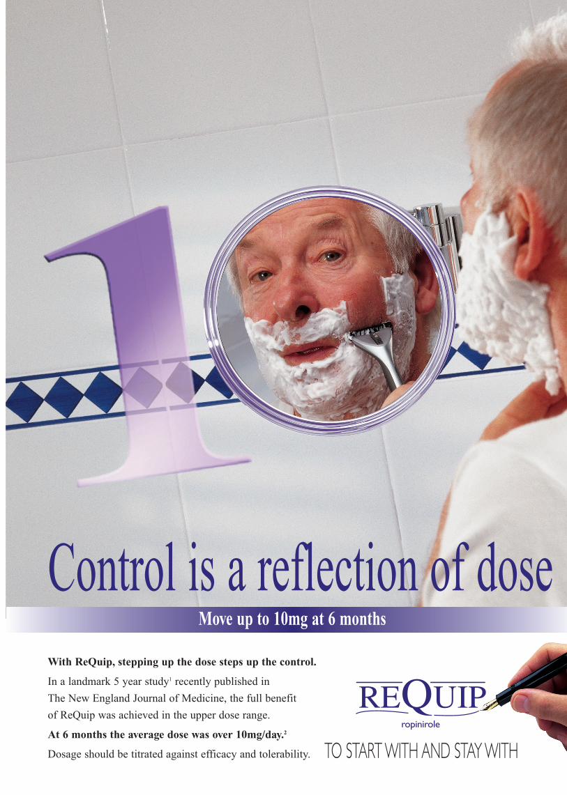
Control is a reflection of doseMove up to 10mg at 6 months
With ReQuip, stepping up the dose steps up the control.
In a landmark 5 year study1 recently published in
The New England Journal of Medicine, the full benefit
of ReQuip was achieved in the upper dose range.
At 6 months the average dose was over 10mg/day.2
Dosage should be titrated against efficacy and tolerability. TO START WITH AND STAY WITH
Section
e
REQUIP ropinirole Prescribing InformationPresentation ‘Requip’ Tablets, PL 10592/0085, 0087-0089, each containingropinirole hydrochloride equivalent to either 0.25, 1, 2 or 5 mg ropinirole. 0.25mg tablets – 210 tablets starting pack, £43.12, 1 mg tablets – 84 tablets, £46.20,2 mg tablets – 84 tablets, £92.40, 5 mg tablets – 84 tablets, £184.80.Indications Treatment of idiopathic Parkinson’s disease. May be used alone(without L-dopa) or in addition to L-dopa to control “on-off” fluctuations andpermit a reduction in the L-dopa dose. Dosage Adults: Three times a day, withmeals. Titrate dose against efficacy and tolerability. Initial dose for 1st weekshould be 0.25 mg t.i.d., 2nd week 0.5 mg t.i.d., 3rd week 0.75 mg t.i.d., 4thweek 1 mg t.i.d. After initial titration, dose may be increased in gradual week-ly increments until acceptable therapeutic response established. Do notexceed 24 mg/day. Concurrent L-dopa dose may be reduced gradually byaround 20%.When switching from another dopamine agonist follow manufac-turer’s guidance on discontinuation. Discontinue ropinirole by reducing dosesover one week. Renal or hepatic impairment: No change needed in mild to mod-erate renal impairment. Not studied in severe renal or hepatic impairment –administration not recommended. Elderly: Titrate dose in normal manner.Children: Parkinson’s disease does not occur in children – do not give to chil-dren. Contra-indications Hypersensitivity to ropinirole, pregnancy, lactationand women of child-bearing potential unless using adequate contraception.Precautions Caution advised in patients with severe cardiovascular diseaseand when co-administering with anti-hypertensive and anti-arrhythmic agents.Patients with major psychotic disorders should be treated with dopamine ago-nists only if potential benefits outweigh the risks. Patients should avoid drivingor other potentially dangerous activities, since rarely, sudden onset of sleephas been reported during daily activities. Caution advised when taking othersedating medication or alcohol in combination with ropinirole. If sudden onsetof sleep occurs in patients, consider dose reduction or drug withdrawal. Druginteractions Neuroleptics and other centrally active dopamine antagonistsmay diminish effectiveness of ropinirole – avoid concomitant use. No dosageadjustment needed when co-administering with L-dopa or domperidone. Nointeraction seen with other Parkinson’s disease drugs but take care whenadding ropinirole to treatment regimen. Should not be given with otherdopamine agonists. In a study with concurrent digoxin, no interaction seenwhich would require dosage adjustment. Metabolised by cytochrome P450enzyme CYP1A2 therefore potential for interaction with substrates orinhibitors of this enzyme – ropinirole dose may need adjustment when thesedrugs are introduced or withdrawn. Increased plasma levels of ropinirole havebeen observed with high oestrogen doses – in patients on hormone replace-ment therapy (HRT) ropinirole treatment may be initiated in normal manner,however, if HRT is stopped or introduced during ropinirole treatment, dosageadjustment may be required. No information on interaction with alcohol – aswith other centrally active medications, caution patients against taking ropini-role with alcohol. Pregnancy and lactation Do not use during pregnancy –based on results of animal studies. There have been no studies of ropinirolein human pregnancy. Do not use in nursing mothers as lactation may be inhib-ited. Adverse reactions In early therapy: nausea, somnolence, leg oedema,abdominal pain, vomiting and syncope. In adjunct therapy: dyskinesia, nausea,hallucinations and confusion. Incidence of postural hypotension (commonlyassociated with dopamine agonists), not markedly different from placebo,however, decreases in systolic blood pressure have been noted; symptomatichypotension and bradycardia, occasionally severe, may occur. As with anotherdopamine agonist, extreme somnolence/ and/or sudden onset of sleep havebeen reported rarely, occasionally when driving (see Precautions and Effects on ability to drive and use machines).Effects on ability to drive and use machines Patients must be informednot to drive and to avoid other potentially dangerous activities, since rarely,cases of sudden onset of sleep have been reported. If this event occurs, con-sider dose reduction or drug withdrawal. Overdosage No incidences report-ed. Symptoms of overdose likely to be related to dopaminergic activity. Legalcategory POM. 8.11.99 ‘Requip’ and the SB logo are registered trade marks.References: 1. Rascol O et al. N Engl J Med 2000; 342(20); 1484-1491. 2. Dataon file (Study 056 Report Synopsis) SmithKline Beecham 2000.Further information is available on request from:
Welwyn Garden City, Hertfordshire AL7 1EY© 2000 SmithKline Beecham Pharmaceuticals
GEN 26839
Editorial Board andregular contributors
Roger Barker is editor of Advances in ClinicalNeuroscience & Rehabilitation (ACNR), and isHonorary Consultant in Neurology at TheCambridge Centre for Brain Repair. He trained inneurology at Cambridge and at the NationalHospital in London. His main area of research is intoneurodegenerative and movement disorders, in par-
ticular parkinson's and Huntington's disease. He is also the uni-versity lecturer in Neurology at Cambridge where he continues todevelop his clinical research into these diseases along with hisbasic research into brain repair using neural transplants.
Stephen Kirker is editor of the Rehabilitation sec-tion of ACNR and Consultant in RehabilitationMedicine in Addenbrooke's NHS Trust, Cambridge.He graduated from Trinity College, Dublin in 1985and trained in neurology in Dublin, London andEdinburgh before moving to rehabilitation inCambridge and Norwich. His main research has
been into postural responses after stroke. His particular interestsare in prosthetics, orthotics, gait training and neurorehabilitation.
Mark Manford contributes our EpilepsyManagement Feature. He has been Consultant atAddenbrooke's Hospital, Cambridge and at BedfordHospital for 3 years. He was an undergraduate atUniversity College London and trained in Neurologyin London at The National Hospital for Neurologyand Neurosurgery, Charing Cross Hospital and at
Southampton. His special interest is epilepsy and he is closelyinvolved with undergraduate training in neurology. He has co-authored an undergraduate textbook of neurology and is current-ly working on a guide to epilepsy.
Alasdair Coles contributes our Anatomy Primer.He is a Wellcome Advanced Fellow working onexperimental immunological therapies in multiplesclerosis, based at the Dunn School of Pathology inOxford and Department of Neurology inCambridge
.Niall Pender is a new recruit to the editorialboard. He is a Neuropsychologist and ClinicalLeader of the Neuro-behavioural RehabilitationUnit at the Royal Hospital for Neuro-disability,London. He is also Neuropsychologist to theHuntington’s Disease Unit at the Royal Hospital. Inaddition he is an Honorary Lecturer in Psychology
at the Institute of Psychiatry, King’s College. Following degrees inPsychology and Neuropsychology he completed his training inClinical Psychology at the Institute of Psychiatry. His researchinterests include cognition in Huntington’s disease, behaviourmanagement in brain injury rehabilitation, memory, and visualimpairments after brain injury.
Andrew Larner is the editor of our Book ReviewSection. He is a Consultant Neurologist at theWalton Centre for Neurology and Neurosurgery inLiverpool, with a particular interest in dementia andcognitive disorders. He is also an HonoraryApothecaries' Lecturer in the History of Medicineat the University of Liverpool.
David J Burn is Consultant and Senior Lecturer inNeurology at the Regional Neurosciences Centre,Newcastle upon Tyne. He qualified from OxfordUniversity and Newcastle upon Tyne MedicalSchool in 1985. His MD was in the functional imag-ing of parkinsonism. He runs Movement Disordersclinics in Newcastle upon Tyne and Sunderland.
Research interests include progressive supranuclear palsy anddementia with Lewy bodies. He is also involved in several drugsstudies for Parkinson's Disease.

A F I R S T C H O I C E A D D - O N T H E R A P Y F O R M O S T S E I Z U R E T Y P E S
topiramate
Because life without seizures is so much better.
TOPAMAX® Abbreviated PrescribingInformation.Please read Summary of ProductCharacteristics before prescribing.Presentation: Tablets: 25, 50, 100, 200 mgtopiramate. Sprinkle Capsules: 15, 25, 50 mgtopiramate. Uses: Adjunctive therapy ofseizures: partial, Lennox Gastaut Syndromeand primary generalised tonic-clonic. Dosageand Administration: Oral administration (notto be chewed). Over 16 years: Usually 200-400mg/day (two divided doses; maximum 800mg/day). Initiate at 25 mg daily with weeklyincrements of 25 mg. Renal disease mayrequire a dose modification. Children 2 to 16:Approx. 5 - 9 mg/kg/day (two divided doses).Initiate at 25 mg nightly with weekly incrementsof 1 - 3 mg/kg.Sprinkle Capsules should be taken whole orsprinkled on a small amount (teaspoon) of softfood and swallowed immediately. Contra-indi-cations: Hypersensitivity to any component.Precautions and Warnings: May cause seda-tion; so caution if driving or operating machin-ery. Contraception recommended for womenof childbearing potential (oral contraceptivesshould contain at least 50 µg oestrogen).SideEffects: Abdominal pain, ataxia, anorexia,CNS side effects, diplopia, fatigue, nausea,nystagmus, weight decrease, agitation, per-sonality disorder, insomnia, increased saliva,hyperkinesia depression, apathy, leucopenia,psychotic symptoms (such as hallucinations),venous thrombo-embolic events, nephrolithia-sis. Pharmaceutical Precautions:Tablets: Store in a dry place at or below 25°C.Sprinkle Capsules: Store below 25°C.Legal Category: POMPackage Quantities and Prices:Bottles of 60 tablets. 25 mg (PL0242/0301) =£22.02, 50 mg (PL0242/0302) = £36.17;100 mg (PL0242/0303) = £64.80; 200 mg(PL0242/0304) = £125.83. Containers of 60capsules. 15 mg (PL0242/0348) = £16.88, 25mg (PL0242/0349) = £25.32, 50 mg(PL0242/0350) = £41.60.Product licence holder: JANSSEN-CILAGLIMITED, SAUNDERTON, HIGH WYCOMBE,BUCKINGHAMSHIRE HP14 4HJ UKDate of text revision: August 2000APIVER250800
Date of preparation: July 200101759
®
ANCR • VOLUME 1 NUMBER 4 SEPTEMBER/OCTOBER 2001 7
The mean duration of disease at death of patho-logically confirmed cases of Parkinson’s Disease
is around 17 years, with a mortality ratio of around2:1. In the earliest phase of the malady, symptomatictreatment may not be needed but within five yearsof diagnosis virtually all patients will be receiving acombination of anti-Parkinsonian medication includ-ing L-dopa.
This is the easiest period of medical managementand after the initial confirmation of diagnosis by amovement disorder hospital specialist many peoplecan be effectively cared for and treated by their gen-eral practitioner.After ten years of disease, howev-er, most patients are running into difficulties eitheras a result of increasing gait, balance, speech andmobility problems, neuropsychiatric symptomsincluding visual hallucinations, confusion, psychosisand dementia or the emergence of the long-term L-dopa syndrome with unpredictable and disablingmotor fluctuations and dyskinesias.
Management of these late phase disabilities are amajor therapeutic challenge and unfortunately thenewer adjuvant drugs including the latest commer-cially available dopamine agonists, COMT inhibitorsand selective MAOIs have had a relatively modestimpact on improving overall quality of life.
Management of Refractory Off periods and Dyskinesias(a) AmantidineIt is remarkable that the anti-dyskinetic effects of amantidinewere completely missed until basic scientific studies related tothe potential therapeutic role of glutamate antagonists led to thedemonstration of its anti-dyskinetic potential in L-dopa primedMPTP-lesioned primates. In 1998 its clinical potential was high-lighted, and sustained reduction in dyskinesias for up to a yearreported. Doses as high as 600mg/day have been recommendedbut many patients derive benefit at doses of only 300mg a day.The beneficial effects seem to occur without any substantialaggravation of the Parkinsonian syndrome and there is a wealthof literature demonstrating modest anti-Parkinsonian effects at200mg/day. All patients with disabling dyskinesias should there-fore be given a therapeutic trial with amantidine before pro-ceeding to consideration of apomorphine or neurosurgery,although it should be stressed that amantidine should be built upand withdrawn slowly to avoid toxic confusional states.
(b) Subcutaneous apomorphine infusionsContinuous subcutaneous apomorphine infusions markedlyimprove off period disability when optimum oral therapy hasfailed and benefit can be maintained for up to 5 years. Despite itsundoubted efficacy this therapeutic approach has been relativelyslow to gain acceptance by physicians in much the same way asinsulin infusions has in Type 1 diabetes. For a successful apomor-phine programme, proper facilities for pump training and medicalsupervision must be in place and there must be a commitmentand belief in the concept of continuous dopaminergic stimulationas a pharmacological goal in treating these complications. Thiseffectively limits its use to regional neuroscience centres andfunding issues have also led to delays in its application in someregions. Furthermore there are a number of patients who do notwarm to the idea of daily injections and a pump, some of who aremuch keener on exploring neurosurgical avenues. Neverthelessall patients with refractory motor fluctuations and disabling dysk-inesias should be enthusiastically offered a trial of subcutaneous
continuous apomorphine.The procedure should befully explained in the out patient clinic and for thosewho are keen to proceed it is generally advisable toadmit the patient to hospital for a 5 day period ofinstruction and treatment initiation. During the waitto be admitted, adjuvant therapy can start to bereduced. Once admitted and with domperidonecover, a subcutaneous challenge with apomorphineis administered to guide the initial pump startingdose, following which the infusion is started and thedose built up with the aim of minimising off periods.During the in-patient admission the patient andtheir family need to be familiarised with the tech-nique by the neurologist and skilled nurse practi-tioner, and the remainder of the adjuvant anti-Parkinsonian medication can be tailed off(Dopamine agonists, COMT inhibitors, amantidine,anti-cholinergics and selegiline). The patient mustthen be seen at weekly intervals for the first fort-night in out patients and then at slowly increasingtime intervals but no less frequently than threemonthly when blood should be taken to check forany haemolysis - a very rare complication of thistherapy. During the first six months of pump treat-ment patients must be strongly encouraged to slow-
ly reduce their L-dopa at a rate of about 50mg/day/week untilthey are only receiving a single nocturnal dose of L-dopa, afterthe pump is removed for the day and possibly also a kick-startdose while the pump is being set up in the morning. Withencouragement about 75% of patients can discontinue all oralanti-Parkinsonian medication while the pump is running. Thisstrategy results in immediate marked reduction in off periods (3-4 hours a day on average or 60% reduction in total off time) andover the ensuing months a marked and sustained diminution indyskinesia duration and severity. Subcutaneous abdominal wallpanniculitis with itchy nodules which may scab and ulcerate arethe commonest complication.These seem to be in part idiosyn-cratic and can be helped by aseptic technique of needle insertion,abdominal wall ultrasound, and silicone gel patches.When severethey may lead to erratic absorption of apomorphine and a returnof off period disability and dyskinesias. In addition some patientsbecome unacceptably drowsy on treatment and a few develop adopamine dysregulation syndrome with behavioural disordersand neuropsychiatric complications. However generally thetreatment is well tolerated and the beneficial effects continue forover five years. These results cannot be reproduced by oraldopamine agonist monotherapy as the clinical potency of theavailable medications is less than both L-dopa and apomorphine.
(c) Functional neurosurgeryNew insights into basal ganglia circuitry scientifically underpinnedthe resurgence of interest in ventrolateral pallidotomy.A decadelater the value of this procedure in the long-term appears some-what clearer in that it appears to be an extremely effective pro-cedure for abolishing contralateral L-dopa induced dyskinesiasand rest tremor. However, its long-term beneficial effects onbradykinesia are uncertain. Furthermore there is a significantmorbidity, even in the most proficient hands, with some fatalities.Speech problems and neuropsychological sequelae are the com-monest unwanted effects and balance and posture are notimproved. Most functional neurosurgeons now seemed to haveabandoned lesioning, including subthalomotomy which showedconsiderable early promise, in favour of bilateral deep brain stim-ulation.Although expensive and labour intensive (because of the
Author
Professor Andrew Leesholds a personal chair in neu-rology at University CollegeLondon. He is director of theReta Lila Weston Institute ofNeurological Studies, UCL andalso directs the Brain ResearchCentre, Institute of Neurology,Queen Square, London.Professor Lees is co-editor ofMovement Disorders Journaland President of the EuropeanSection of the MovementDisorder Society.
New advances in the management oflate stage Parkinson’s Disease
Review Article
8 ANCR • VOLUME 1 NUMBER 4 SEPTEMBER/OCTOBER 2001
need for frequent post-operative adjustments of the stimulationfrequency), extremely impressive results with up to 2 years fol-low-up have been reported with subthalamic and pallidal stimu-lation. At least half the patients who have received subthalamicstimulation are able to withdraw anti-Parkinsonian medicationcompletely and the rest markedly reduce it.Tremor, rigidity andbradykinesia are all helped and the marked reduction of L-dopaabolishes dyskinesias. Marked improvements in alertness anddrive are reported but a few patients have experienced unwant-ed neuropsychiatric complications such as suicidal depression,and incontinent laughter or crying. It is unclear whether postur-al instability can be helped in the long-term. Fewer pallidal pro-cedures have so far been reported but comparable results can beseen.
Management of Neuropsychiatric ComplicationsVisual hallucinations occur in at least 40% of patients and increas-ing age, duration of disease, and deteriorating cognition andvision are predisposing factors although some patients developthem early in the course of the illness in the absence of demen-tia.Anticholinergics and dopaminergic agents may cause, or aggra-vate, them but there is increasing evidence to suggest that thepathological substrate of Parkinson’s Disease is primarily respon-sible. Daytime somnolence may be another marker for visual hal-lucinations and a REM sleep behaviour disorder akin to nar-colepsy has been also suggested as a pathophysiological compo-nent. In many instances the visual hallucinations are minor, non-frightening and are evaluated by the patient as being fictitious.However, the presence of shadey strangers, beasts and insectsand Capgras phenomena may cause great distress and requiretherapy.
Dopaminometic psychoses resembling those seen in ampheta-mine and cocaine addicts can occur with paranoid delusions andhypomania and a few patients develop dopamine dysregulationsyndromes with excessive and destructive overuse of medicationleading to hyperlibidinous behaviour, compulsive gambling,hoarding behaviour and errors of judgement. Toxic confusionalstates are also common and intercurrent causes such as infec-tions, metabolic disturbances, drug interactions or falls with sub-dural haematomata need to be excluded.
Psychotic behaviour and dysregulation may be helped bydopamine blocking drugs. Most of these unfortunately aggravatemotor disability and may induce severe akinetic crises which maytake many weeks to reverse even after discontinuation of theoffending neuroleptic. Clozapine is an exception but the need forregular white cell monitoring limits its routine practicability.However, quetiapine another novel neuroleptic in doses from 25-400mg/day holds out promise as a second selective anti-psychot-ic agent. Like clozapine it has marked effects on 5HT2A recep-tors. Occasionally unilateral electroshock therapy can be usefulfor confusional states especially on a background of severe drugrefractory major depressive episodes. There is also now greatinterest in the use of the central cholinesterase inhibitors,
Correspondence Address
Professor AJ Lees, The National Hospital for Neurology &Neurosurgery, Queen Square, London, WC1N 3BG. [email protected]
donepezil, rivastigmine and galantamine to help confusion, visualhallucinations and attention in Parkinsons disease with dementia.Many of these patients have severe central cholinergic deficitsand cerebral Lewy bodies.
Management of Freezing, Falls, Speech and SwallowingdifficultiesThese symptoms which become increasingly common and dis-abling as the disease progresses often defy all available medicaland surgical approaches. However intensive physical, speech andswallowing therapy programmes conducted by skilled and spe-cialised therapists can be of great help. Gastrostomies are stillprobably underused and silent aspiration, a common cause ofdeath in Parkinson’s Disease underdiagnosed.
ReferencesVerhagen Metman L, Del Dotto P, LePooleK et al. Amantidine for L-dopa-induced dyskinesias. A one year follow-up study. ArchNeurol.1999;56:1382-1386.Colzi A, Turner K, Lees AJ. Continuous subcutaneous waking day apomor-phine in the long-term treatment of levodopa induced dyskinesias in Parkinsonsdisease. J.Neurol.Neurosurg.Psychiatry 1998;64:573-576.McKeith I, Del Ser T , Spano P et al Efficacy of rivastigmine in dementia withLewy bodies :a randomised double-blind controlled international study. Lancet2000;256:2031-2036.Limousin P, Krack P, Pollak P et al. Electrical stimulation of the subthalamicnucleus in advanced Parkinsons disease. N.Engl.J.Med. 1998;339:1105-111.Friedman J and Factor S. Management of psychosis in Parkinsons disease.Mov Disorders 2000;15:201-211.Iansek RT and Morris M. Rehabilitation of gait in Parkinsons disease.J.Neurol.Neurosurg Psychiat.1997;62: 22-26.
Management of off period Dystonia� Introduce a dopamine agonist (at night if early
morning disability main problem)� Trials of baclofen and lithium� Consideration of botulinus toxin therapy� Apomorphine pump� Functional neurosurgery
Management of Disabling L-Dopa Provoked PeakDose Dyskinesias� Reduce L-dopa dose to minimum required to control
mobility � Partially replace L-dopa with a dopamine agonist� Add amantidine� Trial of continuous waking day subcutaneous
apomorphine therapy� Consider functional neurosurgery
Review Article
Would you like to receive your own regularcomplimentary copy of ACNR magazine?To add yourself to the mailing list either photocopy or tear out this form, completeyour details, and fax or post the sheet to the address below.
Yes, please add me to the mailing list (BLOCK CAPS PLEASE)Name: . . . . . . . . . . . . . . . . . . . . . . . . . . . . . . . . . . . . . . . . . . . . . . . . . . . . . . . . . .Job Title: . . . . . . . . . . . . . . . . . . . . . . . . . . . . . . . . . . . . . . . . . . . . . . . . . . . . . . . .Address: . . . . . . . . . . . . . . . . . . . . . . . . . . . . . . . . . . . . . . . . . . . . . . . . . . . . . . . .City: . . . . . . . . . . . . . . . . . . . . . . . . . . . . . . . . . . . . . . . . . . . . . . . . . . . . . . . . . . .Postcode . . . . . . . . . . . . . . . . . . . . . . . . . . . . . . . . . . . . . . . . . . . . . . . . . . . . . . . .Tel: . . . . . . . . . . . . . . . . . . . . . . . . . . . . . . . . . . . . . . . . . . . . . . . . . . . . . . . . . . . .Fax: . . . . . . . . . . . . . . . . . . . . . . . . . . . . . . . . . . . . . . . . . . . . . . . . . . . . . . . . . . .
NOW FAX TO: 0131 313 1110 or post to: ACNR Magazine, 7 Alderbank Terrace, Edinburgh EH11 1SX
APG.AD.V2A4
apomorphine hydrochloride
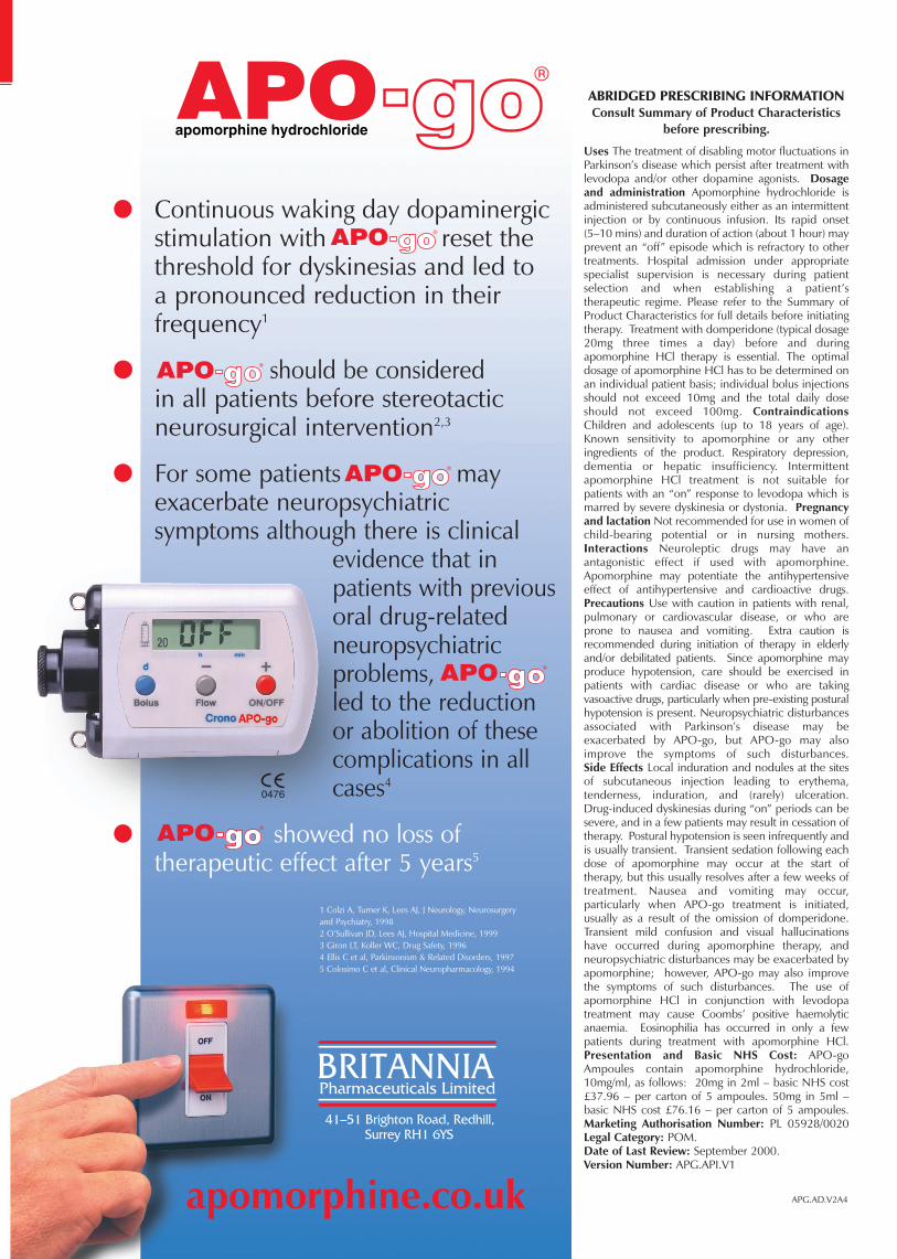
� Continuous waking day dopaminergicstimulation with reset thethreshold for dyskinesias and led to a pronounced reduction in their frequency1
� should be consideredin all patients before stereotactic neurosurgical intervention2,3
� For some patients may exacerbate neuropsychiatric symptoms although there is clinical
evidence that in patients with previousoral drug-relatedneuropsychiatric problems,led to the reduction or abolition of thesecomplications in allcases4
� showed no loss of therapeutic effect after 5 years5
1 Colzi A, Turner K, Lees AJ, J Neurology, Neurosurgeryand Psychiatry, 19982 O’Sullivan JD, Lees AJ, Hospital Medicine, 19993 Giron LT, Koller WC, Drug Safety, 19964 Ellis C et al, Parkinsonism & Related Disorders, 19975 Colosimo C et al, Clinical Neuropharmacology, 1994
ABRIDGED PRESCRIBING INFORMATIONConsult Summary of Product Characteristics
before prescribing.
Uses The treatment of disabling motor fluctuations inParkinson’s disease which persist after treatment withlevodopa and/or other dopamine agonists. Dosageand administration Apomorphine hydrochloride is administered subcutaneously either as an intermittentinjection or by continuous infusion. Its rapid onset (5–10 mins) and duration of action (about 1 hour) mayprevent an “off” episode which is refractory to othertreatments. Hospital admission under appropriatespecialist supervision is necessary during patient selection and when establishing a patient’s therapeutic regime. Please refer to the Summary ofProduct Characteristics for full details before initiatingtherapy. Treatment with domperidone (typical dosage20mg three times a day) before and during apomorphine HCl therapy is essential. The optimaldosage of apomorphine HCl has to be determined onan individual patient basis; individual bolus injectionsshould not exceed 10mg and the total daily doseshould not exceed 100mg. ContraindicationsChildren and adolescents (up to 18 years of age).Known sensitivity to apomorphine or any other ingredients of the product. Respiratory depression,dementia or hepatic insufficiency. Intermittent apomorphine HCl treatment is not suitable forpatients with an “on” response to levodopa which ismarred by severe dyskinesia or dystonia. Pregnancyand lactation Not recommended for use in women ofchild-bearing potential or in nursing mothers.Interactions Neuroleptic drugs may have an antagonistic effect if used with apomorphine.Apomorphine may potentiate the antihypertensiveeffect of antihypertensive and cardioactive drugs.Precautions Use with caution in patients with renal,pulmonary or cardiovascular disease, or who areprone to nausea and vomiting. Extra caution is recommended during initiation of therapy in elderlyand/or debilitated patients. Since apomorphine mayproduce hypotension, care should be exercised inpatients with cardiac disease or who are taking vasoactive drugs, particularly when pre-existing posturalhypotension is present. Neuropsychiatric disturbancesassociated with Parkinson’s disease may be exacerbated by APO-go, but APO-go may alsoimprove the symptoms of such disturbances. Side Effects Local induration and nodules at the sitesof subcutaneous injection leading to erythema, tenderness, induration, and (rarely) ulceration.Drug-induced dyskinesias during “on” periods can besevere, and in a few patients may result in cessation oftherapy. Postural hypotension is seen infrequently andis usually transient. Transient sedation following eachdose of apomorphine may occur at the start of therapy, but this usually resolves after a few weeks oftreatment. Nausea and vomiting may occur, particularly when APO-go treatment is initiated, usually as a result of the omission of domperidone.Transient mild confusion and visual hallucinationshave occurred during apomorphine therapy, and neuropsychiatric disturbances may be exacerbated byapomorphine; however, APO-go may also improvethe symptoms of such disturbances. The use of apomorphine HCl in conjunction with levodopa treatment may cause Coombs’ positive haemolyticanaemia. Eosinophilia has occurred in only a fewpatients during treatment with apomorphine HCl.Presentation and Basic NHS Cost: APO-goAmpoules contain apomorphine hydrochloride,10mg/ml, as follows: 20mg in 2ml – basic NHS cost£37.96 – per carton of 5 ampoules. 50mg in 5ml –basic NHS cost £76.16 – per carton of 5 ampoules.Marketing Authorisation Number: PL 05928/0020 Legal Category: POM.Date of Last Review: September 2000.Version Number: APG.API.V1
BRITANNIAPharmaceuticals Limited
41–51 Brighton Road, Redhill,Surrey RH1 6YS
apomorphine.co.uk
0476
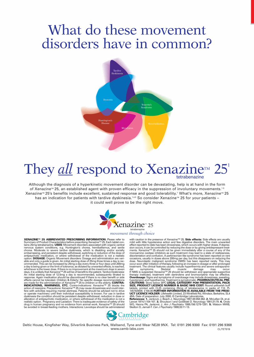
Although the diagnosis of a hyperkinetic movement disorder can be devastating, help is at hand in the form of Xenazine™ 25, an established agent with proven efficacy in the suppression of involuntary movements.1-5
Xenazine™ 25’s benefits include excellent, sustained response and good tolerability.1 What's more, Xenazine™ 25has an indication for patients with tardive dyskinesia.1,4,5 So consider Xenazine™ 25 for your patients –
it could well prove to be the right move.
They all respond to Xenazine™ 251
Deltic House, Kingfisher Way, Silverlink Business Park, Wallsend, Tyne and Wear NE28 9NX. Tel: 0191 296 9300 Fax: 0191 296 9368www.camb-labs.com
What do these movement disorders have in common?
tetrabenazine
XENAZINE™ 25 ABBREVIATED PRESCRIBING INFORMATION: Please refer toSummary of Product Characteristics before prescribing Xenazine™ 25. Each tablet con-tains 25mg tetrabenazine. USES: Movement disorders associated with organic centralnervous system conditions, e.g. Huntington’s chorea, hemiballismus, and senilechorea. Moderate to severe tardive dyskinesia, which is disabling and/or sociallyembarrassing, and persistent despite withdrawal, switching or reduction of the dose ofantipsychotic medication, or where withdrawal of the medication is not a realisticoption. DOSAGE: Organic Movement disorders: Dosage and administration are vari-able and only a guide is given. An initial starting dose of 25mg three times a day is rec-ommended. This can be increased by 25mg a day every three or four days until 200mga day is being given or the limit of tolerance, as dictated by unwanted effects, is reached,whichever is the lower dose. If there is no improvement at the maximum dose in sevendays, it is unlikely that Xenazine™ 25 will be of benefit to the patient. Tardive Dyskinesia:An initial starting dose of 12.5mg a day is recommended, subsequently titrated toresponse. Again medication should be discontinued if there is no clear benefit or sideeffects cannot be tolerated. Children & Elderly: No specific dosage recommendationsare made for the administration of Xenazine™ 25 to children or the elderly. CONTRA-INDICATIONS, WARNINGS, ETC. Contra-indications: Xenazine™ 25 blocks theaction of reserpine. Precautions: Xenazine™ 25 may cause drowsiness and could inter-fere with activities requiring mental alertness. Patients should be advised not to driveor operate machinery until their individual susceptibility is known. For use in tardivedyskinesia the condition should be persistent despite withdrawal, reduction in dose oralteration of antipsychotic medication, or where withdrawal of the medication is not arealistic option. Pregnancy and Lactation: There is inadequate evidence of safety of thedrug in human pregnancy and no evidence from animal work. Xenazine™ 25 shouldbe avoided in breast-feeding mothers. Interactions: Levodopa should be administered
with caution in the presence of Xenazine™ 25. Side effects: Side effects are usuallymild with little hypotensive action and few digestive disorders. The main unwantedeffect reported to date has been drowsiness, which occurs with higher doses. If depres-sion occurs, it can be controlled by reducing the dose or by giving antidepressant treat-ments. Xenazine™ 25 should not be given immediately after a course of any of themonoamine oxidase inhibitors as such treatment may lead to a state of restlessness,disorientation and confusion. A parkinsonian-like syndrome has been reported on rareoccasions, usually in doses above 200mg per day, but this disappears on reducing thedose. Neuroleptic malignant syndrome (NMS) has been reported rarely. This mayoccur soon after initiation of therapy, following an increase in dosage or after prolongedtreatment. The clinical features usually include hyperthermia and severe extrapyrami-dal symptoms. Skeletal muscle damage may occur. If NMS is suspected Xenazine™ 25 should be withdrawn and appropriate supportive therapy instituted, treatment with dantrolene and bromocriptine may be effective.Overdosage: Signs and symptoms of overdosage may include drowsiness, sweating,hypotension and hypothermia. Treatment is symptomatic. PHARMACEUTICAL PRE-CAUTIONS: Store below 30ºC LEGAL CATEGORY POM PRESENTATION, PACKSIZE, PRODUCT LICENCE NUMBER & BASIC NHS COST: Round yellowish bufftablets, printed with CL25 containing 25mg of tetrabenazine in packs of 112. PL14576/0005 £100.00 FURTHER INFORMATION IS AVAILABLE FROM THE PROD-UCT LICENCE HOLDER: Lifehealth Limited, 23 Winkfield Rd, Windsor, Berkshire, SL44BA. Date of preparation: July 2000. © Cambridge Laboratories. References: 1. Jankovic J, Beach J. Neurology 1997;48:358-362. 2. McLellan DL et al.Lancet 1974;1:104-107. 3. Shoulson I and Goldblatt D. Neurology 1981;31:79. 4. OndoWG, Hanna PA, Jankovic J. Am J Psychiatry 1999;156:1279-1281. 5. Watson MWB,Skelton D, Jamali F. Can J Psychiatry 1988;33:11-13.
CL/TET019
ANCR • VOLUME 1 NUMBER 4 SEPTEMBER/OCTOBER 2001 11
Section
Introduction
Huntington's Disease (HD) is a rare, complexand challenging condition to manage. It occurs
at a rate of 5-10 per 100, 000 but this rate varieswithin and between countries. It is estimated thatthere are approximately 5000 people with the con-dition in the UK.The core genetic anomaly in HDis the expansion of the trinucleotide repeat CAG1 .
The central triad of deficits in HD has changedlittle since Huntington's description (see Table 1)but the nature, speed and specificity of presenta-tion of these symptoms is variable and inconsis-tent. The inconsistency of cognitive impairmentsremains problematic.
Frequent symptoms of HDThere are many clear descriptions of the neurobi-ology and genetics of HD2.The principal structuresinvolved include the substantia nigra, internal andexternal portions of the globus pallidus, sub-thala-mic nucleus, amygdala, thalamus and hypothalamus.Extensive cortical damage has also been noted.Studies using procedures such as Proton MagneticResonance Spectroscopy (pMRS) have foundincreased lactate levels and decreased N-AcetylAspartate (NAA) in the frontal and occipital lobes3.Moreover, PET studies have shown reduced glu-cose and oxygen metabolism in the basal gangliaand cortex in HD before structural scanningchanges or clinical signs become apparent.
It is clear that HD produces damage that,although markedly apparent in the basal ganglia,can be widespread and may involve many corticalregions particularly as the disease progresses.This has consider-able impact on the nature and presentation of cognitive impair-ments. However, it is only recently that detailed descriptions ofcognitive dysfunction are emerging.
Cognitive impairments in HDWith the advent and increasing use of diagnostic technology forHD there has been a growth in studies of cognitive functioning.We are still far from being clear about the nature of such impair-ments in HD and this is particularly important given the need toevaluate outcome following treatment trials for HD4,5.
Early cognitive impairmentsIn the early stages of the condition patients with HD are consis-tently impaired on general intellectual and cognitive measuresrelative to other neurodegenerative groups such as multiple scle-rosis, Parkinson's Disease and AIDS dementia6. Several tests havebeen noted to successfully discriminate HD from healthy partic-ipants (See Table 2).
Executive functions in HDRecently there has been increased interest in theexecutive deficits experienced by patients with HD.This not only results from the earlier application ofmore sophisticated diagnostic testing but alsoreflects the improved resolution of current neu-roimaging techniques. Lesions are seen to extendbeyond the territory of the basal ganglia and encom-pass the frontal lobes. Moreover, the nature of thereciprocal connections between the basal ganglia andthe oculomotor region, dorsolateral prefrontal cor-tex and lateral orbitofrontal area would suggest thatan exploration of executive deficits in HD would beprudent. This is supported by behavioural evidenceand clinical reports of patients at various stages ofthe disease.
HD patients appear to be impaired on tests ofplanning, organising, initiating, executing and sustain-ing behaviour7. Impairments of verbal fluency andbehavioural features of perseveration have alsobeen reported.
Marked perseveration can be observed and HDpatients are impaired at learning new stimulus-response associations relative to healthy subjectsand more importantly patients with Dementia ofthe Alzheimer Type (DAT)8. Double dissociationswithin the class of executive functions have alsobeen noted between HD and DAT patients9.
HD patients are particularly impaired on tests ofplanning and this translates directly into their dailyfunctioning where they often appear to lead chaot-ic and disorganised lives. HD patients are impaired,
in particular at shifting set, again an obvious feature of the con-dition where patients become fixed on particular ideas andissues.
Attentional function in HDIn terms of attentional functioning, alertness, divided attentionand response flexibility are markedly impaired in patients withHD patients10. These fundamental impairments are exacerbatedby their extensive executive impairments and contribute to sig-nificant difficulties in activities of daily living and occupation.
Emotional Perception in HDFew studies have addressed perceptual skills directly. By far themost frequent are the intriguing studies of emotional and facialperception in this group.
HD patients have been shown to be impaired at recognisingfacial and vocal expressions of emotion, especially fear and dis-gust11. Furthermore, patients are impaired at comprehendingemotional prosody in speech, matching facial affect, facial recog-
SectionSectionReview Article
Author
Niall Pender is a Neuro-psychologist and ClinicalLeader of the Neuro-behav-ioural Rehabilitation Unit atthe Royal Hospital for Neuro-disability, London. He is alsoNeuropsychologist to theHuntington’s Disease Unit atthe Royal Hospital. In additionhe is an Honorary Lecturer inPsychology at the Institute ofPsychiatry, King’s College.Following degrees inPsychology and Neuro-psychology he completed histraining in Clinical Psychologyat the Institute of Psychiatry.His research interests includecognition in Huntington’sDisease, behaviour manage-ment in brain injury rehabilita-tion, memory, and visualimpairments after brain injury.
Cognition in Huntington's Disease
Chorea Executive dysfunctionAthetosis Episodic memory impairmentsParanoia Relatively better recognitionSelf-neglect Flat temporal gradient in autobiographical memory30% experience major depressive episode Impairments in emotional perceptionHigher than base rate suicide Impairments with focused, sustained and divided attentionPersonality changesDysarthria
Neurological/Psychiatric Cognitive
1Table 1. Frequent symptoms of HD
12 ANCR • VOLUME 1 NUMBER 4 SEPTEMBER/OCTOBER 2001
Correspondence Address
Niall Pender, Royal Hospital for Neuro-Disability, West Hill,Putney, London SW15 3SW.Tel. 020 8780 4500,E-Mail. [email protected]
SectionSection
nition and discriminating faces12. These impairments have directrelationships to patients' ability to cope in their daily lives andinterpret and use facial expression accurately.
Memory functioningThere has been a great deal of debate concerning the nature ofthe memory impairment in HD and the pattern of impaired andpreserved skills in this patient group.
However, some clear details have consistently been reported.Global memory deficits are common in HD13. Implicit motortasks are more impaired than lexical tasks14. It has been suggest-ed that recognition memory is preferentially preserved untillater stages and therefore a retrieval deficit is favoured by manyauthors7. However, some controversy remains as to whether theprincipal deficit is in fact an executive dysfunction mediating aretrieval or even an encoding deficit.
Interestingly a flat temporal gradient is seen in retrogradememory functions with equal impairments observed acrossdecades. Cueing, in particular phonemic cues, seem to improveperformance for recently diagnosed patients but not for thelater stage patients.
Longitudinal changes in cognitionLong-term evaluations of patients with HD are currently under-way and in the most recent and best-conducted studies thereappear to be some particular consistencies among the data.
In a recent three-year follow-up study of HD patients15 signif-icant impairments were noted at the end of the first year onexecutive and memory tasks, particularly verbal fluency, theStroop test and object recall.At the final follow-up, a similar pat-tern of scores was obtained. Speed based tasks and memorychanged significantly over time. The authors suggested that theobserved impairments in memory relate to a primary deficit inexecutive functioning which impacts on encoding and retrieval.
A further recent study16 reported that performance on testsof attention, executive functioning, language comprehension andvisual-spatial memory deteriorated over a four-year period.Early episodic memory impairments remained stable over thecourse of the study.
Thus studies of cognitive functioning in HD report decline ona range of measures over the course of the condition. Morerecent longitudinal studies appear to be finding consistentchanges in executive functioning and a less dramatic deteriora-tion in memory functioning over time. However, there stillappears to be marked variability in the experimental designswith different studies employing different measures of cognitivefunctioning. More importantly, many studies use insufficientpatient numbers to achieve adequate power.
SummaryHD is a complex and challenging condition. The care of suchpatients generally requires the expertise of multi-disciplinaryteams. Complicating this picture is the patient's fluctuating yetdeteriorating level of cognition, which subsequently leads to areduction of the patient's mental capacity. Clear and obvious
Review Article
deficits in executive functioning and memory are noted early inthe course of the disease but impairments in other areas of abil-ity are apparent as the disease progresses. Such deficits com-pound the existing impairments in motor functioning, communi-cation, nutrition and psychiatry and can exacerbate any behav-ioural difficulties. A detailed neuropsychological assessment inthe context of an MDT evaluation can provide much usefulinformation for the care of such patients.
AcknowledgementNiall Pender is supported by a grant from the Neuro-disabilityResearch Trust.
References1. Harper, P.S. (1993). A specific mutation for Huntington's disease. Journal
of Medical Genetics, 30, 975-977.2. Lowe, J., Lennox, G., Leigh, P.N. (1997). Disorders of movement and sys-
tem degeneration. In Graham, D.I. and P.L. Lantos (Eds). Greenfield'sNeuropathology. 6th edn.Volume 2.Arnold Press:UK. Chpt. 6.
3. Harms, L., Meierkord, H., Timm, G., Pfeiffer, L., Ludolph, A.C. (1997).Decreased N-acetyl-aspartate/choline ratio and increased lactate in thefrontal lobe of patients with Huntington's disease: A proton magnetic res-onance spectroscopy study. Journal of Neurology, Neurosurgery andPsychiatry, 62, 27-30.
4. Quinn, N., Brown, R., Craufurd, D., Goldman, S., Hodges, J., Kieburtz,K., Lindvall, O., MacMillan, J., Roos, R. (1996). Core Assessment Programfor Intracerebral Transplantation in Huntington's Disease (CAPIT-HD).Movement Disorders, 11, 2, 143-150.
5. Kieburtz, K., Penney., J.B., Como., P. et al., (1996). Unified Huntington'sDisease Rating Scale: Reliability and consistency. Movement Disorders,11, 2, 136-142.
6. Clark, C.M., Jacova, C., Klonoff, H., Kremer, B., Hayden, M., Paty, D.(1997). Pathological association and dissociation of functional systems inMultiple Sclerosis and Huntington's Disease. Journal of Clinical andExperimental Neuropsychology, 19,1, 63-76
7. Purdon, S.E., Chase, T. Mohr E. (1996). Huntington's Disease. In J.G.Beaumont, P.K. Kenealy, M.J.C. Rogers (Eds).The Blackwell Dictionaryof Neuropsychology. Blackwell Publishers: UK.
8. Lange, K.W., Sahakian, B.J., Quinn, N.P., Marsden, C.D., Robbins, T.W.(1995). Comparison of executive and visuospatial memory function inHuntington's disease and dementia of Alzheimer type matched for degreeof dementia. Journal of Neurology, Neurosurgery and Psychiatry, 58,598-606.
9. Rosser,A., Hodges, J.R. (1994). Initial letter and semantic category fluen-cy in Alzheimer's disease, Huntington's disease and progressive supranu-clear palsy. Journal of Neurology, Neurosurgery and Psychiatry, 57,1389-1394.
10. Sprengelmeyer, R., Lange, H., Homberg,V. (1995).The pattern of atten-tional deficits in Huntington's Disease. Brain, 118, 1, 145-152.
11. Gray, J., Young, A.W., Barker, W.A., Curtis, A., Gibson, D. (1997).Impaired recognition of disgust in Huntington's disease gene carriers.Brain, 120, 2029-2038.
12. Jacobs, D.H., Shuren, J., Heilman, K.H. (1995). Impaired perception offacial identity and facial affect in Huntington's disease. Neurology, 45,1217-1218.
13. Ginovart, N., Lundin, A., Farde, L., Halldin, C., Backman, L., Swahn,C.G., Pauli, S., Sedvall, G. (1997). PET study of the pre- and post-synap-tic dopaminergic markers for the neurodegenerative process inHuntington's disease. Brain, 120, 503-514.
14. Heindel, W.C., Salmon, D.P., Shults, C.W., Walicke, P.A., Butters, N.(1989). Neuropsychological evidence for multiple implicit memory sys-tems: A comparison of Alzheimer's, Huntington's and Parkinson's diseasepatients. The Journal of Neurosciences, 9, 2, 582-587.
15. Snowden, J., Crauford, D., Griffiths, H.,Thompson, J., Neary, D. (2001).Longitudinal evaluation of cognitive disorder in Huntington's disease.Journal of the International Neuropsychological Society, 7, 33-44.
16. Bachoud-Levi, A., C., Maison, P., Bartolomeo, P., Boisse, M.F. et al.,(2001). Retest effects and cognitive decline in longitudinal follow-up ofpatients with early HD. Neurology, 56, 1052-1058.
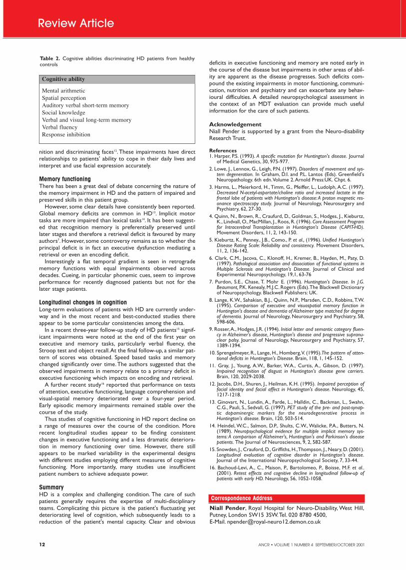
Mental arithmetic Spatial perceptionAuditory verbal short-term memorySocial knowledgeVerbal and visual long-term memory Verbal fluencyResponse inhibition
Cognitive ability
Table 2. Cognitive abilities discriminating HD patients from healthycontrols
ANCR • VOLUME 1 NUMBER 4 SEPTEMBER/OCTOBER 2001 13
Management Topic
Introduction
Epilepsy creates some real risks for patients that need to beanticipated and some imagined risks, for which the clinician
must try to allay the fears of patients and relatives. Many socialspheres need to be addressed, including, education, work, hob-bies, relationships, sexuality, marriage, parenting and driving.Stigmatization may spread beyond the patient to other familymembers, employers and schools.
Social effects of epilepsyPredictors of psychosocial underfunctioning� Biological aspects of the epilepsy partly determine the quality
of life measures in some but not all studies.� Social aspects, especially supportive family dynamics are impor-
tant in enabling patients to cope with their epilepsy (table 1).Some patients with severe epilepsy lead remarkably normallives, whilst others with relatively mild epilepsy may becomeover-protected and socially isolated.
Education� School failure may affect one third of intellectually normal chil-
dren with epilepsy, and one third may require special educa-tional support.
� Education may be impaired by organic factors, by the socialconsequences of the epilepsy or by the attitudes of teachers.
� Psychiatric support may be needed by nearly a quarter duringthe education years.
Employment� The number of patients who are unemployed or on perma-
nent sick pay in the UK (table 2) correlates with epilepsyseverity but differs substantially between countries and cul-tures. Reduced educational achievement contributes to unem-ployment.
Leisure activities� Individuals with epilepsy are less likely to pursue leisure activ-
ities than the unaffected population. Reasons include low self-esteem and fear of seizures in company. Epilepsy sufferers areless physically fit than the rest of the population.
Stigma of epilepsy� Cross-cultural differences are marked.Around 30% of patients
feel stigmatized in Spain or Poland and 52% in the UK.
� Factors associated with stigma include worry, negative feel-ings, long-term health problems, injuries and adverse effects ofAED.
� Prejudice against epilepsy also varies greatly. In China 57% ofparents object to their children playing with a child withepilepsy, compared with 7% in Denmark.
Marriage and fertility� Marriage rates are inversely correlated and divorce rates are
positively correlated with epilepsy severity in the UK.� Fertility is 60-80% of normal in women with epilepsy and is
also reduced in men with epilepsy. Reasons include reducedmarriage rates, reduced sexual arousal or satisfaction, alteredgonadal function, effects of AED and fear of the effects ofepilepsy on the ability to care for children.
Epilepsy and driving� The responsibility of reporting the onset of epilepsy to the
driving authorities (DVLA in the UK) rests with the patient inthe UK. However, if a clinician knows that a patient is puttingothers at risk by continuing to drive, their duty to the com-munity usually takes precedence over their duty of confiden-tiality.
� UK regulations are summarised in table 3.These are regularlyupdated and are available from DVLAhttp://www.dvla.gov.uk/drivers/drivers.htm
� Group 2 licences (heavy goods or passenger-carrying vehicles)generally require the patient to be free of attacks for at leastten years without medication.
� The aetiology of the seizures may modify the ruling. Malignanttumours carry more stringent regulations.
� All seizures are the same to the DVLA, no matter how mild.� Insurance companies should be informed as well as the DVLA.
Situations of risk - advice to patients and carersEach seizure carries a small risk. A balance needs to be struckbetween alarmist over-protection and sensible precautions. Thiswill depend on the frequency of seizures, whether they are noc-turnal or diurnal and whether there is an aura, which allows thepatient to take evasive action.
Reducing the risk from seizuresIn the kitchen 1. Cooker guards reduce the risk of falling onto a cooker.
Social effects of epilepsy Mark Manford
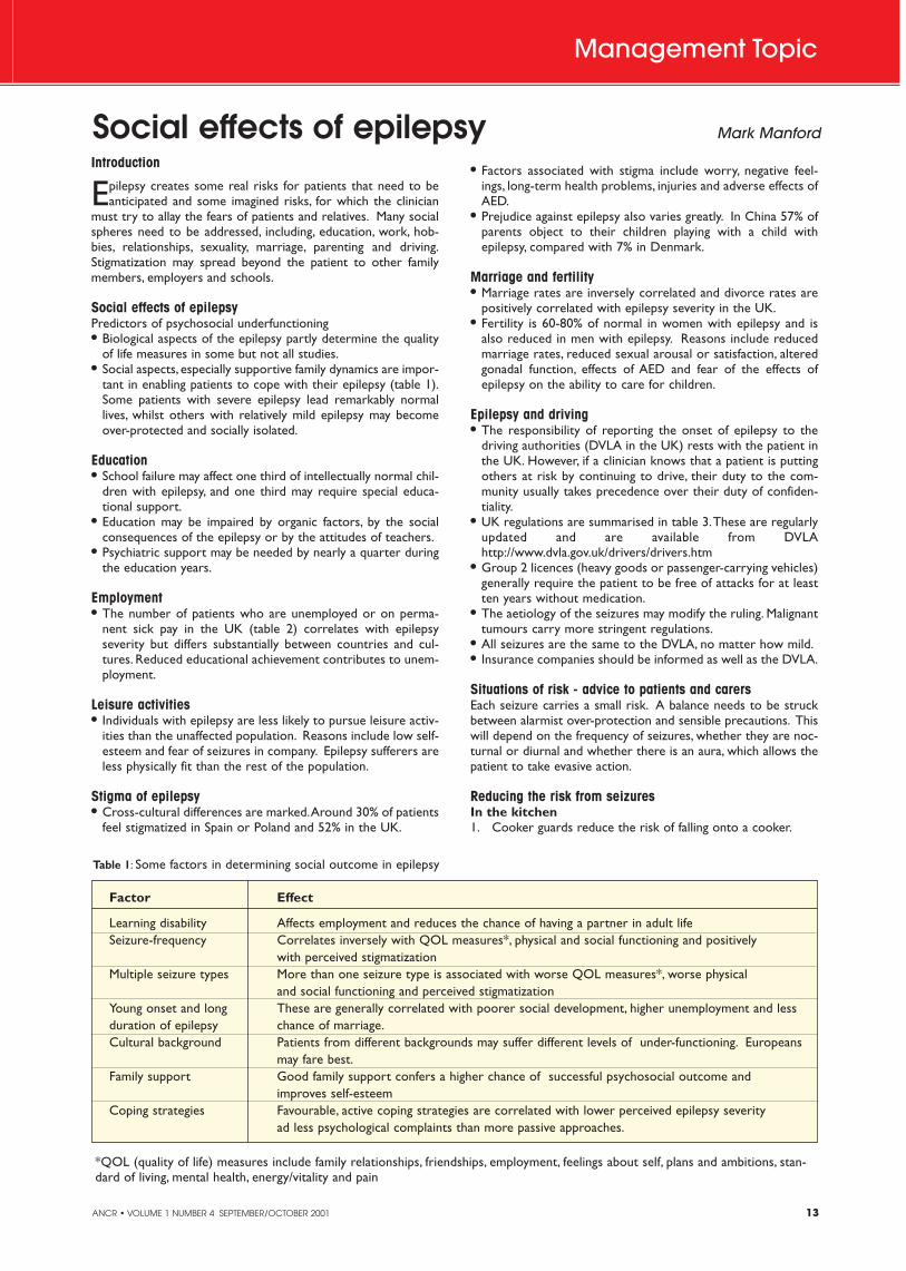
Table 1: Some factors in determining social outcome in epilepsy
Factor Effect
Learning disability Affects employment and reduces the chance of having a partner in adult life Seizure-frequency Correlates inversely with QOL measures*, physical and social functioning and positively
with perceived stigmatizationMultiple seizure types More than one seizure type is associated with worse QOL measures*, worse physical
and social functioning and perceived stigmatizationYoung onset and long These are generally correlated with poorer social development, higher unemployment and less duration of epilepsy chance of marriage.Cultural background Patients from different backgrounds may suffer different levels of under-functioning. Europeans
may fare best.Family support Good family support confers a higher chance of successful psychosocial outcome and
improves self-esteemCoping strategies Favourable, active coping strategies are correlated with lower perceived epilepsy severity
ad less psychological complaints than more passive approaches.
*QOL (quality of life) measures include family relationships, friendships, employment, feelings about self, plans and ambitions, stan-dard of living, mental health, energy/vitality and pain

14 ANCR • VOLUME 1 NUMBER 4 SEPTEMBER/OCTOBER 2001
Management Topic
� Fear of seizures may make some jobs particularly stressful,for example where the individual is on show as a musician.
� Misunderstanding of epilepsy in the workplace may be helpedby education of colleagues.
Travel� There is no restriction on travel for patients with epilepsy
but it is advisable that they are accompanied.� Medication should be to hand at all times, a back-up set may
be needed. Check whether medication is available at the des-tination, especially if travelling for a long period.
� A doctor's letter may help clarify the problems for cliniciansabroad.
� Long-haul flights and jet lag may cause sleep deprivation andtrigger seizures. A benzodiazepine may ensure good sleepand act as an AED for the journey but should first be tried athome on a "dummy run" to make sure there are no unto-ward effects.
2. Microwave cooking is much safer.3 Hot water taps can have temperature control devices.4. Electric tea- or coffee makers are safer than kettles.
In the bathroom1. Showering is safer than bathing.2. If there is only a bath, only fill to 3-5cm depth.3. Only wash when someone else is in the house.4. Don't lock the door when washing.5. It is better if the door of the bathroom opens outwards so
can be opened easily from outside.
In the bedroom1. Sleep with a firm pillow to avoid suffocation.2. Sleep on a mattress on the floor if seizures are very violent.
Elsewhere1. Avoid sharp furniture.2. Avoid glass or other dangerous ornaments.3. Avoid unguarded fires or heating appliances.4. Use reinforced glass for windows and door panels.
Sports� Some sports pose unacceptable risks such as motor sports or
aerial sports and many are completely safe� Some sports are intermediate. For example individuals with
epilepsy should not swim alone and probably should not swimin open waters where first aid is more difficult.
� Someone in a position of responsibility, such as a lifeguard,should be made aware of the problem.
� Many sports have regulatory authorities that can give advice.
Social activities� There is no reason why patients with epilepsy should not
engage fully in social activities.� Sleep deprivation and alcohol withdrawal may trigger seizures
in susceptible individuals. It is reasonable to advise patients toavoid binge drinking.
� Photosensitivity affects only about 3% of patients with epilep-sy and can be excluded by an EEG with photic stimulation.
Employment� Few jobs are closed to epilepsy sufferers. Examples involve
flying, driving or working at heights or close to dangerousmachinery. An employer in the UK has a duty to try andaccommodate an employee with a disability, where possible.
Table 3. Summary of DVLA guidelines for epilepsy
Situation Regulation for an ordinary driving licence
Newly diagnosed epilepsy Driving ban until one year after seizures have ceasedRecurrent blackouts of uncertain cause Driving ban until one year after blackouts have ceasedSingle blackout of uncertain cause Driving ban for one yearwith epileptic features eg. tongue-biting.Blackout of uncertain cause with no Driving ban for 6 monthsepileptic featuresSingle provoked seizure or bout of Driving ban is discretionary, sometimes until 6 months after the seizure providing thestatus epilepticus cause has been removed unless alcohol or illicit drugs were implicated.Single provoked seizure related Driving ban until one year after seizures have ceased. A medicalto alcohol or illicit drugs report and urine toxicology may be required to confirm current drug status before a
licence is issuedRecurring seizures whilst awake Driving ban until one year after seizures have ceasedRecurring seizures whilst asleep Even if seizures continue to occur, a patient may resume driving where it has been
established for at least three years that they only occur in sleepWithdrawal of all medication in a The clinician should advise the patient not to drive until 6 months after completion ofseizure-free patient drug withdrawal
Table 2. Unemployment among patients with epilepsy
Seizure severity Percentage unemployment
Seizure-free 19%
<1 seizure per month 35%
>1 seizure per month 52%
Table 4. Useful organisations
National Society for Epilepsy, Chalfont St Peter, GerrardsCross, Bucks, SL9 ORJ A source of information for patients, rela-tives, health care professionals and other interested parties. UKEpilepsy Helpline tel 01494 601 400.http://www.epilepsynse.org.uk/
British Epilepsy Association. A source of information forpatients, relatives, health care professionals and other interestedparties. Freephone UK Helpline on 0808 800 5050http://www.epilepsy.org.uk/index.htmlAmerican Epilepsy Society. Mostly a source of informationfor clinicianshttp://www.aesnet.org/Epilepsy bereaved? PO Box 1777, Bournemouth BH5 1YRPatient support group for relatives of those bereaved throughepilepsyhttp://www.bodley.ox.ac.uk/external/epilepsy/
Section
UCB-K-01-16A © 2001, UCB Pharma Ltd.
S I M P L I F Y I N G S E I Z U R E C O N T R O L
N E W A D J U N C T I V E T H E R A P Y F O R PA R T I A L S E I Z U R E S I N A D U LT S
KEPPRA™ Prescribing Information:Presentation: Keppra 250 mg, 500 mg and 1,000 mg film-coated tablets containing 250 mg, 500 mg and 1,000 mg levetiracetam respectively. Uses: Adjunctive therapy in thetreatment of partial onset seizures with or without secondary generalisation in patientswith epilepsy. Dosage and administration: Adults and adolescents older than 16 years:The initial therapeutic dose is 500 mg twice daily which can be started on the first day oftreatment. Depending upon clinical response and tolerance the dose can be increased upto 1,500 mg twice daily. Dose changes can be made in 500 mg twice daily increments ordecrements every two to four weeks. Elderly: Adjustment of the dose is recommended inelderly patients with compromised renal function. Children (under 16 years): Not recom-mended. Patients with renal impairment: Adjust dose according to creatinine clearance asadvised in SPC. Patients with hepatic impairment: No dose adjustment with mild to mod-erate hepatic impairment. In patients with severe hepatic impairment and creatinine clear-ance <70 ml/min a 50% reduction of the daily maintenance dose is recommended.Contraindications: Hypersensitivity to levetiracetam, other pyrrolidone derivatives or excip-ients. Warnings and special precautions for use: If discontinuing treatment reduce dosegradually as advised in SPC. Interactions: Keppra did not affect serum concentrations ofphenytoin, carbamazepine, valproic acid, phenobarbital, lamotrigine, gabapentin or prim-idone. Drugs excreted by active tubular secretion could reduce the renal clearance of themetabolite. Levetiracetam 1,000 mg daily did not affect the pharmacokinetics of oral con-traceptives (ethinyl-estradiol and levonorgestrel) or levels of luteinizing hormone or prog-esterone. Levetiracetam 2,000 mg daily did not affect the pharmacokinetics of digoxin andwarfarin and prothrombin times were not modified. Pregnancy and lactation: Should notbe used during pregnancy unless clearly necessary. Breast-feeding not recommended.Driving, etc: Caution recommended when performing skilled tasks, e.g. driving vehicles oroperating machinery. Undesirable effects: The most commonly reported undesirableeffects are somnolence, asthenia and dizziness. In the pooled safety analysis there was no
clear dose-response relationship but incidence and severity of the central nervous systemrelated undesirable effects decreased over time. Incidence of undesirable effects consid-ered to be at least possibly related in controlled clinical studies: Very common (>10%):asthenia and somnolence. Common (between 1%–10%): accidental injury, headache,anorexia, diarrhoea, dyspepsia, nausea, amnesia, ataxia, convulsion, depression, dizzi-ness, emotional lability, hostility, insomnia, nervousness, tremor, vertigo, rash and diplop-ia. Legal category: POM. Marketing Authorisation numbers: 250 mg x 60 tablets:EU/1/00/146/004. 500 mg x 60 tablets: EU/1/00/146/010. 1,000 mg x 60 tablets:EU/1/00/146/024. Basic NHS cost: 250 mg x 60 tablets: £27.00. 500 mg x 60 tablets: £49.50. 1,000 mg x 60 tablets: £94.50.Further information is available from: UCB Pharma Ltd., 3 George Street, Watford, HertsWD18 0UH. Tel: 01923 – 211811.Date of Preparation: October 2000.References:1. Shorvon S et al. Pooled efficacy and safety data of levetiracetam (LEV) used as adjunctive
therapy in patients with partial onset seizures. Epilepsia 1999;40,S7:76,abstract B.01. 2. Cereghino J et al. Levetiracetam for partial seizures. Results of a double-blind, randomized
clinical trial. Neurology 2000;55:236-242. 3. Ben-Menachem E et al. Efficacy and tolerability of levetiracetam 3000 mg in patients with
refractory partial seizures: a multicenter, double-blind, responder-selected study evaluat-ing monotherapy. Epilepsia 2000;41,10, 1276-1283.
4. Shorvon S et al. Multicenter, double-blind, randomized, placebo-controlled trial of leve-tiracetam as add-on therapy in patients with refractory partial seizures. Epilepsia2000;41,9,1179 -1186.
5. Data on file, UCB Pharma Ltd. 6. Patsalos P. Pharmacokinetic profile of levetiracetam: towards ideal characteristics.
Pharmacol Ther 2000;85(2):77-85.
ADD-ON THERAPY STARTS WITH
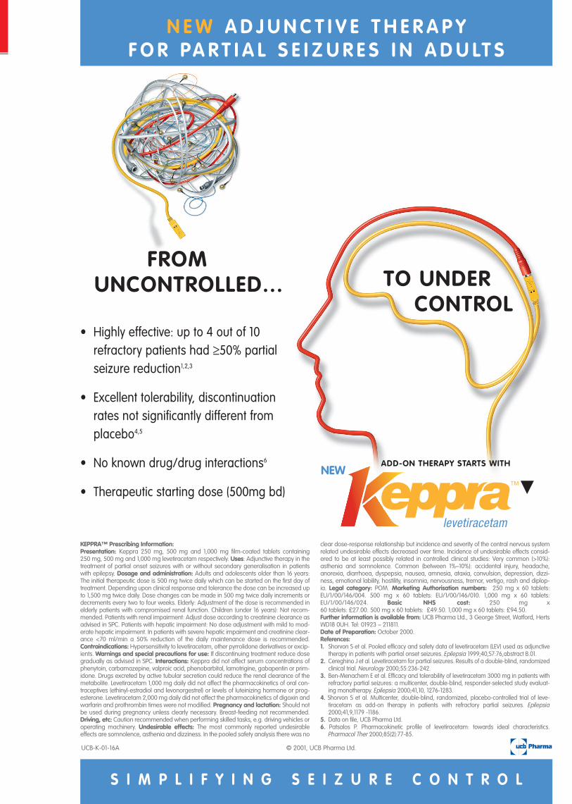
FROM UNCONTROLLED... TO UNDER
CONTROL• Highly effective: up to 4 out of 10
refractory patients had ≥50% partialseizure reduction1,2,3
• Excellent tolerability, discontinuationrates not significantly different fromplacebo4,5
• No known drug/drug interactions6
• Therapeutic starting dose (500mg bd)
16 ANCR • VOLUME 1 NUMBER 4 SEPTEMBER/OCTOBER 2001
The Seventh Cranial Nerve Alasdair Coles
Anatomy Primer
The Basics. The facial nerve consists of two “roots”, the motor root and the inter-mediate nerve (that is sometimes called the sensory root, a poor name as it containsmotor fibres as well as sensory).The motor root is easy to understand: it supplies themuscles derived from the second branchial arch, which are mainly the muscles of facialexpression. The intermediate nerve is complex and consists of taste fibres, parasym-pathetic efferents to lacrimal and salivary glands as well as a minor cutaneous sensorybranch. The anatomical course of the seventh nerve is characterised by four sharpturns, two of which are described as genu (knee). It emerges at the lower end of thepons, passes through the petrous portion of the temporal bone and exits the styolo-mastoid foramen to bury its fibres in the parotid gland.The commonest lesion of thisnerve is a “Bell’s Palsy”, an idiopathic condition with a lifetime prevalence of 6/1000.
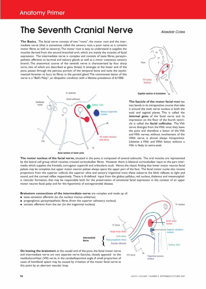
The fascicle of the motor facial root hastwo bends in its intrapontine course that takeit around the sixth nerve nucleus in both theaxial and sagittal planes. This is called theinternal genu of the facial nerve and itsimpression on the floor of the fourth ventri-cle is called the facial colliculus. The VIthnerve diverges from the VIIth once they leavethe pons and therefore a lesion of the VIthand VIIth nerves, without involvement of theVIIIth nerve, is almost always intrapontine.Likewise a VIIth and VIIIth lesion without aVIth is likely to extra-axial.
Facial colliculusVestibular
nuclei
VIII
VI
VI
Sensory trigeminalnucleus
Solitary Tract
IV ventricle
VII motor nucleusand nerve
The motor nucleus of the facial nerve, situated in the pons, is composed of several subnuclei. The oral muscles are representedby the lateral cell group which receives crossed corticobulbar fibres. However there is bilateral corticobulbar input to the pars inter-media which supplies the frontalis, corrugator supercilii and orbicularis oculi. Hence the classic finding that lower motor neuron facialpalsies may be complete, but upper motor neuron palsies always spare the upper part of the face. The facial motor nuclei also receiveprojections from the superior colliculi, the superior olive and sensory trigeminal tract; these subserve the blink reflexes to light andsound, and the corneal reflex respectively. There is ill-defined input from the globus pallidus, red nucleus, thalamus and mesencephal-ic reticular formation, that may be responsible both for the preservation of emotional facial expression in the context of an uppermotor neuron facial palsy and for the hypomimia of extrapyramidal disease.
On leaving the brainstem at the caudal end of the pons, the facial motor nerveand intermediate nerve are two separate nerve fascicles, closely apposed to thevestibulocochlear (VIII) nerve, in the cerebellopontine angle.A small proportion ofcases of hemifacial spasm may be caused by irritation of the motor facial nerve atthis point by an aberrant vascular loop.
Brainstem connections of the intermediate nerve are complex and made up of:� taste sensation afferents (to the nucleus tractus solitarius)� preganglionic parasympathetic fibres (from the superior salivatory nucleus)� somatic afferents from the ear (to the trigeminal nucleus)
Axial section of lower pons
Intermediate Nerve
Sagittal section of brainstem
VII motornucleus
Pons
III
IV
V VI
VIII
V! nucleus
V! nerve
V!II nerve
Facial motornucleur
Facial motor fibresSuperior salivatorynucleus
Parasympathetic fibres
Trigeminal sensorynucleus
Somatic efferents Solitary Tract
Taste fibres
{
ANCR • VOLUME 1 NUMBER 4 SEPTEMBER/OCTOBER 2001 17
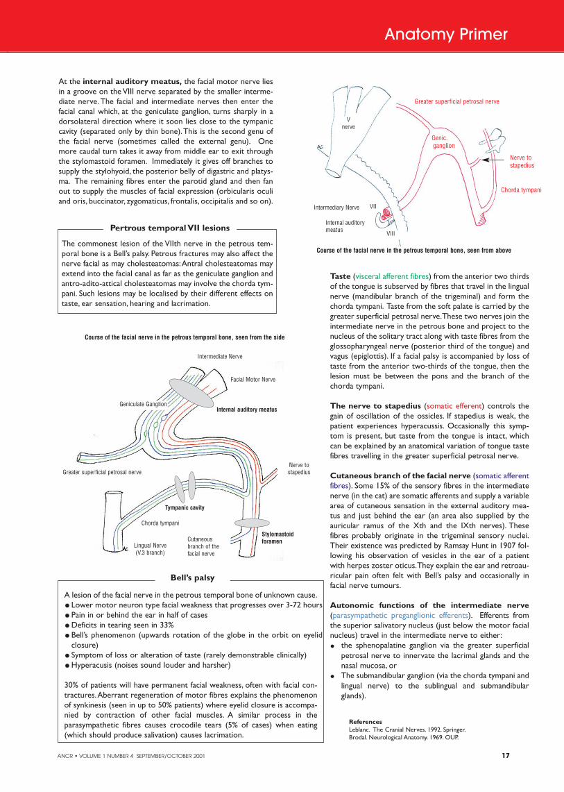
SectionSectionAnatomy Primer
The commonest lesion of the VIIth nerve in the petrous tem-poral bone is a Bell’s palsy. Petrous fractures may also affect thenerve facial as may cholesteatomas:Antral cholesteatomas mayextend into the facial canal as far as the geniculate ganglion andantro-adito-attical cholesteatomas may involve the chorda tym-pani. Such lesions may be localised by their different effects ontaste, ear sensation, hearing and lacrimation.
Pertrous temporal VII lesions
A lesion of the facial nerve in the petrous temporal bone of unknown cause.� Lower motor neuron type facial weakness that progresses over 3-72 hours� Pain in or behind the ear in half of cases� Deficits in tearing seen in 33%� Bell’s phenomenon (upwards rotation of the globe in the orbit on eyelid
closure) � Symptom of loss or alteration of taste (rarely demonstrable clinically) � Hyperacusis (noises sound louder and harsher)
30% of patients will have permanent facial weakness, often with facial con-tractures.Aberrant regeneration of motor fibres explains the phenomenonof synkinesis (seen in up to 50% patients) where eyelid closure is accompa-nied by contraction of other facial muscles. A similar process in theparasympathetic fibres causes crocodile tears (5% of cases) when eating(which should produce salivation) causes lacrimation.
Bell’s palsy
At the internal auditory meatus, the facial motor nerve liesin a groove on the VIII nerve separated by the smaller interme-diate nerve. The facial and intermediate nerves then enter thefacial canal which, at the geniculate ganglion, turns sharply in adorsolateral direction where it soon lies close to the tympaniccavity (separated only by thin bone).This is the second genu ofthe facial nerve (sometimes called the external genu). Onemore caudal turn takes it away from middle ear to exit throughthe stylomastoid foramen. Immediately it gives off branches tosupply the stylohyoid, the posterior belly of digastric and platys-ma. The remaining fibres enter the parotid gland and then fanout to supply the muscles of facial expression (orbicularis oculiand oris, buccinator, zygomaticus, frontalis, occipitalis and so on).
Taste (visceral afferent fibres) from the anterior two thirdsof the tongue is subserved by fibres that travel in the lingualnerve (mandibular branch of the trigeminal) and form thechorda tympani. Taste from the soft palate is carried by thegreater superficial petrosal nerve.These two nerves join theintermediate nerve in the petrous bone and project to thenucleus of the solitary tract along with taste fibres from theglossopharyngeal nerve (posterior third of the tongue) andvagus (epiglottis). If a facial palsy is accompanied by loss oftaste from the anterior two-thirds of the tongue, then thelesion must be between the pons and the branch of thechorda tympani.
The nerve to stapedius (somatic efferent) controls thegain of oscillation of the ossicles. If stapedius is weak, thepatient experiences hyperacussis. Occasionally this symp-tom is present, but taste from the tongue is intact, whichcan be explained by an anatomical variation of tongue tastefibres travelling in the greater superficial petrosal nerve.
Cutaneous branch of the facial nerve (somatic afferentfibres). Some 15% of the sensory fibres in the intermediatenerve (in the cat) are somatic afferents and supply a variablearea of cutaneous sensation in the external auditory mea-tus and just behind the ear (an area also supplied by theauricular ramus of the Xth and the IXth nerves). Thesefibres probably originate in the trigeminal sensory nuclei.Their existence was predicted by Ramsay Hunt in 1907 fol-lowing his observation of vesicles in the ear of a patientwith herpes zoster oticus.They explain the ear and retroau-ricular pain often felt with Bell’s palsy and occasionally infacial nerve tumours.
Autonomic functions of the intermediate nerve(parasympathetic preganglionic efferents). Efferents fromthe superior salivatory nucleus (just below the motor facialnucleus) travel in the intermediate nerve to either:� the sphenopalatine ganglion via the greater superficial
petrosal nerve to innervate the lacrimal glands and thenasal mucosa, or
� The submandibular ganglion (via the chorda tympani andlingual nerve) to the sublingual and submandibularglands).
Course of the facial nerve in the petrous temporal bone, seen from above
Course of the facial nerve in the petrous temporal bone, seen from the side
ReferencesLeblanc. The Cranial Nerves. 1992. Springer.Brodal. Neurological Anatomy. 1969. OUP.
Genic.ganglion
Greater superficial petrosal nerve
Chorda tympani
Nerve to stapedius
Internal auditory meatus
Intermediary Nerve
Intermediate Nerve
Facial Motor Nerve
Geniculate Ganglion
Chorda tympani
Greater superficial petrosal nerve
Lingual Nerve(V.3 branch)
Nerve tostapedius
Cutaneousbranch of thefacial nerve
Internal auditory meatus
Tympanic cavity
Stylomastoid foramen
V nerve
VII
VIII
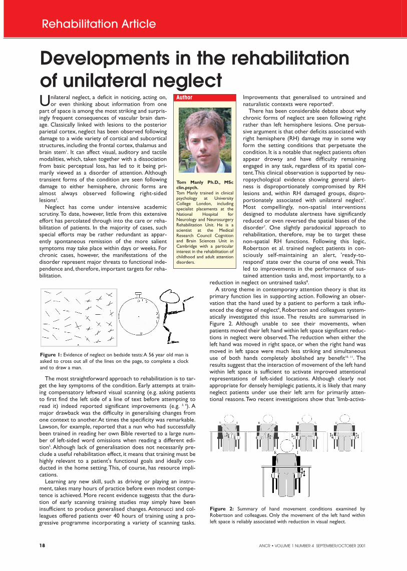
18 ANCR • VOLUME 1 NUMBER 4 SEPTEMBER/OCTOBER 2001
Section
Unilateral neglect, a deficit in noticing, acting on,or even thinking about information from one
part of space is among the most striking and surpris-ingly frequent consequences of vascular brain dam-age. Classically linked with lesions to the posteriorparietal cortex, neglect has been observed followingdamage to a wide variety of cortical and subcorticalstructures, including the frontal cortex, thalamus andbrain stem1. It can affect visual, auditory and tactilemodalities, which, taken together with a dissociationfrom basic perceptual loss, has led to it being pri-marily viewed as a disorder of attention. Althoughtransient forms of the condition are seen followingdamage to either hemisphere, chronic forms arealmost always observed following right-sidedlesions2.
Neglect has come under intensive academicscrutiny. To date, however, little from this extensiveeffort has percolated through into the care or reha-bilitation of patients. In the majority of cases, suchspecial efforts may be rather redundant as appar-ently spontaneous remission of the more salientsymptoms may take place within days or weeks. Forchronic cases, however, the manifestations of thedisorder represent major threats to functional inde-pendence and, therefore, important targets for reha-bilitation.
The most straightforward approach to rehabilitation is to tar-get the key symptoms of the condition. Early attempts at train-ing compensatory leftward visual scanning (e.g. asking patientsto first find the left side of a line of text before attempting toread it) indeed reported significant improvements (e.g. 3, 4). Amajor drawback was the difficulty in generalising changes fromone context to another.At times the specificity was remarkable.Lawson, for example, reported that a nun who had successfullybeen trained in reading her own Bible reverted to a large num-ber of left-sided word omissions when reading a different edi-tion5. Although lack of generalisation does not necessarily pre-clude a useful rehabilitation effect, it means that training must behighly relevant to a patient's functional goals and ideally con-ducted in the home setting.This, of course, has resource impli-cations.
Learning any new skill, such as driving or playing an instru-ment, takes many hours of practice before even modest compe-tence is achieved. More recent evidence suggests that the dura-tion of early scanning training studies may simply have beeninsufficient to produce generalised changes. Antonucci and col-leagues offered patients over 40 hours of training using a pro-gressive programme incorporating a variety of scanning tasks.
Improvements that generalised to untrained andnaturalistic contexts were reported6.
There has been considerable debate about whychronic forms of neglect are seen following rightrather than left hemisphere lesions. One persua-sive argument is that other deficits associated withright hemisphere (RH) damage may in some wayform the setting conditions that perpetuate thecondition. It is a notable that neglect patients oftenappear drowsy and have difficulty remainingengaged in any task, regardless of its spatial con-tent.This clinical observation is supported by neu-ropsychological evidence showing general alert-ness is disproportionately compromised by RHlesions and, within RH damaged groups, dispro-portionately associated with unilateral neglect7.Most compellingly, non-spatial interventionsdesigned to modulate alertness have significantlyreduced or even reversed the spatial biases of thedisorder7. One slightly paradoxical approach torehabilitation, therefore, may be to target thesenon-spatial RH functions. Following this logic,Robertson et al. trained neglect patients in con-sciously self-maintaining an alert, 'ready-to-respond' state over the course of one week.Thisled to improvements in the performance of sus-tained attention tasks and, most importantly, to a
reduction in neglect on untrained tasks8.A strong theme in contemporary attention theory is that its
primary function lies in supporting action. Following an obser-vation that the hand used by a patient to perform a task influ-enced the degree of neglect9, Robertson and colleagues system-atically investigated this issue. The results are summarised inFigure 2. Although unable to see their movements, whenpatients moved their left hand within left space significant reduc-tions in neglect were observed.The reduction when either theleft hand was moved in right space, or when the right hand wasmoved in left space were much less striking and simultaneoususe of both hands completely abolished any benefit10, 11. Theresults suggest that the interaction of movement of the left handwithin left space is sufficient to activate improved attentionalrepresentations of left-sided locations. Although clearly notappropriate for densely hemiplegic patients, it is likely that manyneglect patients under use their left arm for primarily atten-tional reasons.Two recent investigations show that 'limb-activa-
Rehabilitation Article
Author
Tom Manly Ph.D., MScclin.psych.Tom Manly trained in clinicalpsychology at UniversityCollege London, includingspecialist placements at theNational Hospital forNeurology and NeurosurgeryRehabilitation Unit. He is ascientist at the MedicalResearch Council Cognitionand Brain Sciences Unit inCambridge with a particularinterest in the rehabilitation ofchildhood and adult attentiondisorders.
Developments in the rehabilitationof unilateral neglect
Figure 2: Summary of hand movement conditions examined byRobertson and colleagues. Only the movement of the left hand withinleft space is reliably associated with reduction in visual neglect.
Figure 1: Evidence of neglect on bedside tests:A 56 year old man isasked to cross out all of the lines on the page, to complete a clockand to draw a man.
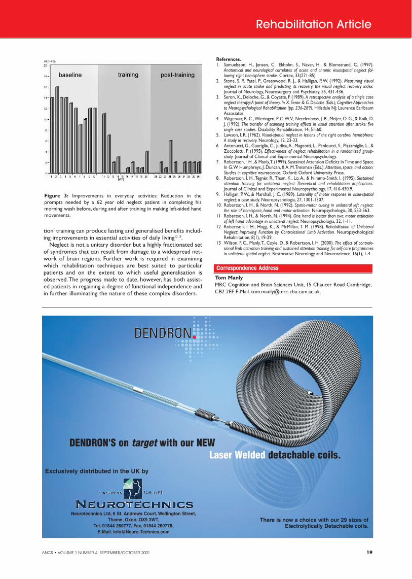
ANCR • VOLUME 1 NUMBER 4 SEPTEMBER/OCTOBER 2001 19
Section
Correspondence AddressTom ManlyMRC Cognition and Brain Sciences Unit, 15 Chaucer Road Cambridge,CB2 2EF. E-Mail. [email protected].
SectionSection
tion' training can produce lasting and generalised benefits includ-ing improvements in essential activities of daily living12,13.
Neglect is not a unitary disorder but a highly fractionated setof syndromes that can result from damage to a widespread net-work of brain regions. Further work is required in examiningwhich rehabilitation techniques are best suited to particularpatients and on the extent to which useful generalisation isobserved.The progress made to date, however, has both assist-ed patients in regaining a degree of functional independence andin further illuminating the nature of these complex disorders.
Rehabilitation Article
Figure 3: Improvements in everyday activities: Reduction in theprompts needed by a 62 year old neglect patient in completing his morning wash before, during and after training in making left-sided handmovements.
References.1. Samuelsson, H., Jensen, C., Ekholm, S., Naver, H., & Blomstrand, C. (1997).
Anatomical and neurological correlates of acute and chronic visuospatial neglect fol-lowing right hemisphere stroke. Cortex, 33(271-85).
2. Stone, S. P., Patel, P., Greenwood, R. J., & Halligan, P. W. (1992). Measuring visualneglect in acute stroke and predicting its recovery: the visual neglect recovery index.Journal of Neurology, Neurosurgery and Psychiatry, 55, 431-436.
3. Seron, X., Deloche, G., & Coyette, F. (1989). A retrospective analysis of a single caseneglect therapy:A point of theory. In X. Seron & G. Deloche (Eds.), Cognitive Approachesto Neuropsychological Rehabilitation (pp. 236-289). Hillsdale NJ: Laurence EarlbaumAssociates.
4. Wagenaar, R. C.,Wieringen, P. C.W.V., Netelenboss, J. B., Meijer, O. G., & Kuik, D.J. (1992). The transfer of scanning training effects in visual attention after stroke: fivesingle case studies. Disability Rehabilitation, 14, 51-60.
5. Lawson, I. R. (1962). Visual-spatial neglect in lesions of the right cerebral hemisphere:A study in recovery. Neurology, 12, 23-33.
6 Antonucci, G., Guariglia, C., Judica,A., Magnotti, L., Paoloucci, S., Pizzamiglio, L., &Zoccolotti, P. (1995). Effectiveness of neglect rehabilitation in a randomized group-study. Journal of Clinical and Experimental Neuropsychology.
7. Robertson, I. H., & Manly,T. (1999). Sustained Attention Deficits in Time and SpaceIn G.W. Humphreys, J. Duncan, & A. M.Treisman (Eds.), Attention, space, and action:Studies in cognitive neuroscience. Oxford: Oxford University Press.
8. Robertson, I. H.,Tegnér, R., Tham, K., Lo, A., & Nimmo-Smith, I. (1995). Sustainedattention training for unilateral neglect: Theoretical and rehabilitation implications.Journal of Clinical and Experimental Neuropsychology, 17, 416-430.9.
9. Halligan, P.W., & Marshall, J. C. (1989). Laterality of motor response in visuo-spatialneglect: a case study. Neuropsychologia, 27, 1301-1307.
10. Robertson, I. H., & North, N. (1992). Spatio-motor cueing in unilateral left neglect:the role of hemispace, hand and motor activation. Neuropsychologia, 30, 553-563.
11 Robertson, I. H., & North, N. (1994). One hand is better than two: motor extinctionof left hand advantage in unilateral neglect. Neuropsychologia, 32, 1-11.
12 Robertson, I. H., Hogg, K., & McMillan, T. M. (1998). Rehabilitation of UnilateralNeglect: Improving Function by Contralesional Limb Activation. NeuropsychologicalRehabilitation, 8(1), 19-29.
13 Wilson, F. C., Manly,T., Coyle, D., & Robertson, I. H. (2000). The effect of contrale-sional limb activation training and sustained attention training for self-care programmesin unilateral spatial neglect. Restorative Neurology and Neuroscience, 16(1), 1-4.
DENDRON'S on target with our NEWLaser Welded detachable coils.
There is now a choice with our 29 sizes ofElectrolytically Detachable coils.
Exclusively distributed in the UK by
Neurotechnics Ltd, 6 St. Andrews Court, Wellington Street,Thame, Oxon, OX9 3WT.
Tel. 01844 260777, Fax. 01844 260778,E-Mail. [email protected]
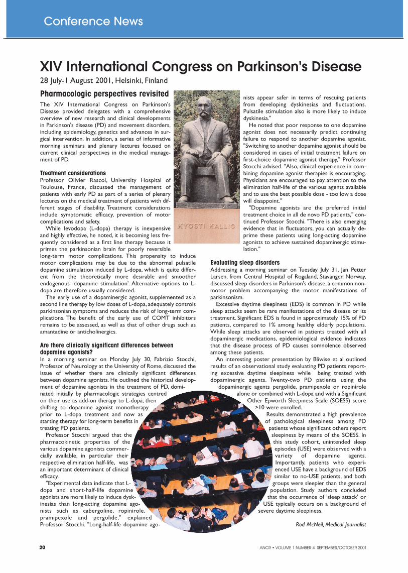
20 ANCR • VOLUME 1 NUMBER 4 SEPTEMBER/OCTOBER 2001
nists appear safer in terms of rescuing patientsfrom developing dyskinesias and fluctuations.Pulsatile stimulation also is more likely to inducedyskinesia."
He noted that poor response to one dopamineagonist does not necessarily predict continuingfailure to respond to another dopamine agonist."Switching to another dopamine agonist should beconsidered in cases of initial treatment failure onfirst-choice dopamine agonist therapy," ProfessorStocchi advised. "Also, clinical experience in com-bining dopamine agonist therapies is encouraging.Physicians are encouraged to pay attention to theelimination half-life of the various agents availableand to use the best possible dose - too low a dosewill disappoint."
"Dopamine agonists are the preferred initialtreatment choice in all de novo PD patients," con-tinued Professor Stocchi. "There is also emergingevidence that in fluctuators, you can actually de-prime these patients using long-acting dopamineagonists to achieve sustained dopaminergic stimu-lation."
Evaluating sleep disordersAddressing a morning seminar on Tuesday July 31, Jan PetterLarsen, from Central Hospital of Rogaland, Stavanger, Norway,discussed sleep disorders in Parkinson's disease, a common non-motor problem accompanying the motor manifestations ofparkinsonism.
Excessive daytime sleepiness (EDS) is common in PD whilesleep attacks seem be rare manifestations of the disease or itstreatment. Significant EDS is found in approximately 15% of PDpatients, compared to 1% among healthy elderly populations.While sleep attacks are observed in patients treated with alldopaminergic medications, epidemiological evidence indicatesthat the disease process of PD causes somnolence observedamong these patients.
An interesting poster presentation by Bliwise et al outlinedresults of an observational study evaluating PD patients report-ing excessive daytime sleepiness while being treated withdopaminergic agents. Twenty-two PD patients using the
dopaminergic agents pergolide, pramipexole or ropinirolealone or combined with L-dopa and with a Significant
Other Epworth Sleepiness Scale (SOESS) score>10 were enrolled.
Results demonstrated a high prevalenceof pathological sleepiness among PD
patients whose significant others reportsleepiness by means of the SOESS. Inthis study cohort, unintended sleepepisodes (USE) were observed with avariety of dopamine agents.Importantly, patients who experi-enced USE have a background of EDSsimilar to no-USE patients, and bothgroups were sleepier than the general
population. Study authors concludedthat the occurrence of 'sleep attack' or
USE typically occurs on a background ofsevere daytime sleepiness.
Rod McNeil, Medical Journalist
SectionSectionConference News
XIV International Congress on Parkinson's Disease28 July-1 August 2001, Helsinki, Finland
Pharmacologic perspectives revisitedThe XIV International Congress on Parkinson'sDisease provided delegates with a comprehensiveoverview of new research and clinical developmentsin Parkinson's disease (PD) and movement disorders,including epidemiology, genetics and advances in sur-gical intervention. In addition, a series of informativemorning seminars and plenary lectures focused oncurrent clinical perspectives in the medical manage-ment of PD.
Treatment considerationsProfessor Olivier Rascol, University Hospital ofToulouse, France, discussed the management ofpatients with early PD as part of a series of plenarylectures on the medical treatment of patients with dif-ferent stages of disability. Treatment considerationsinclude symptomatic efficacy, prevention of motorcomplications and safety.
While levodopa (L-dopa) therapy is inexpensiveand highly effective, he noted, it is becoming less fre-quently considered as a first line therapy because itprimes the parkinsonian brain for poorly reversiblelong-term motor complications. This propensity to inducemotor complications may be due to the abnormal pulsatiledopamine stimulation induced by L-dopa, which is quite differ-ent from the theoretically more desirable and smootherendogenous 'dopamine stimulation'. Alternative options to L-dopa are therefore usually considered.
The early use of a dopaminergic agonist, supplemented as asecond line therapy by low doses of L-dopa, adequately controlsparkinsonian symptoms and reduces the risk of long-term com-plications. The benefit of the early use of COMT inhibitorsremains to be assessed, as well as that of other drugs such asamantadine or anticholinergics.
Are there clinically significant differences betweendopamine agonists?In a morning seminar on Monday July 30, Fabrizio Stocchi,Professor of Neurology at the University of Rome, discussed theissue of whether there are clinically significant differencesbetween dopamine agonists. He outlined the historical develop-ment of dopamine agonists in the treatment of PD, domi-nated initially by pharmacologic strategies centredon their use as add-on therapy to L-dopa, thenshifting to dopamine agonist monotherapyprior to L-dopa treatment and now asstarting therapy for long-term benefits intreating PD patients.
Professor Stocchi argued that thepharmacokinetic properties of thevarious dopamine agonists commer-cially available, in particular theirrespective elimination half-life, wasan important determinant of clinicalefficacy.
"Experimental data indicate that L-dopa and short-half-life dopamineagonists are more likely to induce dysk-inesias than long-acting dopamine ago-nists such as cabergoline, ropinirole,pramipexole and pergolide," explainedProfessor Stocchi. "Long-half-life dopamine ago-
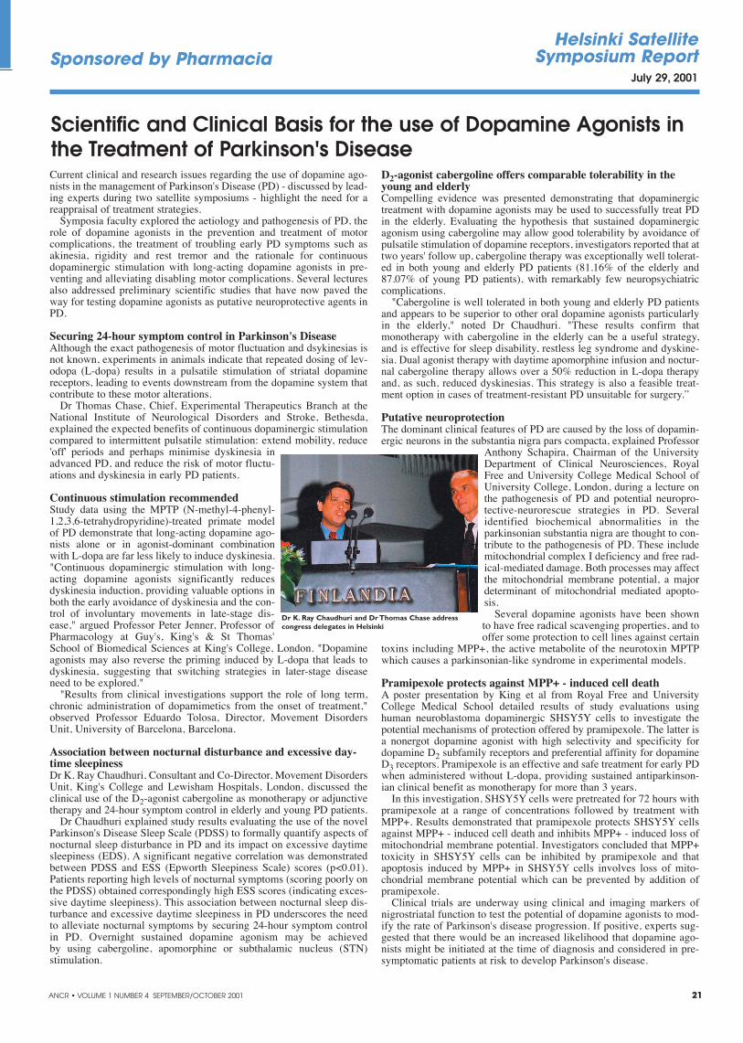
ANCR • VOLUME 1 NUMBER 4 SEPTEMBER/OCTOBER 2001 21
Current clinical and research issues regarding the use of dopamine ago-nists in the management of Parkinson's Disease (PD) - discussed by lead-ing experts during two satellite symposiums - highlight the need for areappraisal of treatment strategies.
Symposia faculty explored the aetiology and pathogenesis of PD, therole of dopamine agonists in the prevention and treatment of motorcomplications, the treatment of troubling early PD symptoms such asakinesia, rigidity and rest tremor and the rationale for continuousdopaminergic stimulation with long-acting dopamine agonists in pre-venting and alleviating disabling motor complications. Several lecturesalso addressed preliminary scientific studies that have now paved theway for testing dopamine agonists as putative neuroprotective agents inPD.
Securing 24-hour symptom control in Parkinson's DiseaseAlthough the exact pathogenesis of motor fluctuation and dsykinesias isnot known, experiments in animals indicate that repeated dosing of lev-odopa (L-dopa) results in a pulsatile stimulation of striatal dopaminereceptors, leading to events downstream from the dopamine system thatcontribute to these motor alterations.
Dr Thomas Chase, Chief, Experimental Therapeutics Branch at theNational Institute of Neurological Disorders and Stroke, Bethesda,explained the expected benefits of continuous dopaminergic stimulationcompared to intermittent pulsatile stimulation: extend mobility, reduce'off' periods and perhaps minimise dyskinesia inadvanced PD, and reduce the risk of motor fluctu-ations and dyskinesia in early PD patients.
Continuous stimulation recommendedStudy data using the MPTP (N-methyl-4-phenyl-1,2,3,6-tetrahydropyridine)-treated primate modelof PD demonstrate that long-acting dopamine ago-nists alone or in agonist-dominant combinationwith L-dopa are far less likely to induce dyskinesia."Continuous dopaminergic stimulation with long-acting dopamine agonists significantly reducesdyskinesia induction, providing valuable options inboth the early avoidance of dyskinesia and the con-trol of involuntary movements in late-stage dis-ease," argued Professor Peter Jenner, Professor ofPharmacology at Guy's, King's & St Thomas'School of Biomedical Sciences at King's College, London. "Dopamineagonists may also reverse the priming induced by L-dopa that leads todyskinesia, suggesting that switching strategies in later-stage diseaseneed to be explored."
"Results from clinical investigations support the role of long term,chronic administration of dopamimetics from the onset of treatment,"observed Professor Eduardo Tolosa, Director, Movement DisordersUnit, University of Barcelona, Barcelona.
Association between nocturnal disturbance and excessive day-time sleepinessDr K. Ray Chaudhuri, Consultant and Co-Director, Movement DisordersUnit, King's College and Lewisham Hospitals, London, discussed theclinical use of the D2-agonist cabergoline as monotherapy or adjunctivetherapy and 24-hour symptom control in elderly and young PD patients.
Dr Chaudhuri explained study results evaluating the use of the novelParkinson's Disease Sleep Scale (PDSS) to formally quantify aspects ofnocturnal sleep disturbance in PD and its impact on excessive daytimesleepiness (EDS). A significant negative correlation was demonstratedbetween PDSS and ESS (Epworth Sleepiness Scale) scores (p<0.01).Patients reporting high levels of nocturnal symptoms (scoring poorly onthe PDSS) obtained correspondingly high ESS scores (indicating exces-sive daytime sleepiness). This association between nocturnal sleep dis-turbance and excessive daytime sleepiness in PD underscores the needto alleviate nocturnal symptoms by securing 24-hour symptom controlin PD. Overnight sustained dopamine agonism may be achieved by using cabergoline, apomorphine or subthalamic nucleus (STN)stimulation.
D2-agonist cabergoline offers comparable tolerability in theyoung and elderlyCompelling evidence was presented demonstrating that dopaminergictreatment with dopamine agonists may be used to successfully treat PDin the elderly. Evaluating the hypothesis that sustained dopaminergicagonism using cabergoline may allow good tolerability by avoidance ofpulsatile stimulation of dopamine receptors, investigators reported that attwo years' follow up, cabergoline therapy was exceptionally well tolerat-ed in both young and elderly PD patients (81.16% of the elderly and87.07% of young PD patients), with remarkably few neuropsychiatriccomplications.
"Cabergoline is well tolerated in both young and elderly PD patientsand appears to be superior to other oral dopamine agonists particularlyin the elderly," noted Dr Chaudhuri. "These results confirm thatmonotherapy with cabergoline in the elderly can be a useful strategy,and is effective for sleep disability, restless leg syndrome and dyskine-sia. Dual agonist therapy with daytime apomorphine infusion and noctur-nal cabergoline therapy allows over a 50% reduction in L-dopa therapyand, as such, reduced dyskinesias. This strategy is also a feasible treat-ment option in cases of treatment-resistant PD unsuitable for surgery.”
Putative neuroprotectionThe dominant clinical features of PD are caused by the loss of dopamin-ergic neurons in the substantia nigra pars compacta, explained Professor
Anthony Schapira, Chairman of the UniversityDepartment of Clinical Neurosciences, RoyalFree and University College Medical School ofUniversity College, London, during a lecture onthe pathogenesis of PD and potential neuropro-tective-neurorescue strategies in PD. Severalidentified biochemical abnormalities in theparkinsonian substantia nigra are thought to con-tribute to the pathogenesis of PD. These includemitochondrial complex I deficiency and free rad-ical-mediated damage. Both processes may affectthe mitochondrial membrane potential, a majordeterminant of mitochondrial mediated apopto-sis.
Several dopamine agonists have been shownto have free radical scavenging properties, and tooffer some protection to cell lines against certain
toxins including MPP+, the active metabolite of the neurotoxin MPTPwhich causes a parkinsonian-like syndrome in experimental models.
Pramipexole protects against MPP+ - induced cell deathA poster presentation by King et al from Royal Free and UniversityCollege Medical School detailed results of study evaluations usinghuman neuroblastoma dopaminergic SHSY5Y cells to investigate thepotential mechanisms of protection offered by pramipexole. The latter isa nonergot dopamine agonist with high selectivity and specificity fordopamine D2 subfamily receptors and preferential affinity for dopamineD3 receptors. Pramipexole is an effective and safe treatment for early PDwhen administered without L-dopa, providing sustained antiparkinson-ian clinical benefit as monotherapy for more than 3 years.
In this investigation, SHSY5Y cells were pretreated for 72 hours withpramipexole at a range of concentrations followed by treatment withMPP+. Results demonstrated that pramipexole protects SHSY5Y cellsagainst MPP+ - induced cell death and inhibits MPP+ - induced loss ofmitochondrial membrane potential. Investigators concluded that MPP+toxicity in SHSY5Y cells can be inhibited by pramipexole and thatapoptosis induced by MPP+ in SHSY5Y cells involves loss of mito-chondrial membrane potential which can be prevented by addition ofpramipexole.
Clinical trials are underway using clinical and imaging markers ofnigrostriatal function to test the potential of dopamine agonists to mod-ify the rate of Parkinson's disease progression. If positive, experts sug-gested that there would be an increased likelihood that dopamine ago-nists might be initiated at the time of diagnosis and considered in pre-symptomatic patients at risk to develop Parkinson's disease.
Helsinki SatelliteSymposium Report
Scientific and Clinical Basis for the use of Dopamine Agonists inthe Treatment of Parkinson's Disease
July 29, 2001
Dr K. Ray Chaudhuri and Dr Thomas Chase addresscongress delegates in Helsinki
Sponsored by Pharmacia
22 ANCR • VOLUME 1 NUMBER 4 SEPTEMBER/OCTOBER 2001
Section
2001September
Visible Solutions for InvisibleInjury - Headway/HIRE11-12 September, 2001; Grantham, UKTel. 0115 947 1913,E-Mail [email protected]
Association of British Neurologists12-14 September, 2001; Durham, UKTel. 020 7405 4060, Fax. 020 7405 4070,E-Mail. [email protected]
International Brain InjuryAssociation12-15 September, 2001; Edinburgh, UKTel. 001 703 683 8400,Fax. 001 703 683 8996,E-Mail. [email protected]
ECTRIMS 200112-15 September, 2001; Dublin, IrelandFax. 00353 1 676 9088,E-Mail. [email protected]
XXVII Congress of the EuropeanSociety of Neuroradiology13-16 September, 2001;Ancona, ItalyTel. 0039 02 56601212, Fax. 0039 0256609045, E-Mail. [email protected]
European Paediatric NeurologySociety13-16 September, 2001; GermanyFax. 0049 7621 78 714
XII International Congress of theWorld Federation ofNeurosurgical Societies15-21 September, 2001; Sydney,Australia Tel. 0061 2 9241 1478,Fax. 0061 2 9251 3552
World Congress of Neurosurgery16-20 September, 2001; Sydney,Australia Tel. 0061 2 9241 1478,Fax. 0061 2 9251 3552, [email protected]
126th Annual Meeting of theAmerican NeurologicalAssociation30 September-3 October, 2001;Chicago, US Tel. 001 612 545 6284,Fax. 001 612 545 6073,E-Mail. [email protected]
International Multiple SclerosisConference30 September - 4 October, 2001;Melbourne,Australia Tel. 0061 3 98287222, Fax. 0061 3 9828 9054,[email protected]
ACTRIMS30 September, 2001; Chicago, USTel. 001 212 476 0452,[email protected]
OctoberEpilepsy Research Foundation10th Anniversary Meeting17 October, 2001; London, UKTel/Fax. 020 8995 4781,E-Mail. [email protected]
British Geriatric Society18-19 October, 2001; London, UKTel. 01825 768902,E-Mail: [email protected]
2001 European CharcotFoundation Symposium18-20 October, 2001;Venice, ItalyTel. +31 24 356 1954,Fax. +31 24 354 0920,E-Mail. [email protected]
✶ Amended Date ✶Neuro-behavioural Rehabilitationof Severe Brain Injury - Theory &Practise23 October, 2001; London, UKTel. 020 8780 4500 ext 5236,E-Mail. [email protected]
17th Alzheimer's DiseaseInternational Conference25-27 October, 2001; Christchurch,New Zealand Tel. 0064 3 364 2534,[email protected]
NovemberAlzheimer's Society (UK)5-8 November, 2001; London, UKTel. 020 7306 0606, Fax. 020 7306 0808,E-Mail. [email protected]
British NeuropsychologicalSociety Autumn Meeting8 November, 2001; London, UKwww.hop.man.ac.uk/bns,Tel. 0161 275 3401
12th International Symposium onALS/MND18-20 November, 2001; Oakland, USATel. 01604 250505, Fax. 01604 638289,E-Mail. [email protected]
National Society of EpilepsyAdvanced Lecture Series22 November, 2001; London, UKTel. 01494 601300, Fax. 01494 871977.
Ageing of the Brain - Dementia23-24 November, 2001; Florence, ItalyTel. 00390 55 43 68 455,E-Mail. [email protected]
BSRM Autumn 2001 Meeting26-27 November, 2001; Manchester, UKTel/Fax. 01992 638865,E-Mail [email protected]
PDS Conference - Scotland29 November, 2001; ScotlandTel. 020 7932 1343,E-Mail. [email protected]
The Changing Brain29 November, 2001; Milan, Italywww.armenise.meditech-media.com
55th Annual Meeting of theAmerican Epilepsy Society30 November - 5 December, 2001;Philadelphia, USATel. 001 860 586 7505,Fax. 001 860 586 7550
UKABIF Annual Conference30 November, 2001; London, UKTel. 020 8780 4569,E-Mail [email protected]
2002March
Brain Awareness Week 200212-17 March, 2002; UKE-Mail. [email protected]
April3rd World Congress inNeurological Rehabilitation3-6 April, 2002;Venice, ItalyTel. 0039 06 844 98364,Fax. 0039 06 844 98332,E-Mail. [email protected]
54th Annual Meeting of theAmerican Academy of Neurology13-20 April, 2002; Denver, USATel. 001 651 695 1940,Fax. 001 651 695 2791
5th European Parkinson's DiseaseAssociation Conference21-24 April, 2002; Jerusalem, IsraelTel. 01273 686889, Fax. 01273 570082,E-Mail. [email protected]
1st Mediterranean Congress ofNeurology26-28 April, 2002; Limassol, CyprusTel. 00357 5 749919, Fax. 00357 5749744, [email protected]
MayXIV International Neuro-Ophthalmology Society Meeting5-8 May, 2002; Buenos Aires,ArgentinaFax. 0054 11 4331 0223,E-Mail. [email protected]
6th Congress of the EuropeanSociety for ClinicalNeuropharmacology (ESCNP)14-18 May, 2002; Budapest, HungaryTel. 0036 1 311 6687,Fax. 0036 1 383 7918,E-Mail. [email protected]
7th Meeting of the EuropeanSociety of Neurosonology andCerebral Hemodynamics26-28 May, 2002; Bern, SwitzerlandTel. 0041 41 767 34 49,Fax. 0041 41 767 34 00,E-Mail. [email protected]
33rd Scandinavian NeurologyCongress29 May-1 June, 2002; Reykjavik, IcelandTel. 00354 585 3900, Fax. 00354 5853901, E-Mail. [email protected],www.congress.is
11th European Stroke Conference29 May-1 June, 2002; Geneva,SwitzerlandTel. 0041 22 33 99 624,Fax. 0041 22 33 99 621,E-Mail. [email protected]
JuneInternational Association ofGerontology: European Section6th European Congress of ClinicalGerontologyJune 2002; Moscow, RussiaProf L B Lazebnik,E-Mail. [email protected]
6th European Headache Congress17-22 June, 2002; Istanbul,TurkeyTel. 0090 312 4420700,E-Mail. [email protected]
July10th International Congress ofNeuromuscular Diseases7-12 July, 2002;Vancouver, CanadaTel. 001 604 681 5226,Fax. 001 604 681 2503,E-Mail. [email protected]
7th European Congress ofNeuropathology, Neuropathology200214-17 July, 2002; Helskinki, FinlandTel. + 3 58 9 56 07-5 00,Fax. + 3 58 9 56 07-50 20,E-Mail. Neuropathology2002:congrex.fi,www.congrex.fi/neuropathology2002
8th International Conference onAlzheimer's Disease and RelatedDisorders20-25 July, 2002; Stockholm, SwedenTel. 001 312 335 5813,Fax. 001 312 335 5781,www.alx.org/internationalconference
AugustWFNRS SymposiumNeuroradiologicum XVII18-24 August, 2002; Paris, FranceTel. 0033 3 83851456,Fax. 0033 3 838 51391,
E-Mail. l.picard:chu-nancy.fr
Events Diary
If you would like your event listed in the next issue, send details to: Rachael Hansford on Fax: 0131 3131110 or E-Mail: [email protected] by October 8th, 2001.
Hosted by
The British Society ofRehabilitation Medicine
Society for Researchin Rehabilitation
13th European Congress of Physical and Rehabilitation Medicine
28–31 May 2002 Brighton UKPromoting Standards
in Rehabilitation
Congress Organisers, Concorde Services Ltd42 Canham Road, London W3 7SR, UK
Tel: +44 (0)20 8743 3106 Fax: +44 (0)20 8743 1010E-mail: [email protected] Website: www.ecprm2002.co.uk
ANCR • VOLUME 1 NUMBER 4 SEPTEMBER/OCTOBER 2001 23
SectionJournal Reviews
Panel of Reviewers Roger Barker, Honorary Consultant in Neurology,Cambridge Centre of Brain RepairAlasdair Coles, Wellcome Advanced Fellow, Dunn School ofPathology, Oxford and Dept of Neurology, Cambridge Tom Foltynie, Neurology Research Registrar, CambridgeTarek Gaber, Specialist Registrar in Rehabilitation, LewinRehabilitation Unit, Cambridge Tim Harrower, Wellcome Clinical Research Fellow andHonorary Neurology Clinical Fellow, Addenbrooke’s Hospital Andrew Larner, Consultant Neurologist, Walton Centre forNeurology & Neurosurgery, Liverpool Simon J G Lewis, Honorary Clinical Research Fellow,Cambridge Centre for Brain Repair Mark Manford, Consultant Neurologist, Addenbrooke'sHospital, Cambridge, and Bedford Hospital Peter Martin, Consultant Neurologist, Addenbrooke'sHospital, Cambridge Brian McNamara, SpR in Clinical Neurophysiology,Addenbrooke's Hospital, Cambridge Jane Mickelborough, Research Fellow, University of Salford Fiona Ritchie, Speech & Language Therapist, OliverZangwill Neurorehabilitation Unit, Ely John Thorpe, Addenbrooke's Hospital, Cambridge, andPeterborough Ailie Turton, Research Fellow, Burden Neurological Institute,Bristol
For more information on joining our panel of reviewers,E-Mail [email protected] or Tel. Rachael Hansfordon 0131 477 2335.
EDITOR'S CHOICE
Neglect is due to temporal -not parietal- lobedamage!All neurological teaching and textbooks tell us that hemi-neglect is a feature of right posterior parietal lobe dam-age. But that has always been hard to square with thefact that primates get neglect from lesions of either tem-poral, rather than parietal, lobes. This careful study ofstroke patients from Tubingen provides an explanation.Over 5 years the authors collected 49 patients with purespatial neglect (that is without visual field lesions). Ofthese, 25 had cortical lesions only. This subgroup wascompared, by lesion mapping from magnetic resonanceimages, with patients with a variety of right-sidedlesions; the lesions causing pure spatial neglect wereonly congruent over the superior temporal gyrus not theparietal lobe. Next they compared lesions causing purespatial neglect with those responsible for combined spa-tial neglect and visual field defects. They found that thesuperior temporal gyrus was always affected, and exten-sion to the posterior horn of the lateral ventricle causedthe field defect. It seems then that patients with morewidespread deficits, especially visual field defects, haveconfounded previous studies of neglect. We can alsoconclude that the evolutionary change from monkey tohuman spatial awareness processing has been to later-alise it to the right temporal lobe rather than move it fromthe temporal to the parietal lobe. -AJCKarnath HO, Ferber S, Himmelbach M.Spatial awareness is a function of the temporal notthe posterior parietal lobe.NATURE 2001 Jun 21;411(6840):950-3
'Mind the doors please' - Virtual realitywheelchair training for patients with unilateralneglectPatients with unilateral neglect take many weeks to reha-bilitate, are prone to accidents and have difficulty achievingindependence in daily life. A group from California hasdeveloped an imaginative and safe aid to improving wheel-chair mobility in such patients. A computer assisted and vir-tual reality system of training in which images are project-ed onto a large (8x6ft) area of wall is used. Patients aretrained on a hierarchical series of modules that aredesigned to improve scanning to the left space, decreaseattentional pull to the right and improve visuospatial plan-ning.
Webster et al tested their training protocol on 20 patientswith left unilateral neglect and compared performance ofthese patients with 20 previous patients who had not expe-rienced the computer assisted training. The patients whotraining with the computer and virtual reality simulation per-formed significantly better on a wheelchair obstacle coursethan the control subjects. They also had fewer reportedaccidents during their hospital admission.
Webster et al acknowledge the limitation of using a ret-rospective group as control subjects. This study is excitingbecause it demonstrates that wheelchair mobility and safe-ty in neglect patients can be improved with tasks that donot require wheelchair propulsion. This means that trainingcan begin before patients are ready to self propel in wheel-chairs. Although it will be some time before virtual reality isin common use in rehabilitation. -AJTComputer assisted training for improving wheelchairmobility in unilateral neglect patients.Webster JS, McFarland PT, Rapport LJ, Morrill B,Roades LA, Abadee PS.ARCHIVES OF PHYSICAL MEDICINE AND REHABILITATION2001: 82: 769-75
Would you like to joinour panel of reviewers?
We are looking for reviewers to scan thefollowing journals on a regular basis andprovide short summaries and commenton interesting papers from:
Neurology European Journal of Neurology Journal of Neurology
If you have access to any of the abovejournals and would like to get involved,please contact Rachael Hansford onTel. 0131 477 2335,Fax. 0131 313 1110,E-Mail. [email protected],stating which journal you would prefer.
24 ANCR • VOLUME 1 NUMBER 4 SEPTEMBER/OCTOBER 2001
COGNITIVE NEUROLOGY'Stroop effect' is related to a specific pattern offrontal lobe damage rather than frontal damageper se.
The Stroop test is one of the most widely utilised para-digms in neuropsychological testing. The classic version ofthe test consists of three conditions: the first requires thesubject to read colour words, written in black; the secondrequires the naming of coloured patches using the coloursidentified by their written names in the first condition andthe third condition requires naming the colour of ink inwhich a colour word is printed when the colour is incongru-ent with the name (e.g. 'red' printed in the colour green).The test results in the general pattern of results whereword reading is performed the fastest, colour naming isslower and the incongruent condition takes the longest timeto complete. The great interest in this paradigm lies in thedisproportionately impaired performance of various patientgroups in the incongruent condition ('Stroop effect').Performance of this aspect of the test is likely to dependheavily on intact frontal lobe function and functional neu-roimaging has proposed a variety of key frontal loberegions. In this study, Stuss et al, report a series of fifty-onepatients with focal brain lesions (37 frontal, 14 posterior)and compared their performance on the Stroop test with 26normal control subjects.
These investigations confirmed that only the group withfrontal lobe lesions demonstrated a significant Stroopeffect, with no effect seen in any patient with a posteriorlobe lesion. Analysis of the frontal lobe lesion patientsrevealed that only 12 of the 37 had performed poorly on theincongruity aspect of the test. Comparison betweenimpaired and non-impaired groups of frontal lobe lesionpatients showed no difference between any demographicvariables or neuropsychological measures. Thus Stroopeffect was not related to frontal lobe damage in general,nor by undifferentiated unilateral or bilateral lesions.Patients with frontal lobe pathology were slower across allconditions of the task however; patients with superior medi-al frontal lesions (usually bilateral) did show an exaggera-tion of the Stroop effect. This region maps to the supple-mentary motor area bilaterally and is thought to be animportant intersection between neural pathways receivingafferent subcortical activation pathways and projectingbilaterally to frontal cortex and striatum.
Greater understanding of the pathophysiologicalchanges underlying this neuropsychological tool may allowfor future therapeutic modulation of neurotransmitter sys-tems (e.g. dopaminergic) to improve the cognitive deficitobserved across a variety of neurological conditions.-SJGLStroop performance in focal lesion patients:dissocia-tion of processes and frontal lobe lesion location.Stuss DT, Floden D, Alexander MP, Levine B, Katz DNEUROPSYCHOLOGIA2001:39:771-786
Midlife systolic blood pressure and serumcholesterol linked to development ofAlzheimer's disease
Previous studies have linked midlife blood pressure andserum cholesterol to the development in later life ofAlzheimer's dementia, suggesting a possible vascular rolein the pathophysiology of the disease. In this paperKivipelto et al have performed a longitudinal populationbased study in Finland assessing both men and women to
evaluate the role of these risk factors in this context. Theyidentified a cohort of 1449 patients, mean age 71.3 years(65-80y) who had previously assessed between 1972-1987when their mean age was 50.4years (40-64y). They wereassessed by means of questionnaire, clinical examinationand laboratory measurements. A diagnosis of dementiawas based on the Diagnostic and Statistical Manual ofMental Disorders, fourth edition with diagnosis ofAlzheimer's disease being made according to the criteria ofthe National Institute of Neurological and CommunicativeDisorders and Stroke and the Alzheimer Disease andRelated Disorders Association.
In total, forty-eight cases of Alzheimer's disease wereidentified. All of these cases had generalised or medialtemporal atrophy on magnetic resonance scans whereasnone showed appreciable vascular pathology. People withraised systolic blood pressure (>160 mm Hg) or high serumcholesterol concentration (>6.5 mmol/l) in midlife had a sig-nificantly higher risk of Alzheimer's disease in later life,even after adjustment for age, body mass index, education,vascular events, smoking status, and alcohol consumption,compared with those individuals with normal systolic bloodpressure (odds ratio 2.3, 95% confidence interval 1.0 to5.5) or serum cholesterol (odds ratio 2.1, 1.0 to 4.4).Participants with both of these risk factors in midlife had asignificantly higher risk of developing Alzheimer's diseasethan those with either of the risk factors alone (odds ratio3.5, 1.6 to 7.9). Diastolic blood pressure in midlife had nosignificant effect on the risk of Alzheimer's disease.
The results presented here emphasise the clinical impor-tance in the aggressive management of these risk factorsduring midlife and highlight a possible vascular role under-lying pathophysiology in this common neurodegenerativecondition. -SJGLMidlife vascular risk factors and Alzheimer's disease inlater life: longitudinal, population based study.Kivipelto M, Helkala E, Laakso MP, Hänninen T,Hallikainen M, Alhainen K, Soininen H, Tuomilehto J,Nissinen A.BRITISH MEDICAL JOURNAL2001:322:1447-1451
MULTIPLE SCLEROSISChlamydia pneumonia and multiple sclerosis
Pierre Marie, a pupil of Charcot, was perhaps the first per-son to suggest an infective aetiology for multiple sclerosisand there have been no end of candidate pathogens pro-posed since then. Most recently, attention has turned onChlamydia pneumoniae, an organism that was first identi-fied in 1986 and is established to be pathogenic in athero-sclerosis. In 1999, Sriram and colleagues foundChlamydiae pneumoniae DNA in a high percentage ofpatients with multiple sclerosis, but not controls, by poly-merase chain reaction. However replication of this findingby other laboratories has proved controversial.
A group from Lubeck in Germany have published a studyin Annals, which may illuminate the controversy.They foundC. pneumoniae DNA in the CSF of 21% of 58 multiple scle-rosis patients and none present in 67 healthy controls. But43% of 47 patients with other neurological diseases werealso positive for C. pneumoniae in the CSF. Gieffers et althen examined peripheral blood mononuclear cells in someof their patients, because anything up to 50% of healthyblood donors may be positive for C. pneumoniae by PCR.Of 6 multiple sclerosis patients with positive CSF, 2 hadpositive blood C. pneumoniae and, of 10 patients other
Journal Reviews
ANCR • VOLUME 1 NUMBER 4 SEPTEMBER/OCTOBER 2001 25
Section
neurological diseases, 8 had positive blood. In thosepatients with paired CSF and blood results, it may be thatChlamydial DNA had simply leaked across a disruptedblood-brain-barrier and its presence was an epiphenome-non of the disease process. But, the devil's advocate mightsay, the data is also consistent with the possibility that fail-ure to clear C. pneumoniae from the CSF, having eliminat-ed it from the blood, is seen in the majority of patients withmultiple sclerosis.
The controversy is further fuelled by a study by Lenz andcolleagues from Detroit. They looked for sequence similar-ity between C. pneumoniae proteins and myelin basic pro-tein. They found a 20-mer peptide from one C. pneumoniaeprotein that shares a 7-aa motif with a critical epitope ofmyelin basic protein (rat MBP 68-86). They injected this C.pneumoniae peptide, with complete Freund's adjuvant, intoLewis rats and found that it produced typical EAE, withperivascular cuffing and parenchymal mononuclear cellinfiltration on pathology (but not demyelination). The impli-cation is that immune responses against C. pneumoniaecan induce central nervous system inflammation in labora-tory animals- in which case why not in man? -AJCGieffers J, Pohl D,Treib J, Dittmann R, Stephan C, KlotzK, Hanefeld F, Solbach W, Haass A, Maass M.Presence of Chlamydia pneumoniae DNA in the cere-bral spinal fluid is a common phenomenon in a varietyof neurological diseases and not restricted to multi-plebsclerosis.ANNALS OF NEUROLOGY 2001 May;49(5):585-9Lenz, D, Lu L, Conant S, Wolf N, Gerard H, Whittum-Hudson J, Hudson A., Swanborg, R.A Chlamydia pneumoniae-Specific Peptide InducesExperimental Autoimmune Encephalomyelitis in Rats.JOUNRAL OF IMMUNOLOGY 2001 167: 1803-1808
Early intervention with interferon in suspectedMultiple Sclerosis: Treatment or Prevention?Earlier treatment in suspected multiple sclerosis (MS)would seem to be important in preventing conversion toclinically definite MS (CDMS), but currently little evidenceconfirms this logical notion. In an attempt to address thisissue the multi-centre ETOMS (Early treatment of MultipleSclerosis) study group undertook a randomised doubleblind placebo controlled trial of interferon after first presen-tation of neurological events, in patients with MRI findingsstrongly suggestive of multiple sclerosis. With 154 patientsassigned to receive 22mcg of Interferon beta 1a (now gen-erally considered a low dose) and 155 patients assigned to
receive placebo injections, follow up for 2 years was under-taken. 34% in the treatment group developed CDMS, whichwas significantly less (p=0.047) than the 45% in the place-bo group. It took 569 days for 30% of the interferon groupto develop CDMS, which differed significantly (p=0.034)from the placebo group. Annual relapse rate and lesionburden on MRI were also significantly less in the treatmentgroup. The EDSS score did not differ, but this was to beexpected at such an early stage in the disease. These find-ings taken with results from the CHAMPS (Controlled High-risk Subjects Avonex Multiple Sclerosis Prevention Study)study suggest interferon has positive effects on clinical andMRI outcomes in suspected MS. But the ultimate targets ofintervention on the long-term course of MS should be theprevention of the onset of progression and reduction in dis-ability. Given the short follow up times this can not be fullyassessed in these studies and will require further informa-tion, which may become available on these patients duecourse. As it now stands 6 years of treatment would berequired to prevent one relapse, which would be hard tojustify in the current funding environments. - THEffect of early interferon treatment on the conversionto definite multiple sclerosis: a randomised study.Comi G, Fillipi M, Barkhof F, Durelli L, Edan G, FernadezO, Hartung HP, Seeldrayers P, Sorensen PS, Rovaris M,Martinelli V, Hpmmes OR, and the Early Treatment ofMultiple Sclerosis Study Group.LANCET(2001);357: 1576-82 Preventing multiple sclerosis.Ebers G.LANCET(2001);357: 1547
Cigarette Smoking and Incidence of MultipleSclerosis
The causes of multiple sclerosis remain unknown althoughboth genetic and environmental factors such as viruses arelikely to play a role in the development of the disease.Epidemiological studies continue to play an important rolein attempting to identify possible risk factors.
This paper presents data from the ongoing NursesHealth Studies (NHS) I and II, in which exposure data hasbeen collected prospectively from over 230,000 USwomen. Every 2 years follow up questionnaires are used toupdate exposure assessments and identify incident diag-noses, with written confirmation of diagnoses being soughtfrom the subject's specialist physician.
Three hundred and fifteen "definite" or "probable" diag-noses of MS were identified from these studies during fol-low up to 1995. Analysis with adjustment for age, latitude ofresidence and ancestry, has shown a significantly
We would like to thank the following publishers who have provided us with complimentary review subscriptions to their journals. Forsubscription details please contact the relevant publisher direct.
Cerebrovascular Diseases, Neuroepidemiology S. Karger AG - Medical and Scientific Publishers,Allschwilerstrasse 10, CH - 4009 Basel, SWITZERLAND.Tel. 0041 61 306 11 11, Fax. 0041 61 306 12 34, E-Mail. [email protected] www.karger.com
Cerebral Cortex Oxford University Press, Great Clarendon Street, Oxford, OX2 6DP. Tel. 01865 267154, Fax. 01865 267985.
Clinical Rehabilitation, Multiple Sclerosis Arnold, 338 Euston Road, London NW1 3BH. Tel. 020 7873 6339, Fax. 020 7873 6325,[email protected],www.arnoldpublishers.com/journals
Complimentary Journals Reviewed
✭✭✭ RECOMMENDED
Journal Reviews

26 ANCR • VOLUME 1 NUMBER 4 SEPTEMBER/OCTOBER 2001
increased relative rate of MS (1.6) for current smokerscompared to never smokers. This was consistently found inboth of these studies individually and when the data werepooled. When the analysis incorporated the number ofpack-years smoked before the diagnosis, a significantdose-response effect was also observed, but the relativerate of MS for past smokers was not significantly increased.Although significant, the magnitude of the increased risk ofMS in these studies due to smoking is small. Previousprospective cohort studies have also identified increasedrisks for MS among smokers but with only borderline sig-nificance. Restriction of analysis to only "definite" cases ofMS, or further adjustment for alcohol, caffeine intake orBody Mass Index exerted little change on the results, whichexcludes false association due to some, but not all possibleconfounders.
Although by no means proof of a cause and effect rela-tionship, these studies raise the possibility that the smallpositive association between smoking and MS is real.Possible mechanisms include immuno-modulation by nico-tine or tobacco glycoprotein, direct CNS toxicity, orincreased permeability of the blood brain barrier.-TFCigarette Smoking and Incidence of Multiple Sclerosis.Hernan MA, Olek MJ, Ascherio A.AMERICAN JOURNAL OF EPIDEMIOLOGY2001:154:1:69-74
EPILEPSY
Looking beyond the hippocampus
Hippocampal atrophy can be detected preoperatively in85% of patients with mesial temporal epilepsy. Proving thepresence of hippocampal disease is a challenge in theremaining 15% and methods such as magnetic resonancespectroscopy and T2 relaxometry may increase sensitivity.There is increasing evidence however, that mesial tempo-ral epilepsy is not just a disease of the hippocampus.Ultrastructural studies have shown there are also dysplas-tic changes elsewhere within the temporal lobes. Moran etal compared the volumes of different temporal lobe struc-tures ipsilateral and contralateral to the affected hippocam-pus to volumes in normal controls. They found an averagevolume reduction of 13% in the temporal cortex ipsilateralto hippocampal volume reduction, which averaged 38%.The greatest reduction was in the temporal pole.Bernasconi et al studied patients with TLE but no evidenceof reduction in hippocampal volume on MRI. They found a20% reduction in entorhinal cortex volume ipsilateral to theTLE focus. These patients went on to have temporal lobesurgery and despite the normal hippocampal volumes, thegreat majority had histological evidence of mesial temporalsclerosis. Taken together these studies show that TLE isnot restricted to the hippocampus and extrahippocampalatrophy can be a marker of hippocampal disease, evenwhen the hippocampus itself appears normal. To whatextent the extratemporal cortex is involved in the genera-tion of seizures is unclear but the best seizure outcome isobtained if entorhinal cortex as well as hippocampus isresected.These findings will help chip away at the numbersof patients with no structural imaging abnormality prior totemporal lobe epilepsy surgery, but they are very time-con-suming and can only be performed in some centres.- MRAMExtrahippocampal temporal lobe atrophy in
temporal lobe epilepsy and mesial temporal sclerosis.Moran N, Lemieux L, Kitchen N, Fish D, Shorvon S.BRAIN 2001;124:167-75Entorhinal cortex atrophy in epilepsy patients exhibit-ing normal hippocampal volumes.Bernasconi N, Bernasconi A, Caramanos Z, Dubeau F,Richardson J, Andermann F, Arnold D.NEUROLOGY 2001;56:1335-39
NEUROPHYSIOLOGYNerve conduction studies and carpal tunnelsyndrome, an old controversy laid to rest?
Most neurophysiology journals have at least one paper oncarpal tunnel syndrome in each edition, so yet anotherpaper on carpal tunnel syndrome tends to attract a 'sowhat' response. However Jeremy Bland's enormous(n=3336) and thorough clinical study of the subject isessential reading for anyone involved in the treatment ofcarpal tunnel syndrome. He examined the role of nerveconduction studies in predicting the outcome of surgicaldecompression. He found that those patients with absentsensory and motor responses from the median nerve tend-ed to respond poorly, suggesting that in these patients thedamage to the median nerve may be irreversible. Patientswith normal responses or mild abnormalities tended to dobadly also, implying that this group contains a significantnumber of false positives. Those patients with moderateabnormalities had a very good surgical outcome, this maybe due to a combination of a treatable neuropathy and alow number of false positives. Nerve conduction studieswere of greater predictive value than pre-operative symp-tom score, this is very reassuring for those of us whobelieve that nerve conduction studies should be an essen-tial part of the pre-operative evaluation of anyone with sus-pected carpal tunnel syndrome. - BMcNBland J. Do Nerve conduction studies predict the out-come of carpal tunnel decompression?MUSCLE AND NERVEJuly 2001; 24:935-940
Functional MRI and simultaneous EEG
One of the difficulties with EEG is that while the clinical cor-relates of many sorts of abnormal EEG activity are welldescribed, the pathophysiological mechanisms that pro-duce these EEG features are not. A possible solution to thisproblem is to combine EEG with other means of studyingbrain function such as functional MRI. Unfortunately this iseasier said than done, MRI scanning tends to produce hor-rendous EEG artefacts, also timing the scanning to corre-late the scanning period with the precise period of abnor-mal EEG activity can be a logistical and computationalnightmare. Baudewig et al present a simple approach tothese problems that allowed them to correlate EEG record-ings with fMRI bold responses in a patient with generalisedepilepsy. The precise details of the study are not important,however this and other recent reports show that combinedEEG and fMRI studies are becoming technically possiblewhich has exciting implications for the clinical application ofEEG. - BMcNSimulaneous EEG and functional MRI of Epilepticactivity: a case report. Baudewig J, Bittermann HJ,Paulus W, Frahm J.CLINICAL NEUROPHYSIOLOGYJuly 2001;112:1196-2000.
✭✭✭ RECOMMENDED
Journal Reviews
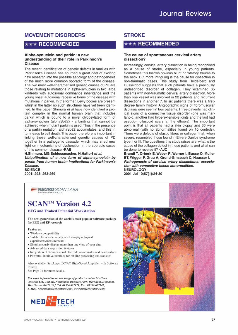
ANCR • VOLUME 1 NUMBER 4 SEPTEMBER/OCTOBER 2001 27
Section
EEG and Evoked Potential Workstation
SCANTM Version 4.2
The next generation of the world’s most popular software packagefor EEG and EP research
Features:� Windows compatibility� Suitable for a wide variety of electrophysiological
experiments/measurements� Simultaneously display more than one view of your data� Advanced data acquisition features� Integration of 3-dimensional electrode co-ordinates and head surface� Powerful, intuitive interface for off-line processing and statistics
Also available: SynAmps: DC/AC High-Speed Amplifier with SoftwareControl. See Page 31 for more details.
For more information on our range of products contact MedTechSystems Ltd, Unit 2E, Northlands Business Park, Warnham, Horsham,
West Sussex RH12 3SJ. Tel. 01306 627171, Fax. 01306 627141, E-Mail. [email protected], www.medtechsystems.com
MOVEMENT DISORDERS
Alpha-synuclein and parkin: a newunderstanding of their role in Parkinson'sDiseaseThe recent identification of genetic defects in families withParkinson's Disease has spurned a great deal of excitingnew research into the possible aetiology and pathogenesisof the much more common sporadic form of the disease.The two most well-characterised genetic causes of PD arethose relating to mutations in alpha-synuclein in two largekindreds with autosomal dominance inheritance and theyoung onset autosomal recessive forms of the disease withmutations in parkin. In the former, Lewy bodies are presentwhilst in the latter no such structures have yet been identi-fied. In this paper Shimura et al have now identified a pro-tein complex in the normal human brain that includesparkin which is bound to a novel glycosylated form ofalpha-synuclein (alphaSp22) - a binding that cannot beachieved when mutant parkin is used. Thus in the presenceof a parkin mutation, alphaSp22 accumulates, and this inturn leads to cell death. This paper therefore is important inlinking these well-characterised genetic causes of PDtogether in a pathogenic cascade, which may shed newlight on mechanisms of dysfunction in the sporadic casesof this common disease -RABH.Shimura, MG Schlossmacher, N.Hattori et al.Ubiquitination of a new form of alpha-synuclein byparkin from human brain: Implications for Parkinson’sDisease.SCIENCE 2001: 293: 263-269
STROKE
The cause of spontaneous cervical arterydissection?Increasingly, cervical artery dissection is being recognisedas a cause of stroke, especially in young patients.Sometimes this follows obvious blunt or rotatory trauma tothe neck. But more intriguing is the cause for dissection innon-traumatic cases. This study from Heidelberg andDüsseldorf suggests that such patients have a previouslyundescribed disorder of collagen. They examined 65patients with non-traumatic cervical artery dissection. Morethan one vessel was involved in 22 patients and recurrentdissections in another 7. In six patients there was a first-degree family history. Angiographic signs of fibromusculardyplasia were seen in four patients.Three patients had clin-ical signs of a connective tissue disorder (one was mar-fanoid, another had hyperextensible joints and the last hadpseudo-molluscoid scars at the elbows). The importantpoint is that all patients had a skin biopsy and 36 wereabnormal (with no abnormalities found on 10 controls).There were defects of elastic fibres or collagen that, whensevere, resembled those found in Ehlers-Danlos syndrometype II or III. The questions this study raises are: what is thecause of the collagen defect in these patients and what canbe done to reverse it? -AJCBrandt T, Orberk E, Weber R, Werner I, Busse O, MullerBT, Wigger F, Grau A, Grond-Ginsbach C, Hausser I.Pathogenesis of cervical artery dissections: associa-tion with connective tissue abnormalities.NEUROLOGY2001 Jul 10;57(1):24-30
✭✭✭ RECOMMENDED ✭✭✭ RECOMMENDED
Journal Reviews
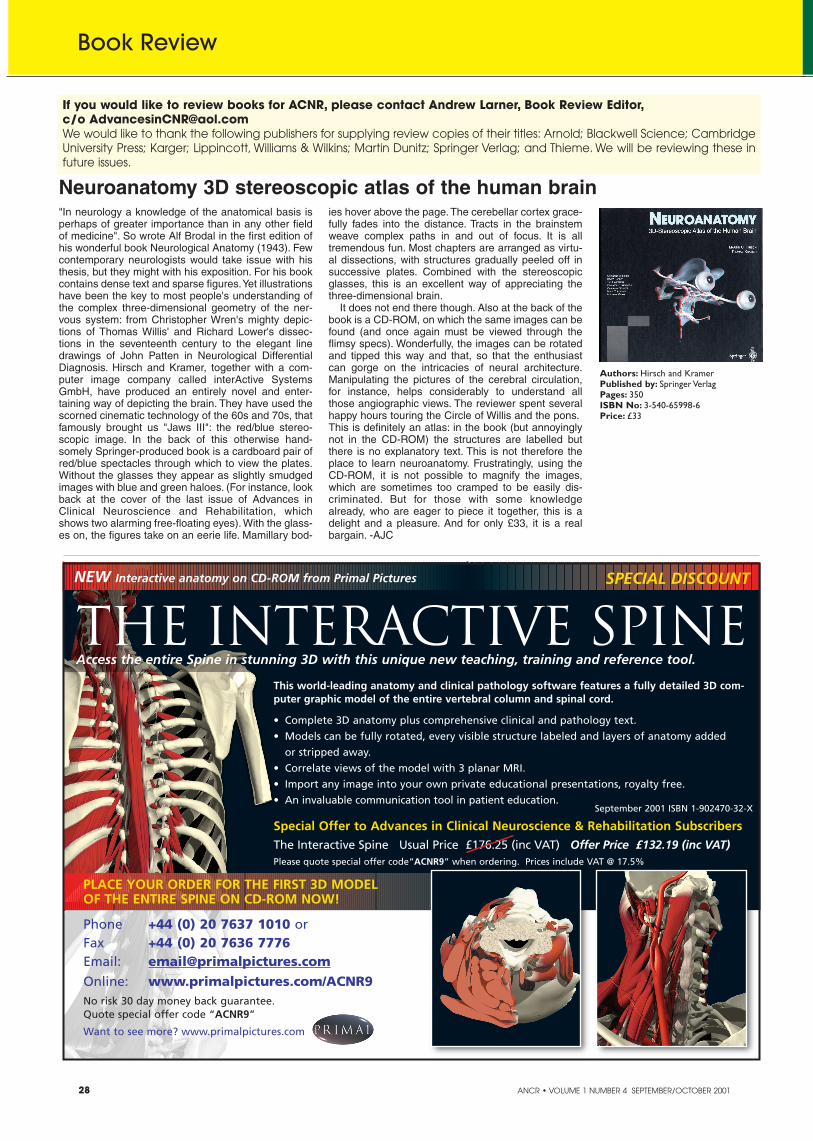
28 ANCR • VOLUME 1 NUMBER 4 SEPTEMBER/OCTOBER 2001
SectionSection
If you would like to review books for ACNR, please contact Andrew Larner, Book Review Editor,c/o [email protected] would like to thank the following publishers for supplying review copies of their titles: Arnold; Blackwell Science; CambridgeUniversity Press; Karger; Lippincott, Williams & Wilkins; Martin Dunitz; Springer Verlag; and Thieme. We will be reviewing these infuture issues.
SectionBook Review
"In neurology a knowledge of the anatomical basis isperhaps of greater importance than in any other fieldof medicine". So wrote Alf Brodal in the first edition ofhis wonderful book Neurological Anatomy (1943). Fewcontemporary neurologists would take issue with histhesis, but they might with his exposition. For his bookcontains dense text and sparse figures.Yet illustrationshave been the key to most people's understanding ofthe complex three-dimensional geometry of the ner-vous system: from Christopher Wren's mighty depic-tions of Thomas Willis' and Richard Lower's dissec-tions in the seventeenth century to the elegant linedrawings of John Patten in Neurological DifferentialDiagnosis. Hirsch and Kramer, together with a com-puter image company called interActive SystemsGmbH, have produced an entirely novel and enter-taining way of depicting the brain. They have used thescorned cinematic technology of the 60s and 70s, thatfamously brought us "Jaws III": the red/blue stereo-scopic image. In the back of this otherwise hand-somely Springer-produced book is a cardboard pair ofred/blue spectacles through which to view the plates.Without the glasses they appear as slightly smudgedimages with blue and green haloes. (For instance, lookback at the cover of the last issue of Advances inClinical Neuroscience and Rehabilitation, whichshows two alarming free-floating eyes). With the glass-es on, the figures take on an eerie life. Mamillary bod-
ies hover above the page. The cerebellar cortex grace-fully fades into the distance. Tracts in the brainstemweave complex paths in and out of focus. It is alltremendous fun. Most chapters are arranged as virtu-al dissections, with structures gradually peeled off insuccessive plates. Combined with the stereoscopicglasses, this is an excellent way of appreciating thethree-dimensional brain.
It does not end there though. Also at the back of thebook is a CD-ROM, on which the same images can befound (and once again must be viewed through theflimsy specs). Wonderfully, the images can be rotatedand tipped this way and that, so that the enthusiastcan gorge on the intricacies of neural architecture.Manipulating the pictures of the cerebral circulation,for instance, helps considerably to understand allthose angiographic views. The reviewer spent severalhappy hours touring the Circle of Willis and the pons.This is definitely an atlas: in the book (but annoyinglynot in the CD-ROM) the structures are labelled butthere is no explanatory text. This is not therefore theplace to learn neuroanatomy. Frustratingly, using theCD-ROM, it is not possible to magnify the images,which are sometimes too cramped to be easily dis-criminated. But for those with some knowledgealready, who are eager to piece it together, this is adelight and a pleasure. And for only £33, it is a realbargain. -AJC
Neuroanatomy 3D stereoscopic atlas of the human brain
Authors: Hirsch and KramerPublished by: Springer VerlagPages: 350ISBN No: 3-540-65998-6Price: £33
THE INTERACTIVE SPINEAccess the entire Spine in stunning 3D with this unique new teaching, training and reference tool.
SPECIAL DISCOUNTNEW Interactive anatomy on CD-ROM from Primal Pictures
This world-leading anatomy and clinical pathology software features a fully detailed 3D com-puter graphic model of the entire vertebral column and spinal cord.
• Complete 3D anatomy plus comprehensive clinical and pathology text.
• Models can be fully rotated, every visible structure labeled and layers of anatomy added
or stripped away.
• Correlate views of the model with 3 planar MRI.
• Import any image into your own private educational presentations, royalty free.
• An invaluable communication tool in patient education.September 2001 ISBN 1-902470-32-X
Special Offer to Advances in Clinical Neuroscience & Rehabilitation SubscribersThe Interactive Spine Usual Price £176.25 (inc VAT) Offer Price £132.19 (inc VAT)Please quote special offer code“ACNR9” when ordering. Prices include VAT @ 17.5%
PLACE YOUR ORDER FOR THE FIRST 3D MODELOF THE ENTIRE SPINE ON CD-ROM NOW!
Phone +44 (0) 20 7637 1010 orFax +44 (0) 20 7636 7776Email: [email protected]: www.primalpictures.com/ACNR9No risk 30 day money back guarantee. Quote special offer code “ACNR9”
Want to see more? www.primalpictures.com
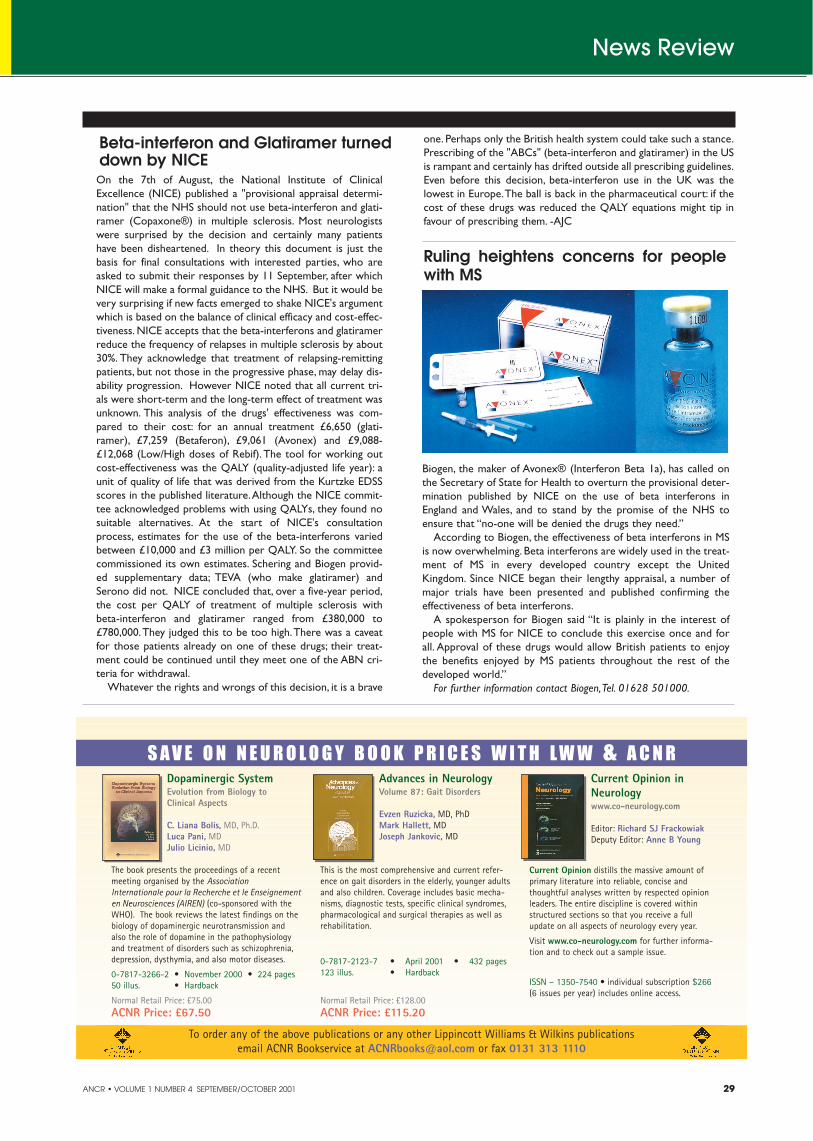
ANCR • VOLUME 1 NUMBER 4 SEPTEMBER/OCTOBER 2001 29
SectionSection News Review
On the 7th of August, the National Institute of ClinicalExcellence (NICE) published a "provisional appraisal determi-nation" that the NHS should not use beta-interferon and glati-ramer (Copaxone®) in multiple sclerosis. Most neurologistswere surprised by the decision and certainly many patientshave been disheartened. In theory this document is just thebasis for final consultations with interested parties, who areasked to submit their responses by 11 September, after whichNICE will make a formal guidance to the NHS. But it would bevery surprising if new facts emerged to shake NICE's argumentwhich is based on the balance of clinical efficacy and cost-effec-tiveness. NICE accepts that the beta-interferons and glatiramerreduce the frequency of relapses in multiple sclerosis by about30%. They acknowledge that treatment of relapsing-remittingpatients, but not those in the progressive phase, may delay dis-ability progression. However NICE noted that all current tri-als were short-term and the long-term effect of treatment wasunknown. This analysis of the drugs' effectiveness was com-pared to their cost: for an annual treatment £6,650 (glati-ramer), £7,259 (Betaferon), £9,061 (Avonex) and £9,088-£12,068 (Low/High doses of Rebif). The tool for working outcost-effectiveness was the QALY (quality-adjusted life year): aunit of quality of life that was derived from the Kurtzke EDSSscores in the published literature.Although the NICE commit-tee acknowledged problems with using QALYs, they found nosuitable alternatives. At the start of NICE's consultationprocess, estimates for the use of the beta-interferons variedbetween £10,000 and £3 million per QALY. So the committeecommissioned its own estimates. Schering and Biogen provid-ed supplementary data; TEVA (who make glatiramer) andSerono did not. NICE concluded that, over a five-year period,the cost per QALY of treatment of multiple sclerosis withbeta-interferon and glatiramer ranged from £380,000 to£780,000.They judged this to be too high.There was a caveatfor those patients already on one of these drugs; their treat-ment could be continued until they meet one of the ABN cri-teria for withdrawal.
Whatever the rights and wrongs of this decision, it is a brave
Beta-interferon and Glatiramer turneddown by NICE
S A V E O N N E U R O L O G Y B O O K P R I C E S W I T H L W W & A C N R
To order any of the above publications or any other Lippincott Williams & Wilkins publications email ACNR Bookservice at [email protected] or fax 0131 313 1110
Dopaminergic SystemEvolution from Biology to Clinical Aspects
C. Liana Bolis, MD, Ph.D.Luca Pani, MDJulio Licinio, MD
The book presents the proceedings of a recentmeeting organised by the AssociationInternationale pour la Recherche et le Enseignementen Neurosciences (AIREN) (co-sponsored with theWHO). The book reviews the latest findings on thebiology of dopaminergic neurotransmission andalso the role of dopamine in the pathophysiologyand treatment of disorders such as schizophrenia,depression, dysthymia, and also motor diseases.
0-7817-3266-2 • November 2000 • 224 pages50 illus. • Hardback
Normal Retail Price: £75.00ACNR Price: £67.50
Advances in NeurologyVolume 87: Gait Disorders
Evzen Ruzicka, MD, PhDMark Hallett, MDJoseph Jankovic, MD
This is the most comprehensive and current refer-ence on gait disorders in the elderly, younger adultsand also children. Coverage includes basic mecha-nisms, diagnostic tests, specific clinical syndromes,pharmacological and surgical therapies as well asrehabilitation.
0-7817-2123-7 • April 2001 • 432 pages123 illus. • Hardback
Normal Retail Price: £128.00ACNR Price: £115.20
Current Opinion inNeurologywww.co-neurology.com
Editor: Richard SJ FrackowiakDeputy Editor: Anne B Young
Current Opinion distills the massive amount ofprimary literature into reliable, concise andthoughtful analyses written by respected opinionleaders. The entire discipline is covered withinstructured sections so that you receive a fullupdate on all aspects of neurology every year.
Visit www.co-neurology.com for further informa-tion and to check out a sample issue.
ISSN – 1350-7540 • individual subscription $266(6 issues per year) includes online access.
Ruling heightens concerns for peoplewith MS
Biogen, the maker of Avonex® (Interferon Beta 1a), has called onthe Secretary of State for Health to overturn the provisional deter-mination published by NICE on the use of beta interferons inEngland and Wales, and to stand by the promise of the NHS toensure that “no-one will be denied the drugs they need.”
According to Biogen, the effectiveness of beta interferons in MSis now overwhelming. Beta interferons are widely used in the treat-ment of MS in every developed country except the UnitedKingdom. Since NICE began their lengthy appraisal, a number ofmajor trials have been presented and published confirming theeffectiveness of beta interferons.
A spokesperson for Biogen said “It is plainly in the interest ofpeople with MS for NICE to conclude this exercise once and forall. Approval of these drugs would allow British patients to enjoythe benefits enjoyed by MS patients throughout the rest of thedeveloped world.”
For further information contact Biogen,Tel. 01628 501000.
one. Perhaps only the British health system could take such a stance.Prescribing of the "ABCs" (beta-interferon and glatiramer) in the USis rampant and certainly has drifted outside all prescribing guidelines.Even before this decision, beta-interferon use in the UK was thelowest in Europe.The ball is back in the pharmaceutical court: if thecost of these drugs was reduced the QALY equations might tip infavour of prescribing them. -AJC

30 ANCR • VOLUME 1 NUMBER 4 SEPTEMBER/OCTOBER 2001
New guidelines to improve the outcome forpeople with spasticity were launched recentlyat the RCP in London.
‘The Management of Adults withSpasticity using BotulinumToxin: A Guide to ClinicalPractice’ was produced by aneminent panel comprising spe-cialists from neurological rehabili-tation, neurology, physiotherapyand occupational therapy andchaired by Dr Anthony Ward,Consultant in RehabilitationMedicine at the North StaffordshireRehabilitation Centre.
Spasticity often occurs in people fol-lowing stroke, traumatic brain injuryand in moderate to severe multiple sclerosis.
Botulinum toxin is an innovative treatment in thepharmacological management of spasticity, and if
used appropriately, has the potential toreduce the cost burden.
The guide will assist clinicians treatingadults with spasticity by providing guidanceon the correct use of botulinum toxin, aspart of an overall patient managementprogramme. It also aims to ensure allpatients treated with botulinum toxinachieve optimum benefit when used aspart of a co-ordinated multi-discipli-nary approach.For a free copy of the guidelines contact Radius Healthcare, Tel. 01932
350006, Fax. 01932 353336, [email protected], or return the read-er enquiry form enclosed with this magazine.
New fever controltechniquereduces nurseworkload inneuro ICU
Activity monitor for use in neurological research and clinical assessment
A new medical technique toprotect critically-ill patientsfrom developing harmfulfever is also helping to reducethe workload on nurses inNeuro ICU.
The revolutionary intravas-cular products developed byAlsius Corporation ofCalifornia allow doctors toclosely control the core tem-perature of their patients -particularly critical forpatients with stroke, braintrauma or other severe neu-ronal injury, who commonlyexperience elevated temper-ature or ‘fever’.
The use of the Cool LineTM
catheter in conjunction withthe CoolGardTM temperaturemanagement system providesa quantum leap over conven-tional methods of fever man-agement that are primitive,labour-intensive and of limit-ed effectiveness.
According to TrinaCunningham, NeurosurgeryNurse Coordinator at JohnHopkins Hospital: “Our tem-peratures are on a constantmonitoring schedule with theCoolGardTM, so the nursedoesn’t have to be at the bed-side every hour, or every 30minutes, taking the tempera-ture.”
Aslius Corporation is theworldwide leader in thedevelopment of intravasculartemperature control sys-tems. The systems havereceived CE Clearance inEurope and are currentlyinvestigational in the US.
Alsius Corporation, 15770Laguna Canyon Road, Suite150, Irvine, California USA92618. www.alsius.com
Management of adults with spasticity
Britannia Pharmaceuticals Ltd have re-branded theirapomorphine range, with the introduction of 2mland 5ml APO-go ampoules.
APO-go is used in the treatmentof Parkinson’s Disease and providesadditional or alternative benefit forthose individuals who have foundthat each dose of their existing treat-ment has become shorter and whoare experiencing ‘wearing off ’, or verysudden and unpredictable ‘on-off ’ fluctuations.
Britannia has developed, with the assistance ofthe Italian company, Cane, the APO-go Pump, which
is used to administer a continuous infusionof apomorphine.
The APO-go pump is small and “stateof the art”. It is discreet to wear andonce programmed with the dosage
details, it is simple to use.For further information contact Britannia
Pharmaceuticals Ltd on Tel. 01737 773741, Fax.01737 773973.
APO-go (apomorphine)
The Government's response to the HSC report intorehabilitation following head injury was describedrecently as "Complacent, ambiguous and very disap-pointing" by Headway.
Graham Nickson, Policy and Campaigns Manager,said, "Although action is promised by 2005 with theimplementation of a national service framework (NSF)there is nothing in the Government response to tack-le the problem which exists now of the lottery of careor rehabilitation services across England for peoplewho survive a head injury. Early, appropriate and wellresourced rehabilitation following head injury is vital ifpatients are to maximise their recovery and reinte-grate into their community".
"The Government response perpetuates the lot-tery by creating a four-year roll over until 2005, when
the NSF will ensure legally enforceable minimumstandards are in place across England." Headwaywants the Government to address the followingpoints immediately:� Research to establish the prevalence of head injuryacross the country.� social services departments to adopt a separatecategory of user group of people with complex neu-rological conditions, recognising head injury as a sep-arate disability and so design services which addresstheir real - rather than perceived - needs� improve the quality of information offered to peo-ple who survive a head injury, their families and carersand professional clinical and social care staff.
For further information contact Headway on Tel. 01159240800.
4-year roll over in the brain injury lottery
FR
EE
RE
SO
UR
CE
The Actiwatch-Neurologica®, recently exhibitedby Cambridge Neurotechnology Ltd at the WorldCongress of Neurology, is one of a series of ultra-lightweight activity monitors designed to be wornby people of all ages, typically on the wrist. Anaccelerometer measures movement.
Each monitor is easily programmed via aReader connected to a PC with data transferredby telemetry and then analysed by specific soft-ware.As well as monitoring general activity levels,tremor levels and duration can be detected, anddata collected on the periodicity and frequency ofspecific movements.
The applications for Actiwatch - Neurologica®include use in the detection and assessment oftremor and dyskinesias associated withParkinson’s Disease and other movement disor-
ders.Also in the monitoring of the effectiveness ofdifferent treatment regimes, and the effects ofdrugs on the CNS system, as well as in strokerehabilitation programmes.
For further information contact Dr CarolineReynolds, Business Development Manager, CambridgeNeurotechnology Limited, Upper Pendrill Court,Papworth Everard, Cambridge, CB3 8UY. Tel. 01480831223, Fax. 01480 831733, E-Mail. [email protected]
News Review
ANCR • VOLUME 1 NUMBER 4 SEPTEMBER/OCTOBER 2001 31
Section
Dendron GmbH. of Germany haslaunched its product range of emboli-sation coils and accessories into theUK market.
Dendron (originally EFMT) has beenmanufacturing the coils for ten yearsand was previously an EEC fundedresearch company looking into thetreatments for AVMs and various typesof aneurysms.
The German company was recentlyprivatised in order to allow it to concentrate onexpanding the international markets for the coils.“There has previously only been one main supplierfor these embolisation coils’’ said Ian Graney,Managing Director of Neurotechnics Ltd, the exclu-sive UK & Ireland distributor of the coils. “We arenow in a position to offer the customer a wide
choice of the highestquality EEC manufac-tured product at avery competitiveprice, which can onlyserve to help theNHS in meeting its’challenging fundinggoals for the future.’’Mr Graney went onto add that ‘’Further
unique products are planned for release towardsthe end of this year that will add to the neuroRadiologists armoury and Neurotechnics plan to bea key supplier in this marketplace within 24months’’.
For further information contact Ian Graney on Tel.01844 260777, Fax. 01844 260778.
The X-ray department at thenew Bristol Royal Hospital forChildren was recently commis-sioned with the installation ofnew General X-ray and DigitalFluoroscopy facilities.
The new Siemens SIRESKOPSX Digital Fluoroscopy unitincorporates many of the dosesaving features relevant fora dedicated PaediatricDepartment. The combinationof ‘Carevision’ low dose pulsedfluoroscopy together with‘Careprofile’ collimation with-out radiation, Carewatch dosedisplay and ‘Carefilter’ auto-
matic copper filtration adap-tation depending on thepatient size and the new‘Harmonization’ feature, allcontribute to the resultantlower doses now achievedwith excellent image quality.
The department also hous-es two new MULTIX TOPgeneral x-ray units one withthe new ORBIX TOP isocen-tric skull unit.
For further information con-tact Siemens Medical Solutions,Tel. 01344 396317,Fax. 01344 396337. [email protected]
Siemens supplies X-ray equipment for new Bristol Children’s Hospital Consensusreached onRiluzolecost-effectiveness
UK Launch of Dendron (GmbH) embolisation coils
Sheffield hunts for better diagnosis of brain disorder
Pictured alongside the new fluoroscopy facilityat the Bristol Children’s Hospital are: (L to R)Mark Waterhouse, Radiographer and DonnaDimond, Superintendent Radiographer.
Positive NICE guidance hasconfirmed that riluzole is cost-effective in the management ofpatients with ALS / MND.However, there has been debateas to what is the true figure ofcost-effectiveness (measured ascost per quality adjusted lifeyear (QALY)). The ratio hasranged from £34,000 to £58,000at the upper level (based on theHealth Technology Assessmentreport), and £16,500 to £20,904at the lower level (based on themanufacturer submission).
At the recent InternationalHealth Economics Association(IHEA) meeting in York, StirlingBryan (a co-author of the HTAreport that estimated the ratiosat the higher level) stated thathaving incorporated the long-term evidence for riluzole (48months), their revised estimateof the cost per QALY wasbetween £16,500 and £20,000.This is now consistent with themanufacturers evaluation.
Along with the impressivesurvival advantage that riluzoleoffers (an additional 3 to 6months over best supportivecare), the consensus on cost-effectiveness provides strongersupport for prescribing.
The NICE guidance on rilu-zole (Rilutek) and the HTAreport are available atwww.nice.org.uk.
For further information contactAventis Pharma on Tel: 08705239604.
SynAmps: speed, power and flexibilityCombining the flexibility of software with the power andspeed of the latest electronics, SynAmps is a software-programmable amplifier capable of true DC or ACrecording. Each unit is an integrated physiological dataacquisition system that can be configured for 8,16, 24 or32 channels and includes ultra-low-noise amplification,16-bit analogue-to-digital conversion, and digital filtering.Multiple units can be interconnected or ‘daisy-chained’together to provide synchronised acquisition for up to256 channels.
SynAmps contains the analogue components needed to amplifylow-level neurophysiological signals and the digital componentsrequired to log external events and digitise, DC-correct, digitally filter
and transfer data to a host computer.With its distributedprocessing approach, SynAmps can acquire data in dis-crete epochs at rates of up to 20 KHz per each of 32channels, 50 KHz per each of 8 channels, or 100 KHz pereach of 4 channels.
It performs simultaneous sampling via multiple individ-ual sample-and-holds, thereby assuring zero phase erroracross all channels.This is a significant problem that mostmulti-channel systems do not address.
For more information contact MedTech Systems Ltd,Tel. 01306 627171, Fax. 01306 627141,E-Mail. [email protected] also our advertisement on page 27.
Sheffield is leading a new study designed to helpmedics more accurately diagnose Parkinson'sDisease. Lead researcher Professor Paul Griffiths,head of neuroimaging at the University ofSheffield, says, 'The exact cause of the illness is stillunknown, and as yet there is no accurate way ofdiagnosing whether a living patient has PD. Only apost-mortem can define the disease.'
He adds, 'Current techniques - in which medicscarry out an assessment based on a set of clinicalcriteria - mean that almost one in five patients arewrongly diagnosed with PD when they haveanother condition with similar symptoms such asmultiple system atrophy (MSA).'
A good reliable diagnostic technique is there-fore needed and a new two-year study funded byAction Research is designed to do just that.
The researchers will be looking at how muchiron might play a role in the illness. Iron is essen-tial for a number of brain processes but increasedlevels may be damaging to the individual, resultingin neurodegeneration.
The Action Research team will carry out aseries of tests using brain scanning to determinewhether iron content in the brain can be used asa 'measuring stick' to assess PD progression.
For more information on Action Research seewww.actionresearch.co.uk
News Review
Prescribing informationLamictal (lamotrigine)Brief Prescribing Information. Presentation: Pale yellow tablets containing 25mg, 50mg, 100mg and 200mg lamotrigine, and white dispersible/chewable tablets containing 2mg, 5mg, 25mg and100mg lamotrigine.Uses: Monotherapy: Not recommended in children under 12 years. Adults and children over 12 yearsfor partial epilepsy with or without secondarily generalised tonic-clonic seizures and in primary gen-eralised tonic-clonic seizures. Add-on therapy: Adults and children over 2 years for partial epilepsy withor without secondary generalised tonic-clonic seizures and in primary generalised tonic-clonicseizures. Seizures associated with Lennox-Gastaut syndrome.Dosage and Administration: Initial dose and subsequent dose escalation should not be exceeded tominimise the risk of rash. Monotherapy: Initial dose is 25mg daily for two weeks, followed by 50mgdaily for two weeks. Dose should be increased by a maximum of 50-100mg every 1-2 weeks until opti-mal response. Usual maintenance dose is 100-200mg/day in one dose, or two divided doses. Add-on therapy: Adults and Children over 12 years: To sodium valproate with or without ANY otherantiepileptic drug (AED), initial dose 25mg every alternate day for two weeks, followed by 25mg/dayfor two weeks. Dose should be increased by 25-50mg every 1-2 weeks until optimal response. Usualmaintenance dose 100 to 200mg/day in one dose, or two divided doses. To enzyme inducing AEDswith or without other AEDs (but NOT valproate), initial dose is 50mg daily for two weeks, followedby 100mg/day in two divided doses for two weeks. Dose should be increased by 100mg every 1-2 weeks until optimal response. Usual maintenance dose is 200 to 400mg/day given in two divideddoses. Children aged 2-12 years: To be dosed on a mg/kg basis until the adult recommended titrationdose is reached. Add-on to sodium valproate with or without ANY other AED, initial dose is0.15mg/kg bodyweight/day given once a day for two weeks, followed by 0.3mg/kg/day given once aday for two weeks. Dose should then be increased by a maximum of 0.3mg/kg every 1-2 weeks untiloptimal response. Usual maintenance dose is 1 to 5mg/kg/day given in one dose, or two divideddoses. Add-on to enzyme-inducing AEDs with or without other AEDs (but NOT valproate) is0.6mg/kg bodyweight/day given in two divided doses for two weeks, followed by 1.2mg/kg/day for twoweeks given in two divided doses. Dose should then be increased by a maximum of 1.2mg/kg every1-2 weeks until optimal response. Usual maintenance dose is 5-15mg/kg/day given in two divideddoses. Weight of child should be monitored and dose adjusted as appropriate. If calculated dose is 1-2mg/day then 2mg may be taken on alternate days for the first two weeks. Dose Escalation:Starter packs covering the first four weeks treatment are available for adults and children over 12 years. When the pharmacokinetic interaction of any AED with Lamictal is unknown the dose esca-lation for Lamictal and concurrent sodium valproate should be used. Elderly patients: No dose adjust-ment required.Contra-indications: Hypersensitivity to lamotrigine.Precautions: Adverse skin reactions, mostly mild and self-limiting, may occur generally during thefirst 8 weeks of treatment. Rarely, serious, potentially life threatening rashes including Stevens-Johnson syndrome (SJS) and toxic epidermal necrolysis (TEN) have been reported. Patients shouldbe promptly evaluated and Lamictal withdrawn unless the rash is clearly not drug related. High ini-tial dose, exceeding the recommended dose escalation rate, and concomitant use of sodium valproatehave been associated with an increased risk of rash. Patients who acutely develop symptoms sugges-tive of hypersensitivity such as rash, fever, lymphadenopathy, facial oedema, blood and liver abnor-malities, flu-like symptoms, drowsiness or worsening seizure control, should be evaluated imme-diately and Lamictal discontinued if an alternative aetiology cannot be established. Hepaticimpairment: Dose reductions recommended. Withdrawal: Avoid abrupt withdrawal, exceptfor safety reasons. Pregnancy: Lamictal was not carcinogenic, mutagenic, teratogenicor shown to impair fertility in animal studies. There are insufficient data availableon the use of Lamictal in human pregnancy to evaluate its safety. Lamictal shouldnot be used during pregnancy unless, in the opinion of the physician, the potentialbenefits of treatment to the mother outweigh any possible risk to the developing foe-tus. Driving: As with all AEDs, the individual response should be considered. Interactions: Antiepileptic drugs which alter certain metabolising enzymes in the liveraffect the pharmacokinetics of Lamictal (see Dosage and Administration). This is also impor-tant during AED withdrawal.Side and Adverse Effects: With monotherapy: headache, tiredness, rash, nausea, dizziness, drowsi-ness, and insomnia. Other adverse experiences have included diplopia, blurred vision, conjunctivi-tis, GI disturbances, irritability/aggression, agitation, confusion, hallucinations and haematologicalabnormalities. Also movement disorders such as tics, unsteadiness, ataxia, nystagmus and tremor. Severe skin reactions including SJS and TEN have occurred rarely, with orwithout signs of hypersensitivity syndrome. Elevations of liver function tests and rare reports ofhepatic dysfunction.Legal category: POM.Basic NHS costs: £16.45 for Monotherapy Starter Pack of 42 x 25mg tablets (PL0003/0272); £27.98 for Non-Valproate Starter Pack of 42 x 50mg tablets (PL0003/0273); £8.23 for ValproateStarter Pack of 21 x 25mg tablets (PL0003/0272). £64.37 for pack of 56 x 100mg tablets(PL0003/0274); £109.42 for pack of 56 x 200mg tablets (PL0003/0297). £21.95 for pack of 56 x 25mgtablets (PL0003/0272). £37.31 for pack of 56 x 50mg tablets (PL0003/0273). £8.75 for pack of 28 x 5mg dispersible tablets (PL0003/0346). £21.95 for pack of 56 x 25mg dispersible tablets(PL0003/0347). £64.37 for pack of 56 x 100mg dispersible tablets (PL0003/0348). £9.37 for pack of30 x 2mg dispersible tablets (PL0003/0375).Product Licence Holder: The Wellcome Foundation Ltd, Middlesex UB6 0NN. Lamictal is a Trademark of the Glaxo Wellcome Group of Companies.Further information is available from GlaxoSmithKline UK Limited, Stockley Park West, Uxbridge,Middlesex UB11 1BT.Note: If changes in AED medication are to be made they should be completed before conception.9
The UK Pregnancy Register (0800 389 1248) is collecting prospective data on the effects of allAEDs in pregnancy. Please phone for information or to register a patient.©Glaxo Wellcome UK Limited, [email protected] Services Freephone 0800 221441
References:1. Holdich T et al. Epilepsia 1991; 32 (Suppl. 1): 96.2. Morrell MJ. Neurology 1998; 51 (Suppl. 4): S21-S27.3. Fitton A, Goa KL. Drugs 1995; 50 (4): 691-713.4. Patsalos PN, Sander JWAS. Drug Safety 1994; 11 (1): 37-67.5. Messenheimer J et al. Drug Safety 1998; 18 (4): 281-296.6. Brodie MJ et al. The Lancet 1995; 345: 476-479.7. Reunanen M et al. Epilepsy Research 1996; 23: 149-155.8. Steiner TJ et al. Epilepsia 1999; 40 (5): 601-607.9. Crawford P et al. Seizure 1999; 8: 201-217.
Epilepsy treatment with women in mind
Imagine you’re a woman diagnosed with epilepsy.
There are certain things you need to be assured of before starting monotherapy.
Will it affect my periods? Will I put on weight?
Unlike some other therapies, Lamictal can offer the reassurance a woman seeks.
Lamictal does not interact with the contraceptive pill.1,2
It is not associated with cosmetic side effects or menstrual disorders.3-5
Lamictal causes significantly less sedation than carbamazepine6,7 and phenytoin.8
In addition to these benefits – essential to women – it still provides the effective
seizure control you expect.6-8 What other AED can offer a woman so much?
Before youtreat her epilepsy,
put yourself in these.
lamotrigine
TM
GEN 26792-ALP/May 2001