Embed Size (px)
Citation preview

Yangtze Medicine, 2017, 1, 127-188 http://www.scirp.org/journal/ym
ISSN Online: 2475-7349 ISSN Print: 2475-7330
Table of Contents Volume 1 Number 3 September 2017 Application of T-Wave Alternans in Evaluation of Prognosis in Patients with Intracerebral Hemorrhage
X. Li, X. L. Cheng………………………………………………………………………………………………………127
EVI1 Mediated Stimulation of 3T3-L1 Preadipocyte Differentiation Is CtBP Dependent
M. J. Ireland, M. Al-Hasan, J. A. Craft, A. Graham, C. Bartholomew………………………………………………133
Clinical Performance of ADNEX (The Assessment of Different NEoplasias in the adneXa) Model in Early Diagnosis and Staging of Benign and Malignant Ovarian Tumors
J. M. Hu, Y. S. Shi, M. X. Li, C. J. Yi…………………………………………………………………………………148
Nursing Students’ Experience with Information Literacy Skill
H. Osman………………………………………………………………………………………………………………157
Logistic Regression Analysis the Risk Factors of Peripherally Inserted Central Catheter Related Blood Stream Infection of Tumor Patients
J. Song, Y. Yan, H. Yan, C. L. Wang, J.-E. Hu………………………………………………………………………169
Prevalence of Coinfection with Malaria and HIV among Children in Yaoundé, Cameroon: A Cross-Sectional Survey Performed in Three Communities in Yaoundé
T. E. Kwenti, E. Edo, B. S. Ayuk, T. D. B. Kwenti……………………………………………………………………178

Yangtze Medicine (YM)
Journal Information
SUBSCRIPTIONS
The Yangtze Medicine (Online at Scientific Research Publishing, www.SciRP.org) is published quarterly by Scientific Research
Publishing, Inc., USA.
Subscription rates:
Print: $39 per issue.
To subscribe, please contact Journals Subscriptions Department, E-mail: [email protected]
SERVICES
Advertisements
Advertisement Sales Department, E-mail: [email protected]
Reprints (minimum quantity 100 copies)
Reprints Co-ordinator, Scientific Research Publishing, Inc., USA.
E-mail: [email protected]
COPYRIGHT
Copyright and reuse rights for the front matter of the journal:
Copyright © 2017 by Scientific Research Publishing Inc.
This work is licensed under the Creative Commons Attribution International License (CC BY).
http://creativecommons.org/licenses/by/4.0/
Copyright for individual papers of the journal:
Copyright © 2017 by author(s) and Scientific Research Publishing Inc.
Reuse rights for individual papers:
Note: At SCIRP authors can choose between CC BY and CC BY-NC. Please consult each paper for its reuse rights.
Disclaimer of liability
Statements and opinions expressed in the articles and communications are those of the individual contributors and not the statements
and opinion of Scientific Research Publishing, Inc. We assume no responsibility or liability for any damage or injury to persons or
property arising out of the use of any materials, instructions, methods or ideas contained herein. We expressly disclaim any implied
warranties of merchantability or fitness for a particular purpose. If expert assistance is required, the services of a competent
professional person should be sought.
PRODUCTION INFORMATION
For manuscripts that have been accepted for publication, please contact:
E-mail: [email protected]
Yangtze Medicine, 2017, 1, 127-132 http://www.scirp.org/journal/ym
ISSN Online: 2475-7349 ISSN Print: 2475-7330
DOI: 10.4236/ym.2017.13013 Sep. 21, 2017 127 Yangtze Medicine
Application of T-Wave Alternans in Evaluation of Prognosis in Patients with Intracerebral Hemorrhage
Xian Li, Xianglin Cheng*
Department of Neurology, The Clinical Medicine School of Yangtze University, The First Affiliated Hospital of Yangtze University, Jingzhou, China
Abstract Objective: To explore the application value of electrocardiograph (ECG) T-wave Alternans (TWA) anomaly in acute stage of intracerebral hemorrhage pa-tients. Methods: We choose 1175 intracerebral hemorrhage patients whose con-ventional 12-lead ECG has TWA in our hospital from January 2011 to De-cember 2015, 751 patients without TWA in the same period as the control group, compared the volume of intracerebral hemorrhage, bleeding site and mortality between the 2 groups. Results: In TWA group, 247 cases died, 361 cases with massive intracerebral hemorrhage, 298 cases with brain stem he-morrhage; in TWA negative group (control group), 41 cases died, 93 cases with massive brain hemorrhage, 64 cases with brain stem hemorrhage. There are statistical differences between two groups (P < 0.05). Multi factor Logistic regression analysis showed that massive intracerebral hemorrhage, brain stem hemorrhage, Glasgow score and TWA were the independent factors in the prognosis of intracerebral hemorrhage (P < 0.05). Conclusion: The occur-rence of TWA is significantly related to the volume of bleeding, the bleeding site and mortality, and can be used as an important parameter in the progno-sis of intracerebral hemorrhage.
Keywords Intracerebral Hemorrhage, T-Wave Alternans, Prognosis
1. Introduction
Intracerebral hemorrhage is a disease with high mortality. In cerebrovascular events, the ECG often changes, which is related to the high mortality of intrace-rebral hemorrhage. It can provide useful clues to the doctors to assess a patient
How to cite this paper: Li, X. and Cheng, X.L. (2017) Application of T-Wave Alternans in Evaluation of Prognosis in Patients with Intracerebral Hemorrhage. Yangtze Medi-cine, 1, 127-132. https://doi.org/10.4236/ym.2017.13013 Received: May 19, 2017 Accepted: September 18, 2017 Published: September 21, 2017 Copyright © 2017 by authors and Scientific Research Publishing Inc. This work is licensed under the Creative Commons Attribution International License (CC BY 4.0). http://creativecommons.org/licenses/by/4.0/
Open Access

X. Li, X. L. Cheng
DOI: 10.4236/ym.2017.13013 128 Yangtze Medicine
and judge the prognosis. In addition to the general severity index, another indi-cator TWA, such as the S-T changes, severe arrhythmia and so on, can be a sign of myocardial repolarization abnormalities. Although we discussed the relation-ship between TWA and cerebral hemorrhage site and hemorrhage volume of 65 cases [1], the sample size is small, and it’s not clear whether it can also be used as an indicator of prognosis of patients with cerebral hemorrhage. We did not ex-plore, as we retrospectively analyze our hospital intracerebral hemorrhage pa-tients in conventional 12 lead ECG TWA incidence and its relationship with prognosis from January 2011 to December 2015. We judge its value to evaluate the prognosis of intracerebral hemorrhage. Now it is reported as follows.
2. Data and Methods 2.1. Case Selection
First, 2417 intracerebral hemorrhage patients in our hospital were selected from January 2011 to December 2015, all patients were diagnosed as intracerebral hemorrhage by CT, excluding patients who had coronary heart disease (typical angina symptoms and ECG changes, or cardiac angiography evidence), arr-hythmia (ECG changes), electrolyte disturbance (typical clinical symptom and blood electrolyte examination), cardiac hypertrophy (cardiac color ultrasound evidence), and used anti-arrhythmia drugs and digoxin drugs (patients readme). The remaining 1926 patients, including 1039 males and 887 females, aged 34 - 91 years, average age (61.27 ± 21.23) years. Among them, 1175 patients with the TWA were the experimental group, including 634 males and 541 females, aged 39 - 91 years, the average age (63.75 ± 20.17) years, Glasgow score (9.1 ± 2.1), of which 286 patients died, Glasgow score (7.9 ± 1.8); the remaining 751 patients as control group, including 405 males and 346 females, aged 34 to 87 years, average age (60.32 ± 22.74) years, Glasgow score (12.3 ± 4.1). There were no significant differences in age and sex between the two groups (P > 0.05). The basic informa-tion between the experimental group and the control group is showed in Table 1.
2.2. Method
1) The Bleeding volume calculation: by coniglobus formula [2], select the maximum amount of bleeding plane according to the calculation, the volume of bleeding (ml) = (length × width × layer)/2, the basal ganglia and lobe hemorr-hage > 50 ml, cerebellar hemorrhage > 15 ml, the brain stem hemorrhage > 15
Table 1. Comparison of basic information between the experimental group and the con-trol group.
Total Average age Gender male female Glasgow score
The experimental group 1175 63.75 ± 20.17 634 541 9.1 ± 2.1
The control group 751 60.32 ± 22.74 405 346 12.3 ± 4.1
P value P > 0.05 P > 0.05 P > 0.05

X. Li, X. L. Cheng
DOI: 10.4236/ym.2017.13013 129 Yangtze Medicine
ml are massive hemorrhage, the rest is not. 2) All intracerebral hemorrhage patients synchronous scanned routine 12-lead
ECG examination on the 1st day in hospital. TWA calculation: the same lead T- wave amplitude difference 0.1 mv is TWA, calculate the incidence of TWA.
2.3. Statistical Analysis
Intracerebral hemorrhage site, intracerebral hemorrhage volume and mortality between the two groups were compared by the χ2 test, using multiple factors lo-gistic regression analysis to determine the independent predictive factor of prog-nosis, P < 0.05 has a statistically significant difference.
3. Results
1) In 1926 brain hemorrhage patients, 1175 cases with TWA, the occurrence rate was 61.0%, 454 cases with massive brain hemorrhage, accounting for 23.6%, 1472 cases without massive Intracerebral hemorrhage, accounting for 76.4%; 96 cases with lobe hemorrhage, accounting for 5%; 1272 cases with basal ganglia he-morrhage, accounting for 66.0%; 173 cases with cerebellar hemorrhage, account-ing for 9% and 362 cases with brain stem hemorrhage, accounting for 18.8%, 23 cases with intraventricular hemorrhage, accounting for 1.2%; 286 cases died, ac-counting for 14.8%.
2) Compared with the control group, in the experimental group, the patients with massive intracerebral hemorrhage, brain stem hemorrhage and death rate were statistically significant (P < 0.01), as showed in Table 2.
3) Multivariate logistic regression analysis showed that massive intracerebral hemorrhage, brain stem hemorrhage, Glasgow score and TWA were the inde-pendent factors in the prognosis of intracerebral hemorrhage (P < 0.05).
4. Discussion
Intracerebral hemorrhage is a disease of high death rate and high disability rate [3]. Some patients did not die of primary brain dysfunction, but die of secondary cardiac causes, and this part of patients had no organic heart damage, but were secondary to intracranial damage. So those patients with coronary heart disease, arr-hythmia, electrolyte disorder, cardiac hypertrophy, and the use of anti-arrhythmia
Table 2. Comparison between experimental group and control group in 1104 cases mas-sive intracerebral hemorrhage patients, brain stem hemorrhage patients and death pa-tients (n = 1104).
Total The experimental
group The control
group X2
value P value
Massive intracerebral hemorrhage 454 361 93 85.539 <0.01
Brain stem hemorrhage 362 298 64 85.125 <0.01
Death 288 247 41 87.247 <0.01
Total 1104 906 198
X. Li, X. L. Cheng
DOI: 10.4236/ym.2017.13013 130 Yangtze Medicine
drugs and digoxin drugs are excluded. We only analyze the influence on cardiac repolarization by intracerebral hemorrhage, and find an independent predictor of the prognosis with intracerebral patients hemorrhage. Because cardiac death is always caused by ventricular arrhythmias, and the influence of intracerebral hemorrhage on cardiac repolarization is obvious [4], repolarization abnormali-ties must lead to ventricular arrhythmia [5]. On the 12 lead ECG, the objective indicators are not much used to direct observe repolarization abnormalities, in-cluded heart rate variability, Q-T dispersion, TWA etc. The prediction effect of heart rate variability and Q-T dispersion has often been reported. Therefore, we mainly analyze the prediction function of the prognosis of intracerebral he-morrhage by TWA.
We retrospectively analyzed all patients who were hospitalized in our hospital from January 2011 to December 2015. The patients who had coronary heart disease, arrhythmia, electrolyte disorder, cardiac hypertrophy, and the use of an-ti-arrhythmia drugs and digoxin drugs were excluded. There were 1926 patients, in which 1175 patients had TWA on the conventional 12-lead ECG on the first day of admission, accounting for 61%. There was a great difference between the reported normal healthy people [1], which further explained that intracerebral hemorrhage obviously has a direct impact on cardiac repolarization. And in intracerebral hemorrhage patients, the performance of the heart is significant and meaningful. At the same time, we analyzed the relationship between the TWA generator with the intracerebral hemorrhage site, the volume of intracere-bral hemorrhage and the death time, and compared with the non TWA genera-tor. In the experimental group, 247 patients died, accounting for 12.8%; in the control group, 41 patients died, accounting for 2%. They were statistically sig-nificant (P < 0.05), further confirmed that the experimental group with TWA had a high mortality rate, at the same time we conducted a multifactor Logistic regression analysis, it showed that massive intracerebral hemorrhage, brain stem hemorrhage, Glasgow score and TWA were the independent factors for the prognosis of intracerebral hemorrhage, they could be used as a basis for judging the prognosis of intracerebral hemorrhage. But in our experiments we also found that in the experimental group, 361 cases with massive intracerebral hemorr-hage, accounting for 18.7%, 93 cases without massive intracerebral hemorrhage, accounting for 4.9%; in the experimental group, 298 cases with brain stem he-morrhage, accounting for 15.5%, in control group, 64 cases with brain stem he-morrhage, accounting for 3.3%. The comparison between the two group was also statistically significant, it also showed that several predictive factors have a mu-tual influence on the occurrence mechanism. Massive intracerebral hemorrhage, brain stem hemorrhage could influence cardiac repolarization and lead to the occurrence of TWA.
The mechanism that intracerebral hemorrhage induces TWA is not very clear. Studies have found that the increase of sympathetic nerve tension can induce TWA, sympathetic nervous excitement makes the action potential morphology

X. Li, X. L. Cheng
DOI: 10.4236/ym.2017.13013 131 Yangtze Medicine
and amplitude change, thus inducing ventricular repolarization heterogeneity and leading to TWA. Therefore, we speculate that the mechanism of TWA in intracerebral hemorrhage is mainly caused by affecting the autonomic nervous function, which leads to the dysfunction of the sympathetic and parasympathet-ic. Autonomic nerve cortical representation known to regulate cardiac activity is in the orbital surface of the frontal lobe and the anterior cingulate cortex (i.e. 13 and 24 districts), and hypothalamus as a higher subcortical autonomic nerve center, regulates the cardiac activity. The insular lobe is closely related to the occurrence of TWA. Brain stem as the descending pathway of autonomic nerve, also has an important effect on the cardiac activity. So we speculate that massive intracerebral hemorrhage on the basal ganglia and lobes affect the autonomic nerve center of cingulate, insular lobe and hypothalamus. The brain stem he-morrhage can directly destroy autonomic nerve descending pathway, causing the change of autonomic nerve center and the imbalance of sympathetic and para-sympathetic and affecting of cardiac conduction system and myocardial repola-rization, producing TWA and leading to the occurrence of ventricular arrhyth-mia, but the specific mechanism needs to be further confirmed in our later expe-riments.
Our study has some limitations. First of all, a variety of other prognostic fac-tors for intracerebral hemorrhage have been studied. However, in different models it is difficult to be fully taken into account, it may cause some deviation. We should try to judge comprehensively in the future analysis. Second, autonomic nerve dysfunction could be studied by other methods, such as the change of blood pressure and heart rate variability, but these studies need to observe for a long time and special analysis software, but our purpose is to provide a fast and effective prediction index to the grassroots medical staff, so we choose TWA, hoping to replace the application of heart rate variability and blood pressure changes. In short, TWA occurrence is an independent predictor of intracerebral hemorr-hage patients’ hospitalized death, TWA occurrence reminds that intracerebral hemorrhage patients have severe autonomic dysfunction and brain damage, these patients should get more attention in the follow-up treatment.
Acknowledgements
We thank the hospital medical records department’s support in providing ECG information.
References [1] Cheng, X.-L., Zhao, C.-S. and Ma, L. (2005) The Analysis of T Wave Alternations
and QT Dispersion on Early Cerebral Hemorrhage Patient. Jilin Medicine, 26, 578-579.
[2] Xu, X., Chen, X., Zhang, J., Zheng, Y., Sun, G., Yu, X. and Xu, B. (2014) Compari-son of the Tada Formula with software Slicer: Precise and Low-Cost Method for Volume Assessment of Intracerebral Hematoma. Stroke, 45, 3433-3535. https://doi.org/10.1161/STROKEAHA.114.007095
[3] Hemphill 3rd, J.C., Greenberg, S.M., Anderson, C.S., Becker, K., Bendok, B.R.,

X. Li, X. L. Cheng
DOI: 10.4236/ym.2017.13013 132 Yangtze Medicine
Cushman, M., Fung, G.L., Goldstein, J.N., Macdonald, R.L., Mitchell, P.H., Scott, P.A., Selim, M.H. and Woo, D. (2015) Guidelines for the Management of Sponta-neous Intracerebral Hemorrhage: A Guideline for Health Care Professionals from the American Heart Association/American Stroke Association. Stroke, 46, 2032-2060. https://doi.org/10.1161/STR.0000000000000069
[4] Junttila, E., Vaara, M., Koskenkari, J., Ohtonen, P., Karttunen, A., Raatikainen, P. and Ala-Kokko, T. (2013) Repolarization Abnormalities in Patients with Subarach-noid and Intraintracerebral Hemorrhage: Predisposing Factors and Association with Outcome. Anesthesia & Analgesia, 116, 190-197. https://doi.org/10.1213/ANE.0b013e318270034a
[5] Sachs, K.V., Harnke, B., Mehler, P.S. and Krantz, M.J. (2015) Cardiovascular Com-plications of Anorexia Nervosa: A Systematic Review. International Journal of Eat-ing Disorders, 49, 238-248. https://doi.org/10.1002/eat.22481
Submit or recommend next manuscript to SCIRP and we will provide best service for you:
Accepting pre-submission inquiries through Email, Facebook, LinkedIn, Twitter, etc. A wide selection of journals (inclusive of 9 subjects, more than 200 journals) Providing 24-hour high-quality service User-friendly online submission system Fair and swift peer-review system Efficient typesetting and proofreading procedure Display of the result of downloads and visits, as well as the number of cited articles Maximum dissemination of your research work
Submit your manuscript at: http://papersubmission.scirp.org/ Or contact [email protected]

Yangtze Medicine, 2017, 1, 133-147 http://www.scirp.org/journal/ym
ISSN Online: 2475-7349 ISSN Print: 2475-7330
DOI: 10.4236/ym.2017.13014 Sep. 21, 2017 133 Yangtze Medicine
EVI1 Mediated Stimulation of 3T3-L1 Preadipocyte Differentiation Is CtBP Dependent
M. J. Ireland, M. Al-Hasan, J. A. Craft, A. Graham, C. Bartholomew
Department of Life Sciences, School of Health & Life Sciences, Glasgow Caledonian University, City Campus, Glasgow, Scotland
Abstract Myelodysplasia syndrome 1 (MDS1) and Ecotropic viral integration site 1 (EVI1) complex (MECOM) locus encode multiple isoforms of the EVI1 pro-tein that are essential for normal vertebrate development and when inappro-priately expressed play a significant role in malignancy and in particular leu-kaemias. However, the function of individual EVI1 isoforms is not fully un-derstood. Recently, EVI1 or PRDM3, which is structurally closely related to the brown adipose tissue determining factor PRDM16, was shown to be required for differentiation of adipocytes. In this study, we show that 3T3-L1 preadipo-cytes sustain expression of all Evi1 isoforms examined, including Mds1-Evi1, Evi1FL, Evi1Δ324, Evi1FL + 9 and Evi1Δ105 throughout the adipogenesis dif-ferentiation programme. We also show that differentiation markers are en-hanced by enforced expression of either Evi1, Evi1FL + 9 or Evi1Δ105 iso-forms. Interestingly 3T3-L1 differentiation markers are also moderately en-hanced by enforced expression of Evi1Δ324, which lacks part of the N-ter- minal zinc finger domain (ZF1), demonstrating a biological activity for this particular isoform. Enforced expression of an Evi1 mutant lacking C-terminal binding protein (CtBP) co-repressor protein binding activity fails to stimulate 3T3-L1 differentiation markers and may have dominant negative activity, caus-ing partial inhibition of this developmental programme. These studies show that multiple EVI1 isoforms are expressed in adipocytes and can stimulate adi-pogenic markers in a manner that is partially independent of the ZF1 DNA binding domain but fully dependent upon interaction with co-repressor CtBP proteins.
Keywords MECOM, PRDM3, EVI1 Isoforms, C-Terminal Binding Proteins, Adipogenesis
How to cite this paper: Ireland, M.J., Al-Hasan, M., Craft, J.A., Graham, A. and Bartholomew, C. (2017) EVI1 Mediated Sti-mulation of 3T3-L1 Preadipocyte Differen-tiation Is CtBP Dependent. Yangtze Medi-cine, 1, 133-147. https://doi.org/10.4236/ym.2017.13014 Received: June 5, 2017 Accepted: September 18, 2017 Published: September 21, 2017 Copyright © 2017 by authors and Scientific Research Publishing Inc. This work is licensed under the Creative Commons Attribution International License (CC BY 4.0). http://creativecommons.org/licenses/by/4.0/
Open Access

M. J. Ireland et al.
DOI: 10.4236/ym.2017.13014 134 Yangtze Medicine
1. Introduction
Myelodysplasia syndrome 1 (MDS1) and Ecotropic virus integration site 1 (EVI1) complex (MECOM) locus gene transcripts include MDS1, EVI1 and a fusion of part of MDS1 with EVI1 [1] and their inappropriate expressions are associated with poor prognosis leukaemias and other malignancies [2] [3]. Those tran-scripts containing EVI1 encode transcription factors with multiple cys2his2 zinc finger DNA binding motifs [4] and are required for mammalian development [5]. EVI1 has been shown to contribute to a number of developmental programmes including maintenance of haemopoietic stem cells and various committed pro-genitor cells in haemopoiesis [6], neuroectodermal cell differentiation [7], neph-rogenesis [8] and cardiac development [9].
EVI1 is also known as positive regulatory domain I-binding factor 1, retinob-lastoma protein-binding zinc finger protein (PR) domain protein 3 (PRDM3) and the structurally similar PRDM16 is a key regulator of brown adipose tissue development [10]. Recent studies show that EVI1 also participates in adipogene-sis [11] [12]. These studies show that EVI1 converts nonadipogenic cells to adi-pocytes and knockdown (KD) suppresses preadipocyte differentiation by im-pairing CCAAT/Enhancer-binding protein-beta (CEBPβ) assisted induction of peroxisome proliferator-activated receptor-gamma 2 (PPARγ2).
There are multiple naturally occurring isoforms of EVI1 but it is not known which are expressed in preadipocytes and which might participate in adipogene-sis as all are potentially affected in the knockdown (KD) study of Ishibashi et al., 2012. The isoforms include MDS1-EVI1, EVI1FL, EVI1Δ324 [13], EVI1RP+ and EVI1Δ105 (murine specific) [14]. MDS1-EVI1 comprises intergenic transcripts containing coding exons of both the MDS1 and the EVI1 genes and encodes an EVI1 protein with an N-terminal PR domain. EVI1FL is the original full length murine protein encoded by the cDNA first isolated from leukaemia cells [4]. EVI1RP+ is similar to EVI1FL but has an additional 9 amino acids inserted within the repressor domain (RP). EVI1Δ324 lacks 324 amino acids, including part of the first zinc finger domain up to, but excluding, RP and EVI1Δ105 has 105 C-terminal amino acids deleted. Various properties have been attributed to some of these isoforms and in some instances they have been shown to have opposing activities. For example, MDS1-EVI1 has been associated with tumor suppressing activity whereas EVI1FL is oncogenic. MDS1-EVI1 activates AGATA motif promoters whereas EVI1FL represses [1], EVI1FL inhibits 32Dcl3 cell re-sponse to granulocyte-colony stimulating factor (G-CSF) and transforming growth factor beta (TGFβ1) whereas MDS1-EVI1 has no effect on G-CSF response and enhances TGFβ1 signalling [15] and EVI1FL enhances proliferation of haemo-poietic colonies from differentiating embryonal stem (ES) cells whereas MDS1-EVI1 represses these activities [16]. The MDS1-EVI1 isoform has a PR domain [17] which confers intrinsic histone H3 lysine 9 monomethyltransferase catalytic ac-tivity [18] which is absent from other EVI1 isoforms.
The significance of the remaining isoforms remains unclear. Studies show ex-
M. J. Ireland et al.
DOI: 10.4236/ym.2017.13014 135 Yangtze Medicine
pression of each isoform in all tissues examined but little difference in DNA binding, CtBP protein binding, transcriptional repression or cell transformation activities between EVI1FL, EVI1RP+ or EVI1Δ105 [14]. EVI1Δ324 however lacks 3 N-terminal zinc fingers (ZF1), neither binds nor represses transcription via ZF1 DNA binding sites, does not transform fibroblasts [19] and to date no bio-logical activity has been assigned to this isoform.
In this study we investigate the profile of expression and biological activity of EVI1 isoforms in 3T3-L1 preadipocytes and throughout the adipocyte differen-tiation programme.
2. Materials and Methods 2.1. Cell Culture
Plat-E (Cambridge Bioscience, Cambridge, UK, RV-101) and 3T3-L1 (ATCC
CL-173) cells were cultured in complete medium (CM) comprising Dulbecco’s Modified Eagle’s Medium (Lonza Group Ltd., Basel, Switzerland, BE12-604F) sup-plemented with 10% (v/v) newborn calf serum (3T3-L1) (Sigma-Aldrich, Poole, UK, N4637) or 10% (v/v) foetal calf serum (Plat-E cells) (FCS, Lonza, DE14-801F) and 2.5 mM glutamine, 50 μg/ml penicillin, 50 units/ml streptomycin (Lonza Group Ltd., BE17-605E and BE17-603E), 37˚C, 5% CO2. For differentiation 3T3-L1 were cultured with induction medium 1 (IM1), comprising CM with 10% (v/v) FCS, 5 μg/ml insulin (Sigma-Aldrich, I9278), 0.25 μM dexamethasone (Sigma-Aldrich, D4902), 0.5 mM Isobutylmethylxanthine (IBMX, Sigma-Aldrich I5879), for 48 h fol-lowed by a further 48 h incubation with induction medium 2 (IM2) comprising CM supplemented with 10% FCS and 5 μg/ml insulin. Culture medium was subse-quently replaced with fresh IM2 every 48 h for up to 10 days. For retrovirus pro-duction, Plat-E cells were transiently transfected with retroviral plasmid DNA using Fugene6® (Roche Diagnostics GmbH, Mannheim, Germany, 11815091001); virus was harvested and used to infect 3T3-L1 as described before [20].
2.2. Preparation of Total Cellular RNA, cDNA Synthesis and Quantitative Real-Time Polymerase Chain Reaction QPCR
RNA was prepared from cultures of cells by the TRI Reagent method (Sigma-Aldrich, 93289). Total cellular RNA (1 μg) was used to synthesise cDNA using Maxima re-verse transcriptase (Thermo Fisher Scientific Inc., St. Leon-Rot, Germany, EP0742) with random hexamer (Thermo Fisher Scientific, S0142) and oligo dT (Thermo Fisher Scientific, S0131) primers according to the manufacturer’s instructions. The cDNA reaction (5%) was used for real time quantitative polymerase chain reaction using QPCR SYBR Green mix (Thermo Fisher Scientific, 11873913), gene specific oligonucleotide primers (Integrated DNA Technologies, Leuven, Belgium), 95˚C, 15 min followed by 40 cycles 95˚C, 30 s, 60˚C, 30 s in a CFX96 C1000 Thermal cyc-ler (BIO-RAD Laboratories Ltd., Hemel Hempstead, UK).
The efficiency of the Q-PCR reactions were calculated by using the formula Efficiency = −1 + 10(−1/slope) against the standard curve of each assay over a gra-
M. J. Ireland et al.
DOI: 10.4236/ym.2017.13014 136 Yangtze Medicine
dient of template concentration with each gene. The efficiency of primers are Ca3 (88%), C/ebpα (75%), Pparγ2 (92%), Fabp4 (91%), Evi1 (101%) and Gapdh (90%). Relative expression levels between target and Gapdh were determined using the arithmetic comparative 2−∆∆Ct method [21] and were determined rela-tive to the target gene in MX infected 3T3-L1 cells (calibrator). Oligonucleotide primers were supplied by Integrated DNA Technologies (Leuven, Belgium) Pparγ2FP: GCCCACCAACTTCGGAATC, Pparγ2RP: TGCGAGTGGTCTTCC ATCAC, C/ebpαFP: GAGCTGAGTGAGGCTCTCATTCT, C/ebpαRP: TGGGA GGCAGACGAAAAAAC, Fabp4FP: GGGCGTGGAATTCGATGAAATCA, Fab p4RP: CCCGCCATCTAGGGTTATGAT, Evi1FP: CGCTTGAAGCTTTGAAAG AAAAATA, Evi1RP: TGTTCTCAATTGCTGACATTTGC, Evi1 probe (HEX): TTGAGACCTTCTCCAGGATTCTTGTTTCACC, Ca3FP: CCGGGACTATTGG ACCTATCAC, Ca3RP: TTGAGCAGCAGCCACACAA, Ca3 probe (FAM): CTCC TTCACCACGCCGCCCTG, GapdhFP: GGGCTGCCCAGAACATCA, GapdhRP: CCGTTCAGCTCTGGGATGAC, Gapdh probe (FAM): CCCTGCATCCACTG GTGCTGCC.
2.3. Endpoint PCR
cDNA (0.5 μl) was amplified by PCR with 140 ng/μl forward and reverse primers using ReddyMix PCR master mix [1.5 mM MgCl2] (Thermoscientific) 95˚C, 5 min followed by 40 cycles 95˚C, 15 s, 60˚C, 60 s in a PTC-100™ Thermal cycler (MJ Research, Inc.). Products were analysed by 3% (w/v) agarose gel electrophore-sis in 40 mM Tris-acetate, 1 mM EDTA (pH 8.0) buffer (1XTAE). Oligonucleo-tide primers were supplied by Integrated DNA Technologies. Mds1/Evi1 and Evi1 specific primers were EF, MF1 and GSP3 [22], RP+ primers were ME1/ME3 and Δ105 primers were ME2/ME4 [14] and Δ324 primers were Δ324F: CGTCA GGGCCTCAAACAGC, Δ324R: GGGTACATTGATTGAGAGAATGAGA. CtBP 1 and 2 primers were: CtBP1FP; CACACAGGAGATCCATGAGAAG, CtBP1RP; CTCTGGTCAGTGTGATGGTATG, CtBP2FP; GCACAGTCCACTCAGGAAAT, CtBP2RP; CCTTGAACTTCTCCAGGTCTTC.
2.4. Western Blot Analysis
Protein extracts, SDS polyacrylamide gel electrophoresis and western blotting were performed as described previously [23] with either α-EVI1 (1806) or α-GAPDH (Fitzgerald Industries, North Acton, MA, USA, 6C5) diluted 1/1000 (1806) or 1/5000 (6C5) respectively. Appropriate IRDye 800CW conjugated anti-rabbit (Li-Cor Biosciences, 926-32211) or IRDye 680RD conjugated anti-mouse (Li-Cor Biosciences, 926-68072) IgG secondary antibodies were used at 1/15000 dilu-tions and detection was performed by fluorescence using an Odyssey Fc Imaging System (Li-Cor Biosciences).
2.5. Statistical Analysis
Unpaired Student’s t-test was used to determine the significance of data using

M. J. Ireland et al.
DOI: 10.4236/ym.2017.13014 137 Yangtze Medicine
Graphpad Prism® 6.0 software. P ≤ 0.05 was considered significant. *P ≤ 0.05, **P ≤ 0.01, ***P ≤ 0.001, ns not significant.
3. Results 3.1. Enforced Expression of EVI1 in 3T3-L1 Preadipocytes
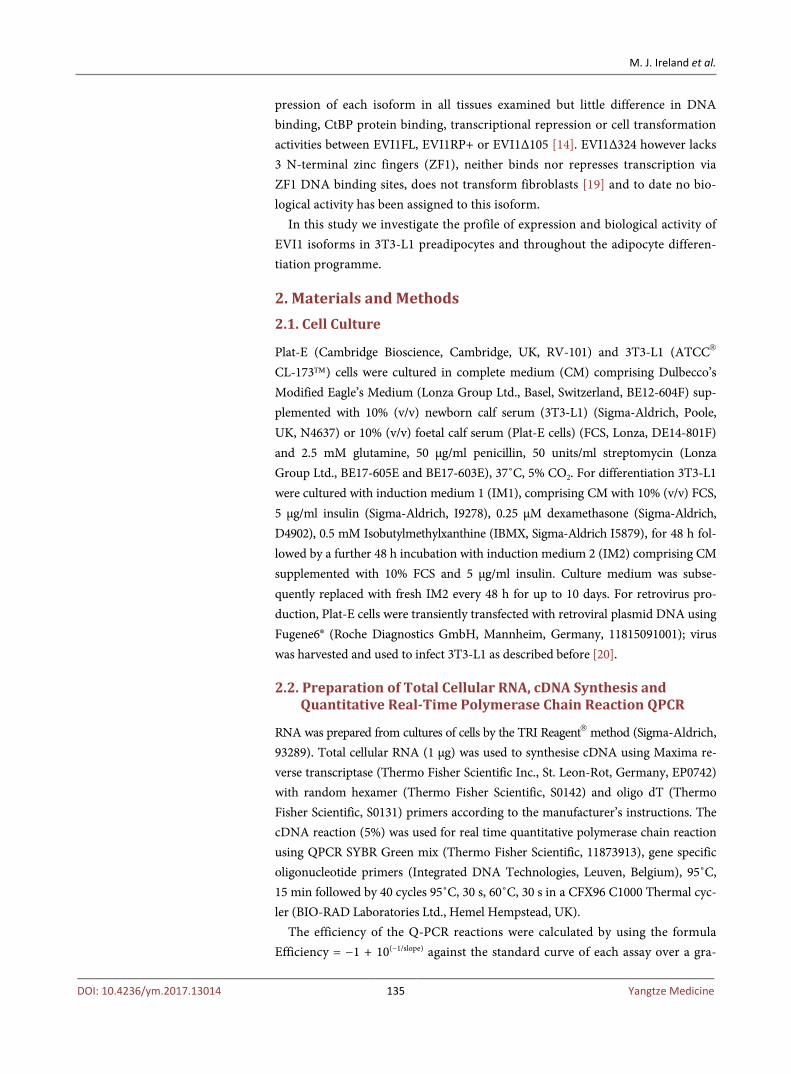
In order to investigate the effect of EVI1 expression on adipogenesis it was ex-pressed in 3T3-L1 cells. Initially Plat-E cells were transiently transfected with the previously described Evi1FL encoding p50M5.6-neo retroviral vector [20] and the resulting virus containing supernatants used to infect varying numbers of 3T3-L1 cells (Materials and Methods). The 3T3-L1 cells were re-infected with virus con-taining supernatant again 24 hrs later. After virus infection (48 h) cells were examined for Evi1 expression by western blot analysis with α-EVI1. The results show production of the 145 kd Evi1 protein in cells infected with the 5.6 retro-viral vector (Figure 1(a)). Even loading of samples was confirmed by western blot analysis with α-GAPDH (Figure 1(a)). Highest Evi1 expression is observed when either 2 × 10⁴ or 5 × 10⁴ cells were used for virus infection and therefore 5 × 10⁴ cells were chosen for further experiments.
3.2. EVI1 Enhances 3T3-L1 Adipocyte Differentiation
To investigate the impact of enforced EVI1FL expression on adipogenesis it was expressed in 3T3-L1 cells using the transient retroviral infection scheme and subsequent induction of adipocyte differentiation programme outlined in Figure 1(b). Cells were transiently infected with either p50MX-neo (MX, empty vector control) or p50M5.6-neo (5.6) virus, induced to differentiate and RNA prepared from cell extracts at various time points. Initially, expression of the adipocyte differentiation marker gene Fabp4 at days 0 and 10 were examined. The results show induction of this marker for both control infected cells as well as cells with enforced expression of Evi1 (Figure 1(c), MX, 5.6), however the induction of Fabp4 is significantly increased in cells with enforced Evi1 expression at day 10 compared to MX infected cells on the same day (Figure 1(c), 5.6).
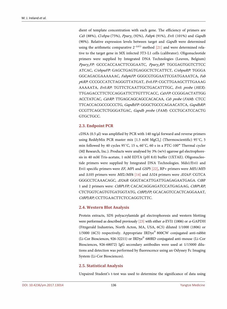
We next examined expression of key regulators of adipocyte differentiation Pparγ2 and C/ebpα in the presence (5.6) or absence (MX) of Evi1 at days 0, 1, 2, 3 and 4 of induction. The results show that both markers are induced during the 4 day period but accumulate to significantly higher levels in the presence of Evi1 (Figure 2(a), Figure 2(b), 5.6) when compared with empty vector infected cells at each point examined (Figure 2(a), Figure 2(b), MX). Pparγ2 expression in-itially declines between day 0 and days 1 and 2 in MX and 5.6 cells but one-way ANOVA and Dunnett’s multiple comparison post-test using MX day 0 or 5.6 day 0 as control group confirms significant increases by day 4 [P ≤ 0.001 (MX), P ≤ 0.05 (5.6)]. Other studies have shown that the enzyme carbonic anhydrase III (Ca3) is induced during adipocyte differentiation [24] and is either a marker or regulator of this process. Ca3 gene expression increases significantly (D1 p ≤ 0.01, D2, D3 and D4, P ≤ 0.001) in control MX cells compared to levels at D0 in-
M. J. Ireland et al.
DOI: 10.4236/ym.2017.13014 138 Yangtze Medicine
Figure 1. (a) Western blot analysis of whole cell protein extracts derived from 3T3-L1 cells transiently infected with p50M5.6neo retrovirus. The number of cells exposed to re-trovirus is shown at the top of each lane. The size of Evi-1 and Gapdh proteins observed with α-Evi1 and α-Gapdh are indicated; (b) Strategy for transient retroviral infection of 3T3-L1 cells and timeline for induction of differentiation. Complete media (CM1), induc-tion media 1 (IM1) and 2 (IM2) are described in materials & methods; (c) Histogram showing relative gene expression of Fabp4 in empty vector control (MX, clear bars) and Evi1 vector (5.6, black bars) infected 3T3-L1 cells at days 0 (D0) and 10 (D10) of differen-tiation. Error bars are the standard deviation of 3 (n = 3) independent virus infection and differentiation experiments. ***P ≤ 0.001 indicates statistical significance of MXD10 vs. MXD0 and 5.6D10 vs. MXD10.
Figure 2. Histograms showing relative gene expression of C/ebpα (a), Pparγ2 (b), Ca3 (c) and Evi1 (d) in empty vector control (MX, white bars) and Evi1 vector (5.6, black bars) virus infected 3T3-L1 cells at days 0 (D0), 1 (D1), 2 (D2), 3 (D3) and 4 (D4) of differen-tiation. Error bars are the standard deviation of 3 (n = 3) independent virus infection and differentiation experiments. *P ≤ 0.05, **P ≤ 0.01, ***P ≤ 0.001 indicates statistically sig-nificant differences in expression of the indicated gene for EVI1 expressing cells (5.6) compared to MX infected cells on the same day.
M. J. Ireland et al.
DOI: 10.4236/ym.2017.13014 139 Yangtze Medicine
dicating progression through the differentiation programme. Comparison of Ca3 gene expression in MX and 5.6 cells shows its expression is significantly elevated in 3T3-L1 cells with enforced expression of Evi-1 (Figure 2(c), 5.6) compared with cells examined at the same time point that were infected with the empty vector (Figure 2(c), MX). Finally, Evi1 transgene expression was maintained for at least the 4 day duration of the transient expression and differentiation system as its mRNA expression is significantly higher in 5.6 infected cells compared with low, but detectable, endogenous Evi1 expression observed in MX infected 3T3-L1 cells at each time point examined (Figure 2(d), MX, 5.6).
3.3. Naturally Occurring EVI1 Splice Variants, RP+, Δ105 and Δ324 Stimulate 3T3-L1 Adipocyte Differentiation
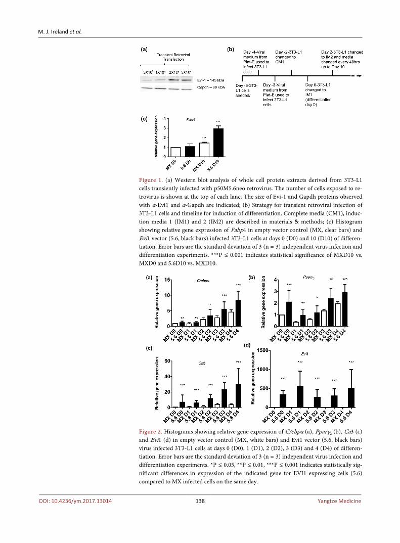
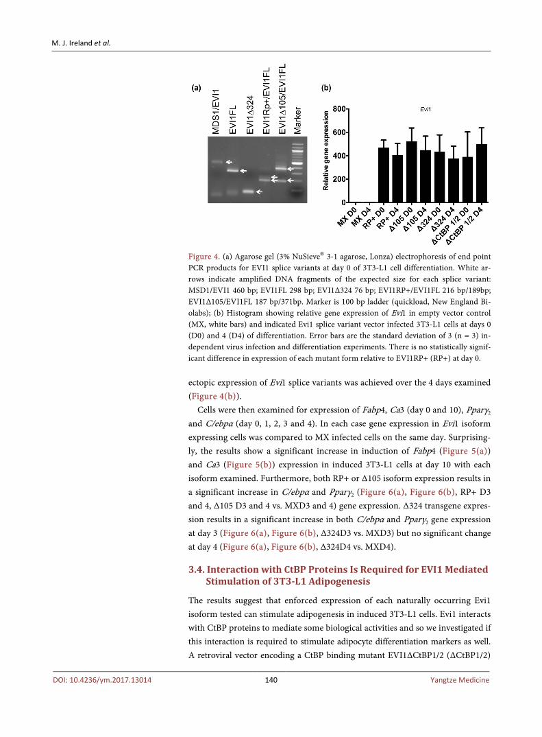
These data suggest enforced expression of Evi1 accelerates adipocyte differentia-tion of induced 3T3-L1 cells. Multiple, naturally occurring Evi1 splice variants exist in murine cells [14]. A schematic representation of the isoform shown to stimulate adipocyte differentiation (Figure 2) is shown in Figure 3, designated EVI1FL, along with other splice variants MDS1/EVI1, EVI1RP+ (RP+), EVI1Δ324 (Δ324) and EVI1Δ105 (Δ105). Endogenous expression of each of these in 3T3-L1 cells, in preadipocytes (Figure 4(a)) and throughout 10 days of differentiation (data not shown), was confirmed using isoform specific oligonucleotide primers (Materials and Methods) by end point PCR.
Since all isoforms examined are expressed in 3T3-L1 cells we investigated which can induce adipocyte differentiation. Previously described retroviral vec-tors [14] [19] were used to transiently express each isoform (RP+, Δ324 and Δ105) in 3T3-L1 cells. Infected cells were induced to differentiate and similar levels of
Figure 3. Schematic representation of the domain structure of the indicated EVI1 splice variant encoded proteins showing the PR domain (PR), 1st and second 2nd zinc finger domains (ZF1 & ZF2), repressor domain (Rp), acidic domain (Ac), CtBP binding sites 1 & 2 and the additional 9 amino acids (single letter amino acid code) found in the repres-sor domain of Rp+. X indicates CtBP binding inactivating point mutations.
M. J. Ireland et al.
DOI: 10.4236/ym.2017.13014 140 Yangtze Medicine
Figure 4. (a) Agarose gel (3% NuSieve® 3-1 agarose, Lonza) electrophoresis of end point PCR products for EVI1 splice variants at day 0 of 3T3-L1 cell differentiation. White ar-rows indicate amplified DNA fragments of the expected size for each splice variant: MSD1/EVI1 460 bp; EVI1FL 298 bp; EVI1Δ324 76 bp; EVI1RP+/EVI1FL 216 bp/189bp; EVI1Δ105/EVI1FL 187 bp/371bp. Marker is 100 bp ladder (quickload, New England Bi-olabs); (b) Histogram showing relative gene expression of Evi1 in empty vector control (MX, white bars) and indicated Evi1 splice variant vector infected 3T3-L1 cells at days 0 (D0) and 4 (D4) of differentiation. Error bars are the standard deviation of 3 (n = 3) in-dependent virus infection and differentiation experiments. There is no statistically signif-icant difference in expression of each mutant form relative to EVI1RP+ (RP+) at day 0.
ectopic expression of Evi1 splice variants was achieved over the 4 days examined (Figure 4(b)).
Cells were then examined for expression of Fabp4, Ca3 (day 0 and 10), Pparγ2 and C/ebpα (day 0, 1, 2, 3 and 4). In each case gene expression in Evi1 isoform expressing cells was compared to MX infected cells on the same day. Surprising-ly, the results show a significant increase in induction of Fabp4 (Figure 5(a)) and Ca3 (Figure 5(b)) expression in induced 3T3-L1 cells at day 10 with each isoform examined. Furthermore, both RP+ or Δ105 isoform expression results in a significant increase in C/ebpα and Pparγ2 (Figure 6(a), Figure 6(b), RP+ D3 and 4, Δ105 D3 and 4 vs. MXD3 and 4) gene expression. Δ324 transgene expres-sion results in a significant increase in both C/ebpα and Pparγ2 gene expression at day 3 (Figure 6(a), Figure 6(b), Δ324D3 vs. MXD3) but no significant change at day 4 (Figure 6(a), Figure 6(b), Δ324D4 vs. MXD4).
3.4. Interaction with CtBP Proteins Is Required for EVI1 Mediated Stimulation of 3T3-L1 Adipogenesis
The results suggest that enforced expression of each naturally occurring Evi1 isoform tested can stimulate adipogenesis in induced 3T3-L1 cells. Evi1 interacts with CtBP proteins to mediate some biological activities and so we investigated if this interaction is required to stimulate adipocyte differentiation markers as well. A retroviral vector encoding a CtBP binding mutant EVI1ΔCtBP1/2 (ΔCtBP1/2)

M. J. Ireland et al.
DOI: 10.4236/ym.2017.13014 141 Yangtze Medicine
Figure 5. Histograms showing relative gene expression of Fabp4 (a) and Ca3 (b) in empty vector control (MX) and EVI1RP+ (RP+), EVI1Δ105 (Δ105), EVI1Δ324 (Δ324) and EVI1ΔCtBP1/2 (ΔCtBP1/2) virus infected 3T3-L1 cells at days 0 (D0) and 10 (D10) of differentiation. Error bars are the standard deviation of 3 (n = 3) independent virus infec-tion and differentiation experiments. *P ≤ 0.05, **P ≤ 0.01, ***P ≤ 0.001 indicates statisti-cally significant differences in expression of the indicated gene for each form of EVI1 rel-ative to MX infected cells at day 10.
(Figure 3) that is unable to bind CtBP proteins [25] was transiently expressed in 3T3-L1 cells. The cells were induced to differentiate and examined for expression of the same molecular markers as before. ΔCtBP1/2 mutant transgene expres-sion was observed at similar levels to the other Evi1 isoforms studied (Figure 4(b), ΔCtBP1/2 D0 and D4). The results show that instead of an increase, there is a significant decrease in Fabp4 (Figure 5(a), Figure 5(b), ΔCtBP1/2 D10), C/ebpα and Pparγ2 (Figure 6(a), Figure 6(b), ΔCtBP1/2 D4) gene expression in ΔCtBP1/2 expressing cells when compared to cells infected with the empty vec-tor (MX) control on the same days. Only Ca3 gene expression shows a small in-crease in expression in cells with enforced ΔCtBP1/2 expression (Figure 5(b), ΔCtBP1/2 D10). Expression of both CtBP1 and CtBP2 genes are observed throughout the 3T3-L1 cell differentiation programme (Figure 6(c)). These data show that Evi1 mediated stimulation of 3T3-L1 cell differentiation markers is dependent on interaction with CtBP binding proteins.
4. Discussion
In this study a transient retroviral infection system was developed to investigate the effect of enforced EVI1 expression on 3T3-L1 pre-adipocyte cell differentia-tion to adipocytes. Results show that under these conditions EVI1 enhances chemi-cally induced 3T3-L1 differentiation as measured by characteristic gene markers and mediators of this process (Fabp4, Ca3, C/ebpα and Pparγ2). Furthermore, we show that all previously described and naturally occurring EVI1 splice va-riants are expressed in 3T3-L1 preadipocytes as well as throughout the differen-tiation programme and that enforced expression of splice variants EVI1RP+, EVI1Δ105 and EVI1Δ324 similarly enhance the process. Finally, we demonstrate

M. J. Ireland et al.
DOI: 10.4236/ym.2017.13014 142 Yangtze Medicine
Figure 6. Histograms showing relative gene expression of C/ebpα (a) and Pparγ2 (b), in empty vector control (MX) and EVI1Rp+9 (Rp+), EVI1Δ105 (Δ105), EVI1Δ324 (Δ324) and EVI1ΔCtBP1/2 (ΔCtBP1/2) virus infected 3T3-L1 cells at days 0 (D0), 1 (D1), 2 (D2), 3 (D3) and 4 (D4) of differentiation. Error bars are the standard deviation of 3 (n = 3) in-dependent virus infection and differentiation experiments. Statistical analysis of days 3 (D3) and 4 (D4) data only are shown. **P ≤ 0.01, ***P ≤ 0.001 indicates statistically sig-nificant differences in expression of the indicated gene for each form of EVI1 relative to MX infected cells on the same day. ns indicates no significant difference in expression relative to MX infected cells on the same day. (c) Agarose gel (3% NuSieve® 3-1 agarose) electrophoresis of end point PCR products for CtBP1 and CtBP2 gene expression at indi-cated days 0, 1, 2, 3, 4, 6 and 10 of 3T3-L1 cell differentiation. M indicates 100 bp ladder marker (quickload) and C the negative control.
that a mutant of EVI1, which no longer binds CtBP proteins, is unable to sti-mulate the 3T3-L1 differentiation markers that are observed with wild type va-riants.

M. J. Ireland et al.
DOI: 10.4236/ym.2017.13014 143 Yangtze Medicine
The efficiency of differentiation of empty vector control 3T3-L1 cells is subop-timal as indicated by relatively small changes in molecular marker gene expres-sion shown in Figure 1(c); Figures 2(a)-(c); Figure 5(a) & Figure 5(b) and Figure 6(a) & Figure 6(b). In the virus infection and differentiation scheme used here (Figure 1(b)) 3T3-L1 cells are unlikely to be confluent for 48 hrs prior to induction of differentiation as is normally the case [26] because of the need to optimize retroviral infection in dividing cells [27]. However, our results clearly show that differentiation is significantly enhanced by enforced expression of EVI1 under these conditions, based on the molecular markers examined. Following growth arrest, efficient induction of 3T3-L1 cell differentiation is accompanied by mitotic clonal expansion (MCE) [28]. Studies have shown that EVI1 stimulates cell proliferation [29] and this property may stimulate MCE, contributing to the enhanced expression of adipogenic markers observed here, which are consistent with previous observations [11].
All known naturally occurring EVI1 splice variants are expressed in preadi-pocytes and throughout differentiation of 3T3-L1 cells. The relative abundance of splice variants has not been determined in this study but others have shown that general EVI1 expression is low in proliferating preadipocytes, transiently peaks during chemical stimulation of differentiation then is low again for the remaining programme [11].
Enforced expression of splice variants EVI1FL, EVI1RP+, EVI1Δ105 and EVI1Δ324 are each capable of stimulating adipocyte differentiation based on relative increases in programme mediator (Cebpα, Pparγ2) and marker (Fabp4, Ca3) gene expression (Figure 2, Figure 5, Figure 6). It is interesting that EVI1Δ324 can stimulate adipogenic markers as this represents one of the few biological ac-tivities associated with the isoform to date. This splice variant lacks part of zinc finger 6 and all of zinc finger 7 of the ZF1 domain as well as 275 intervening amino acids to the Rp domain [30]. Recent studies show EVI1FL and EVI1Δ324 co-regulate largely the same genes in cells and that EVI1Δ324 can induce an-chorage independent growth in HeLa cells [31]. However, our results show EVI1Δ324 cannot fully complement the activity of the other EVI1 splice variants studied as stimulation of gene expression of the markers examined is less in most cases when compared with the other isoforms. This indicates the missing amino acids, including the ZF1 domain, are important for optimal EVI1 me-diated stimulation of adipogenesis.
The interaction of EVI1 with CtBP proteins has previously been shown to be essential for biological activities including cell transformation [25] and inhibi-tion of TGFβ signaling [32]. This study shows EVI1 mediated stimulation of adi-pogenic markers is also CtBP binding dependent. Interestingly, EVI1ΔCtBP1/2 not only fails to stimulate adipogenic markers in 3T3-L1 cells but it actually ap-pears to repress them when compared with MX infected cells. Gene expression of Fabp4, C/ebpα and Pparγ2 are all significantly repressed in EVI1ΔCtBP1/2 expressing cells (Figure 5 and Figure 6) which suggests it has dominant negative
M. J. Ireland et al.
DOI: 10.4236/ym.2017.13014 144 Yangtze Medicine
activity with regard to adipocyte differentiation. Other regulators of adipogene-sis are also dependent upon CtBP complexes including Klf3 [33], Fog1 and Fog2 [34]. Both CtBP1 and CtBP2 are expressed throughout adipocyte differentiation (Figure 6(c)) and their binding is required for both negative (Klf3, Fog1 and Fog2) and positive (EVI1) regulation. Furthermore, the EVI1 related protein PRDM16 also binds CtBP proteins to repress white fat specific genes and are displaced to promote brown adipose tissue development [35]. CtBP proteins bind NAD+ and NADH with higher affinity for the latter which promotes interaction with part-ner proteins [36]. CtBP proteins have been proposed to have a role in metabolic sensing [37]. High calorie intake is associated with increased levels of NADH. Based on our study this would be predicted to promote association of EVI1 and CtBP and stimulate adipogenesis.
Obesity, the expansion of adipose tissue depots, is one underlying cause of major health conditions worldwide including both type 2 diabetes mellitus and cardiovascular disease, but the mechanisms involved are not fully understood. Understanding the molecular mechanisms regulating adipogenesis might iden-tify novel targets for therapeutic intervention. Regulation of the adipogenesis developmental programme is controlled by a complex network of transcription factors and EVI1 has only recently been identified to be involved in this process. These studies show for the first time that multiple EVI1 isoforms are expressed in adipocytes and can stimulate adipogenic markers in a manner that is partially independent of the ZF1 DNA binding domain but fully dependent upon interac-tion with co-repressor CtBP proteins. Blocking EVI1/CtBP interaction may be a target for drug development controlling obesity.
Acknowledgements
This work was fully funded by a Glasgow Caledonian University PhD student-ship awarded to Mark Ireland.
References [1] Soderholm, J., Kobayashi, H., Mathieu, C., Rowley, J.D. and Nucifora, G. (1997) The
Leukemia-Associated Gene MDS1/EVI1 Is a New Type of GATA-Binding Transac-tivator. Leukemia, 11, 352-358. https://doi.org/10.1038/sj.leu.2400584
[2] Koos, B., et al. (2011) The Transcription Factor Evi-1 Is Overexpressed, Promotes Proliferation, and Is Prognostically Unfavorable in Infratentorial Ependymomas. Clin. Cancer Research, 17, 3631-3637. https://doi.org/10.1158/1078-0432.CCR-11-0175
[3] Morishita, K., et al. (1992) Activation of EVI1 Gene Expression in Human Acute Myelogenous Leukemias by Translocations Spanning 300-400 Kilobases on Chro-mosome Band 3q26. Proceedings of the National Academy of Sciences of the United States of America, 89, 3937-3941. https://doi.org/10.1073/pnas.89.9.3937
[4] Morishita, K., Parker, D.S., Mucenski, M.L., Jenkins, N.A., Copeland, N.G. and Ihle, J.N. (1988) Retroviral Activation of a Novel Gene Encoding a Zinc Finger Protein in IL-3-Dependent Myeloid Leukemia Cell Lines. Cell, 54, 831-840. https://doi.org/10.1016/S0092-8674(88)91175-0

M. J. Ireland et al.
DOI: 10.4236/ym.2017.13014 145 Yangtze Medicine
[5] Hoyt, P.R., et al. (1997) The Evi1 Proto-Oncogene Is Required at Midgestation for Neural, Heart, and Paraxial Mesenchyme Development. Mechanisms of Develop-ment, 65, 55-70. https://doi.org/10.1016/S0925-4773(97)00057-9
[6] Goyama, S., et al. (2008) Evi-1 Is a Critical Regulator for Hematopoietic Stem Cells and Transformed Leukemic Cells. Cell Stem Cell, 3, 207-220. https://doi.org/10.1016/j.stem.2008.06.002
[7] Kazama, H., Kodera, T., Shimizu, S., Mizoguchi, H. and Morishita, K. (1999) Eco-tropic Viral Integration Site-1 Is Activated During, and Is Sufficient for, Neuroec-todermal P19 Cell Differentiation. Cell Growth & Differentiation, 10, 565-573.
[8] Van Campenhout, C., et al. (2006) Evi1 Is Specifically Expressed in the Distal Tu-bule and Duct of the Xenopus Pronephros and Plays a Role in Its Formation. Deve-lopmental Biology, 294, 203-219. https://doi.org/10.1016/j.ydbio.2006.02.040
[9] Bard-Chapeau, E.A., et al. (2014) Mice Carrying a Hypomorphic Evi1 Allele Are Embryonic Viable but Exhibit Severe Congenital Heart Defects. PLoS One, 9, e89397. https://doi.org/10.1371/journal.pone.0089397
[10] Seale, P., et al. (2008) PRDM16 Controls a Brown Fat/Skeletal Muscle Switch. Na-ture, 454, 961-967. https://doi.org/10.1038/nature07182
[11] Ishibashi, J., et al. (2012) An Evi1-C/EBPβ Complex Controls Peroxisome Prolife-rator-Activated Receptor γ2 Gene Expression to Initiate White Fat Cell Differentia-tion. Molecular and Cellular Biology, 32, 2289-2299. https://doi.org/10.1128/MCB.06529-11
[12] An, Q., Wu, D., Ma, Y., Zhou, B. and Liu, Q. (2015) Suppression of Evi1 Promotes the Osteogenic Differentiation and Inhibits the Adipogenic Differentiation of Bone Marrow-Derived Mesenchymal Stem Cells in Vitro. International Journal of Mole-cular Medicine, 36, 1615-1622. https://doi.org/10.3892/ijmm.2015.2385
[13] Morishita, K., Parganas, E., Douglass, E.C. and Ihle, J.N. (1990) Unique Expression of the Human Evi-1 Gene in an Endometrial Carcinoma Cell Line: Sequence of cDNAs and Structure of Alternatively Spliced Transcripts. Oncogene, 5, 963-971.
[14] Alzuherri, H., McGilvray, R., Kilbey, A. and Bartholomew, C. (2006) Conservation and Expression of a Novel Alternatively Spliced Evi1 Exon. Gene, 384, 154-162. https://doi.org/10.1016/j.gene.2006.07.027
[15] Sood, R., Chakrabarti, S.R. and Nucifora, G. (1999) MDS1/EVI1 Enhances TGF-Boldβ1 Signaling and Strengthens Its Growth-Inhibitory Effect, but the Leukemia-Associated Fusion Protein AML1/MDS1/EVI1, Product of the T(3;21), Abrogates Growth-Inhibition in Response to TGF-Boldβ1. Leukemia, 13, 348-357. https://doi.org/10.1038/sj.leu.2401360
[16] Sitailo, S., Sood, R., Barton, K. and Nucifora, G. (1999) Forced Expression of the Leukemia-Associated Gene EVI1 in ES Cells: A Model for Myeloid Leukemia with 3q26 Rearrangements. Leukemia, 13, 1639-3645. https://doi.org/10.1038/sj.leu.2401585
[17] Fog, C.K., Galli, G.G. and Lund, A.H. (2012) PRDM Proteins: Important Players in Differentiation and Disease. Bioessays, 34, 50-60. https://doi.org/10.1002/bies.201100107
[18] Pinheiro, I., et al. (2012) Prdm3 and Prdm16 Are H3K9me1 Methyltransferases Required for Mammalian Heterochromatin Integrity. Cell, 150, 948-960. https://doi.org/10.1016/j.cell.2012.06.048
[19] Kilbey, A. and Bartholomew, C. (1998) Evi-1 ZF1 DNA Binding Activity and a Second Distinct Transcriptional Repressor Region Are both Required for Optimal

M. J. Ireland et al.
DOI: 10.4236/ym.2017.13014 146 Yangtze Medicine
Transformation of Rat1 Fibroblasts. Oncogene, 16, 2287-2291. https://doi.org/10.1038/sj.onc.1201732
[20] Bartholomew, C., Kilbey, A., Clark, A.M. and Walker, M. (1997) The Evi-1 Pro-to-Oncogene Encodes a Transcriptional Repressor Activity Associated with Trans-formation. Oncogene, 14, 569-577. https://doi.org/10.1038/sj.onc.1200864
[21] Livak, K.J. and Schmittgen, T.D. (2001) Analysis of Relative Gene Expression Data Using Real-Time Quantitative PCR and the 2(-Delta Delta C(T)) Method. Methods, 25, 402-408. https://doi.org/10.1006/meth.2001.1262
[22] Wimmer, K., Vinatzer, U., Zwirn, P., Fonatsch, C. and Wieser, R. (1998) Compara-tive Expression Analysis of the Antagonistic Transcription Factors EVI1 and MDS1-EVI1 in Murine Tissues and during in Vitro Hematopoietic Differentiation. Biochemical and Biophysical Research Communications, 252, 691-696. https://doi.org/10.1006/bbrc.1998.9588
[23] Roy P., et al. (2010) Enhanced Sensitivity to Hydrogen Peroxide-Induced Apoptosis in Evi1 Transformed Rat1 Fibroblasts Due to Repression of Carbonic Anhydrase III. The FEBS Journal, 277, 441-452. https://doi.org/10.1111/j.1742-4658.2009.07496.x
[24] Lynch, C.J., Hazen, S.A., Horetsky, R.L. and Carter, N.D. (1993) Differentia-tion-Dependent Expression of Carbonic Anhydrase II and III in 3T3 Adipocytes. American Journal of Physiology, 265, 234-243.
[25] Palmer S., et al. (2001) Evi-1 Transforming and Repressor Activities Are Mediated by CtBP Co-Repressor Proteins. Journal of Biological Chemistry, 276, 25834-25840. https://doi.org/10.1074/jbc.M102343200
[26] Student, A.K., Hsu, R.Y. and Lane, M.D. (1980) Induction of Fatty Acid Synthetase Synthesis in Differentiating 3T3-L1 Preadipocytes. Journal of Biological Chemistry, 255, 4745-4750.
[27] Miller, D.G., Adam, M.A. and Miller, A.D. (1990) Gene Transfer by Retrovirus Vectors Occurs only in Cells that Are Actively Replicating at the Time of Infection. Molecular and Cellular Biology, 10, 4239-4242. https://doi.org/10.1128/MCB.10.8.4239
[28] Otto, T.C. and Lane, M.D. (2005) Adipose Development: From Stem Cell to Adi-pocyte. Critical Reviews in Biochemistry and Molecular Biology, 40, 229-242. https://doi.org/10.1080/10409230591008189
[29] Kilbey, A., Stephens, V. and Bartholomew, C. (1999) Loss of Cell Cycle Control by Deregulation of Cyclin-Dependent Kinase 2 Kinase Activity in Evi-1 Transformed Fibroblasts. Cell Growth & Differentiation, 10, 601-610.
[30] Morishita, K., Parganas, E., Parham, D.M., Matsugi, T. and Ihle, J.N. (1990) The Evi-1 Zinc Finger Myeloid Transforming Gene Is Normally Expressed in the Kidney and in Developing Oocytes. Oncogene, 5, 1419-1423.
[31] Sayadi, A., et al. (2015) Functional Features of EVI1 and EVI1Delta324 Isoforms of MECOM Gene in Genome-Wide Transcription Regulation and Oncogenicity. On-cogene, 35, 2311-2321.
[32] Izutsu, K., Kurokawa, M., Imai, Y., Maki, K., Mitani, K. and Hirai, H. (2001) The Corepressor CtBP Interacts with Evi-1 to Repress Transforming Growth Factor β Signaling. Blood, 97, 2815-2822. https://doi.org/10.1182/blood.V97.9.2815
[33] Sue, N., et al. (2008) Targeted Disruption of the Basic Krüppel-Like Factor Gene (Klf3) Reveals a Role in Adipogenesis. Molecular and Cellular Biology, 28, 3967- 3978. https://doi.org/10.1128/MCB.01942-07
[34] Jack, B.H.A. and Crossley, M. (2010) GATA Proteins Work Together with Friend of

M. J. Ireland et al.
DOI: 10.4236/ym.2017.13014 147 Yangtze Medicine
GATA (FOG) and C-Terminal Binding Protein (CTBP) Co-Regulators to Control Adipogenesis. Journal of Biological Chemistry, 285, 32405-32414. https://doi.org/10.1074/jbc.M110.141317
[35] Kajimura, S., et al. (2008) Regulation of the Brown and White Fat Gene Programs through a PRDM16/CtBP Transcriptional Complex. Genes & Development, 22, 1397-1409. https://doi.org/10.1101/gad.1666108
[36] Zhang, Q., Piston, D.W. and Goodman, R.H. (2002) Regulation of Corepressor Function by Nuclear NADH. Science, 295, 1895-1897.
[37] Jack, B.H.A., Pearson, R.C. and Crossley, M. (2011) C-Terminal Binding Protein: A Metabolic Sensor Implicated in Regulating Adipogenesis. The International Journal of Biochemistry & Cell Biology, 43, 693-696. https://doi.org/10.1016/j.biocel.2011.01.017
Submit or recommend next manuscript to SCIRP and we will provide best service for you:
Accepting pre-submission inquiries through Email, Facebook, LinkedIn, Twitter, etc. A wide selection of journals (inclusive of 9 subjects, more than 200 journals) Providing 24-hour high-quality service User-friendly online submission system Fair and swift peer-review system Efficient typesetting and proofreading procedure Display of the result of downloads and visits, as well as the number of cited articles Maximum dissemination of your research work
Submit your manuscript at: http://papersubmission.scirp.org/ Or contact [email protected]

Yangtze Medicine, 2017, 1, 148-156 http://www.scirp.org/journal/ym
ISSN Online: 2475-7349 ISSN Print: 2475-7330
DOI: 10.4236/ym.2017.13015 Sep. 21, 2017 148 Yangtze Medicine
Clinical Performance of ADNEX (The Assessment of Different NEoplasias in the adneXa) Model in Early Diagnosis and Staging of Benign and Malignant Ovarian Tumors
Jumei Hu, Yushuang Shi, Mengxiong Li, Cunjian Yi*
Department of Obstetrics and Gynecology, The First Affiliated Hospital of Yangtze University, Jingzhou, China
Abstract Objective: To investigate the clinical value of ADNEX model in early diagno-sis and staging of benign and malignant ovarian tumors. Method: 136 cases of ovarian cancer patients treated in our hospital were retrospectively analyzed using the ADNEX risk model and MRI data. The accuracy of the two diagnos-tic methods was compared with the results of pathological examination as gold standard. Results: For qualitative assessment, the accuracy and sensitivi-ty of the ADNEX model were 78.70% and 93%, while the accuracy and sensi-tivity of MRI examination were 80.1%, and 90.7%, respectively. The diagnos-tic values of the two methods were not statistically different (P > 0.05). For ovarian tumor staging, the ADNEX model was significantly less accurate and specific for staging borderline tumor than MRI examination, although it had significantly higher sensitivity (P < 0.05). For tumors at other stages, there were no diagnostic differences between the methods (P > 0.05). Conclusion: ADNEX risk model has certain diagnostic and predictive value to distinguish benign from malignant ovarian tumors. It is useful to detect and exclude ova-rian tumor. However, for early diagnosis, it is not accurate enough and fur-ther study is needed to validate this usefulness.
Keywords Ovarian Cancer, ADNEX Risk Model, MRI Examination
1. Introduction
The incidence of ovarian cancer ranks the third in gynecological malignancies with the highest mortality [1]. Early diagnosis and cytoreductive surgery im-
How to cite this paper: Hu, J.M., Shi, Y.S., Li, M.X. and Yi, C.J. (2017) Clinical Per-formance of ADNEX (the Assessment of Different NEoplasias in the adneXa) Model in Early Diagnosis and Staging of Benign and Malignant Ovarian Tumors. Yangtze Medicine, 1, 148-156. https://doi.org/10.4236/ym.2017.13015 Received: May 19, 2017 Accepted: September 18, 2017 Published: September 21, 2017 Copyright © 2017 by authors and Scientific Research Publishing Inc. This work is licensed under the Creative Commons Attribution International License (CC BY 4.0). http://creativecommons.org/licenses/by/4.0/
Open Access
J. M. Hu et al.
DOI: 10.4236/ym.2017.13015 149 Yangtze Medicine
prove 5-year survival rate [2] [3]. However, early screening and diagnosis of ovarian cancer is a hot but difficult spot in ovarian cancer research. The aux-iliary diagnosis of ovarian cancer mainly includes the use of imaging examina-tion and serum markers. Since ultrasound examination is simple and cost effec-tive, it is most widely used in gynecological examination. To maximize the effi-ciency of early diagnosis of ovarian cancer, a number of ultrasound models have been proposed [4] [5] [6]. In 2014, the Assessment of Different NEoplasias in the adneXa (ADNEX) model was proposed to differentiate between benign, border-line, early and advanced stage invasive, and secondary metastatic tumors [7]. It can automatically provide differentiation between benign and malignant and tumor staging information on mobile devices or websites using clinical informa-tion and ultrasound data. At present, the clinical performance of the model has not been reported in China. The aim of this study is to investigate the clinical value of the model in the early diagnosis and staging of benign and malignant ovarian tumors.
2. Subjects and Methods 2.1. Subjects
223 cases of patients enrolled at the First Affiliated Hospital of Yangtze Univer-sity from January 2011 to October 2015 with ovarian cancer were retrospectively analyzed. The patients were preoperatively diagnosed using color Doppler ul-trasound and postoperatively confirmed pathologically to have epithelial tumors. Of them, 136 patients had ultrasound data for the ADNEX modeling and were examined using pelvic MRI examination. Among them, there were benign in 93 cases and malignant in 43 cases. The age ranged from 19 to 74 years with an av-erage age of 45.6 years. MRI and pathological staging were preformed based on 2013 FIGO [8]. Inclusive criteria: 1) From January 2011 to October 2015, we have admitted ovarian cancer to the Department of Obstetrics and gynecology in our hospital; 2) Histopathological diagnosis of ovarian tumors is clear, and the nature and pathological staging of ovarian tumors are determined; 3) Ovarian tumors are epithelial; 4) All the patients were examined by transvaginal ultra-sound before operation. The ultrasonic image data can be read out or recorded, and all the index data needed for the ADNEX model can be read out; 5) MRI ex-amination was performed before or after operation, with or without abdominal distension, abdominal pain and other clinical symptoms; 6) Preoperative serum CA125 examination is available or not. Exclusion criteria: 1) No MRI examination was performed before the operation; 2) Non epithelial ovarian tumor; 3) The ul-trasonic inspection record is incomplete or missing image; 4) Exclusion of en-dometriosis, tuberculous peritonitis, tumors outside the reproductive tract (re-troperitoneal neoplasms, rectal cancer, sigmoid colon cancer, etc.).
2.2. Methods
Ultrasound examinations were conducted using Philips ClearVue 580 system
J. M. Hu et al.
DOI: 10.4236/ym.2017.13015 150 Yangtze Medicine
and reported by the same physician. The married patients were examined by transvaginal examination, while the unmarried patients were examined by rectal examination. If the tumors were large, transabdominal examinations were pre-formed.
MRI axial, sagittal and coronal scans were preformed using GE Signal 1.5 T magnetic resonance imaging system. The field of view (FOV) was 28 - 36 cm and the layer thickness/spacing were 5 mm/1mm. T1WI was obtained using SE se-quence at TR/TE: 350 - 550/10ms, with a matrix of 256 × (192 - 128). The num-ber of acquisition was 2. T2WI was generated using FSE sequence at TR/TE: 3000/108ms with a matrix of 320 × 224. The number of scans was 4. The en-hanced scanning was preformed once at TR/TE: 80 to 150 ms/minimum with a matrix of 256 × (192 - 224). The number of scans was 1. The contrast agent was acyclic, ionic gadolinium (GD-DTPA), used at a dose of 0.1 to 0.2 mmol/kg, in-jected at a rate of 2.5 ml/s rate through elbow vein.
Serum CA125 was detected using ADVIA Centaur XP automated chemilumi-nescence analysis system and associated kit (Siemens, Germany).
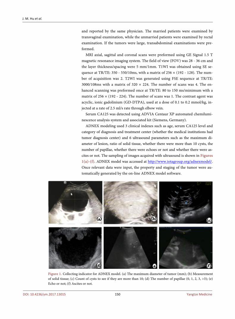
ADNEX modeling used 3 clinical indexes such as age, serum CA125 level and category of diagnosis and treatment center (whether the medical institutions had tumor diagnosis center) and 6 ultrasound parameters such as the maximum di-ameter of lesion, ratio of solid tissue, whether there were more than 10 cysts, the number of papillae, whether there were echoes or not and whether there were as-cites or not. The sampling of images acquired with ultrasound is shown in Figures 1(a)-(f). ADNEX model was accessed at http://www.iotagroup.org/adnexmodel/. Once relevant data were input, the property and staging of the tumor were au-tomatically generated by the on-line ADNEX model software.
Figure 1. Collecting indicator for ADNEX model. (a) The maximum diameter of tumor (mm); (b) Measurement of solid tissue; (c) Count of cysts to see if they are more than 10; (d) The number of papillae (0, 1, 2, 3, >3); (e) Echo or not; (f) Ascites or not.

J. M. Hu et al.
DOI: 10.4236/ym.2017.13015 151 Yangtze Medicine
2.3. Statistical Analysis
Data were processed using SPSS 17.0 statistical software. Enumeration data were tested using X2 test. The data were considered statistically different when P is <0.05 and were tested using a receiver operating characteristic curve (ROC).
3. Results 3.1. Distinguishment of Benign and Malignant Ovarian Tumors
Among the 136 cases, 93 and 43 (including were classified as benign and malig-nant), (including borderline malignant) based on FIGO (2013), respectively. Based on the ADNEX model software, 70 cases were benign, and 66 cases were malig-nant. The accuracy, sensitivity and specificity of the ADNEX model were 78.7%, 93%, and 72%, respectively, as compared to the FIGO system. The positive and negative predictive values were 60.6% and 95.7%, respectively (Table 1).
3.2. Staging of Ovarian Cancer by the ADNEX Model
Compared to the pathological results, the ADNEX model classified the tumors into five stages benign, borderline, I stage, II to IV stage and metastatic tumor (Table 2).
Table 1. The outcome of the ADNEX modeling on benign and malignant ovarian tu-mors.
Pathological examination ADNEX modeling
Total Malignant Benign
Malignant 40 3 43
Benign 26 67 93
Total 66 70 136
Table 2. Prediction of tumor stage using the ADNEX model.
Benign Borderline I stage II to IV
stage Metastatic
tumor
Accuracy (%) 78.6 86.0 91.9 86.8 93.4
Sensitivity (%) 72.0 50.0 25.0 90.9 14.3
Specificity (%) 93.0 87.8 96.1 86.0 97.7
Positive predictive value (%) 95.7 15.8 28.6 55.6 25.0
Negative predictive value (%) 60.6 97.4 95.3 98.0 95.5
Remark: ACC: Accuracy; SENS: Sensitivity, SPEC: Specificity; PPV: Positive predictive value; NPV: Nega-tive predictive value.
J. M. Hu et al.
DOI: 10.4236/ym.2017.13015 152 Yangtze Medicine
3.3. Comparison of the ADNEX Model and MRI in the Diagnosis of Benign and Malignant Ovarian Tumors
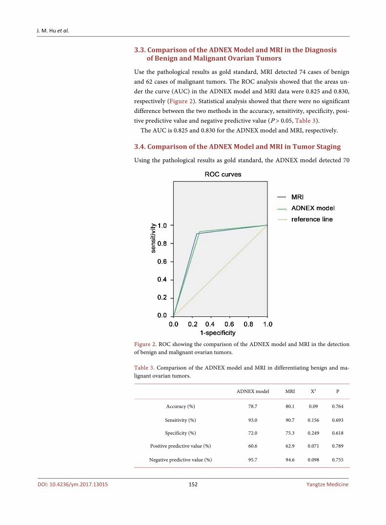
Use the pathological results as gold standard, MRI detected 74 cases of benign and 62 cases of malignant tumors. The ROC analysis showed that the areas un-der the curve (AUC) in the ADNEX model and MRI data were 0.825 and 0.830, respectively (Figure 2). Statistical analysis showed that there were no significant difference between the two methods in the accuracy, sensitivity, specificity, posi-tive predictive value and negative predictive value (P > 0.05, Table 3).
The AUC is 0.825 and 0.830 for the ADNEX model and MRI, respectively.
3.4. Comparison of the ADNEX Model and MRI in Tumor Staging
Using the pathological results as gold standard, the ADNEX model detected 70
Figure 2. ROC showing the comparison of the ADNEX model and MRI in the detection of benign and malignant ovarian tumors.
Table 3. Comparison of the ADNEX model and MRI in differentiating benign and ma-lignant ovarian tumors.
ADNEX model MRI X2 P
Accuracy (%) 78.7 80.1 0.09 0.764
Sensitivity (%) 93.0 90.7 0.156 0.693
Specificity (%) 72.0 75.3 0.249 0.618
Positive predictive value (%) 60.6 62.9 0.071 0.789
Negative predictive value (%) 95.7 94.6 0.098 0.755
J. M. Hu et al.
DOI: 10.4236/ym.2017.13015 153 Yangtze Medicine
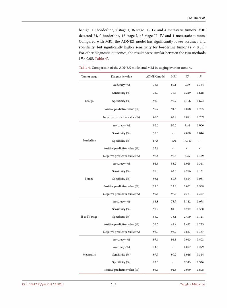
benign, 19 borderline, 7 stage I, 36 stage II - IV and 4 metastatic tumors. MRI detected 74, 0 borderline, 18 stage I, 43 stage II- IV and 1 metastatic tumors. Compared with MRI, the ADNEX model has significantly lower accuracy and specificity, but significantly higher sensitivity for borderline tumor (P < 0.05). For other diagnostic outcomes, the results were similar between the two methods (P > 0.05, Table 4).
Table 4. Comparison of the ADNEX model and MRI in staging ovarian tumors.
Tumor stage Diagnostic value ADNEX model MRI X2 P
Benign
Accuracy (%) 78.6 80.1 0.09 0.764
Sensitivity (%) 72.0 75.3 0.249 0.618
Specificity (%) 93.0 90.7 0.156 0.693
Positive predictive value (%) 95.7 94.6 0.098 0.755
Negative predictive value (%) 60.6 62.9 0.071 0.789
Borderline
Accuracy (%) 86.0 95.6 7.44 0.006
Sensitivity (%) 50.0 ‐ 4.000 0.046
Specificity (%) 87.8 100 17.049 ‐
Positive predictive value (%) 15.8 ‐ ‐ ‐
Negative predictive value (%) 97.4 95.6 6.26 0.429
I stage
Accuracy (%) 91.9 88.2 1.028 0.311
Sensitivity (%) 25.0 62.5 2.286 0.131
Specificity (%) 96.1 89.8 3.824 0.051
Positive predictive value (%) 28.6 27.8 0.002 0.968
Negative predictive value (%) 95.3 97.5 0.781 0.377
II to IV stage
Accuracy (%) 86.8 78.7 3.112 0.078
Sensitivity (%) 90.9 81.8 0.772 0.380
Specificity (%) 86.0 78.1 2.409 0.121
Positive predictive value (%) 55.6 41.9 1.472 0.225
Negative predictive value (%) 98.0 95.7 0.847 0.357
Metastatic
Accuracy (%) 93.4 94.1 0.063 0.802
Accuracy (%) 14.3 ‐ 1.077 0.299
Sensitivity (%) 97.7 99.2 1.016 0.314
Specificity (%) 25.0 ‐ 0.313 0.576
Positive predictive value (%) 95.5 94.8 0.059 0.808
J. M. Hu et al.
DOI: 10.4236/ym.2017.13015 154 Yangtze Medicine
4. Discussion
Ovarian cancer is a common malignant tumor in female reproductive systems, the incidence rate ranks the third and only seconds to cervical cancer and ute-rine cancer. Furthermore, the incidence has been increasing recently. It has been a hot but challenging spot to find effective early diagnosis method. The advan-tage of the ADNEX model is that it is designed specifically for predicting and staging benign and malignant ovarian tumors in a cost effective way. It uses convention-al clinical information and ultrasound data for on-line prediction, irrespective of the availability of CA125 data. For ultrasound examination, data corrected by inexperienced physician are sufficient for modeling. MRI provides images at various directions and layers, and is especially suitable for soft tissue. It can dis-play the relationship between the various organs in the pelvic cavity and guide surgical operation although the reports may be somewhat subjective.
4.1. The Clinical Significance of the ADNEX Model in the Diagnosis of Benign and Malignant Ovarian Tumors
It was reported that when the two sets of data were used for the diagnosis of be-nign and malignant ovarian tumors, the accuracy of the ADNEX model was 79.9%, and 81.3%, respectively [9]. We found that the accuracy, sensitivity and specialty of the ADNEX model were 78.70%, 93%, and 72%, respectively with the positive predictive value of 60.6% and negative predictive value of 95.7%. The accuracy is similar to the previous report. The accuracy, sensitivity, special-ty, positive predictive value and negative predictive value of MRI were 80.1%, 90.7%, 75.3%, 62.9% and 94.6%, respectively. AUC was 0.825 and 0.830 for the ADNEX model and MRI, suggesting that both methods have excellent diagnosis value, although MRI is slightly better than the ADNEX model. Statistically, the two methods are the same in the tumor diagnosis. The outcomes of the ADNEX model showed that it is better for the detection and exclusion of ovarian tumors. It is clear that the ADNEX model is clinically valuable for the diagnosis of be-nign and malignant ovarian tumors as MRI technology.
4.2. The Clinical Significance of the ADNEX Model in Staging Ovarian Tumors
Traditionally, ovarian tumor staging is mainly depended on pathological ex-amination, not on ultrasound data. The staging results based on the ADNEX model are similar to those reported previously [9]. The accuracy and sensitivity of the ADNEX model on early stage tumors were less than on late stage tumors. For early stage tumors, the ADNEX model and MRI are similar. For borderline tumor staging, the ADNEX model is less accurate and specific but more sensitive as compared with MRI (P < 0.05). For staging tumors at other stages, the out-comes from the two methods are slightly, and statistically insignificantly differ-ent (P > 0.05). Therefore, the ADNEX model is better at ovarian tumor staging, while MRI cannot directly stage the tumors, particularly for borderline tumor.
J. M. Hu et al.
DOI: 10.4236/ym.2017.13015 155 Yangtze Medicine
Although the ADNEX model is not perfect but it is a big step forward in tumor staging, despite its low sensitivity to early stage tumor. For better clinical use of the ADNEX model and higher qualitative assessment and staging of benign and malignant ovarian tumors, we have identified a number of shortcomings in the ADNEX model. For example, the age input has to be ≥14; the maximum diame-ter of tumor must be ≥8 mm. It is desirable to improve the model making it possi-ble to accommodate the data outside the current range for better applicability. In addition, parameters used in the model may be expended to include indexes de-scribing lymph node enlargement, nodes in pelvic cavity and posterior fornix, blood flow signal and resistance if any. Finally, due to the retrospective nature of the study, the ultrasound data parameters collected did not strictly follow what are required in the model, and some of the data were estimated. The limited sample size may also affect the diagnostic efficacy of the ADNEX model and MRI examination. It is likely that the model would have better diagnosis per-formance for differentiating benign and malignant ovarian tumors and their staging if the model is modified, clinical data are collected according to the model requirement, and further prospective study is conducted.
In conclusion, our study shows the ADNEX model is clinically value for diffe-rentiating benign and malignant ovarian tumors and their staging. It is useful for detection and exclusion of ovarian tumors, although its staging ability for early stage tumor needs further improvement and validation.
References [1] Jemal, A., Siegel, R., Xu, J., et al. (2010) Cancer Statistics. CA: A Cancer Journal for
Clinicians, 60, 277-300. https://doi.org/10.3322/caac.20073
[2] Badgwell, D. and Bast, Jr. R.C. (2007) Early Detection of Ovarian Cancer. Disease Markers, 23, 397-410. https://doi.org/10.1155/2007/309382
[3] Liang, M.L. and Wang, Z.H. (2012) The Screening and Early Diagnosis of Ovarian Cancer. Chinese Journal of Practical Gynecology and Obstetrics, 28, 166-169.
[4] Alcazar, J.L., Guerriero, S., Laparte, C., et al. (2011) Contribution of Power Doppler Blood Flow Mapping to Grayscale Ultrasound for Predicting Malignancy of Adnex-al Masses in Symptomatic and Asymptomatic Women. European Journal of Obste-trics, Gynecology, and Reproductive Biology, 155, 99-105. https://doi.org/10.1016/j.ejogrb.2010.11.010
[5] Meng, L., Shi, T.M. (2015) IOTA Simple Rules in Differentiating between Benign and Malignant Ovarian Tumors. Journal of Chinese Clinical Medical Imaging, 26, 502-504.
[6] Liu, F. (2015) Evaluation of Ultrasonic Exam in Differentiation Diagnosis of Ova-rian Tumors Using Logistic Regression. Journal of Modern Oncology, 23, 264-266.
[7] Van Calster, B., Van Hoorde, K., Valentin, L., Testa, A.C., Fischerova, D., Van Holsbeke, C., et al. (2014) Evaluating the Risk of Ovarian Cancer before Surgery Using the ADNEX Model to Differentiate between Benign, Borderline, Early and Advanced Stage Invasive, and Secondary Metastatic Tumours: Prospective Multi-centre Diagnostic Study. BMJ, 349, Article No: g5920. https://doi.org/10.1136/bmj.g5920
[8] Lin, Z.Q., (2013) FIGO 2013 New Stage of Ovarian Cancer, Fallopian Tube Cancer,

J. M. Hu et al.
DOI: 10.4236/ym.2017.13015 156 Yangtze Medicine
Peritoneal Cancer. Chinese Journal of Practical Gynecology and Obstetrics, 29, 921-923.
[9] Szubert, S. (2016) External Validation of the IOTA ADNEX Model Performed by Two Independent Gynecologic Centers. Gynecologic Oncology, 142, 490-495. https://doi.org/10.1016/j.ygyno.2016.06.020
Submit or recommend next manuscript to SCIRP and we will provide best service for you:
Accepting pre-submission inquiries through Email, Facebook, LinkedIn, Twitter, etc. A wide selection of journals (inclusive of 9 subjects, more than 200 journals) Providing 24-hour high-quality service User-friendly online submission system Fair and swift peer-review system Efficient typesetting and proofreading procedure Display of the result of downloads and visits, as well as the number of cited articles Maximum dissemination of your research work
Submit your manuscript at: http://papersubmission.scirp.org/ Or contact [email protected]
Yangtze Medicine, 2017, 1, 157-168 http://www.scirp.org/journal/ym
ISSN Online: 2475-7349 ISSN Print: 2475-7330
DOI: 10.4236/ym.2017.13016 Sep. 21, 2017 157 Yangtze Medicine
Nursing Students’ Experience with Information Literacy Skill
Hawa Osman
Library, University of Health and Allied Sciences, Ho, Ghana
Abstract This study examined the searching skills and extent of usage of electronic da-tabases by Nursing, Midwifery and Public Health Nursing students in the University of Health and Allied Science (UHAS). The focus was on forty (40) level 300 students drawn from a universe of two hundred and forty six (246) of the School of Public Health (SPH). The study used quantitative method approach and the survey instruments were questionnaire, interview and ob-servation. The data collected were analyzed and classified into the following themes: usefulness, extent of use, determinants of use of e-databases, search-ing skills, and main drawbacks of learning information literacy skills (ILS). Although all the respondents strongly agreed that e-databases are indispensa-ble for academic and professional practice, findings revealed that majority of them have low quality of searching skills and that accounts for the sparse use of the e-databases. This positive association is proven by Pearson’s chi square test (0.000). The study also established that students’ attitude, academic loads and methodology of teaching were the challenges hindering the acquisition of ILS of students. As a consequence, the study recommends that Academic li-brarians should intensify their education on e-databases, the development of research guides and encourages stronger collaboration with faculty members in the teaching of ILS so that student nurses would be more adept in searching for information to enhanced scholarship and professional practice.
Keywords Information Literacy Skills, Academic Librarians, Faculty, Electronic-Database, Nursing, Midwifery, Public Health Nursing, Information Professionals
1. Introduction
Universities prepare students for professional careers. This enables individuals
How to cite this paper: Osman, H. (2017) Nursing Students’ Experience with Infor-mation Literacy Skill. Yangtze Medicine, 1, 157-168. https://doi.org/10.4236/ym.2017.13016 Received: March 9, 2017 Accepted: September 18, 2017 Published: September 21, 2017 Copyright © 2017 by author and Scientific Research Publishing Inc. This work is licensed under the Creative Commons Attribution International License (CC BY 4.0). http://creativecommons.org/licenses/by/4.0/
Open Access
H. Osman
DOI: 10.4236/ym.2017.13016 158 Yangtze Medicine
to participate with greater understanding of issues that affect region, community and their chosen fields. The 21st century has brought tremendous changes in higher education globally as a consequence of new information and technical developments. According to [1], these changes exclusively affect every facets of society and levels of education. New ways of learning, acquisition, storage and retrieval of information are evolving based on e-resources teaching, e-library or-ganization and life-long learning. The [2] points out the academic significance of the e-library as an organized collection of selected digital resources created to support scholarship, research and teaching. In the light of these developments, students need high level of information literacy skills (ILS) in every phase of their education in order to function productively in the ever changing informa-tion environment.
As a result of these demands, universities are undergoing major changes glo-bally in relation to information literacy (IL). Key among the structures is elec-tronic database. Huge cost is sunk in the e-resources to satisfy the teaching, learning and research needs of its faculty and students. To compensate the effort of the university and the funding agents, students are expected to optimally ap-preciate and use the e-resources for the benefit of all. However, there is wide-spread concern about lack of searching and evaluation skills, particularly among students; this is evident in the literature [3]. In another study, [4] confirmed stu-dents’ repertoire of poor search skills, which include selecting search terms, eva-luating web sites, and citing sources appropriately. [5] posited that IL is con-ceivably the foundation for learning in our contemporary environment which experiences continuous technological change. But regrettably, due to lack of skills, students are not able to avail themselves with the numerous advantageous e-databases which are presented to them.
[6] asserts that students need some levels of ILS to make decisions about aca-demic matters. [7] observes that ILS enables one to make efficient and effective use of information sources, and that an information literate person should pos-sess specific online searching skills. [8] concurred that ILS has created limitless opportunities for open access to information. [9] reiterated that, effective deci-sion making in health care delivery relies on timely and accurate information. This assertion by McNeill makes a strong case for the acquisition of searching skills for all professionals. [10] echoed similar view that Nurses specifically deal with an increasingly complex information and decision on continuous basis. Therefore a major goal of nursing programs must include online searching com-petencies for higher knowledge [11] argued that IL is a cumulative experience of a range of courses, activities and assessments. Thus, it requires collaboration between faculty and academic librarians on one hand and the management of institutions on the other. [12] held a similar view that, integrated curriculum approach results in advanced IL skills, increased access to and use of evidence to support decision making. Students’ high searching skills is a precursor of ex-tensive use of library electronic resources including e-databases. Some draw-
H. Osman
DOI: 10.4236/ym.2017.13016 159 Yangtze Medicine
backs of learning ILS are attributable to methodology of teaching, students’ at-titude and other academic loads. In a related study [13] posits that, users’ be-haviour influences the usage of e-databases, and that other factors that stimu-late usage of e-databases are; ease and speed of access, preferred types of mate-rials, good searching skills, and limitless access among others. Factors that account for the low patronage of the e-database are akin to environment and circumstantial. [14] indicated that, usage of e-databases in developing coun-tries is generally low because of poor ICT infrastructure. [15] hold a strong view that if respondents were not aware of most of the e-databases provided them, they are inclined to use common search engines to satisfy their infor-mation needs.
Although there are copious insights into students ILS learning approach and other educational factors that enhances skill mastery [16] but there is virtually no study conducted on same with nursing students of School of Nursing and Midwifery (SONAM) UHAS. Whether or not these numerous studies on the ILS assessment and usage of e-database have a direct effect on the respondents, re-mains an open question. This necessitates research to better understand the factors responsible for the use or lack thereof of subscribed e-databases (e-databases) by the participants. The result of this study is what the management of the institu-tion, faculty and the academic librarians need for policy direction in order to prosecute their core educational mandate.
This study attempted to contribute to the knowledge base by examining the ILS of students and its effect on utilization of e-databases, factors that influ-ence the use of school subscribed e-databases and challenges encountered in learning ILS. Further research is expected to validate the association between ILS and increase evidence-base practice in the field of work [17].
The purpose of this study is to investigate the ILS and its effect on utilization of e-databases, identify challenges and prospects the institution is facing with the e-resource funding for policy direction. Further research is expected to validate the association between information literacy skills and increase evidence base practice in the field of work.
1.1. Problem Statement
In an information society, where access to information and critical evaluation of that information is central to economic and personal well-being, ILS are as es-sential as basic reading and writing. The value thus attached to ILS in relation to formal education cannot be overemphasized. This culminated in the huge in-vestment made by the institution in the area of ICT in order to harness its bene-fit for the general development of all [18], puts it succinctly in his study that, ILS leads to independent learning and creates a greater responsibility on the learner of becoming dynamic thinker with creative, analytical and efficient mind instead of mere regurgitation of facts.
Sampling several nursing courses, assignments and exams, it revealed that
H. Osman
DOI: 10.4236/ym.2017.13016 160 Yangtze Medicine
students are limited in ideas and arguments, in-text citations, referencing and poor evaluation skills just to mention a few. It is possible that students are not taken advantage of the library databases and other materials that their lecturers recommend for further reading. Evidence are abound that sophisticated infor-mation literacy skills are beneficial to academic success, therefore it suffice to say that students are generally not doing what is expected of them to achieve the de-sired goal. This phenomenon is worrying and had consequently engaged the lecturers and school authorities for a swift and lasting solution. Also, the library staff had made an observation regarding the use of library e-materials particu-larly the subscribed e-databases. It revealed that the library patrons who visited library seldom use the e-databases and the few who use it often sought the assis-tance of the library staff in order to accomplish a task. This was confirmed in a study [17] conducted in KUVEMPU University to assess the computer literacy and information literacy of the post graduate students reveal that majority of them do not possess the ability to identify the key concepts in the given informa-tion environment. Majority of the respondents in the above study opined that the computer literacy and information literacy programmes are very important for them. In the light of this, the academic librarian is motivated to conduct an empirical research to confirm or refute the assertion that students lack of search-ing skills accounts for the low patronage of e-databases.
1.2. Significance of the Study
Accessing information by Nursing, Midwifery and Public Health Nursing stu-dents of the University of Health and Allied Sciences will be of interest to a large number of institutions that are unaware of the students searching skills and the extent of utilization of the subscribed e-databases the schools have invested in for the promotion of scholarship and research work. Although this study con-centrates on few students for reasons of economy and time, it will be useful to a wide range of situations particularly where factors are similar to the studied in-stitution.
Notwithstanding the contribution to new knowledge, policy decisions regard-ing investments in the area of IL programmes and activities that could accelerate the achievement of a desired outcome shall be taken by the school authorities. Whereas findings will help academic librarians and faculty members in rede-signing and developing IL instructions that are compelling, students will also renew their interest in ILS acquisition for academic and professional develop-ment.
1.3. Objectives of the Study
Given the importance of e-databases as a valuable basis of information to teach-ing and learning as well as research, the main purpose of this study was to assess students of SONAM’s ILS and utilization of e-databases of UHAS Library. As a complement of the main issues investigated, challenges of navigation or access to

H. Osman
DOI: 10.4236/ym.2017.13016 161 Yangtze Medicine
e-databases are also matters of concern. The following are the specific objectives of the study. • To examine whether students use the school subscribed e-databases. • To determine the level of searching skills of students. • To examine factors that influence the use of school subscribed e-databases. • To examine the challenges encountered in learning ILS.
1.4. Research Questions
The study attempted to answer the following research questions: • Do you use the school subscribed e-databases? • What levels of searching skills have you? • What are the factors that influence the use of the school subscribed e-data-
bases? • What are the challenges encountered in learning ILS?
1.5. Limitation of the Study
The scope of study is not only narrowed to a single university in Ghana but to one school of Nursing and Midwifery (SONAM) of UHAS. It also examined on-ly level 300 students who were assumed to be of the same age bracket. Higher levels of students (e.g. level 400, and graduate students) are likely to have altered the results of the study. This is corroborated by [19], who found that doctoral research scholars of Goa University, India are consummate users of e-databases. [20] concurred that usage of e-databases is predicated on the purpose and level of study.
2. Methodology
Positivist approach is the research philosophy adopted for this study. This scien-tific approach allows the use of quantitative data to answer research questions. [21] defined quantitative method as a process of assigning numbers to observe events on a phenomenon and using the rules of mathematics, probability and statistics to make statement about a phenomenon. The study made use of a sur-vey and a quantitative design and utilized data from primary and secondary sources to justify the use of the above approach. While the primary data were obtained through the questionnaires on 40 level 300 students of the SONAM, the secondary data were derived from relevant articles and papers. In selecting the respondents, one-sixth of the population was taken as a sample fraction from each stratum [Nursing (190), Midwifery (50) and Public Health Nursing (6)]. A purposive sampling technique was adopted for observing 10 students who vi-sited physical library at the time of collecting data. The study sample from the population was admissible against the backdrop of the position of [22], which refers to Krejcie and Morgan's sampling formula, which suggests a sample should be about 10% of the population size. Retrieval and completion rate of the ques-tionnaire was 100% and this was attributable to spot on collection of the instru-

H. Osman
DOI: 10.4236/ym.2017.13016 162 Yangtze Medicine
ment. The questionnaire was made up of four (4) sections consisting of 17 items. Section one comprises the levels of students and their awareness of e-databases. Section two has to do with usage/access of e-databases. Whereas section three dealt with library service and importance of e-databases to academic and profes-sional work, section four covered searching skills and challenges. Data was criti-cally analyzed using SPSS software according to objectives set out to be ex-amined.
2.1. Data Presentation and Analysis
Findings from the fieldwork realized from the solicited views of respondents to appropriately address the hypothesis (research question) are discussed below. Analysis of data and the results were presented in tables and charts using fre-quencies and percentages. The results of the analysis are grouped under the fol-lowing sub-headings: • Usefulness of e-databases. • Determinants of use of subscribed e-databases. • Searching skills versus use of subscribed databases. • Main drawbacks of learning ILS.
In addition to the analyses are some considerations in the literature.
2.2. Usefulness of E-Databases to Academic and Professional Practice
Table 1 below summarizes the responses of opinion of respondents in relation to the usefulness of subscribed e-databases. The result indicated that 40 respon-dents representing 100% were of the view that, subscribed e-databases are indis-pensable for academic and professional practice.
2.3. Searching Skills and the Extent of Use of Subscribed E-Databases
The first and the second objectives of the study sought to examine the level of searching skills of respondents and its impacts on the use of s-e-databases. It was however found from the study that 4 and 5 of the respondents have very high and high skills respectively use at least 2 times a day of the s-e-databases. Of the 13 respondents with high skills, 7 of them representing 54% use the e-databases for at most 2 times a day. Only 1 respondent representing 7% who had high search-ing skills accesses subscribed e-db once a week. Whereas, none of the respon-dents with low searching skills access e-db for at least 2 times a day 4 of them do access it for at most 2 times a day and 11 of them representing 73% do so for only once a week. From Table 2 below, it follows that 9 out of 40 respondents
Table 1. Usefulness of subscribed e-db to academic and professional practice (N = 40).
Opinion Frequency Percent
Strongly agree 40 100
Source: field data, 2016.

H. Osman
DOI: 10.4236/ym.2017.13016 163 Yangtze Medicine
representing 22.5%, 11 out of 40 respondents representing 27.5% and 20 out 40 respondents representing 50% use subscribed e-db for at least 2 times, at most 2 times and once a week respectively. The findings clearly shows that majority of respondents (19 out 20 have low and very low searching skills) this means they do not use subscribed data as expected. Table 2 below summarizes searching skills of respondents versus degree of use of subscribed e-db.
Statistically, there is strong association between the above variables (searching skills of respondents and degree of use of subscribed e-db). Chi-Square Tests which measure the degree of relationship between variables indicated a positive signi-ficance with Pearson (significance value of 0.000) with an internal consistency and reliability of Cronbach Alpha of 0.890. It follows that a change in one varia-ble has a corresponding effects on the other. In this case however, there is a di-rect effect on usage of subscribed e-db given the searching skills of a respondent. This is seen in Table 3 below.
The cross-tabulation of searching skills by extent of use of subscribed e-databases yields the following joint frequency which is shown in Table 2 below.
2.4. Factors That Influence the Use of Subscribed E-Databases
In relation to the determinants of usage of subscribed e-db, opinion of respon-dents were collected and organized in Table 4. The opinions of respon-dents were of three (3) categories. Ease and speed of access, good searching skills and unlimited access to the e-databases. Majority of respondents, 20 of them
Table 2. Chi-square tests.
Chi-Square Tests
Pearson Chi-Square Value 36.197
df 6
Asymptotic Significance (2-sided) 0.000
Result of Table 2 above, SPSS generated.
Table 3. Extent of use of subscribed e-databases (N = 40).
Searching skills Extent of use of subscribed e-databases
Total At least 2 times a day At least 2 times a day Once a week
Very high 4 0 0 4
High 5 7 1 13
Low 0 4 11 15
Very low 0 0 8 8
Total 9 11 20 40
Source: field data, 2016.
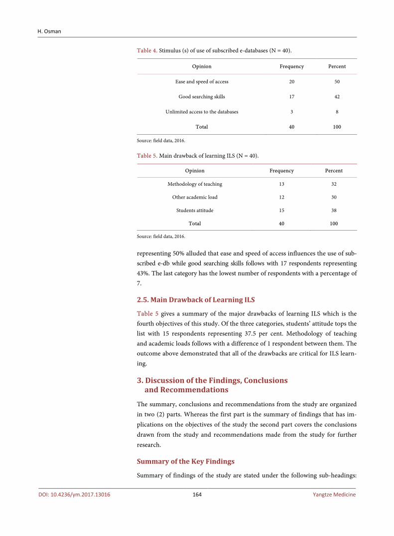
H. Osman
DOI: 10.4236/ym.2017.13016 164 Yangtze Medicine
Table 4. Stimulus (s) of use of subscribed e-databases (N = 40).
Opinion Frequency Percent
Ease and speed of access 20 50
Good searching skills 17 42
Unlimited access to the databases 3 8
Total 40 100
Source: field data, 2016.
Table 5. Main drawback of learning ILS (N = 40).
Opinion Frequency Percent
Methodology of teaching 13 32
Other academic load 12 30
Students attitude 15 38
Total 40 100
Source: field data, 2016.
representing 50% alluded that ease and speed of access influences the use of sub-scribed e-db while good searching skills follows with 17 respondents representing 43%. The last category has the lowest number of respondents with a percentage of 7.
2.5. Main Drawback of Learning ILS
Table 5 gives a summary of the major drawbacks of learning ILS which is the fourth objectives of this study. Of the three categories, students’ attitude tops the list with 15 respondents representing 37.5 per cent. Methodology of teaching and academic loads follows with a difference of 1 respondent between them. The outcome above demonstrated that all of the drawbacks are critical for ILS learn-ing.
3. Discussion of the Findings, Conclusions and Recommendations
The summary, conclusions and recommendations from the study are organized in two (2) parts. Whereas the first part is the summary of findings that has im-plications on the objectives of the study the second part covers the conclusions drawn from the study and recommendations made from the study for further research.
Summary of the Key Findings
Summary of findings of the study are stated under the following sub-headings:
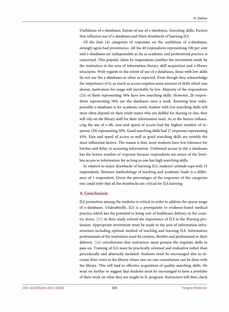
H. Osman
DOI: 10.4236/ym.2017.13016 165 Yangtze Medicine
Usefulness of e-databases, Extent of use of e-databases, Searching skills, Factors that influence use of e-databases and Main drawbacks of learning ILS.
Of the four (4) categories of responses on the usefulness of e-databases, strongly agree had prominence. All the 40 respondents representing 100 per cent said e-databases are indispensable as far as academic and professional practice is concerned. This popular claim by respondents justifies the investment made by the institution in the area of information literacy skill acquisition and e-library structures. With regards to the extent of use of e-databases, those with low skills do not use the e-databases as often as expected. Even though they acknowledge the importance of it, as much as access requires some amount of skills which was absent, motivation for usage will inevitably be low. Majority of the respondents (23) of them representing 58% have low searching skills. However, 20 respon-dents representing 50% use the databases once a week. Knowing how indis-pensable e-databases is for academic work, student with low searching skills will most often depend on their study mates who are skillful for sharing or else, they will rely on the library staff for their information need. As to the factors influen-cing the use of e-db, ease and speed of access had the highest number of re-sponse (20) representing 50%. Good searching skills had 17 responses representing 43%. Ease and speed of access as well as good searching skills are notably the most influential factors. The reason is that, most students have low tolerance for hitches and delay in accessing information. Unlimited access to the e-databases has the lowest number of response because respondents are aware of the limit-less access to information for as long as one has high searching skills.
In relation to major drawbacks of learning ILS, students’ attitude tops with 15 respondents. Between methodology of teaching and academic loads is a differ-ence of 1 respondent. Given the percentages of the responses of the categories one could infer that all the drawbacks are critical for ILS learning.
4. Conclusion
ILS promotion among the students is critical in order to address the sparse usage of e-databases. Undoubtedly, ILS is a prerequisite to evidence-based medical practice which has the potential to bring cost of healthcare delivery in the coun-try down. [23] in their study echoed the importance of ILS to the Nursing pro-fession. Appropriate investment must be made in the area of information infra-structure including optimal method of teaching and learning ILS. Information professionals of the institution must be creative, flexible and professional in their delivery. [24] corroborates that instructors must possess the requisite skills to pass on. Training of ILS must be practically oriented and evaluative rather than procedurally and abstractly modeled. Students must be encouraged also to in-crease their visits to the library where one-on-one consultation can be done with the library. This will lead to effective acquisition of quality searching skills. He went on further to suggest that students must be encouraged to have a portfolio of their work on what they are taught in IL program. Instructors will then check
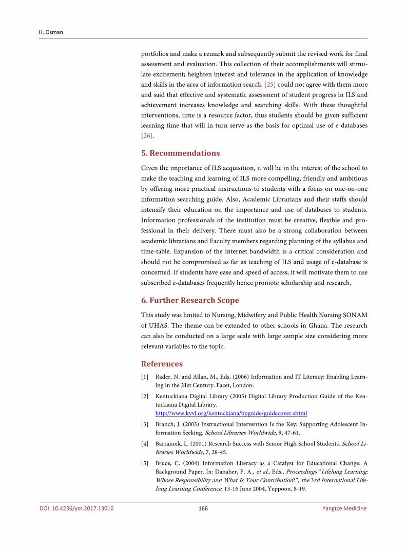
H. Osman
DOI: 10.4236/ym.2017.13016 166 Yangtze Medicine
portfolios and make a remark and subsequently submit the revised work for final assessment and evaluation. This collection of their accomplishments will stimu-late excitement; heighten interest and tolerance in the application of knowledge and skills in the area of information search. [25] could not agree with them more and said that effective and systematic assessment of student progress in ILS and achievement increases knowledge and searching skills. With these thoughtful interventions, time is a resource factor, thus students should be given sufficient learning time that will in turn serve as the basis for optimal use of e-databases [26].
5. Recommendations
Given the importance of ILS acquisition, it will be in the interest of the school to make the teaching and learning of ILS more compelling, friendly and ambitious by offering more practical instructions to students with a focus on one-on-one information searching guide. Also, Academic Librarians and their staffs should intensify their education on the importance and use of databases to students. Information professionals of the institution must be creative, flexible and pro-fessional in their delivery. There must also be a strong collaboration between academic librarians and Faculty members regarding planning of the syllabus and time-table. Expansion of the internet bandwidth is a critical consideration and should not be compromised as far as teaching of ILS and usage of e-database is concerned. If students have ease and speed of access, it will motivate them to use subscribed e-databases frequently hence promote scholarship and research.
6. Further Research Scope
This study was limited to Nursing, Midwifery and Public Health Nursing SONAM of UHAS. The theme can be extended to other schools in Ghana. The research can also be conducted on a large scale with large sample size considering more relevant variables to the topic.
References [1] Rader, N. and Allan, M., Eds. (2006) Information and IT Literacy: Enabling Learn-
ing in the 21st Century. Facet, London.
[2] Kentuckiana Digital Library (2005) Digital Library Production Guide of the Ken-tuckiana Digital Library. http://www.kyvl.org/kentuckiana/bpguide/guidecover.shtml
[3] Branch, J. (2003) Instructional Intervention Is the Key: Supporting Adolescent In-formation Seeking. School Libraries Worldwide, 9, 47-61.
[4] Barranoik, L. (2001) Research Success with Senior High School Students. School Li-braries Worldwide, 7, 28-45.
[5] Bruce, C. (2004) Information Literacy as a Catalyst for Educational Change: A Background Paper. In: Danaher, P. A., et al., Eds., Proceedings “Lifelong Learning: Whose Responsibility and What Is Your Contribution?”, the 3rd International Life-long Learning Conference, 13-16 June 2004, Yeppoon, 8-19.
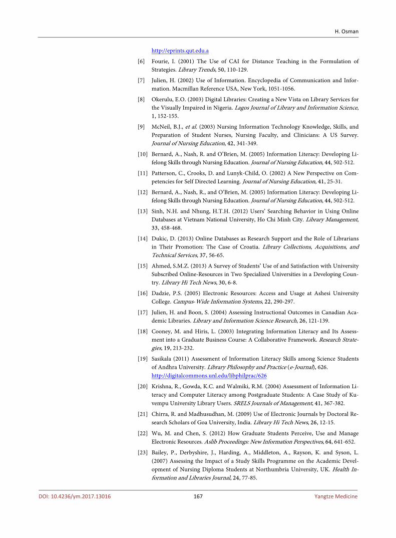
H. Osman
DOI: 10.4236/ym.2017.13016 167 Yangtze Medicine
http://eprints.qut.edu.a
[6] Fourie, I. (2001) The Use of CAI for Distance Teaching in the Formulation of Strategies. Library Trends, 50, 110-129.
[7] Julien, H. (2002) Use of Information. Encyclopedia of Communication and Infor-mation. Macmillan Reference USA, New York, 1051-1056.
[8] Okerulu, E.O. (2003) Digital Libraries: Creating a New Vista on Library Services for the Visually Impaired in Nigeria. Lagos Journal of Library and Information Science, 1, 152-155.
[9] McNeil, B.J., et al. (2003) Nursing Information Technology Knowledge, Skills, and Preparation of Student Nurses, Nursing Faculty, and Clinicians: A US Survey. Journal of Nursing Education, 42, 341-349.
[10] Bernard, A., Nash, R. and O’Brien, M. (2005) Information Literacy: Developing Li-felong Skills through Nursing Education. Journal of Nursing Education, 44, 502-512.
[11] Patterson, C., Crooks, D. and Lunyk-Child, O. (2002) A New Perspective on Com-petencies for Self Directed Learning. Journal of Nursing Education, 41, 25-31.
[12] Bernard, A., Nash, R., and O’Brien, M. (2005) Information Literacy: Developing Li-felong Skills through Nursing Education. Journal of Nursing Education, 44, 502-512.
[13] Sinh, N.H. and Nhung, H.T.H. (2012) Users’ Searching Behavior in Using Online Databases at Vietnam National University, Ho Chi Minh City. Library Management, 33, 458-468.
[14] Dukic, D. (2013) Online Databases as Research Support and the Role of Librarians in Their Promotion: The Case of Croatia. Library Collections, Acquisitions, and Technical Services, 37, 56-65.
[15] Ahmed, S.M.Z. (2013) A Survey of Students’ Use of and Satisfaction with University Subscribed Online-Resources in Two Specialized Universities in a Developing Coun-try. Library Hi Tech News, 30, 6-8.
[16] Dadzie, P.S. (2005) Electronic Resources: Access and Usage at Ashesi University College. Campus-Wide Information Systems, 22, 290-297.
[17] Julien, H. and Boon, S. (2004) Assessing Instructional Outcomes in Canadian Aca-demic Libraries. Library and Information Science Research, 26, 121-139.
[18] Cooney, M. and Hiris, L. (2003) Integrating Information Literacy and Its Assess-ment into a Graduate Business Course: A Collaborative Framework. Research Strate-gies, 19, 213-232.
[19] Sasikala (2011) Assessment of Information Literacy Skills among Science Students of Andhra University. Library Philosophy and Practice (e-Journal), 626. http://digitalcommons.unl.edu/libphilprac/626
[20] Krishna, R., Gowda, K.C. and Walmiki, R.M. (2004) Assessment of Information Li-teracy and Computer Literacy among Postgraduate Students: A Case Study of Ku-vempu University Library Users. SRELS Journals of Management, 41, 367-382.
[21] Chirra, R. and Madhusudhan, M. (2009) Use of Electronic Journals by Doctoral Re-search Scholars of Goa University, India. Library Hi Tech News, 26, 12-15.
[22] Wu, M. and Chen, S. (2012) How Graduate Students Perceive, Use and Manage Electronic Resources. Aslib Proceedings: New Information Perspectives, 64, 641-652.
[23] Bailey, P., Derbyshire, J., Harding, A., Middleton, A., Rayson, K. and Syson, L. (2007) Assessing the Impact of a Study Skills Programme on the Academic Devel-opment of Nursing Diploma Students at Northumbria University, UK. Health In-formation and Libraries Journal, 24, 77-85.
H. Osman
DOI: 10.4236/ym.2017.13016 168 Yangtze Medicine
[24] Edem, M.B. (2005) Library Acquisitions of Indigenous Law Textbooks and Its Uti-lization in Selected Federal Universities as Some Factors Influencing Indigenous Law Textbooks Publishing in Nigeria. Post Field Seminar. Department of LARIS.
[25] Eisenberg, M., Lowe, A. and Spitzer, K. (2004) Information: Essential Skills for the Information Age. Conn: Library Unlimited, Westport.
[26] Cheney, D. (1991) Evaluation-Based Training: Improving the Quality of End-User Searching. Journal of Academic Librarianship, 17, 152-155.
Submit or recommend next manuscript to SCIRP and we will provide best service for you:
Accepting pre-submission inquiries through Email, Facebook, LinkedIn, Twitter, etc. A wide selection of journals (inclusive of 9 subjects, more than 200 journals) Providing 24-hour high-quality service User-friendly online submission system Fair and swift peer-review system Efficient typesetting and proofreading procedure Display of the result of downloads and visits, as well as the number of cited articles Maximum dissemination of your research work
Submit your manuscript at: http://papersubmission.scirp.org/ Or contact [email protected]
Yangtze Medicine, 2017, 1, 169-177 http://www.scirp.org/journal/ym
ISSN Online: 2475-7349 ISSN Print: 2475-7330
DOI: 10.4236/ym.2017.13017 Sep. 21, 2017 169 Yangtze Medicine
Logistic Regression Analysis the Risk Factors of Peripherally Inserted Central Catheter Related Blood Stream Infection of Tumor Patients
Jian Song1, Yan Yan2, Huang Yan2, Chunlin Wang2, Jun-e Hu2*
1School of Medicine, Yangtze University, Jing Zhou, China 2Department of Oncology, Jing Zhou Central Hospital, Jing Zhou, China
Abstract Objective: Our object is to study risk factors of tumor patients’ PICC catheter- related blood stream infection. Method: a retrospective analysis of data of 586 PICC catheterized patients was implemented, a univariate analysis of general data and catheterizing data of tumor patients was then carried out, and data of single factors with statistical significance were incorporated into multi-factor Logistic regression model for analysis. Results: PICC catheter-related blood stream infection occurred to 16 patients, and occurrence rate was 2.73%. Multi- factor Logistic regression analysis results showed that number of puncturing times, positioning method and maintenance frequency were risk factors for tumor patients’ peripherally inserted central catheter catheter-related blood stream infection, and odds risk values were respectively 8.762, 9.253 and 10.324. Conclusion: for tumor patients implanted with peripherally inserted central catheters, using ECG positioning during strict sterile operation and cathete-rizing process to avoid repeated puncturing and increasing maintenance fre-quency could effectively reduce occurrence of PICC catheter-related blood stream infection.
Keywords PICC Related Blood Stream Infection, Logistic Regression Analysis, Risk Factor
1. Introduction
By virtue of advantages like once implantation with long-term utilization, few com-plications, possible bedside operation, reduction and prevention of risks caused by drug extravasation, etc., peripherally inserted central catheter (PICC) cathete-
How to cite this paper: Song, J., Yan, Y., Yan, H., Wang, C.L. and Hu, J.-E. (2017) Paper Title. Yangtze Medicine, 1, 169-177. https://doi.org/10.4236/ym.2017.13017 Received: July 17, 2017 Accepted: September 18, 2017 Published: September 21, 2017 Copyright © 2017 by authors and Scientific Research Publishing Inc. This work is licensed under the Creative Commons Attribution International License (CC BY 4.0). http://creativecommons.org/licenses/by/4.0/
Open Access

J. Song et al.
DOI: 10.4236/ym.2017.13017 170 Yangtze Medicine
rization has been clinically applied within a large scope [1]. With extensive ap-plication of PICC catheter, PICC catheter-related blood stream infection has been gradually recognized. PICC catheter-related blood stream infection (PICC-CRBSI) refers to that patients, who carry intravascular catheters or whose intravascular catheters are removed within 48 hours, suffer from bacteremia or fungemia ac-companied by infectious manifestations like fever (>38˚C) and shivering and there is no any other infection source except intravascular catheter [2]. PICC-CRBSI, an important complication of long-term retention of vein catheterization, will leng-then length of stay (LOS) of patients and cause waste of medical resources in less severe case once it happens, and more seriously, it will cause patient death and medical dispute. Research shows that CRBSI occurrence rate of PICC patients is 12.7% - 32.7%, infection rates of Gram-positive bacteria and Gram-negative bacteria are respectively 38.8% - 64.4% and 35.56% - 41.2%, fungal infection rate is 0% - 20.0% and average death rate among infected patients is 12% - 25% [3] [4]. Therefore, it’s of great importance to actively explore into risk factors caus-ing PICC-CRBSI. A retrospective analysis of 586 patients carrying PICC cathe-ters was implemented in this paper and occurrence rate of PICC-CRBSI to tu-mor patients and its relevant risk factors were discussed so as to provide basis for clinical prevention of tumor patients’ PICC catheter-related blood stream infection.
2 Data and Method 2.1. Research Object
PICC-catheterized patients in central venous indwelling catheter registration da-tabase in one of the level-three class-A hospital from February in 2016 to February in 2017. During this time, a total of 756 patients were applied with a PICC ca-theterization. For the research purposes, we set up inclusion and exclusion crite-ria, and inclusion criteria were: 1) more than 18 years old; 2) diagnosed as ma-lignant tumor for the first time through histopathology; 3) implanted with tri-valve single-cavity high voltage-resistant PICC catheters by clinical nurse spe-cialists; 4) PICC catheterizing time lasted at least 3 months. Exclusion criteria: 1) patients with preexistence of catheter-related thrombosis, deep venous thrombo-sis, catheter-related blood stream infection or lymphatic backflow obstruction; 2) Those who don’t have obtain information such as: death, abandon treatment and con-tact lost were deleted. Diagnostic criteria of PICC catheter-related blood stream in-fection were subjected to Prevention and Treatment Guidelines of Intravascular Catheter-Related Infection (2007) [5]. Because this study is a retrospective study, so no patients required to sign informed consent. Under the approval of the Hos-pital Academic Ethics Committee authors found out the medical clinical records, finally 586 patients meeting the criteria were selected, where 16 ones suffered from PICC catheter-related blood stream infection, the occurrence rate was 2.73%. However this occurrence rate was lower than the previous reported 12% - 25% of the incidence [3] [4].

J. Song et al.
DOI: 10.4236/ym.2017.13017 171 Yangtze Medicine
2.2. Methodology 2.2.1. Observational Indexes General Data (Gender, age, occupation, degree of education, pathological type, clinical staging, complications, operation history and antibacterial agent application) and cathe-terizing data (number of puncturing times, positioning method, punctured limbs, catheterizing time, maintenance frequency and maintenance plane) of tu-mor patients.
2.2.2. Data Collection Method Through unified training, 2 postgraduates and PICCC clinical nurse specialists looked up and recorded general data and catheterizing data of research objects who met criteria according to incorporation & exclusion criteria and medical record number.
2.2.3. Statistical Method SPSS 19.0 software was used for statistical analysis, measured data were ex-pressed by x s± , enumeration data were expressed by percentages, and com-parison of means of two samples was implemented through t test. χ2 test was used for comparison of enumeration data. Logistic regression analysis of dichotom-ous outcome variables was used for multi-factor analysis.
3 Results 3.1. Occurrence of PICC Catheter-Related Blood Stream Infection
16 cases among 586 patients suffered from PICC catheter-related blood stream infection, therefore occurrence rate was 2.73%: which was lower than what’s re-ported in past researches [2] [3], and this might be related to the fact that only 16 patients infected with typical clinical manifestations like fever (>38˚C) and shivering were observed in this study but screening of patients without any symp-tom was not implemented.
3.2. Univariate Analysis of Influence on Occurrence of PICC Catheter-Related Blood Stream Infection (Table 1)
Univariate analysis results showed that the differences of PICC catheter-related blood stream infection from pathological type, complications, number of punc-turing times, positioning method, maintenance frequency and maintenance place had statistical significance (P < 0.05), but differences of ICC catheter-related blood stream infection from gender, age, occupation, degree of education, clini-cal staging, operation history, use of antibacterial agents, punctured limbs and catheterizing time didn’t have statistical significance (P > 0.05).
3.3. Multi-Factor Analysis of Influence on Occurrence of PICC Catheter-Related Blood Stream Infection (Table 2 and Table 3)
Whether PICC-CRBSI happened was taken as a dependent variable (dependent va-riable: patients without occurrence of PICC catheter-related blood stream infection
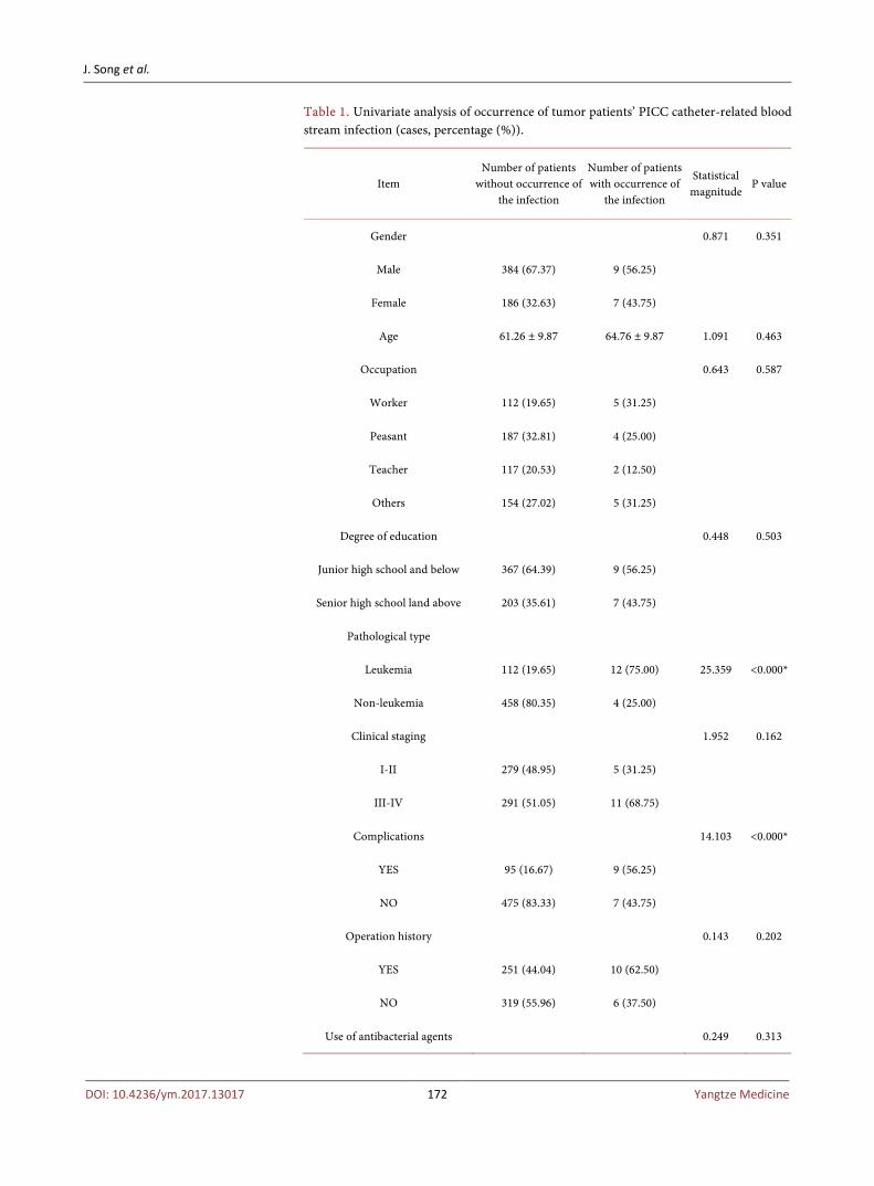
J. Song et al.
DOI: 10.4236/ym.2017.13017 172 Yangtze Medicine
Table 1. Univariate analysis of occurrence of tumor patients’ PICC catheter-related blood stream infection (cases, percentage (%)).
Item Number of patients
without occurrence of the infection
Number of patients with occurrence of
the infection
Statistical magnitude
P value
Gender 0.871 0.351
Male 384 (67.37) 9 (56.25)
Female 186 (32.63) 7 (43.75)
Age 61.26 ± 9.87 64.76 ± 9.87 1.091 0.463
Occupation 0.643 0.587
Worker 112 (19.65) 5 (31.25)
Peasant 187 (32.81) 4 (25.00)
Teacher 117 (20.53) 2 (12.50)
Others 154 (27.02) 5 (31.25)
Degree of education 0.448 0.503
Junior high school and below 367 (64.39) 9 (56.25)
Senior high school land above 203 (35.61) 7 (43.75)
Pathological type
Leukemia 112 (19.65) 12 (75.00) 25.359 <0.000*
Non-leukemia 458 (80.35) 4 (25.00)
Clinical staging 1.952 0.162
I-II 279 (48.95) 5 (31.25)
III-IV 291 (51.05) 11 (68.75)
Complications 14.103 <0.000*
YES 95 (16.67) 9 (56.25)
NO 475 (83.33) 7 (43.75)
Operation history 0.143 0.202
YES 251 (44.04) 10 (62.50)
NO 319 (55.96) 6 (37.50)
Use of antibacterial agents 0.249 0.313
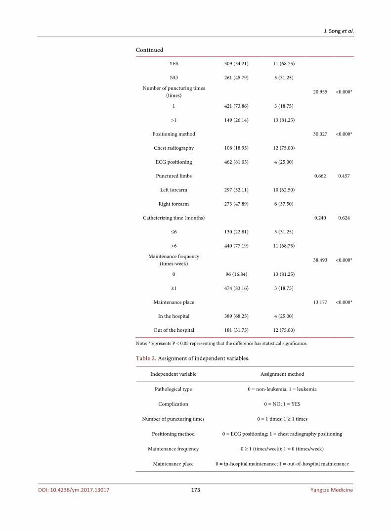
J. Song et al.
DOI: 10.4236/ym.2017.13017 173 Yangtze Medicine
Continued
YES 309 (54.21) 11 (68.75)
NO 261 (45.79) 5 (31.25)
Number of puncturing times (times)
20.955 <0.000*
1 421 (73.86) 3 (18.75)
>1 149 (26.14) 13 (81.25)
Positioning method 30.027 <0.000*
Chest radiography 108 (18.95) 12 (75.00)
ECG positioning 462 (81.05) 4 (25.00)
Punctured limbs 0.662 0.457
Left forearm 297 (52.11) 10 (62.50)
Right forearm 273 (47.89) 6 (37.50)
Catheterizing time (months) 0.240 0.624
≤6 130 (22.81) 5 (31.25)
>6 440 (77.19) 11 (68.75)
Maintenance frequency (times-week)
38.493 <0.000*
0 96 (16.84) 13 (81.25)
≥1 474 (83.16) 3 (18.75)
Maintenance place 13.177 <0.000*
In the hospital 389 (68.25) 4 (25.00)
Out of the hospital 181 (31.75) 12 (75.00)
Note: *represents P < 0.05 representing that the difference has statistical significance.
Table 2. Assignment of independent variables.
Independent variable Assignment method
Pathological type 0 = non-leukemia; 1 = leukemia
Complication 0 = NO; 1 = YES
Number of puncturing times 0 = 1 times; 1 ≥ 1 times
Positioning method 0 = ECG positioning; 1 = chest radiography positioning
Maintenance frequency 0 ≥ 1 (times/week); 1 = 0 (times/week)
Maintenance place 0 = in-hospital maintenance; 1 = out-of-hospital maintenance

J. Song et al.
DOI: 10.4236/ym.2017.13017 174 Yangtze Medicine
Table 3. Multi-factor logistic regression of tumor patients concurrent with PICC ca-theter-related blood stream infection.
Independent variable B SE P OR 95%CI
Constant term 13.42 4.652 <0.001 <0.001 4.652 - 23.632
Number of puncturing times 3.782 0.746 <0.001 8.762 4.053 - 23.685
Positioning method 6.322 0.674 <0.001 9.253 5.693 - 32.157
Maintenance frequency 5.736 0.324 0.003 10.324 2.329 - 24.956
= 0, and patients with occurrence of PICC catheter-related blood stream infec-tion = 1) and variables with statistical significance (P < 0.05) in univariate analy-sis were incorporated into Logistic regression mode, and results showed that number of puncturing times, positioning method and maintenance frequency were risk factors of tumor patients with concurrence of PICC catheter-related blood stream infection. Odds risk values of multiple times of puncturing, chest radiography positioning and low maintenance frequency (≤1 times/week) were respectively 8.762, 9.253 and 10.324.
4. Discussion 4.1. Occurrence of PICC Catheter-Related Blood Stream Infection
to Tumor Patients
The study results showed that occurrence rate of symptomatic PICC-CRBSI was 2.73%, which was similar to what’s reported by many of the other scholars. 16 patients with occurrence of PICC-CRDSI in this study experienced obvious clin-ical symptoms, but PICC-CRBSI without delete typical clinical symptoms might exist in practical work, The occurrence rate of PICC-CRBSI might be far higher than existing reports, which might be related to the fact that high fever occurring in chemoradiotherapy of tumor patients was usually deemed as tumor-related fever without consideration of PICC-CRBSI. This indicated that medical per-sonnel should blindly use antibiotics when faced with high fever of tumor pa-tients carrying catheters not simply considering it as tumor-related fever, but in-stead, comprehensively considering whether patients had risks of PICC-CRBSI and make etiological quantitative examination when necessary. Antibiotics should be reasonably used according to etiological examination results or optimal in-tervention measures should be selected according to standard treatment process of PICC-CRBSI to ensure patient safety.
4.2. Repeated Puncturing Is a Risk Factor of PICC-CRBSI Occurrence
The study results indicated that repeated puncturing was a risk factor of PICC-CRBSI occurrence with similar finding to past studies, all of which believed that the more times of puncturing during catheterizing process, the higher the risks of in-ducing PICC-CRBSI would be [6] [7]. The risk of PICC-CRBSI occurrence when
J. Song et al.
DOI: 10.4236/ym.2017.13017 175 Yangtze Medicine
number of puncturing times was greater than 1 was 8.762 times of 1 successful puncturing, possibly because repeated puncturing, difficult catheterizing and ca-theter misplacement during catheterizing process would result in damage of blood vessel wall and subcutaneous tissues, lengthened tissue repair time and increased possibility for bacterial invasion to cause infection, and moreover, catheter may be polluted before entering human body due to lengthened operation time and too long exposure of sterile articles. Therefore, administrative department should enhance qualification access system of PICC catheterizing nurses and ensure that PICC specialized nurses had excellent puncturing technology. Meanwhile, “blind puncturing” should be avoided during catheterizing process, catheterizing suc-cess rate was improved by 98% with B-ultrasound guidance combining improved Seldinger technique [8]. After three times of unsuccessful puncturing, puncturing might not be implemented, or otherwise complications would present fold in-crease [9].
4.3. X-Ray Chest Radiograph Positioning Is a Risk Factor of PICC-CRBSI Occurrence
Study results found that PICC-CRBSI risk of X-ray chest radiography position-ing was 9.253 times of ECG positioning. To reach the effect of once implantation with long-term use, the position of the head of PICC is of vital importance. No-wadays most hospitals in China still take X-ray chest radiograph positioning as the only “gold standard” but its disadvantages have gradually been highlighted with disciplinary progress, The following X-ray chest radiograph and may be used only after catheterization. If PICC catheter head has malposition, then it’s necessary to readjust the catheter or re-implant the catheter after removal. Dur-ing the process, the catheter exposed in the air has been polluted so that it’s dif-ficult to achieve strict sterilization during catheter debugging process, conse-quently bacteria adhered onto the catheter will possibly cause bacterial infection [10], and bacterial infection is the most important original inducing factor of PICC-CRBSI. Hence, in future work, ECG positioning method should be widely applied to positioning of PICC catheter head, and advantages of ECG which can timely observe catheter position with integrated catheterizing and positioning should be taken fully.
4.4. Irregular Maintenance Is a Risk Factor of PICC-CRBSI Occurrence
Risk of PICC-CRBSI occurrence due to irregular maintenance in this study was 10.324 times of regular maintenance. As a new venous treatment means, PICC has been widely carried out in superior hospitals and have penetrated into pri-mary hospitals gradually. At present, most county-level hospitals and all healthy clinics in towns lack personnel mastering PICC maintenance technique due to disadvantages in aspects of hardware and personnel. Consequently, regular main-tenance can’t be achieved during therapeutic intermission of many patients so

J. Song et al.
DOI: 10.4236/ym.2017.13017 176 Yangtze Medicine
that loose herbal application, bacterial reproduction and infection are then caused. Most research objects selected in this study came from rural areas, great difficul-ties existed in maintenance per week because of factors like their economic sta-tus, traffic condition and scattered regional distribution. Many patients chose self-care at home, which not achieve sterilization and increased infection risk [11] [12] [13]. Irregular maintenance namely maintenance frequency which was low-er than 1 time/week was a risk factor of PICC-CRBSI occurrence. Therefore, we should establish PICC maintenance network centered on regional medical treat-ment and covering peripheral counties and cities, strengthen training of medical care personnel at basic level, so as to improve maintenance level of nursing staff at basic level, patients compliance with PICC maintenance and reduce occurrence rate of PICC infection due to medical care personnel during treatment intermis-sion.
5. Limitation
Only a retrospective analysis of 586 PICC catheterized patients was made in this study. Logistic regression model was used to analyze risk factors of patients with PICC-CRBSI, but in practical work, many risk factors exist before, in the middle of and after catheterization. No final conclusion has been formed for many po-tential risk factors and relative risk degree of each risk factor has not been fur-ther verified. Therefore, large-sample case-control study should be carried out in future study to accurately explore into risk factors of PICC-CRBSI so as to real-ize early-stage recognition of risk factors and judge possibility of CRBSI occur-rence. Intervention should be made in key link to ensure patient safety.
References [1] Shi, Y., Zheng, Y.P., Li, Y., et al. (2017) Application of Simultaneous Single-Hand
Double-Cavity Catheter Locking in Prevention of PICC Catheter Plugging. Chinese Journal of Nursing Science, 52, 621-623.
[2] Li, R.Q., Jiang, D.Q. and Lv, Y.J. (2016) Observation and Nursing of Different Pa-thogenic Bacteria-Induced PICC Catheter-Related Blood Stream Infection. Chinese Journal of Nursing Science, 51, 1368-1370.
[3] Xu, X.F., Zhao, X.H. and Wu, L.H. (2015) Comparative Study of Application of Different Infusion Connectors to CVC-Related Blood Stream Infection. Chinese Journal of Nosocomial Infection, 25, 2025-2027.
[4] Zheng, X., Liu, W., Liu, X.M., et al. (2012) Analysis of CVC-Related Pathogenic Bacterial Distribution of 520 Tumor Patients Peripherally Implanted with Cathe-ters. Modern Diagnosis and Treatment, 23, 1554-1555.
[5] Fang, Q. (2008) Prevention and Treatment Guidelines for Intravascular Cathe-ter-Related Infection. Chinese Journal of Practical Surgery, 17, 597-605.
[6] Gao, A.Y. (2012) Cause Analysis and Intervention Measures of Vein Catheteriza-tion-Related Blood Stream Infection. Chinese Journal of Nosocomial Infection, 22, 3758-3759.
[7] Jiang, W. and Zeng, D.F. (2015) Research Progress of Risk Factors and Preventive Measures of PICC-Related Blood Infection. Chinese Nursing Management, 15, 218-221.

J. Song et al.
DOI: 10.4236/ym.2017.13017 177 Yangtze Medicine
[8] Johnson, M.A., Mckenzie, L., Tussey, S., et al. (2009) Portable Ultrasound: A Cost-Effective Process Improvement Tool for PICC Placement. Nursing Manage-ment, 40, 47-50. https://doi.org/10.1097/01.NUMA.0000343984.23505.37
[9] Song, P. and Li, Z.D. (1999) Enlightenment of CVC Complications. Journal of Prac-tical Nursing, 15, 7-8.
[10] Yuan, L., Li, R.M., Li, S.P., et al. (2015) Effect Comparison of Two Methods on Tri-valve PICC-Guided Intracardiac Electrocardiogram. Chinese Journal of Nursing Science, 50, 1055-1059.
[11] Luo, H., Ren, D.Q., Tan, J., et al. (2013) Construction and Implementation of Hos-pital-Community-Family Integrated PICC Maintenance Network Pattern. Chinese Journal of Nursing Science, 28, 16-18.
[12] Lin, N. and Wu, A.Z. (2015) Prevalence Survey of Website Maintenance of Dis-charged Patients Carrying Catheters. Modern Nurse Periodical (Every Ten Days), No. 6, 88-89.
[13] Wu, Y., Hu, Q.X., Hu, S.R., et al. (2015) Construction of PICC Maintenance Net-work and Nursing Practice. Sichuan Medical Journal, 36, 1183-1187.
Submit or recommend next manuscript to SCIRP and we will provide best service for you:
Accepting pre-submission inquiries through Email, Facebook, LinkedIn, Twitter, etc. A wide selection of journals (inclusive of 9 subjects, more than 200 journals) Providing 24-hour high-quality service User-friendly online submission system Fair and swift peer-review system Efficient typesetting and proofreading procedure Display of the result of downloads and visits, as well as the number of cited articles Maximum dissemination of your research work
Submit your manuscript at: http://papersubmission.scirp.org/ Or contact [email protected]

Yangtze Medicine, 2017, 1, 178-188 http://www.scirp.org/journal/ym
ISSN Online: 2475-7349 ISSN Print: 2475-7330
DOI: 10.4236/ym.2017.13018 Sep. 27, 2017 178 Yangtze Medicine
Prevalence of Coinfection with Malaria and HIV among Children in Yaoundé, Cameroon: A Cross-Sectional Survey Performed in Three Communities in Yaoundé
Tebit E. Kwenti1,2,3, Emilienne Edo2, Besong S. Ayuk3, Tayong D. B. Kwenti1
1Department of Medical Laboratory Sciences, University of Buea, Buea, Cameroon 2Department of Microbiology and Parasitology, University of Buea, Buea, Cameroon 3Regional Hospital of Buea, Buea, Cameroon
Abstract Background: Malaria and HIV are endemic in Cameroon. But data on the prevalence of coinfection with malaria and HIV in Cameroonian children are essentially absent. This study was aimed at determining the prevalence of coinfection with malaria and HIV among children in Yaoundé, so as to advice control policies. Methods: In a cross-sectional survey, children (≤15 years) were recruited from 3 communities in Yaoundé namely: Efoulan, Biyem-assi and Cité-verte. A semi-structured questionnaire was used to collect demo-graphic data. Participants were screened for malaria parasites by the examina-tion of Giemsa-stained blood films meanwhile participants were screened for HIV following Cameroon’s national algorithm. The Pearson’s chi-square test was performed as part of the statistical analyses. Statistical significance was set at p < 0.05. Result: Three hundred and ten (310) children took part in the study. The mean age (±SD) of the participants was 75.64 (±63.23) months and a majority of them were males (56.1%). The prevalence was 19.7%, 4.8% and 1.2% for malaria, HIV, and coinfection with malaria and HIV respectively. The prevalence of malaria was associated with age (p = 0.009) meanwhile the prevalence of HIV was associated with study site (p = 0.024). Plasmodium fal-ciparum was the only species identified as causing malaria in the target popu-lation. Conclusion: A substantial prevalence of malaria, HIV and coinfection with malaria and HIV was observed in this study. Efforts should be streng-thened to control and eventually eliminate these diseases in the target popula-tion.
How to cite this paper: Kwenti, T.E., Edo, E., Ayuk, B.S. and Kwenti, T.D.B. (2017) Prevalence of Coinfection with Malaria and HIV among Children in Yaoundé, Came-roon: A Cross-Sectional Survey Performed in Three Communities in Yaoundé. Yangtze Medicine, 1, 178-188. https://doi.org/10.4236/ym.2017.13018 Received: July 28, 2017 Accepted: September 24, 2017 Published: September 27, 2017 Copyright © 2017 by authors and Scientific Research Publishing Inc. This work is licensed under the Creative Commons Attribution International License (CC BY 4.0). http://creativecommons.org/licenses/by/4.0/
Open Access

T. E. Kwenti et al.
DOI: 10.4236/ym.2017.13018 179 Yangtze Medicine
Keywords Malaria, HIV, Coinfection, Plasmodium falciparum, Prevalence, Children, Yaoundé, Cameroon
1. Introduction
Malaria is a mosquito-borne infectious disease affecting humans and other ani-mals. Globally there were an estimated 212 million cases of malaria and 429,000 deaths attributed to malaria in 2015 [1]. The incidence rate of malaria is estimated to have decreased by 41% globally between 2000 and 2015, meanwhile the mala-ria mortality rates have declined by 62% within the same timeframe [1]. The ma-jority of cases and deaths attributed to malaria occur in sub-Saharan Africa (SSA) [1]. Although there has been a decline in malaria recently, malaria still remains a significant cause of morbidity and mortality in SSA, claiming the live of a child every 2 minutes [2]. In Cameroon, malaria is a major cause of morbidity and mortality among children [3] [4] [5]. Malaria is caused by parasitic protozoans of the genus Plasmodium. Five species are known to cause disease in humans namely; Plasmodium ovale, P. malariae, P. knowlesi, P. vivax, and P. falciparum, with the latter being the most virulent species accounting for the majority of cases and deaths attributed to malaria. Like in other SSA countries, P. falciparum is the predominant species in Cameroon [4] [6] [7].
SSA also has the highest burden of the human immunodeficiency virus (HIV). In 2014, there were 25.8 million people living with HIV/AIDS (PLWHA) in SSA [8], accounting for approximately 70% of the global total. In Cameroon the overall prevalence of HIV is estimated at 4.5% [9]. There are about 39,000 child-ren (<14 years) living with HIV in Cameroon and about 310,000 children or-phaned due to AIDS [9]. HIV in Cameroon affects typically the poor and less privileged [10].
Because of the presence of all the factors favouring transmission in SSA in-cluding poverty, malaria and HIV are common in the region. Due to their over-lapping distribution, coinfection with malaria and HIV is therefore bound to be common in the area. Coinfection with malaria and HIV is thought to have a syn-ergistic effect, with studies reporting that repeated infection with malaria leads to a more rapid decline in CD4+ T cells overtime, meanwhile malaria coinfection with HIV results in more episodes of symptomatic malaria [11], and more epi-sodes of severe or complicated malaria including death in both children and adults [12] [13] [14] [15] [16]. The risk of severe anaemia is also higher in HIV patients coinfected with malaria compared to HIV patients without malaria [17] [18]. In Cameroon, the prevalence of coinfection with malaria and HIV among adolescence and adults has been reported to range between 2.24% to 29.4% [15] [16] [19]. But studies reporting the prevalence of coinfection with malaria and HIV in children are very limited in the country.

T. E. Kwenti et al.
DOI: 10.4236/ym.2017.13018 180 Yangtze Medicine
This study was therefore designed to determine the prevalence of coinfection with malaria and HIV among children in Yaoundé, in order to generate data for clinico-epidemiological purposes which will improve on the control of both dis-eases in the country.
2. Materials and Methods 2.1. Study Area
This study was performed in Yaoundé in the Centre region (Figure 1). Yaoundé (3˚52'N 11˚31'E) with an average elevation of 750 m, is the capital of Cameroon. With a population of roughly 2.5 million, Yaoundé is second only to Douala as the largest city in Cameroon [20]. Yaoundé is a very diverse city with people from different works of life and is home to most of the administrative structures in the country. The climate of Yaoundé is tropical with 2 rainy (March to June, Sep-tember to November) and 2 dry seasons (December to February, July-August). Malaria transmission in Yaoundé is holoendemic and seasonal with Anopheles gambiae as the principle vector [21]. According to hospital records, peak malaria transmission occurs at the beginning of the rainy seasons. The prevalence of malaria in the general population of Yaoundé is estimated at 35% [22] mean-while the prevalence of HIV is estimated at 8.3% [10].
2.2. Study Design and Setting
This was a cross-sectional study performed between May and June 2017, involv-ing children randomly selected from 3 communities in Yaoundé.
Figure 1. Map of Yaoundé. The study sites are delineated.

T. E. Kwenti et al.
DOI: 10.4236/ym.2017.13018 181 Yangtze Medicine
2.3. Sample Size Estimation
The sample size was estimated using the formula for sample size calculation de-scribed by Swinscow [23] as follows;
( )2
2
1Z p pn
e× −
=
Z = 1.96 p = prevalence of malaria in children in Cameroon = 17.6% [7]. e = error rate = 0.05
( )2
2
1.96 0.176 1 0.1760.0
22 95
2 .n×
=−
=
Thus we recruited 310 participants to adjust for possible loss of samples.
2.4. Sampling Technique
Two stage sampling was done. In the first stage, 3 communities were randomly selected in Yaoundé including: Efoulan, Biyem-assi, and Cité-verte (Figure 1). In the second stage, houses in the communities were randomly selected and children aged 15 years and below within selected houses were enrolled.
2.5. Study Population
Children (≤15 years) of both sexes were eligible to participate in the study. The participants were recruited from 3 communities in Yaoundé namely: Efoulan, Biyem-assi and Cité-verte. Written informed consent was obtained from the parents or guidance of the children after explaining to them the study protocol and objectives. Excluded from the study were children not residing in the se-lected communities as well as those on any antimalarial drug 2 weeks prior to the study commencing.
2.6. Ethical Consideration
Authorization to carry out this research was obtained from the Faculty of Health Sciences, University of Buea, and from the Delegation of Public Health, Yaoundé, Center region. Written informed consent was obtained from all participants prior to their inclusion.
2.7. Data Collection
A semi-structured questionnaire was used to collect demographic characteristics (age, gender, etc.). The questionnaire was administered to the parents or guid-ance of the children by members of the research team.
2.8. Sample Collection
About 3 ml of blood was collected from the children into EDTA anticoagulated tubes following antiseptic techniques. The blood was used to perform the com-plete blood count, preparation of blood films as well as screening for HIV.
T. E. Kwenti et al.
DOI: 10.4236/ym.2017.13018 182 Yangtze Medicine
2.9. Laboratory Analysis 2.9.1. Performance of Complete Blood Count (CBC) CBC was performed using the Mindray® Auto haematology analyzer (BC-2800, Shenzhen Mindray Bio-Medical Electronics Co., Ltd.). The white blood cell counts were obtained from the CBC results and used in the estimation of the parasite density.
2.9.2. Detection of Malaria Parasite The prepared blood films were air-dried and stained with 10% Giemsa (1 in 20 dilutions) for 25 - 30 minutes [24]. The blood films were read by two expert mi-croscopists who were blinded from the results of the other. In the case of any discrepancy with the results obtained by the two microscopists, a third was brought in and the results he gave were considered as final. At least 200 fields were screened for malaria parasite using the 100X (oil immersion) objective and where parasites were seen, they were counted until 500 WBC were reached. The slides were only declared negative after counting to 2500 WBC. Malaria parasite density was estimated by dividing the parasites counted by 500 WBC and then multiplied by the actual WBC count of the participant to give numbers in para-site per μl [7].
2.9.3. Screening for HIV HIV screening was done in accordance with the Cameroon’s national algorithm for HIV screening by detecting anti-HIV antibodies [25]. Briefly, a first line rapid test was used and if positive, a second line test was used to confirm the result as well as determine the HIV type. Where the first line test was positive and the second line test was negative, a third line test was brought in. In this study, the first line test used was Determine™ HIV (Abbott Laboratories, Abbott Park, IL, USA), the second line test was First Response® (Kachigam, India) and the third line test was Immuno-Comb® (Orgenics Ltd., Israel).
2.10. Statistical Analyses
Data collected was entered into Excel spreadsheet and analyzed using Stata® ver-sion 12.1 (StataCorp LP) statistical package and group comparisons were per-formed using the Pearson’s Chi-square test. Statistical significance was set at p ≤ 0.05.
3. Results
Three hundred and ten (310) participants successfully took part in the study. Their ages ranged between 0 and 180 months, with mean (±SD) = 75.64 (±63.23) months. Among them were 136 (43.9%) females and 174 (56.1%) males (Table 1).
Among the 310 participants, 61 were positive for malaria giving a prevalence of 19.7% (95% CI: 15.4 - 24.6). Malaria prevalence was associated with age (p = 0.01) but not with gender (p = 0.427) nor study site (p = 0.337) (Table 2). Plas-
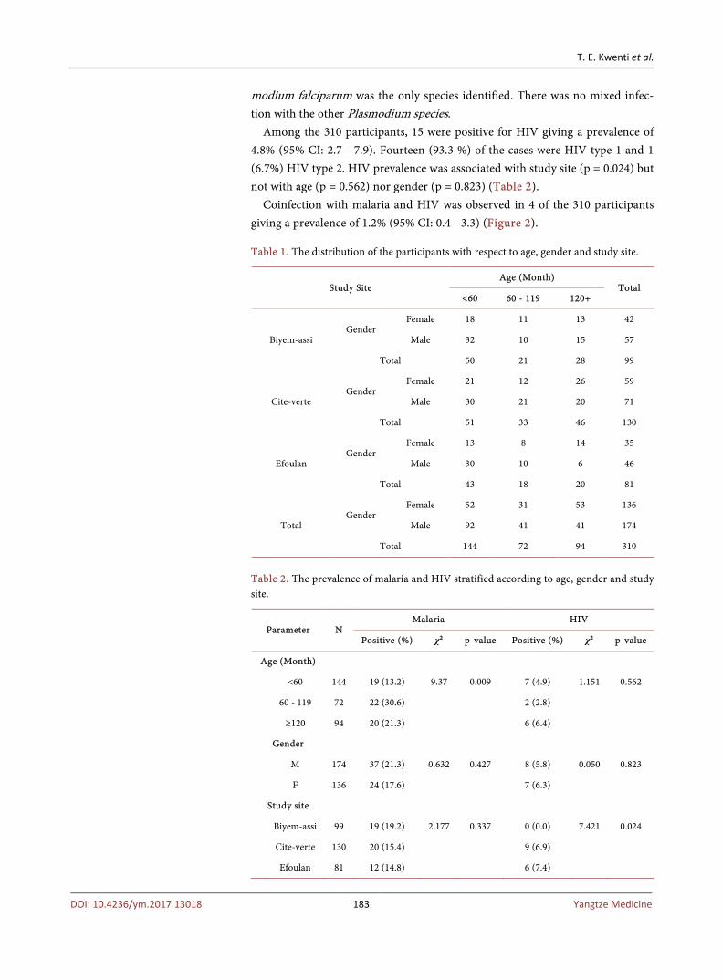
T. E. Kwenti et al.
DOI: 10.4236/ym.2017.13018 183 Yangtze Medicine
modium falciparum was the only species identified. There was no mixed infec-tion with the other Plasmodium species.
Among the 310 participants, 15 were positive for HIV giving a prevalence of 4.8% (95% CI: 2.7 - 7.9). Fourteen (93.3 %) of the cases were HIV type 1 and 1 (6.7%) HIV type 2. HIV prevalence was associated with study site (p = 0.024) but not with age (p = 0.562) nor gender (p = 0.823) (Table 2).
Coinfection with malaria and HIV was observed in 4 of the 310 participants giving a prevalence of 1.2% (95% CI: 0.4 - 3.3) (Figure 2).
Table 1. The distribution of the participants with respect to age, gender and study site.
Study Site Age (Month)
Total <60 60 - 119 120+
Biyem-assi Gender
Female 18 11 13 42
Male 32 10 15 57
Total 50 21 28 99
Cite-verte Gender
Female 21 12 26 59
Male 30 21 20 71
Total 51 33 46 130
Efoulan Gender
Female 13 8 14 35
Male 30 10 6 46
Total 43 18 20 81
Total Gender
Female 52 31 53 136
Male 92 41 41 174
Total 144 72 94 310
Table 2. The prevalence of malaria and HIV stratified according to age, gender and study site.
Parameter N Malaria HIV
Positive (%) χ² p-value Positive (%) χ² p-value
Age (Month)
<60 144 19 (13.2) 9.37 0.009 7 (4.9) 1.151 0.562
60 - 119 72 22 (30.6) 2 (2.8)
≥120 94 20 (21.3) 6 (6.4)
Gender
M 174 37 (21.3) 0.632 0.427 8 (5.8) 0.050 0.823
F 136 24 (17.6) 7 (6.3)
Study site
Biyem-assi 99 19 (19.2) 2.177 0.337 0 (0.0) 7.421 0.024
Cite-verte 130 20 (15.4) 9 (6.9)
Efoulan 81 12 (14.8) 6 (7.4)
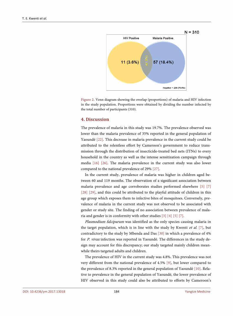
T. E. Kwenti et al.
DOI: 10.4236/ym.2017.13018 184 Yangtze Medicine
Figure 2. Venn diagram showing the overlap (proportions) of malaria and HIV infection in the study population. Proportions were obtained by dividing the number infected by the total number of participants (310).
4. Discussion
The prevalence of malaria in this study was 19.7%. The prevalence observed was lower than the malaria prevalence of 35% reported in the general population of Yaoundé [22]. This decrease in malaria prevalence in the current study could be attributed to the relentless effort by Cameroon’s government to reduce trans-mission through the distribution of insecticide-treated bed nets (ITNs) to every household in the country as well as the intense sensitization campaign through media [16] [26]. The malaria prevalence in the current study was also lower compared to the national prevalence of 29% [27].
In the current study, prevalence of malaria was higher in children aged be-tween 60 and 119 months. The observation of a significant association between malaria prevalence and age corroborates studies performed elsewhere [5] [7] [28] [29], and this could be attributed to the playful attitude of children in this age group which exposes them to infective bites of mosquitoes. Conversely, pre-valence of malaria in the current study was not observed to be associated with gender or study site. The finding of no association between prevalence of mala-ria and gender is in conformity with other studies [3] [4] [5] [7].
Plasmodium falciparum was identified as the only species causing malaria in the target population, which is in line with the study by Kwenti et al. [7], but contradictory to the study by Mbenda and Das [30] in which a prevalence of 4% for P. vivax infection was reported in Yaoundé. The differences in the study de-sign may account for this discrepancy; our study targeted mainly children mean-while theirs targeted adults and children.
The prevalence of HIV in the current study was 4.8%. This prevalence was not very different from the national prevalence of 4.5% [9], but lower compared to the prevalence of 8.3% reported in the general population of Yaoundé [10]. Rela-tive to prevalence in the general population of Yaoundé, the lower prevalence of HIV observed in this study could also be attributed to efforts by Cameroon’s
T. E. Kwenti et al.
DOI: 10.4236/ym.2017.13018 185 Yangtze Medicine
government to control the disease largely through programs to prevent moth-er-to-child transmission as well as regular sensitization campaigns. There was an association between prevalence of HIV and study site in the current study, being highest in Efoulan (7.4%). An immediate explanation for this observation was not imminent. However, HIV prevalence in Cameroon has been reported to vary from one location to another and is influenced by the socio-cultural characteris-tics of the different populations. There was no association between HIV preva-lence and age or gender in the current study.
The prevalence of coinfection with malaria and HIV observed in the current study was 1.2%. This finding is similar to the prevalence of 2.24% reported in Bamenda in the Northwest Region [19]. However, the prevalence of coinfection with malaria and HIV was lower than the 7.3% and 29.4% reported by Njunda et al. [16] and Nkuo-Akenji et al. [15] respectively. The difference in the prevalence of coinfection reported in these studies and ours could be attributed to differences in the study designs; our study targeted children in the community meanwhile theirs targeted known HIV patients recruited from HIV treatment facilities.
This study revealed the prevalence of malaria, HIV, and coinfection of malaria with HIV among children in 3 communities in Yaoundé. It has generated data that may be useful in designing control policies. The study is however limited in that participants were recruited from only 3 communities in Yaoundé and the findings may not be generalizable to the entire population of children in Yaoundé. Larger studies will therefore be required in the study area to give a clearer pic-ture.
5. Conclusion
In the current study, a prevalence of 19.7%, 4.8%, and 1.2% was observed for malaria, HIV, and coinfection with malaria and HIV respectively. The preva-lence of malaria was observed to be associated with age meanwhile the preva-lence of HIV was associated with study site, being highest in Efoulan. Plasmo-dium falciparum was the only species identified as the cause of malaria in the target population. Efforts should be strengthened to control and eventually elimi-nate malaria and HIV in children in the study area.
Acknowledgements
Our sincere gratitude goes to all the children who voluntarily took part in this study.
Competing Interests
The authors declare that they have no competing interests.
Authors’ Contributions
This work was carried out in collaboration between all authors. TEK conceived the study; participated in its design, coordination and data collection; took part
T. E. Kwenti et al.
DOI: 10.4236/ym.2017.13018 186 Yangtze Medicine
in the analyses and interpretation; conducted literature search and review; per-formed the statistical analysis and co-wrote the first draft. EE and BSA partici-pated in the data collection, took part in the analyses and interpretation, con-ducted the literature search and review and co-wrote the first draft. TDBK con-ceived, designed and coordinated the study; participated in the statistical analy-sis; and critically revised the manuscript. All authors read and approved the final manuscript.
References [1] World Health Organisation (2016) World Malaria Report 2016. World Health Or-
ganisation, Geneva. http://apps.who.int/iris/bitstream/10665/252038/1/9789241511711-eng.pdf?ua=1
[2] World Health Organisation (2015) World Malaria Report 2015. World Health Or-ganisation, Geneva. http://apps.who.int/iris/bitstream/10665/200018/1/9789241565158_eng.pdf?ua=1
[3] Njunda, A.L., Fon, S.G., Assob, J.C.N., Nsagha, D.S., Kwenti, T.D.B. and Kwenti, E.T. (2015) Malaria and Intestinal Parasitic Coinfection and Their Contribution to Anaemia in Children in Cameroon. Infectious Diseases of Poverty, 4, 43. https://doi.org/10.1186/s40249-015-0078-5
[4] Kwenti, T.E., Nkume, F.A., Tanjeko, A.T. and Kwenti, T.D.B. (2016) The Effect of Intestinal Parasitic Infection on the Clinical Outcome of Malaria in Coinfected Children in Cameroon. PLoS Neglected Tropical Diseases, 10, Article ID: e0004673. https://doi.org/10.1371/journal.pntd.0004673
[5] Kwenti, T.E., Kwenti, T.D.B., Latz, A., Njunda, L.A. and Nkuo-Akenji, T. (2017) Epidemiological and Clinical Profile of Paediatric Malaria: A Cross Sectional Study Performed on Febrile Children in Five Epidemiological Strata of Malaria in Came-roon. BMC Infectious Diseases, 17, 499. https://doi.org/10.1186/s12879-017-2587-2
[6] World Health Organisation (2010) Cameroon: Epidemiological Profile. World Ma-laria Report. WHO, Geneva.
[7] Kwenti, T.E., Kwenti, T.D.B., Njunda, L.A., Latz, A., Tufon, K.A. and Nkuo-Akenji, T. (2017) Identification of the Plasmodium Species in Clinical Samples from Child-ren Residing in Five Epidemiological Strata of Malaria in Cameroon. Tropical Med-icine and Health, 45, 14. https://doi.org/10.1186/s41182-017-0058-5
[8] UNAIDS (2015a) Fact Sheet 2015. UNAIDS, Geneva. http://www.unaids.org/sites/default/files/media_asset/20150901_FactSheet_2015_en.pdf
[9] UNAIDS (2015b) Cameroon: Epidemiological Fact Sheet on HIV and AIDS. UNAIDS, Geneva. http://www.unaids.org/en/regionscountries/countries/cameroon/
[10] Kwenti, T.E., Nsagha, D.S., Kwenti, B.D.T. and Njunda, A.L. (2014) Sexual Risk Behaviours among People Living with HIV and Implications for Control in the Northwest Region of Cameroon. World Journal of AIDS, 4, 198-205. https://doi.org/10.4236/wja.2014.42025
[11] Kamya, M.R., Gasasira, A.F., Yeka, A., Bakyaita, N., Nsobya, S.L., Francis, D., et al. (2006) Effect of HIV-1 Infection on Antimalarial Treatment Outcomes in Uganda: A Population-Based Study. Journal of Infectious Disease, 193, 9-15. https://doi.org/10.1086/498577

T. E. Kwenti et al.
DOI: 10.4236/ym.2017.13018 187 Yangtze Medicine
[12] Grimwade, K., French, N., Mbatha, D.D., Zungu, D.D., De-Dicoat, M. and Gilks, C.F. (2004) HIV Infection as a Cofactor for Severe Falciparum Malaria in Adults Living in a Region of Unstable Malaria Transmission in South Africa. AIDS, 18, 547-554. https://doi.org/10.1097/00002030-200402200-00023
[13] Cohen, C., Karstaedt, A., Frean, J., Thomas, J., Govender, N., Prentice, E., et al. (2005) Increased Prevalence of Severe Malaria in HIV-Infected Adults in South Africa. Clinical Infection Disease, 41, 1631-1637. https://doi.org/10.1086/498023
[14] Otieno, R.O., Ouma, C., Ong’echa, J.M., Keller, C.C., Were, T., Waindi, E.N., et al. (2006) Increased Severe Anemia in HIV-1-Exposed and HIV-1-Positive Infants and Children during Acute Malaria. AIDS, 20, 275-280. https://doi.org/10.1097/01.aids.0000200533.56490.b7
[15] Nkuo-Akenji, T., Tevoufouet, E.M., Nzang, F., Ngufor, N. and Fon, E. (2008) High Prevalence of HIV and Malaria Co-Infection in Urban Douala, Cameroon. African Journal of AIDS Research, 7, 229-235. https://doi.org/10.2989/AJAR.2008.7.2.8.525
[16] Njunda, A.L., Njumkeng, C., Nsagha, S.D., Assob, J.C.N. and Kwenti, E.T. (2016) The Prevalence of Malaria in People Living with HIV in Yaounde, Cameroon. BMC Public Health, 16, 964. https://doi.org/10.1186/s12889-016-3647-z
[17] Saracino, A., Nacarapa, E.A., da Costa, M.E.A., Martinelli, D., Scacchetti, M., de Oliveira, C., et al. (2012) Prevalence and Clinical Features of HIV and Malaria Coinfection in Hospitalized Adults in Beira, Mozambique. Malaria Journal, 11, 241. https://doi.org/10.1186/1475-2875-11-241
[18] Tay, S.C.K., Badu, K., Mensah, A.A. and Gbedema, S.Y. (2015) The Prevalence of Malaria among HIV Seropositive Individuals and the Impact of the Co-Infection on Their Hemoglobin Levels. Annals of Clinical Microbiology and Antimicrobials, 14, 10. https://doi.org/10.1186/s12941-015-0064-6
[19] Njunda, L.A., Kamga, H.L.F., Nsagha, D.S., Assob, J.C.N. and Kwenti, T.E. (2012) Low Malaria Prevalence in HIV-Positive Patients in Bamenda, Cameroon. Journal of Microbiology Research, 2, 56-59. https://doi.org/10.5923/j.microbiology.20120203.03
[20] World Gazetteer (2013) Cameroon: Largest Cities and Towns and Statistics of Their Population.
[21] Craig, M., Snow, R. and le Sueur, D. (1999) A Climate-Based Distribution Model of Malaria Transmission in Sub-Saharan Africa. Parasitology Today, 15, 105-111. https://doi.org/10.1016/S0169-4758(99)01396-4
[22] van der Kolk, M., Etti T.A., Nimpaye, H., Ngo, N.D., Sauerwein, R. and Eling, W. (2003) Transmission of Plasmodium falciparum in Urban Yaoundé Cameroon Is Seasonal and Age-Dependent. Transactions of the Royal Society of Tropical Medi-cine and Hygiene, 97, 375-379. https://doi.org/10.1016/S0035-9203(03)90059-9
[23] Swinscow, T.D.V. and Campbell, M.J. (2002) Statistics at Square. 10th Edition, BMJ Books, London.
[24] Njunda, A.L., Assob, N.J.C., Nsagha, S.D., Kamga, F.H.L., Mokenyu, M.D. and Kwenti, E.T. (2013) Comparison of Capillary and Venous Blood Using Blood Film Microscopy in the Detection of Malaria Parasites: A Hospital Based Study. Scientific Journal of Microbiology, 2, 89-94.
[25] Sumbele, I.U.N., Ning, T.R., Bopda, O.S.M. and Nkuo-Akenji, T. (2014) Variation in Malariometric and Red Cell Indices in Children in the Mount Cameroon Area Following Enhanced Malaria Control Measures: Evidence from a Repeated Cross- Sectional Study. Malaria Journal, 13, 334. https://doi.org/10.1186/1475-2875-13-334

T. E. Kwenti et al.
DOI: 10.4236/ym.2017.13018 188 Yangtze Medicine
[26] Kwenti, E.T., Njouom, R., Njunda, L.A. and Kamga, H.L.F. (2011) Comparison of an Immunochromatographic Rapid Strip Test, ELISA and PCR in the Diagnosis of Hepatitis C in HIV Patients in Hospital Settings in Cameroon. Clinical Medicine and Diagnostics, 1, 21-27. https://doi.org/10.5923/j.cmd.20110101.04
[27] Mangham, L.J., Cundill, B., Achonduh, O.A., Ambebila, J.N., Lele, A.K., Metoh, T.N., et al. (2012) Malaria Prevalence and Treatment of Febrile Patients at Health Facilities and Medicine Retailers in Cameroon. Tropical Medicine and International Health, 17, 330-342.
[28] Degarege, A., Legesse, M., Medhin, G., Animut, A. and Erko, B. (2012) Malaria and Related Outcomes in Patients with Intestinal Helminths: A Cross-Sectional Study. BMC Infectious Disease, 12, 291. https://doi.org/10.1186/1471-2334-12-291
[29] Alemu, A., Shiferaw, Y., Ambachew, A. and Hamid, H. (2012) Malaria Helminth Co-Infections and Their Contribution for Anaemia in Febrile Patients Attending Azzezo Health Center, Gondar, Northwest Ethiopia: A Cross Sectional Study. Asian Pacific Journal Tropical Medicine, 5, 803-809. https://doi.org/10.1016/S1995-7645(12)60147-3
[30] Ngassa, M.H.G. and Das, A. (2014) Molecular Evidence of Plasmodium Vivax Mono and Mixed Malaria Parasite Infections in Duffy-Negative Native Cameroo-nians. PLoS One, 9, e103262. https://doi.org/10.1371/journal.pone.0103262
Submit or recommend next manuscript to SCIRP and we will provide best service for you:
Accepting pre-submission inquiries through Email, Facebook, LinkedIn, Twitter, etc. A wide selection of journals (inclusive of 9 subjects, more than 200 journals) Providing 24-hour high-quality service User-friendly online submission system Fair and swift peer-review system Efficient typesetting and proofreading procedure Display of the result of downloads and visits, as well as the number of cited articles Maximum dissemination of your research work
Submit your manuscript at: http://papersubmission.scirp.org/ Or contact [email protected]


![A Case of Benign Meningioma Presented with Subdural Hemorrhage · Meningioma with Subdural Hemorrhage Martínez-Lage et al. [4] studied 57 cases of meningioma with hemorrhagic onset](https://img.pdfslide.net/doc/110x75/5eca99262fcc5c7ee06897d3/a-case-of-benign-meningioma-presented-with-subdural-hemorrhage-meningioma-with-subdural.jpg)


![Case_G1-G8[1] cases chapter 10 Managerial accounting](https://img.pdfslide.net/doc/110x75/5695d1881a28ab9b0296e6cd/caseg1-g81-cases-chapter-10-managerial-accounting.jpg)