Embed Size (px)
Citation preview
To obtain CME credit for this activity, go to http://cme.ufl .edu/ed/self-study/toai/ Topics in OCULAR ANTIINFLAMMATORIES 1Supported by an unrestricted educational grant from Shire.
Managing In� ammation in Patients with Serious Allergic Conditions of the Ocular Sur faceJAY S. PEPOSE, MD, PhD The two most serious allergic conditions of the ocular surface are vernal keratoconjunctivitis (VKC) and atopic keratoconjunctivitis (AKC) due to their tissue remodelling and sight-threatening potential. Although both conditions may be managed using mast cell-stabilizing, antihistamine, or corticosteroid agents—with varying degrees of success depending upon disease severity—new agents capable of modifying the allergic response in a more targeted manner are needed.
Allergic conditions of the ocular service include several clinically diverse conditions with diff erent forms of patho-genesis, hypersensitivity mechanisms, and diagnostic criteria, although an individual allergic condition of the ocular surface is characterized by a single or dominant presentation of a local, allergic sensitization.1 Ocular allergies are a common problem, with approximately 15% to 20% of the global population being aff ected by some form of allergic disease. Of those aff ected, it is estimated that 40% to 60% have ocular symptoms, which may progress to have a negative impact on patient quality of life.2-4
Ocular allergy encompasses the two acute disorders of seasonal allergic conjunctivitis (SAC) and perennial allergic conjunctivitis (PAC), as well as the more serious chronic conditions of vernal keratoconjunctivitis (VKC) and atopic keratoconjunctivitis (AKC). All of these conditions are char-
acterized by early acute phase infl ammation of the ocular surface resulting in itching, tearing, conjunctival and lid edema-redness and photophobia. In some individuals, this may progress to the late phase responses of eosinophilia and neutrophilia. Chronic disease, which can develop in some patients, is associated with tissue remodeling of the ocular surface and, in severe cases, sustained damage.5 Ocular al-lergy is oft en associated with other allergic conditions, and thus optimal management may require the involvement of both an ophthalmologist and an allergist.
DIAGNOSING SERIOUS OCULAR ALLERGYAn accurate medical history and a clinical examination are
fundamental to determining if the patient has acute or chronic allergic conjunctivitis. VKC is a condition most commonly observed in hot, dry environments such as West Africa, parts of India, and Mexico.6 In the US, it mostly occurs during the warmer months of spring and summer in children and young adults. Typically observed more frequently in males than females (at a ration of 3:1), VKC usually resolves at puberty. Th e reasons for this variance in incidence with age and geo-graphical region remain unclear. Symptoms include intense itching, tearing, and photophobia but may progress to mucous
ISSUE 18
A CONTINUINGMEDICAL EDUCATION
PUBLICATIONCME
CONTINUING MEDICAL EDUCATION
See INSIDE for:Infl ammation Control in Corticosteroid Respondersby Ronald M. Caronia, MD, FACS
To obtain CME credit for this activity, go to http://cme.ufl .edu/ed/self-study/toai/2 Topics in OCULAR ANTIINFLAMMATORIES
STATEMENT OF NEEDThe control of ocular infl ammation is a critical aspect of medical and surgical ophthalmic practice. Despite their side eff ects, antiinfl ammatory drugs are used to treat a very wide range of conditions throughout the eye, from ocular surface disease and allergic conjunctivitis to poste-rior segment conditions. Use of antiinfl ammatory agents is also critical in ocular surgery, contributing greatly to patient comfort and positive outcomes.The ocular antiinfl ammatory landscape is changing as research reveals more about the role of infl ammation in a range of ocular conditions and as new antiinfl ammatory agents enter the market.1,2 Twenty years ago, for example, the idea of using a topical corticosteroid to treat dry eye and/or allergic conjunctivitis was viewed with alarm; today, it is accepted practice. Although corticosteroids and nonsteroidal antiinfl am-matory drugs (NSAIDs) have been the mainstays of the ocular anti-infl ammatory armamentarium, a number of new agents with novel mechanisms of action (and new ocular drug delivery systems) have come to market or are being made ready for market.3,4
As indications expand and change, and as new drugs, formulations, and delivery systems become available, clinicians require up-to-date protocols for drug selec-tion and use. Such protocols are also needed for routine (but nevertheless off -label) uses of corticosteroids and NSAIDs because important diff erences in effi cacy, safety, and tolerability exist between these classes and among formulations within each of these classes.5,6
By putting the latest published evidence into the context of current clinical practice, Topics in Ocular Antiinfl amma-tories equips ophthalmologists to maintain competen-cies and narrow gaps between their actual and optimal infl ammation management practices, across the range of clinical situations in which current and novel ocular antiinfl ammatories may be used.
REFERENCES 1. Song JS, Hyon JY, Lee D, et al. Current practice pattern
for dry eye patients in South Korea: a multicenter study. Korean Journal of Ophthalmology. 2014;28(2):115-21.
2. Ciulla TA, Harris A, McIntyre N, Jonescu-Cuypers C. Treat-ment of diabetic macular edema with sustained-release glucocorticoids: intravitreal triamcinolone acetonide, dexamethasone implant, and fl uocinolone acetonide implant. Expert Opin Pharmacother. 2014;15(7):953-9.
3. Maya JR, Sadiq MA, Zapata LJ, et al. Emerging therapies for noninfectious uveitis: what may be coming to the clinics. J Ophthalmol. 2014;2014:310329.
4. Sheppard JD, Torkildsen GL, Lonsdale JD, et al, and the OPUS-1 Study Group. Lifi tegrast ophthalmic solu-tion 5.0% for treatment of dry eye disease: results of the OPUS-1 phase 3 study. Ophthalmology. 2014 Feb;121(2):475-83.
5. Fong R, Leitritz M, Siou-Mermet R, Erb T. Loteprednol etabonate gel 0.5% for postoperative pain and infl am-mation after cataract surgery: results of a multicenter trial. Clin Ophthalmol. 2012;6:1113-24.
6. Singer M, Cid MD, Luth J, et al. Incidence of corneal melt in clinical practice: our experience vs a meta-analysis of the literature. Clin Exp Ophthalmol. 2012;S1:003.
OFF-LABEL USE STATEMENTThis work may discuss off -label uses of medications.
GENERAL INFORMATIONThis CME activity is sponsored by the University of Florida College of Medicine and is supported by an unrestricted educational grant from Shire.The University of Florida College of Medicine designates this activity for a maximum of 1 AMA PRA Category 1 Credit™. There is no fee to participate in this activity. In order to receive CME credit, participants should read the report, and then take the posttest. A score of 80% is required to qualify for CME credit. Estimated time to complete the activity is 60 minutes. On completion, take the test online at http://cme.ufl .edu/ed/self-study/toai/System requirements for this activity are: For PC us-ers: Windows® 2000, XP, 2003 Server, or Vista; Internet Explorer® 6.0 or newer, or Mozilla® Firefox® 2.0 or newer
(JavaScript™ and Java™ enabled). For Mac® users: Mac OS® X 10.4 (Tiger®) or newer; Safari™ 3.0 or newer, Mozilla® Firefox® 2.0 or newer; (JavaScript™ and Java™ enabled). Internet connection required: Cable modem, DSL, or better.
DATE OF ORIGINAL RELEASE November 2017. Ap-proved for a period of 12 months.
ACCREDITATION STATEMENTThis activity has been planned and implemented in accordance with the accreditation requirements and policies of the Accreditation Council for Continuing Medical Education through the joint providership of the University of Florida College of Medicine and Candeo Clinical/Science Communications, LLC. The University of Florida College of Medicine is accredited by the ACCME to provide continuing medical education for physicians.
CREDIT DESIGNATION STATEMENTThe University of Florida College of Medicine designates this enduring material for a maximum of 1 AMA PRA Cate gory 1 Credit™. Physicians should claim only the credit commensurate with the extent of their participa-tion in the activity.
EDITORIAL BOARD / FACULTY ADVISORSMarguerite B. McDonald, MD, FACS, practices at Ophthalmic Consultants of Long Island, and is a clinical professor of ophthalmology at the New York University School of Medicine. She is also an adjunct clinical profes-sor of ophthalmology at Tulane University Health Sciences Center. She’s a consultant to Allergan, Alcon, Abbott Medi-cal Optics, Bausch + Lomb, FOCUS Laboratories, Shire, OCuSOFT, Altaire, Bio-Tissue, BlephEx, Oculus USA, and Optical Express.Victor L. Perez, MD, is a professor of ophthalmology at the Duke University School of Medicine. He is also the director of Duke Eye Center's Ocular Immunology Cen-ter and Ocular Surface Program. He has received grant/research support from the National Institutes of Health, and is a consultant for Allergan, EyeGate, and Shire. He is also a stock shareholder for EyeGate.Matthew J. Gray, MD, is an assistant professor in the department of ophthalmology at the University of Florida College of Medicine. He states that in the past 12 months, he has not had a fi nancial relationship with any commercial organization that produces, markets, resells, or distributes healthcare goods or services consumed by or used on patients relevant to this manuscript.Ronald M. Caronia, MD, FACS, is a partner at Ophthalmic Consultants of Long Island and assistant clinical professor of ophthalmology at the Albert Einstein School of Medi-cine. He states that in the past 12 months, he has not had a fi nancial relationship with any commercial organization that produces, markets, resells, or distributes healthcare goods or services consumed by or used on patients relevant to this manuscript.Jay S. Pepose, MD, PhD, is the director of Pepose Vision Institute and professor of clinical ophthalmology at Washington University School of Medicine in St. Louis, Missouri. He is a consultant for Shire and Sun Pharma-ceutical Industries Ltd. Medical writer Denise Campbell, PhD, of Markey Medical Consulting Pty Ltd, assisted in the preparation of this manuscript.
DISCLAIMERParticipants have an implied responsibility to use the new-ly acquired information to enhance patient outcomes and professional development. The information presented in this activity is not meant to serve as a guideline for patient care. Procedures, medications, and other courses of diag-nosis and treatment discussed or suggested in this activity should not be used by clinicians without evaluation of their patients’ conditions and possible contraindications or dangers in use, applicable manufacturer’s product information, and comparison with recommendations of other authorities.
COMMERCIAL SUPPORTERS This activity is supported by an unrestricted educational grant from Shire.
TOPICS IN OCULAR ANTIINFLAMMATORIES, ISSUE 18discharge and, in some instances, reac-tive ptosis and blepharospasm. The tarsal form of VKC commonly involves the upper tarsus with the development of cobblestone papillae.1 Th rough exces-sive rubbing against the upper lid, these papillae may result in the development of shield ulcers and plaques on the cor-nea. Th e limbal form of VKC is seen in subtropical countries and characteristi-cally has transient, multiple conjunctival yellow-grey infi ltrates with overlaying white deposits (Horn0er-Trantas dots) and papillae at the limbus.1
AKC occurs equally between the sexes. Onset occurs in young adults and continues through to the fi ft h decade of life, with a peak incidence between the ages of 30 to 50 years. Th is chronic con-dition, which can develop at any time of the year, is characterized by persistent infl ammation involving the eyelids, the conjunctiva, and sometimes the cornea. Th e lower tarsus is oft en involved with patients showing cicatrization (this may be so severe as to be associated with entropion or ectropion) and persistent epithelial defects that have vision-threat-ening potential. Other features may include madarosis or trichiasis, kerato-conus and/or subcapsular cataract, and peripheral neovascularization. Patients often present with atopic dermatitis and/or asthma.6 Interestingly, AKC is a risk factor for developing herpes sim-plex keratitis, which typically presents unilaterally while in patients with AKC oft en presents bilaterally.
INFLAMMATORY MECHANISMSTh e mechanisms underlying severe
ocular allergy are complex and involve the upregulation of multiple pro-infl am-matory mediators that cause breakdown of the corneal epithelial cell barrier. In the case of VKC, IgE-mediated mecha-nisms are involved in approximately 50% of patients.7 Increased numbers of CD4+ Th 2 cells, costimulatory mol-ecules, and multiple chemokines and cytokines all act synergistically to en-hance ocular surface production of IgE,8 leading to corneal impact and tissue remodelling.9,10 Potent T-cell-mediated responses are also observed for VKC in association with a massive infi ltration
To obtain CME credit for this activity, go to http://cme.ufl .edu/ed/self-study/toai/ Topics in OCULAR ANTIINFLAMMATORIES 3
of macrophages, neutrophils, and eosinophils, the latter of which plays an important role in producing eosinophilic-derived factors (notably eosinophil cationic protein) that are cytotoxic to the corneal epithelium.1 Th e involvement of matrix metalloproteinases MMP-1 and MMP-9 also contrib-utes to the enzymatic degradation of the corneal basement membrane and stroma. In many patients, non-IgE-mediated activation pathways are present, confi rming the complexity of this condition.6
AKC is associated with alterations of the mucous com-ponent of the tear film and leads to mucous discharge, epithelial disease, dry eye signs/symptoms, and reduced tear fi lm stability.11 It is thought that mechanisms linked to the activation of innate immunity receptors play a key role in the chronic infl ammatory response that occurs.12 In the case of AKC, specifi c allergen(s) are likely involved, and an accurate medical history and allergy testing is advised to identify allergen(s) to which the patient is sensitized. Both a genetic predisposition to atopy and environmental allergens/irritants may be implicated.6 Although 45% of patients do not have any specifi c sensitization, high serum IgE levels and polysensitization are oft en found.13 In addition, it is believed that Th 1- and Th 2-lymphocyte-mediated mechanisms may be involved in AKC.14,15
MANAGING OCULAR INFLAMMATIONManagement of ocular allergy typically includes allergen
avoidance, an air-conditioned environment, pharmacological treatment, immunotherapy, and patient education. Th e use of artifi cial tears may enhance the function of the ocular surface barrier, and cold compresses oft en give relief from symptoms.1
With AKC, treatment options include topical ophthalmic drops (mast cell stabilizers, antihistamines, corticosteroids, calcineurin inhibitors), topical ointments (ophthalmic corti-costeroids, calcineurin inhibitors), and systemic medications (antihistamines, corticosteroids, calcineurin inhibitors).16
Topical mast cell stabilizer ophthalmic drops (cromoglycate and lodoxamide 0.1%) are eff ective as fi rst-line treatments due to their ability to block histamine release from mast cells and are primarily used as maintenance therapy in chronic disease. Th e use of agents that have both mast cell-stabilizing eff ects and H1 receptor-blocking eff ects can be eff ective, particularly in cases where single-acting agents fail.16
Topical corticosteroids are usually required to control severe signs and symptoms of AKC.17 Th e use of topical cyclo-sporine 2% has been observed to eff ectively reduce the amount of topical corticosteroid needed to treat severe AKC,18-20 while topical cyclosporine A 0.05% may be eff ective in alleviating signs and symptoms in cases that are refractory to topical steroid treatment.21 Supratarsal injection of a corticosteroid may also be considered in sight-threatening cases of AKC.22 In cases where systemic disease accompanies the presentation of AKC, treatment of the disease with systemic medications may be very helpful. AKC is a complex condition and the choice of therapy needs to be individualized for each patient.
Patients with mild VKC disease may be managed with
CORE CONCEPTS ✦ The two most serious allergic conditions of the ocular
surface are AKC and VKC.
✦ When chronic, these diseases can result in ocular surface tissue remodeling and threaten sight.
✦ First treatment choice should include topical antihistamines, mast cell stabilizers, or double-action medications.
✦ When the cornea is involved, topical corticosteroids should be used as short, pulsed therapy.
allergen avoidance and use of lubricants, antihistamines, and mast cell stabilizers. For those with more serious disease, the use of repeat short-term therapy with topical corticosteroids is advised, if needed. Maintenance treatment can include a topical cyclosporine (1%) or a tacrolimus ointment (0.03%).6,23 Patients should be informed of the potential complications of corticosteroid therapy with the aim being to minimize corti-costeroid use. Systemic immunosuppression is rarely needed for the management of VKC. For cases in which there is eyelid involvement, pimecrolimus cream 1% or topical tacrolimus ointment may be applied.24,25 Tacrolimus ointment 0.03% may be used for children aged 2 years to 15 years and either 0.03% or 0.1% for patients aged 16 years and older.26
Patients with blinding disease may require the continuous use of potent steroids in addition to the above-stated therapies, although use should be judicious to avoid further epithelial degradation and complications such as infection, cataract, and glaucoma.23 Either cyclosporine drops 2% or tacrolimus ointment (0.03%, or 0.01% available in Japan) can be used as adjuncts. Th ese patients may also need supratarsal steroids and debridement of any shield ulcers, and some patients may require systemic steroids to treat highly refractory infl am-mation.23
FUTURE THERAPIESLifitegrast ophthalmic solution 5%, which is currently
indicated in the US for the treatment of dry eye,27 is under investigation for the treatment of allergic conjunctivitis (NCT00882687). Th is product is a novel, small molecule, integ-rin antagonist that blocks the binding of intercellular adhesion molecule 1 (ICAM-1) to lymphocyte function-associated anti-gen 1 (LFA-1). In addition, mapracorat is under investigation for the prevention of allergic conjunctivitis (NCT01289431). Th is new type of antiinfl ammatory is a selective glucocorticoid receptor agonist designed with similar antiinfl ammatory and immunosuppressive eff ects as the glucocorticoids but with a decreased potential of the steroid side eff ects.28
Certainly, new investigational products that target specifi c immunomodulation of the T-cell-mediated response or have targeted vasoconstriction and anti-histamine eff ects would be valuable additions to the therapies currently available to treat serious ocular allergy.
To obtain CME credit for this activity, go to http://cme.ufl .edu/ed/self-study/toai/4 Topics in OCULAR ANTIINFLAMMATORIES
Jay S. Pepose, MD, PhD, is the director of Pepose Vision Institute and professor of clinical ophthalmology at Washington University School of Medicine in St. Louis, Missouri. He is a consultant for Shire and Sun Pharmaceutical Industries Ltd. Medical writer Denise Campbell, PhD, of Markey Medical Consulting Pty Ltd, assisted in the preparation of this manuscript.
REFERENCES 1. Leonardi A, Bogacka E, Fauqert JL, et al. Ocular allergy: recognizing
and diagnosing hypersensitivity disorders of the ocular surface. Allergy. 2012;67:1327-37.
2. Pitt AD, Smith AF, Lindsell L, Voon LW, Rose PW, Bron AJ. Economic and quality-of-life impact of seasonal allergic conjunctivitis in Oxfordshire. Ophthalmic Epidemiol. 2004;11:17-33.
3. Smith AF, Pitt AD, Rodriguez AE, et al. Th e economic and quality of life impact of seasonal allergic conjunctivitis in a Spanish setting. Ophthalmic Epidemiol. 2005;12:233-42.
4. Petricek I, Prost M, Popova A. Th e diff erential diagnosis of red eye: a survey of medical practitioners from Eastern Europe and the Middle East. Ophthal-mologica. 2006;220:229-37.
5. Ono SJ, Abelson MB. Allergic conjunctivitis: update on pathophysiology and prospects for future treatment. J Allergy Clin Immunol. 2005;115:118-22.
6. American Academy of Ophthalmology Cornea/External Disease Panel. Preferred Practice Pattern® Guidelines. Conjunctivitis. San Francisco, CA: American Academy of Ophthalmology; 2013. www.aao.org/ppp. Accessed 30 July 2017.
7. Leonardi A, Busca F, Motterle L, et al. Case series of 406 vernal keratocon-junctivitis patients: a demographic and epidemiological study. Acta Ophthal-mol Scand. 2006;84:406-10.
8. Abu El-Asrar AM, Fatani RA, Missotten L, Geboes K. Expression of CD23/CD21 and CD40/CD40 ligand in vernal keratoconjunctivitis. Eye (Lond.) 2001;15(Pt 2):217-24.
9. Leonardi A, Brun P, Abatangelo G, Plebani M, Secchi AG. Tear levels and activity of matrix metalloproteinase (MMP)-1 and MMP-9 in vernal kera-toconjunctivitis. Invest Ophthalmol Vis Sci. 2003;44:3052-8.
10. Shoji J, Inada N, Sawa M. Antibody array-generated cytokine profi les of tears of patients with vernal keratoconjunctivitis or giant papillary conjunctivitis. Jpn J Ophthalmol. 2006;50:195-204.
11. Dogru M, Okada N, Asano-Kato N, et al. Alterations of the ocular surface epithelial mucins 1, 2, 4 and the tear functions in patients with atopic kera-toconjunctivitis. Clin Exp Allergy. 2006;36:1556-65.
12. Bonini S, Micera A, Iovieno A, Lambiase A. Expression of Toll-like receptors
in healthy and allergic conjunctiva. Ophthalmology. 2005;112:1528. 13. Bonini S. Atopic keratoconjunctivitis. Allergy. 2004; 59(Suppl 78):71-3. 14. Nivenius E, Montan PG, Chryssanthou E, Jung K, van Hage-Hamsten M,
van der Ploeg I. No apparent association between periocular and ocular microcolonization and the degree of infl ammation in patients with atopic keratoconjunctivitis. Clin Exp Allergy. 2004;34:725-30.
15. Yamagami S, Ebihara N, Amano SY. Chemokine receptor gene expression in giant papillae of atopic keratoconjunctivitis. Mol Vis. 2005;11:192-200.
16. Chen JJ, Applebaum DS, Sun GS, Pfl ugfelder SC. Atopic keratoconjuncti-vitis: A review. J Am Acad Dermatol. 2014;70:569-75.
17. Mantelli F, Santos MS, Pettiti T, et al. Systematic review and meta-analysis of randomised clinical trials on topical treatments for vernal keratoconjunc-tivitis. Br J Ophthalmol. 2007;91:1656-61.
18. Hingorani M, Moodaley L, Calder VL, Buckley RJ, Lightman S. A random-ized, placebo-controlled trial of tropical cyclosporin A in steroid-dependent atopic keratoconjunctivitis. Ophthalmology. 1998;105:1715-20.
19. Avunduk AM, Avunduk MC, Erdol H, Kapicioglu K, Akyol N. Cyclosporine eff ects on clinical fi ndings and impression cytology specimens in severe vernal keratoconjunctivitis. Ophthalmologica. 2001;215:290-3.
20. Gupta V, Sahu PK. Topical cyclosporin A in the management of vernal keratoconjunctivitis. Eye. 2001;15:39-41.
21. Akpek EK, Dart JK, Watson S, et al. A randomized trial of topical cyclosporin 0.05% in topical steroid-resistant atopic keratoconjunctivitis. Ophthalmology. 2004;111:476-82.
22. Holsclaw DS, Whitcher JP, Wong IG, Margolis TP. Supratarsal injection of corticosteroid in the treatment of refractory vernal keratoconjunctivitis. Am J Ophthalmol. 1996;121:243-9.
23. Gokhale NS. Systematic approach to managing vernal keratoconjunctivitis in clinical practice: Severity grading system and a treatment algorithm. Indian J Ophthalmol. 2016;64(2):145-8.
24. Rikkers SM, Holland GN, Drayton GE, Michel FK, Torres MF, Takhashi S. Topical tacrolimus treatment of atopic eyelid disease. Am J Ophthalmol. 2003;135:297-302.
25. Eichenfi eld LF, Th aci D, de Prost Y, et al. Clinical management of atopic eczema with pimecrolimus cream 1% (Elidel) in paediatric patients. Derma-tology. 2007;215(Suppl 1):3-17.
26. Miyazaki D, Tominaga T, Kakimaru-Hasegawa A, Nagata Y, Hasegawa J, Inoue Y. Th erapeutic eff ects of tacrolimus ointment for refractory ocular surface infl ammatory diseases. Ophthalmology. 2008;115:988-92.
27. Keating GM. Lifi tegrast ophthalmic solution 5%: a review in dry eye disease. Drugs. 2017;77:201-8.
28. Ehrchen J, Steinmüller L, Barczyk K, et al. Glucocorticoids induce dif-ferentiation of a specifi cally activated, anti-infl ammatory subtype of human monocytes. Blood. 2007 1;109(3):1265-74.
To obtain CME credit for this activity, go to http://cme.ufl .edu/ed/self-study/toai/ Topics in OCULAR ANTIINFLAMMATORIES 5
In� ammation Control in Corticosteroid RespondersRONALD M. CARONIA, MD, FACSCorticosteroids are highly e� ective at controlling in� ammation after cataract removal or glaucoma surgery. However, the risk of elevated intraocular pressure and steroid-induced glaucoma requires balancing the bene� ts with the potential complications.
Increases in intraocular pressure (IOP) due to topical cor-ticosteroids have been reported in approximately one third of the general population, with 5% of the population exhibiting high-level responses.1,2 Elevated IOP carries the risk of dam-age to the optic nerve and, although IOP will usually return to normal aft er withdrawal of steroids, secondary glaucoma ensues in up to 4% of patients.3
Th e criteria for a high-level steroid response are an IOP of greater than 31 mm Hg1 or an increase of more than 16 mm Hg above baseline4 following topical steroid application. In practice, the health of the patient’s optic nerve should be considered when deciding how to treat a steroid response. A patient with a healthy optic nerve will be able to tolerate an elevated IOP for a short time while infl ammation is being con-trolled; whereas for a patient who has optic nerve damage, even a relatively small increase may require prompt intervention.3
RISK FACTORS FOR STEROID RESPONSESMore than 90% of patients diagnosed with primary open-
angle glaucoma (POAG) will exhibit a high-level steroid re-sponse.1,2 Patients with a fi rst-degree relative with POAG are also at increased risk,2 with 19% of patients whose parents were diagnosed with POAG reported to be high-level responders.1
Other risk factors include diabetes, (62.5% of responders5) connective tissue disease such as rheumatoid arthritis (35% of responders, including 15% of high-level responders, who were notably all male6) and myopia (21.9% of patients with axial length greater than 29 mm were steroid responders7). Th ere is confl icting evidence regarding the relationship between age and steroid response, with evidence that children under 10 and older adults are at greater risk;2 and another study showing an inverse relationship between age and steroid response.7
Risk factors for persistent IOP elevation include a family history of POAG and steroid use for more than 4 years.8 One can speculate that patients with elevated IOP that does not re-solve aft er withdrawal of steroids had previously undiagnosed POAG exacerbated by steroid treatment.
MECHANISMS OF STEROID RESPONSETh e mechanism underlying the steroid response has not yet
been completely elucidated. Several possible mechanisms have
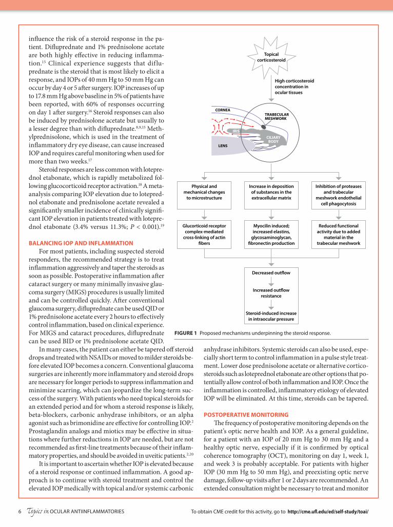
been proposed (Figure 1) and some or all of these mechanisms may be involved. One possibility is that corticosteroids reduce aqueous outfl ow through the trabecular meshwork (TM) by increasing the deposition or decreasing the degradation of the extracellular matrix.9 Excess glycosaminoglycan accumulates in the TM of steroid responders and in cultured cells exposed to dexamethasone.9 Dexamethasone also aff ects the structure of F-actin and causes contraction of TM cells, reducing the intercellular space available for aqueous fl ow. TM cells are phagocytic and remove debris from the TM. Dexamethasone inhibits phagocytosis by TM cells, which could result in re-duced clearing of debris and increased IOP as the trabecular outfl ow channels become blocked.2,8
Genetic factors including myocilin (MYOC) have been implicated in the steroid response. MYOC is expressed in TM cells, and mutations causing defective MYOC secretion lead to decreased trabecular outfl ow.2 Overexpression of glaucoma-causing mutants of human MYOC induces raised IOP in mice10 and MYOC is strongly upregulated aft er dexamethasone exposure.11 Mutations aff ecting expression and secretion of the product of the mucin-encoding gene HCG22 were identifi ed as potentially pathogenic in steroid-induced elevations in IOP,12 and an additional 14 genes induced by dexamethasone in human TM cells have been identifi ed in genomic regions associated with glaucoma.13
Another potential mechanism of the steroid response is inhibition of prostaglandin activity. Prostaglandins are part of the infl ammatory cascade, and prostaglandin analogues are eff ective in reducing IOP.9 Prostaglandin production is inhibited by dexamethasone in cultured human TM cells,14
so it is possible that reduced prostaglandin production in the TM of steroid responders could contribute to elevated IOP.
PROPENSITY FOR STEROID RESPONSE WITH DIFFERENT DRUGS
Both the type of corticosteroid and the duration of usage
CORE CONCEPTS ✦ Diagnosis or family history of glaucoma are the strongest
risk factors for high-level steroid response.
✦ Possible mechanisms may involve changes to cytoplasm, inhibition of phagocytosis, prostaglandins, or mutations in MYOC.
✦ Diff erent corticosteroids have diff erent propensities to elicit a response.
✦ Infl ammation should be treated quickly with steroids and then tapered as soon as possible.
✦ Treatment strategies should take into account patient history, and the level of IOP elevation, including health of the optic nerve.
To obtain CME credit for this activity, go to http://cme.ufl .edu/ed/self-study/toai/6 Topics in OCULAR ANTIINFLAMMATORIES
anhydrase inhibitors. Systemic steroids can also be used, espe-cially short term to control infl ammation in a pulse style treat-ment. Lower dose prednisolone acetate or alternative cortico-steroids such as loteprednol etabonate are other options that po-tentially allow control of both infl ammation and IOP. Once the infl ammation is controlled, infl ammatory etiology of elevated IOP will be eliminated. At this time, steroids can be tapered.
POSTOPERATIVE MONITORINGTh e frequency of postoperative monitoring depends on the
patient’s optic nerve health and IOP. As a general guideline, for a patient with an IOP of 20 mm Hg to 30 mm Hg and a healthy optic nerve, especially if it is confi rmed by optical coherence tomography (OCT), monitoring on day 1, week 1, and week 3 is probably acceptable. For patients with higher IOP (30 mm Hg to 50 mm Hg), and preexisting optic nerve damage, follow-up visits aft er 1 or 2 days are recommended. An extended consultation might be necessary to treat and monitor
infl uence the risk of a steroid response in the pa-tient. Difl uprednate and 1% prednisolone acetate are both highly eff ective in reducing infl amma-tion.15 Clinical experience suggests that dif lu-prednate is the steroid that is most likely to elicit a response, and IOPs of 40 mm Hg to 50 mm Hg can occur by day 4 or 5 aft er surgery. IOP increases of up to 17.8 mm Hg above baseline in 5% of patients have been reported, with 60% of responses occurring on day 1 aft er surgery.16 Steroid responses can also be induced by prednisolone acetate but usually to a lesser degree than with difl uprednate.8,9,15 Meth-ylprednisolone, which is used in the treatment of infl ammatory dry eye disease, can cause increased IOP and requires careful monitoring when used for more than two weeks.17
Steroid responses are less common with lotepre-dnol etabonate, which is rapidly metabolized fol-lowing glucocorticoid receptor activation.18 A meta-analysis comparing IOP elevation due to lotepred-nol etabonate and prednisolone acetate revealed a signifi cantly smaller incidence of clinically signifi -cant IOP elevation in patients treated with lotepre-dnol etabonate (3.4% versus 11.3%; P < 0.001).19
BALANCING IOP AND INFLAMMATIONFor most patients, including suspected steroid
responders, the recommended strategy is to treat infl ammation aggressively and taper the steroids as soon as possible. Postoperative infl ammation aft er cataract surgery or many minimally invasive glau-coma surgery (MIGS) procedures is usually limited and can be controlled quickly. Aft er conventional glaucoma surgery, difl uprednate can be used QID or 1% prednisolone acetate every 2 hours to eff ectively control infl ammation, based on clinical experience. For MIGS and cataract procedures, difl uprednate can be used BID or 1% prednisolone acetate QID.
In many cases, the patient can either be tapered off steroid drops and treated with NSAIDs or moved to milder steroids be-fore elevated IOP becomes a concern. Conventional glaucoma surgeries are inherently more infl ammatory and steroid drops are necessary for longer periods to suppress infl ammation and minimize scarring, which can jeopardize the long-term suc-cess of the surgery. With patients who need topical steroids for an extended period and for whom a steroid response is likely, beta-blockers, carbonic anhydrase inhibitors, or an alpha agonist such as brimonidine are eff ective for controlling IOP.2 Prostaglandin analogs and miotics may be eff ective in situa-tions where further reductions in IOP are needed, but are not recommended as fi rst-line treatments because of their infl am-matory properties, and should be avoided in uveitic patients.2,20
It is important to ascertain whether IOP is elevated because of a steroid response or continued infl ammation. A good ap-proach is to continue with steroid treatment and control the elevated IOP medically with topical and/or systemic carbonic
CORNEATRABECULARMESHWORK
CILIARYBODY
IRIS
LENS
Physical and mechanical changes
to microstructure
Increase in depositionof substances in theextracellular matrix
Inhibition of proteasesand trabecular
meshwork endothelialcell phagocytosis
Glucorticoid receptorcomplex-mediated
cross-linking of actin�bers
Myocilin induced;increased elastins,
glycosaminoglycan,�bronectin production
Reduced functionalactivity due to added
material in thetrabecular meshwork
Decreased out�ow
Increased out�owresistance
Steroid-induced increasein intraocular pressure
Topicalcorticosteroid
High corticosteroidconcentration in ocular tissues
FIGURE 1 Proposed mechanisms underpinning the steroid response.
To obtain CME credit for this activity, go to http://cme.ufl .edu/ed/self-study/toai/ Topics in OCULAR ANTIINFLAMMATORIES 7
IOP. Th e timing of the follow-up visit should be decided upon based on the examination results.
Steroid medications can also be delivered via intraocular injections or slow-release implants. Th ese approaches are ef-fective in controlling infl ammation, are convenient for the patient, and remove any issues with patient compliance.21 Treatment of steroid responses, however, can be more chal-lenging because of the diffi culty in removing residual steroid. Implant removal may be eff ective,2 but when steroids have been injected into the eye, treatment could require anterior chamber washout to remove residual steroid.
One possible approach to treating sustained elevated IOP is selective laser trabeculoplasty (SLT). Th e eff ectiveness of SLT has been tested in patients with elevated IOP following sub-tenon and intravitreal steroid injections, resulting in a greater than 50% reduction in IOP aft er 9 months.22 On rare occasions incisional surgery might be required to control persistent IOP elevation that is refractory to other approaches.
Treatment with biologic agents and antimetabolites, in col-laboration with a uveitis specialist or a rheumatologist, are oth-er options for managing infl ammation that can’t be controlled by steroids or to allow steroids to be tapered when a steroid response is developing. Methotrexate, which inhibits rapidly dividing cells, and mycophenolate, which selectively targets B and T lymphocytes, are well established agents for controlling ocular infl ammation.23 B cells can also be targeted with the monoclonal antibody rituximab and TNFα, an infl ammatory cytokine, can be targeted by a soluble receptor (etanercept) or monoclonal antibody (infl iximab, adalimumab).23
OTHER CAUSES OF ELEVATED IOP Alternative causes of postsurgical elevated IOP should
be investigated in addition to steroid responses. Viscoelastic agents are known to decrease outfl ow facility, and insuffi cient removal at the end of surgery can cause elevated IOP in the early postoperative period.24 Trabecular precipitates, cellular debris, and infl ammation of the TM can also decrease tra-becular outfl ow.25 Elevated IOP aft er cataract surgery is more likely in patients who also have glaucoma, perhaps because their TM is already compromised and cannot tolerate the ad-ditional insult from cataract surgery. Th e elevated IOP may not be secondary to steroid use, but to already compromised TM. Aqueous misdirection syndrome occurs most commonly aft er surgery for angle-closure glaucoma but can also occur aft er routine cataract surgery; it presents as a shallow anterior chamber with raised IOP.26
Elevated IOP following conventional glaucoma surgery and MIGS can have the same etiology as for cataract surgery. An additional cause of elevated IOP aft er trabeculectomy is overtightening of the fl ap sutures, which restricts outfl ow.27 All glaucoma procedures can have short term fl uctuations in IOP, in addition to early and late failure to control IOP.
CONCLUSIONPerhaps the most important aspect of treating potential
steroid responders is to be aware of the risk of a steroid response
occurring. Considering the patient’s history will prepare you to eff ectively control a response should it occur. NSAIDS and cyclosporine are useful for treating infl ammation; using them in combination might be considered as an alternative to steroid treatment. However, these drugs are less eff ective than steroids and, in most cases, changes in dose or type of steroid will result in lower IOP. Monitor the patient aft er surgery and treat the problem accordingly. Th is will prevent the sequelae of a steroid response which can lead to a major, debilitating condition for the patient.
Ronald M. Caronia, MD, FACS, is a partner at Ophthalmic Consultants of Long Island and assistant clinical professor of ophthalmology at the Albert Ein-stein School of Medicine. He states that in the past 12 months, he has not had a fi nancial relationship with any commercial organization that produces, markets, resells, or distributes healthcare goods or services consumed by or used on patients relevant to this manuscript. Medical writer David Loebel, PhD, of Markey Medical Consulting Pty Ltd, assisted in the preparation of this manuscript.
REFERENCES 1. Becker B. Intraocular Pressure Response to topical corticosteroids. Invest
Ophthalmol Vis Sci. 1965;4(2):198-205. 2. Razeghinejad MR, Katz LJ. Steroid-induced iatrogenic glaucoma. Ophthalmic
Res. 2012;47(2):66-80. 3. Tranos P, Bhar G, Little B. Postoperative intraocular pressure spikes: the
need to treat. Eye (Lond). 2004;18(7):673-9. 4. Armaly MF. Statistical attributes of the steroid hypertensive response in the
clinically normal eye I. Th e demonstration of three levels of response. Invest Ophthalmol. 1965;4:187-97.
5. Hirooka K, Shiraga F, Tanaka S, Baba T, Mandai H. Risk factors for elevated intraocular pressure after trans-tenon retrobulbar injections of triamcinolone. Jpn J Ophthalmol. 2006;50(3):235-8.
6. Gaston H, Absolon MJ, Th urtle OA, Sattar MA. Steroid responsiveness in connective tissue diseases. Brit J Ophthalmol. 1983;67(7):487-90.
7. Chang DF, Tan JJ, Tripodis Y. Risk factors for steroid response among cataract patients. J Cataract Refract Surg. 2011;37(4):675-81.
8. Kersey JP, Broadway DC. Corticosteroid-induced glaucoma: a review of the literature. Eye (Lond). 2006;20(4):407-16.
9. Pleyer U, Ursell PG, Rama P. Intraocular pressure eff ects of common topical steroids for post-cataract infl ammation: are they all the same? Ophthalmol Th er. 2013;2(2):55-72.
10. Shepard AR, Jacobson N, Millar JC, et al. Glaucoma-causing myocilin mu-tants require the Peroxisomal targeting signal-1 receptor (PTS1R) to elevate intraocular pressure. Hum Mol Genet. 2007;16(6):609-17.
11. Shepard AR, Jacobson N, Fingert JH, et al. Delayed secondary glucocorti-coid responsiveness of MYOC in human trabecular meshwork cells. Invest Ophthalmol Vis Sci. 2001;42(13):3173-81.
12. Jeong S, Patel N, Edlund CK, et al. Identifi cation of a novel mucin gene HCG22 associated with steroid-induced ocular hypertension. Invest Oph-thalmol Vis Sci. 2015;56(4):2737-48.
13. Lo WR, Rowlette LL, Caballero M, et al. Tissue diff erential microarray analysis of dexamethasone induction reveals potential mechanisms of steroid glaucoma. Invest Ophthalmol Vis Sci. 2003;44(2):473-85.
14. Weinreb RN, Mitchell MD, Polansky JR. Prostaglandin production by hu-man trabecular cells: in vitro inhibition by dexamethasone. Invest Ophthalmol Vis Sci. 1983;24(12):1541-5.
15. Sheppard JD, Toyos MM, Kempen JH, Kaur P, Foster CS. Difl uprednate 0.05% versus prednisolone acetate 1% for endogenous anterior uveitis: a phase III, multicenter, randomized study. Invest Ophthalmol Vis Sci. 2014;55:2993-3002.
16. Marsh P, Pf lugfelder SC. Topical nonpreserved methylprednisolone therapy for keratoconjunctivitis sicca in Sjögren syndrome. Ophthalmology. 1999;106(4):811-6.
17. Cable MM. Intraocular pressure spikes using difl uprednate 0.05% for post-operative cataract infl ammation. Invest Ophthalmol Vis Sci. 2010;51(13):1981.
18. Bodor N, Buchwald P. Ophthalmic drug design based on the meta-bolic activity of the eye: soft drugs and chemical delivery systems. AAPS J.
CARONIA REFERENCES continue on page 8
To obtain CME credit for this activity, go to http://cme.ufl.edu/ed/self-study/toai/8 Topics in OCULAR ANTIINFLAMMATORIES
1. Which of the following antiinflammatory therapies is recommended to treat vernal keratoconjunctivitis (moderate disease)? A. Topical timolol B. NSAID C. Topical lubricant D. Short-term therapy with topical
corticosteroids
2. The underlying pathogenesis of vernal keratoconjunctivitis may include: A. Ig-E-mediated mechanisms B. Infiltration of macrophages,
neutrophils and eosinophils C. Eosinophil cationic protein and
MMP-9 D. All of the above
3. Which one of the following statements is NOT correct about atopic keratoconjunctivitis? A. It develops during spring and is
self-limiting B. It occurs equally between the
sexes C. Patients often have dermatitis
and may have asthma D. Ectropion or entropion may be
observed
4. Rapid steroid responses have been reported most frequently with: A. Loteprednol etabonate B. 1% prednisolone acetate C. Difluprednate D. 0.1% prednisolone acetate
5. Possible mechanisms for the steroid response include: A. Increased glycoasaminoglycans
and reduced phagocytosis B. Decreased glycosaminoglycans
and reduced phagocytosis C. Increased intercellular space D. Excess prostaglandin activity
6. Which one of the following statements is NOT correct about vernal keratoconjunctivitis? A. It occurs in children and young
adults B. It affects more females than
males C. It is more common in hot, dry
environments and with upper tarsus involvement
D. Typical symptoms include intense itching, tearing, and photophobia
7. Which one of the following pathological changes may be found on the ocular surface of patients with vernal keratoconjunctivitis? A. Reduced mucin production B. Disrupted barrier function of the
intact corneal epithelium C. Shield ulcer on the cornea D. All of the above
8. A high-level steroid response has been reported in what proportion of POAG patients? A. 62% B. 21.5% C. more than 90% D. 35%
9. Other potential risk factors for high-level steroid response include: A. Myopia B. Diabetes C. Family history of glaucoma D. All of the above
10. What are the criteria for a high-level steroid response? A. IOP greater than 31 mm Hg B. IOP rise of more than 31 mm Hg
above baseline C. IOP rise of more than 16 mm Hg
above baseline D. A or C
This CME activity is sponsored by the University of Florida College of Medicine and is supported by an unrestricted educational grant from Shire. Participants must score at least 80% on this exam in order to receive credit. The University of Florida College of Medicine designates this enduring material for a maximum of 1 AMA PRA Category 1 Credit™. To take this exam and obtain credit, please take the test online at http://cme.ufl.edu/ed/self-study/toai/. Expires October 31, 2018.
EXAMINATION QUESTIONS TOPICS IN OCULAR ANTIINFLAMMATORIES | ISSUE 18
CARONIA continued from page 7
2005;7(4):E820-33. 19. Sheppard JD, Comstock TL, Cavet ME. Impact
of the topical ophthalmic corticosteroid lotepre-dnol etabonate on intraocular pressure. Adv Ther. 2016;33(4):532-52.
20. Bodh SA, Kumar V, Raina UK, Ghosh B, Thakar M. Inflammatory glaucoma. Oman J Ophthalmol. 2011;4(1):3-9.
21. Fisher BL, Potvin R. Transzonular vitreous injection vs a single drop compounded topical pharmaceutical regimen after cataract surgery.
Clin Ophthalmol. 2016;18(10):1297-1303. 22. Yuki K, Inoue M, Shiba D, et al. Selective laser
trabeculoplasty for elevated intraocular pressure following subtenon injection of triamcinolone acetonide. Clin Ophthalmol. 2010;26(4):247-9.
23. Larson T, Nussenblatt RB, Sen HN. Emerg-ing drugs for uveitis. Expert Opin Emerg Drugs. 2011;16(2):309-22.
24. Higashide T, Sugiyama K. Use of viscoelastic sub-stance in ophthalmic surgery – focus on sodium hyaluronate. Clin Ophthalmol. 2008;2(1):21-30.
25. Baneke AJ, Lim KS, Stanford M. The pathogen-esis of raised intraocular pressure in uveitis. Curr Eye Res. 2016;41(2):137-49.
26. Ruben S, Tsai J, Hitchings RA. Malignant glaucoma and its management. Br J Ophthalmol. 1997;81(2):163-7.
27. Vijaya L, Manish P, Ronnie G, Shantha B. Man-agement of complications in glaucoma surgery. Indian J Ophthalmol. 2011;59 Suppl:S131-40.