Embed Size (px)
Citation preview

216
OPTOMETRY VOLUME 75/NUMBER 4/APRIL 2004
ISSUE HIGHLIGHT
Age-related macular degeneration (ARMD) is the lead-ing cause of untreated vision loss in aging Westernsocieties, accounting for 45% of all visual disability
in the United States.1 Increasing age is associated withincreasing prevalence of ARMD.2 Atrophic ARMD consti-tutes 90% of all cases. It results in a chronic, painless, bilat-eral asymmetric, paracentral, or central photoreceptor-retinalpigment epithelium (RPE) disturbance.3 ARMD hasincreased in prevalence in Great Britain in the last 60 years,suggesting that genetic predisposition is not the primary eti-ologic factor.4 In Japan, the prevalence of ARMD is increas-ing, possibly from a shift to a more-Westernized diet.5
ARMD is a complex disorder that involves genetic, car-diovascular, environmental, and nutritional components.Recent identification of a locus on chromosome 1q31 asso-ciated with increased susceptibility to ARMD may some dayallow testing of high-risk individuals.6 After aging, smok-ing remains the most significant risk factor for ARMD.7,8
Smoking is known to deplete serum antioxidants, alter bloodviscosity, alter the auto regulation flow mechanism of bloodvessels, and is associated with lower levels of macular xan-thophylls pigments, such as lutein.9,10 Environmental riskfactors include exposure to solar radiation/blue light andphotosensitizing drugs.11-14
Low fruit and vegetable consumption increases the risk ofARMD.15,16 A population-based cross-sectional surveyrevealed that ARMD is lower in a self-sustained farmingcommunity than elsewhere in the industrial world.17 Thelong-term, large-population National Eye Institute Age-related Eye Disease Study (NIH AREDS, 2001) showed thatin people with intermediate or moderately advanced ARMD
Double-masked, placebo-controlled, randomizedtrial of lutein and antioxidant supplementation inthe intervention of atrophic age-related maculardegeneration: the Veterans LAST study (LuteinAntioxidant Supplementation Trial)Stuart Richer, O.D., Ph.D.a,c; William Stiles, M.D., J.D.a; Laisvyde Statkute, M.D.a,*;Jose Pulido, M.D.b; James Frankowski, M.S., Ph.D. candidatea,*; David Rudy, M.D.,M.P.H.c; Kevin Pei, B.S.c; Michael Tsipursky, M.S.c; and Jill Nyland, R.N.a
aDepartment of Veterans’ Affairs, Medical Center Eye Clinic, North Chicago, Illinois; bDepartment of Ophthalmology, University ofIllinois Eye and Ear Infirmary Retina Service, Chicago, Illinois; and cFamily Medicine/Chicago Medical School, Chicago, Illinois
* Dr. Statkute is currently affiliated with Internal Medicine, Cook County Hospital, Chicago, Illinois, and Dr. Frankowski is currentlyaffiliated with Department of Research and Scientific Affairs, American Academy of Orthopaedic Surgeons, Rosemont, Illinois.
Richer S, Stiles W, Statkute L, et al. Double-masked, placebo-controlled, randomized trial of lutein and antioxidant supple-mentation in the intervention of atrophic age-related maculardegeneration: the Veterans LAST study (Lutein AntioxidantSupplementation Trial). Optometry 2004;75:216-30.
Background: Age-related macular degeneration (ARMD) is theleading cause of vision loss in aging Western societies. Theobjective of the lutein antioxidant supplementation trial (LAST)is to determine whether nutritional supplementation with luteinor lutein together with antioxidants, vitamins, and minerals,improves visual function and symptoms in atrophic ARMD.
Methods: The study was a prospective, 12-month, randomized,double-masked, placebo-controlled trial conducted at an urbanmidwestern Veterans Administration Hospital from August1999 to May 2001. Ninety patients with atrophic ARMD werereferred by ophthalmologists at two Chicago-area veteransmedical facilities. Patients in Group 1 received lutein 10 mg(L); in Group 2, a lutein 10 mg/antioxidants/vitamins and min-erals broad spectrum supplementation formula (L/A); and inGroup 3, a maltodextrin placebo (P) over 12 months.
Results: In Groups 1 L and 2 L/A, mean eye macular pigment opti-cal density increased approximately 0.09 log units from base-line, Snellen equivalent visual acuity improved 5.4 letters forGroup 1 L and 3.5 letters for Group 2 L/A, and contrast sensi-tivity improved. There was a net subjective improvement inAmsler grid in Group 1 L. VFQ–14 questionnaires concerning sub-jective glare recovery were nearly significant at 4 months forGroup 2 L/A. Patients who received the placebo (Group 3) hadno significant changes in any of the measured findings.
Conclusion: In this study, visual function is improved with luteinalone or lutein together with other nutrients. Further studies areneeded with more patients, of both genders, and for longer peri-ods of time to assess long-term effects of lutein or lutein togetherwith a broad spectrum of antioxidants, vitamins, and mineralsin the treatment of atrophic age-related macular degeneration.
Key Words: Age-related macular degeneration, antioxidants,lutein, nutrition

ISSUE HIGHLIGHT
in one eye (but not the opposite eye), the com-bination of vitamins C, E, beta-carotene, zinc, andcopper reduced 5-year risk of severe visual lossby 25%.18,19 As with AREDS, we also found thatbroad-spectrum antioxidant/mineral supplementsdelayed the progression of the disease, but did notreverse visual loss.20,21
Low intake of lutein, the primary dietarycarotenoid xanthophyll pigment responsible formacular pigment optical density (MPOD) in pri-mates, is a major risk factor for advancedARMD.16,22-24 The eyes of the elderly, smokers,blue-eyed individuals, post-menopausal women,and the retinas of autopsied ARMD patients havereduced lutein.10,24-28 Lutein intake and highMPOD are hypothesized to play a preventive andtherapeutic role in optimizing eye health.29-31
AREDS did not evaluate lutein nor its potentialeffects on MPOD and ARMD.18
The objective of the lutein antioxidant supple-mentation trial (LAST study) is to evaluate theeffect of lutein alone or a more complete nutri-tional supplement of lutein in combination withadditional carotenoids, antioxidants, vitamins, andminerals (referred to as L/A), on MPOD and cen-tral vision outcome measures in atrophic ARMD.
MethodsThe study was approved by the R&D and HumanSubjects Committees at the Department of Vet-erans Affairs (DVA) North Chicago and HinesHospitals and took place between August 1999and May 2001. Eligibility included diagnosis ofatrophic ARMD (ICD9 362.51) by stereo bio-oph-thalmoscopy and at least one vision-degradingvisual-psychophysical abnormality associatedwith ARMD in one or both eyes.32,33 The latterinclude depressed contrast sensitivity (CSF, dis-cussed later) at one or more of four spatial fre-quencies (3, 6, 12, or 18 cc/degree) not attributableto cataract or ocular disease; an abnormalphoto-stress glare recovery (GR) deficit or deficitsof the Amsler grid (i.e., intermittent/permanentvisual spots [scotomas]); or distortions of lines(metamorphopsia). Inclusion criteria also includedclear non-lenticular ocular media (cornea, aque-ous, and vitreous), free of advanced glaucoma anddiabetes or any other ocular or systemic diseasethat could affect central or parafoveal macularvisual function. Subjects were excluded if theyhad undergone recent (within 6 months) cataract
or retinal surgery, were taking photosensitizingdrugs (such as phenothiazines and chloroquine),or did not meet ophthalmic/visual entrance cri-teria. Subjects were excluded if they had takenlutein supplements within the previous sixmonths.
Study designThis was a 12-month randomized, double-masked, placebo-controlled clinical trial. One hun-dred nine subjects were registered by anoptometrist and ophthalmologist, with 19excluded because they: were fundus-positive, buthad no visual psychophysical abnormality (n =9); voluntarily withdrew during baseline workup(n = 6); received ARMD laser treatment (n = 2),or had pre-retinal membrane (n = 1) orAlzheimer’s disease (n = 1). Following writteninformed consent, 90 subjects (86 men and 4women) were randomly assigned to one of threecapsule groups by consecutive random card—3-choice, allocation sequence.
Randomization was applied to the entire sampleof 90 subjects, with 29 in Group 1 L, 30 in Group2 L/A, and 31 in Group 3 P. Serial eye examina-tions were performed on 28, 28, and 30 subjectsafter 4 months of intervention; 26, 26, and 28 sub-jects after 8 months of intervention; 25, 24, and27 subjects after 12 months of intervention (forGroups 1 L, 2 L/A, and 3 P, respectively). Duringthe 12-months’ clinical trial, 1, 2, and 1 subjectswere lost to followup; 1, 0, and 2 subjects died;and 2, 4, and 1 subjects had other reasons forwithdrawal (for Groups 1 L, 2 L/A, and 3 P,respectively). Compliance was assessed by tele-phone at 1 week, 2 weeks, 4 weeks, 6 weeks, 3months, and 12 months. During the one-yearstudy, 96% of the subjects took approximately92% of their assigned capsules. There were no dif-ferences in compliance among the three groups.
We selected a dose of 10-mg non-esterified luteinto approximate food-equivalent lutein intake fromspinach in our pilot case series experiments.33-35
Group 1 L received only lutein (FloraGlo® fromKemin Foods International, Des Moines, Iowa);Group 2 L/A received lutein plus additionalantioxidants and nutrients (OcuPower® fromNutraceutical Sciences Institute (NSI) , BoyntonBeach, Florida), and Group 3 P received mal-todextrin. The L/A OcuPower® supplement con-sists of 10-mg lutein (FloraGlo); 2,500 IU vitamin A;
217
VOLUME 75/NUMBER 4/APRIL 2004 OPTOMETRY

218
OPTOMETRY VOLUME 75/NUMBER 4/APRIL 2004
ISSUE HIGHLIGHT
15,000 IU natural beta carotene (Betatene®);1,500-mg vitamin C (as calcium ascorbate–EsterC®); 400 IU vitamin D3; 500 IU natural vitaminE (d-alpha tocopherol succinate); 50-mg vitaminB1; 10-mg vitamin B2; 70-mg vitamin B3; 50-mgvitamin B5; 50-mg vitamin B6; 500-mcg vitaminB12; 800-mcg folic acid; 300-mcg biotin; 500-mgcalcium; 300-mg magnesium; 75-mcg iodine; 25-mg zinc (as zinc L-methionine—L-OptiZinc®); 1-mg copper; 2-mg manganese; 200-mcg selenium;200-mcg chromium; 75-mcg molybdenum; 600-mcg lycopene; 160-mg bilberry extract (stan-dardized to 25% anthocyanosides); 150-mg alphalipoic acid; 200-mg N-acetyl cysteine; 100-mgquercetin; 100-mg rutin; 250-mg citrusbioflavonoids; 50-mg plant enzymes; 5-mg blackpepper extract (Bioperine®); 325-mg malic acid;900-mg taurine; 100-mg L-glycine; 10-mg L-glu-tathione; and 2-mg boron. Nutraceutical SciencesInstitute prepared the lutein capsules, the L/A cap-sules, and the P capsules and also maintained andconcealed the blinding and four-digit allocationcodes. Bottles with masked four-digit allocationcodes were sent to the assigned research phar-macist at DVA Medical Center, North Chicago. Allpersonnel at the DVA Medical Center wereunaware of the masked allocation codes duringthe 12-month clinical study. Subjects were pro-vided with opaque capsules of identical appear-ance in numbered containers taken as threecapsules twice per day with food. Independentverification of lutein content was performed viarandom sampling and high-performance liquidchromatography at Kemin Foods, Inc..
Nutritional status was assessed using Food Fre-quency Questionnaires (FFQs), which the subjectand/or spouse completed at baseline and final 12-month visit. All FFQs were analyzed by HarvardUniversity, School of Public Health.36,37 Subjectswere encouraged not to alter their diets.
Objective measuresOphthalmic testing was conducted at baseline andat 4 months, 8 months, and 12 months. Baselineand final ocular lens opacification (cataract) wereevaluated subjectively by a single observer, usinga Haag–Streit slit-lamp biomicroscope. A 7-incre-ment validated Lens Opacity Cataract Scale (LOC-SIII) photographic transparency was placed on a5000 K color temperature, 8-watt fluorescent pho-tographic light box, according to standard pro-cedures.38 Retinal images were taken with aCanon model CF-600 Vi Fundus camera at base-
line and study completion, and were rated by asingle retinologist, masked as to image date orintervention group.39
MPOD was measured by heterochromatic flickerphotometry using a MacularMetrics® instrument(Rehoboth, Massachusetts), according to the man-ufacturers’ protocol.40 It was determined by sub-jects matching a one-degree 460/540 nanometerflickering stimulus for perceived brightnessfoveally and at a retinal location seven degreesextra-foveally, where the MPOD is virtually zero.
Refractive error was neutralized with lenses priorto acuity and glare testing at each visit, and result-ant trial lenses were used for all vision tests. Glarerecovery (GR) is a measure of both macular func-tion and retinal health.41,42 Subjects monocularlyviewed a 6500 lux, 5000 K color temperature lightbox held at 40 cm from their eye for one minutepre-adaptation. Following this visual stress, thesubject was asked to attempt to read a super-threshold low-contrast line of print. Examinersmonitored accuracy and time required for mac-ular function to return to normal following thephoto-stressed condition. One minute or less torecovery is normal.42,43,44
Monocular visual acuity at distance was measuredvia random presentation of distance Snellen let-ters on a Mentor Opththalmics BVAT–II® com-puter screen under mesopic lighting conditions.Low-vision patients with Snellen acuity less than20/200 were tested with a Designs for Vision®Feinbloom distance acuity test for the partiallysighted. In both cases, fractional Snellen acuitywas converted to Log minimal angle of resolution(LogMAR). Visual acuity at near was measuredfor each eye using low- and high-contrast SKILLtest targets.41
Contrast sensitivity function (CSF) measures theleast amount of contrast needed to detect visualstimuli at different spatial frequencies. It identi-fies selective retinal deficits in visual processingat an earlier stage than is possible with conven-tional visual acuity testing.45 The Vector VisionCSV 1000 ® contrast sensitivity test system wasused. For low-vision patients, the CSV 1000L–VPelli–Robson type single large letter chart of vary-ing contrasts was used.46
Activities of daily living, night driving, and glarerecovery symptoms were evaluated on a 4- to

ISSUE HIGHLIGHT
20-point VFQ–14 rating scale used by the NationalEye Institute and described previously.33,47 Subjectswere also provided an integrated instructionsheet/questionnaire/Amsler grid to monitor changesin vision over time. Subjects at each visit wereasked if they felt their overall vision had worsened,remained the same, or improved, and the numberof boxes missing (scotomas) or distorted (meta-morphopsia) were counted. Reports of visionchange were digitally videotaped.
Statistical analysisRepeated measures analysis of variance (ANOVA)assessed main ophthalmic outcome variables attrib-utable to the time of measurement, treatment con-dition, and their interaction. Follow-up analyses forsignificant group differences were done by pair-wise
comparisons between baseline and each time point(using pooled error terms) for unequal n’s, and aBonferroni correction factor. The Friedman’s two-way ANOVA by rank test, a non-parametric test, wasused in place of the repeated measures ANOVAwhen the assumptions for a parametric ANOVAwere not satisfied (i.e., severity of disease compar-isons with small sample sizes). Finally, all analyseswere conducted for each eye separately. At no timewere results averaged across both eyes.
Power analysis was based on the guidelines put forthby Kirk.48 A minimum group size of 30 would beneeded to detect a standard effect size of 1.0 at anerror rate (Alpha) of 0.01, with a desired power (Beta)of 80 for three groups. Similarly, a minimum groupsize of 30 is considered adequate to detect a standardeffect size of 1.0 with an error rate (Alpha) of 0.05
Table 1. Baseline characteristicsLutein Lutein/A Placebo
Variable (n = 29) (n = 30) (n = 31) P value
Sex Male 27 29 30 —Female 2 1 1 —
Age, mean (SD), yrs. 74.4 (6.4) 73.5 (8.5) 76.1 (6.4) 0.34ARMD Dx mean (SD), yrs. 4.1 (5.2) 4.4 (4.4) 4.9 (5.9) 0.82Smoking pack-years 5.2 (14.1) 7.1 (17.3) 9.2 (22.6) 0.71Alcohol grams 11.0 (26.7) 11.9 (17.8) 6.3 (11.8) 0.52Caffeine mg 231 (192) 225 (247) 211 (171) 0.32Body Mass Index 28.5 (4.2) 30.4 (4.8) 27.3 (5.7) 0.06Iris color
Blue/Gray – light (n) 13 14 18 0.63Gray/Hazel – light (n) 9 6 3 0.22Brown/Black – dark (n) 7 10 9 0.76
Multivitamin useNone (n) 14 13 14 0.97Pabulum (n) 7 8 9 0.88RDA+ (n) 8 9 8 0.96
Dietary Zn include 18.5 (16) 16.3 (13) 30.7 (33) 0.04Supplements mg
Dietary lutein mg 3.0 (2.6) 2.1 (1.4) 1.9 (1.6) 0.13Dietary iron mg * 17.7 (18) 22.2 (34) 23.7 (19) 0.70
Ocular Baseline Data and Significance
Cataract (R LOCSIII rating)Nuclear color 28.3 (1.03) 3.26 (1.13) 2.86 (1.09) 0.28Nuclear opalescence 2.73 (0.96) 3.30 (1.14) 2.76 (1.12) 0.11Cortical 1.83 (1.07) 1.56 (0.80) 1.55 (0.82) 0.48Posterior subcapsular 1.04 (0.21) 1.04 (0.19) 1.00 (0.00) 0.56
Cataract (L LOCSIII rating)Nuclear color 2.81 (0.85) 3.15 (0.99) 3.00 (1.31) 0.51Nuclear opalescence 2.73 (0.87) 3.15 (0.99) 2.92 (1.36) 0.39Cortical 1.73 (0.87) 1.41 (0.70) 1.75 (1.01) 0.27Posterior subcapsular 1.12 (0.43) 1.04 (0.19) 1.21 (0.78) 0.47
219
VOLUME 75/NUMBER 4/APRIL 2004 OPTOMETRY

220
OPTOMETRY VOLUME 75/NUMBER 4/APRIL 2004
ISSUE HIGHLIGHT
and a desired power (Beta) of 0.90 for three groups.Repeated measures involve multiple instances ofsampling the same subjects, thereby substituting forlarger samples consisting of independent subjects.
Baseline characteristicsThe L, L/A, and P groups did not differ in age,years diagnosed with ARMD, smoking, caf-feine/alcohol use, iris color, multivitamin use, anddietary lutein and iron intake (see Table 1). Therewere also no statistical differences among the threeintervention groups in cataract or ocular lens opaci-fication parameters, visual acuity, glare recovery
ability, quality of vision as measured by CSF, or thepresence or absence of scotomas/metamorphopsiason Amsler grid testing. Right eyes Group 2 L/Ahave 0.7 log units less MPOD at baseline (P = 0.05)with a commensurate trend in increased lens colorand decreased low spatial frequency CSF (P =0.10), relationships reported previously.49,50 All ofthis is consistent with a higher body mass indexwhich was found in Group 2 L/A (P = 0.06).
Nutritional characteristics at baseline and 12 monthsIntervention groups were similar as to caloric andnutrient intake at baseline and 12 months, except
AREDS, mean retinal photographic grade and distribution (percent)
R AREDS retinal grade, mean 3.33 (0.62) 2.88 (1.03) 3.05 (0.90) 0.51R Eyes % Grade I 4.5 11.1 0.0 0.002
% Grade II 18.2 16.7 12.0 0.51% Grade III 45.4 44.4 56.0 0.43% Grade IV 31.8 27.8 32.0 0.83
L Eyes retinal grade, mean 2.85 (0.55) 2.71 (1.2) 3.28 (0.83) 0.13L Eyes % Grade I 0.0 4.5 4.2 0.10
% Grade II 36.4 31.8 16.7 0.02% Grade III 54.5 40.1 37.5 0.15% Grade IV 9.1 22.7 41.7 0.0002
MPOD † R 0.23 (0.14) 0.16 (0.08) 0.23 (0.14) 0.05L 0.24 (0.16) 0.21 (0.12) 0.24 (0.15) 0.63
Visual Baseline Data and Significance
Visual acuity R (LogMar) 0.359 0.324 0.445 0.19L (LogMar) 0.279 0.303 0.286 0.15
Glare recovery R (sec) 100.7 (65.1) 88.7 (58.2) 73.4 (54.4) 0.34recovery L (sec) 83.4 (59.2) 82.2 (64.0) 89.7 (65.2) 0.92
Contrast sensitivity R3 cc/degree (log) 1.55 (0.28) 1.53 (0.23) 1.62 (0.30) 0.526 cc/degree (log) 1.56 (0.35) 1.46 (0.33) 1.65 (0.28) 0.14
12 cc/degree (log) 1.10 (0.34) 1.06 (0.43) 1.20 (0.42) 0.4718 cc/degree (log) 0.60 (0.38) 0.55 (0.34) 0.64 (0.44) 0.70
Contrast sensitivity L3 cc/degree (log) 1.63 (0.24) 1.51 (0.20) 1.62 (0.21) 0.106 cc/degree (log) 1.55 (0.21) 1.51 (0.32) 1.56 (0.25) 0.80
12 cc/degree (log) 1.07 (0.36) 1.08 (0.36) 1.10 (0.36) 0.9718 cc/degree (log) 0.54 (0.42) 0.50 (0.29) 0.51 (0.32) 0.95
Amsler grid defects R (n) 15 10 11 0.56L (n) 11 18 11 0.29
SD, Standard deviation; ARMD Dx, years diagnosed with atrophic age-related macular degeneration; RDA+, vitamin consumption beyond the Recom-mended Daily Allowance; LOCSIII, Lens Opacity Classification System, 3rd revision; AREDS, NIH National Eye Institute Age-Related Eye Disease Study;MPOD, macular pigment optical density; and LogMar, log minimal angle of resolution.Smoking status included present and past smoking, and not exposure to passive (second-hand) smoke. Eye color was assessed by categorical visualinspection (blue/gray; gray/hazel, and brown/black) under identical lighting conditions.* Serum iron, transferrin saturation, ferritin, and total iron binding capacity (n = 75) published separately.65 Additional postulated independent ARMD
(and cardiovascular) risk factors obtained (n = 29) by GSDL Laboratories, Asheville, North Carolina. Fibrinogen was elevated at 409 ± 89%; homocysteine was elevated at 13.6 ± 9.2 mg %; and C-reactive protein was slightly elevated at 2.3 ± 4.9 mg % against GSDL normative data.
† R (right eye) and L (left eye) baseline MPOD are correlated, describing 40% of the variance of macular pigmentation in this veteran population. Low spatialfrequency contrast sensitivity (3 cc/degree – large object vision) is weakly positively correlated with MPOD (Pearson r = 0.28; P = 0.02).49
Table 1, continued
ISSUE HIGHLIGHT
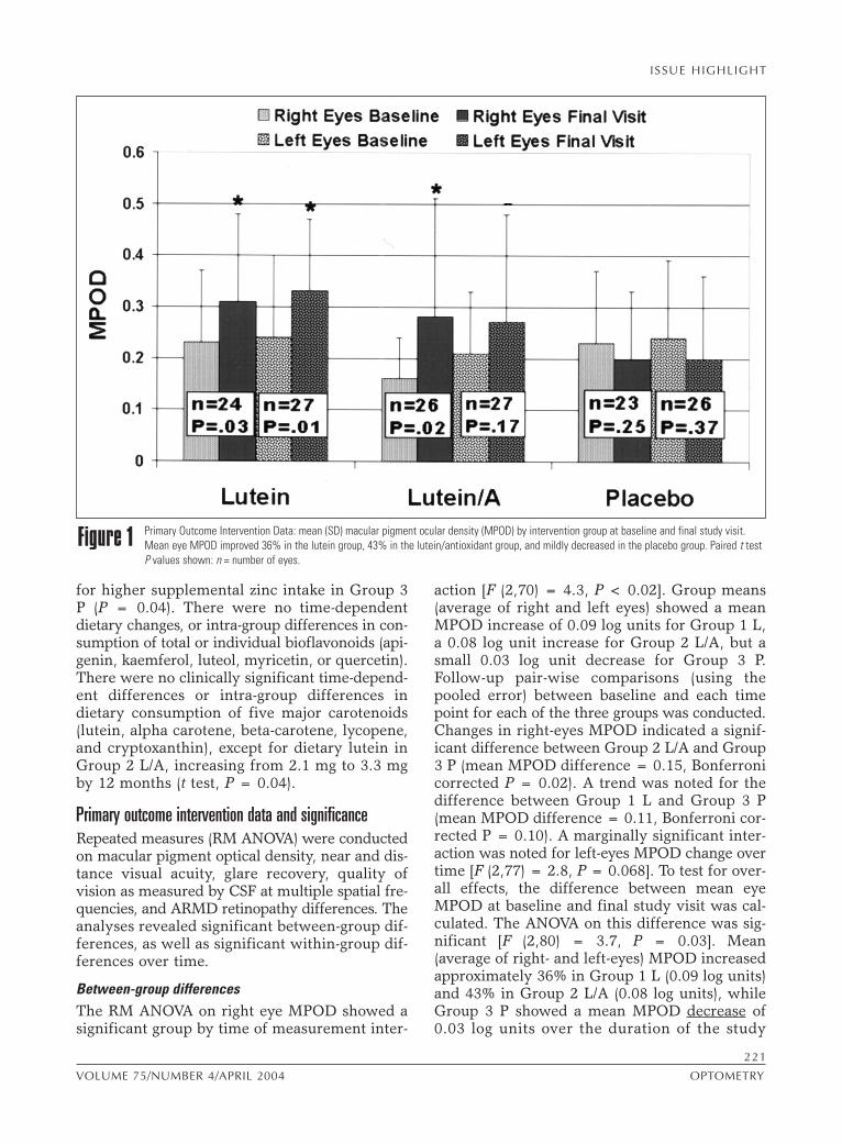
for higher supplemental zinc intake in Group 3P (P = 0.04). There were no time-dependentdietary changes, or intra-group differences in con-sumption of total or individual bioflavonoids (api-genin, kaemferol, luteol, myricetin, or quercetin).There were no clinically significant time-depend-ent differences or intra-group differences indietary consumption of five major carotenoids(lutein, alpha carotene, beta-carotene, lycopene,and cryptoxanthin), except for dietary lutein inGroup 2 L/A, increasing from 2.1 mg to 3.3 mgby 12 months (t test, P = 0.04).
Primary outcome intervention data and significanceRepeated measures (RM ANOVA) were conductedon macular pigment optical density, near and dis-tance visual acuity, glare recovery, quality ofvision as measured by CSF at multiple spatial fre-quencies, and ARMD retinopathy differences. Theanalyses revealed significant between-group dif-ferences, as well as significant within-group dif-ferences over time.
Between-group differencesThe RM ANOVA on right eye MPOD showed asignificant group by time of measurement inter-
action [F (2,70) = 4.3, P < 0.02]. Group means(average of right and left eyes) showed a meanMPOD increase of 0.09 log units for Group 1 L,a 0.08 log unit increase for Group 2 L/A, but asmall 0.03 log unit decrease for Group 3 P. Follow-up pair-wise comparisons (using thepooled error) between baseline and each timepoint for each of the three groups was conducted.Changes in right-eyes MPOD indicated a signif-icant difference between Group 2 L/A and Group3 P (mean MPOD difference = 0.15, Bonferronicorrected P = 0.02). A trend was noted for thedifference between Group 1 L and Group 3 P(mean MPOD difference = 0.11, Bonferroni cor-rected P = 0.10). A marginally significant inter-action was noted for left-eyes MPOD change overtime [F (2,77) = 2.8, P = 0.068]. To test for over-all effects, the difference between mean eyeMPOD at baseline and final study visit was cal-culated. The ANOVA on this difference was sig-nificant [F (2,80) = 3.7, P = 0.03]. Mean(average of right- and left-eyes) MPOD increasedapproximately 36% in Group 1 L (0.09 log units)and 43% in Group 2 L/A (0.08 log units), whileGroup 3 P showed a mean MPOD decrease of0.03 log units over the duration of the study
Primary Outcome Intervention Data: mean (SD) macular pigment ocular density (MPOD) by intervention group at baseline and final study visit.Mean eye MPOD improved 36% in the lutein group, 43% in the lutein/antioxidant group, and mildly decreased in the placebo group. Paired t testP values shown: n = number of eyes.
Figure 1
221
VOLUME 75/NUMBER 4/APRIL 2004 OPTOMETRY

222
OPTOMETRY VOLUME 75/NUMBER 4/APRIL 2004
(see Figure 1). The MPOD increased in Group 1L, independent of the severity of disease (see Fig-ure 2).
The RM ANOVA on left-eyes near visual acuityshowed a significant group by time of measure-ment interaction [F (2,81) = 4.7, P = 0.01].Between baseline and final study measurement,Group 1 L showed a mean eye 5.4 Snellen letterequivalent improvement (95% CI = 2.7 – 9.0, P= 0.01); Group 2 L/A showed a mean eye 3.5Snellen letter equivalent improvement (95% CI =0.8 – 6.1, P = 0.04), while Group 3 P showed a2.1 Snellen letter equivalent decrease (95% CI =–6.7 to 2.4). Follow-up pair-wise comparisons(using a pooled error term) on changes in nearvisual acuity over time showed a significant dif-ference between Group 1 L and Group 3 P (Bon-feronni corrected P = 0.01) and a marginallysignificant difference between Group 2 L/A andGroup 3 P (Bonferroni corrected P = 0.08). TheRM ANOVA for right-eyes near visual acuity wasnon-significant [F (2,65) = 0.3]. To test for over-all effects, an ANOVA on the difference between
mean right-/left-eyes near visual acuity ratings atbaseline and final study visit was performed andfound to be significant [F (2,83) = 4.7, P = 0.013].These results reveal that near visual acuity aver-aged over both eyes increased a mean of 5.4Snellen equivalent letters (about 1 line of visualacuity) in Group 1 L (95% CI = 2.5 – 8.2), and3.5 Snellen equivalent letters in Group 2 L/A (95%CI = 1.2 – 5.8), but decreased 0.2 Snellen equiv-alent letters in Group 3 P (95% CI = –3.0 – 2.7).
Within-group differencesRM ANOVAS indicated significant main effects fortime of measurement on glare recovery, qualityof vision, and distance acuity measurements.Between baseline and final study visit, photo-stress recovery (in seconds) quickened by 25.9seconds (SD = 64.6) in the right eyes and 29.5seconds (SD = 61.3) in left eyes, regardless ofgroup membership. Descriptive statistics withineach group for each eye showed that photo-stress-recovery quickened by approximately 34.8 sec-onds in Group 1 L right-eyes (95% confidenceinterval [CI] 2.9 – 66.8) and 20.3 seconds in group
ISSUE HIGHLIGHT
Macular pigment ocular density (MPOD) of Group 1 (lutein) segregated by retinal specialist into AREDS subgroups (Stage 2 = mild age-relatedmacular degeneration [ARMD]; Stage 3 = moderate ARMD, and Stage 4 = advanced ARMD), and subjected to Friedman’s two-way analysis ofvariance (ANOVA) by rank test. Regardless of ARMD disease stage, MPOD increased over time with lutein supplementation. MD, Median; R, range.
Figure 2

ISSUE HIGHLIGHT
1 left-eyes (95% CI –0.3 – 40.9). In Group 2 L/A,there was a 30.2-second improvement in right-eyes (95% CI 1.0 – 59.4) and a 37.0-second changein left-eyes (95% CI 5.4 – 68.7). In Group 3 P,there was a 15.4-second improvement in right-eyes (95% CI –7.0 – 38.3) and 20.4 seconds forleft-eyes (95% CI –12.5 – 53.2).
Averaging the data from the right and left photo-stress-recovery times, it can be seen that therewas a mean improvement in recovery time of 27.3seconds (SD = 52.7). Within the separate treat-ment groups, this is seen as a 23.7 second morerapid glare recovery for Group 1 L, a 34.7 secondmean eye quicker glare recovery for Group 2 L/A,and a 22.7 second more rapid recovery for Group3 P from the baseline values shown in Table 1.
Lutein also improved glare recovery, independ-ent of AREDS retinal stage, at baseline and after12 months of lutein: stage II, n = 10, median 135sec (range, 15 to 180) versus 35 (15 to 140), P =
0.02; stage III, n = 11, 75 (15 to 180) versus 30(12 to 90), P = 0.28; and stage IV, n = 9, median90 and mean 102 (20 to 180) versus median 90 andmean 80 (5 to 135), P = 0.05; Friedman’s non-parametric statistics.
RM ANOVAs on quality of vision, as measuredby CSF at multiple spatial frequencies over time,indicated significant within-group differences overtime for the right eyes, measured at 3, 6, and 12cycles(cc)/degree, and for the left eye, measuredat 6 and 12 cc/degree. For each of these effects,within-group t-tests comparing baseline to finalstudy visit showed the quality of vision improvedsignificantly in both Group 1 L ,and especiallywith a greater effect in Group 2 L/A (see Figure3). Repeated factors ANOVA of VFQ–14 ques-tionnaires concerning night driving were not sig-nificant for any group. However, glare recoveryVFQ–14 ANOVA subscale data, from baseline(score 15.4 ± 4.0, n = 30), over time, showed atrend toward significance by 4 months (15.7 ±
Primary Outcome Intervention Data: change in quality of vision (contrast sensitivity) by intervention group (denoted pictorially as bars of increas-ing spatial frequency) over 1 year at four spatial frequencies (3, 6, 12, and 18 cc/degree), by eye, with positive numbers denoting improvementbetween baseline and final visit. Number of eyes in each treatment group (Lutein, Lutein/A, and Placebo) and significance levels are as follows:Lutein (R eyes significant at 3 cc/degree [paired t test; P = 0.04; n = 21], 6 cc/degree [P = 0.07; n = 21], and 12 cc/degree [P = 0.01, n = 21]). In theLutein/A group, five of eight R eye/L eye spatial frequency combinations were significant at (P < 0.05) and two were near-significant (P < 0.10). Inthe Placebo group, no statistically significant changes in contrast sensitivity occurred over the 1-year study.
Figure 3
223
VOLUME 75/NUMBER 4/APRIL 2004 OPTOMETRY

224
OPTOMETRY VOLUME 75/NUMBER 4/APRIL 2004
ISSUE HIGHLIGHT
3.7, n = 28, P = 0.06, repeated factors ANOVA)and 8 months (16.3 ± 3.3, n = 26, P = 0.09) forGroup 2 L/A.
Lutein supplementation had no significant effecton CSF for AREDS retinal stage II or III. Forexploratory purposes, we reviewed whetherlutein significantly improved contrast sensitivityin patients with advanced AREDS stage IV dis-ease. At three of four spatial frequencies, thissmall group of patients with advanced AREDSstage IV disease—who were also taking lutein (n = 5)—showed mean contrast sensitivityimprovements at 6, 12, and 18 cc/dgeree (all P’s< 0.05) as follows:
mean (log) ± SD at baselineand after 12 months of lutein
3 cc/degree 1.56 ± 0.24 vs. 1.70 ± 0.37, NS6 cc/degree 1.27 ± 0.22 vs. 1.90 ± 0.38, P = 0.0212 cc/degree 0.79 ± 0.20 vs. 1.45 ± 0.28, P = 0.0318 cc/degree 0.17 ± 0.01 vs. 0.85 ± 0.49, P = 0.006
RM ANOVAS on distance visual acuity revealeda significant time of measurement effect for right-eyes values only (F [1,83] = 6.7, P = 0.01). Dis-tance acuity showed a mild (usually notsignificant) improvement, equivalent to a 2.5 to5 Snellen distance letter improvement over thecourse of the study (negative numbers of LogMARdenote improvement). Over the course of thestudy, all participants showed a change in their
right-eyes of –0.09 LogMAR (SD = 0.32) and a–0.01 LogMAR (SD = 0.32) change in their left-eyes.
Descriptive statistics within each group showedGroup 1 L right-eyes change of neg 0.10 LogMAR(95% CI neg 0.19 – neg 0.01) and left-eyes changeof neg 0.03 (95% CI neg 0.09 – pos 0.03). Group2 L/A showed a LogMAR change of neg 0.03 inright-eyes (95% CI neg 0.12 – pos 0.07) and neg0.06 in left-eyes (95% CI neg 0.14 – pos 0.03).Group 3 P showed a neg 0.14 right-eyes improve-ment (95%CI neg 0.30 – pos 0.03), but a left-eyesworsening of pos 0.05 (95% CI neg 0.14 – pos 0.23).
Retinopathy and lens changeThere was no progression in ARMD retinopathyfor either eye in any group over the course of thestudy. Across all participants, right-eyes baselineAREDS stage was 3.06 (SD = 0.87) and right-eyesfinal AREDS stage was 3.10 (SD = 0.89); left-eyesbaseline AREDS stage was 2.98 (SD = 0.92) andleft-eyes final AREDS stage was 3.04 (SD = 0.90).Descriptive statistics within each group showedthat Group 1 L had no change in right-eyes, butan average 0.07 increase in AREDS stage for left-eyes; Group 2 L/A showed no changes inAREDS stage for either eye; and Group 3 Pshowed a 0.09 increase in AREDS stage for right-eyes and a 0.07 increase in AREDS stage in left-
Table 2.Video-documented change in Amsler grid findings (scotomas and/or metamorphopsias)Improve Worsen Net Improve Worsen Net
Group (no. of eyes) (no. of eyes) effect (no. of veterans) (no. of veterans) effect
4 months Lutein 9 4 5 8 3 5Lutein/A 9 3 6 8 2 6Placebo 7 3 4 7 3 4
8 months Lutein 10 4 6 9 4 5Lutein/A 8 4 4 7 4 3Placebo 2 3 –1 1 3 –2
12 months Lutein 7 1 6 6 1 5Lutein/A 8 7 1 8 7 1Placebo 3 1 2 3 1 2
Totals Lutein 26 9 17 23 8 15Lutein/A 25 14 11 23 13 10Placebo 12 7 5 11 7 4
Table 2 presents the number of single eyes and veterans, by intervention group, self-reporting improvement or worsening in the Amsler grid in terms ofscotoma(s) or metamorphopsia(s) at 4, 8, and 12 months, as compared to the previous visit. Only veterans having a change in Amsler grid status are listed.Six veterans in Group 3 placebo confounded the results by mean dietary intake of lutein of 9.9 mg/day, with improvement in three of these veterans forscotomas/metamorphopsias. For this post hoc analysis, these six veterans were removed from the data analysis. The net changes over time (gray high-light)—taking into account both improvement and worsening of the Amsler grid, by eyes or by number of reporting vetereans—was significant for Group 1Lutein, Chi-square; P = 0.01).

ISSUE HIGHLIGHT
eyes. Nonetheless, there were no between-groupdifferences in retinopathy during the study (uni-variate RM ANOVA, R eyes, P = 0.35; L eyes, P=0.13). Statistical analysis also revealed no signif-icant within- and between-group changes in lensopacification.
Post-hoc analysis of subjective visual change and Amsler gridAfter analyses were completed, we revieweddietary lutein intake and found that six partici-pants in the placebo group ingested a higher-than-average amount. In fact, the amount theyconsumed (9.9 mg/d by food frequency analysis)was similar to what was included in the other twotreatment groups. To examine how this may haveinfluenced results, we removed these six subjects.Subjects at the final visit were asked whethertheir vision had improved, remained the same, orworsened. Though not statistically significant,there was a trend in subjective visual improve-ment in Groups L and L/A by Chi-square analy-sis (P = 0.10) following elimination of these sixsubjects from the placebo group (data notshown).
Serial evaluation of the Amsler grid at each 4-month study visit indicated net improvements inboth Group 1 L and 2 L/A. Following eliminationof these six subjects, the net change—based onthese reports of improvement or worsening of theAmsler grid—was significant for Group 1 L (Chi-square, P = 0.01), but not for Group 2 L/A (seeTable 2).
Adverse effectsThere were no significant between-group differ-ences in minor side effects among Groups L, L/A,and P (data not shown). None of the 30 patientsassigned to Group 2 L/A had a major cardiovas-cular event (myocardial infarction, sudden death,angioplasty, coronary artery bypass surgery, orstroke) or death from any cause. In contrast, inthe 31 patients assigned to Group 3 P, one patienthad angioplasty, one patient died from cardiac dis-ease, and one patient died from metastatic ade-nocarcinoma. In the 29 patients assigned to Group1 L, two patients had angioplasty, one patient hada stroke, and one patient died from cerebrovas-cular disease and pneumonia. Thus, there was anon-significant trend for fewer major cardiovas-cular events or death in Group L/A (0/30) as com-pared to Groups L and P combined (7/60) (seeAppendix).
CommentNutritional treatment of retinal disease hasproved at least partially successful in commonretinitis pigmentosa (vitamin A), Bassen–Kowzweigdisease (vitamins A, E, and K), gyrate atrophy (lowprotein, low arginine diet, and/or vitamin B6), Ref-sum disease (low phytol, low phytanic acid), andSorsby fundus dystrophy (vitamin A).51 Small-scale, prospective, double-masked, randomized,placebo-controlled studies have demonstrated thatthe progression of ARMD can be slowed witheither zinc alone, or the combination of betacarotene, vitamin C, vitamin E, and zinc.21,52
AREDS confirmed these findings.18
We constructed LAST to evaluate the effect oflutein alone or lutein combined with additionalcarotenoids and antioxidants/minerals (includingzinc, beta carotene, and vitamins C and E) onMPOD and objective visual outcome measures inatrophic ARMD. This study demonstrates thatlutein alone or lutein combined with additionalcarotenoids and antioxidants/minerals (includingzinc) significantly improved macular pigment opti-cal density and glare recovery, improved nearvisual acuity, and significantly improved mostmeasures of quality of vision (contrast sensitivityfunction), with L/A having a broader effect. L aloneresulted in a net improvement in Amsler grid sco-tomas and metamorphopsia. These results areimportant because lutein is an essential carotenoidnot produced by the body. Therapeutic loading andmaintenance dosing of lutein and other nutrientsmay be required to treat atrophic ARMD.
The observation of no progression of ARMDretinopathy in patients receiving L/A must be con-sidered a preliminary result, given the small num-ber of patients studied, the short time period ofobservation-one year, and the lack of statistical sig-nificance among the three groups. The increasedmacular pigment and improved visual functionwith lutein and lutein together with antioxidantsin the present LAST study—together with previ-ous studies showing the importance of zinc andseveral vitamins in slowing the progression ofARMD retinopathy and visual loss—raise the pos-sibility that lutein, together with a broad spectrumof antioxidants, vitamins, and minerals, may bea more effective nutritional supplement for treat-ment of ARMD.
There are major population, methodological, andoutcome differences to consider in comparing the
225
VOLUME 75/NUMBER 4/APRIL 2004 OPTOMETRY

226
OPTOMETRY VOLUME 75/NUMBER 4/APRIL 2004
ISSUE HIGHLIGHT
results of this small, brief study with the NIHAREDS. Both studies evaluate white populationswith a similar percentage of smokers, patientswith diabetes, and patients on pre-existing mul-tivitamin and mineral formulations. However, the90 patients in the one-year LAST study were pri-marily male subjects with a mean age about 6years older, compared to the 4,757 mixed genderpatients enrolled in the multicenter, seven-yearAREDS trial. Fully a third of the subjects hadvision worse than 20/32 in the better-functioningeye and would not have qualified for AREDS.Indeed, the mean eye retinal stage for veteransin our LAST study is an AREDS moderatelyadvanced “category 3.” Our study evaluated dis-tinctly different outcome measures. In AREDS,progression to advanced ARMD was based onretinal photographs and visual acuity. In contrast,LAST used a battery of low-contrast visual psy-chophysical tests known to be more sensitive toclinical concerns of patients, compared to retinalappearance and traditional visual acuity tests usedin AREDS and most ophthalmology offices (i.e.,Snellen acuity measurement). We believe thestrength of our methodological approach lies inthe use of basic measurements of functional mac-ular integrity related to ARMD symptoms.
Analogous to the findings in AREDS of antioxi-dants adding to zinc’s effect, subjects in our trialreceiving L/A did better in overall visual quality(CSF) than those who received L alone. Previ-ously, using a non–lutein-containing formulation,we observed a tendency for cataracts to developin subjects over time.21 In contrast, no statisticallysignificant lens opacification occurred in the one-year LAST study. In AREDS, use of isolated beta-carotene was associated with declines in serumconcentration of other carotenoids, includinglutein (as well as lycopene and beta-cryptoxan-thin). Beta-carotene may compete with nutritionalor supplemental lutein in both duodenum trans-port/absorption and hepatic lipoprotein segrega-tion and fractionization.53-57 In contrast, thelutein/antioxidant formulation used in LAST con-tains a broader array of dietary carotenoids thatmay, in part, explain improvements in thevisual outcome, compared with AREDS. Theseimprovements occurred despite subjects beingolder—and with more severe disease—thanpatients enrolled in AREDS.
Increased MPOD is weakly correlated withquicker glare recovery in healthy women, which
suggests that macular pigment may be importantin visual function.58 In our LAST study, theimprovement of visual function by lutein andlutein/antioxidant supplementation may, in part,be due to the increased MPOD. Part of the salu-tary effect of lutein or lutein/antioxidant inter-vention might result from two additionalbiophysical factors: reduction in chromatic aber-ration and reduction in glare sensitivity.22,59 Mac-ular pigment, primarily positioned betweenincoming light and the photoreceptor outer seg-ments, filters blue light, which is particularlydegrading to image quality as well as damagingto photoreceptors and retinal pigment epithe-lium.60 Contrast sensitivity is enhanced bypatients using simple yellow, amber, and orangefilters.61 Nonetheless, this would not explain res-olution of distortions and blind spots, which aredistinct visual phenomena and represent healing.These events may relate to free oxyradicalquenching and the rescue of cells undergoingapoptosis. Lutein is a powerful antioxidant, ableto quench the triplet state of photosensitizers andsinglet oxygen. Increased MPOD would beexpected to limit oxidant stress from light in thelow pO2 environment of the anterior retina.12,29
Lutein or related isomers might have other ben-eficial retinal effects. Mesozeaxanthin—an isomerof lutein, hypothesized to provide structural sup-port to photoreceptors62—may explain the sco-tomas and metamorphopsia improvementsencountered in our present LAST study. Finally,a recent study demonstrated that lutein protectsagainst atherosclerosis.24 These findings on therole of lutein in the retina—together with ourobservations of lutein intervention increasingmacular pigment and improving glare recovery,quality of vision, visual acuity, andscotomas/metamorphopsias in patients withARMD—raise the possibility that lutein inter-vention may be useful in elderly people (withoutmacular degeneration), to protect the retina andpreserve visual function.
In LAST, resources limited patient enrollment.The sample was comprised mostly of male sub-jects, while the prevalence of ARMD is higher inolder women. In addition to higher risk for arte-riosclerosis, female post-menopausal hormonalshifts affect fat-soluble nutrients, like lutein, thatare carried on lipoprotein molecules, from theliver to ocular tissues, and sequestered in adiposetissue.26,50 We also did not look for gene mutationsin these subjects. Trying to increase lutein or reti-

ISSUE HIGHLIGHT
nal antioxidants does not address the physiolog-ical dysfunction of a gene mutation.
In the future, development and refinement ofmeaningful atrophic ARMD severity scales,related to visual impairment, will be needed toevaluate nutritional or pharmaceutical interven-tion. We believe LAST supports this idea—thatsubtle signs of photoreceptor–retinal pigmentepithelium disturbance characteristic of ARMD,such as glare recovery difficulty, degraded con-trast sensitivity, scotomas/metamorphopsias, andpossibly reading speed and comprehension dif-ficulties—often occur, long before the appearanceof obvious ophthalmoscopic signs, when up to80% of photoreceptor–retinal pigment epitheliumcomplexes are already gone.63
The past 20 years has produced many advancesin the understanding of the pathophysiology andtreatment of ARMD. Despite these advances, thenumber of patients worldwide with ARMD—par-ticularly, atrophic ARMD—continues to grow. Thenegative psychosocial impact of ARMD is great,with diminishing vision, increasing emotional dis-tress, and loss of independent activity of daily liv-ing.64 In the absence of cure, any therapeuticintervention that extends the time an affected indi-vidual retains good central vision can have a sig-nificant impact on quality of life. The results ofour LAST study support the results of our pilotspinach data,33,35 that lutein may be useful in thenutritional intervention of atrophic ARMD in mid-western male subjects. In LAST, lutein enhancedmacular pigment and visual function with AREDSstages II, III, and IV. Thus lutein supplementationmay be beneficial at all stages of ARMD. Furtherstudies with more patients of both genders areneeded to determine the long-term effect of luteinalone or lutein together with a broad spectrum ofantioxidants, vitamins, and minerals on patientswith atrophic age-related macular degeneration.
AcknowledgmentsThis material is based on work supported by the DVA Medical Center, NorthChicago, Illinois and the Department of Veteran’s Affairs, Hines, Illinois. This workis in partial fulfillment of an alpha omega alpha fellowship/mentor project forKevin Pei and the PI.We wish to thank Debbie Bourdo in Research Service; Linda Dowell, Chief, Clin-ical Laboratory Service; Bernhard Blom, Ph.D., Chief of Psychology; Herschel Ryalesin Pharmacy Service; Craig Hanks and Mary Waterman in Graphic Arts/Pho-tography at DVA Medical Center, North Chicago, Illinois. Grant sponsors are KeminFoods, Inc. (Des Moines, Iowa); Vitacost.com, with its subsidiary NutraceuticalSciences Institute (NSI: Boynton Beach, Florida); and Great Smokies DiagnosticLaboratory (Asheville, North Carolina).FloraGlo® non-esterified lutein is a product of Kemin Foods. The FloraGlo®
lutein/antioxidant supplement evaluated is known as OcuPower®, U.S. Patent#6,103,756—Wayne Gorsek, inventor; Vitacost.com assignee.We wish to express special thanks to Aaron Purdue, medical student/researchassistant; Hy Nagirner Weinstein, Ph.D., Department of Medicine, FUHS/ChicagoMedical School; Peter Russo, O.D., at VA Hines Medical Center, OphthalmologySection, Hines, Illinois; Mr. Bill Sardi at Knowledge of Health, Inc. ,San Dimas,California; and David N. Ilfeld, M.D., Maccabi Healthcare Services, Tel Aviv, Israel.
This material was presented to the Retina Section, Association for Research in Vision and Ophthalmology (ARVO) Annual Meeting, Ft. Lauderdale, Florida, May 5, 2003 (Paper 969).
References1. Klein R, Klein BE, Linton KL. Prevalence of age-related
maculopathy. The Beaver Dam Study. Ophthalmology1992;99:933-43.
2. Klein R, Klein B, Jensen S. The five-year incidence andprogression of age-related maculopathy: The Beaver DamStudy. Ophthalmology 1997;104:7-21.
3. Ryan S. Age-related macular degeneration. Retina, vol.2. St. Louis: CV Mosby, 1994.
4. Evans J, Wormald R. Is the incidence of registrable age-related macular degeneration increasing? Br J Ophthal-mol 1996;80:9-14.
5. Maruo T, Ikebukuro N, Kawanabe K, et al. Changes incauses of visual handicaps in Tokyo. Jpn J Ophthalmol1991;35:268-72.
6. Weeks DE, Conley YP, Tsai HJ, et al. Age-related macu-lopathy: an expanded genome-wide scan with evidenceof susceptability loci within the 1q31 and 17q25regions. Am J Ophthalmol 2001;132:682-92.
7. Christen W, Glynn R, Manson J, et al. A prospectivestudy of cigarette smoking and risk of age-related mac-ular degeneration in men. JAMA 1996;276:1147-51.
8. Seddon J, Willett W, Speizer F, et al. A prospective studyof cigarette smoking and age-related macular degenera-tion in women. JAMA 1996;276:1141-6.
9. Handelman G, Packer L, Cross C. Destruction of toco-pherols, carotenoids, and retinol in human plasma by cig-arette smoke. Am J Clin Nutr 1996;63:559-65.
10. Hammond B, Wooten B, Snodderly D. Cigarette smok-ing and retinal carotenoids: implications for age-relatedmacular degeneration. Vis Res 1996;36:3003-9.
11. Lerman S. Photosensitizing drugs and their possible rolein enhancing ocular toxicity. Ophthalmology 1986;93:304-18.
12. Khachik F, Bernstein P, Garland D. Identification of luteinand zeaxanthin oxidation products in human and mon-key retina. Invest Ophthalmol Vis Sci 1997;38:1802-11.
13. Organisciak D, Darrow R, Barsalou L. Light history andage-related changes in retinal light damage. Invest Oph-thalmol Vis Sci 1998;39:1107-16.
14. Sujak A, Gabrielska J, Grudzinski W, et al. Lutein andzeaxanthin as protectors of lipid membranes againstoxidative damage: the structural aspects. Arch BiochemBiophys 1999;371:301-7.
15. Goldberg J, Flowerdew G, Smith E, et al. Factors asso-ciated with age-related macular degeneration. An analy-sis of data from the first National Health and NutritionExamination Survey. Am J Epidemiol 1988;128:700-10.
16. Seddon J, Ajani U, Sperduto R, et al. Dietary carotenoids,vitamins A, C, and E, and advanced age-related macu-lar degeneration. JAMA 1994;272:1413-20. Erratum in:JAMA 1995;273:622.
227
VOLUME 75/NUMBER 4/APRIL 2004 OPTOMETRY

228
OPTOMETRY VOLUME 75/NUMBER 4/APRIL 2004
ISSUE HIGHLIGHT
17. Pagliarini S, Moramarco A, Wormald R, et al. Age-relatedmacular disease in rural southern Italy. Arch Ophthalmol1997;115:616-22.
18. Age-related Eye Disease Study Research Group. A ran-domized, placebo-controlled, clinical trial of high-dose sup-plementation with vitamins C and E, beta carotene, andzinc for age-related macular degeneration and vision loss:AREDS report no. 9. Arch Ophthalmol 2001;119: 1439-52.
19. Jampol LM. Antioxidants, zinc, and age-related maculardegeneration. Results and recommendations (editorial).Arch Ophthalmol 2001;119:1533-4.
20. Age-related Macular Degeneration Study Group. Multi-center ophthalmic/nutritional ARMD Study—Part 1:Design, subjects, and procedures. J AM OPTOM ASSOC1996;67:12-29.
21. Richer S. Multicenter ophthalmic and nutritional age-related macular degeneration study—Part 2: Antioxidantintervention and conclusions. J AM OPTOM ASSOC 1996;67:30-49.
22. Pratt S. What is lutein, and what is its role in the mac-ula. Paper presented at: Age-related macular degeneration(AMD) and lutein: assessing the evidence. London: RoyalSociety of Medicine, June 7, 2001.
23. Shao A. The role of lutein in human health. JAMA 2001;4:8-24.
24. Dwyer J, Navab M, Dwyer K, et al. Oxygenatedcarotenoid lutein and progression of early atherosclero-sis: The Los Angeles Atherosclerosis Study. Circulation2001;103:2922-7.
25. Hammond BR Jr, Fuld K, Snodderly DM. Iris color and mac-ular pigment optical density. Exp Eye Res 1996;62:293-7.
26. Hammond BR Jr, Curran–Celentano J, Judd S, et al. Sexdifferences in macular pigment optical density: relationof plasma carotenoid concentrations and dietary patterns.Vis Res 1996;36:2001-12.
27. Bone RA, Landrum JT, Mayne ST, et al. Macular pigmentin donor eyes with and without AMD: a case controlstudy. Invest Ophthalmol Vis Sci 2001;42:235-40. Erratumin: Invest Ophthalmol Vis Sci 2001;42:548.
28. Beatty S, Murray JJ, Henson DB, et al. Macular pigmentand risk for age-related macular degeneration in subjectsfrom a northern European population. Invest OphthalmolVis Sci 2001;42:439-46.
29. Schalch W. Carotenoids in the retina—a review of their pos-sible role in preventing or limiting damage caused by lightand oxygen. Switzerland: Birkhauser Verlag Basel, 1992.
30. Pratt S. Dietrary prevention of age-related macular degen-eration. J AM OPTOM ASSOC 1999;70:39-47.
31. Snodderly DM. Evidence for protection against age-relatedmacular degeneration by carotenoids and antioxidantvitamins. Am J Clin Nutr 1995;62(Suppl):1448s-1461s.
32. Midena E, Degli Angeli C, Blarzino MC, et al. Macular func-tion impairment in eyes with early age-related maculardegeneration. Invest Ophthalmol Vis Sci 1997;38:469-77.
33. Richer S. Part I: A protocol for the evaluation and treat-ment of atrophic age-related macular degeneration. J AMOPTOM ASSOC 1999;70:13-23.
34. Hammond BR Jr, Johnson EJ, Russel RM, et al. Dietarymodification of human macular pigment density. InvestOphthalmol Vis Sci 1997;38:1795-801.
35. Richer S. Part II: ARMD-Pilot (case series) environmentalintervention data. J AM OPTOM ASSOC 1999;70:24-36.
36. Willett W. Assessment of antioxidant intake in epidemio-logical studies: Department of Nutrition, Harvard Schoolof Public Health. ARVO, Ft. Lauderdale, Florida, #899.
37. Willett WC, Sampson L, Stampfer MJ, et al. Repro-ducibility and validity of a semiquantitative food fre-quency questionnaire. Am J Epidemiol 1985;122:51-65.
38. Chylack LT. Function of the lens and methods of quan-tifying cataract. In: Taylor A, ed. Nutritional and envi-ronmental influences on the eye. Boca Raton, Fla.: CRCPress, 1999:25-52.
39. Bird AC, Bressler NM, Bressler SB, et al. An internationalclassification and grading system for age-related macu-lopathy and age-related macular degeneration. The Inter-national ARM Epidemiological Study Group. SurvOphthalmol 1995;39:367-74.
40. Wooten BR, Hammond BR Jr, Land R, et al. A practicalmethod for measuring macular pigment optical density.Invest Ophthalmol Vis Sci 1999;40:2481-9.
41. Haegerstrom–Portnoy G, Brabyn J, Schneck ME, et al.The SKILL Card: an acuity test of reduced luminance andcontrast. Invest Ophthalmol Vis Sci 1997;38:207-18.
42. Glaser JS, Savino PJ, Sumers KD, et al. The photostressrecovery test in the clinical assessment of visual function.Am J Ophthalmol 1977;83:255-60.
43. Haegerstrom–Portnoy G. Personal communication, 1999.44. Haegerstrom–Portnoy G, Schneck ME, Brabyn JA. See-
ing into old age: vision function beyond acuity. Optom VisSci 1999;76:141-58.
45. Jindra LF, Zemon V. Contrast sensitivity testing: a morecomplete assessment of vision. J Cataract Refract Surg1989;15:141-8.
46. Mantyjarvi M, Laitinen T. Normal values for thePelli–Robson contrast sensitivity test. J Cataract RefractSurg 2001;27:261-6.
47. Mangione CM, Phillips RS, Seddon JM, et al. Develop-ment of the ‘Activities of Daily Vision Scale.’ A measureof visual functional status. Med Care 1992;30:1111-26.
48. Kirk R. Experimental design, 2nd ed. Pacific Grove, Calif.:Brooks/Cole Publishing Company, 1982.
49. Richer S, Statkute L, Frankowski J, et al. Simple contrast sen-sitivity (CSF) testing at 3 cc/degree is a surrogate marker forhuman macula pigment density. DVA Medical Center, NorthChicago, Ill.: ARVO 2001, Ft. Lauderdale, Florida, #3804.
50. Hammond BR Jr, Ciulla TA, Snodderly DM. Macula pig-ment density is reduced in obese subjects. Invest Oph-thalmol Vis Sci 2002;43:47-50.
51. Berson EL. Nutrition and retinal degenerations. Int Oph-thalmol Clin 2000;40:93-111.
52. Newsome DA, Swartz M, Leone NC, et al. Oral zinc inmacular degeneration. Arch Ophthalmol 1988;106:192-8.
53. van den Berg H. Carotenoid interactions. Nutr Rev 1999;57:1-10.
54. Thurnham DI, Northrop–Clewes CA. Optimal nutrition: vita-min A and the carotenoids. Proc Nutr Soc 1999;58:449-57.
55. Paiva SA, Russell RM. Beta-carotene and other carotenoidsas antioxidants. J Am Col Nutr 1999;18:426-33.
56. Castenmiller JJ, West CE. Bioavailability and biocon-version of carotenoids. Ann Rev Nutr 1998;18:19-38.
57. Palace VP, Khaper N, Qin Q, et al. Antioxidant potentialsof vitamin A and carotenoids and their relevance to heartdisease. Free Radic Biol and Med 1999;26:746-61.
58. Pratt VP, Richer S, Tornabe P, et al. Macula pigment den-sity is negatively correlated with glare recovery in femalepatients with normal acuity. Scripps Institute of IntegrativeMedicine. ARVO 2002, Ft. Lauderdale, Florida, #2540.
59. Beatty S, Boulton M, Henson D. Macular pigment andage-related macular degeneration. Br J Ophthalmol 1999;83:867-77.

ISSUE HIGHLIGHT
60. Snodderly DM, Auran JD, Delori F, et al. The macularpigment. II. Spatial distribution in primate retinas. InvestOphthalmol Vis Sci 1984;25:674-85.
61. de Fez MD, Luque MJ, Viqueira V. Enhancement of con-trast sensitivity and losses of chromatic discriminationwith tinted lenses. Optom Vis Sci 2002;79:590-7.
62. Sommerburg OG, Siems WG, Hurst JS, et al. Lutein andzeaxanthin are associated with photoreceptors in thehuman retina. Curr Eye Res 1999;19:491-5.
63. Sarks JP, Sarks SH, Killingsworth MC. Evolution of geo-graphic atrophy of the retinal pigment epithelium. Eye1988;2:552-77.
64. Williams RA, Brody BL, Thomas RG, et al. The psy-chosocial impact of macular degeneration. Arch Oph-thalmol 1998;116:514-20.
65. Richer S, Rudy D, Statkute L, et al. Serum iron, trans-ferrin saturation, ferritin, and dietary data in age-relatedmacular degeneration. Am J Ther 2002;9:25-8.
Corresponding author:
Stuart Richer, O.D., Ph.D.Eye Clinic/Operative and
Invasive Procedures, 112eDVA Medical Center
3001 Green Bay RoadNorth Chicago, Illinois 60064-3095
229
VOLUME 75/NUMBER 4/APRIL 2004 OPTOMETRY

ISSUE HIGHLIGHT
Event Group 1 (Lutein) Group 2 (Lutein/A) Group 3 (Placebo)
Diarrhea 2 0 0
Rash 1 0 1
Headache 2 1 0
Seizure 0 1 0
Malaise 0 1 0
Dyspepsia 0 1 0
Chest pain 0 0 1
Gout 0 1 0
Hematuria 0 0 1
Pericarditis 0 0 1
Colon cancer 0 0 1
Cholecystectomy 0 0 1
Angioplasty 2 0 1
Stroke 1 0 0
Cardiac failure 0 1 1
Death 1 0 2
Appendix
230
OPTOMETRY VOLUME 75/NUMBER 4/APRIL 2004


![How to Use - helpguide.sony.net · Creating a highlight movie (MP4 format) with Highlight Movie Maker [32] Adding a highlight point during recording [33] Playing highlight movies](https://img.pdfslide.net/doc/110x75/5cbc22f788c99348568c2888/how-to-use-creating-a-highlight-movie-mp4-format-with-highlight-movie-maker.jpg)