Embed Size (px)
Citation preview
Bisciotti GN, et al. BMJ Open Sport Exerc Med 2018;4:e000323. doi:10.1136/bmjsem-2017-000323 1
Open Access Consensus statement
Italian consensus conference on guidelines for conservative treatment on lower limb muscle injuries in athlete
Gian Nicola Bisciotti,1,2 Piero Volpi,3,4 Maurizio Amato,5 Giampietro Alberti,6 Francesco Allegra,7 Alessandro Aprato,8 Matteo Artina,6 Alessio Auci,9 Corrado Bait,10 Gian Matteo Bastieri,2 Luca Balzarini,3 Andrea Belli,4 Gianandrea Bellini,11 Pierfrancesco Bettinsoli,12 Alessandro Bisciotti,2 Andrea Bisciotti,2 Stefano Bona,3 Lorenzo Brambilla,13 Marco Bresciani,14 Michele Buffoli,15 Filippo Calanna,16 Gian Luigi Canata,17 Davide Cardinali,2 Giulia Carimati,3 Gabriella Cassaghi,2 Enrico Cautero,18 Emanuele Cena,1 Barbara Corradini,2 Alessandro Corsini,4 Cristina D'Agostino,3 Massimo De Donato,3 Giacomo Delle Rose,3 Francesco Di Marzo,19 Francesco Di Pietto,20 Drapchind Enrica,2 Cristiano Eirale,1 Luigi Febbrari,21 Paolo Ferrua,16 Andrea Foglia,22 Alberto Galbiati,4 Alberto Gheza,15 Carlo Giammattei,23 Francesco Masia,3 Gianluca Melegati,13 Biagio Moretti,24 Lorenzo Moretti,24 Roberto Niccolai,4 Antonio Orgiani,3 Claudio Orizio,25 Andrea Pantalone,26 Federica Parra,2 Paolo Patroni,27 Maria Teresa Pereira Ruiz,28 Marzio Perri,29 Stefano Petrillo,30 Luca Pulici,16 Alessandro Quaglia,3 Luca Ricciotti,2 Francesco Rosa,3 Nicola Sasso,31 Claudio Sprenger,4 Chiara Tarantola,2 Fabio Gianpaolo Tenconi,32 Fabio Tosi,2 Michele Trainini,25 Agostino Tucciarone,33 Ali Yekdah,34 Zarko Vuckovic,1 Raul Zini,35 Karim Chamari1
To cite: Bisciotti GN, Volpi P, Amato M, et al. Italian consensus conference on guidelines for conservative treatment on lower limb muscle injuries in athlete. BMJ Open Sport & Exercise Medicine 2018;4:e000323. doi:10.1136/bmjsem-2017-000323
► Additional material is published online only. To view please visit the journal online (http:// dx. doi. org/ 10. 1136/ bmjsem- 2017- 000323).
Accepted 2 March 2018
For numbered affiliations see end of article.
Correspondence toDr Alessandro Corsini; sirconi@ gmail. com
AbsTrACTProvide the state of the art concerning (1) biology and aetiology, (2) classification, (3) clinical assessment and (4) conservative treatment of lower limb muscle injuries (MI) in athletes. Seventy international experts with different medical backgrounds participated in the consensus conference. They discussed and approved a consensus composed of four sections which are presented in these documents. This paper represents a synthesis of the consensus conference, the following four sections are discussed: (i) The biology and aetiology of MIs. A definition of MI was formulated and some key points concerning physiology and pathogenesis of MIs were discussed. (ii) The MI classification. A classification of MIs was proposed. (iii) The MI clinical assessment, in which were discussed anamnesis, inspection and clinical examination and are provided the relative guidelines. (iv) The MI conservative treatment, in which are provided the guidelines for conservative treatment based on the severity of the lesion. Furthermore, instrumental therapy and pharmacological treatment were discussed. Knowledge of the aetiology and biology of MIs is an essential prerequisite in order to plan and conduct a rehabilitation plan. Another important aspect is the use of a rational MI classification on prognostic values. We propose a classification based on radiological investigations performed by ultrasonography and MRI strongly linked to prognostic factors. Furthermore, the consensus conference results will able to provide fundamental guidelines for diagnostic and rehabilitation practice, also considering instrumental therapy and pharmacological treatment of MI. Expert opinion, level IV.
InTroduCTIonMuscle injuries (MI) are the most common trauma both in team and individual sports and are responsible for most of the time lost in both training and competition.1–3 In profes-sional football (soccer), they account for 30%4 and in track and field 48% of all inju-ries recorded.1 In soccer, the muscle groups most prone to injury are the hamstrings (37% of all the MIs),1–5 followed by the adductors (23%),4 rectus femoris (19%)4 and calf muscles (13%).4 6 However, despite the strong interest of clinicians and researchers in this topic, there is still no consensus conference (CC) which simultaneously covered the aetiopathogenesis, classification, clinical examination and treat-ment of MIs.3 6
bACkground of ITAlIAn Consensus ConferenCe on guIdelInes for ConservATIve TreATmenT on lower lImb InjurIes In AThleTeThe first ‘Italian Consensus Conference on Guidelines for Conservative Treatment on Lower Limb Injuries in Athlete’ was organ-ised by the Italian Society of Arthroscopy in Milan, on 8 April 2017, with the participa-tion of 70 national experts with different
copyright. on A
pril 27, 2020 by guest. Protected by
http://bmjopensem
.bmj.com
/B
MJ O
pen Sport E
xerc Med: first published as 10.1136/bm
jsem-2017-000323 on 24 M
ay 2018. Dow
nloaded from

2 Bisciotti GN, et al. BMJ Open Sport Exerc Med 2018;4:e000323. doi:10.1136/bmjsem-2017-000323
Open Access
medical backgrounds: orthopaedic surgeons (25), sports physicians (8), radiologists (5), rehabilitation physicians (3), sport physiologists (3), general surgeons (2), family physicians (2), physiotherapists (11) and physical trainers (11). The selection was based on their Hirsch index, the number of publications concerning MI and experience in the clinical evaluation, medical treatment and rehabilitation of MI. The experts did not represent any organisations. All experts who partic-ipated in the CC are included as authors of this report. Three authors (EC, KC and ZV) though not present at CC were invited to give their intellectual contribution to the study.
This paper represents the synthesis of the Italian Consensus Conference on Guidelines for Conservative Treatment on Lower Limb Injuries in Athlete, while the complete document (about 200 pages in Italian language) can be consulted at the official website of the Italian Society of Arthroscopy ().
CC lITerATure revIew proCessPrior to the CC, two senior authors (GNB and PV) performed a systematic literature review on the biology and aetiology, classification, clinical evaluation and conservative treatment of MI. The review process was conducted as follows:1. An independent research was performed inde-
pendently by two authors, with no language limitation applied.
2. Databases used were MEDLINE, EMBASE, EXCERPTA MEDICA, Cochrane Central Register of Controlled Trials and Cochrane Database of Systematic Review. The so-called ‘grey literature’ (ie, conferences, ab-stracts, thesis and unpublished reports) was not taken into consideration.
Following the review, all studies that did not report rele-vant information to the above-mentioned four specific clinical points were excluded.
After the review, the authors provided a comprehensive summary document divided into four distinct sections: (1) biology and aetiology; (2) classification; (3) clinical evaluation; and (4) conservative treatment regarding MI. This document was delivered in advance (7 days) to each expert participating at the CC and was considered as a starting point for the discussion. The two senior authors had the role of facilitator (GNB) and chairman (PV) during the CC.
CC presenTATIonAll the CC participants approved the four following sections of the summary document:1. Biology and aetiology of MI.2. MI classification.3. MI clinical assessment.4. MI conservative treatment.
During the discussion, each document was initially presented by the facilitator (GNB), followed by a plenary discussion guided by the chairman (PV), and finally approved by a vote.
The CC participants voted for each document, util-ising a Likert scale of 0–10, where 0 reflected complete disagreement, 5 neither agreement nor disagree-ment and 10 complete agreement. The discussions continued until a mean score of >7.5 was reached7 8 and the voting process enabled the chairman to interrupt the discussion if in his opinion final decision could not be reached. The first document required three separate discussions and three voting rounds, while the second, third and fourth documents required 17, 1 and 4 discussions of subparts and voting rounds, respectively. During the discussions, the document was modified and then the final version was voted on. Consensus was reached in all cases at the end of each discussion phase, where the majority of partici-pants reached an agreement (ie, when a mean score of >7.5 was reached). The voting results are shown in tables 1–4.
Table 1 The results of the different voting rounds concerning document 1 (biology and aetiology of muscle injury document consensus)
Voting (n=3) Score
Voting 1 9.2±0.5
Voting 2 9.6±0.4
Voting 3 9.4±0.5
Table 2 The results of the different voting rounds concerning document 2 (muscle injury classification document consensus)
Voting (n=17) Score
Voting 1 9.4±0.5
Voting 2 9±0.6
Voting 3 8.8±0.6
Voting 4 9±0.5
Voting 5 9.2±0.4
Voting 6 9.2±0.5
Voting 7 9±0.6
Voting 8 9.2±0.3
Voting 9 9.2±0.5
Voting 10 9.2±0.5
Voting 11 9.2±0.5
Voting 12 9.2±0.5
Voting 13 9.2±0.5
Voting 14 9.2±0.5
Voting 15 9.2±0.5
Voting 16 9.4±0.4
Voting 17 9.2±0.5
copyright. on A
pril 27, 2020 by guest. Protected by
http://bmjopensem
.bmj.com
/B
MJ O
pen Sport E
xerc Med: first published as 10.1136/bm
jsem-2017-000323 on 24 M
ay 2018. Dow
nloaded from
3Bisciotti GN, et al. BMJ Open Sport Exerc Med 2018;4:e000323. doi:10.1136/bmjsem-2017-000323
Open Access
seCTIon 1. summAry of bIology And AeTIology of mI doCumenT ConsensusFew authors have explicitly defined the term ‘muscular injury’, although some have attempted to correlate the concept of injury to that of loss of proper muscle func-tions.9 10 However, identifying MI with the simple loss of function is not entirely correct, since muscle function can be affected by events such as fatigue or atrophy, which has nothing to do with the injury mechanism. For these reasons, during the CC, we formulated the following defi-nition of muscle lesion:
‘MI is a loss of function caused by a damage of the anatomical structure that generates (muscle) and transmits (tendon) force.’
key pointsThe central role of eccentric contraction in the pathogenesis of indirect MI (indMI; ie, a lesion due to an overstretching of the muscle fibres) was underlined.9 11–15 Furthermore, the significance of overlapping mechanical causes11–18 and metabolic path-ways was discussed.19–21 The importance of the calcium overload stage and the consequent self-aggravation of the lesion in the immediate postinjury period was stressed.21–23 Finally, the importance of the role of stem cells (SC) in skeletal muscle repair and regeneration processes was also discussed24–29 and, in agreement with other studies,28 29 the use of stem cells as treatment for MIs was disapproved because scientific evidence does not confirm its effectiveness and safety.
The importance of three different phases when planning conservative treatment programmes was acknowledged: (1) destruction phase, (2) repair phase and (3) remodelling phase.27 30–32
seCTIon 2. summAry of mI ClAssIfICATIon doCumenT ConsensusWe provide guidance on:i. The classification of indMI.ii. The classification of direct MI (dirMI; ie, an injury
caused by a blunt trauma at muscle belly level).iii. The timing of imaging exams.
The classification of mIClassification should respect the following points:i. It must be reproducible.ii. It must provide a clear distinction between different
categories.iii. It must be easy to remember.iv. It must be concise and easily understandable.v. It must be linked to prognostic factors.
The CC, carrying out a synthesis of the main and most recent classifications in literature,33–48 proposed the following classification for the indMI, subdividing it in accordance with the radiological investigation performed by ultrasonography (US) or MRI.
staging of indmI by us examinationDelayed muscle sorenessThe delayed-onset muscle soreness (DOMS) does not give any US abnormalities. Diagnosis should therefore be based on the clinical examination, and imaging is not recommended for 48 hours. DOMS is to be consid-ered as a substructural (ie, not detectable by radiological examination) lesion of the muscle, although due to a physiological adaptation.49
Clinical-history criteriai. The athlete has no recollection of any injury event. ii. The pain appears about 24–48 hours after the trig-
gering event (training or competition). iii. The pain is usually bilateral.iv. The athlete does not indicate a specific point of pain,
but rather the entire muscle belly.v. Usually, the athlete feels pain at rest as well as
during the activity which includes minimal muscle activation.
vi. Usually, the pain decreases after warm-up and during training sessions.
vii. The triggering event is usually a high-intensity training session or competition, related to unusual exercises often with high eccentric loading, or from unfavourable environmental conditions such as par-ticularly heavy playing grounds.
PrognosisThe usual return to full participation to training and competition (RFPTC) prognosis for DOMS (with optimal treatment) is between 48 and 72 hours. In case of extremely severe DOMS, the prognosis for RFPTC is subjective.
Fatigue-induced muscular disorderFatigue-induced muscular disorder (FIMD) is a substruc-tural lesion normally not causing US abnormalities, although occasionally there may be a slight change in structural echogenicity.
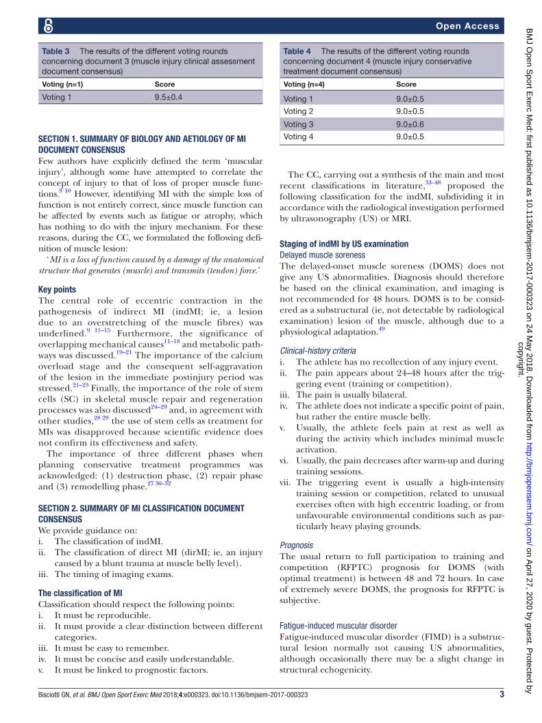
Table 3 The results of the different voting rounds concerning document 3 (muscle injury clinical assessment document consensus)
Voting (n=1) Score
Voting 1 9.5±0.4
Table 4 The results of the different voting rounds concerning document 4 (muscle injury conservative treatment document consensus)
Voting (n=4) Score
Voting 1 9.0±0.5
Voting 2 9.0±0.5
Voting 3 9.0±0.6
Voting 4 9.0±0.5
copyright. on A
pril 27, 2020 by guest. Protected by
http://bmjopensem
.bmj.com
/B
MJ O
pen Sport E
xerc Med: first published as 10.1136/bm
jsem-2017-000323 on 24 M
ay 2018. Dow
nloaded from
4 Bisciotti GN, et al. BMJ Open Sport Exerc Med 2018;4:e000323. doi:10.1136/bmjsem-2017-000323
Open Access
Clinical-anamnestic criteriai. In FIMD the athlete does not correlate onset of pain
to a precise movement and/or situation.ii. In opposite to DOMS, symptomatology is usually
monolateral. iii. In opposite to DOMS, initial cramping is often
perceived during the game, or the symptoms are per-ceived at the end of the activity.
iv. In opposite to DOMS, pain is not perceived at rest but only during activity.
v. The athlete does not indicate a precise point of pain, rather an extensive muscle area but still less than the entire muscular belly.
PrognosisThe prognosis for RFPTC in the case of FIMD (with optimal treatment) is 3–5 days.
Grade 0 lesion (indMI 0°)Given the difficulty of detecting objective oedema through US examination and especially given the impossibility of differentiating an oedema from a haem-orrhagic zone, especially in small lesions, the ‘grade 0’ staging via US examination is not advisable.
Grade I lesion (indMI I°)The indMI I° is as macroscopic (ie, detectable by radio-logical examination) structural damage.
Clinical-anamnestic criteriai. The onset is acute and can be referred to a precise
event.ii. In most cases, the athlete is unable to continue the
sporting activity he is engaged in.iii. Pain, localised and well reproducible, is perceived
during activity only.
US imaging criteriaThe US shows a lesion at the primary and secondary muscle fascicles and the presence of a haematoma. The lesion extension has a diameter smaller than that of a secondary fascicle (ie, less than 5 mm). Perifascial liquid can be present.
PrognosisThe prognosis for RFPTC (with optimal treatment) is about 15 days.
Grade II lesion (indMI II°)The indMI II° is as a macroscopic structural damage.
Clinical-anamnestic criteriai. The onset is acute and can be traced back to a very
precise event.ii. In almost all cases, the athlete fails to continue the
sporting activity in which he is engaged, and often has to stop immediately.
iii. Localised and well-reproducible pain is often perceived at rest. Sports activities are impossible.
US imaging criteriaThere is a substantial muscle discontinuity area, associated with a visible haematoma distal and proximal to the lesion. The extension of the lesion involves more than one secondary fascicle (ie, greater than 5 mm). Muscle areas adjacent to the injured zone appear to be hyperecho-genic. Commonly, a considerable amount of intermuscular, perifascial and subcutaneous fluid collection is observed.
Often, a heterogeneous haematoma may overshadow the terminal tendon area, increasing the difficulty in reaching a precise diagnosis. In these cases, the terminal tendon area is better observed by MRI exam.
Sometimes, it is objectively difficult to differentiate an indMI grade I from II by MRI exam.50 In these cases, the US exam is a valuable complementary tool to confirm the accuracy of the lesion18 45 50 with the possibility that the US itself allows to highlight the macroscopic struc-tural alterations of the muscle at the level of the lesion site occurring during an eccentric dynamic contraction (during which the US imaging shows an increase in the lesion gap) typical of the grade II lesions.45 50
PrognosisThe prognosis for RFPTC (with optimal treatment) has been estimated to be between 20 and 60 days.
Grade III lesion (indMI III°)IndMI III° is a macroscopic structural damage.
Clinical-anamnestic criteriai. The onset is acute and can be traced back to a very
precise event by the athlete.ii. In all cases, the athlete is forced to stop the sport-
ing activity in which he is engaged, at the time of the injury.
iii. The pain, localised and well reproducible, is also per-ceived at rest.
iv. Sports activities are impossible.
US imaging criteriaIn US examination, an indMI III° involves more than 85% of the total muscle diameter. Muscle areas adjacent to the lesion zone are hyperechogenic. Commonly, a considerable amount of intermuscular, perifascial and subcutaneous fluid collection is observed.
PrognosisWith optimal treatment the prognosis for RFPTC can be estimated between 60 and 90 days (in cases of serious injuries of muscles particularly involved in the sporting activity).
staging of indmI by mrI examinationDelayed-onset muscle soreness Clinical-anamnestic criteriaThe criteria stated for the US exam remain valid.
MRI criteriaA low-grade DOMS does not involve alterations of the MRI signal either in the anatomic or fluid-sensitive sequences.
copyright. on A
pril 27, 2020 by guest. Protected by
http://bmjopensem
.bmj.com
/B
MJ O
pen Sport E
xerc Med: first published as 10.1136/bm
jsem-2017-000323 on 24 M
ay 2018. Dow
nloaded from

5Bisciotti GN, et al. BMJ Open Sport Exerc Med 2018;4:e000323. doi:10.1136/bmjsem-2017-000323
Open Access
There may sometimes be transient oedema with no trace of blood. On the contrary, in the case of extremely severe DOMS, oedema in fluid-sensitive sequences appears to be superimposable to the one present in the case of indMI I°–II° (without fascicle damages) and persist for a period that may last even for 80 days.
Fatigue-induced muscular disorder Clinical-anamnestic criteriaThe criteria stated for the US exam remain valid.
MRI criteriaFIMD generally does not result in alteration of the MRI signal neither in the anatomic nor in fluid-sensitive sequences. Transient oedema with no trace of blood may occasionally be present. The difference between DOMS and DMFI is therefore in principal basically based on the clinical-anamnestic investigation.
PrognosisThe criteria given for the US examination remain valid.
Grade 0 lesion (indMI 0°)The indMI 0° is a substructural MI with functional conse-quences and not a macroscopic structural damage.
Clinical-anamnestic criteriai. The athlete can recall of a traumatic event in a repro-
ducible anatomically well-located point.ii. The athlete is often able to continue the sports
activity.iii. Pain is not felt at rest but only during the activity.
MRI criteriaNo macroscopic alterations in the muscular architecture are visible. There is a muscle oedema without disconti-nuity of the fibres and without the presence of blood. The absence of blood is the comparative parameter that allows differentiating indMI 0° from an indMI I°.
PrognosisWith optimal treatment the prognosis for RFPTC is about 8 days.
Grade I lesion (indMI I°)The indMI I° is a macroscopic structural damage.
Clinical-anamnestic criteriaThe clinical-anamnestic criteria identified for the US exam remain valid.
MRI criteriaAn increased signal within the lesion zone is visible in high-contrast gradient sequences (STIR Short-tau inver-sion recovery (STIR) and T2 weighted image). The signal increase is due to oedema and blood flow, usually from the muscle-tendon junction, which expands along the muscular fascicles producing a classic ‘bird pen’ pattern. The lesion extension has a maximum diameter,
however, smaller than that of a secondary bundle (ie, less than 5 mm). In the indMI I° perifascial fluid is also visible.
PrognosisWith optimal treatment the prognosis for RFPTC is about 15 days.
Grade II lesion (indMI II°)In opposite to US, MRI examination, thanks to its high-contrast gradient, allows to differentiate indMI II° in three variants: types A, B and C. The indMI II° (in its variants A, B and C) must be considered a macroscopic structural damage.
Clinical-anamnestic criteriaThe clinical-anamnestic criteria identified for the US exam remain valid.
MRI criteriaMacroscopic alterations of muscular architecture can be observed within the lesion zone. The extension of the lesion involves more than one secondary bundle (ie, greater than 5 mm). Frequently, the perifascial liquid may be very abundant and may expand in accordance to the anatomical structure of the injured muscle, allowing to easily see the secondary fascicles remaining structur-ally intact.
A further classification of the grade II lesions can be performed by staging the extension of the lesion (and indirectly the number of secondary fascicles)36 43:i. Grade II lesion A-type (indMI II° A) injury involv-
ing less than one-third (ie, less than ~35%) of the muscular cross-sectional area (CSA).
ii. Grade II lesion B-type (indMI II° B) injury involv-ing more than one-third but less than two-thirds (ie, between ~35% and ~65%) of CSA.
iii. Grade II lesion C-type (indMI II° C) injury involving more than two-thirds (ie, between ~65% and~85%) of CSA.
The prognosis for RFPTC (with optimum treatment) is ~20 days for an indMI II° A, ~40 days for an indMI II° B and ~60 days for an indMI II° C.
Grade III lesion (indMI III°)Generally, third-degree lesions are observed more frequently at the origin of the free proximal tendon, while they are less frequent distally. Grade III lesions are rare and represent on average 3% of all lesions observed.51 Surgery is rarely needed and is normally performed in the case of avulsion trauma with a retraction greater than 2–3 cm.3 6 45 IndMI III° is a macroscopic structural damage.
Clinical-anamnestic criteriaThe clinical-anamnestic criteria identified for the US exam remain valid.
copyright. on A
pril 27, 2020 by guest. Protected by
http://bmjopensem
.bmj.com
/B
MJ O
pen Sport E
xerc Med: first published as 10.1136/bm
jsem-2017-000323 on 24 M
ay 2018. Dow
nloaded from
6 Bisciotti GN, et al. BMJ Open Sport Exerc Med 2018;4:e000323. doi:10.1136/bmjsem-2017-000323
Open Access
MRI criteriaA third-degree injury involves more than 85% of the CSA with a haematoma that fully fills the gap of the lesion. The secondary bundles show a clear ripple and retrac-tion.
PrognosisWith optimal treatment the prognosis for RFPTC is on average between 60 and 90 days.
direct mIDirMIs are divided into contusions and lacerations. The contusions are caused by the impact of a generally rounded foreign body on the muscle surface. The lacera-tions are the result of the effect of a sharp body (cutting wounds) or the penetration of a pointed body (tip inju-ries) in the muscle.
As far as the second case is concerned, for the tears, CC does not propose a specific classification of their gravity. In this category only contusions were considered.
The CC adopted a classification of dirMI divided into three categories: minor, moderate and severe; this classifi-cation is mainly based on functional (clinical) evaluation. All the three categories of dirMI are to be considered a structural lesion.i. Minor dirMI (dirMI GMi) where movement is
possible for more than half of the range of motion (ROM) of the involved joints is possible. The US and MRI exam show the presence of a limited haemato-ma. The prognosis (with optimal treatment) is be-tween 3 and 10 days.
ii. Moderate dirMI (dirMI GMo) where movement is possible for less than half but more than one-third of the entire ROM of the involved joints is possible. The US and MRI exams show the presence of a diffuse haematoma. The prognosis (with optimal treatment) ranges from 10 to 50 days depending on the exten-sion of the haematoma itself.
iii. Severe dirMI (dirMI GS) where the movement is possible for less than one-third of the total range of movement of the involved joints is allowed. The US and MRI exam show the presence of widespread haematoma and muscle fibre crushing. The progno-sis (with optimal treatment) is between 50 and 70 days.
On examination of US and MRI, the dirMI does not differ significantly from the indMI, showing a more or less circumscribed oedema or haematoma, with a possible discontinuity of the secondary fascicles. In the US examination, the appearance of a haematoma is quite variable and depends on the trauma dating. A recent and high-pressure haematoma is sometimes more echogenic because of its high cellular content and can, therefore, remain difficult to differentiate from muscle tissue. In any case, it important to note that a haematoma in the first 24 hours post-trauma may have a very variable appearance, from not echogenic at all or hypoechogenic to a frankly hyperechogenic appearance. Finally, in the following 2–3
days the haematoma moves to hypoechogenicity or not echogenicity appearance.52
It should be noted that in dirMI the extent of haema-toma is not necessarily correlated, as opposed to indMI, with the severity of the lesion and consequently with the severity of the prognosis. In fact, a direct trauma causes vascular damage and internal bleeding. Structural fibre lesion is rare since the contusing force tends to crush them.43 In other words, a diffused haematoma can only be caused by a rupture of the vessels and not necessarily by the loss of continuity of muscle fibres, as it is the case with indMI. For this reason, in dirMI the prognosis is usually better than the indMI of equal size.43 45
The timing of the imaging examThere is no consensus in the literature as to the optimal timing to perform the MRI or the US after the injury event.42 53 As far as the US examination is concerned, the timing of the exam execution that is more consensual would seem to be ~48 hours from the injury.41 50 54 There-fore, this is the indication provided by the CC.
Regarding MRI examination, some authors55 demon-strated the signal invariance in the fluid-sensitive sequences within the so-called ‘acute phase’, that is, during the first 7 days after injury. However, in the present study, the MRI signal recorded in the fluid-sensi-tive sequences is considered in its entirety, that is, without differentiating the oedema from the blood. Indeed, one of the most important parameters regarding the choice of timing of the imaging is that it can accurately discern whether the fluid content inside and around the area is oedema and/or haematic in nature. This aspect is funda-mental when it comes to differentiating an indMI 0° from an indMI I°, given in particular the important prognostic difference between the two different types of lesion (8 days vs 15 days) and the resulting obvious diversity of the rehabilitation programmes to be adopted. As already mentioned, such differentiation is only possible through MRI assessment, provided that it is carried out within a proper ‘time window’. Based on studies performed on brain bleeding states, and in absence of any study concerning muscle haematoma in different states,56–61 this CC suggests that the ideal observation window lies between the third and fifth postlesion days. During this period, it is possible to differentiate the oedema from the blood by comparing the T1 and the T2 sequences.56–61 In any case, it is important to note that muscle haematoma sometimes shows, at MRI examination, a rather variable appearance compared with the lesion dating; therefore, the appearance may sometimes differ from that observed in a haematoma at the cerebral tissue level.56 57 61 It is, therefore, advisable to exercise caution in data interpre-tation.
seCTIon 3. summAry of mI ClInICAl AssessmenT doCumenT ConsensusDespite the increasing imaging utilisation in MI diagnos-tics, clinical skills remain a fundamental point. According
copyright. on A
pril 27, 2020 by guest. Protected by
http://bmjopensem
.bmj.com
/B
MJ O
pen Sport E
xerc Med: first published as 10.1136/bm
jsem-2017-000323 on 24 M
ay 2018. Dow
nloaded from

7Bisciotti GN, et al. BMJ Open Sport Exerc Med 2018;4:e000323. doi:10.1136/bmjsem-2017-000323
Open Access
to the CC conclusion, clinical evaluation must basically consist of three distinct, but highly complementary phases:i. Anamnesis (AN; history). ii. Inspection (IS). iii. Clinical examination.
AnamnesisAN is a fundamental part of the clinical evaluation. In order to rationalise the anamnestic examination, the CC recommends to follow the key points below:i. Pay attention whether in the past or recent AN similar
lesion to the one currently evaluated has occurred, or in an anatomically adjacent area: this finding is crucial to identify possible recent or late reinjuries.
ii. The fact that the patient can easily recall of the moment of trauma and indicates with exact accu-racy the precise area of pain is highly suggestive of a structural muscular injury. This diagnostic suspi-cion could be confirmed by the fact that the athlete has failed to complete the rest of training session or competition.
iii. Conversely, a cramping sensation, though progres-sive, would be strongly suggestive of an ultrastructur-al lesion.
iv. The young age and the particular anatomic location of the pain site (anterior superior iliac spine, anterior inferior iliac spine, ischial tuberosity) may be highly suggestive of the diagnosis of an apophyseal injury.
InspectionIS is the second phase of the clinical evaluation. The CC recommends the following key points to be respected during the IS:i. Check for swelling.ii. Check for the presence of haematoma (strongly sug-
gestive of the indirect lesion) or ecchymosis (strongly suggestive of direct trauma).
iii. Check for the presence of gap or clear muscle retraction.
iv. Check for changes in the muscular profile in the region of the suspected lesion in comparison to the contralateral muscle.
Clinical examinationA proper clinical examination must be based on the following points:i. Palpation.ii. Checking of ROM.iii. Stretching.iv. Functional manoeuvres.
PalpationThe palpation exam (PE) requires specific skill set and experience of the clinician. The PE shall be conducted in two ways:i. PE mode 1: The patient is positioned in a way that
the examined muscle is in a slightly elongated posi-tion (hence slightly contracted eccentrically).
ii. PE mode 2: The muscle to be examined must be completely relaxed. The PE should be repeated sev-eral times, according to the two above-mentioned modalities, both on the entire muscle belly and on the area/point of pain indicated by the patient. The clinician should get information, obvious-ly subjective, from the skin, subcutaneous tissue, fascia and muscle. The PE should be performed, when applicable, in a comparative manner and in both modalities described above, exerting a mod-erate pressure, and in a proximal-distal direction and vice versa, always following the orientation of the fibres.
The PE aims to verify:i. The tone of the muscles affected by the alleged le-
sion compared with the contralateral one. In addi-tion, it is of utmost importance to check the tone of the muscles adjacent to the presumably injured one, as a tone alteration may suggest a high-grade lesion.18 32
ii. The presence of gap or clear muscle retraction.iii. The existence of stiff zones.iv. The existence of previous fibrotic areas or altered
myofascial adhesion.v. The existence of painful areas or otherwise
impaired perception by the patient.
Checking of ROMThe ROM of the proximal and distal joints (when appli-cable) to the injured muscle should be checked (eg, hip and knee in case of a rectus femoris lesion).
StretchingA stretching of the injured muscle should be performed in the following modalities:i. Active modality.ii. Passive modality.
Generally, a structural lesion results in pain on both passive and active stretching, while in a functional pathology (such as the DOMS) stretching may provide a pleasant feeling to the patient.
Functional manoeuvresThe injured muscle should be tested in three ways:i. Maximum isometric contraction.ii. Concentric contraction of medium intensity against
the operator’s resistance. iii. Medium effort eccentric contraction against opera-
tor’s resistance with no pain.Quantification (from 0 to 10) of the perceived pain
in accordance with the visual analogue scale62 will be required for each type of contraction.
Finally, in order to facilitate a summary of the main information of the clinical evaluation exam, CC proposes the adoption of two dedicated tabs of which we provide an example in online supplementary annex 1 and annex 2.
copyright. on A
pril 27, 2020 by guest. Protected by
http://bmjopensem
.bmj.com
/B
MJ O
pen Sport E
xerc Med: first published as 10.1136/bm
jsem-2017-000323 on 24 M
ay 2018. Dow
nloaded from

8 Bisciotti GN, et al. BMJ Open Sport Exerc Med 2018;4:e000323. doi:10.1136/bmjsem-2017-000323
Open Access
seCTIon 4. summAry of mI ConservATIve TreATmenT doCumenT ConsensusIn the fourth document, all the non-surgical treatments of MI are considered as conservative treatments. The fourth document provided the following points:i. The conservative treatment of indMI.ii. The conservative treatment of dirMI.iii. The physical therapy into the conservative treatment
of MI.iv. The pharmacological treatment of MI.
The treatment of doms, fImd and grade 0 lesion (indmI 0°)While there is no evidence in the literature concerning the conservative treatment of DOMS, FIMD and indMI 0°, the CC merely proposes treatment guidelines based on expert advice (level of evidence IV).
It is possible to summarise the main points of the rehabilitation programme as follows:i. Suspension or reduction of the functional load
until total or partial resolution of symptoms.31 63
ii. Contrast therapy (hot/cold).64 65
iii. Hydrokinesitherapy.64 65
iv. Endogenous thermotherapy.66
v. Massage.67 68
It is important to note that, despite the controversy in literature69–71 of the above-mentioned treatment, many athletes are seeking them because they feel their bene-fits.67 68
Treatment guidelines according to the biological tissue repair steps for grade I indirect lesions (indmI I°) and onwardsAs already discussed, muscle tissue repair/regen-eration process is completed in a longer or shorter period depending on the severity of the lesion. During this period, there are well-defined biolog-ical phases (first phase of destruction, the second phase of repair/regeneration and third phase of remodelling).31 63 The CC, therefore, recommends a three-phase break-up rehabilitation protocol. Each of these phases must be characterised by a well-defined type of muscular contraction that is consistent with the biological condition observed within the injured area.31 63 Therefore, the rehabilitation programme should be set as follows:
The first phase (approximately between the second and the fifth to seventh postlesion days)The biological situation within the injured areaAt the beginning of the first phase (second postle-sion day), the necrotised parts of the muscle fibres are removed by the macrophages. At the same time, the formation of the scarring connective tissue within the central lesion zone (CZ; ie, the gap of the lesion) by fibroblasts starts. At the end of the first phase (approxi-mately seventh postlesion day), the repairing processes of the muscle cells extend beyond the old basal laminae cylinders in the CZ and begin to penetrate through the scar zone.10 27 31 32 63 72 73
Type of contraction in the first phaseConsidering that the first 5–7 postlesion days are char-acterised by the fact that the CZ has not yet developed a sufficiently dense and compact scarring connective tissue, the major risk in this period is that an excessive muscle contraction, especially with eccentric modality, increases the already existing lesion gap. Therefore, the type of contraction recommended in this first phase is an isometric modality. In fact, during the isometric contraction there is no myofilaments slip-page and, therefore, there is no macrochange of the muscle length. For this reason, the CZ lesion gap is not structurally stressed.74 The intensity of the isometric contraction required must, however, remain below the threshold of pain.63
In this first phase, the CC also recommends:i. In the immediate postinjury period (24–72 hours) it
is advisable to apply the PRICE (Protection, Rest, Ice, Compression, Elevation) principle.75
ii. It is possible to consider having a short rest period and/or relative immobilisation immediately after the injury. This rest period optimises the formation of connective tissue by fibroblasts within the CZ, there-by reducing the risk of recurrences.31 63 76 However, rest and immobilisation should be reduced to only the first postlesion days (3–5 days).31 63 76
iii. In the first 72 hours postlesion, physical therapies that induce endothermia should be avoided in order to avoid the possible increase in blood extravasation.
iv. All forms of massage on the affected area should be avoided. The practice of massage is allowed only af-ter the completion of tissue healing processes.77
v. If there is an excessive haematoma formation with-in the injured area, it is advisable to proceed to an echo-guided aspiration before the haematoma or-ganisation.76 78
second rehabilitation phase (approximately between 8th and 14th postlesion days)Clinical and functional data and imaging techniques are the criteria for proceeding from the first to the third phase of rehabilitation.
Clinic criteriai. Resolution of swelling, if initially present.77 79
ii. Absence of pain in response to maximal isometric contraction.77 79
iii. Absence of pain in response to end-range stretching tests carried out in the active and passive modes.49 77 79
iv. Complete ROM of the joints involved in the move-ment.77 79
Imaging criteriai. Reduction of the lesion gap as observed with US or
MRI imaging.80 81
ii. The presence of granulation repair tissue within the CZ as revealed by the US.80 81
copyright. on A
pril 27, 2020 by guest. Protected by
http://bmjopensem
.bmj.com
/B
MJ O
pen Sport E
xerc Med: first published as 10.1136/bm
jsem-2017-000323 on 24 M
ay 2018. Dow
nloaded from

9Bisciotti GN, et al. BMJ Open Sport Exerc Med 2018;4:e000323. doi:10.1136/bmjsem-2017-000323
Open Access
The biological situation within the injured areaAt this stage, the scar zone in the CZ is further condensed and reduced in size, and myofibres fill the residual gap of the CZ.10 27 31 32 63 72 73 During this phase, the granulation tissue gains compactness and elasticity.82 However, the possible weak points of the lesion repair in this and the next phase44 45 could be:i. Immature scar tissue within the CZ.80
ii. Healthy muscle tissue adjacent to the CZ which can be overtensioned.80
Type of contraction in the second phaseThe CC recommends that the second phase of treatment respects the following points:i. The introduction of progressively intense exercises
based on concentric contractions. During a concen-tric contraction, the bulk of the muscle shortens due to the sliding motion of the myofilaments with a rel-atively constant force proportional to the external load. Subsequently, the CZ is not subjected to trac-tion and the jagged muscle edges, undergoing reor-ganisation/repair, avoiding diastasis.83
ii. The concentric contraction can be manual at the beginning and subsequently with isotonic equip-ment (ie, Olympic weightlifting devices).84
iii. The eccentric (ie, negative) phase of the move-ment must, in all cases, be reduced to the minimum possible intensity.77
iv. Since there is evidence85 that a rehabilitation protocol, following an indMI which includes exer-cises aimed to stabilise and strengthen the ‘core’ muscles (in particular abdominal, quadratus lum-borum and paravertebral muscles) yields better re-sults (in terms of reduced relapses and in an earlier return to sport), a ‘core stability program’ should be systematically introduced in the rehabilitation plan.86
v. Stationary bike and deloaded run can be introduced during this phase.79
Third rehabilitation phase (~14th–21st day, postinjury)Clinical data and imaging techniques are the criteria for proceeding from the second to the third phase of reha-bilitation.
Clinical criteriai. The absence of pain in response to concentric con-
traction performed at increasing intensity against re-sistance performed by the physiotherapist.77 79
ii. The absence of pain in response to submaximal eccentric contraction.77 79
Imaging criteriai. The substantial disappearance of the lesion gap on
US or MR examination.80 81 ii. The presence of compact granulation repair tissue
within the CZ as revealed by US or MR.80 81
The biological situation within the injured areaIn this phase, the myofibres intertwining is effectively completed by the interposition of a small amount of scar tissue. The remodelling phase may last more than 60 days, depending on the anatomical extent of the injury.
Treatment during the third week of the postinjury should, therefore, respect the following points:i. The inclusion of isokinetic exercises76 followed by: ii. The inclusion of elastic resistance exercises where
the intensity of the eccentric phase is progressively increased, followed by77 79:
iii. Exercises predominantly based on eccentric contrac-tions of progressively increasing intensity.77 79
iv. The introduction of stretching. Stretching must be introduced gradually and exercises must not cause the onset of pain. The time of elongation initially is 10–15 s and subsequently up to 1 min, in order to induce a durable, and not just a transient, plastic deformation within the area of structural reorganisa-tion.77 79 87
v. Running may be introduced during this phase, on the condition that dynamometric values of the in-jured muscle have been reinstated to at least 70% of the preinjury level or that of the opposite limb.77
vi. Sport-specific exercises can be introduced with caution at the end of the third phase.77
phase durationThe duration of each phase is consistent with the dynamics of the healing processes occurring in the muscle tissue affected by the injury and therefore, the duration of each of the three phases is directly related to the severity of the injury. In other words, for every grade 3 lesion, the duration of each of the three phases will vary. The duration of each phase has to be determined ad personam, in accordance with the clinical and imaging criteria required for proceeding from one phase to the next. Therefore, although rehabilitation indMI I°, indMI II° and indMI III° will be subdivided into three phases, the duration of phases will be different, and progression is not time based, but clinical, functional and imaging criteria based.77 79
The conservative treatment of haematomaSince the blood loss causes the formation of a haematoma, it is necessary to distinguish between intermuscular haematoma (interH) and intramuscular haematoma (intraH).
interH: In this case, the fascia appears to be injured so that the blood can be released from the muscle belly. Pain generally persists for a period ranging from 24 to 48 hours.8889
intraH: In this case, the muscle fascia remains intact and the blood extravasation remains confined within the injured muscle. Since the blood collection within the muscle produces an osmotic gradient, the swelling can persist, or increase, beyond 48 hours postlesions.90
copyright. on A
pril 27, 2020 by guest. Protected by
http://bmjopensem
.bmj.com
/B
MJ O
pen Sport E
xerc Med: first published as 10.1136/bm
jsem-2017-000323 on 24 M
ay 2018. Dow
nloaded from

10 Bisciotti GN, et al. BMJ Open Sport Exerc Med 2018;4:e000323. doi:10.1136/bmjsem-2017-000323
Open Access
The CC recommends that the treatment of the haema-toma follows the following points:i. Into the case of intraH, the PRICE principle needs
to be changed. Indeed, into the intraH compression therapy should be avoided in order to avoid the onset of a compartmental syndrome.22 Furthermore, there is no need to apply the principle of the ‘protection’ of the injured limb. On the contrary, in order to fa-cilitate the haematoma reabsorption in intraH, early mobilisation is preferable since from the second-day postlesion.79 91
ii. In the presence of excessive haematoma formation, it is advisable to proceed to an ultrasound-guided aspiration before the solid haematoma organisa-tion.76 78
iii. The so-called ‘contrast therapies’ (ie, the alternate administration of hot and cold in order to speed up the absorption of haematoma) can be initiated only at the end of the haemorrhagic phase, so usually not before 72 hours.92
iv. Massage must be avoided in the first postlesion days, in order to avoid a possible exacerbation of the hae-morrhagic process.79 Massage can be started when the haematoma has completely disappeared.79
Instrumental therapy in the conservative treatment of MIIn the first postlesion days, it is of paramount importance to allow the implantation of muscle resident stem cells (MRSC) inside the injured area.93–95 The MRSC implant process is strongly dependent on the injured muscle angiogenesis process. This process is stimulated by both voluntary exercise and neuromuscular electrostimula-tion (NMES).93–95 For this reason, the use of NMES by promoting an early angiogenesis in the post-traumatic regeneration phase could increase both the presence of MRSC and postnatal muscle-derived stem cells (MDSC) resulting from the vascular endothelium. In fact, some authors have advanced the hypothesis that MDSCs originate from blood vessels.96–98 The use of NMES for vascularisation should be encouraged until the end of the regeneration phase (ie, up to about the third postlesion week).99 100
Ultrasound therapy There is limited evidence that ultrasound therapy (UST) is able to increase the levels of basic fibroblastic growth factor and vascular endothelial growth factor (VEGF).101 The use of UST in MI is justified by the fact that tissue micromassage induced by the UST frequencies can generate an antalgic effect.102 However, it is important to note that UST can inhibit the expression of the RNA messenger (mechano growth factor (MGF) mRNA).102 The MGF mRNA plays an important role in the insulin-like growth-1 (IGF)-1 upregulation mechanism. Therefore, it is advisable not to use UST in the first 24 hours postlesion.102 In summary, UST may be recom-mended in the conservative treatment of MI after the first 24 hours postinjury.101 102
Laser therapy Many studies have shown that laser therapy (LT) can reduce the inflammatory process of the damaged muscle tissue,103 speed up the tissue regeneration/repair processes by stimulating myogenic processes,104 optimise the oxidative metabolism by increasing ATP synthesis105–107 and stimulate the synthesis of RNA and regulatory protein cycles in cell proliferation.108 Finally, LT could increase the expression of VEGF.109 110 There-fore, the use of LT in the treatment of MIs appears to be justified by sufficient evidence.111
Hyperthermia (endogenous thermotherapy)Hyperthermia therapy (HT) (ie, microwave diathermy) increases the temperature of the deep tissue up to 41°C–45°C, through electromagnetic energy. HT has proven to be able to stimulate the tissue repair processes, diminish pain symptoms, increase tissue flexibility and reduce muscular and joint stiffness.112 113 Some studies point out its effectiveness even in the specific field of skeletal muscle repair/regeneration processes.112–117 HT may also facilitate the resolution of haematoma following MI.118 Furthermore, HT increase increases pain threshold through a direct action of heat on free nerve endings119 and trunks120 sufficiently to block the transmission of pain.119 120 Finally, HT reduces the muscle spasm resulting from MI contributing to the pain decreasing.117 121 122 Therefore, for CC experts’ opinion, the use of HT in the treatment of MI is justified by a sufficient evidence.
Hyperbaric oxygen therapyCurrently, in the literature, there is a total lack of evidence on the effectiveness of hyperbaric oxygen therapy (HOT) in the MI conservative treatment, both indirect and direct. Therefore, the HOT needs further evidence to prove its effectiveness in MI treatment.
Extracorporeal shock wave therapyExtracorporeal shock wave therapy (ESWT) is a ‘mechan-otherapy’, whose biological effects have been now widely described: modulation of inflammation and macro-phages activity, angiogenesis and tissue-specific growth factor (GF) induction, SC activity stimulation, besides antioedema and antinociceptive effect.123–125 Based on its ‘trophic’ effects and in accordance with the Consensus Statement of the International Society for Medical Shockwave Treatment126 regarding clinical indications, the CC suggests to apply ESWT in DOMS, reserving to ‘ESWT skilled hands’ its application after muscle lesions as possible adjuvant therapy, in order to reduce oedema and pain, improve healing and accelerate remodelling of tissue fibrosis regardless of the degree of the injury.123–127
pharmacological treatment of mIThe most commonly used pharmacological therapies have been taken into consideration by the CC. The CC’s recommendations in this specific field can be summarised as follows:
copyright. on A
pril 27, 2020 by guest. Protected by
http://bmjopensem
.bmj.com
/B
MJ O
pen Sport E
xerc Med: first published as 10.1136/bm
jsem-2017-000323 on 24 M
ay 2018. Dow
nloaded from

11Bisciotti GN, et al. BMJ Open Sport Exerc Med 2018;4:e000323. doi:10.1136/bmjsem-2017-000323
Open Access
Non-steroidal anti-inflammatory drugsNon-steroidal anti-inflammatory drug (NSAID) prescrip-tion in the early postinjury period, especially in the case of a medium to severe MI (II°–III°), can be considered as a justified medical treatment.128 It should be remem-bered, however, that there is still no evidence of the benefits and the adverse effects that the intake of non-se-lective NSAIDs may provide.129
The use of analgesic drugsThe most commonly used analgesic drug is parac-etamol.129 Its analgesic effect is mainly related to the action on the systems involved in pain modulation, such as serotonin system. Paracetamol does not act at periph-eral level and therefore does not have anti-inflammatory activity. Some studies recommend the use of analgesics for low-intensity pain in neuromuscular disorders in the first postlesion days.130 In conclusion, the CC suggests that analgesics can be used in case of pain in the first postlesion days.
The use of calcium chelantsChelation therapy is a drug therapy utilising chelation to treat some forms of intoxication due to heavy metals. In the case of MI, chelators may limit the calcium over-load phase (calcium is an alkaline earth metal). The most widely used and well known of chelants is EDTA. EDTA would seem to be able to limit the histopatholog-ical changes present in the context of muscular lesions.131 However, the use of calcium chelants needs further evidence to be justified for the treatment of MIs.
The use of corticosteroidsThe use of corticosteroid drugs should be avoided. Indeed, among different adverse effects,67 they involve delayed absorption of haematoma, increased necrosis in the injured myofibrils, delay in muscle tissue regenera-tion processes and loss of muscle strength.132
The use of muscle relaxant drugsThe use of muscle relaxant drugs for a limited period could, in theory, reduce, or at least contain, the self-aggra-vation stage of the lesion. In any case, CC recommends that the use of muscle relaxants requires further evidence-based studies regarding their efficacy in MI treatment.
ActoveginActovegin is a deproteinised derivative—ultrafiltrate of calf serum from animals under 8 months of age. It is important to remember that the use of Actovegin may, although in rare cases, have serious adverse effects (anaphylactic shock with multiorgan failure in a cyclist after intravenous administration of Actovegin),133 also if in effect this effect is most likely not due to Actovegin administration but due to a bacterial/infection contam-ination. In the literature , it is still yet to define the Actovegin’s active compounds,134 in any case current literature suggests that Actovegin shows antioxidant and antiapoptotic properties, and may also have an
upregulating action on macrophage responses following muscle repair.134 However, the Actovegin’s clinical efficacy is supported by only one new original research article.135 Finally, the CC, after the examination of the current liter-ature, believes that Actovegin needs further evidence to demonstrate its efficacy in the MI treatment.133–139
TraumeelTraumeel is a homeopathic medicine marketed in Italy under the name of Arnica compositum (most recently under the name of Hell Traumeels), in the form of a gel or solution for injection. In literature, there are studies (randomised controlled trials) that show its efficacy in the reduction of pain and swelling following MIs,140 in rotator cuff syndrome141 and in ankle and knee sprains.142 143 Finally, Traumeel has proven to be a well-tol-erated product with little adverse effects.144–146 However, since Traumeel is a homeopathic product, this CC still expresses a certain perplexity about its effectiveness and hopes that further evidence in the future will confirm its therapeutic validity.
LosartanLosartan is an angiotensin II antagonist drug used to treat essential hypertension. Some studies show that losartan, if used in a time period ranging from 3 to 7 days on mouse model,147 in addition to platelet rich plasma (PRP) therapy (PRPt),148 is able to reduce fibrotic processes in MI, promote tissue repair/regeneration processes and stimulate angiogenesis. Further and more thorough studies will have to demonstrate the effective-ness and safety in humans.
The use of mesenchymal stem cellsMesenchymal stem cells (MSC) are adult, immature, undifferentiated and multipotent SCs that originate from mesoderm, and which can be extracted from the adipose and synovial tissues, blood, skeletal muscle, umbilical cord, placenta and bone marrow.28 The Australasian College of Sport and Exercise Physicians, formulating a change29 to the previously stated position statement on the use of MSCs,28 strongly advised to restrict the use of MSCs only into well-controlled clinical research trials. This recommenda-tion is the result of serious complications (glioproliferative lesion of the spinal cord arising from the use of MSCs).149 Therefore, the CC does not advocate the use of MSCs as part of MI care, as long as scientific evidence does not confirm its effectiveness and safety.
The use of platelet GFsThe methodology using PRP is based on the rationality that GFs contained in platelets stimulate the proliferation of skeletal muscle cell progenitors, guide cell differenti-ation and modify the local inflammatory response.150–154 On this rationale of application, research investigating the efficacy of PRP in cartilage, bone and muscular-tendon diseases was carried out.150–154 However, there are many problems actually still unresolved as well as many doubts about the actual efficacy of PRPt.
copyright. on A
pril 27, 2020 by guest. Protected by
http://bmjopensem
.bmj.com
/B
MJ O
pen Sport E
xerc Med: first published as 10.1136/bm
jsem-2017-000323 on 24 M
ay 2018. Dow
nloaded from
12 Bisciotti GN, et al. BMJ Open Sport Exerc Med 2018;4:e000323. doi:10.1136/bmjsem-2017-000323
Open Access
Synthetically, the various fields of investigation still in progress can be summarised as follows: i. Nomenclature and classification.155–158 ii. Standardisation and preparation of the prod-
uct.153 159 160 iii. The specific indications of PRPt.161 162 iv. The release kinetics of the various GFs.163–165 v. The timing of administration.163–165
After discussing the above points, CC formulates the following recommendations:i. As there is currently no consensus concerning the
PRP classification,155–158 166 a universally shared classi-fication of all different autologous blood and plasma preparations of PRPt has to be urgently developed.
ii. Since the effects of various PRP preparations in func-tion of their different formulations are not yet well understood,153 159 160 166 there is a need for a consen-sus on standardised product preparation.
iii. Since a key point in PRPt is the full understanding of the different roles of the various GFs and differ-ent cytokines within the different biological con-texts considered,161 162 167 168 the CC underlines the strong need for further major studies to clarify the specific indications of different types of GF and cy-tokines present in different types of PRPt currently proposed.
iv. Since any pathological conditions may have a ther-apeutic window within which PRPt would be able to make the greatest benefit,163–165 the CC hopes that further studies will be implemented in the future to elucidate the kinetics of release and activation of cytokines and GFs, and the subsequent activation methods according to different tissues and differ-ent pathological conditions.
v. As the optimum timing of PRP administration remains an essentially unknown aspect,163–165 the CC underlines the strong need for further high level of evidence studies to clarify the timing of admin-istration, according to the specific indications of the different types of GF and cytokines present in different types of PRPs currently proposed.
CC ConClusIonsThe main strength of the Italian CC on guidelines for conservative treatment of lower limb injuries in athlete was the presence of recognised leading experts in this field with different backgrounds. This multidisciplinary expert attendance guaranteed a deep and comprehen-sive approach to the topic.
During the CC some important points of discussion and reflection emerged. They can be summarised as follows:i. The CC underlined the importance of a full
understanding of biological and biomechanical mechanisms of MI aetiology. This is a key point in applying rational conservative treatment protocols for MI.
ii. The presented MI classification is reproducible, clearly distinct, easy to memorise, concise, easy to un-derstand and linked to prognostic factors.
iii. The CC referred clear references concerning the presented clinical evaluation of MI.
iv. The CC presented a rehabilitation programme plan conceived in accordance with the biological repair phases of muscle tissue.
v. The CC presented a clear overview of the state of the art of pharmacological and instrumental therapy concerning MI.
fuTure dIreCTIonsThe CC hopes in the future to develop and study the following points:i. A greater control of the actual effectiveness and
scientific rationality of the application of physical therapy regarding MI.
ii. The implementation of research projects searching for more evidence regarding pharmacological ther-apies in general and especially regarding the use of PRPt and MSCs.
Author affiliations1Qatar Orthopaedic and Sport Medicine Hospital, Doha, Qatar2Centro Studi Kinemove Rehabilitation Centers, Pontremoli, Italy3Istituto Clinico Humanitas, Milano, Italy4FC Internazionale, Milano, Italy5Istituto Clinico S Anna, Brescia, Italy6Università Statale, Milano, Italy7Polo Pontino, Università La Sapienza di Roma, ICOT, Latina, Italy8CTO Hospital, Torino, Italy9UOS Angiografia e Radiologia Interventistica, Ospedale delle Apuane, Massa-Carrara, Italy10Istituto Clinico Villa Aprica, Como, Italy11Atalanta BC, Bergamo, Italy12Ospedale Carlo Poma, Mantova, Italy13Istituto Ortopedico Galeazzi AC Milan, Milan, Italy14Vicenza Calcio 1902, Vicenza, Italy15Feralpisaló, Salò, Italy16Istituto Ortopedico Gaetano Pini, Milano, Italy17Ospedale e Casa di Cura Koelliker, Torino, Italy18Ospedale di Latisana, Latisana, Italy19Ospedale Unico della Versilia, Asl Nordovest, Lido di Camaiore, Italy20AORNA Cardarelli, Napoli, Italy21Università Cattolica, Milano, Italy22Studio Riabilita, Civitanova Marche, Italy23Azienda USL Toscana Nord Ovest, Lucca, Italy24Azienda Ospedaliero-Universitaria “Policlinico”, Bari, Italy25Università di Brescia, Brescia, Italy26Università di Chieti, Pescara, Italy27Centro Kinetic, Bergamo, Italy28Clinica Montallegro, Genova, Italy29Centro Sanitario, Livorno, Italy30Università Campus Biomedico, Roma, Italy31Federazione Monegasca di Sci, Monaco, Monaco32Studio FKT Tenconi, Genova, Italy33Istituto ‘Marco Pasquali’-ICOT, Latina, Italy34FAF Jenia Centre Med Sport, Algeri, Algeria35Maria Cecilia Hospital, Cotignola, Italy
funding The authors have not declared a specific grant for this research from any funding agency in the public, commercial or not-for-profit sectors.
Competing interests None declared.
copyright. on A
pril 27, 2020 by guest. Protected by
http://bmjopensem
.bmj.com
/B
MJ O
pen Sport E
xerc Med: first published as 10.1136/bm
jsem-2017-000323 on 24 M
ay 2018. Dow
nloaded from

13Bisciotti GN, et al. BMJ Open Sport Exerc Med 2018;4:e000323. doi:10.1136/bmjsem-2017-000323
Open Access
patient consent Not required.
provenance and peer review Not commissioned; externally peer reviewed.
open Access This is an Open Access article distributed in accordance with the Creative Commons Attribution Non Commercial (CC BY-NC 4.0) license, which permits others to distribute, remix, adapt, build upon this work non-commercially, and license their derivative works on different terms, provided the original work is properly cited and the use is non-commercial. See: http:// creativecommons. org/ licenses/ by- nc/ 4. 0/
Published by the BMJ Publishing Group Limited. For permission to use (where not already granted under a licence) please go to http://www. bmj. com/ company/ products- services/ rights- and- licensing/
referenCes 1. Alonso JM, Edouard P, Fischetto G, et al. Determination of future
prevention strategies in elite track and field: analysis of Daegu 2011 IAAF Championships injuries and illnesses surveillance. Br J Sports Med 2012;46:505–14.
2. Freckleton G, Pizzari T. Risk factors for hamstring muscle strain injury in sport: a systematic review and meta-analysis. Br J Sports Med 2013;47:351–8.
3. Pollock N, James SL, Lee JC, et al. British athletics muscle injury classification: a new grading system. Br J Sports Med 2014;48:1347–51.
4. Ekstrand J, Hägglund M, Waldén M. Epidemiology of muscle injuries in professional football (soccer). Am J Sports Med 2011;39:1226–32.
5. Hägglund M, Waldén M, Ekstrand J. Risk factors for lower extremity muscle injury in professional soccer: the UEFA Injury Study. Am J Sports Med 2013;41:327–35.
6. Maffulli N, Oliva F, Frizziero A, et al. ISMuLT Guidelines for muscle injuries. Muscles Ligaments Tendons J 2013;3:241–9.
7. Griffin DR, Dickenson EJ, O'Donnell J, et al. The Warwick Agreement on femoroacetabular impingement syndrome (FAI syndrome): an international consensus statement. Br J Sports Med 2016;50:1169–76.
8. Vanbelle S, Lesaffre E. Modeling agreement on bounded scales. Stat Methods Med Res 2017;1:096228021770570.
9. Brooks SV, Zerba E, Faulkner JA. Injury to muscle fibres after single stretches of passive and maximally stimulated muscles in mice. J Physiol 1995;488(Pt 2):459–69.
10. Brooks SV. Current topics for teaching skeletal muscle physiology. Adv Physiol Educ 2003;27(1-4):171–82.
11. Garrett WE. Muscle strain injuries: clinical and basic aspects. Med Sci Sports Exerc 1990;22:439–43.
12. Armstrong RB. Initial events in exercise-induced muscular injury. Med Sci Sports Exerc 1990;22:429–37.
13. Armstrong RB, Warren GL, Warren JA. Mechanisms of exercise-induced muscle fibre injury. Sports Med 1991;12:184–207.
14. Aärimaa V, Rantanen J, Best T, et al. Mild eccentric stretch injury in skeletal muscle causes transient effects on tensile load and cell proliferation. Scand J Med Sci Sports 2004;14:367–72.
15. Patel TJ, Das R, Fridén J, et al. Sarcomere strain and heterogeneity correlate with injury to frog skeletal muscle fiber bundles. J Appl Physiol 2004;97:1803–13.
16. Takala TE, Virtanen P. Biochemical composition of muscle extracellular matrix: the effect of loading. Scand J Med Sci Sports 2000;10:321–5.
17. Schache AG, Wrigley TV, Baker R, et al. Biomechanical response to hamstring muscle strain injury. Gait Posture 2009;29:332–8.
18. Volpi P, Bisciotti GN. The hamstring muscles: anatomy, biomechanics and risk injury. Med Sports 2016;69:297–307.
19. Horáková L, Strosová M, Skuciová M. Antioxidants prevented oxidative injury of SR induced by Fe2+/H2O2/ascorbate system but failed to prevent Ca2+-ATPase activity decrease. Biofactors 2005;24:105–9.
20. Anitua E, Sanchez M, Nurden AT, et al. Autologous fibrin matrices: a potential source of biological mediators that modulate tendon cell activities. J Biomed Mater Res A 2006;77A77:285–93.
21. Clanton TL. Hypoxia-induced reactive oxygen species formation in skeletal muscle. J Appl Physiol 2007;102:2379–88.
22. Kerkweg U, Petrat F, Korth HG, et al. Disruption of skeletal myocytes initiates superoxide release: contribution of NADPH oxidase. Shock 2007;27:552–8.
23. Voss P, Engels M, Strosova M, et al. Protective effect of antioxidants against sarcoplasmic reticulum (SR) oxidation by Fenton reaction, however without prevention of Ca-pump activity. Toxicol In Vitro 2008;22:1726–33.
24. Dreyfus PA, Chretien F, Chazaud B, et al. Adult bone marrow-derived stem cells in muscle connective tissue and satellite cell niches. Am J Pathol 2004;164:773–9.
25. Musaro A, Giacinti C, Borsellino G, et al. Stem cell-mediated muscle regeneration is enhanced by local isoform of insulin-like growth factor 1. Proceedings of the National Academy of Sciences 2004;101:1206–10.
26. Chazaud B. Macrophages: Supportive cells for tissue repair and regeneration. Immunobiology 2014;219:172–8.
27. Laumonier T, Menetrey J. Muscle injuries and strategies for improving their repair. J Exp Orthop 2016;3:15.
28. Osborne H, Anderson L, Burt P, et al. Australasian college of sports physicians-position statement: the place of mesenchymal stem/stromal cell therapies in sport and exercise medicine. Br J Sports Med 2016;50:1237–44.
29. Osborne H, Castricum A. Change to Australasian College of Sport and Exercise Physicians-position statement: the place of mesenchymal stem/stromal cell therapies in sport and exercise medicine. Br J Sports Med 2016;50:1229.
30. Chargé SB, Rudnicki MA. Cellular and molecular regulation of muscle regeneration. Physiol Rev 2004;84:209–38.
31. Järvinen TA, Järvinen M, Kalimo H. Regeneration of injured skeletal muscle after the injury. Muscles Ligaments Tendons J 2013;3:337–45.
32. Delos D, Maak TG, Rodeo SA. Muscle injuries in athletes: enhancing recovery through scientific understanding and novel therapies. Sports Health 2013;5:346–52.
33. Jackson DW, Feagin JA. Quadriceps contusions in young athletes. Relation of severity of injury to treatment and prognosis. J Bone Joint Surg Am 1973;55:95–105.
34. Peetrons P, Creteur P. Echographies et traumatismes musculaires aigus. Imagerie Des Parties Molles De L'Appareil Locomoteur. Montpellier: Sauramps Medical, 1993:229–35.
35. Takebayashi S, Takasawa H, Banzai Y, et al. Sonographic findings in muscle strain injury: clinical and MR imaging correlation. Journal of Ultrasound in Medicine 1995;14:899–905.
36. Boutin RD, Fritz RC, Steinbach LS. Imaging of sports-related muscle injuries. Radiol Clin North Am 2002;40:333–62.
37. Blankenbaker DG, De Smet AA. MR imaging of muscle injuries. Appl Radiol 2004;33:14–26.
38. Bordalo-Rodrigues M, Rosenberg ZS. MR imaging of the proximal rectus femoris musculotendinous unit. Magn Reson Imaging Clin N Am 2005;13:717–25.
39. Gyftopoulos S, Rosenberg ZS, Schweitzer ME, et al. Normal anatomy and strains of the deep musculotendinous junction of the proximal rectus femoris: MRI features. AJR Am J Roentgenol 2008;190:W182–W186.
40. Bryan Dixon J, Dixon J. Gastrocnemius vs. soleus strain: how to differentiate and deal with calf muscle injuries. Curr Rev Musculoskelet Med 2009;2:74–7.
41. Chan O, Del Buono A, Best TM, et al. Acute muscle strain injuries: a proposed new classification system. Knee Surg Sports Traumatol Arthrosc 2012;20:2356–62.
42. Ekstrand J, Healy JC, Waldén M, et al. Hamstring muscle injuries in professional football: the correlation of MRI findings with return to play. Br J Sports Med 2012;46:112–7.
43. Böck J, Mundinger P, Luttke G. Magnetic resonance imaging. Muscle injuries in the sport. New York: Thieme Editions, 2013:203–20.
44. Mueller-Wohlfahrt HW, Haensel L, Mithoefer K, et al. Terminology and classification of muscle injuries in sport: the Munich consensus statement. Br J Sports Med 2013;47:342–50.
45. Mueller-Wohlfart H, Ueblacker P, Garrett WE. Muscle injuries in sports. New York: Thieme Editions, 2013.
46. Bisciotti GN, Balzarini L, Volpi P. The classification of muscle injuries: a critical review. Med Sport 2015;68:165–77.
47. Patel A, Chakraverty J, Pollock N, et al. British athletics muscle injury classification: a reliability study for a new grading system. Clin Radiol 2015;70:1414–20.
48. Valle X, Alentorn-Geli E, Tol JL, et al. Muscle injuries in sports: a new evidence-informed and expert consensus-based classification with clinical application. Sports Med 2017;47:1241–53.
49. Bisciotti GN, Eirale C. Le lesioni muscolari indotte dall’esercizio: il delayed muscle soreness. Med Sport 2012;65:423–35.
50. Lee JC, Healy J. Sonography of lower limb muscle injury. AJR Am J Roentgenol 2004;182:341–51.
51. Lee J, Healy J. Imaging of lower limb muscle injury. ASPETAR Sports Medicine Journal 2012;2:142–7.
52. Koh ES, McNally EG. Ultrasound of skeletal muscle injury. Semin Musculoskelet Radiol 2007;11:162–73.
copyright. on A
pril 27, 2020 by guest. Protected by
http://bmjopensem
.bmj.com
/B
MJ O
pen Sport E
xerc Med: first published as 10.1136/bm
jsem-2017-000323 on 24 M
ay 2018. Dow
nloaded from

14 Bisciotti GN, et al. BMJ Open Sport Exerc Med 2018;4:e000323. doi:10.1136/bmjsem-2017-000323
Open Access
53. Kerkhoffs GM, van Es N, Wieldraaijer T, et al. Diagnosis and prognosis of acute hamstring injuries in athletes. Knee Surg Sports Traumatol Arthrosc 2013;21:500–9.
54. Connell DA, Schneider-Kolsky ME, Hoving JL, et al. Longitudinal study comparing sonographic and MRI assessments of acute and healing hamstring injuries. AJR Am J Roentgenol 2004;183:975–84.
55. Wangensteen A, Bahr R, Van Linschoten R, et al. MRI appearance does not change in the first 7 days after acute hamstring injury—a prospective study. Br J Sports Med 2017;51:1087–92.
56. Bradley WG. MR appearance of hemorrhage in the brain. Radiology 1993;189:15–26.
57. Colosimo C. Neuroradiologia. Milano, 2013. 58. Imajo Y, Kanchiku T, Suzuki H, et al. Intracranial epidural
hemorrhage during lumbar spinal surgery. Spinal Cord Ser Cases 2016;2(Apr 71):15040.
59. Carroll JJ, Lavine SD, Meyers PM. Imaging of subdural hematomas. Neurosurg Clin N Am 2017;28:179–203.
60. Williamson MR, Dietrich K, Hackett MJ, et al. Rehabilitation augments hematoma clearance and attenuates oxidative injury and ion dyshomeostasis after brain hemorrhage. Stroke 2017;48:195–203.
61. Allkemper T, Tombach B, Schwindt W, et al. Acute and subacute intracerebral hemorrhages: comparison of MR imaging at 1.5 and 3.0 T–initial experience. Radiology 2004;232:874–81.
62. Joyce CR, Zutshi DW, Hrubes V, et al. Comparison of fixed interval and visual analogue scales for rating chronic pain. Eur J Clin Pharmacol 1975;8:415–20.
63. Järvinen TA, Järvinen TL, Kääriäinen M, et al. Muscle injuries: biology and treatment. Am J Sports Med 2005;33:745–64.
64. Gill ND, Beaven CM, Cook C. Effectiveness of post-match recovery strategies in rugby players. Br J Sports Med 2006;40:260–3.
65. Bailey DM, Erith SJ, Griffin PJ, et al. Influence of cold-water immersion on indices of muscle damage following prolonged intermittent shuttle running. J Sports Sci 2007;25:1163–70.
66. Jalluerry C, Bataini JP, Brunin F, et al. Present clinical status of hyperthermia associated with radiotherapy. Bull Cancer 1981;68:261–7.
67. Nunes GS, Bender PU, de Menezes FS, et al. Massage therapy decreases pain and perceived fatigue after long-distance Ironman triathlon: a randomised trial. J Physiother 2016;62:83–7.
68. Poppendieck W, Wegmann M, Ferrauti A, et al. Massage and performance recovery: a meta-analytical review. Sports Med 2016;46:183–204.
69. Barnett A. Using recovery modalities between training sessions in elite athletes: does it help? Sports Med 2006;36:781–96.
70. Nédélec M, McCall A, Carling C, et al. Recovery in soccer: part I - post-match fatigue and time course of recovery. Sports Med 2012;42:997–1015.
71. Nédélec M, McCall A, Carling C, et al. Recovery in soccer : part ii-recovery strategies. Sports Med 2013;43:9–22.
72. Garg K, Corona BT, Walters TJ. Therapeutic strategies for preventing skeletal muscle fibrosis after injury. Front Pharmacol 2015;6:87.
73. Gordon T, English AW. Strategies to promote peripheral nerve regeneration: electrical stimulation and/or exercise. Eur J Neurosci 2016;43:336–50.
74. Dias da Silva SR, Gonçalves M. Dynamic and isometric protocols of knee extension: effect of fatigue on the EMG signal. Electromyogr Clin Neurophysiol 2006;46:35–42.
75. Bleakley C, McDonough S, MacAuley D. The use of ice in the treatment of acute soft tissue injury: a systematic review of randomized controlled trials. Am J Sports Med 2004;34:251–61.
76. Järvinen TAH, Järvinen TLN, Kääriäinen M, et al. Muscle injuries: optimising recovery. Best Pract Res Clin Rheumatol 2007;21:317–31.
77. Volpi P, Bisciotti GN, Corsini A. FC Internazionale medical department: indirect muscle injuries rehabilitation. Milano: Ciemme-Group Editions, 2016.
78. Smith TO, Hunt NJ, Wood SJ. The physiotherapy management of muscle haematomas. Physical Therapy in Sport 2006;7:201–9.
79. Bisciotti GN. Returnto play after a muscle lesion. Switzerland: Arthroscopy in Sports Springer International Publishing, 2015.
80. Megliola A, Eutropi F, Scorzelli A, et al. Ultrasound and magnetic resonance imaging in sports-related muscle injuries. Radiol Med 2006;111:836–45.
81. Alessandrino F, Balconi G. Complications of muscle injuries. J Ultrasound 2013;16:215–22.
82. Kannus P, Parkkari J, Järvinen TL, et al. Basic science and clinical studies coincide: active treatment approach is needed after a sports injury. Scand J Med Sci Sports 2003;13:150–4.
83. Chaudhry S, Morrissey D, Woledge RC, et al. Eccentric and concentric loading of the triceps surae: an in vivo study of dynamic muscle and tendon biomechanical parameters. J Appl Biomech 2015;31:69–78.
84. Chaouachi A, Hammami R, Kaabi S, et al. Olympic weightlifting and plyometric training with children provides similar or greater performance improvements than traditional resistance training. J Strength Cond Res 2014;28:1483–96.
85. Sherry MA, Best TM. A comparison of 2 rehabilitation programs in the treatment of acute hamstring strains. J Orthop Sports Phys Ther 2004;34:116–25.
86. Huxel Bliven KC, Anderson BE. Core stability training for injury prevention. Sports Health 2013;5:514–22.
87. Petersen J, Hölmich P. Evidence based prevention of hamstring injuries in sport. Br J Sports Med 2005;39:319–23.
88. Lynch SA, Renström PA. Groin injuries in sport: treatment strategies. Sports Med 1999;28:137–44.
89. Çağatay HH, Ekinci M, Ulusal Ş, et al. Isolated rectus muscle rupture following trauma. Nepal J Ophthalmol 2015;7:182–5.
90. Martinez-Zapata MJ, Orozco L, Balius R, et al. Efficacy of autologous platelet-rich plasma for the treatment of muscle rupture with haematoma: a multicentre, randomised, double-blind, placebo-controlled clinical trial. Blood Transfus 2016;14:245–54.
91. Prentice WE. Rehabilitation techniques for sports medicine and athletic training. 4th edn. London: McGrow-Hill, 2004.
92. Bisciotti GN. Le terapie di recupero muscolare post-esercizio. Strenght & Conditioning 2013;4:35–40.
93. Ljubicic V, Adhihetty PJ, Hood DA. Application of animal models: chronic electrical stimulation-induced contractile activity. Can J Appl Physiol 2005;30:625–43.
94. Efthimiadou A, Asimakopoulos B, Nikolettos N, et al. The angiogenetic effect of intramuscular administration of b-FGF and a-FGF on cardiac muscle: the influence of exercise on muscle angiogenesis. J Sports Sci 2006;24:849–54.
95. Bellafiore M, Sivverini G, Palumbo D, et al. Increased cx43 and angiogenesis in exercised mouse hearts. Int J Sports Med 2007;28:749–55.
96. Hudlicka O, Milkiewicz M, Cotter MA, et al. Hypoxia and expression of VEGF-A protein in relation to capillary growth in electrically stimulated rat and rabbit skeletal muscles. Exp Physiol 2002;87:373–81.
97. Asakura A. Stem cells in adult skeletal muscle. Trends Cardiovasc Med 2003;13:123–8.
98. Peng H, Huard J. Muscle-derived stem cells for musculoskeletal tissue regeneration and repair. Transpl Immunol 2004;12(3-4):311–9.
99. Pomeranz SJ, Heidt RS. MR imaging in the prognostication of hamstring injury. Work in progress. Radiology 1993;189:897–900.
100. Quintero AJ, Wright VJ, Fu FH, et al. Stem cells for the treatment of skeletal muscle injury. Clin Sports Med 2009;28:1–11.
101. Reher P, Doan N, Bradnock B, et al. Effect of ultrasound on the production of IL-8, basic FGF and VEGF. Cytokine 1999;11:416–23.
102. McBrier NM, Lekan JM, Druhan LJ, et al. Therapeutic ultrasound decreases mechano-growth factor messenger ribonucleic acid expression after muscle contusion injury. Arch Phys Med Rehabil 2007;88:936–40.
103. Cressoni MD, Dib Giusti HH, Casarotto RA, et al. The effects of a 785-nm AlGaInP laser on the regeneration of rat anterior tibialis muscle after surgically-induced injury. Photomed Laser Surg 2008;26:461–6.
104. Alves AN, Fernandes KP, Deana AM, et al. Effects of lowel-laser therapy on skeletal muscle repair: a systematic review. J Phys Med Rehabil 2014;93:1073–85.
105. Grossman N, Schneid N, Reuveni H, et al. 780 nm low power diode laser irradiation stimulates proliferation of keratinocyte cultures: Involvement of reactive oxygen species. Lasers Surg Med 1998;22:212–8.
106. Karu T. Primary and secondary mechanisms of action of visible to near-IR radiation on cells. J Photochem Photobiol B 1999;49:1–17.
107. Pastore D, Greco M, Passarella S. Specific helium–neon laser sensitivity of the purified cytochrome c oxidase, Int. J. Radiat. Biol 2000;6:863–70.
108. Renno AC, McDonnell PA, Parizotto NA, Parizotto LEL, et al. The effects of laser irradiation on osteoblast and osteosarcoma cell proliferation and differentiation in vitro. Photomed Laser Surg 2007;25:275–80.
109. Silveira PCL, da Silva LA, Pinho CA, et al. Effects of low-level laser therapy (GaAs) in an animal model of muscular damage induced by trauma. Lasers Med Sci 2013;28:431–6.
110. Martignago CCS, Oliveira RF, Pires-Oliveira DAA, et al. Effect of low-level laser therapy on the gene expression of collagen and
copyright. on A
pril 27, 2020 by guest. Protected by
http://bmjopensem
.bmj.com
/B
MJ O
pen Sport E
xerc Med: first published as 10.1136/bm
jsem-2017-000323 on 24 M
ay 2018. Dow
nloaded from

15Bisciotti GN, et al. BMJ Open Sport Exerc Med 2018;4:e000323. doi:10.1136/bmjsem-2017-000323
Open Access
vascular endothelial growth factor in a culture of fibroblast cells in mice. Lasers Med Sci 2015;30:203–8.
111. de Freitas CE, Bertaglia RS, Vechetti Júnior IJ, et al. High final energy of low-level gallium arsenide laser therapy enhances skeletal muscle recovery without a positive effect on collagen remodeling. Photochem Photobiol 2015;91:957–65.
112. Giombini A, Casciello G, Di Cesare MC, et al. A controlled study on the effects of hyperthermia at 434 MHz and conventional ultrasound upon muscle injuries in sport. J Sports Med Phys Fitness 2001;41:521–7.
113. Giombini A, Giovannini V, Di Cesare A, et al. Applicazione della termoterapia endogena nel trattamento delle lesioni muscolari da sport: studio comparativo con ultrasuoni. Br Med Bull 2007;83:379–96.
114. Sorrenti D, Casciello D, Dragoni S. Applicazione della termoterapia endogena nel trattamento delle lesioni muscolari da sport: studio comparativo con ultrasuoni. Med Sport 2000;53:59–67.
115. Lehmann JF, DeLateur BJ. Therapeutic heat. Therapeutic heat and cold. 3rd edn. Baltimore: Williams and Wilkins, 1982:404–562.
116. Kloth L. Shortwave and microwave diathermy. Thermal agents in rehabilitation. Philadelphia: F A Davis Company, 1986:177–216.
117. Goats GC. Continuous short-wave (radio-frequency) diathermy. Br J Sports Med 1989;23:123–7.
118. Lehmann JF, Dundore DE, Esselman PC, et al. Microwave diathermy: effects on experimental muscle hematoma resolution. Arch Phys Med Rehabil 1983;64:127–9.
119. Lehmann JF, Brunner GD, McMillan JA, et al. Modification of heating patterns produced by microwaves at the frequencies of 2456 and 900 mc. by physiologic factors in the human. Arch Phys Med Rehabil 1964;45:555–63.
120. Lehmann JF, Brunner GD, Stow RW. Pain threshold measurements after therapeutic application of ultrasound. microwaves and infra-red Arch Phys Med Rehabil 1958;39:560–5.
121. Currier DP, Kramer JF. Sensory nerve conduction: heating effects of ultrasound and infra-red. Physiother Can 1982;34:241–6.
122. Food and Drug Administration, HHS. Physical Medicine Devices; Reclassification of shortwave diathermy for all other uses, henceforth to be known as nonthermal shortwave therapy. final order; technical correction. Fed Regist 2015;80:61298–302.
123. d'Agostino MC, Craig K, Tibalt E, et al. Shock wave as biological therapeutic tool: From mechanical stimulation to recovery and healing, through mechanotransduction. Int J Surg 2015;24(Pt B):147–53.
124. Sukubo NG, Tibalt E, Respizzi S, et al. Effect of shock waves on macrophages: a possible role in tissue regeneration and remodeling. Int J Surg 2015;24(Pt B):124–30.
125. Zissler A, Steinbacher P, Zimmermann R, et al. Extracorporeal shock wave therapy accelerates regeneration after acute skeletal muscle injury. Am J Sports Med 2017;45:676–84.
126. ISMST – International Society for Medical Shockwave Treatment. Consensus statement on ESWT indications and contraindications naples. Italy: International Society for Medical Shockwave Treatment, 2016.
127. Fleckenstein J, Friton M, Himmelreich H, et al. Effect of a single administration of focused extracorporeal shock wave in the relief of delayed-onset muscle soreness: results of a partially blinded randomized controlled trial. Arch Phys Med Rehabil 2017;98:923–30.
128. Mackey AL, Rasmussen LK, Kadi F, et al. Activation of satellite cells and the regeneration of human skeletal muscle are expedited by ingestion of nonsteroidal anti-inflammatory medication. Faseb J 2016;30:2266–81.
129. Jones P, Dalziel SR, Lamdin R, et al. Oral non-steroidal anti-inflammatory drugs versus other oral analgesic agents for acute soft tissue injury. Cochrane Database Syst Rev 2015:CD007789.
130. Oliva F G. Ibuprofen pharmacologic and its implication for muskoloskeletal disorders. Functional neurology: New trends in Interventional Neurosciences 2010;5:1–19.
131. Willems ME, Stauber WT. Streptomycin and EDTA decrease the number of desmin-negative fibers following stretch injury. Muscle Nerve 2005;32:310–5.
132. Bialas MC, Routledge PA. Adverse effects of corticosteroids. Adverse Drug React Toxicol Rev 1998;17:227–35.
133. Maillo L. Anaphylactic shock with multiorgan failure in a cyclist after intravenous administration of Actovegin. Ann Intern Med 2008;148:407.
134. Brock J, Golding D, Smith PM. Update on the role of actovegin in musculoskeletal medicine: a review of the past 10 Years. Clin J Sport Med 2018.
135. Reichl FX, Holdt LM, Teupser D, et al. Comprehensive analytics of actovegin® and its effect on muscle cells. Int J Sports Med 2017;38:809–18.
136. Pfister A, Koller W. [Treatment of fresh muscle injury]. Sportverletz Sportschaden 1990;4:41–4.
137. Wright-Carpenter T, Klein P, Schäferhoff P, et al. Treatment of muscle injuries by local administration of autologous conditioned serum: a pilot study on sportsmen with muscle strains. Int J Sports Med 2004;25:588–93.
138. Wright-Carpenter T, Opolon P, Appell HJ, et al. Treatment of muscle injuries by local administration of autologous conditioned serum: animal experiments using a muscle contusion model. Int J Sports Med 2004;25:582–7.
139. Lee P, Kwan A, Nokes L. Actovegin--Cutting-edge sports medicine or "voodoo" remedy? Curr Sports Med Rep 2011;10:186–90.
140. Muders K, Pilat C, Deuster V, et al. Effects of Traumeel (Tr14) on Exercise-Induced Muscle Damage Response in Healthy Subjects: A Double-Blind RCT. Mediators Inflamm 2016;2016:1–9.
141. Vanden Bossche L, Vanderstraeten G, A multi-center VG. A multi-center, double-blind, randomized, placebo-controlled trial protocol to assess Traumeel injection vs dexamethasone injection in rotator cuff syndrome: the TRAumeel in ROtator cuff syndrome (TRARO) study protocol. BMC Musculoskelet Disord 2015;16:8.
142. Zell J, Connert WD, Mau J. Treatment of acute sprains of the ankle: controlled double-blind trial to test the effectiveness of a homeopathic preparation. Fortschr Med 1988;106:96–100.
143. Orizola AJ, Vargas F. The Efficacy of traumeels versus diclofenac and placebo ointment in tendinous pain in elite athletes. Medicine & Science in Sports & Exercise 2007;39(Suppl):S79.
144. Arora S, Harris T, Scherer C. Clinical safety of a homeopathic preparation. Biomed Ther 2000;18:222–5.
145. Birnesser H, Oberbaum M, Klein P, et al. The homeopathic preparation traumeel ® s compared with nsaids for symptomatic treatment of epicondylitis. J Musculoskelet Res 2004;08(02n03):119–28.
146. Schneider C, Klein P, Stolt P, et al. A homeopathic ointment preparation compared with 1% diclofenac gel for acute symptomatic treatment of tendinopathy. Explore 2005;1:446–52.
147. Kobayashi T, Uehara K, Ota S, et al. The timing of administration of a clinically relevant dose of losartan influences the healing process after contusion induced muscle injury. J Appl Physiol 2013;114:262–73.
148. Terada S, Ota S, Kobayashi M, et al. Use of an antifibrotic agent improves the effect of platelet-rich plasma on muscle healing after injury. J Bone Joint Surg Am 2013;95:980–8.
149. Berkowitz AL, Miller MB, Mir SA, et al. Glioproliferative lesion of the spinal cord as a complication of “Stem-Cell Tourism”. N Engl J Med 2016;375:196–8.
150. Hall MP, Band PA, Meislin RJ, et al. Platelet-rich plasma: current concepts and application in sports medicine. J Am Acad Orthop Surg 2009;17:602–8.
151. Andia I, Sanchez M, Maffulli N. Tendon healing and platelet-rich plasma therapies. Expert Opin Biol Ther 2010;10:1415–26.
152. Kon E, Mandelbaum B, Buda R, et al. Platelet-rich plasma intra-articular injection versus hyaluronic acid viscosupplementation as treatments for cartilage pathology: from early degeneration to osteoarthritis. Arthroscopy 2011;27:1490–501.
153. Mazzocca AD, McCarthy MB, Chowaniec DM, et al. Platelet-rich plasma differs according to preparation method and human variability. J Bone Joint Surg Am 2012;94:308–16.
154. A Hamid MS, Mohamed Ali MR, Yusof A, et al. Platelet-rich plasma injections for the treatment of hamstring injuries: a randomized controlled trial. Am J Sports Med 2014;42:2410–8.
155. DeLong JM, Russell RP, Mazzocca AD. Platelet-rich plasma: the PAW classification system. Arthroscopy 2012;28:998–1009.
156. Dohan Ehrenfest DM, Bielecki T, Mishra A, et al. In search of a consensus terminology in the field of platelet concentrates for surgical use: platelet-rich plasma (PRP), platelet-rich fibrin (PRF), fibrin gel polymerization and leukocytes. Curr Pharm Biotechnol 2012;13:1131–7.
157. Mishra A, Harmon K, Woodall J, et al. Sports medicine applications of platelet rich plasma. Curr Pharm Biotechnol 2012;13:1185–95.
158. LaPrade RF, Dragoo JL, Koh JL, et al. AAOS Research symposium updates and consensus: biologic treatment of orthopaedic injuries. J Am Acad Orthop Surg 2016;24:e62–78.
159. Mazzocca AD, McCarthy MB, Chowaniec DM, et al. The positive effects of different platelet-rich plasma methods on human muscle, bone, and tendon cells. Am J Sports Med 2012;40:1742–9.
160. Andia I, Maffulli N. Platelet-rich plasma for managing pain and inflammation in osteoarthritis. Nat Rev Rheumatol 2013;9:721–30.
copyright. on A
pril 27, 2020 by guest. Protected by
http://bmjopensem
.bmj.com
/B
MJ O
pen Sport E
xerc Med: first published as 10.1136/bm
jsem-2017-000323 on 24 M
ay 2018. Dow
nloaded from
16 Bisciotti GN, et al. BMJ Open Sport Exerc Med 2018;4:e000323. doi:10.1136/bmjsem-2017-000323
Open Access
161. Li Y, Fu FH, Huard J. Cutting-edge muscle recovery: using antifibrosis agents to improve healing. Phys Sportsmed 2005;33:44–50.
162. Dragoo JL, Braun HJ, Durham JL, et al. Comparison of the acute inflammatory response of two commercial platelet-rich plasma systems in healthy rabbit tendons. Am J Sports Med 2012;40:1274–81.
163. Kobayashi M, Itoi E, Minagawa H, et al. Expression of growth factors in the early phase of supraspinatus tendon healing in rabbits. J Shoulder Elbow Surg 2006;15:371–7.
164. Otis JS, Niccoli S, Hawdon N, et al. Pro-inflammatory mediation of myoblast proliferation. PLoS One 2014;9:e92363.
165. Oh JH, Kim W, Park KU, et al. Comparison of the cellular composition and cytokine-release kinetics of various platelet-rich plasma preparations. Am J Sports Med 2015;43:3062–70.
166. Hamilton B, Tol JL, Almusa E, et al. Platelet-rich plasma does not enhance return to play in hamstring injuries: a randomised controlled trial. Br J Sports Med 2015;49:943–50.
169. Bohmer D, Ambrus P. Treatment of sports injuries with traumeel ointment: a controlled double blind study. Biomed Ther 1992;10:290–300.
170. Thiel W. The treatment of recent traumatic blood effusions of the knee joint. Biol Ther 1994;12:242–8.
167. Murata M, Yudoh K, Masuko K. The potential role of vascular endothelial growth factor (VEGF) in cartilage: how the angiogenic factor could be involved in the pathogenesis of osteoarthritis? Osteoarthritis Cartilage 2008;16:279–86.
168. Moojen DJ, Everts PA, Schure RM, et al. Antimicrobial activity of platelet-leukocyte gel against Staphylococcus aureus. J Orthop Res 2008;26:404–10.
copyright. on A
pril 27, 2020 by guest. Protected by
http://bmjopensem
.bmj.com
/B
MJ O
pen Sport E
xerc Med: first published as 10.1136/bm
jsem-2017-000323 on 24 M
ay 2018. Dow
nloaded from