Embed Size (px)
Citation preview

IV. Myology THE MUSCLES 75 are connected with the bones, cartilages, ligaments, and skin, either directly, or through the intervention of fibrous structures called tendons or aponeuroses. Where a muscle is attached to bone or cartilage, the fibers end in blunt extremities upon the periosteum or perichondrium, and do not come into direct relation with the osseous or cartilaginous tissue. Where muscles are connected with its skin, they lie as a flattened layer beneath it, and are connected with its areolar tissue by larger or smaller bundles of fibers, as in the muscles of the face.
1
The muscles vary extremely in their form. In the limbs, they are of considerable length, especially the more superficial ones; they surround the bones, and constitute an important protection to the various joints. In the trunk, they are broad, flattened, and expanded, and assist in forming the walls of the trunk cavities. Hence the reason of the terms, long, broad, short, etc., used in the description of a muscle.
2
There is considerable variation in the arrangement of the fibers of certain muscles with reference to the tendons to which they are attached. In some muscles the fibers are parallel and run directly from their origin to their insertion; these are quadrilateral muscles, such as the Thyreohyoideus. A modification of these is found in the fusiform muscles, in which the fibers are not quite parallel, but slightly curved, so that the muscle tapers at either end; in their actions, however, they resemble the quadrilateral muscles. Secondly, in other muscles the fibers are convergent; arising by a broad origin, they converge to a narrow or pointed insertion. This arrangement of fibers is found in the triangular muscles—e. g., the Temporalis. In some muscles, which otherwise would belong to the quadrilateral or triangular type, the origin and insertion are not in the same plane, but the plane of the line of origin intersects that of the line of insertion; such is the case in the Pectineus. Thirdly, in some muscles (e. g., the Peronei) the fibers are oblique and converge, like the plumes of a quill pen, to one side of a tendon which runs the entire length of the muscle; such muscles are termed unipennate. A modification of this condition is found where oblique fibers converge to both sides of a central tendon; these are called bipennate, and an example is afforded in the Rectus femoris. Finally, there are muscles in which the fibers are arranged in curved bundles in one or more planes, as in the Sphincters. The arrangement of the fibers is of considerable importance in respect to the relative strength and range of movement of the muscle. Those muscles where the fibers are long and few in number have great range, but diminished strength; where, on the other hand, the fibers are short and more numerous, there is great power, but lessened range.
3
The names applied to the various muscles have been derived: (1) from their situation, as the Tibialis, Radialis, Ulnaris, Peronæus; (2) from their direction, as the Rectus abdominis, Obliqui capitis, Transversus abdominis; (3) from their uses, as Flexors, Extensors, Abductors, etc.; (4) from their shape, as the Deltoideus, Rhomboideus; (5) from the number of their divisions, as the Biceps and Triceps; (6) from their points of attachment, as the Sternocleidomastoideus, Sternohyoideus, Sternothyreoideus.
4
In the description of a muscle, the term origin is meant to imply its more fixed or central attachment; and the term insertion the movable point on which the force of the muscle is applied; but the origin is absolutely fixed in only a small number of muscles, such as those of the face which are attached by one extremity to immovable bones, and by the other to the movable integument; in the greater number, the muscle can be made to act from either extremity.
5
In the dissection of the muscles, attention should be directed to the exact origin, insertion,and actions of each, and to its more important relations with surrounding parts. While accurate knowledge of the points of attachment of the muscles is of great importance in the determination of their actions, it is not to be regarded as conclusive. The action of the muscle deduced from its attachments, or even by pulling on it in the dead subject, is not necessarily its action in the living. By pulling, for example, on the Brachioradialis in the cadaver the hand may be slightly supinated when in the prone position and slightly pronated when in the supine position, but there is no evidence that these actions are performed by the muscle during life. It is impossible for an individual to throw into action any one muscle; in other words, movements, not muscles, are represented in the central nervous system. To carry out a movement a definite combination of muscles is called into play, and the individual has no power either to leave out a muscle from this combination or to add one to it. One (or more) muscle of the combination is the
6

chief moving force; when this muscle passes over more than one joint other muscles (synergic muscles) come into play to inhibit the movements not required; a third set of muscles (fixation muscles) fix the limb—i. e., in the case of the limb-movements—and also prevent disturbances of the equilibrium of the body generally. As an example, the movement of the closing of the fist may be considered: (1) the prime movers are the Flexores digitorum, Flexor pollicis longus, and the small muscles of the thumb; (2) the synergic muscles are the Extensores carpi, which prevent flexion of the wrist; while (3) the fixation muscles are the Biceps and Triceps brachii, which steady the elbow and shoulder. A further point which must be borne in mind in considering the actions of muscles is that in certain positions a movement can be effected by gravity, and in such a case the muscles acting are the antagonists of those which might be supposed to be in action. Thus in flexing the trunk when no resistance is interposed the Sacrospinales contract to regulate the action of gravity, and the Recti abdominis are relaxed. 76 By a consideration of the action of the muscles, the surgeon is able to explain the causes of displacement in various forms of fracture, and the causes which produce distortion in various deformities, and, consequently, to adopt appropriate treatment in each case. The relations, also, of some of the muscles, especially those in immediate apposition with the larger bloodvessels, and the surface markings they produce, should be remembered, as they form useful guides in the application of ligatures to those vessels.
7
1. Mechanics of Muscle
In studying the mechanical action of muscles 77 the individual muscle cannot always be treated as a single unit, since different parts of the same muscle may have entirely different actions, as with the Pectoralis major, the Deltoid, and the Trapezius where the nerve impulses control and stimulate different portions of the muscle in succession or at different times. Most muscles are, however, in a mechanical sense units. But in either case the muscle fibers constitute the elementary motor elements.
8
FIG. 361– No caption. (See enlarged image)
The Direction of the Muscle Pull.—In those muscles where the fibers always run in a straight line from origin to insertion in all positions of the joint, a straight line joining the middle of the surface of origin with the middle of the insertion surface will give the direction of the pull (Fig. 361). If, however, the muscle or its tendon is bent out of a straight line by a bony process or ligament so that it runs over a pulley-like arrangement, the direction of the muscle pull is naturally bent out of line. The direction of the pull in such cases is from the middle point of insertion to the middle point of the pulley where the muscle or tendon is bent. Muscles or tendons of muscles which pass over more than one joint and pass through more than one pulley may be resolved, so far as the direction of the pull is concerned, into two or more units or single-
9

joint muscles (Fig. 362). The tendons of the Flexor profundus digitorum, for example, pass through several pulleys formed by fibrous sheaths. The direction of the pull is different for each joint and varies for each joint according to the position of the bones. The direction is determined in each case, however, by a straight line between the centers of the pulleys on either side of the joint (Fig. 363).The direction of the pull in any of the segments would not be altered by any change in the position or origin of the muscle belly above the proximal pulley.
FIG. 362– No caption. (See enlarged image)
FIG. 363– No caption. (See enlarged image)
The Action of the Muscle Pull on the Tendon.—Where the muscle fibers are parallel or nearly parallel to the direction of the tendon the entire strength of the muscle contraction acts in the direction of the tendon.
10
In pinnate muscles, however, only a portion of the strength of contraction is efficient in the direction of the tendon, since a portion of the pull would tend to draw the tendon to one side, this is mostly annulled by pressure of surrounding parts. In bipinnate muscles this lateral pull is
11

counterbalanced. If, for example, the muscle fibers are inserted into the tendon at an angle of 60 degrees (Fig. 364), it is easy to determine by the parallelogram of forces that the strength of the pull along the direction of the tendon is equal to one-half the muscle pull. T = tendon, m = strength and direction of muscle pull. 12 t = component acting in the direction of the tendon. 13 φ = angle of insertion of muscle fibers into tendon. 14 cos φ = t/m cos ∠ 60° = 0.50000 15 0.5 = t/m t = 1/2 m 16
If < φ = 72° 30' cos = 1/3 < φ = 41° 20' cos = 3/4 < φ = 90° cos = 0 < φ = 0° cos = 1
The more acute the angle φ, that is the smaller the angle, the greater the component acting in the direction of the tendon pull. At 41° 20’ three-fourths of the pull would be exerted in the direction of the tendon and at 0° the entire strength. On the other hand, the greater the angle the smaller the tendon component; at 72° 30’ one-third the muscle strength would act in the direction of the tendon and at 90° the tendon component would be nil.
17
FIG. 364– No caption. (See enlarged image)
The Strength of Muscles.—The strength of a muscle depends upon the number of fibers in what is known as the physiological cross-section, 18
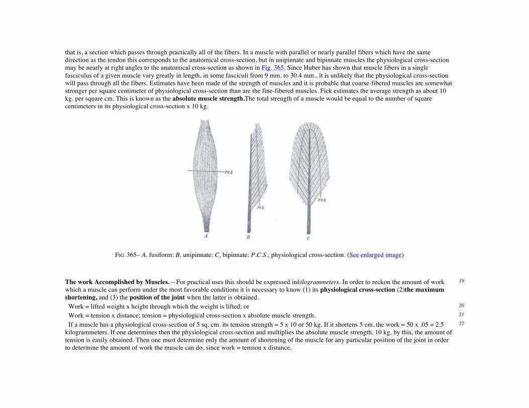
that is, a section which passes through practically all of the fibers. In a muscle with parallel or nearly parallel fibers which have the same direction as the tendon this corresponds to the anatomical cross-section, but in unipinnate and bipinnate muscles the physiological cross-section may be nearly at right angles to the anatomical cross-section as shown in Fig. 365. Since Huber has shown that muscle fibers in a single fasciculus of a given muscle vary greatly in length, in some fasciculi from 9 mm. to 30.4 mm., it is unlikely that the physiological cross-section will pass through all the fibers. Estimates have been made of the strength of muscles and it is probable that coarse-fibered muscles are somewhat stronger per square centimeter of physiological cross-section than are the fine-fibered muscles. Fick estimates the average strength as about 10 kg. per square cm. This is known as the absolute muscle strength.The total strength of a muscle would be equal to the number of square centimeters in its physiological cross-section x 10 kg.
FIG. 365– A, fusiform; B, unipinnate; C, bipinnate; P.C.S., physiological cross-section. (See enlarged image)
The work Accomplished by Muscles.—For practical uses this should be expressed inkilogrammeters. In order to reckon the amount of work which a muscle can perform under the most favorable conditions it is necessary to know (1) its physiological cross-section (2)the maximum shortening, and (3) the position of the joint when the latter is obtained.
19
Work = lifted weight x height through which the weight is lifted; or 20 Work = tension x distance; tension = physiological cross-section x absolute muscle strength. 21 If a muscle has a physiological cross-section of 5 sq. cm. its tension strength = 5 x 10 or 50 kg. If it shortens 5 cm. the work = 50 x .05 = 2.5 kilogrammeters. If one determines then the physiological cross-section and multiplies the absolute muscle strength, 10 kg. by this, the amount of tension is easily obtained. Then one must determine only the amount of shortening of the muscle for any particular position of the joint in order to determine the amount of work the muscle can do, since work = tension x distance.
22

The tension of a muscle is, however, not constant during the course of contraction but is continually decreasing during contraction. It is at a maximum at the beginning and gradually decreases.
23
This can be illustrated by the work diagram Fig. 366. A M D (ordinate) = tension. A V X (abscissa) = shortening. A D = tension of muscle in extended or antagonistic position. A V = amount of actual shortening. A M = tension in midposition = absolute muscle strength. D V = shows how the tension sinks from maximum (in the extended position of the muscle) where it is about double that in the midposition (M) to nothing on complete contraction. Δ A D V = work diagram, in reality the hypothenose is not straight but has a concave curve. The Δ has the same area as the rectangle A M M’ V. A M = the average tension. Work = A M x A V kilogrammeters if the size of the ordinate as expressed in kilograms and the abscissa in meters.
24
FIG. 366– No caption. (See enlarged image)
Although the muscle works with a changing tension, yet the accomplishment is the same as if it were contracting with the tension of the midposition.
25
In reality the amount of work is somewhat greater since even in extreme contraction the muscle still retains a certain amount of tension so that the maximum amount of work is more nearly like A D X. We know that a muscle may have an extreme actual shortening of about 80 per cent. of its length when the tendon of insertion is cut.
26
The trapezoid A D S V represents more nearly the amount of work, but since there are only approximate values and A D S V is not much larger than A M M’ V, we may use the latter.
27
Only the tension and amount of shortening are needed to determine the amount of work of the muscle. Neither the lever arm nor the fiber 28
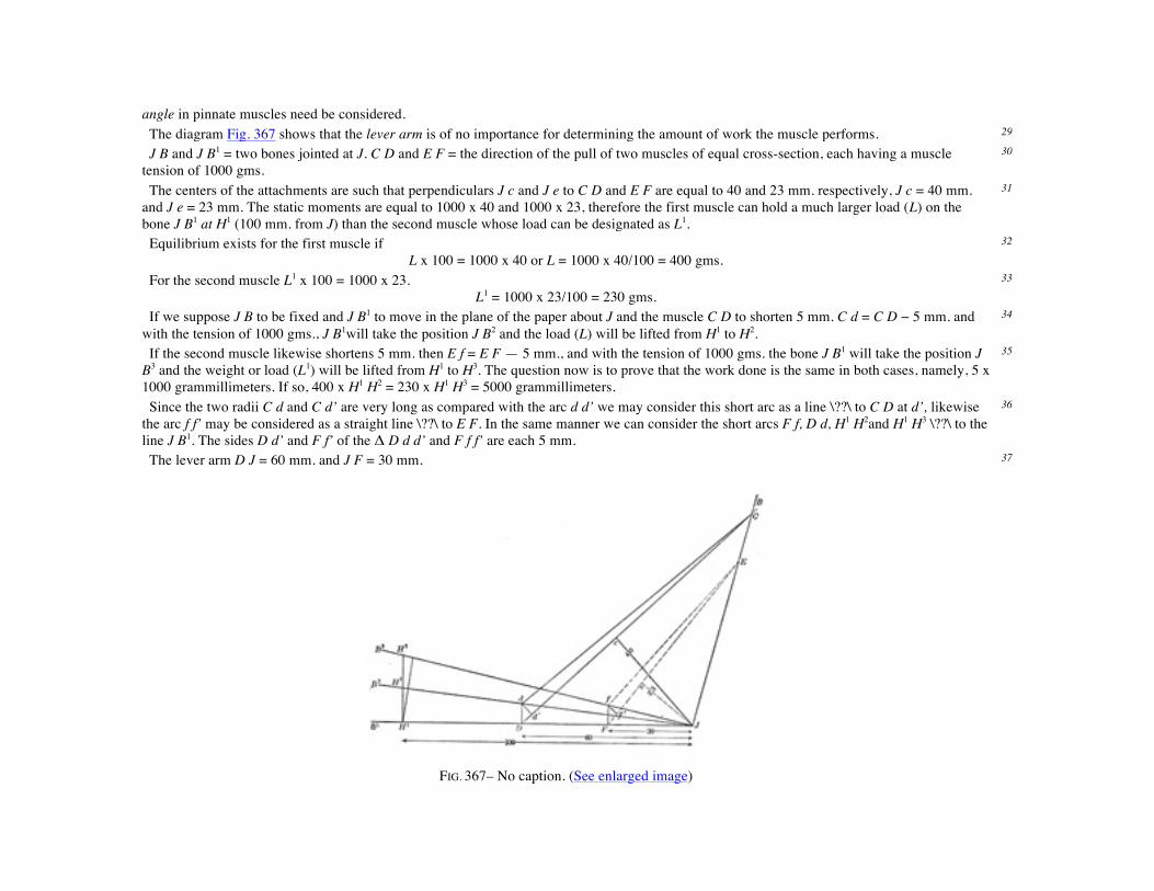
angle in pinnate muscles need be considered. The diagram Fig. 367 shows that the lever arm is of no importance for determining the amount of work the muscle performs. 29 J B and J B1 = two bones jointed at J. C D and E F = the direction of the pull of two muscles of equal cross-section, each having a muscle tension of 1000 gms.
30
The centers of the attachments are such that perpendiculars J c and J e to C D and E F are equal to 40 and 23 mm. respectively, J c = 40 mm. and J e = 23 mm. The static moments are equal to 1000 x 40 and 1000 x 23, therefore the first muscle can hold a much larger load (L) on the bone J B1 at H1 (100 mm. from J) than the second muscle whose load can be designated as L1.
31
Equilibrium exists for the first muscle if L x 100 = 1000 x 40 or L = 1000 x 40/100 = 400 gms.
32
For the second muscle L1 x 100 = 1000 x 23. L1 = 1000 x 23/100 = 230 gms.
33
If we suppose J B to be fixed and J B1 to move in the plane of the paper about J and the muscle C D to shorten 5 mm. C d = C D − 5 mm. and with the tension of 1000 gms., J B1will take the position J B2 and the load (L) will be lifted from H1 to H2.
34
If the second muscle likewise shortens 5 mm. then E f = E F — 5 mm., and with the tension of 1000 gms. the bone J B1 will take the position J B3 and the weight or load (L1) will be lifted from H1 to H3. The question now is to prove that the work done is the same in both cases, namely, 5 x 1000 grammillimeters. If so, 400 x H1 H2 = 230 x H1 H3 = 5000 grammillimeters.
35
Since the two radii C d and C d’ are very long as compared with the arc d d’ we may consider this short arc as a line \??\ to C D at d’, likewise the arc f f’ may be considered as a straight line \??\ to E F. In the same manner we can consider the short arcs F f, D d, H1 H2and H1 H3 \??\ to the line J B1. The sides D d’ and F f’ of the Δ D d d’ and F f f’ are each 5 mm.
36
The lever arm D J = 60 mm. and J F = 30 mm. 37
FIG. 367– No caption. (See enlarged image)

The Δ D d d' is similar to the Δ D c J 38
hence D d : 5 :: 60 : 40 D d = 300/40 also H1 H2 : D d :: 100 : 60
H1 H2 : 300/40 :: 100 : 60 H1 H2 = 300/24
hence F f : 5 :: 30 : 23 F f = 150/23 also H1 H3 : F f :: 100 : 30
H1 H3 :150/23::100:30 H1 H3 = 1500/69 … 400 x 300/24 = 230 x 1500/69 = 5000
Thus we see that the work of the two muscles depends on the size of the contraction and on the tension and not on the lever arm in very small contractions or in the summation of such contractions and therefore for large contractions. In the first muscle a large load is moved through a short distance and in the second muscle a lighter load is moved through a greater distance.
39
The amount of work accomplished by pinnate muscles is not dependent upon the angle of insertion of the muscle fibers into the tendon, as will be seen by the following diagram Fig. 368.
40
T' T = direction of the tendon pull. w a = direction of muscle fiber before contraction. m’ = direction of muscle fiber after contraction.
v = amount of contraction. m = tension of the muscle. φ = angle of insertion of muscle fiber. t = tendon component = m x cos φ = the weight carried by the tendon to balance the muscle tension. d = distance tendon is drawn up.
(1) m x v = work done by the muscle fiber. (2) t x d = work done by the movement of the tendon.
If we consider the distance v as being very short then the line b c can be dealt with as though it were perpendicular to a c. 41
then v = d x cos φ or d = v/cos φ since t = m x cos φ or m = t/cos φ m x v = t/cos φ x d x cos φ = t x d
42
If this is true for very minute contractions it is likewise true for a series of such contraction and hence for larger contractions. 43 If we assume that φ = 60°, m = 10 kg. and v = 5 mm., the work done by the contracting muscle fiber = m v or 10 x 5 kilogrammillimeters. 44

FIG. 368– No caption. (See enlarged image) cos ∠ 60° = 1/2; hence t = 1/2 m; and d = v/1/2 = 2 v; 1/2 m = 5 kg.; and 2 v = 10 mm. hence t d = 50 kilogrammillimeters or the work done by the movement of the tendon in lifting the load of 5 kg. a distance of 10 mm., and is exactly the same as that done by the muscle fiber. The load on the tendon is but one-half the tension of the muscle, but the distance through which the load is lifted is twice that of the amount of shortening of the muscle. If φ = 41° 20’ then cos φ = 3/4 hence t = 3/4 m and d = 4/3 v and t d = m v
45
In pinnate muscles, then, we have the rather unexpected condition in which the same amount of movement of the tendon can be accomplished with less contraction of the muscle than in muscles where the fibers have the same direction as the tendon.
46
The Action of Muscles on Joints.—If we consider now the action of a single muscle extending over a single joint in which one bone is fixed and the other movable, we will find that muscle pull can be resolved into two components, a turning component and a friction or pressure component as shown in Fig. 369.
47

FIG. 369– No caption. (See enlarged image) D F = the fixed bone from which the muscle takes its origin. 48 D K = the movable bone. 49 O I = a line from the middle of origin to the middle of insertion. 50 I M = size and direction of the muscle pull. 51
If the parallelogram is constructed with I t and M b ⊥ to D K, then I t = the turning component and I b = the component which acts against the joint.
52
The size of the two components depends upon the insertion angle φ. The smaller this angle the smaller the turning component, and the nearer this angle φ is to 90° the larger the turning component.
53
I t = I M x sin φ 54 I b = I M x cos φ 55 If φ = 90° cos φ = 0, sine φ = 1 hence I b = 0 and I t = I m 56 If φ = 0° cos φ = 1, sine φ = 0 hence I b = 1 and I t = 0 57 With movements of the bone D K the angle of insertion is continually changing, and hence the two components are changing in value. 58

FIG. 370– No caption. (See enlarged image) If, for example, the distance from origin 0 to the joint D is greater than from D to I, as in the Brachialis or Biceps muscles, the turning component increases until the insertion angle φ = 90°, which is the optimum angle for muscle action, while the pressure component gradually decreases. If the movement continues beyond this point the turning component gradually decreases and the pressure component changes into a component which tends to draw the two bones apart and which gradually increases as shown in Fig. 370.
59
When the bone D K is in such a position that the insertion angle φ = 41° 20’ the pressure component = 3/4 I m and the turning component 1/4 I m, at 60° the two components are equal, at 90° the pressure component = 0 and the turning component = I M and at 131° 21’ the pressure component has been converted into a pulling component = 1/4 I M and the turning component = 3/4 I M.
60
FIG. 371– No caption. (See enlarged image)
If, for example, the distance from the origin O to the joint D is less than the distance from the insertion I to the joint D, as in the Brachioradialis muscle, the insertion angle increases with the flexion but never reaches 90°. The turning component gradually increases to a certain point and then slowly decreases as shown in Fig. 371, while the pressure component gradually decreases and then slowly increases. It always remains large
61
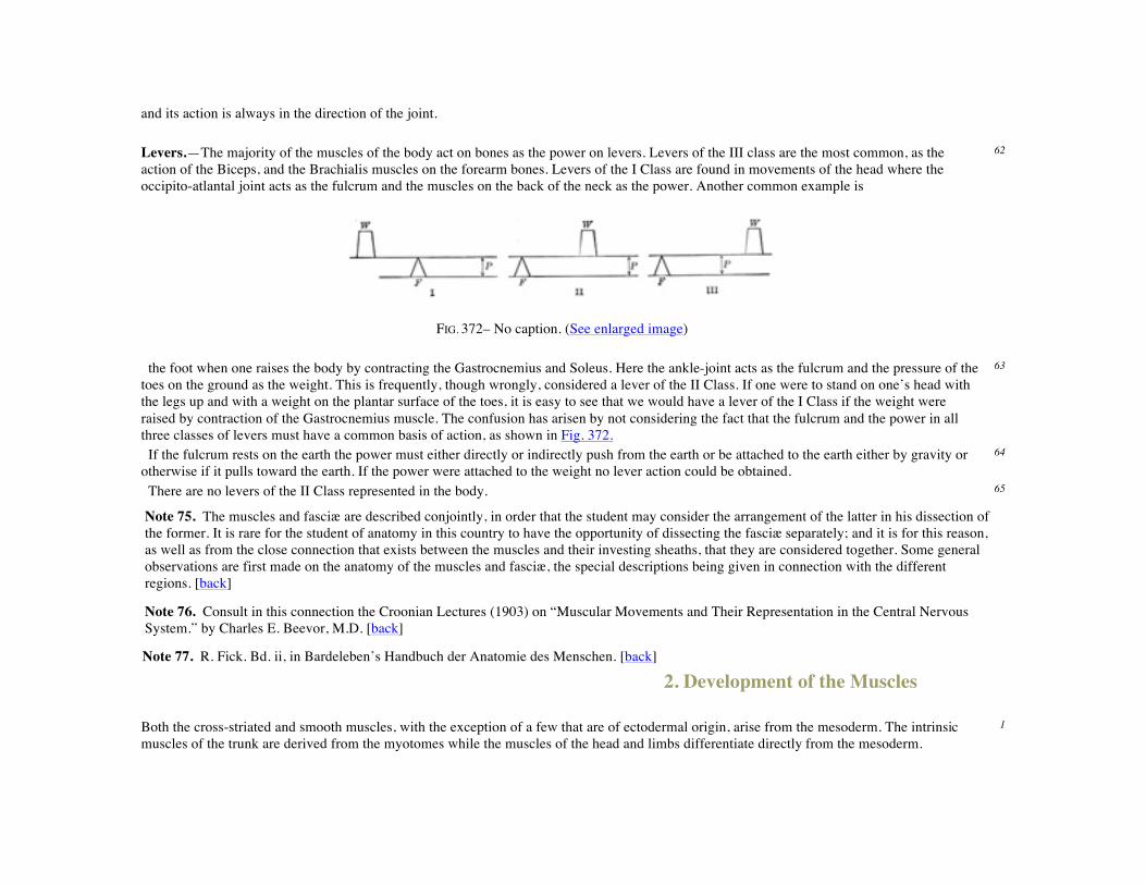
and its action is always in the direction of the joint. Levers.—The majority of the muscles of the body act on bones as the power on levers. Levers of the III class are the most common, as the action of the Biceps, and the Brachialis muscles on the forearm bones. Levers of the I Class are found in movements of the head where the occipito-atlantal joint acts as the fulcrum and the muscles on the back of the neck as the power. Another common example is
62
FIG. 372– No caption. (See enlarged image)
the foot when one raises the body by contracting the Gastrocnemius and Soleus. Here the ankle-joint acts as the fulcrum and the pressure of the toes on the ground as the weight. This is frequently, though wrongly, considered a lever of the II Class. If one were to stand on one’s head with the legs up and with a weight on the plantar surface of the toes, it is easy to see that we would have a lever of the I Class if the weight were raised by contraction of the Gastrocnemius muscle. The confusion has arisen by not considering the fact that the fulcrum and the power in all three classes of levers must have a common basis of action, as shown in Fig. 372.
63
If the fulcrum rests on the earth the power must either directly or indirectly push from the earth or be attached to the earth either by gravity or otherwise if it pulls toward the earth. If the power were attached to the weight no lever action could be obtained.
64
There are no levers of the II Class represented in the body. 65
Note 75. The muscles and fasciæ are described conjointly, in order that the student may consider the arrangement of the latter in his dissection of the former. It is rare for the student of anatomy in this country to have the opportunity of dissecting the fasciæ separately; and it is for this reason, as well as from the close connection that exists between the muscles and their investing sheaths, that they are considered together. Some general observations are first made on the anatomy of the muscles and fasciæ, the special descriptions being given in connection with the different regions. [back]
Note 76. Consult in this connection the Croonian Lectures (1903) on “Muscular Movements and Their Representation in the Central Nervous System.” by Charles E. Beevor, M.D. [back]
Note 77. R. Fick. Bd. ii, in Bardeleben’s Handbuch der Anatomie des Menschen. [back]
2. Development of the Muscles
Both the cross-striated and smooth muscles, with the exception of a few that are of ectodermal origin, arise from the mesoderm. The intrinsic muscles of the trunk are derived from the myotomes while the muscles of the head and limbs differentiate directly from the mesoderm.
1
The Myotomic Muscles.—The intrinsic muscles of the trunk which are derived directly from the myotomes are conveniently treated in two groups, the deep muscles of the back and the thoraco-abdominal muscles.
2
The deep muscles of the back extend from the sacral to the occipital region and vary much in length and size. They act chiefly on the vertebral column. The shorter muscles, such as the Interspinales, Intertransversarii, the deeper layers of the Multifidus, the Rotatores, Levatores costarum, Obliquus capitis inferior, Obliquus capitis superior and Rectus capitis posterior minor which extend between adjoining vertebræ, retain the primitive segmentation of the myotomes. Other muscles, such as the Splenius capitis, Splenius cervicis, Sacrospinalis, Semispinalis, Multifidus, Iliocostalis, Longissimus, Spinales, Semispinales, and Rectus capitis posterior major, which extend over several vertebræ, are formed by the fusion of successive myotomes and the splitting into longitudinal columns.
3
The fascia lumbo-dorsalis develops between the true myotomic muscles and the more superficial ones which migrate over the back such as the Trapezius, Rhomboideus, and Latissimus.
4
The anterior vertebral muscles, the Longus colli, Longus capitis, Rectus capitis anterior and Rectus capitis lateralis are derived from the ventral part of the cervical myotomes as are probably also the Scaleni.
5
The thoraco-abdominal muscles arise through the ventral extension of the thoracic myotomes into the body wall. This process takes place coincident with the ventral extension of the ribs. In the thoracic region the primitive myotomic segments still persist as the intercostal muscles, but over the abdomen these ventral myotomic processes fuse into a sheet which splits in various ways to form the Rectus, the Obliquus externus and internus, and the Transversalis. Such muscles as the Pectoralis major and minor and the Serratus anterior do not belong to the above group.
6
The Ventrolateral Muscles of the Neck.—The intrinsic muscles of the tongue, the Infrahyoid muscles and the diaphragm are derived from a more or less continuous premuscle mass which extends on each side from the tongue into the lateral region of the upper half of the neck and into it early extend the hypoglossal and branches of the upper cervical nerves. The two halves which form the Infrahyoid muscles and the diaphragm are at first widely separated from each other by the heart. As the latter descends into the thorax the diaphragmatic portion of each lateral mass is carried with its nerve down into the thorax and the laterally placed Infrahyoid muscles move toward the midventral line of the neck.
7
Muscles of the Shoulder Girdle and Arm.—The Trapezius and Sternocleidomastoideus arise from a common premuscle mass in the occipital region just caudal to the last branchial arch; as the mass increases in size it spreads downward to the shoulder girdle to which it later becomes attached. It also spreads backward and downward to the spinous processes, gaining attachment at a still later period.
8
The Levator scapulæ, Serratus anterior and the Rhomboids arise from premuscle tissue in the lower cervical region and undergo extensive migration.
9
The Latissimus dorsi and Teres major are associated in their origin from the premuscle sheath of the arm as are also the two Pectoral muscles when the arm bud lies in the lower cervical region.
10
The intrinsic muscles of the arm develop in situ from the mesoderm of the arm bud and probably do not receive cells or buds from the myotomes. The nerves enter the arm bud when it still lies in the cervical region and as the arm shifts caudally over the thorax the lower cervical nerves which unite to form the brachial plexus, acquire a caudal direction.
11
The Muscles of the Leg.—The muscles of the leg like those of the arm develop in situfrom the mesoderma of the leg bud, the myotomes apparently taking no part in their formation.
12

The Muscles of the Head.—The muscles of the orbit arise from the mesoderm over the dorsal and caudal sides of the optic stalk. 13 The muscles of mastication arise from the mesoderm of the mandibular arch. The mandibular division of the trigeminal nerve enters this premuscle mass before it splits into the Temporal, Masseter and Pterygoideus.
14
The facial muscles (muscles of expression) arise from the mesoderm of the hyoid arch. The facial nerve enters this mass before it begins to split, and as the muscle mass spreads out over the face and head and neck it splits more or less incompletely into the various muscles.
15
The early differentiation of the muscular system apparently goes on independently of the nervous system and only later does it appear that muscles are dependent on the functional stimuli of the nerves for their continued existence and growth. Although the nervous system does not influence muscle differentiation, the nerves, owing to their early attachments to the muscle rudiments, are in a general way indicators of the position of origin of many of the muscles and likewise in many instances the nerves indicate the paths along which the developing muscles have migrated during development. The muscle of the diaphragm, for example, has its origin in the region of the fourth and fifth cervical segments. The phrenic nerve enters the muscle mass while the latter is in this region and is drawn out as the diaphragm migrates through the thorax. The Trapezius and Sternocleidomastoideus arise in the lateral occipital region as a common muscle mass, into which at a very early period the nervus accessorius extends and as the muscle mass migrates and extends caudally the nerve is carried with it. The Pectoralis major and minor arise in the cervical region, receive their nerves while in this position and as the muscle mass migrates and extends caudally over the thorax the nerves are carried along. The Latissimus dorsi and Serratus anterior are excellent examples of migrating muscles whose nerve supply indicates their origin in the cervical region. The Rectus abdominis and the other abdominal muscles migrate or shift from a lateral to a ventrolateral or abdominal position, carrying with them the nerves.
16
The facial nerve, which early enters the common facial muscle mass of the second branchial or hyoid arch, is dragged about with the muscle as it spreads over the head and face and neck, and as the muscle splits into the various muscles of expression, the nerve is correspondingly split. The mandibular division of the trigeminal nerve enters at an early time the muscle mass in the mandibular arch and as this mass splits and migrates apart to form the muscles of mastication the nerve splits into its various branches.
17
The nerve supply then serves as a key to the common origin of certain groups of muscles. The muscles supplied by the oculomotor nerve arise from a single mass in the eye region; the lingual muscles arise from a common mass supplied by the hypoglossal nerve.
18
Striped or Voluntary Muscle.—Striped or voluntary muscle is composed of bundles of fibers each enclosed in a delicate web called the perimysium in contradistinction to the sheath of areolar tissue which invests the entire muscle, the epimysium. The bundles are termed fasciculi; they are prismatic in shape, of different sizes in different muscles, and are for the most part placed parallel to one another, though they have a tendency to converge toward their tendinous attachments. Each fasciculus is made up of a strand of fibers, which also run parallel with each other, and are separated from one another by a delicate connective tissue derived from the perimysium and termed endomysium. This does not form the sheath of the fibers, but serves to support the bloodvessels and nerves ramifying between them.
19
A muscular fiber may be said to consist of a soft contractile substance, enclosed in a tubular sheath named by Bowman the sarcolemma. The fibers are cylindrical or prismatic in shape (Fig. 373), and are of no great length, not exceeding, as a rule, 40 mm. Huber 78has recently found that the muscle fibers in the adductor muscle of the thigh of the rabbit vary greatly in length even in the same fasciculus. In a fasciculus 40 mm. in length the fibers varied from 30.4 mm. to 9 mm. in length. Their breadth varies in man from 0.01 to 0.1 mm. As a rule, the fibers do not divide or anastomose; but occasionally, especially in the tongue and facial muscles, they may be seen to divide into several branches. In the substance of the muscle, the fibers end by tapering extremities which are joined to the ends of other fibers by the sarcolemma. At the tendinous end of the muscle the sarcolemma appears to blend with a small bundle of fibers, into which the tendon becomes subdivided, while the muscular substance ends abruptly and can be readily made to retract from the point of junction. The areolar tissue between the fibers appears to be prolonged more
20

or less into the tendon, so as to form a kind of sheath around the tendon bundles for a longer or shorter distance. When muscular fibers are attached to skin or mucous membranes, their fibers become continuous with those of the areolar tissue.
FIG. 373– Transverse section of human striped muscle fibers. x 255. (See enlarged image)
FIG. 374– Striped muscle fibers from tongue of cat. x 250. (See enlarged image)
The sarcolemma, or tubular sheath of the fiber, is a transparent, elastic, and apparently homogeneous membrane of considerable toughness, so that it sometimes remains entire when the included substance is ruptured. On the internal surface of the sarcolemma in mammalia, and also in the substance of the fiber in frogs, elongated nuclei are seen, and in connection with these is a little granular protoplasm.
21

Upon examination of a voluntary muscular fiber by transmitted light, it is found to be marked by alternate light and dark bands or striæ, which pass transversely across the fiber(Fig. 374). When examined by polarized light the dark bands are found to be doubly refracting (anisotropic), while the clear stripes are singly refracting (isotropic). The dark and light bands are of nearly equal breadth, and alternate with great regularity; they vary in breadth from about 1 to 2μ. If the surface be carefully focussed, rows of granules will be detected at the points of junction of the dark and light bands, and very fine longitudinal lines may be seen running through the dark bands and joining these granules together. By treating the specimen with certain reagents (e. g., chloride of gold) fine lines may be seen running transversely between the granules and uniting them together. This appearance is believed to be due to a reticulum or network of interstitial substance lying between the contractile portions of the muscle. The longitudinal striation gives the fiber the appearance of being made up of a bundle of fibrils which have been termed sarcostyles or muscle columns, and if the fiber be hardened in alcohol, it can be broken up longitudinally and the sarcostyles separated from each other (Fig. 375.) The reticulum, with its longitudinal and transverse meshes, is called sarcoplasm.
22
FIG. 375– A. Portion of a medium-sized human muscular fiber. Magnified nearly 800 diameters. B. Separated bundles of fibrils, equally magnified. a, a. Larger, and b, b, smaller collections. c. Still smaller. d, d. The smallest which could be detached. (See enlarged image)
In a transverse section, the muscular fiber is seen to be divided into a number of areas, called the areas of Cohnheim, more or less polyhedral in shape and consisting of the transversely divided sarcostyles, surrounded by transparent sarcoplasm (Fig. 373).
23

FIG. 376– Diagram of a sarcomere. (After Schäfer.) A. In moderately extended condition. B.In a contracted condition. k, k. Membranes of Krause. H. Line or plane of Hensen. S.E.Poriferous sarcous element. (See enlarged image)
Upon closer examination, and by somewhat altering the focus, the appearances become more complicated, and are susceptible of various interpretations. The transverse striation, which in Fig. 374 appears as a mere alternation of dark and light bands, is resolved into the appearance seen in Fig. 375, which shows a series of broad dark bands, separated by light bands, each of which is divided into two by a dark dotted line. This line is termed Dobie’s line or Krause’s membrane (Fig. 376, k), because it was believed by Krause to be an actual membrane, continuous with the sarcolemma, and dividing the light band into two compartments. In addition to the membrane of Krause, fine clear lines may be made out, with a sufficiently high power, crossing the center of the dark band; these are known as thelines of Hensen (Fig. 376, H).
24
Schäfer has worked out the minute anatomy of muscular fiber, particularly in the wing muscles of insects, which are peculiarly adapted for this purpose on account of the large amount of interstitial sarcoplasm which separates the sarco-styles. In the following description that given by Schäfer will be closely followed.
25
A sarcostyle may be said to be made up of successive portions, each of which is termed asarcomere. The sarcomere is situated between two membranes of Krause and consists of (1) a central dark part, which forms a portion of the dark band of the whole fiber, and is named a sarcous element. This sarcous element really consists of two parts, superimposed one on the top of the other, and when the fiber is stretched these two parts become separated from each other at the line of Hensen (Fig. 376, A). (2) On either side of this central dark portion is a clear layer, most visible when the fiber is extended; this is situated between the dark center and the membrane of Krause, and when the sarcomeres are joined together to form the sarcostyle, constitutes the light band of the striated muscular fiber.
26
When the sarcostyle is extended, the clear intervals are well-marked and plainly to be seen; when, on the other hand, the sarcostyle is contracted, that is to say, when the muscle is in a state of contraction, these clear portions are very small or they may have disappeared altogether (Fig. 376, B). When the sarcostyle is stretched to its full extent, not only is the clear portion well-marked, but the dark portion—the sarcous element—is separated into its two constituents along the line of Hensen. The sarcous element does not lie free in the sarcomere, for when the sarcostyle is stretched, so as to render the clear portion visible, very fine lines, which are probably septa, may be seen running through it from the sarcous element to the membrane of Krause.
27
Schäfer explains these phenomena in the following way: He considers that each sarcous element is made up of a number of longitudinal channels, which open into the clear part toward the membrane of Krause but are closed at the line of Hensen. When the muscular fiber is contracted the clear part of the muscular substance is driven into these channels or tubes, and is therefore hidden from sight, but at the same time
28

it swells up the sarcous element and widens and shortens the sarcomere. When, on the other hand, the fiber is extended, this clear substance is driven out of the tubes and collects between the sarcous element and the membrane of Krause, and gives the appearance of the light part between these two structures; by this means it elongates and narrows the sarcomere. If this view be true, it is a matter of great interest, and, as Schäfer has shown, harmonizes the contraction of muscle with the ameboid action of protoplasm. In an ameboid cell, there is a framework of spongioplasm, which stains with hematoxylin and similar reagents, enclosing in its meshes a clear substance, hyaloplasm, which will not stain with these reagents. Under stimulation the hyaloplasm passes into the pores of the spongioplasm; without stimulation it tends to pass out as in the formation of pseudopodia. In muscle there is the same thing, viz., a framework of spongioplasm staining with hematoxylin—the substance of the sarcous element—and this encloses a clear hyaloplasm, the clear substance of the sarcomere, which resists staining with this reagent. During contraction of the muscle—i.e., stimulation—this clear substance passes into the pores of the spongioplasm; while during extension of the muscle—i.e., when there is no stimulation—it tends to pass out of the spongioplasm.
29
In this way the contraction is brought about: under stimulation the protoplasmic material (the clear substance of the sarcomere) recedes into the sarcous element, causing the sarcomere to widen out and shorten. The contraction of the muscle is merely the sum total of this widening out and shortening of these bodies.
30
Vessels and Nerves of Striped Muscle.—The capillaries of striped muscle are very abundant, and form a sort of rectangular network, the branches of which run longitudinally in the endomysium between the muscular fibers, and are joined at short intervals by transverse anastomosing branches. In the red muscles of the rabbit dilatations occur on the transverse branches of the capillary network. The larger vascular channels, arteries and veins, are found only in the perimysium, between the muscular fasciculi. Nerves are profusely distributed to striped muscle. Their mode of termination is described on page 730. The existence of lymphatic vessels in striped muscle has not been ascertained, though they have been found in tendons and in the sheaths of the muscles.
31
Ossification of muscular tissue as a result of repeated strain or injury is not infrequent. It is oftenest found about the tendon of the Adductor longus and Vastus medialis in horsemen, or in the Pectoralis major and Deltoideus of soldiers. It may take the form of exostoses firmly fixed to the bone—e.g., “rider’s bone” on the femur—or of layers or spicules of bone lying in the muscles or their fasciæ and tendons. Busse states that these bony deposits are preceded by a hemorrhagic myositis due to injury, the effused blood organizing and being finally converted into bone. In the rarer disease, progressive myositis ossificans, there is an unexplained tendency for practically any of the voluntary muscles to become converted into solid and brittle bony masses which are completely rigid.
32
Note 78. Anat. Rec., 1916, 11. [back]
3. Tendons, Aponeuroses, and Fasciæ
Tendons are white, glistening, fibrous cords, varying in length and thickness, sometimes round, sometimes flattened, and devoid of elasticity. They consist almost entirely of white fibrous tissue, the fibrils of which have an undulating course parallel with each other and are firmly united together. When boiled in water tendon is almost completely converted into gelatin, the white fibers being composed of the albuminoid collagen, which is often regarded as the anhydride of gelatin. They are very sparingly supplied with bloodvessels, the smaller tendons presenting in their interior no trace of them. Nerves supplying tendons have special modifications of their terminal fibers, named organs of Golgi.
1
Aponeuroses are flattened or ribbon-shaped tendons, of a pearly white color, iridescent, glistening, and similar in structure to the tendons. They are only sparingly supplied with bloodvessels.
2
The tendons and aponeuroses are connected, on the one hand, with the muscles, and, on the other hand, with the movable structures, as the bones, cartilages ligaments, and fibrous membranes (for instance, the sclera). Where the muscular fibers are in a direct line with those of the tendon or aponeurosis, the two are directly continuous. But where the muscular fibers join the tendon or aponeurosis at an oblique angle, they end, according to Kölliker, in rounded extremities which are received into corresponding depressions on the surface of the latter, the connective tissue between the muscular fibers being continuous with that of the tendon. The latter mode of attachment occurs in all the penniform and bipenniform muscles, and in those muscles the tendons of which commence in a membranous form, as the Gastrocnemius and Soleus.
3
The fasciæ are fibroareolar or aponeurotic laminæ, of variable thickness and strength, found in all regions of the body, investing the softer and more delicate organs. During the process of development many of the cells of the mesoderm are differentiated into bones, muscles, vessels, etc.; the cells of the mesoderm which are not so utilized form an investment for these structures and are differentiated into the true skin and the fasciæ of the body. They have been subdivided, from the situations in which they occur, into superficial and deep.
4
The superficial fascia is found immediately beneath the integument over almost the entire surface of the body. It connects the skin with the deep fascia, and consists of fibroareolar tissue, containing in its meshes pellicles of fat in varying quantity. Fibro-areolar tissue is composed of white fibers and yellow elastic fibers intercrossing in all directions, and united together by a homogeneous cement or ground substance, the matrix.
5
The cells of areolar tissue are of four principal kinds: (1) Flattened lamellar cells, which may be either branched or unbranched. The branched lamellar cells are composed of clear cytoplasm, and contain oval nuclei; the processes of these cells may unite so as to form an open network, as in the cornea. The unbranched cells are joined edge to edge like the cells of an epithelium; the “tendon cells,” presently to be described, are examples of this variety. (2) Clasmatocytes, large irregular cells characterized by the presence of granules or vacuoles in their protoplasm, and containing oval nuclei. (3) Granule cells (Mastzellen), which are ovoid or spheroidal in shape. They are formed of a soft protoplasm, containing granules which are basophil in character. (4) Plasma cells of Waldeyer, usually spheroidal and distinguished by containing a vacuolated protoplasm. The vacuoles are filled with fluid, and the protoplasm between the spaces is clear, with occasionally a few scattered basophil granules.
6

FIG. 377– Subcutaneous tissue from a young rabbit. Highly magnified. (Schäfer.) (See enlarged image) In addition to these four typical forms of connective-tissue corpuscles, areolar tissue may be seen to possess wandering cells, i.e., leucocytes which have emigrated from the neighboring vessels; in some instances, as in the choroid coat of the eye cells filled with granules of pigment (pigment cells) are found.
7
The cells lie in spaces in the ground substance between the bundles of fibers, and these spaces may be brought into view by treating the tissue with nitrate of silver and exposing it to the light. This will color the ground substance and leave the cell-spaces unstained.
8
Fat is entirely absent in the subcutaneous tissue of the eyelids, of the penis and scrotum, and of the labia minora. It varies in thickness in different parts of the body; in the groin it is so thick that it may be subdivided into several laminæ. Beneath the fatty layer there is generally another layer of superficial fascia, comparatively devoid of adipose tissue, in which the trunks of the subcutaneous vessels and nerves are found, as the superficial epigastric vessels in the abdominal region, the superficial veins in the forearm, the saphenous veins in the leg and thigh, and the superficial lymph glands. Certain cutaneous muscles also are situated in the superficial fascia, as the Platysma in the neck, and the Orbicularis oculi around the eyelids. This fascia is most distinct at the lower part of the abdomen, perineum, and extremities; it is very thin in those regions where muscular fibers are inserted into the integument, as on the side of the neck, the face, and around the margin of the anus. It is very dense in the scalp, in the palms of the hands, and soles of the feet, forming a fibro-fatty layer, which binds the integument firmly to the underlying structures.
9
The superficial fascia connects the skin to the subjacent parts, facilitates the movement of the skin, serves as a soft nidus for the passage of vessels and nerves to the integument, and retains the warmth of the body, since the fat contained in its areolæ is a bad conductor of heat.
10
The deep fascia is a dense, inelastic, fibrous membrane, forming sheaths for the muscles, and in some cases affording them broad surfaces for attachment. It consists of shining tendinous fibers, placed parallel with one another, and connected together by other fibers disposed in a rectilinear manner. It forms a strong investment which not only binds down collectively the muscles in each region, but gives a separate sheath to
11
each, as well as to the vessels and nerves. The fasciæ are thick in unprotected situations, as on the lateral side of a limb, and thinner on the medial side. The deep fasciæ assist the muscles in their actions, by the degree of tension and pressure they make upon their surfaces; the degree of tension and pressure is regulated by the associated muscles, as, for instance, by the Tensor fasciæ latæ and Glutæus maximus in the thigh, by the Biceps in the upper and lower extremities, and Palmaris longus in the hand. In the limbs, the fasciæ not only invest the entire limb, but give off septa which separate the various muscles, and are attached to the periosteum: these prolongations of fasciæ are usually spoken of as intermuscular septa. The Fasciæ and Muscles may be arranged, according to the general division of the body, into those of the head and neck; of the trunk; of the upper extremity; and of the lower extremity.
12
4. The Fasciæ and Muscles of the Head. a. The Muscles of the Scalp
Epicranius The Skin of the Scalp.—This is thicker than in any other part of the body. It is intimately adherent to the superficial fascia, which attaches it firmly to the underlying aponeurosis and muscle. Movements of the muscle move the skin. The hair follicles are very closely set together, and extend throughout the whole thickness of the skin. It also contains a number of sebaceous glands.
1
The superficial fascia in the cranial region is a firm, dense, fibro-fatty layer, intimately adherent to the integument, and to the Epicranius and its tendinous aponeurosis; it is continuous, behind, with the superficial fascia at the back of the neck; and, laterally, is continued over the temporal fascia. It contains between its layers the superficial vessels and nerves and much granular fat.
2
The Epicranius (Occipitofrontalis) (Fig. 378) is a broad, musculofibrous layer, which covers the whole of one side of the vertex of the skull, from the occipital bone to the eyebrow. It consists of two parts, the Occipitalis and the Frontalis, connected by an intervening tendinous aponeurosis, the galea aponeurotica.
3
The Occipitalis, thin and quadrilateral in form, arises by tendinous fibers from the lateral two-thirds of the superior nuchal line of the occipital bone, and from the mastoid part of the temporal. It ends in the galea aponeurotica.
4
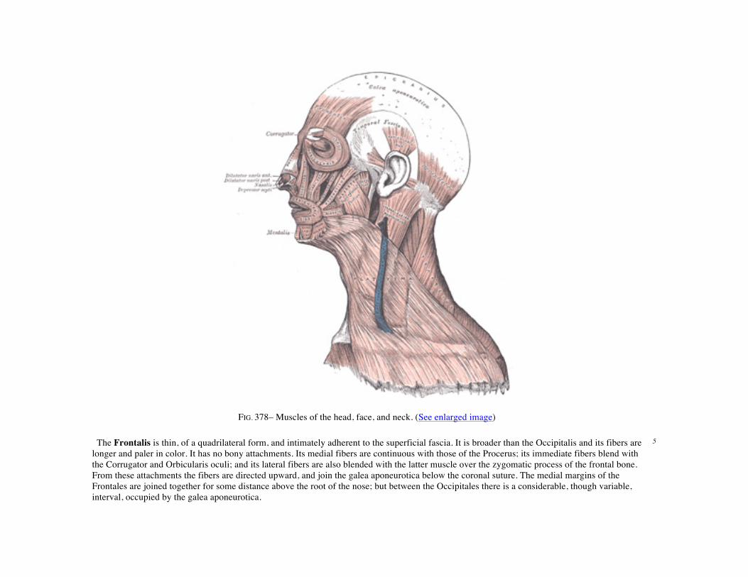
FIG. 378– Muscles of the head, face, and neck. (See enlarged image) The Frontalis is thin, of a quadrilateral form, and intimately adherent to the superficial fascia. It is broader than the Occipitalis and its fibers are longer and paler in color. It has no bony attachments. Its medial fibers are continuous with those of the Procerus; its immediate fibers blend with the Corrugator and Orbicularis oculi; and its lateral fibers are also blended with the latter muscle over the zygomatic process of the frontal bone. From these attachments the fibers are directed upward, and join the galea aponeurotica below the coronal suture. The medial margins of the Frontales are joined together for some distance above the root of the nose; but between the Occipitales there is a considerable, though variable, interval, occupied by the galea aponeurotica.
5

The galea aponeurotica (epicranial aponeurosis) covers the upper part of the cranium; behind, it is attached, in the interval between its union with the Occipitales, to the external occipital protuberance and highest nuchal lines of the occipital bone; in front, it forms a short and narrow prolongation between its union with the Frontales. On either side it gives origin to the Auriculares anterior and superior; in this situation it loses its aponeurotic character, and is continued over the temporal fascia to the zygomatic arch as a layer of laminated areolar tissue. It is closely connected to the integument by the firm, dense, fibro-fatty layer which forms the superficial fascia of the scalp: it is attached to the pericranium by loose cellular tissue, which allows the aponeurosis, carrying with it the integument to move through a considerable distance.
6
Variations.—Both Frontalis and Occipitalis vary considerably in size and in extent of attachment; either may be absent; fusion of Frontalis to skin has been noted.
7
Nerves.—The Frontalis is supplied by the temporal branches of the facial nerve, and the Occipitalis by the posterior auricular branch of the same nerve.
8
Actions.—The Frontales raise the eyebrows and the skin over the root of the nose, and at the same time draw the scalp forward, throwing the integument of the forehead into transverse wrinkles. The Occipitales draw the scalp backward. By bringing alternately into action the Frontales and Occipitales the entire scalp may be moved forward and backward. In the ordinary action of the muscles, the eyebrows are elevated, and at the same time the aponeurosis is fixed by the Occipitales, thus giving to the face the expression of surprise; if the action be exaggerated, the eyebrows are still further raised, and the skin of the forehead thrown into transverse wrinkles, as in the expression of fright or horror.
9
A thin muscular slip, the Transversus nuchæ, is present in a considerable proportion (25 per cent.) of cases; it arises from the external occipital protuberance or from the superior nuchal line, either superficial or deep to the Trapezius; it is frequently inserted with the Auricularis posterior, but may join the posterior edge of the Sternocleidomastoideus.
10
4b. The Muscles of the Eyelid
The muscles of the eyelids are: 1
Levator palpebræ superioris. Orbicularis oculi. Corrugator.
The Levator palpebræ superioris is described with the Anatomy of the Eye. 2

The Orbicularis oculi (Orbicularis palpebrarum) (Fig. 379) arises from the nasal part of the frontal bone, from the frontal process of the maxilla in front of the lacrimal groove, and from the anterior surface and borders of a short fibrous band, the medial palpebral ligament. From this origin, the fibers are directed lateralward, forming a broad and thin layer, which occupies the eyelids or palpebræ, surrounds the circumference of the orbit, and spreads over the temple, and downward on the cheek. The palpebral portion of the muscle is thin and pale; it arises from the bifurcation of the medial palpebral ligament, forms a series of concentric curves, and is inserted into the lateral palpebral raphé. The orbital portion is thicker and of a reddish color; its fibers form a complete ellipse without interruption at the lateral palpebral commissure; the upper fibers of this portion blend with the Frontalis and Corrugator. The lacrimal part (Tensor tarsi) is a small, thin muscle, about 6 mm. in breadth and 12 mm. in length, situated behind the medial palpebral ligament and lacrimal sac (Fig. 379). It arises from the posterior crest and adjacent part of the orbital surface of the lacrimal bone, and passing behind the lacrimal sac, divides into two slips, upper and lower, which are inserted into the superior and inferior tarsi medial to the puncta lacrimalia; occasionally it is very indistinct.
3
The medial palpebral ligament (tendo oculi), about 4 mm. in length and 2 mm. in breadth, is attached to the frontal process of the maxilla in front of the lacrimal groove. Crossing the lacrimal sac, it divides into two parts, upper and lower, each attached to the medial end of the corresponding tarsus. As the ligament crosses the lacrimal sac, a strong aponeurotic lamina is given off from its posterior surface; this expands over the sac, and is attached to the posterior lacrimal crest.
4
The lateral palpebral raphé is a much weaker structure than the medial palpebral ligament. It is attached to the margin of the frontosphenoidal process of the zygomatic bone, and passes medialward to the lateral commissure of the eyelids, where it divides into two slips, which are attached to the margins of the respective tarsi.
5
FIG. 379– Left orbicularis oculi, seen from behind. (See enlarged image)
The Corrugator 79 (Corrugator supercilii) is a small, narrow, pyramidal muscle, placed at the medial end of the eyebrow, beneath the Frontalis and Orbicularis oculi. It arises from the medial end of the superciliary arch; and its fibers pass upward and lateralward, between the palpebral and orbital portions of the Orbicularis oculi, and are inserted into the deep surface of the skin, above the middle of the orbital arch.
6
Nerves.—The Orbicularis oculi and Corrugator are supplied by the facial nerve. 7 Actions.—The Orbicularis oculi is the sphincter muscle of the eyelids. The palpebral portion acts involuntarily, closing the lids gently, as in sleep or in blinking; the orbital portion is subject to the will. When the entire muscle is brought into action, the skin of the forehead, temple, and cheek is drawn toward the medial angle of the orbit, and the eyelids are firmly closed, as in photophobia. The skin thus drawn upon is thrown into folds, especially radiating from the lateral angle of the eyelids; these folds become permanent in old age, and form the so-called “crows’ feet.” The Levator palpebræ superioris is the direct antagonist of this muscle; it raises the upper eyelid and exposes the front of the bulb of the eye. Each time the eyelids are closed through the action of the Orbicularis, the medial palpebral ligament is tightened, the wall of the lacrimal sac is thus drawn lateralward and forward, so that a vacuum is made in it and the tears are sucked along the lacrimal canals into it. The lacrimal part of the Orbicularis oculi draws the eyelids and the ends of the lacrimal canals medialward and compresses them against the surface of the globe of the eye, thus placing them in the most favorable situation for receiving the tears; it also compresses the lacrimal sac. The Corrugator draws the eyebrow downward and medialward, producing the vertical wrinkles of the forehead. It is the “frowning” muscle, and may be regarded as the principal muscle in the expression of suffering.
8
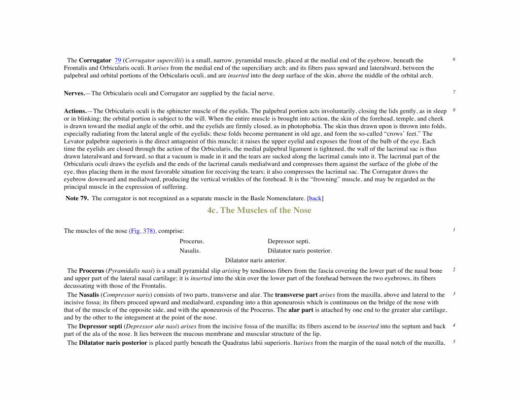
Note 79. The corrugator is not recognized as a separate muscle in the Basle Nomenclature. [back] 4c. The Muscles of the Nose The muscles of the nose (Fig. 378). comprise: 1
Procerus. Depressor septi. Nasalis. Dilatator naris posterior.
Dilatator naris anterior.
The Procerus (Pyramidalis nasi) is a small pyramidal slip arising by tendinous fibers from the fascia covering the lower part of the nasal bone and upper part of the lateral nasal cartilage; it is inserted into the skin over the lower part of the forehead between the two eyebrows, its fibers decussating with those of the Frontalis.
2
The Nasalis (Compressor naris) consists of two parts, transverse and alar. The transverse part arises from the maxilla, above and lateral to the incisive fossa; its fibers proceed upward and medialward, expanding into a thin aponeurosis which is continuous on the bridge of the nose with that of the muscle of the opposite side, and with the aponeurosis of the Procerus. The alar part is attached by one end to the greater alar cartilage, and by the other to the integument at the point of the nose.
3
The Depressor septi (Depressor alœ nasi) arises from the incisive fossa of the maxilla; its fibers ascend to be inserted into the septum and back part of the ala of the nose. It lies between the mucous membrane and muscular structure of the lip.
4
The Dilatator naris posterior is placed partly beneath the Quadratus labii superioris. Itarises from the margin of the nasal notch of the maxilla, 5

and from the lesser alar cartilages, and is inserted into the skin near the margin of the nostril. The Dilatator naris anterior is a delicate fasciculus, passing from the greater alar cartilage to the integument near the margin of the nostril; it is situated in front of the preceding.
6
Variations.—These muscles vary in size and strength or may be absent. 7 Nerves.—All the muscles of this group are supplied by the facial nerve. 8 Actions.—The Procerus draws down the medial angle of the eyebrows and produces transverse wrinkles over the bridge of the nose. The two Dilatatores enlarge the aperture of the nares. Their action in ordinary breathing is to resist the tendency of the nostrils to close from atmospheric pressure, but in difficult breathing, as well as in some emotions, such as anger, they contract strongly. The Depressor septi is a direct antagonist of the other muscles of the nose, drawing the ala of the nose downward, and thereby constricting the aperture of the nares. The Nasalis depresses the cartilaginous part of the nose and draws the ala toward the septum.
9
4d. The Muscles of the Mouth
The muscles of the mouth are: 1
Quadratus labii superioris. Quadratus labii inferioris. Caninus. Triangularis. Zygomaticus. Buccinator. Mentalis. Orbicularis oris.
Risorius.
The Quadratus labii superioris is a broad sheet, the origin of which extends from the side of the nose to the zygomatic bone. Its medial fibers form the angular head, whicharises by a pointed extremity from the upper part of the frontal process of the maxilla and passing obliquely downward and lateralward divides into two slips. One of these is insertedinto the greater alar cartilage and skin of the nose; the other is prolonged into the lateral part of the upper lip, blending with the infraorbital head and with the Orbicularis oris. The intermediate portion or infraorbital head arises from the lower margin of the orbit immediately above the infraorbital foramen, some of its fibers being attached to the maxilla, others to the zygomatic bone. Its fibers converge, to be inserted into the muscular substance of the upper lip between the angular head and the Caninus. The lateral fibers, forming thezygomatic head, arise from the malar surface of the zygomatic bone immediately behind the zygomaticomaxillary suture and pass downward and medialward to the upper lip.
2
The Caninus (Levator anguli oris) arises from the canine fossa, immediately below the infraorbital foramen; its fibers are inserted into the angle of the mouth, intermingling with those of the Zygomaticus, Triangularis, and Orbicularis oris.
3
The Zygomaticus (Zygomaticus major) arises from the zygomatic bone, in front of the zygomaticotemporal suture, and descending obliquely with a medial inclination, is insertedinto the angle of the mouth, where it blends with the fibers of the Caninus, Orbicularis oris, and Triangularis.
4

Nerves.—This group of muscles is supplied by the facial nerve. 5 Actions.—The Quadratus labii superioris is the proper elevator of the upper lip, carrying it at the same time a little forward. Its angular head acts as a dilator of the naris; the infraorbital and zygomatic heads assist in forming the nasolabial furrow, which passes from the side of the nose to the upper lip and gives to the face an expression of sadness. When the whole muscle is in action it gives to the countenance an expression of contempt and disdain. The Quadratus labii superioris raises the angle of the mouth and assists the Caninus in producing the nasolabial furrow. The Zygomaticus draws the angle of the mouth backward and upward, as in laughing.
6
The Mentalis (Levator menti) is a small conical fasciculus, situated at the side of the frenulum of the lower lip. It arises from the incisive fossa of the mandible, and descends to be inserted into the integument of the chin.
7
The Quadratus labii inferioris (Depressor labii inferioris; Quadratus menti) is a small quadrilateral muscle. It arises from the oblique line of the mandible, between the symphysis and the mental foramen, and passes upward and medialward, to be inserted into the integument of the lower lip, its fibers blending with the Orbicularis oris, and with those of its fellow of the opposite side. At its origin it is continuous with the fibers of the Platysma. Much yellow fat is intermingled with the fibers of this muscle.
8
The Triangularis (Depressor anguli oris) arises from the oblique line of the mandible, whence its fibers converge, to be inserted, by a narrow fasciculus, into the angle of the mouth. At its origin it is continuous with the Platysma, and at its insertion with the Orbicularis oris and Risorius; some of its fibers are directly continuous with those of the Caninus, and others are occasionally found crossing from the muscle of one side to that of the other; these latter fibers constitute the Transversus menti.
9
Nerves.—This group of muscles is supplied by the facial nerve. 10 Actions.—The Mentalis raises and protrudes the lower lip, and at the same time wrinkles the skin of the chin, expressing doubt or disdain. The Quadratus labii inferioris draws the lower lip directly downward and a little lateralward, as in the expression of irony. The Triangularis depresses the angle of the mouth, being the antagonist of the Caninus and Zygomaticus; acting with the Caninus, it will draw the angle of the mouth medialward. The Platysma which retracts and depresses the angle of the mouth belongs with this group.
11
The Buccinator (Fig. 380) is a thin quadrilateral muscle, occupying the interval between the maxilla and the mandible at the side of the face. It arises from the outer surfaces of the alveolar processes of the maxilla and mandible, corresponding to the three molar teeth; and behind, from the anterior border of the pterygomandibular raphé which separates it from the Constrictor pharyngis superior. The fibers converge toward the angle of the mouth, where the central fibers intersect each other, those from below being continuous with the upper segment of the Orbicularis oris, and those from above with the lower segment; the upper and lower fibers are continued forward into the corresponding lip without decussation.
12

FIG. 380– Muscles of the pharynx and cheek. (See enlarged image)
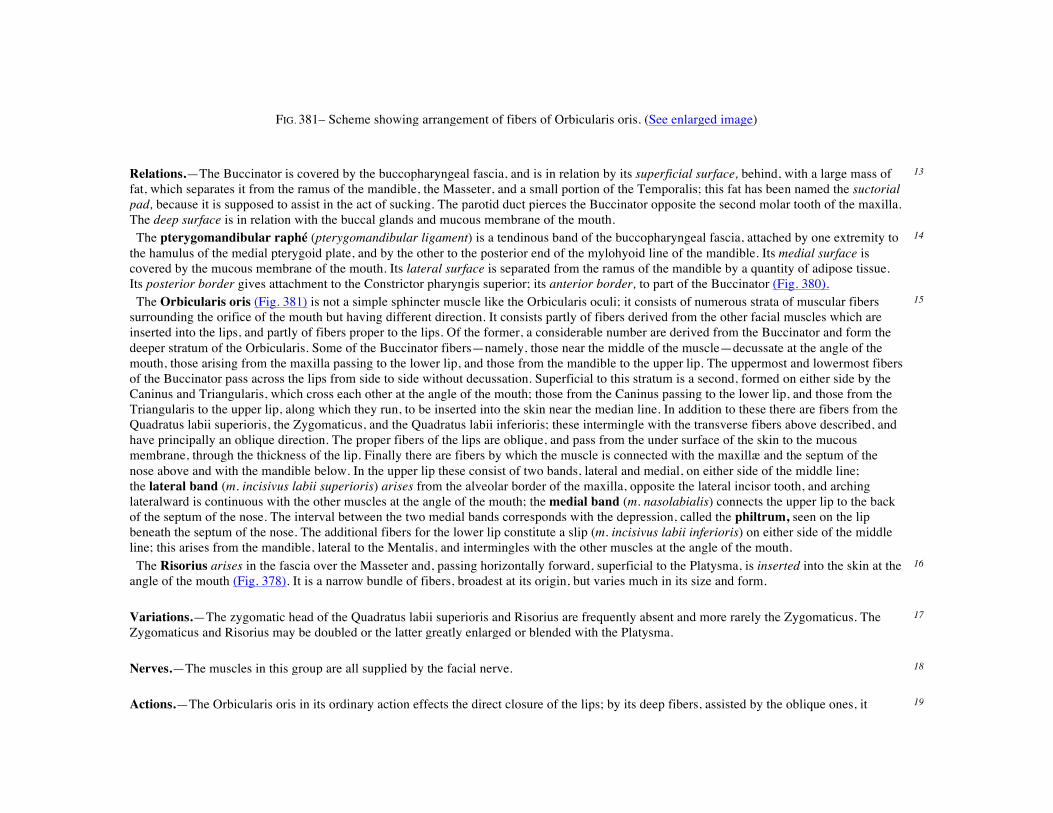
FIG. 381– Scheme showing arrangement of fibers of Orbicularis oris. (See enlarged image)
Relations.—The Buccinator is covered by the buccopharyngeal fascia, and is in relation by its superficial surface, behind, with a large mass of fat, which separates it from the ramus of the mandible, the Masseter, and a small portion of the Temporalis; this fat has been named the suctorial pad, because it is supposed to assist in the act of sucking. The parotid duct pierces the Buccinator opposite the second molar tooth of the maxilla. The deep surface is in relation with the buccal glands and mucous membrane of the mouth.
13
The pterygomandibular raphé (pterygomandibular ligament) is a tendinous band of the buccopharyngeal fascia, attached by one extremity to the hamulus of the medial pterygoid plate, and by the other to the posterior end of the mylohyoid line of the mandible. Its medial surface is covered by the mucous membrane of the mouth. Its lateral surface is separated from the ramus of the mandible by a quantity of adipose tissue. Its posterior border gives attachment to the Constrictor pharyngis superior; its anterior border, to part of the Buccinator (Fig. 380).
14
The Orbicularis oris (Fig. 381) is not a simple sphincter muscle like the Orbicularis oculi; it consists of numerous strata of muscular fibers surrounding the orifice of the mouth but having different direction. It consists partly of fibers derived from the other facial muscles which are inserted into the lips, and partly of fibers proper to the lips. Of the former, a considerable number are derived from the Buccinator and form the deeper stratum of the Orbicularis. Some of the Buccinator fibers—namely, those near the middle of the muscle—decussate at the angle of the mouth, those arising from the maxilla passing to the lower lip, and those from the mandible to the upper lip. The uppermost and lowermost fibers of the Buccinator pass across the lips from side to side without decussation. Superficial to this stratum is a second, formed on either side by the Caninus and Triangularis, which cross each other at the angle of the mouth; those from the Caninus passing to the lower lip, and those from the Triangularis to the upper lip, along which they run, to be inserted into the skin near the median line. In addition to these there are fibers from the Quadratus labii superioris, the Zygomaticus, and the Quadratus labii inferioris; these intermingle with the transverse fibers above described, and have principally an oblique direction. The proper fibers of the lips are oblique, and pass from the under surface of the skin to the mucous membrane, through the thickness of the lip. Finally there are fibers by which the muscle is connected with the maxillæ and the septum of the nose above and with the mandible below. In the upper lip these consist of two bands, lateral and medial, on either side of the middle line; the lateral band (m. incisivus labii superioris) arises from the alveolar border of the maxilla, opposite the lateral incisor tooth, and arching lateralward is continuous with the other muscles at the angle of the mouth; the medial band (m. nasolabialis) connects the upper lip to the back of the septum of the nose. The interval between the two medial bands corresponds with the depression, called the philtrum, seen on the lip beneath the septum of the nose. The additional fibers for the lower lip constitute a slip (m. incisivus labii inferioris) on either side of the middle line; this arises from the mandible, lateral to the Mentalis, and intermingles with the other muscles at the angle of the mouth.
15
The Risorius arises in the fascia over the Masseter and, passing horizontally forward, superficial to the Platysma, is inserted into the skin at the angle of the mouth (Fig. 378). It is a narrow bundle of fibers, broadest at its origin, but varies much in its size and form.
16
Variations.—The zygomatic head of the Quadratus labii superioris and Risorius are frequently absent and more rarely the Zygomaticus. The Zygomaticus and Risorius may be doubled or the latter greatly enlarged or blended with the Platysma.
17
Nerves.—The muscles in this group are all supplied by the facial nerve. 18 Actions.—The Orbicularis oris in its ordinary action effects the direct closure of the lips; by its deep fibers, assisted by the oblique ones, it 19

closely applies the lips to the alveolar arch. The superficial part, consisting principally of the decussating fibers, brings the lips together and also protrudes them forward. The Buccinators compress the cheeks, so that, during the process of mastication, the food is kept under the immediate pressure of the teeth. When the cheeks have been previously distended with air, the Buccinator muscles expel it from between the lips, as in blowing a trumpet; hence the name (buccina, a trumpet). The Risorius retracts the angle of the mouth, and produces an unpleasant grinning expression. For more extensive consideration of the facial muscles, see Charles Darwin, Expression of the Emotions in Man and Animals.
4e. The Muscles of Mastication The chief muscles of mastication are: 1
Masseter. Pterygoideus externus. Temporalis. Pterygoideus internus.
Parotideomasseteric Fascia (masseteric fascia).—Covering the Masseter, and firmly connected with it, is a strong layer of fascia derived from the deep cervical fascia. Above, this fascia is attached to the lower border of the zygomatic arch, and behind, it invests the parotid gland.
2
The Masseter (Fig. 378) is a thick, somewhat quadrilateral muscle, consisting of two portions, superficial and deep. The superficial portion, the larger, arises by a thick, tendinous aponeurosis from the zygomatic process of the maxilla, and from the anterior two-thirds of the lower border of the zygomatic arch; its fibers pass downward and backward, to be inserted into the angle and lower half of the lateral surface of the ramus of the mandible. The deep portion is much smaller, and more muscular in texture; it arises from the posterior third of the lower border and from the whole of the medial surface of the zygomatic arch; its fibers pass downward and forward, to be inserted into the upper half of the ramus and the lateral surface of the coronoid process of the mandible. The deep portion of the muscle is partly concealed, in front, by the superficial portion; behind, it is covered by the parotid gland. The fibers of the two portions are continuous at their insertion.
3
Temporal Fascia.—The temporal fascia covers the Temporalis muscle. It is a strong, fibrous investment, covered, laterally, by the Auricularis anterior and superior, by the galea aponeurotica, and by part of the Orbicularis oculi. The superficial temporal vessels and the auriculotemporal nerve cross it from below upward. Above, it is a single layer, attached to the entire extent of the superior temporal line; but below, where it is fixed to the zygomatic arch, it consists of two layers, one of which is inserted into the lateral, and the other into the medial border of the arch. A small quantity of fat, the orbital branch of the superficial temporal artery, and a filament from the zygomatic branch of the maxillary nerve, are contained between these two layers. It affords attachment by its deep surface to the superficial fibers of the Temporalis.
4
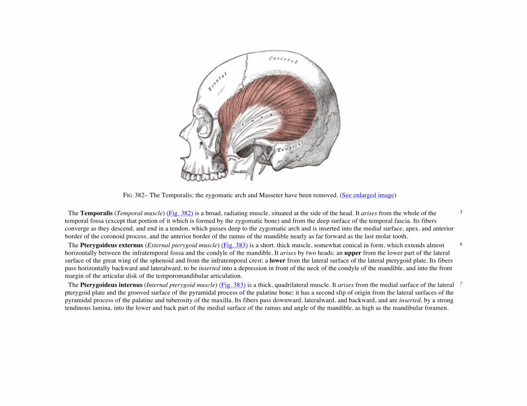
FIG. 382– The Temporalis; the zygomatic arch and Masseter have been removed. (See enlarged image) The Temporalis (Temporal muscle) (Fig. 382) is a broad, radiating muscle, situated at the side of the head. It arises from the whole of the temporal fossa (except that portion of it which is formed by the zygomatic bone) and from the deep surface of the temporal fascia. Its fibers converge as they descend, and end in a tendon, which passes deep to the zygomatic arch and is inserted into the medial surface, apex, and anterior border of the coronoid process, and the anterior border of the ramus of the mandible nearly as far forward as the last molar tooth.
5
The Pterygoideus externus (External pterygoid muscle) (Fig. 383) is a short, thick muscle, somewhat conical in form, which extends almost horizontally between the infratemporal fossa and the condyle of the mandible. It arises by two heads; an upper from the lower part of the lateral surface of the great wing of the sphenoid and from the infratemporal crest; a lower from the lateral surface of the lateral pterygoid plate. Its fibers pass horizontally backward and lateralward, to be inserted into a depression in front of the neck of the condyle of the mandible, and into the front margin of the articular disk of the temporomandibular articulation.
6
The Pterygoideus internus (Internal pterygoid muscle) (Fig. 383) is a thick, quadrilateral muscle. It arises from the medial surface of the lateral pterygoid plate and the grooved surface of the pyramidal process of the palatine bone; it has a second slip of origin from the lateral surfaces of the pyramidal process of the palatine and tuberosity of the maxilla. Its fibers pass downward, lateralward, and backward, and are inserted, by a strong tendinous lamina, into the lower and back part of the medial surface of the ramus and angle of the mandible, as high as the mandibular foramen.
7

FIG. 383– The Pterygoidei; the zygomatic arch and a portion of the ramus of the mandible have been removed. (See enlarged image) Nerves.—The muscles of mastication are supplied by the mandibular nerve. 8 Actions.—The Temporalis, Masseter, and Pterygoideus internus raise the mandible against the maxillæ with great force. The Pterygoideus externus assists in opening the mouth, but its main action is to draw forward the condyle and articular disk so that the mandible is protruded and the inferior incisors projected in front of the upper; in this action it is assisted by the Pterygoideus internus. The mandible is retracted by the posterior fibers of the Temporalis. If the Pterygoidei internus and externus of one side act, the corresponding side of the mandible is drawn forward while the opposite condyle remains comparatively fixed, and side-to-side movements. Such as occur during the trituration of food, take place.
5. The Fasciæ and Muscles of the Anterolateral Region of the Neck. a. The Superficial Cervical Muscle
The antero-lateral muscles of the neck may be arranged into the following groups: 1
I. Superficial Cervical. III. Supra- and Infrahyoid. II. Lateral Cervical. IV. Anterior Vertebral.
V. Lateral Vertebral.
The Superficial Cervical Muscle
Platysma.
The Superficial Fascia of the neck is a thin lamina investing the Platysma, and is hardly demonstrable as a separate membrane.
2
The Platysma (Fig. 378) is a broad sheet arising from the fascia covering the upper parts of the Pectoralis major and Deltoideus; its fibers cross the clavicle, and proceed obliquely upward and medialward along the side of the neck. The anterior fibers interlace, below and behind the symphysis menti, with the fibers of the muscle of the opposite side; the posterior fibers cross the mandible, some being inserted into the bone below the oblique line, others into the skin and subcutaneous tissue of the lower part of the face, many of these fibers blending with the muscles about the angle and lower part of the mouth. Sometimes fibers can be traced to the Zygomaticus, or to the margin of the Orbicularis oculi. Beneath the Platysma, the external jugular vein descends from the angle of the mandible to the clavicle.
3
Variations occur in the extension over the face and over the clavicle and shoulder; it may be absent or interdigitate with the muscle of the opposite side in front of the neck; attachment to clavicle, mastoid process or occipital bone occurs. A more or less independent fasciculus, the Occipitalis minor, may extend from the fascia over the Trapezius to fascia over the insertion of the Sternocleidomastoideus.
4
Nerve.—The Platysma is supplied by the cervical branch of the facial nerve. 5 Actions.—When the entire Platysma is in action it produces a slight wrinkling of the surface of the skin of the neck in an oblique direction. Its anterior portion, the thickest part of the muscle, depresses the lower jaw; it also serves to draw down the lower lip and angle of the mouth in the expression of melancholy.
5b. The Lateral Cervical Muscles
The lateral muscles are: 1
Trapezius and Sternocleidomastoideus. The Trapezius is described on page 432. 2 The Fascia Colli (deep cervical fascia) (Fig. 384).—The fascia colli lies under cover of the Platysma, and invests the neck; it also forms sheaths for the carotid vessels, and for the structures situated in front of the vertebral column.
3
The investing portion of the fascia is attached behind to the ligamentum nuchæ and to the spinous process of the seventh cervical vertebra. It forms a thin investment to the Trapezius, and at the anterior border of this muscle is continued forward as a rather loose areolar layer, covering the posterior triangle of the neck, to the posterior border of the Sternocleidomastoideus, where it begins to assume the appearance of a fascial membrane. Along the hinder edge of the Sternocleidomastoideus it divides to enclose the muscle, and at the anterior margin again forms a single
4

lamella, which covers the anterior triangle of the neck, and reaches forward to the middle line, where it is continuous with the corresponding part from the opposite side of the neck. In the middle line of the neck it is attached to the symphysis menti and the body of the hyoid bone. Above, the fascia is attached to the superior nuchal line of the occipital, to the mastoid process of the temporal, and to the whole length of the inferior border of the body of the mandible. Opposite the angle of the mandible the fascia is very strong, and binds the anterior edge of the Sternocleidomastoideus firmly to that bone. Between the mandible and the mastoid process it ensheathes the parotid gland—the layer which covers the gland extends upward under the name of the parotideomasseteric fascia and is fixed to the zygomatic arch. From the part which passes under the parotid gland a strong band extends upward to the styloid process, forming the stylomandibular ligament. Two other bands may be defined: the sphenomandibular (page 297) and the pterygospinous ligaments.The pterygospinous ligament stretches from the upper part of the posterior border of the lateral pterygoid plate to the spinous process of the sphenoid. It occasionally ossifies, and in such cases, between its upper border and the base of the skull, a foramen is formed which transmits the branches of the mandibular nerve to the muscles of mastication.
5
Below, the fascia is attached to the acromion, the clavicle, and the manubrium sterni. Some little distance above the last it splits into two layers, superficial and deep. The former is attached to the anterior border of the manubrium, the latter to its posterior border and to the interclavicular ligament. Between these two layers is a slit-like interval, thesuprasternal space (space of Burns); it contains a small quantity of areolar tissue, the lower portions of the anterior jugular veins and their transverse connecting branch, the sternal heads of the Sternocleidomastoidei, and sometimes a lymph gland.
6

FIG. 384– Section of the neck at about the level of the sixth cervical vertebra. Showing the arrangement of the fascia coli. (See enlarged image) The fascia which lines the deep surface of the Sternocleidomastoideus gives off the following processes: (1) A process envelops the tendon at the Omohyoideus, and binds it down to the sternum and first costal cartilage. (2) A strong sheath, the carotid sheath,encloses the carotid artery, internal jugular vein, and vagus nerve. (3) The prevertebral fascia extends medialward behind the carotid vessels, where it assists in forming their sheath, and passes in front of the prevertebral muscles. It forms the posterior limit of a fibrous compartment, which contains the larynx and trachea, the thyroid gland, and the pharynx and esophagus. The prevertebral fascia is fixed above to the base of the skull, and below is continued into the thorax in front of the Longus colli muscles. Parallel to the carotid sheath and along its medial aspect the prevertebral fascia gives off a thin lamina, thebuccopharyngeal fascia, which closely invests the Constrictor muscles of the pharynx, and is continued forward from the Constrictor pharyngis superior on to the Buccinator. It is attached to the prevertebral layer by loose connective tissue only, and thus an easily distended space, the retropharyngeal space, is found between them. This space is limited above by the base of the skull, while below it extends behind the esophagus into the posterior mediastinal cavity of the thorax. The prevertebral fascia is prolonged downward and lateralward behind
7

the carotid vessels and in front of the Scaleni, and forms a sheath for the brachial nerves and subclavian vessels in the posterior triangle of the neck; it is continued under the clavicle as the axillary sheath and is attached to the deep surface of the coracoclavicular fascia. Immediately above and behind the clavicle an areolar space exists between the investing layer and the sheath of the subclavian vessels, and in this space are found the lower part of the external jugular vein, the descending clavicular nerves, the transverse scapular and transverse cervical vessels, and the inferior belly of the Omohyoideus muscle. This space is limited below by the fusion of the coracoclavicular fascia with the anterior wall of the axillary sheath. (4) The pretrachial fascia extends medially in front of the carotid vessels, and assists in forming the carotid sheath. It is continued behind the depressor muscles of the hyoid bone, and, after enveloping the thyroid gland, is prolonged in front of the trachea to meet the corresponding layer of the opposite side. Above, it is fixed to the hyoid bone, while below it is carried downward in front of the trachea and large vessels at the root of the neck, and ultimately blends with the fibrous pericardium. This layer is fused on either side with the prevertebral fascia, and with it completes the compartment containing the larynx and trachea, the thyroid gland, and the pharynx and esophagus. 80 The Sternocleidomastoideus (Sternomastoid muscle) (Fig. 385) passes obliquely across the side of the neck. It is thick and narrow at its central part, but broader and thinner at either end. It arises from the sternum and clavicle by two heads. The medial or sternal head is a rounded fasciculus, tendinous in front, fleshy behind, which arises from the upper part of the anterior surface of the manubrium sterni, and is directed upward, lateralward, and backward. The lateral or clavicular head, composed of fleshy and aponeurotic fibers, arises from the superior border and anterior surface of the medial third of the clavicle; it is directed almost vertically upward. The two heads are separated from one another at their origins by a triangular interval, but gradually blend, below the middle of the neck, into a thick, rounded muscle which is inserted, by a strong tendon, into the lateral surface of the mastoid process, from its apex to its superior border, and by a thin aponeurosis into the lateral half of the superior nuchal line of the occipital bone.
8
Variations.—The Sternocleidomastoideus varies much in the extent of its origin from the clavicle: in some cases the clavicular head may be as narrow as the sternal; in others it may be as much as 7.5 cm. in breadth. When the clavicular origin is broad, it is occasionally subdivided into several slips, separated by narrow intervals. More rarely, the adjoining margins of the Sternocleidomastoideus and Trapezius have been found in contact. TheSupraclavicularis muscle arises from the manubrium behind the Sternocleidomastoideus and passes behind the Sternocleidomastoideus to the upper surface of the clavicle.
9
Triangles of the Neck.—This muscle divides the quadrilateral area of the side of the neck into two triangles, an anterior and a posterior. The boundaries of the anterior triangle are, in front, the median line of the neck; above, the lower border of the body of the mandible, and an imaginary line drawn from the angle of the mandible to the Sternocleidomastoideus;behind, the anterior border of the Sternocleidomastoideus. The apex of the triangle is at the upper border of the sternum. The boundaries of the posterior triangle are, in front, the posterior border of the Sternocleidomastoideus; below, the middle third of the clavicle;behind, the anterior margin of the Trapezius. The apex corresponds with the meeting of the Sternocleidomastoideus and Trapezius on the occipital bone. The anatomy of these triangles will be more fully described with that of the vessels of the neck (p. 562).
10
Nerves.—The Sternocleidomastoideus is supplied by the accessory nerve and branches from the anterior divisions of the second and third cervical nerves.
11
Actions.—When only one Sternocleidomastoideus acts, it draws the head toward the shoulder of the same side, assisted by the Splenius and the Obliquus capitis inferior of the opposite side. At the same time it rotates the head so as to carry the face toward the opposite side. Acting together
12

from their sternoclavicular attachments the muscles will flex the cervical part of the vertebral column. If the head be fixed, the two muscles assist in elevating the thorax in forced inspiration.
FIG. 385– Muscles of the neck. Lateral view. (See enlarged image)
Note 80. F.G. Parsons (Journal of Anatomy and Physiology, vol. xliv) regards the carotid sheath and the fascial planes in the neck as structures which are artificially produced by dissection. [back]
5c. The Supra- and Infrahyoid Muscles
The suprahyoid muscles (Figs. 385, 386) are: 1
Digastricus. Mylohyoideus. Stylohyoideus. Geniohyoideus.
The Digastricus (Digastric muscle) consists of two fleshy bellies united by an intermediate rounded tendon. It lies below the body of the mandible, and extends, in a curved form, from the mastoid process to the symphysis menti. The posterior belly, longer than the anterior, arises from the mastoid notch of the temporal bone and passes downward and forward. The anterior belly arises from a depression on the inner side of the lower border of the mandible, close to the symphysis, and passes downward and backward. The two bellies end in an intermediate tendon which perforates the Stylohyoideus muscle, and is held in connection with the side of the body and the greater cornu of the hyoid bone by a fibrous loop, which is sometimes lined by a mucous sheath. A broad aponeurotic layer is given off from the tendon of the Digastricus on either side, to be attached to the body and greater cornu of the hyoid bone; this is termed the suprahyoid aponeurosis.
2
Variations are numerous. The posterior belly may arise partly or entirely from the styloid process, or be connected by a slip to the middle or inferior constrictor; the anterior belly may be double or extra slips from this belly may pass to the jaw or Mylohyoideus or decussate with a similar slip on opposite side; anterior belly may be absent and posterior belly inserted into the middle of the jaw or hyoid bone. The tendon may pass in front, more rarely behind the Stylohoideus. The Mentohyoideus muscle passes from the body of hyoid bone to chin.
3
The Digastricus divides the anterior triangle of the neck into three smaller triangle (1) thesubmaxillary triangle, bounded above by the lower border of the body of the mandible, and a line drawn from its angle to the Sternocleidomastoideus, below by the posterior belly of the Digastricus and the Stylohyoideus, in front by the anterior belly of the Diagastricus; (2) the carotid triangle, bounded above by the posterior belly of the Digastricus and Stylohyoideus, behind by the Sternocleidomastoideus, below by the Omohyoideus; (3) thesuprahyoid or submental triangle, bounded laterally by the anterior belly of the Digastricus, medially by the middle line of the neck from the hyoid bone to the symphysis menti, and inferiorly by the body of the hyoid bone.
4
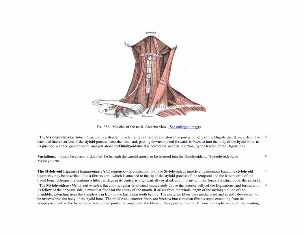
FIG. 386– Muscles of the neck. Anterior view. (See enlarged image) The Stylohyoideus (Stylohyoid muscle) is a slender muscle, lying in front of, and above the posterior belly of the Digastricus. It arises from the back and lateral surface of the styloid process, near the base; and, passing downward and forward, is inserted into the body of the hyoid bone, at its junction with the greater cornu, and just above theOmohyoideus. It is perforated, near its insertion, by the tendon of the Digastricus.
5
Variations.—It may be absent or doubled, lie beneath the carotid artery, or be inserted into the Omohyoideus, Thyreohyoideus, or Mylohyoideus.
6
The Stylohyoid Ligament (ligamentum stylohyoideus).—In connection with the Stylohyoideus muscle a ligamentous band, the stylohyoid ligament, may be described. It is a fibrous cord, which is attached to the tip of the styloid process of the temporal and the lesser cornu of the hyoid bone. It frequently contains a little cartilage in its center, is often partially ossified, and in many animals forms a distinct bone, the epihyal.
7
The Mylohyoideus (Mylohyoid muscle), flat and triangular, is situated immediately above the anterior belly of the Digastricus, and forms, with its fellow of the opposite side, a muscular floor for the cavity of the mouth. It arises from the whole length of the mylohyoid line of the mandible, extending from the symphysis in front to the last molar tooth behind. The posterior fibers pass medialward and slightly downward, to be inserted into the body of the hyoid bone. The middle and anterior fibers are inserted into a median fibrous raphé extending from the symphysis menti to the hyoid bone, where they joint at an angle with the fibers of the opposite muscle. This median raphé is sometimes wanting;
8

the fibers of the two muscles are then continuous. Variations.—It may be united to or replaced by the anterior belly of the Digastricus; accessory slips to other hyoid muscles are frequent. 9 The Geniohyoideus (Geniohyoid muscle) is a narrow muscle, situated above the medial border of the Mylohyoideus. It arises from the inferior mental spine on the back of the symphysis menti, and runs backward and slightly downward, to be inserted into the anterior surface of the body of the hyoid bone; it lies in contact with its fellow of the opposite side.
10
Variations.—It may be blended with the one on opposite side or double; slips to greater cornu of hyoid bone and Genioglossus occur. 11 Nerves.—The Mylohyoideus and anterior belly of the Digastricus are supplied by the mylohyoid branch of the inferior alveolar; the Stylohyoideus and posterior belly of the Digastricus, by the facial; the Geniohyoideus, by the hypoglossal.
12
Actions.—These muscles perform two very important actions. During the act of deglutition they raise the hyoid bone, and with it the base of the tongue; when the hyoid bone is fixed by its depressors and those of the larynx, they depress the mandible. During the first act of deglutition, when the mass of food is being driven from the mouth into the pharynx, the hyoid bone and with it the tongue, is carried upward and forward by the anterior bellies of the Digastrici, the Mylohyoidei, and Geniohyoidei. In the second act, when the mass is passing through the pharynx, the direct elevation of the hyoid bone takes place by the combined action of all the muscles; and after the food has passed, the hyoid bone is carried upward and backward by the posterior bellies of the Digastrici and the Stylohyoidei, which assist in preventing the return of the food into the mouth.
13
The infrahyoid muscles are: 14
Sternohyoideus. Thyreohyoideus. Sternothyreoideus. Omohyoideus.
The Sternohyoideus (Sternohyoid muscle) is a thin, narrow muscle, which arises from the posterior surface of the medial end of the clavicle, the posterior sternoclavicular ligament, and the upper and posterior part of the manubrium sterni. Passing upward and medialward, it is inserted, by short, tendinous fibers, into the lower border of the body of the hyoid bone. Below, this muscle is separated from its fellow by a considerable interval; but the two muscles come into contact with one another in the middle of their course, and from this upward, lie side by side. It sometimes presents, immediately above its origin, a transverse tendinous inscription.
15
Variations.—Doubling; accessory slips (Cleidohyoideus); absence. 16 The Sternothyreoideus (Sternothyroid muscle) is shorter and wider than the preceding muscle, beneath which it is situated. It arises from the posterior surface of the manubrium sterni, below the origin of the Sternohyoideus, and from the edge of the cartilage of the first rib, and sometimes that of the second rib, it is inserted into the oblique line on the lamina of the thyroid cartilage. This muscle is in close contact with its fellow at the lower part of the neck, but diverges somewhat as it ascends; it is occasionally traversed by a transverse or oblique tendinous inscription.
17
Variations.—Doubling; absence; accessory slips to Thyreohyoideus, Inferior constrictor, or carotid sheath. 18

The Thyreohyoideus (Thyrohyoid muscle) is a small, quadrilateral muscle appearing like an upward continuation of the Sternothyreoideus. It arises from the oblique line on the lamina of the thyroid cartilage, and is inserted into the lower border of the greater cornu of the hyoid bone.
19
The Omohyoideus (Omohyoid muscle) consists of two fleshy bellies united by a central tendon. It arises from the upper border of the scapula, and occasionally from the superior transverse ligament which crosses the scapular notch, its extent of attachment to the scapula varying from a few millimetres to 2.5 cm. From this origin, the inferior belly forms a flat, narrow fasciculus, which inclines forward and slightly upward across the lower part of the neck, being bound down to the clavicle by a fibrous expansion; it then passes behind the Sternocleidomastoideus, becomes tendinous and changes its direction, forming an obtuse angle. It ends in the superior belly, which passes almost vertically upward, close to the lateral border of the Sternohyoideus, to be inserted into the lower border of the body of the hyoid bone, lateral to the insertion of the Sternohyoideus. The central tendon of this muscle varies much in length and form, and is held in position by a process of the deep cervical fascia, which sheaths it, and is prolonged down to be attached to the clavicle and first rib; it is by this means that the angular form of the muscle is maintained.
20
Variations.—Doubling; absence; origin from clavicle; absence or doubling of either belly. 21 The inferior belly of the Omohyoideus divides the posterior triangle of the neck into an upper or occipital triangle and a lower or subclavian triangle, while its superior belly divides the anterior triangle into an upper or carotid triangle and a lower or muscular triangle.
22
Nerves.—The Infrahyoid muscles are supplied by branches from the first three cervical nerves. From the first two nerves the branch joins the hypoglossal trunk, runs with it some distance, and sends off a branch to the Thyreohyoideus; it then leaves the hypoglossal to form the descendens hypoglossi and unites with the communicantes cervicalis from the second and third cervical nerves to form the ansa hypoglossi from which nerves pass to the other Infrahyoid muscles.
23
Actions.—These muscles depress the larynx and hyoid bone, after they have been drawn up with the pharynx in the act of deglutition. The Omohyoidei not only depress the hyoid bone, but carry it backward and to one or the other side. They are concerned especially in prolonged inspiratory efforts; for by rendering the lower part of the cervical fascia tense they lessen the inward suction of the soft parts, which would otherwise compress the great vessels and the apices of the lungs. The Thyreohyoideus may act as an elevator of the thyroid cartilage, when the hyoid bone ascends, drawing the thyroid cartilage up behind the hyoid bone. The Sternothyreoideus acts as a depressor of the thyroid cartilage.
24
5d. The Anterior Vertebral Muscles
The anterior vertebral muscles (Fig. 387). are: 1
Longus colli. Rectus capitis anterior. Longus capitis. Rectus capitis lateralis.
The Longus colli is situated on the anterior surface of the vertebral column, between the atlas and the third thoracic vertebra. It is broad in the middle, narrow and pointed at either end, and consists of three portions, a superior oblique, an inferior oblique, and a vertical. The superior oblique portion arises from the anterior tubercles of the transverse processes of the third, fourth, and fifth cervical vertebræ and, ascending obliquely with a medial inclination, is inserted by a narrow tendon into the tubercle on the anterior arch of the atlas. The inferior oblique
2

portion, the smallest part of the muscle, arises from the front of the bodies of the first two or three thoracic vertebræ; and, ascending obliquely in a lateral direction, is inserted into the anterior tubercles of the transverse processes of the fifth and sixth cervical vertebræ. The vertical portion arises, below, from the front of the bodies of the upper three thoracic and lower three cervical vertebræ, and is inserted into the front of the bodies of the second, third, and fourth cervical vertebræ. The Longus capitis (Rectus capitis anticus major), broad and thick above, narrow below,arises by four tendinous slips, from the anterior tubercles of the transverse processes of the third, fourth, fifth, and sixth cervical vertebræ, and ascends, converging toward its fellow of the opposite side, to be inserted into the inferior surface of the basilar part of the occipital bone.
3
FIG. 387– The anterior vertebral muscles. (See enlarged image)
The Rectus capitis anterior (Rectus capitis anticus minor) is a short, flat muscle, situated immediately behind the upper part of the Longus capitis. It arises from the anterior surface of the lateral mass of the atlas, and from the root of its transverse process, and passing obliquely upward
4

and medialward, is inserted into the inferior surface of the basilar part of the occipital bone immediately in front of the foramen magnum. The Rectus capitis lateralis, a short, flat muscle, arises from the upper surface of the transverse process of the atlas, and is inserted into the under surface of the jugular process of the occipital bone.
5
Nerves.—The Rectus capitis anterior and the Rectus capitis lateralis are supplied from the loop between the first and second cervical nerves; the Longus capitis, by branches from thefirst, second, and third cervical; the Longus colli, by branches from the second to the seventh cervical nerves.
6
Actions.—The Longus capitis and Rectus capitis anterior are the direct antagonists of the muscles at the back of the neck, serving to restore the head to its natural position after it has been drawn backward. These muscles also flex the head, and from their obliquity, rotate it, so as to turn the face to one or the other side. The Rectus lateralis, acting on one side, bends the head laterally. The Longus colli flexes and slightly rotates the cervical portion of the vertebral column.
5e. The Lateral Vertebral Muscles
The lateral vertebral muscles (Fig. 387). are: 1
Scalenus anterior. Scalenus medius.
Scalenus posterior.
The Scalenus anterior (Scalenus anticus) lies deeply at the side of the neck, behind the Sternocleidomastoideus. It arises from the anterior tubercles of the transverse processes of the third, fourth, fifth, and sixth cervical vertebræ, and descending, almost vertically, isinserted by a narrow, flat tendon into the scalene tubercle on the inner border of the first rib, and into the ridge on the upper surface of the rib in front of the subclavian groove.
2
The Scalenus medius, the largest and longest of the three Scaleni, arises from the posterior tubercles of the transverse processes of the lower six cervical vertebræ, and descending along the side of the vertebral column, is inserted by a broad attachment into the upper surface of the first rib, between the tubercle and the subclavian groove.
3
The Scalenus posterior (Scalenus posticus), the smallest and most deeply seated of the three Scaleni, arises, by two or three separate tendons, from the posterior tubercles of the transverse processes of the lower two or three cervical vertebræ, and is inserted by a thin tendon into the outer surface of the second rib, behind the attachment of the Serratus anterior. It is occasionally blended with the Scalenus medius.
4
Variations.—The Scaleni muscles vary considerably in their attachments and in the arrangement of their fibers. A slip from the Scalenus anticus may pass behind the subclavian artery. The Scalenus posticus may be absent or extend to the third rib. The Scalenus pleuralis muscle extends from the transverse process of the seventh cervical vertebra to the fascia supporting the dome of the pleura and inner border of first rib.
5
Nerves.—The Scaleni are supplied by branches from the second to the seventh cervical nerves. 6

Actions.—When the Scaleni act from above, they elevate the first and second ribs, and are, therefore, inspiratory muscles. Acting from below, they bend the vertebral column to one or other side; if the muscles of both sides act, the vertebral column is slightly flexed.
6. The Fasciæ and Muscles of the Trunk. a. The Deep Muscles of the Back
The muscles of the trunk may be arranged in six groups: 1
I. Deep Muscles of the Back. IV. Muscles of the Abdomen. II. Suboccipital Muscles. V. Muscles of the Pelvis. III. Muscles of the Thorax. VI. Muscles of the Perineum.
The Deep Muscles of the Back
The deep or intrinsic muscles of the back (Fig. 388). consist of a complex group of muscles extending from the pelvis to the skull. They are:
2
Splenius capitis. Multifidus. Splenius cervicis. Rotatores. Sacrospinalis. Interspinales. Semispinalis. Intertransversarii.
The Lumbodorsal Fascia (fascia lumbodorsalis; lumbar aponeurosis and vertebral fascia).—The lumbodorsal fascia is a deep investing membrane which covers the deep muscles of the back of the trunk. Above, it passes in front of the Serratus posterior superior and is continuous with a similar investing layer on the back of the neck—the nuchal fascia.
3
In the thoracic region the lumbodorsal fascia is a thin fibrous lamina which serves to bind down the Extensor muscles of the vertebral column and to separate them from the muscles connecting the vertebral column to the upper extremity. It contains both longitudinal and transverse fibers, and is attached, medially, to the spinous processes of the thoracic vertebræ; laterally to the angles of the ribs.
4
In the lumbar region the fascia (lumbar aponeurosis) is in two layers, anterior and posterior (Figs. 388, 409). The posterior layer is attached to the spinous processes of the lumbar and sacral vertebræ and to the supraspinal ligament; the anterior layer is attached,medially, to the tips of the transverse processes of the lumbar vertebræ and to the intertransverse ligaments, below, to the iliolumbar ligament, and above, to the lumbocostal ligament. The two layers unite at the lateral margin of the Sacrospinalis, to form the tendon of origin of the Transversus abdominis. The aponeurosis of origin of the Serratus posterior inferior and the Latissimus dorsi are intimately blended with the lumbodorsal fascia.
5
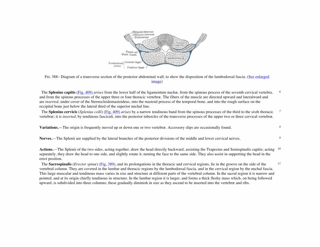
FIG. 388– Diagram of a transverse section of the posterior abdominal wall, to show the disposition of the lumbodorsal fascia. (See enlarged image)
The Splenius capitis (Fig. 409) arises from the lower half of the ligamentum nuchæ, from the spinous process of the seventh cervical vertebra, and from the spinous processes of the upper three or four thoracic vertebræ. The fibers of the muscle are directed upward and lateralward and are inserted, under cover of the Sternocleidomastoideus, into the mastoid process of the temporal bone, and into the rough surface on the occipital bone just below the lateral third of the superior nuchal line.
6
The Splenius cervicis (Splenius colli) (Fig. 409) arises by a narrow tendinous band from the spinous processes of the third to the sixth thoracic vertebræ; it is inserted, by tendinous fasciculi, into the posterior tubercles of the transverse processes of the upper two or three cervical vertebræ.
7
Variations.—The origin is frequently moved up or down one or two vertebræ. Accessory slips are occasionally found. 8 Nerves.—The Splenii are supplied by the lateral branches of the posterior divisions of the middle and lower cervical nerves. 9 Actions.—The Splenii of the two sides, acting together, draw the head directly backward, assisting the Trapezius and Semispinalis capitis; acting separately, they draw the head to one side, and slightly rotate it, turning the face to the same side. They also assist in supporting the head in the erect position.
10
The Sacrospinalis (Erector spinæ) (Fig. 389), and its prolongations in the thoracic and cervical regions, lie in the groove on the side of the vertebral column. They are covered in the lumbar and thoracic regions by the lumbodorsal fascia, and in the cervical region by the nuchal fascia. This large muscular and tendinous mass varies in size and structure at different parts of the vertebral column. In the sacral region it is narrow and pointed, and at its origin chiefly tendinous in structure. In the lumbar region it is larger, and forms a thick fleshy mass which, on being followed upward, is subdivided into three columns; these gradually diminish in size as they ascend to be inserted into the vertebræ and ribs.
11
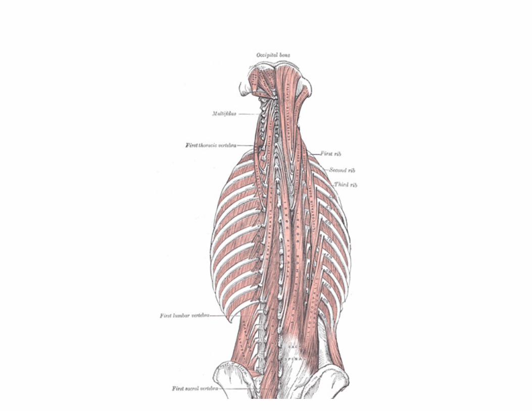

FIG. 389– Deep muscles of the back. (See enlarged image)
The Sacrospinalis arises from the anterior surface of a broad and thick tendon, which is attached to the medial crest of the sacrum, to the spinous processes of the lumbar and the eleventh and twelfth thoracic vertebræ, and the supraspinal ligament, to the back part of the inner lip of the iliac crests and to the lateral crests of the sacrum, where it blends with the sacrotuberous and posterior sacroiliac ligaments. Some of its fibers are continuous with the fibers of origin of the Glutæus maximus. The muscular fibers form a large fleshy mass which splits, in the upper lumbar region into three columns, viz., a lateral, the Iliocostalis,an intermediate, the Longissimus, and a medial, the Spinalis. Each of these consists from below upward, of three parts, as follows:
12
Lateral Column. Intermediate Column. Medial Column. Iliocostalis. Longissimus. Spinalis.
(a) I. lumborum. (a) L. dorsi. (a) S. dorsi. (b) I. dorsi. (b) L. cervicis. (b) S. cervicis. (c) I. cervicis. (c) L. capitis. (c) S. capitis.
The Iliocostalis lumborum (Iliocostalis muscle; Sacrolumbalis muscle) is inserted, by six or seven flattened tendons, into the inferior borders of the angles of the lower six or seven ribs.
13
The Iliocostalis dorsi (Musculus accessorius) arises by flattened tendons from the upper borders of the angles of the lower six ribs medial to the tendons of insertion of the Iliocostalis lumborum; these become muscular, and are inserted into the upper borders of the angles of the upper six ribs and into the back of the transverse process of the seventh cervical vertebra.
14
The Iliocostalis cervicis (Cervicalis ascendens) arises from the angles of the third, fourth, fifth, and sixth ribs, and is inserted into the posterior tubercles of the transverse processes of the fourth, fifth, and sixth cervical vertebræ.
15
The Longissimus dorsi is the intermediate and largest of the continuations of the Sacrospinalis. In the lumbar region, where it is as yet blended with the Iliocostalis lumborum, some of its fibers are attached to the whole length of the posterior surfaces of the transverse processes and the accessory processes of the lumbar vertebræ, and to the anterior layer of the lumbodorsal fascia. In the thoracic region it is inserted, by rounded tendons, into the tips of the transverse processes of all the thoracic vertebræ, and by fleshy processes into the lower nine or ten ribs between their tubercles and angles.
16
The Longissimus cervicis (Transversalis cervicis), situated medial to the Longissimus dorsi, arises by long thin tendons from the summits of the transverse processes of the upper four or five thoracic vertebræ, and is inserted by similar tendons into the posterior tubercles of the transverse processes of the cervical vertebræ from the second to the sixth inclusive.
17
The Longissimus capitis (Trachelomastoid muscle) lies medial to the Longissimus cervicis, between it and the Semispinalis capitis. It arises by tendons from the transverse processes of the upper four or five thoracic vertebræ, and the articular processes of the lower three or four cervical vertebræ, and is inserted into the posterior margin of the mastoid process, beneath the Splenius capitis and Sternocleidomastoideus. It is almost always crossed by a tendinous intersection near its insertion.
18
The Spinalis dorsi, the medial continuation of the Sacrospinalis, is scarcely separable as a distinct muscle. It is situated at the medial side of the Longissimus dorsi, and is intimately blended with it; it arises by three or four tendons from the spinous processes of the first two lumbar and the last two thoracic vertebræ: these, uniting, form a small muscle which isinserted by separate tendons into the spinous processes of the upper
19

thoracic vertebræ, the number varying from four to eight. It is intimately united with the Semispinalis dorsi, situated beneath it. The Spinalis cervicis (Spinalis colli) is an inconstant muscle, which arises from the lower part of the ligamentum nuchæ, the spinous process of the seventh cervical, and sometimes from the spinous processes of the first and second thoracic vertebræ, and is inserted into the spinous process of the axis, and occasionally into the spinous processes of the two vertebræ below it.
20
The Spinalis capitis (Biventer cervicis) is usually inseparably connected with the Semispinalis capitis (see below). 21 The Semispinalis dorsi consists of thin, narrow, fleshy fasciculi, interposed between tendons of considerable length. It arises by a series of small tendons from the transverse processes of the sixth to the tenth thoracic vertebræ, and is inserted, by tendons, into the spinous processes of the upper four thoracic and lower two cervical vertebræ.
22
The Semispinalis cervicis (Semispinalis colli), thicker than the preceding, arises by a series of tendinous and fleshy fibers from the transverse processes of the upper five or six thoracic vertebræ, and is inserted into the cervical spinous processes, from the axis to the fifth inclusive. The fasciculus connected with the axis is the largest, and is chiefly muscular in structure.
23
The Semispinalis capitis (Complexus) is situated at the upper and back part of the neck, beneath the Splenius, and medial to the Longissimus cervicis and capitis. It arises by a series of tendons from the tips of the transverse processes of the upper six or seven thoracic and the seventh cervical vertebræ, and from the articular processes of the three cervical above this. The tendons, uniting, form a broad muscle, which passes upward, and isinserted between the superior and inferior nuchal lines of the occipital bone. The medial part, usually more or less distinct from the remainder of the muscle, is frequently termed theSpinalis capitis; it is also named the Biventer cervicis since it is traversed by an imperfect tendinous inscription.
24
The Multifidus (Multifidus spinæ) consists of a number of fleshy and tendinous fasciculi, which fill up the groove on either side of the spinous processes of the vertebræ, from the sacrum to the axis. In the sacral region, these fasciculi arise from the back of the sacrum, as low as the fourth sacral foramen, from the aponeurosis of origin of the Sacrospinalis, from the medial surface of the posterior superior iliac spine, and from the posterior sacroiliac ligaments; in the lumbar region, from all the mamillary processes; in the thoracic region, from all the transverse processes; and in the cervical region, from the articular processes of the lower four vertebræ. Each fasciculus, passing obliquely upward and medialward, isinserted into the whole length of the spinous process of one of the vertebræ above. These fasciculi vary in length: the most superficial, the longest, pass from one vertebra to the third or fourth above; those next in order run from one vertebra to the second or third above; while the deepest connect two contiguous vertebræ.
25
The Rotatores (Rotatores spinæ) lie beneath the Multifidus and are found only in the thoracic region; they are eleven in number on either side. Each muscle is small and somewhat quadrilateral in form; it arises from the upper and back part of the transverse process, and is inserted into the lower border and lateral surface of the lamina of the vertebra above, the fibers extending as far as the root of the spinous process. The first is found between the first and second thoracic vertebræ; the last, between the eleventh and twelfth. Sometimes the number of these muscles is diminished by the absence of one or more from the upper or lower end.
26
The Interspinales are short muscular fasciculi, placed in pairs between the spinous processes of the contiguous vertebræ, one on either side of the interspinal ligament. In thecervical region they are most distinct, and consist of six pairs, the first being situated between the axis and third vertebra, and the last between the seventh cervical and the first thoracic. They are small narrow bundles, attached, above and below, to the apices of the spinous processes. In the thoracic region, they are found between the first and second vertebræ, and sometimes between the second and third, and between the eleventh and twelfth. In the lumbar region there are four pairs in the intervals between the five lumbar vertebræ. There is also occasionally one between the last thoracic and first lumbar, and one between the fifth lumbar and the sacrum.
27
The Extensor coccygis is a slender muscular fasciculus, which is not always present; it extends over the lower part of the posterior surface of the sacrum and coccyx. It arises by tendinous fibers from the last segment of the sacrum, or first piece of the coccyx, and passes downward to
28

be inserted into the lower part of the coccyx. It is a rudiment of the Extensor muscle of the caudal vertebræ of the lower animals. The Intertransversarii (Intertransversales) are small muscles placed between the transverse processes of the vertebræ. In the cervical region they are best developed, consisting of rounded muscular and tendinous fasciculi, and are placed in pairs, passing between the anterior and the posterior tubercles respectively of the transverse processes of two contiguous vertebræ, and separated from one another by an anterior primary division of the cervical nerve, which lies in the groove between them. The muscles connecting the anterior tubercles are termed the Intertransversarii anteriores; those between the posterior tubercles, the Intertransversarii posteriores; both sets are supplied by the anterior divisions of the spinal nerves (Lickley 81). There are seven pairs of these muscles, the first pair being between the atlas and axis, and the last pair between the seventh cervical and first thoracic vertebræ. In the thoracic region they are present between the transverse processes of the lower three thoracic vertebræ, and between the transverse processes of the last thoracic and the first lumbar. In the lumbar region they are arranged in pairs, on either side of the vertebral column, one set occupying the entire interspace between the transverse processes of the lumbar vertebræ, the Intertransversarii laterales; the other set,Intertransversarii mediales, passing from the accessory process of one vertebra to the mammillary of the vertebra below. The Intertransversarii laterales are supplied by the anterior divisions, and the Intertransversarii mediales by the posterior divisions of the spinal nerves (Lichley, op. cit.).
29
Note 81. Journal of Anatomy and Physiology, 1904, vol. xxxix. [back]
6b. The Suboccipital Muscles
The suboccipital group (Fig. 389). comprises: 1
Rectus capitis posterior major. Obliquus capitis inferior. Rectus capitis posterior minor. Obliquus capitis superior.
The Rectus capitis posterior major (Rectus capitis posticus major) arises by a pointed tendon from the spinous process of the axis, and, becoming broader as it ascends, is insertedinto the lateral part of the inferior nuchal line of the occipital bone and the surface of the bone immediately below the line. As the muscles of the two sides pass upward and lateralward, they leave between them a triangular space, in which the Recti capitis posteriores minores are seen.
2
The Rectus capitis posterior minor (Rectus capitis posticus minor) arises by a narrow pointed tendon from the tubercle on the posterior arch of the atlas, and, widening as it ascends, is inserted into the medial part of the inferior nuchal line of the occipital bone and the surface between it and the foramen magnum.
3
The Obliquus capitis inferior (Obliquus inferior), the larger of the two Oblique muscles,arises from the apex of the spinous process of the axis, and passes lateralward and slightly upward, to be inserted into the lower and back part of the transverse process of the atlas.
4
The Obliquus capitis superior (Obliquus superior), narrow below, wide and expanded above, arises by tendinous fibers from the upper surface of the transverse process of the atlas, joining with the insertion of the preceding. It passes upward and medialward, and isinserted into the occipital bone, between the superior and inferior nuchal lines, lateral to the Semispinalis capitis.
5
The Suboccipital Triangle.—Between the Obliqui and the Rectus capitis posterior major is the suboccipital triangle. It is bounded, above and medially, by the Rectus capitis posterior major; above and laterally, by the Obliquus capitis superior; below and laterally, by the Obliquus capitis inferior. It is covered by a layer of dense fibro-fatty tissue, situated beneath the Semispinalis capitis. The floor is formed by the posterior occipito-atlantal membrane, and the posterior arch of the atlas. In the deep groove on the upper surface of the posterior arch of the
6
atlas are the vertebral artery and the first cervical or suboccipital nerve. Nerves.—The deep muscles of the back and the suboccipital muscles are supplied by the posterior primary divisions of the spinal nerves. 7 Actions.—The Sacrospinalis and its upward continuations and the Spinales serve to maintain the vertebral column in the erect posture; they also serve to bend the trunk backward when it is required to counterbalance the influence of any weight at the front of the body—as, for instance, when a heavy weight is suspended from the neck, or when there is any great abdominal distension, as in pregnancy or dropsy; the peculiar gait under such circumstances depends upon the vertebral column being drawn backward, by the counterbalancing action of the Sacrospinales. The muscles which form the continuation of the Sacrospinales on to the head and neck steady those parts and fix them in the upright position. If the Iliocostalis lumborum and Longissimus dorsi of one side act, they serve to draw down the chest and vertebral column to the corresponding side. The Iliocostales cervicis, taking their fixed points from the cervical vertebræ, elevate those ribs to which they are attached; taking their fixed points from the ribs, both muscles help to extend the neck; while one muscle bends the neck to its own side. When both Longissimi cervicis act from below, they bend the neck backward. When both Longissimi capitis act from below, they bend the head backward; while, if only one muscle acts, the face is turned to the side on which the muscle is acting, and then the head is bent to the shoulder. The two Recti draw the head backward. The Rectus capitis posterior major, owing to its obliquity, rotates the skull, with the atlas, around the odontoid process, turning the face to the same side. The Multifidus acts successively upon the different parts of the column; thus, the sacrum furnishes a fixed point from which the fasciculi of this muscle acts upon the lumbar region; which in turn becomes the fixed point for the fasciculi moving the thoracic region, and so on throughout the entire length of the column. The Multifidus also serves to rotate the column, so that the front of the trunk is turned to the side opposite to that from which the muscle acts, this muscle being assisted in its action by the Obliquus externus abdominis. The Obliquus capitis superior draws the head backward and to its own side. The Obliquus inferior rotates the atlas, and with it the skull, around the odontoid process, turning the face to the same side. When the Semispinales of the two sides act together, they help to extend the vertebral column; when the muscles of only one side act, they rotate the thoracic and cervical parts of the column, turning the body to the opposite side. The Semispinales capitis draw the head directly backward; if one muscle acts, it draws the head to one side, and rotates it so that the face is turned to the opposite side. The Interspinales by approximating the spinous processes help to extend the column. The Intertransversarii approximate the transverse processes, and help to bend the column to one side. The Rotatores assist the Multifidus to rotate the vertebral column, so that the front of the trunk is turned to the side opposite to that from which the muscles act.
8
6c. The Muscles of the Thorax
The muscles belonging to this group are the 1
Intercostales externi. Levatores costarum. Intercostales interni. Serratus posterior superior. Subcostales. Serratus posterior inferior. Transversus thoracis. Diaphragm.
Intercostal Fasciæ.—In each intercostal space thin but firm layers of fascia cover the outer surface of the Intercostalis externus and the inner 2

surface of the Intercostalis internus; and a third, more delicate layer, is interposed between the two planes of muscular fibers. They are best marked in those situations where the muscular fibers are deficient, as between the Intercostales externi and sternum in front, and between the Intercostales interni and vertebral column behind. The Intercostales (Intercostal muscles) (Fig. 411) are two thin planes of muscular and tendinous fibers occupying each of the intercostal spaces. They are named external andinternal from their surface relations—the external being superficial to the internal.
3
The Intercostales externi (External intercostals) are eleven in number on either side. They extend from the tubercles of the ribs behind, to the cartilages of the ribs in front, where they end in thin membranes, the anterior intercostal membranes, which are continued forward to the sternum. Each arises from the lower border of a rib, and isinserted into the upper border of the rib below. In the two lower spaces they extend to the ends of the cartilages, and in the upper two or three spaces they do not quite reach the ends of the ribs. They are thicker than the Intercostales interni, and their fibers are directed obliquely downward and lateralward on the back of the thorax, and downward, forward, and medialward on the front.
4
Variations.—Continuation with the Obliquus externus or Serratus anterior: ASupracostalis muscle, from the anterior end of the first rib down to the second, third or fourth ribs occasionally occurs.
5
The Intercostales interni (Internal intercostals) are also eleven in number on either side. They commence anteriorly at the sternum, in the interspaces between the cartilages of the true ribs, and at the anterior extremities of the cartilages of the false ribs, and extend backward as far as the angles of the ribs, whence they are continued to the vertebral column by thin aponeuroses, the posterior intercostal membranes. Each arises from the ridge on the inner surface of a rib, as well as from the corresponding costal cartilage, and isinserted into the upper border of the rib below. Their fibers are also directed obliquely, but pass in a direction opposite to those of the Intercostales externi.
6
The Subcostales (Infracostales) consist of muscular and aponeurotic fasciculi, which are usually well-developed only in the lower part of the thorax; each arises from the inner surface of one rib near its angle, and is inserted into the inner surface of the second or third rib below. Their fibers run in the same direction as those of the Intercostales interni.
7
The Transversus thoracis (Triangularis sterni) is a thin plane of muscular and tendinous fibers, situated upon the inner surface of the front wall of the chest (Fig. 390). It arises on either side from the lower third of the posterior surface of the body of the sternum, from the posterior surface of the xiphoid process, and from the sternal ends of the costal cartilages of the lower three or four true ribs. Its fibers diverge upward and lateralward, to be inserted by slips into the lower borders and inner surfaces of the costal cartilages of the second, third, fourth, fifth, and sixth ribs. The lowest fibers of this muscle are horizontal in their direction, and are continuous with those of the Transversus abdominis; the intermediate fibers are oblique, while the highest are almost vertical. This muscle varies in its attachments, not only in different subjects, but on opposite sides of the same subject.
8
The Levatores costarum (Fig. 389), twelve in number on either side, are small tendinous and fleshy bundles, which arise from the ends of the transverse processes of the seventh cervical and upper eleven thoracic vertebræ; they pass obliquely downward and lateralward, like the fibers of the Intercostales externi, and each is inserted into the outer surface of the rib immediately below the vertebra from which it takes origin, between the tubercle and the angle (Levatores costarum breves). Each of the four lower muscles divides into two fasciculi, one of which is inserted as above described; the other passes down to the second rib below its origin (Levatores costarum longi).
9
The Serratus posterior superior (Serratus posticus superior) is a thin, quadrilateral muscle, situated at the upper and back part of the thorax. It arises by a thin and broad aponeurosis from the lower part of the ligamentum nuchae, from the spinous processes of the seventh cervical and upper two or three thoracic vertebræ and from the supraspinal ligament. Inclining downward and lateralward it becomes muscular, and is inserted, by four fleshy digitations, into the upper borders of the second, third, fourth, and fifth ribs, a little beyond their angles.
10

Variations.—Increase or decrease in size and number of slips or entire absence. 11 The Serratus posterior inferior (Serratus posticus inferior) (Fig. 409) is situated at the junction of the thoracic and lumbar regions: it is of an irregularly quadrilateral form, broader than the preceding, and separated from it by a wide interval. It arises by a thin aponeurosis from the spinous processes of the lower two thoracic and upper two or three lumbar vertebræ, and from the supraspinal ligament. Passing obliquely upward and lateralward, it becomes fleshy, and divides into four flat digitations, which are inserted into the inferior borders of the lower four ribs, a little beyond their angles. The thin aponeurosis of origin is intimately blended with the lumbodorsal fascia, and aponeurosis of the Latissimus dorsi.
12
FIG. 390– Posterior surface of sternum and costal cartilages, showing Transversus thoracis. (See enlarged image)
Variations.—Increase or decrease in size and number of slips or entire absence. 13 Nerves.—The muscles of this group are supplied by the intercostal nerves. 14 The Diaphragm (Fig. 391) is a dome-shaped musculofibrous septum which separates the thoracic from the abdominal cavity, its convex upper surface forming the floor of the former, and its concave under surface the roof of the latter. Its peripheral part consists of muscular fibers which take origin from the circumference of the thoracic outlet and converge to be inserted into a central tendon.
15
The muscular fibers may be grouped according to their origins into three parts—sternal, costal, and lumbar. The sternal part arises by two fleshy slips from the back of the xiphoid process; the costal part from the inner surfaces of the cartilages and adjacent portions of the lower six ribs on either side, interdigitating with the Transversus abdominis; and the lumbar part from aponeurotic arches, named the lumbocostal arches, and from the lumbar vertebræ by two pillars or crura. There are two lumbocostal arches, a medial and a lateral, on either side.
16
The Medial Lumbocostal Arch (arcus lumbocostalis medialis [Halleri]; internal arcuate ligament) is a tendinous arch in the fascia covering the upper part of the Psoas major; medially, it is continuous with the lateral tendinous margin of the corresponding crus, and is attached to the side of the body of the first or second lumbar vertebra; laterally, it is fixed to the front of the transverse process of the first and, sometimes also, to that of the second lumbar vertebra.
17
The Lateral Lumbocostal Arch (arcus lumbocostalis lateralis [Halleri]; external arcuate ligament) arches across the upper part of the Quadratus lumborum, and is attached, medially, to the front of the transverse process of the first lumbar vertebra, and, laterally, to the tip and lower margin of the twelfth rib.
18
The Crura.—At their origins the crura are tendinous in structure, and blend with the anterior longitudinal ligament of the vertebral column. The right crus, larger and longer than the left, arises from the anterior surfaces of the bodies and intervertebral fibrocartilages of the upper three lumbar vertebræ, while the left crus arises from the corresponding parts of the upper two only. The medial tendinous margins of the crura pass forward and medialward, and meet in the middle line to form an arch across the front of the aorta; this arch is often poorly defined.
19

FIG. 391– The diaphragm. Under surface. (See enlarged image) From this series of origins the fibers of the diaphragm converge to be inserted into the central tendon. The fibers arising from the xiphoid process are very short, and occasionally aponeurotic; those from the medial and lateral lumbocostal arches, and more especially those from the ribs and their cartilages, are longer, and describe marked curves as they ascend and converge to their insertion. The fibers of the crura diverge as they ascend, the most lateral being directed upward and lateralward to the central tendon. The medial fibers of the right crus ascend on the left side of the esophageal hiatus, and occasionally a fasciculus of the left crus crosses the aorta and runs obliquely through the fibers of the right crus toward the vena caval foramen (Low 82).
20
The Central Tendon.—The central tendon of the diaphragm is a thin but strong aponeurosis situated near the center of the vault formed by the muscle, but somewhat closer to the front than to the back of the thorax, so that the posterior muscular fibers are the longer. It is situated immediately below the pericardium, with which it is partially blended. It is shaped somewhat like a trefoil leaf, consisting of three divisions or leaflets separated from one another by slight indentations. The right leaflet is the largest, the middle, directed toward the xiphoid process, the next in size, and the left the smallest. In structure the tendon is composed of several planes of fibers, which intersect one another at various angles and unite into straight or curved bundles—an arrangement which gives it additional strength.
21

Openings in the Diaphragm.—The diaphragm is pierced by a series of apertures to permit of the passage of structures between the thorax and abdomen. Three large openings—theaortic, the esophageal, and the vena caval—and a series of smaller ones are described.
22
The aortic hiatus is the lowest and most posterior of the large apertures; it lies at the level of the twelfth thoracic vertebra. Strictly speaking, it is not an aperture in the diaphragm but an osseoaponeurotic opening between it and the vertebral column, and therefore behind the diaphragm; occasionally some tendinous fibers prolonged across the bodies of the vertebræ from the medial parts of the lower ends of the crura pass behind the aorta, and thus convert the hiatus into a fibrous ring. The hiatus is situated slightly to the left of the middle line, and is bounded in front by the crura, and behind by the body of the first lumbar vertebra. Through it pass the aorta, the azygos vein, and the thoracic duct; occasionally the azygos vein is transmitted through the right crus.
23
The esophageal hiatus is situated in the muscular part of the diaphragm at the level of the tenth thoracic vertebra, and is elliptical in shape. It is placed above, in front, and a little to the left of the aortic hiatus, and transmits the esophagus, the vagus nerves, and some small esophageal arteries.
24
The vena caval foramen is the highest of the three, and is situated about the level of the fibrocartilage between the eighth and ninth thoracic vertebræ. It is quadrilateral in form, and is placed at the junction of the right and middle leaflets of the central tendon, so that its margins are tendinous. It transmits the inferior vena cava, the wall of which is adherent to the margins of the opening, and some branches of the right phrenic nerve.
25
Of the lesser apertures, two in the right crus transmit the greater and lesser right splanchnic nerves; three in the left crus give passage to the greater and lesser left splanchnic nerves and the hemiazygos vein. The gangliated trunks of the sympathetic usually enter the abdominal cavity behind the diaphragm, under the medial lumbocostal arches.
26
On either side two small intervals exist at which the muscular fibers of the diaphragm are deficient and are replaced by areolar tissue. One between the sternal and costal parts transmits the superior epigastric branch of the internal mammary artery and some lymphatics from the abdominal wall and convex surface of the liver. The other, between the fibers springing from the medial and lateral lumbocostal arches, is less constant; when this interval exists, the upper and back part of the kidney is separated from the pleura by areolar tissue only.
27
Variations.—The sternal portion of the muscle is sometimes wanting and more rarely defects occur in the lateral part of the central tendon or adjoining muscle fibers.
28
Nerves.—The diaphragm is supplied by the phrenic and lower intercostal nerves. 29 Actions.—The diaphragm is the principal muscle of inspiration, and presents the form of a dome concave toward the abdomen. The central part of the dome is tendinous, and the pericardium is attached to its upper surface; the circumference is muscular. During inspiration the lowest ribs are fixed, and from these and the crura the muscular fibers contract and draw downward and forward the central tendon with the attached pericardium. In this movement the curvature of the diaphragm is scarcely altered, the dome moving downward nearly parallel to its original position and pushing before it the abdominal viscera. The descent of the abdominal viscera is permitted by the elasticity of the abdominal wall, but the limit of this is soon reached. The central tendon applied to the abdominal viscera then becomes a fixed point for the action of the diaphragm, the effect of which is to elevate the lower ribs and through them to push forward the body of the sternum and the upper ribs. The right cupola of the diaphragm, lying on the liver, has a greater resistance to overcome than the left, which lies over the stomach, but to compensate for this the right crus and the fibers of the right side generally are stronger than those of the left.
30
In all expulsive acts the diaphragm is called into action to give additional power to each expulsive effort. Thus, before sneezing, coughing, laughing, crying, or vomiting, and previous to the expulsion of urine or feces, or of the fetus from the uterus, a deep inspiration takes place. The height of the diaphragm is constantly varying during respiration; it also varies with the degree of distension of the stomach and intestines and with the size of the liver. After a forced expiration the right cupola is on a level in front with the fourth costal cartilage, at the side with the fifth, sixth, and seventh ribs, and behind with the eighth rib; the left cupola is a little lower than the right. Halls Dally 83 states that the absolute range of movement between deep inspiration and deep expiration averages in the male and female 30 mm. on the right side and 28 mm. on the left; in quiet respiration the average movement is 12.5 mm. on the right side and 12 mm. on the left.
31
Skiagraphy shows that the height of the diaphragm in the thorax varies considerably with the position of the body. It stands highest when the body is horizontal and the patient on his back, and in this position it performs the largest respiratory excursions with normal breathing. When the body is erect the dome of the diaphragm falls, and its respiratory movements become smaller. The dome falls still lower when the sitting posture is assumed, and in this position its respiratory excursions are smallest. These facts may, perhaps, explain why it is that patients suffering from severe dyspnœa are most comfortable and least short of breath when they sit up. When the body is horizontal and the patient on his side, the two halves of the diaphragm do not behave alike. The uppermost half sinks to a level lower even than when the patient sits, and moves little with respiration; the lower half rises higher in the thorax than it does when the patient is supine, and its respiratory excursions are much increased. In unilateral disease of the pleura or lungs analogous interference with the position or movement of the diaphragm can generally be observed skiagraphically.
32
It appears that the position of the diaphragm in the thorax depends upon three main factors, viz.: (1) the elastic retraction of the lung tissue, tending to pull it upward; (2) the pressure exerted on its under surface by the viscera; this naturally tends to be a negative pressure, or downward suction, when the patient sits or stands, and positive, or an upward pressure, when he lies; (3) the intra-abdominal tension due to the abdominal muscles. These are in a state of contraction in the standing position and not in the sitting; hence the diaphragm, when the patient stands, is pushed up higher than when he sits.
33
The Intercostales interni and externi have probably no action in moving the ribs. They contract simultaneously and form strong elastic supports which prevent the intercostal spaces being pushed out or drawn in during respiration. The anterior portions of the Intercostales interni probably have an additional function in keeping the sternocostal and interchondral joint surfaces in apposition, the posterior parts of the Intercostales externi performing a similar function for the costovertebral articulations. The Levatores costarum being inserted near the fulcra of the ribs can have little action on the ribs; they act as rotators and lateral flexors of the vertebral column. The Transversus thoracis draws down the costal cartilages, and is therefore a muscle of expiration.
34
The Serrati are respiratory muscles. The Serratus posterior superior elevates the ribs and is therefore an inspiratory muscle. The Serratus posterior inferior draws the lower ribs downward and backward, and thus elongates the thorax; it also fixes the lower ribs, thus assisting the inspiratory action of the diaphragm and resisting the tendency it has to draw the lower ribs upward and forward. It must therefore be regarded as a muscle of inspiration.
35
Mechanism of Respiration.—The respiratory movements must be examined during (a) quiet respiration, and (b) deep respiration. 36 Quiet Respiration.—The first and second pairs of ribs are fixed by the resistance of the cervical structures; the last pair, and through it the eleventh, by the Quadratus lumborum. The other ribs are elevated, so that the first two intercostal spaces are diminished while the others are increased in width. It has already been shown (p. 304) that elevation of the third, fourth, fifth, and sixth ribs leads to an increase in the antero-posterior and transverse diameters of the thorax; the vertical diameter is increased by the descent of the diaphragmatic dome so that the lungs are expanded in all directions except backward and upward. Elevation of the eighth, ninth, and tenth ribs is accompanied by a lateral and backward movement, leading to an increase in the transverse diameter of the upper part of the abdomen; the elasticity of the anterior abdominal wall allows
37
a slight increase in the antero-posterior diameter of this part, and in this way the decrease in the vertical diameter of the abdomen is compensated and space provided for its displaced viscera. Expiration is effected by the elastic recoil of its walls and by the action of the abdominal muscles, which push back the viscera displaced downward by the diaphragm. Deep Respiration.—All the movements of quiet respiration are here carried out, but to a greater extent. In deep inspiration the shoulders and the vertebral borders of the scapulæ are fixed and the limb muscles, Trapezius, Serratus anterior, Pectorales, and Latissimus dorsi, are called into play. The Scaleni are in strong action, and the Sternocleidomastoidei also assist when the head is fixed by drawing up the sternum and by fixing the clavicles. The first rib is therefore no longer stationary, but, with the sternum, is raised; with it all the other ribs except the last are raised to a higher level. In conjunction with the increased descent of the diaphragm this provides for a considerable augmentation of all the thoracic diameters. The anterior abdominal muscles come into action so that the umbilicus is drawn upward and backward, but this allows the diaphragm to exert a more powerful influence on the lower ribs; the transverse diameter of the upper part of the abdomen is greatly increased and the subcostal angle opened out. The deeper muscles of the back, e.g., the Serrati posteriores superiores and the Sacrospinales and their continuations, are also brought into action; the thoracic curve of the vertebral column is partially straightened, and the whole column, above the lower lumbar vertebræ, drawn backward. This increases the antero-posterior diameters of the thorax and upper part of the abdomen and widens the intercostal spaces. Deep expiration is effected by the recoil of the walls and by the contraction of the antero-lateral muscles of the abdominal wall, and the Serrati posteriores inferiores and Transversus thoracis.
38
Halls Dally (op. cit.) gives the following figures as representing the average changes which occur during deepest possible respiration. The manubrium sterni moves 30 mm. in an upward and 14 mm. in a forward direction; the width of the subcostal angle, at a level of 30 mm. below the articulation between the body of the sternum and the xiphoid process, is increased by 26 mm.; the umbilicus is retracted and drawn upward for a distance of 13 mm.
39
Note 82. Journal of Anatomy and Physiology, vol. xlii. [back]
Note 83. Journal of Anatomy and Physiology, 1908, vol. xliii. [back] 6d. The Muscles and Fasciæ of the Abdomen The muscles of the abdomen may be divided into two groups: (1) the anterolateral muscles; (2) the posterior muscles. 1 1. the Antero-lateral Muscles of the Abdomen—The muscles of this group are: 2
Obliquus externus. Transversus. Obliquus internus. Rectus.
Pyramidalis.
The Superficial Fascia.—The superficial fascia of the abdomen consists, over the greater part of the abdominal wall, of a single layer containing a variable amount of fat; but near the groin it is easily divisible into two layers, between which are found the superficial vessels and nerves and the superficial inguinal lymph glands.
3
The superficial layer (fascia of Camper) is thick, areolar in texture, and contains in its meshes a varying quantity of adipose tissue. Below, it passes over the inguinal ligament, and is continuous with the superficial fascia of the thigh. In the male, Camper’s fascia is continued over the
4

penis and outer surface of the spermatic cord to the scrotum, where it helps to form the dartos. As it passes to the scrotum it changes its characteristics, becoming thin, destitute of adipose tissue, and of a pale reddish color, and in the scrotum it acquires some involuntary muscular fibers. From the scrotum it may be traced backward into continuity with the superficial fascia of the perineum. In the female, Camper’s fascia is continued from the abdomen into the labia majora. The deep layer (fascia of Scarpa) is thinner and more membranous in character than the superficial, and contains a considerable quantity of yellow elastic fibers. It is loosely connected by areolar tissue to the aponeurosis of the Obliquus externus abdominis, but in the middle line it is more intimately adherent to the linea alba and to the symphysis pubis, and is prolonged on to the dorsum of the penis, forming the fundiform ligament; above, it is continuous with the superficial fascia over the rest of the trunk; below and laterally, it blends with the fascia lata of the thigh a little below the inguinal ligament; medially and below, it is continued over the penis and spermatic cord to the scrotum, where it helps to form the dartos. From the scrotum it may be traced backward into continuity with the deep layer of the superficial fascia of the perineum (fascia of Colles). In the female, it is continued into the labia majora and thence to the fascia of Colles.
5
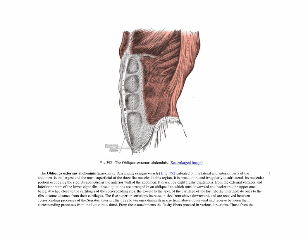
FIG. 392– The Obliquus externus abdominis. (See enlarged image) The Obliquus externus abdominis (External or descending oblique muscle) (Fig. 392),situated on the lateral and anterior parts of the abdomen, is the largest and the most superficial of the three flat muscles in this region. It is broad, thin, and irregularly quadrilateral, its muscular portion occupying the side, its aponeurosis the anterior wall of the abdomen. It arises, by eight fleshy digitations, from the external surfaces and inferior borders of the lower eight ribs; these digitations are arranged in an oblique line which runs downward and backward, the upper ones being attached close to the cartilages of the corresponding ribs, the lowest to the apex of the cartilage of the last rib, the intermediate ones to the ribs at some distance from their cartilages. The five superior serrations increase in size from above downward, and are received between corresponding processes of the Serratus anterior; the three lower ones diminish in size from above downward and receive between them corresponding processes from the Latissimus dorsi. From these attachments the fleshy fibers proceed in various directions. Those from the
6

lowest ribs pass nearly vertically downward, and are inserted into the anterior half of the outer lip of the iliac crest; the middle and upper fibers, directed downward and forward, end in an aponeurosis, opposite a line drawn from the prominence of the ninth costal cartilage to the anterior superior iliac spine. The aponeurosis of the Obliquus externus abdominis is a thin but strong membranous structure, the fibers of which are directed downward and medialward. It is joined with that of the opposite muscle along the middle line, and covers the whole of the front of the abdomen; above, it is covered by and gives origin to the lower fibers of the Pectoralis major; below, its fibers are closely aggregated together, and extend obliquely across from the anterior superior iliac spine to the public tubercle and the pectineal line. In the middle line, it interlaces with the aponeurosis of the opposite muscle, forming the linea alba,which extends from the xiphoid process to the symphysis pubis.
7
That portion of the aponeurosis which extends between the anterior superior iliac spine and the pubic tubercle is a thick band, folded inward, and continuous below with the fascia lata; it is called the inguinal ligament. The portion which is reflected from the inguinal ligament at the pubic tubercle is attached to the pectineal line and is called the lacunar ligament. From the point of attachment of the latter to the pectineal line, a few fibers pass upward and medialward, behind the medial crus of the subcutaneous inguinal ring, to the linea alba; they diverge as they ascend, and form a thin triangular fibrous band which is called the reflected inguinal ligament.
8
In the aponeurosis of the Obliquus externus, immediately above the crest of the pubis, is a triangular opening, the subcutaneous inguinal ring, formed by a separation of the fibers of the aponeurosis in this situation.
9
The following structures require further description, viz., the subcutaneous inguinal ring, the intercrural fibers and fascia, and the inguinal, lacunar, and reflected inguinal ligaments.
10
The Subcutaneous Inguinal Ring (annulus inguinalis subcutaneus; external abdominal ring) (Fig. 393).—The subcutaneous inguinal ring is an interval in the aponeurosis of the Obliquus externus, just above and lateral to the crest of the pubis. The aperture is oblique in direction, somewhat triangular in form, and corresponds with the course of the fibers of the aponeurosis. It usually measures from base to apex about 2.5 cm., and transversely about 1.25 cm. It is bounded below by the crest of the pubis; on eitherside by the margins of the opening in the aponeurosis, which are called the crura of the ring; and above, by a series of curved intercrural fibers. The inferior crus (external pillar) is the stronger and is formed by that portion of the inguinal ligament which is inserted into the pubic tubercle; it is curved so as to form a kind of groove, upon which, in the male, the spermatic cord rests. The superior crus (internal pillar) is a broad, thin, flat band, attached to the front of the symphysis pubis and interlacing with its fellow of the opposite side.
11
The subcutaneous inguinal ring gives passage to the spermatic cord and ilioinguinal nerve in the male, and to the round ligament of the uterus and the ilioinguinal nerve in the female; it is much larger in men than in women, on account of the large size of the spermatic cord.
12
The Intercrural Fibers (fibræ intercrurales; intercolumnar fibers).—The intercrural fibers are a series of curved tendinous fibers, which arch across the lower part of the aponeurosis of the Obliquus externus, describing curves with the convexities downward. They have received their name from stretching across between the two crura of the subcutaneous inguinal ring, and they are much thicker and stronger at the inferior crus, where they are connected to the inguinal ligament, than superiorly, where they are inserted into the linea alba. The intercrural fibers increase the strength of the lower part of the aponeurosis, and prevent the divergence of the crura from one another; they are more strongly developed in the male than in the female.
13
As they pass across the subcutaneous inguinal ring, they are connected together by delicate fibrous tissue, forming a fascia, called the intercrural fascia. This intercrural fascia is continued down as a tubular prolongation around the spermatic cord and testis, and encloses them in a sheath; hence it is also called the external spermatic fascia. The subcutaneous inguinal ring is seen as a distinct aperture only after the
14
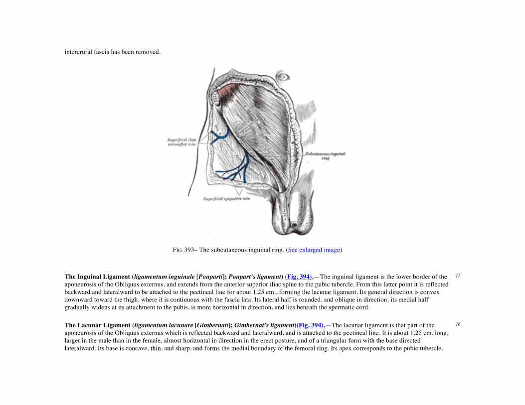
intercrural fascia has been removed.
FIG. 393– The subcutaneous inguinal ring. (See enlarged image)
The Inguinal Ligament (ligamentum inguinale [Pouparti]; Poupart’s ligament) (Fig. 394).—The inguinal ligament is the lower border of the aponeurosis of the Obliquus externus, and extends from the anterior superior iliac spine to the pubic tubercle. From this latter point it is reflected backward and lateralward to be attached to the pectineal line for about 1.25 cm., forming the lacunar ligament. Its general direction is convex downward toward the thigh, where it is continuous with the fascia lata. Its lateral half is rounded, and oblique in direction; its medial half gradually widens at its attachment to the pubis, is more horizontal in direction, and lies beneath the spermatic cord.
15
The Lacunar Ligament (ligamentum lacunare [Gimbernati]; Gimbernat’s ligament)(Fig. 394).—The lacunar ligament is that part of the aponeurosis of the Obliquus externus which is reflected backward and lateralward, and is attached to the pectineal line. It is about 1.25 cm. long, larger in the male than in the female, almost horizontal in direction in the erect posture, and of a triangular form with the base directed lateralward. Its base is concave, thin, and sharp, and forms the medial boundary of the femoral ring. Its apex corresponds to the pubic tubercle.
16
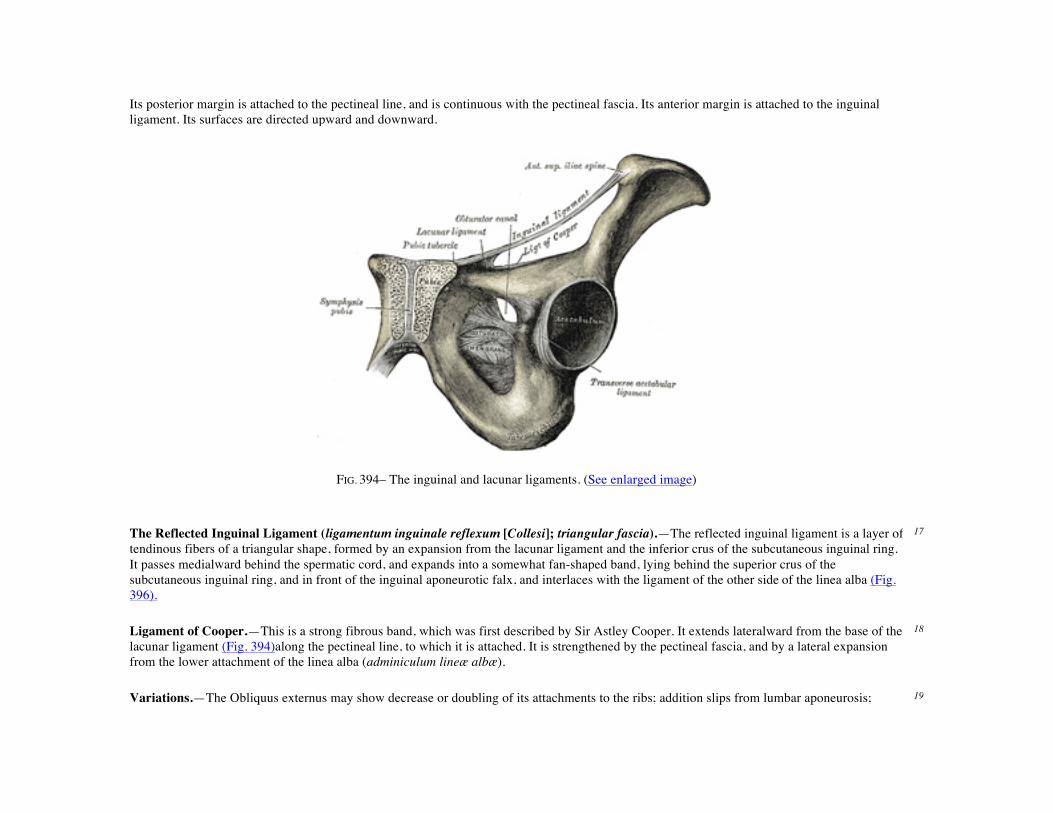
Its posterior margin is attached to the pectineal line, and is continuous with the pectineal fascia. Its anterior margin is attached to the inguinal ligament. Its surfaces are directed upward and downward.
FIG. 394– The inguinal and lacunar ligaments. (See enlarged image)
The Reflected Inguinal Ligament (ligamentum inguinale reflexum [Collesi]; triangular fascia).—The reflected inguinal ligament is a layer of tendinous fibers of a triangular shape, formed by an expansion from the lacunar ligament and the inferior crus of the subcutaneous inguinal ring. It passes medialward behind the spermatic cord, and expands into a somewhat fan-shaped band, lying behind the superior crus of the subcutaneous inguinal ring, and in front of the inguinal aponeurotic falx, and interlaces with the ligament of the other side of the linea alba (Fig. 396).
17
Ligament of Cooper.—This is a strong fibrous band, which was first described by Sir Astley Cooper. It extends lateralward from the base of the lacunar ligament (Fig. 394)along the pectineal line, to which it is attached. It is strengthened by the pectineal fascia, and by a lateral expansion from the lower attachment of the linea alba (adminiculum lineæ albæ).
18
Variations.—The Obliquus externus may show decrease or doubling of its attachments to the ribs; addition slips from lumbar aponeurosis; 19
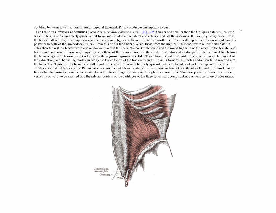
doubling between lower ribs and ilium or inguinal ligament. Rarely tendinous inscriptions occur. The Obliquus internus abdominis (Internal or ascending oblique muscle) (Fig. 395),thinner and smaller than the Obliquus externus, beneath which it lies, is of an irregularly quadrilateral form, and situated at the lateral and anterior parts of the abdomen. It arises, by fleshy fibers, from the lateral half of the grooved upper surface of the inguinal ligament, from the anterior two-thirds of the middle lip of the iliac crest, and from the posterior lamella of the lumbodorsal fascia. From this origin the fibers diverge; those from the inguinal ligament, few in number and paler in color than the rest, arch downward and medialward across the spermatic cord in the male and the round ligament of the uterus in the female, and, becoming tendinous, are inserted, conjointly with those of the Transversus, into the crest of the pubis and medial part of the pectineal line behind the lacunar ligament, forming what is known as the inguinal aponeurotic falx. Those from the anterior third of the iliac origin are horizontal in their direction, and, becoming tendinous along the lower fourth of the linea semilunaris, pass in front of the Rectus abdominis to be inserted into the linea alba. Those arising from the middle third of the iliac origin run obliquely upward and medialward, and end in an aponeurosis; this divides at the lateral border of the Rectus into two lamellæ, which are continued forward, one in front of and the other behind this muscle, to the linea alba: the posterior lamella has an attachment to the cartilages of the seventh, eighth, and ninth ribs. The most posterior fibers pass almost vertically upward, to be inserted into the inferior borders of the cartilages of the three lower ribs, being continuous with the Intercostales interni.
20
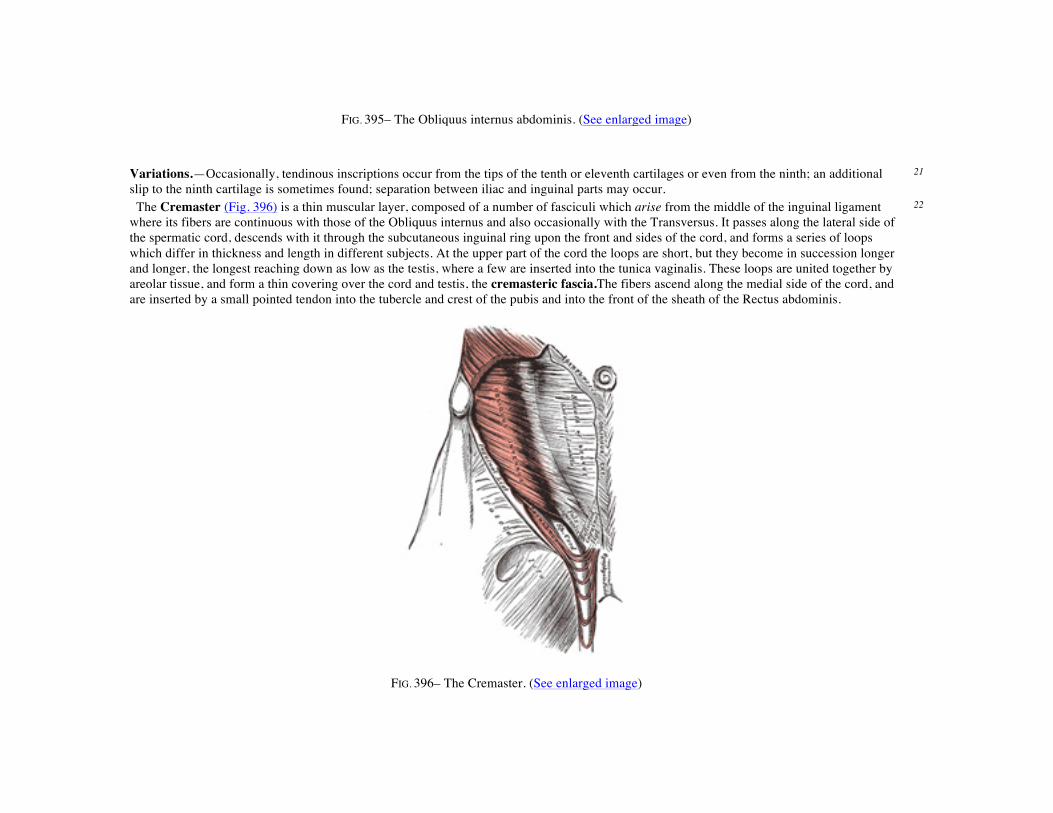
FIG. 395– The Obliquus internus abdominis. (See enlarged image)
Variations.—Occasionally, tendinous inscriptions occur from the tips of the tenth or eleventh cartilages or even from the ninth; an additional slip to the ninth cartilage is sometimes found; separation between iliac and inguinal parts may occur.
21
The Cremaster (Fig. 396) is a thin muscular layer, composed of a number of fasciculi which arise from the middle of the inguinal ligament where its fibers are continuous with those of the Obliquus internus and also occasionally with the Transversus. It passes along the lateral side of the spermatic cord, descends with it through the subcutaneous inguinal ring upon the front and sides of the cord, and forms a series of loops which differ in thickness and length in different subjects. At the upper part of the cord the loops are short, but they become in succession longer and longer, the longest reaching down as low as the testis, where a few are inserted into the tunica vaginalis. These loops are united together by areolar tissue, and form a thin covering over the cord and testis, the cremasteric fascia.The fibers ascend along the medial side of the cord, and are inserted by a small pointed tendon into the tubercle and crest of the pubis and into the front of the sheath of the Rectus abdominis.
22
FIG. 396– The Cremaster. (See enlarged image)
The Transversus abdominis (Transversalis muscle) (Fig. 397), so called from the direction of its fibers, is the most internal of the flat muscles of the abdomen, being placed immediately beneath the Obliquus internus. It arises, by fleshy fibers, from the lateral third of the inguinal ligament, from the anterior three-fourths of the inner lip of the iliac crest, from the inner surfaces of the cartilages of the lower six ribs, interdigitating with the diaphragm, and from the lumbodorsal fascia. The muscle ends in front in a broad aponeurosis, the lower fibers of which curve downward and medialward, and are inserted,together with those of the Obliquus internus, into the crest of the pubis and pectineal line, forming the inguinal aponeurotic falx. Throughout the rest of its extent the aponeurosis passes horizontally to the middle line, and is inserted into the linea alba; its upper three-fourths lie behind the Rectus and blend with the posterior lamella of the aponeurosis of the Obliquus internus; its lower fourth is in front of the Rectus.
23
Variations.—It may be more or less fused with the Obliquus internus or absent. The spermatic cord may pierce its lower border. Slender muscle slips from the ileopectineal line to transversalis fascia, the aponeurosis of the Transversus abdominis or the outer end of the linea semicircularis and other slender slips are occasionally found.
24
The inguinal aponeurotic falx (falx aponeurotica inguinalis; conjoined tendon of Internal oblique and Transversalis muscle) of the Obliquus internus and Transversus is mainly formed by the lower part of the tendon of the Transversus, and is inserted into the crest of the pubis and pectineal line immediately behind the subcutaneous inguinal ring, serving to protect what would otherwise be aweak point in the abdominal wall. Lateral to the falx is a ligamentous band connected with the lower margin of the Transversus and extending down in front of the inferior epigastric artery to the superior ramus of the pubis; it is termed the interfoveolar ligament of Hesselbach (Fig. 398) and sometimes contains a few muscular fibers.
25

FIG. 397– The Transversus abdominis, Rectus abdominis, and Pyramidalis. (See enlarged image) The Rectus abdominis (Fig. 397) is a long flat muscle, which extends along the whole length of the front of the abdomen, and is separated from its fellow of the opposite side by the linea alba. It is much broader, but thinner, above than below, and arises by two tendons; the lateral or larger is attached to the crest of the pubis, the medial interlaces with its fellow of the opposite side, and is connected with the ligaments covering the front of the symphysis pubis. The muscle is inserted by three portions of unequal size into the cartilages of the fifth, sixth, and seventh ribs. The upper portion, attached principally to the cartilage of the fifth rib, usually has some fibers of insertion into the anterior extremity of the rib itself. Some fibers are occasionally connected with the costoxiphoid ligaments, and the side of the xiphoid process.
26
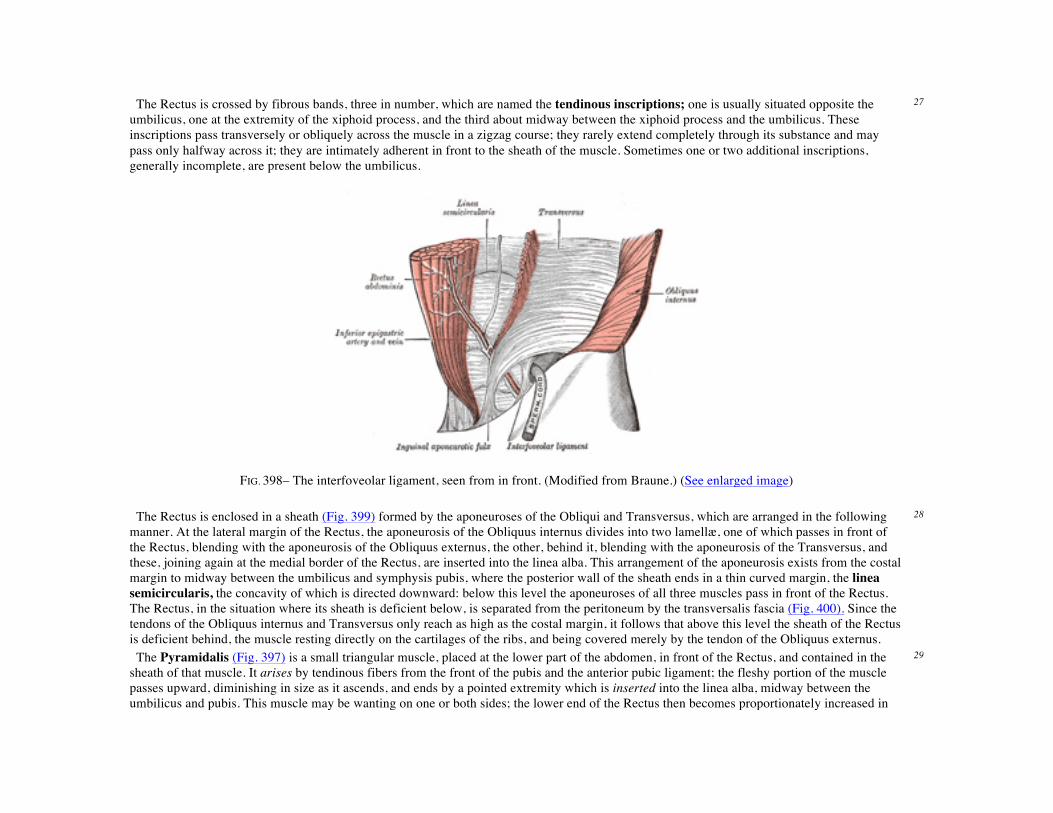
The Rectus is crossed by fibrous bands, three in number, which are named the tendinous inscriptions; one is usually situated opposite the umbilicus, one at the extremity of the xiphoid process, and the third about midway between the xiphoid process and the umbilicus. These inscriptions pass transversely or obliquely across the muscle in a zigzag course; they rarely extend completely through its substance and may pass only halfway across it; they are intimately adherent in front to the sheath of the muscle. Sometimes one or two additional inscriptions, generally incomplete, are present below the umbilicus.
27
FIG. 398– The interfoveolar ligament, seen from in front. (Modified from Braune.) (See enlarged image)
The Rectus is enclosed in a sheath (Fig. 399) formed by the aponeuroses of the Obliqui and Transversus, which are arranged in the following manner. At the lateral margin of the Rectus, the aponeurosis of the Obliquus internus divides into two lamellæ, one of which passes in front of the Rectus, blending with the aponeurosis of the Obliquus externus, the other, behind it, blending with the aponeurosis of the Transversus, and these, joining again at the medial border of the Rectus, are inserted into the linea alba. This arrangement of the aponeurosis exists from the costal margin to midway between the umbilicus and symphysis pubis, where the posterior wall of the sheath ends in a thin curved margin, the linea semicircularis, the concavity of which is directed downward: below this level the aponeuroses of all three muscles pass in front of the Rectus. The Rectus, in the situation where its sheath is deficient below, is separated from the peritoneum by the transversalis fascia (Fig. 400). Since the tendons of the Obliquus internus and Transversus only reach as high as the costal margin, it follows that above this level the sheath of the Rectus is deficient behind, the muscle resting directly on the cartilages of the ribs, and being covered merely by the tendon of the Obliquus externus.
28
The Pyramidalis (Fig. 397) is a small triangular muscle, placed at the lower part of the abdomen, in front of the Rectus, and contained in the sheath of that muscle. It arises by tendinous fibers from the front of the pubis and the anterior pubic ligament; the fleshy portion of the muscle passes upward, diminishing in size as it ascends, and ends by a pointed extremity which is inserted into the linea alba, midway between the umbilicus and pubis. This muscle may be wanting on one or both sides; the lower end of the Rectus then becomes proportionately increased in
29

size. Occasionally it is double on one side, and the muscles of the two sides are sometimes of unequal size. It may extend higher than the level stated.
FIG. 399– Diagram of sheath of Rectus. (See enlarged image)
Besides the Rectus and Pyramidalis, the sheath of the Rectus contains the superior and inferior epigastric arteries, and the lower intercostal nerves.
30
Variations.—The Rectus may insert as high as the fourth or third rib or may fail to reach the fifth. Fibers may spring from the lower part of the linea alba.
31
Nerves.—The abdominal muscles are supplied by the lower intercostal nerves. The Obliquus internus and Transversus also receive filaments from the anterior branch of the iliohypogastric and sometimes from the ilioinguinal. The Cremaster is supplied by the external spermatic branch of the genitofemoral and the Pyramidalis usually by the twelfth thoracic.
32
The Linea Alba.—The linea alba is a tendinous raphé in the middle line of the abdomen, stretching between the xiphoid process and the symphysis pubis. It is placed between the medial borders of the Recti, and is formed by the blending of the aponeuroses of the Obliqui and Transversi. It is narrow below, corresponding to the linear interval existing between the Recti; but broader above, where these muscles diverge from one another. At its lower end the linea alba has a double attachment—its superficial fibers passing in front of the medial heads of the Recti to the symphysis pubis, while its deeper fibers form a triangular lamella, attached behind the Recti to the posterior lip of the crest of the pubis, and named the adminiculum lineæ albæ. It presents apertures for the passage of vessels and nerves; the umbilicus, which in the fetus exists as an aperture and transmits the umbilical vessels, is closed in the adult.
33

FIG. 400– Diagram of a transverse section through the anterior abdomina wall, below the linea semicircularis. (See enlarged image) The Lineæ Semilunares.—The lineæ semilunares are two curved tendinous lines placed one on either side of the linea alba. Each corresponds with the lateral border of the Rectus, extends from the cartilage of the ninth rib to the pubic tubercle, and is formed by the aponeurosis of the Obliquus internus at its line of division to enclose the Rectus, reinforced in front by that of the Obliquus externus, and behind by that of the Transversus.
34
Actions.—When the pelvis and thorax are fixed, the abdominal muscles compress the abdominal viscera by constricting the cavity of the abdomen, in which action they are materially assisted by the descent of the diaphragm. By these means assistance is given in expelling the feces from the rectum, the urine from the bladder, the fetus from the uterus, and the contents of the stomach in vomiting.
35
If the pelvis and vertebral column be fixed, these muscles compress the lower part of the thorax, materially assisting expiration. If the pelvis alone be fixed, the thorax is bent directly forward, when the muscles of both sides act; when the muscles of only one side contract, the trunk is bent toward that side and rotated toward the opposite side.
36
If the thorax be fixed, the muscles, acting together, draw the pelvis upward, as in climbing; or, acting singly, they draw the pelvis upward, and bend the vertebral column to one side or the other. The Recti, acting from below, depress the thorax, and consequently flex the vertebral column; when acting from above, they flex the pelvis upon the vertebral column. The Pyramidales are tensors of the linea alba.
37
The Transversalis Fascia.—The transversalis fascia is a thin aponeurotic membrane which lies between the inner surface of the Transversus and the extraperitoneal fat. It forms part of the general layer of fascia lining the abdominal parietes, and is directly continuous with the iliac and pelvic fasciæ. In the inguinal region, the transversalis fascia is thick and dense in structure and is joined by fibers from the aponeurosis of the Transversus, but it becomes thin as it ascends to the diaphragm, and blends with the fascia covering the under surface of this muscle. Behind, it is lost in the fat which covers the posterior surfaces of the kidneys. Below, it has the following attachments: posteriorly, to the whole length of the iliac crest, between the attachments of the Transversus and Iliacus; between the anterior superior iliac spine and the femoral vessels it is connected to the posterior margin of the inguinal ligament, and is there continuous with the iliac fascia. Medial to the femoral vessels it is thin and attached to the pubis and pectineal line, behind the inguinal aponeurotic falx, with which it is united; it descends in front of the femoral vessels to form the anterior wall of the femoral sheath. Beneath the inguinal ligament it is strengthened by a band of fibrous tissue, which is only loosely connected to the ligament, and is specialized as the deep crural arch. The spermatic cord in the male and the round ligament of the uterus in the female pass through the transversalis fascia at a spot called the abdominal inguinal ring. This opening is not visible externally,
38
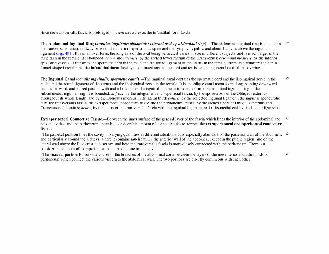
since the transversalis fascia is prolonged on these structures as the infundibuliform fascia. The Abdominal Inguinal Ring (annulus inguinalis abdominis; internal or deep abdominal ring).—The abdominal inguinal ring is situated in the transversalis fascia, midway between the anterior superior iliac spine and the symphysis pubis, and about 1.25 cm. above the inguinal ligament (Fig. 401). It is of an oval form, the long axis of the oval being vertical; it varies in size in different subjects, and is much larger in the male than in the female. It is bounded, above and laterally, by the arched lower margin of the Transversus; below and medially, by the inferior epigastric vessels. It transmits the spermatic cord in the male and the round ligament of the uterus in the female. From its circumference a thin funnel-shaped membrane, the infundibuliform fascia, is continued around the cord and testis, enclosing them in a distinct covering.
39
The Inguinal Canal (canalis inguinalis; spermatic canal).—The inguinal canal contains the spermatic cord and the ilioinguinal nerve in the male, and the round ligament of the uterus and the ilioinguinal nerve in the female. It is an oblique canal about 4 cm. long, slanting downward and medialward, and placed parallel with and a little above the inguinal ligament; it extends from the abdominal inguinal ring to the subcutaneous inguinal ring. It is bounded, in front, by the integument and superficial fascia, by the aponeurosis of the Obliquus externus throughout its whole length, and by the Obliquus internus in its lateral third; behind, by the reflected inguinal ligament, the inguinal aponeurotic falx, the transversalis fascia, the extraperitoneal connective tissue and the peritoneum; above, by the arched fibers of Obliquus internus and Transversus abdominis; below, by the union of the transversalis fascia with the inguinal ligament, and at its medial end by the lacunar ligament.
40
Extraperitoneal Connective Tissue.—Between the inner surface of the general layer of the fascia which lines the interior of the abdominal and pelvic cavities, and the peritoneum, there is a considerable amount of connective tissue, termed the extraperitoneal orsubperitoneal connective tissue.
41
The parietal portion lines the cavity in varying quantities in different situations. It is especially abundant on the posterior wall of the abdomen, and particularly around the kidneys, where it contains much fat. On the anterior wall of the abdomen, except in the public region, and on the lateral wall above the iliac crest, it is scanty, and here the transversalis fascia is more closely connected with the peritoneum. There is a considerable amount of extraperitoneal connective tissue in the pelvis.
42
The visceral portion follows the course of the branches of the abdominal aorta between the layers of the mesenterics and other folds of peritoneum which connect the various viscera to the abdominal wall. The two portions are directly continuous with each other.
43
FIG. 401– The abdominal inguinal ring. (See enlarged image) The Deep Crural Arch.—Curving over the external iliac vessels, at the spot where they become femoral, on the abdominal side of the inguinal ligaments and loosely connected with it, is a thickened band of fibers called the deep crural arch. It is apparently a thickening of the transversalis fascia joined laterally to the center of the lower margin of the inguinal ligament, and arching across the front of the femoral sheath to be inserted by a broad attachment into the pubic tubercle and pectineal line, behind the inguinal aponeurotic falx. In some subjects this structure is not very prominently marked, and not infrequently it is altogether wanting.
44
2. The Posterior Muscles of the Abdomen
Psoas major. Iliacus. Psoas minor. Quadratus lumborum.
The Psoas major, the Psoas minor, and the Iliacus, with the fasciæ covering them, will be described with the muscles of the lower extremity (see page 466).
45
The Fascia Covering the Quadratus Lumborum.—This is a thin layer attached,medially, to the bases of the transverse processes of the lumbar vertebræ; below, to the iliolumbar ligament; above, to the apex and lower border of the last rib. The upper margin of this fascia, which extends
46

from the transverse process of the first lumbar vertebra to the apex and lower border of the last rib, constitutes the lateral lumbocostal arch (page 405). Laterally, it blends with the lumbodorsal fascia, the anterior layer of which intervenes between the Quadratus lumborum and the Sacrospinalis. The Quadratus lumborum (Fig. 389, page 398) is irregularly quadrilateral in shape, and broader below than above. It arises by aponeurotic fibers from the iliolumbar ligament and the adjacent portion of the iliac crest for about 5 cm., and is inserted into the lower border of the last rib for about half its length, and by four small tendons into the apices of the transverse processes of the upper four lumbar vertebræ. Occasionally a second portion of this muscle is found in front of the preceding. It arises from the upper borders of the transverse processes of the lower three or four lumbar vertebræ, and is inserted into the lower margin of the last rib. In front of the Quadratus lumborum are the colon, the kidney, the Psoas major and minor, and the diaphragm; between the fascia and the muscle are the twelfth thoracic, ilioinguinal, and iliohypogastric nerves.
47
Variations.—The number of attachments to the vertebræ and the extent of its attachment to the last rib vary. 48 Nerve Supply.—The twelfth thoracic and first and second lumbar nerves supply this muscle. 49 Actions.—The Quadratus lumborum draws down the last rib, and acts as a muscle of inspiration by helping to fix the origin of the diaphragm. If the thorax and vertebral column are fixed, it may act upon the pelvis, raising it toward its own side when only one muscle is put in action; and when both muscles act together, either from below or above, they flex the trunk.
6e. The Muscles and Fasciæ of the Pelvis
Obturator internus. Levator ani. Piriformis. Coccygeus.
The muscles within the pelvis may be divided into two groups: (1) the Obturator internus and the Piriformis, which are muscles of the lower extremity, and will be described with these (pages 476 and 477); (2) the Levator ani and the Coccygeus, which together form thepelvic diaphragm and are associated with the pelvic viscera. The classification of the two groups under a common heading is convenient in connection with the fasciæ investing the muscles. These fasciæ are closely related to one another and to the deep fascia of the perineum, and in addition have special connections with the fibrous coverings of the pelvic viscera; it is customary therefore to describe them together under the term pelvic fascia.
1
Pelvic Fascia.—The fascia of the pelvis may be resolved into: (a) the fascial sheaths of the Obturator internus, Piriformis, and pelvic diaphragm; (b) the fascia associated with the pelvic viscera.
2
The fascia of the Obturator internus covers the pelvic surface of, and is attached around the margin of the origin of, the muscle. Above, it is loosely connected to the back part of the arcuate line, and here it is continuous with the iliac fascia. In front of this, as it follows the line of origin of the Obturator internus, it gradually separates from the iliac fascia and the continuity between the two is retained only through the periosteum. It arches beneath the obturator vessels and nerve, completing the obturator canal, and at the front of the pelvis is attached to the back of the superior ramus of the pubis. Below, the obturator fascia is attached to the falciform process of the sacrotuberous ligament and to the pubic arch, where it becomes continuous with the superior fascia of the urogenital diaphragm. Behind, it is prolonged into the gluteal region.
3

The internal pudendal vessels and pudendal nerve cross the pelvic surface of the Obturator internus and are enclosed in a special canal—Alcock’s canal—formed by the obturator fascia.
4
The fascia of the Piriformis is very thin and is attached to the front of the sacrum and the sides of the greater sciatic foramen; it is prolonged on the muscle into the gluteal region. At its sacral attachment around the margins of the anterior sacral foramina it comes into intimate association with and ensheathes the nerves emerging from these foramina. Hence the sacral nerves are frequently described as lying behind the fascia. The internal iliac vessels and their branches, on the other hand, lie in the subperitoneal tissue in front of the fascia, and the branches to the gluteal region emerge in special sheaths of this tissue, above and below the Piriformis muscle.
5
FIG. 402– Coronal section of pelvis, showing arrangement of fasciæ. Viewed from behind. (Diagrammatic.) (See enlarged image)
The diaphragmatic part of the pelvic fascia (Fig. 402) covers both surfaces of the Levatores ani. The inferior layer is known as the anal fascia; it is attached above to the obturator fascia along the line of origin of the Levator ani, while below it is continuous with the superior fascia of the urogenital diaphragm, and with the fascia on the Sphincter ani internus. The layer covering the upper surface of the pelvic diaphragm follows, above, the line of origin of the Levator ani and is therefore somewhat variable. In front it is attached to the back of the symphysis pubis about 2 cm. above its lower border. It can then be traced laterally across the back of the superior ramus of the pubis for a distance of about 1.25 cm., when it reaches the obturator fascia. It is attached to this fascia along a line which pursues a somewhat irregular course to the spine of the ischium. The irregularity of this line is due to the fact that the origin of the Levator ani, which in lower forms is from the pelvic brim, is in man lower down, on the obturator fascia. Tendinous fibers of origin of the muscle are therefore often found extending up toward, and in some cases reaching, the pelvic brim, and on these the fascia is carried.
6
It will be evident that the fascia covering that part of the Obturator internus which lies above the origin of the Levator ani is a composite fascia and includes the following: (a) the obturator fascia; (b) the fascia of the Levator ani; (c) degenerated fibers of origin of the Levator ani.
7

The lower margin of the fascia covering the upper surface of the pelvic diaphragm is attached along the line of insertion of the Levator ani. 8 At the level of a line extending from the lower part of the symphysis pubis to the spine of the ischium is a thickened whitish band in this upper layer of the diaphragmatic part of the pelvic fascia. It is termed the tendinous arch or white line of the pelvic fascia, and marks the line of attachment of the special fascia (pars endopelvina fasciæ pelvis) which is associated with the pelvic viscera.
9
FIG. 403– Median sagittal section of pelvis, showing arrangement of fasciæ. (See enlarged image)
The endopelvic part of the pelvic fascia is continued over the various pelvic viscera(Fig. 403) to form for them fibrous coverings which will be described later (see section on Splanchnology). It is attached to the diaphragmatic part of the pelvic fascia along the tendinous arch, and has been subdivided in accordance with the viscera to which it is related. Thus its anterior part, known as the vesical layer, forms the anterior and lateral ligaments of the bladder. Its middle part crosses the floor of the pelvis between the rectum and vesiculæ seminales as the rectovesical
10

layer; in the female this is perforated by the vagina. Its posterior portion passes to the side of the rectum; it forms a loose sheath for the rectum, but is firmly attached around the anal canal; this portion is known as the rectal layer. The Levator ani (Fig. 404) is a broad, thin muscle, situated on the side of the pelvis. It is attached to the inner surface of the side of the lesser pelvis, and unites with its fellow of the opposite side to form the greater part of the floor of the pelvic cavity. It supports the viscera in this cavity, and surrounds the various structures which pass through it. It arises, in front, from the posterior surface of the superior ramus of the pubis lateral to the symphysis; behind, from the inner surface of the spine of the ischium; and between these two points, from the obturator fascia. Posteriorly, this fascial origin corresponds, more or less closely, with the tendinous arch of the pelvic fascia, but in front, the muscle arises from the fascia at a varying distance above the arch, in some cases reaching nearly as high as the canal for the obturator vessels and nerve. The fibers pass downward and backward to the middle line of the floor of the pelvis; the most posterior are inserted into the side of the last two segments of the coccyx; those placed more anteriorly unite with the muscle of the opposite side, in a median fibrous raphé (anococcygeal raphé), which extends between the coccyx and the margin of the anus. The middle fibers are inserted into the side of the rectum, blending with the fibers of the Sphincter muscles; lastly, the anterior fibers descend upon the side of the prostate to unite beneath it with the muscle of the opposite side, joining with the fibers of the Sphincter ani externus and Transversus perinæi, at the central tendinous point of the perineum.
11
FIG. 404– Left Levator ani from within. (See enlarged image)

The anterior portion is occasionally separated from the rest of the muscle by connective tissue. From this circumstance, as well as from its peculiar relation with the prostate, which it supports as in a sling, it has been described as a distinct muscle, under the name ofLevator prostatæ. In the female the anterior fibers of the Levator ani descend upon the side of the vagina.
12
The Levator ani may be divided into iliococcygeal and pubococcygeal parts. 13 The Iliococcygeus arises from the ischial spine and from the posterior part of the tendinous arch of the pelvic fascia, and is attached to the coccyx and anococcygeal raphé; it is usually thin, and may fail entirely, or be largely replaced by fibrous tissue. An accessory slip at its posterior part is sometimes named the Iliosacralis. The Pubococcygeus arisesfrom the back of the pubis and from the anterior part of the obturator fascia, and “is directed backward almost horizontally along the side of the anal canal toward the coccyx and sacrum, to which it finds attachment. Between the termination of the vertebral column and the anus, the two Pubococcygei muscles come together and form a thick, fibromuscular layer lying on the raphé formed by the Iliococcygei” (Peter Thompson). The greater part of this muscle is inserted into the coccyx and into the last one or two pieces of the sacrum. This insertion into the vertebral column is, however, not admitted by all observers. The fibers which form a sling for the rectum are named the Puborectalis or Sphincter recti.They arise from the lower part of the symphysis pubis, and from the superior fascia of the urogenital diaphragm. They meet with the corresponding fibers of the opposite side around the lower part of the rectum, and form for it a strong sling.
14
Nerve Supply.—The Levator ani is supplied by a branch from the fourth sacral nerve and by a branch which is sometimes derived from the perineal, sometimes from the inferior hemorrhoidal division of the pudendal nerve.
15
The Coccygeus (Fig. 404) is situated behind the preceding. It is a triangular plane of muscular and tendinous fibers, arising by its apex from the spine of the ischium and sacrospinous ligament, and inserted by its base into the margin of the coccyx and into the side of the lowest piece of the sacrum. It assists the Levator ani and Piriformis in closing in the back part of the outlet of the pelvis.
16
Nerve Supply.—The Coccygeus is supplied by a branch from the fourth and fifth sacral nerves. 17 Actions.—The Levatores ani constrict the lower end of the rectum and vagina. They elevate and invert the lower end of the rectum after it has been protruded and everted during the expulsion of the feces. They are also muscles of forced expiration. The Coccygei pull forward and support the coccyx, after it has been pressed backward during defecation or parturition. The Levatores ani and Coccygei together form a muscular diaphragm which supports the pelvic viscera.
1F. The Muscles and Fasciæ of the Perineum
The perineum corresponds to the outlet of the pelvis. Its deep boundaries are—in front, the pubic arch and the arcuate ligament of the pubis; behind, the tip of the coccyx; and on either side the inferior rami of the pubis and ischium, and the sacrotuberous ligament. The space is somewhat lozenge-shaped and is limited on the surface of the body by the scrotum in front, by the buttocks behind, and laterally by the medial side of the thigh. A line drawn transversely across in front of the ischial tuberosities divides the space into two portions. The posterior contains the termination of the anal canal and is known as the anal region;the anterior, which contains the external urogenital organs, is termed the urogenital region.
1
The muscles of the perineum may therefore be divided into two groups: 2

1. Those of the anal region. 2. Those of the urogenital region: A, In the male; B, In the female.
1. The Muscles of the Anal Region
Corrugator cutis ani. Sphincter ani externus. Sphincter ani internus. The Superficial Fascia.—The superficial fascia is very thick, areolar in texture, and contains much fat in its meshes. On either side a pad of fatty tissue extends deeply between the Levator ani and Obturator internus into a space known as the ischiorectal fossa.
3
The Deep Fascia.—The deep fascia forms the lining of the ischiorectal fossa; it comprises the anal fascia, and the portion of obturator fascia below the origin of Levator ani.
4
Ischiorectal Fossa (fossa ischiorectalis) (Fig. 405).—The fossa is somewhat prismatic in shape, with its base directed to the surface of the perineum, and its apex at the line of meeting of the obturator and anal fasciæ. It is bounded medially by the Sphincter ani externus and the anal fascia; laterally, by the tuberosity of the ischium and the obturator fascia; anteriorly, by the fascia of Colles covering the Transversus perinæi superficialis, and by the inferior fascia of the urogenital diaphragm; posteriorly, by the Glutæus maximus and the sacrotuberous ligament. Crossing the space transversely are the inferior hemorrhoidal vessels and nerves; at the back part are the perineal and perforating cutaneous branches of the pudendal plexus; while from the forepart the posterior scrotal (or labial) vessels and nerves emerge. The internal pudendal vessels and pudendal nerve lie in Alcock’s canal on the lateral wall. The fossa is filled with fatty tissue across which numerous fibrous bands extend from side to side.
5

FIG. 405– The perineum. The integument and superficial layer of superficial fascia reflected. (See enlarged image) The Corrugator Cutis Ani.—Around the anus is a thin stratum of involuntary muscular fiber, which radiates from the orifice. Medially the fibers fade off into the submucous tissue, while laterally they blend with the true skin. By its contraction it raises the skin into ridges around the margin of the anus.
6
The Sphincter ani externus (External sphincter ani) (Fig 405) is a flat plane of muscular fibers, elliptical in shape and intimately adherent to the integument surrounding the margin of the anus. It measures about 8 to 10 cm. in length, from its anterior to its posterior extremity, and is about 2.5 cm. broad opposite the anus. It consists of two strata, superficial and deep. The superficial, constituting the main portion of the muscle, arises from a narrow tendinous band, the anococcygeal raphé, which stretches from the tip of the coccyx to the posterior margin of the anus; it forms two flattened planes of muscular tissue, which encircle the anus and meet in front to be inserted into the central tendinous point of the perineum, joining with the Transversus perinæi superficialis, the Levator ani, and the Bulbocavernosus. The deeper portion forms a complete sphincter to the anal canal. Its fibers surround the canal, closely applied to the Sphincter ani internus, and in front blend with the other muscles at the central point of the perineum. In a considerable proportion of cases the fibers decussate in front of the anus, and are continuous with the Transversi perinæi superficiales. Posteriorly, they are not attached to the coccyx, but are continuous with those of the opposite side behind the anal canal. The upper edge of the muscle is ill-defined, since fibers are given off from it to join the Levator ani.
7
Nerve Supply.—A branch from the fourth sacral and twigs from the inferior hemorrhoidal branch of the pudendal supply the muscle. 8

Actions.—The action of this muscle is peculiar. (1) It is, like other muscles, always in a state of tonic contraction, and having no antagonistic muscle it keeps the anal canal and orifice closed. (2) It can be put into a condition of greater contraction under the influence of the will, so as more firmly to occlude the anal aperture, in expiratory efforts unconnected with defecation. (3) Taking its fixed point at the coccyx, it helps to fix the central point of the perineum, so that the Bulbocavernosus may act from this fixed point.
9
The Sphincter ani internus (Internal sphincter ani) is a muscular ring which surrounds about 2.5 cm. of the anal canal; its inferior border is in contact with, but quite separate from, the Sphincter ani externus. It is about 5 mm. thick, and is formed by an aggregation of the involuntary circular fibers of the intestine. Its lower border is about 6 mm. from the orifice of the anus.
10
Actions.—Its action is entirely involuntary. It helps the Sphincter ani externus to occlude the anal aperture and aids in the expulsion of the feces. 11 2. A. The Muscles of the Urogenital Region in the Male (Fig. 406).
Transversus perinæi superficialis. Ischiocavernosus. Bulbocavernosus. Transversus perinæi profundus.
Sphincter urethræ membranaceæ.
Superficial Fascia.—The superficial fascia of this region consists of two layers, superficial and deep. 12 The superficial layer is thick, loose, areolar in texture, and contains in its meshes much adipose tissue, the amount of which varies in different subjects. In front, it is continuous with the dartos tunic of the scrotum; behind, with the subcutaneous areolar tissue surrounding the anus; and, on either side, with the same fascia on the inner sides of the thighs. In the middle line, it is adherent to the skin on the raphé and to the deep layer of the superficial fascia.
13
The deep layer of superficial fascia (fascia of Colles) (Fig. 405) is thin, aponeurotic in structure, and of considerable strength, serving to bind down the muscles of the root of the penis. It is continuous, in front, with the dartos tunic, the deep fascia of the penis, the fascia of the spermatic cord, and Scarpa’s fascia upon the anterior wall of the abdomen; on either side it is firmly attached to the margins of the rami of the pubis and ischium, lateral to the crus penis and as far back as the tuberosity of the ischium; posteriorly, it curves around the Transversi perinæi superficiales to join the lower margin of the inferior fascia of the urogenital diaphragm. In the middle line, it is connected with the superficial fascia and with the median septum of the Bulbocavernosus. This fascia not only covers the muscles in this region, but at its back part sends upward a vertical septum from its deep surface, which separates the posterior portion of the subjacent space into two.
14
The Central Tendinous Point of the Perineum.—This is a fibrous point in the middle line of the perineum, between the urethra and anus, and about 1.25 cm. in front of the latter. At this point six muscles converge and are attached: viz., the Sphincter ani externus, the Bulbocavernosus, the two Transversi perinæi superficiales, and the anterior fibers of the Levatores ani.
15
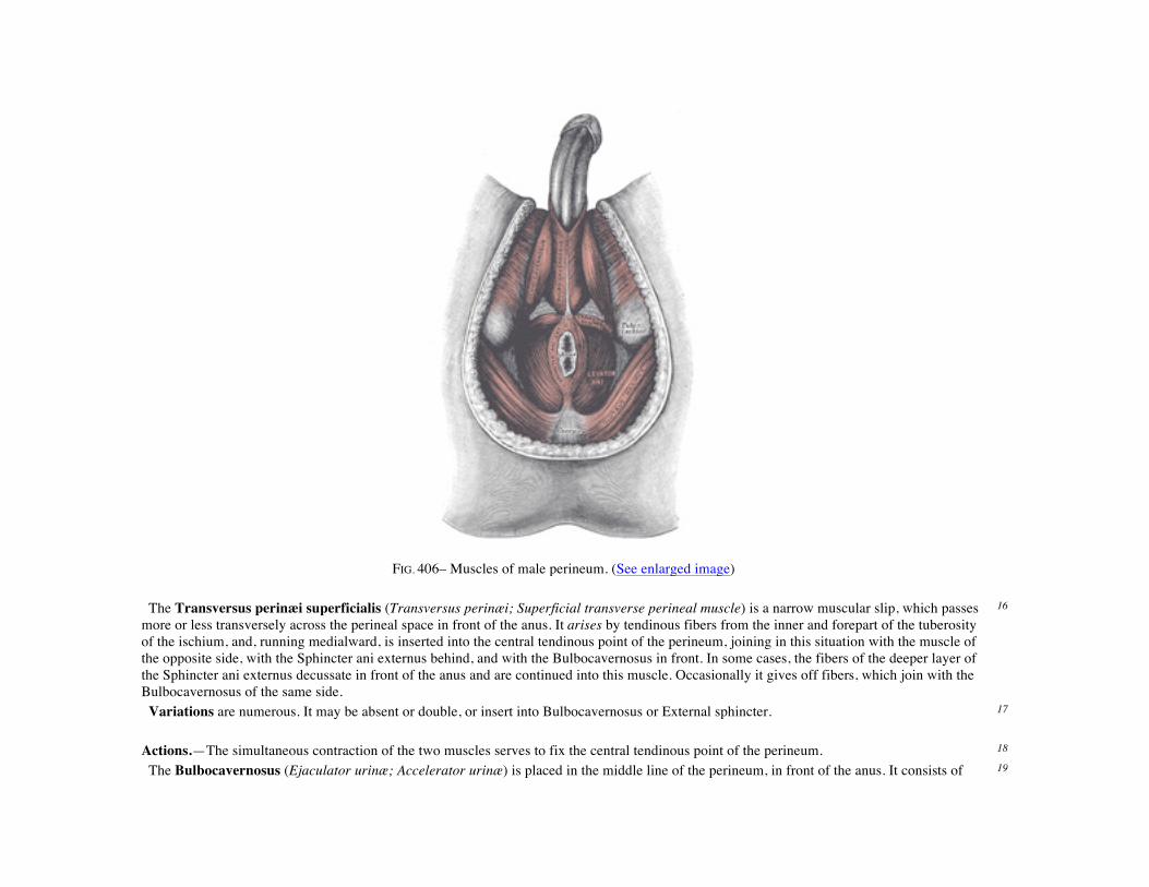
FIG. 406– Muscles of male perineum. (See enlarged image) The Transversus perinæi superficialis (Transversus perinæi; Superficial transverse perineal muscle) is a narrow muscular slip, which passes more or less transversely across the perineal space in front of the anus. It arises by tendinous fibers from the inner and forepart of the tuberosity of the ischium, and, running medialward, is inserted into the central tendinous point of the perineum, joining in this situation with the muscle of the opposite side, with the Sphincter ani externus behind, and with the Bulbocavernosus in front. In some cases, the fibers of the deeper layer of the Sphincter ani externus decussate in front of the anus and are continued into this muscle. Occasionally it gives off fibers, which join with the Bulbocavernosus of the same side.
16
Variations are numerous. It may be absent or double, or insert into Bulbocavernosus or External sphincter. 17 Actions.—The simultaneous contraction of the two muscles serves to fix the central tendinous point of the perineum. 18 The Bulbocavernosus (Ejaculator urinæ; Accelerator urinæ) is placed in the middle line of the perineum, in front of the anus. It consists of 19

two symmetrical parts, united along the median line by a tendinous raphé. It arises from the central tendinous point of the perineum and from the median raphé in front. Its fibers diverge like the barbs of a quill-pen; the most posterior form a thin layer, which is lost on the inferior fascia of the urogenital diaphragm; the middle fibers encircle the bulb and adjacent parts, of the corpus cavernosum urethræ, and join with the fibers of the opposite side, on the upper part of the corpus cavernosum urethræ, in a strong aponeurosis; the anterior fibers, spread out over the side of the corpus cavernosum penis, to be inserted partly into that body, anterior to the Ischiocavernosus, occasionally extending to the pubis, and partly ending in a tendinous expansion which covers the dorsal vessels of the penis. The latter fibers are best seen by dividing the muscle longitudinally, and reflecting it from the surface of the corpus cavernosum urethræ. Actions.—This muscle serves to empty the canal of the urethra, after the bladder has expelled its contents; during the greater part of the act of micturition its fibers are relaxed, and it only comes into action at the end of the process. The middle fibers are supposed by Krause to assist in the erection of the corpus cavernosum urethræ, by compressing the erectile tissue of the bulb. The anterior fibers, according to Tyrrel, also contribute to the erection of the penis by compressing the deep dorsal vein of the penis as they are inserted into, and continuous with, the fascia of the penis.
20
The Ischiocavernosus (Erector penis) covers the crus penis. It is an elongated muscle, broader in the middle than at either end, and situated on the lateral boundary of the perineum. It arises by tendinous and fleshy fibers from the inner surface of the tuberosity of the ischium, behind the crus penis; and from the rami of the pubis and ischium on either side of the crus. From these points fleshy fibers succeed, and end in an aponeurosis which is inserted into the sides and under surface of the crus penis.
21
Action.—The Ischiocavernosus compresses the crus penis, and retards the return of the blood through the veins, and thus serves to maintain the organ erect.
22
Between the muscles just examined a triangular space exists, bounded medially by the Bulbocavernosus, laterally by the Ischiocavernosus, and behind by the Transversus perinæi superficialis; the floor is formed by the inferior fascia of the urogenital diaphragm. Running from behind forward in the space are the posterior scrotal vessels and nerves, and the perineal branch of the posterior femoral cutaneous nerve; the transverse perineal artery courses along its posterior boundary on the Transversus perinæi superficialis.
23
The Deep Fascia.—The deep fascia of the urogenital region forms an investment for the Transversus perinæi profundus and the Sphincter urethræ membranaceæ, but within it lie also the deep vessels and nerves of this part, the whole forming a transverse septum which is known as the urogenital diaphragm. From its shape it is usually termed the triangular ligament, and is stretched almost horizontally across the pubic arch, so as to close in the front part of the outlet of the pelvis. It consists of two dense membranous laminæ (Fig. 407), which are united along their posterior borders, but are separated in front by intervening structures. The superficial of these two layers, the inferior fascia of the urogenital diaphragm, is triangular in shape, and about 4 cm. in depth. Its apex is directed forward, and is separated from the arcuate pubic ligament by an oval opening for the transmission of the deep dorsal vein of the penis. Its lateral margins are attached on either side to the inferior rami of the pubis and ischium, above the crus penis. Its base is directed toward the rectum, and connected to the central tendinous point of the perineum. It is continuous with the deep layer of the superficial fascia behind the Transversus perinæi superficialis, and with the inferior layer of the diaphragmatic part of the pelvic fascia. It is perforated, about 2.5 cm. below the symphysis pubis, by the urethra, the aperture for which is circular and about 6 mm. in diameter by the arteries to the bulb and the ducts of the bulbourethral glands close to the urethral orifice; by the deep arteries of the penis, one on either side close to the pubic arch and about halfway along the attached margin of the fascia; by the dorsal arteries and nerves of the penis near the apex of the fascia. Its base is also perforated by the perineal vessels and nerves, while between its apex and the
24

arcuate pubic ligament the deep dorsal vein of the penis passes upward into the pelvis. If the inferior fascia of the urogenital diaphragm be detached on either side, the following structures will be seen between it and the superior fascia: the deep dorsal vein of the penis; the membranous portion of the urethra; the Transversus perinæi profundus and Sphincter urethræ membranaceæ muscles; the bulbourethral glands and their ducts; the pudendal vessels and dorsal nerves of the penis; the arteries and nerves of the urethral bulb, and a plexus of veins.
25
FIG. 407– Coronal section of anterior part of pelvis, through the pubic arch. Seen from in front. (Diagrammatic.) (See enlarged image)
The superior fascia of the urogenital diaphragm is continuous with the obturator fascia and stretches across the pubic arch. If the obturator fascia be traced medially after leaving the Obturator internus muscle, it will be found attached by some of its deeper or anterior fibers to the inner margin of the pubic arch, while its superficial or posterior fibers pass over this attachment to become continuous with the superior fascia of the urogenital diaphragm. Behind, this layer of the fascia is continuous with the inferior fascia and with the fascia of Colles; in front it is continuous with the fascial sheath of the prostate, and is fused with the inferior fascia to form the transverse ligament of the pelvis.
26
The Transversus perinæi profundus arises from the inferior rami of the ischium and runs to the median line, where it interlaces in a tendinous raphé with its fellow of the opposite side. It lies in the same plane as the Sphincter urethræ membranaceæ; formerly the two muscles were described together as the Constrictor urethræ.
27
The Sphincter urethræ membranaceæ surrounds the whole length of the membranous portion of the urethra, and is enclosed in the fasciæ of the urogenital diaphragm. Its externalfibers arise from the junction of the inferior rami of the pubis and ischium to the extent of 1.25 to 2 cm., and from the neighboring fasciæ. They arch across the front of the urethra and bulbourethral glands, pass around the urethra, and behind it unite
28
with the muscle of the opposite side, by means of a tendinous raphé. Its innermost fibers form a continuous circular investment for the membranous urethra. Nerve Supply.—The perineal branch of the pudendal nerve supplies this group of muscles. 29 Actions.—The muscles of both sides act together as a sphincter, compressing the membranous portion of the urethra. During the transmission of fluids they, like the Bulbocavernosus, are relaxed, and only come into action at the end of the process to eject the last drops of the fluid.
30
2. B. The Muscles of the Urogenital Region in the Female (Fig. 408).
Transversus perinæi superficialis. Ischiocavernosus. Bulbocavernosus. Transversus perinæi profundus.
Sphincter urethræ membranaceæ.
The Transversus perinæi superficialis (Transversus perinæi; Superficial transverse perineal muscle) in the female is a narrow muscular slip, which arises by a small tendon from the inner and forepart of the tuberosity of the ischium, and is inserted into the central tendinous point of the perineum, joining in this situation with the muscle of the opposite side, the Sphincter ani externus behind, and the Bulbocavernosus in front.
31
Action.—The simultaneous contraction of the two muscles serves to fix the central tendinous point of the perineum. 32 The Bulbocavernosus (Sphincter vaginæ) surrounds the orifice of the vagina. It covers the lateral parts of the vestibular bulbs, and is attached posteriorly to the central tendinous point of the perineum, where it blends with the Sphincter ani externus. Its fibers pass forward on either side of the vagina to be inserted into the corpora cavernosa clitoridis, a fasciculus crossing over the body of the organ so as to compress the deep dorsal vein.
33
Actions.—The Bulbocavernosus diminishes the orifice of the vagina. The anterior fibers contribute to the erection of the clitoris, as they are inserted into and are continuous with the fascia of the clitoris, compressing the deep dorsal vein during the contraction of the muscle.
34
The Ischiocavernosus (Erector clitoridis) is smaller than the corresponding muscle in the male. It covers the unattached surface of the crus clitoridis. It is an elongated muscle, broader at the middle than at either end, and situated on the side of the lateral boundary of the perineum. It arises by tendinous and fleshy fibers from the inner surface of the tuberosity of the ischium, behind the crus clitoridis; from the surface of the crus; and from the adjacent portion of the ramus of the ischium. From these points fleshy fibers succeed, and end in an aponeurosis, which is inserted into the sides and under surface of the crus clitoridis.
35
Actions.—The Ischiocavernosus compresses the crus clitoridis and retards the return of blood through the veins, and thus serves to maintain the organ erect.
36
The fascia of the urogenital diaphragm in the female is not so strong as in the male. It is attached to the public arch, its apex being connected with the arcuate pubic ligament. It is divided in the middle line by the aperture of the vagina, with the external coat of which it becomes blended, and in front of this is perforated by the urethra. Its posterior border is continuous, as in the male, with the deep layer of the superficial fascia around the Transversus perinæi superficialis.
37

Like the corresponding fascia in the male, it consists of two layers, between which are to be found the following structures: the deep dorsal vein of the clitoris, a portion of the urethra and the Constrictor urethra muscle, the larger vestibular glands and their ducts; the internal pudendal vessels and the dorsal nerves of the clitoris; the arteries and nerves of the bulbi vestibuli, and a plexus of veins.
38
The Transversus perinæi profundus arises from the inferior rami of the ischium and runs across to the side of the vagina. The Sphincter urethræ membranaceæ (Constrictor urethræ), like the corresponding muscle on the male, consists of external and internal fibers. The external fibers arise on either side from the margin of the inferior ramus of the pubis. They are directed across the pubic arch in front of the urethra, and pass around it to blend with the muscular fibers of the opposite side, between the urethra and vagina. Theinnermost fibers encircle the lower end of the urethra.
39
FIG. 408– Muscles of the female perineum. (Modified from a drawing by Peter Thompson.) (See enlarged image)
Nerve Supply.—The muscles of this group are supplied by the perineal branch of the pudendal. 40 7. The Fascia and Muscles of the Upper Extremity. a. The Muscles Connecting the Upper Extremity to

the Vertebral Column The muscles of the upper extremity are divisible into groups, corresponding with the different regions of the limb. 1
I. Muscles Connecting the Upper Extremity to the Vertebral Column. II. Muscles Connecting the Upper Extremity to the Anterior and Lateral Thoracic Walls. III. Muscles of the Shoulder. V. Muscles of the Forearm. IV. Muscles of the Arm. VI. Muscles of the Hand.
a. The Muscles Connecting the Upper Extremity to the Vertebral ColumnThe muscles of this group are: 2
Trapezius. Rhomboideus major. Latissimus dorsi. Rhomboideus minor.
Levator scapulæ.
Superficial Fascia.—The superficial fascia of the back forms a layer of considerable thickness and strength, and contains a quantity of granular fat. It is continuous with the general superficial fascia.
3
Deep Fascia.—The deep fascia is a dense fibrous layer, attached above to the superior nuchal line of the occipital bone; in the middle line it is attached to the ligamentum nuchæ and supraspinal ligament, and to the spinous processes of all the vertebræ below the seventh cervical; laterally, in the neck it is continuous with the deep cervical fascia; over the shoulder it is attached to the spine of the scapula and to the acromion, and is continued downward over the Deltoideus to the arm; on the thorax it is continuous with the deep fascia of the axilla and chest, and on the abdomen with that covering the abdominal muscles; below, it is attached to the crest of the ilium.
4
The Trapezius (Fig. 409) is a flat, triangular muscle, covering the upper and back part of the neck and shoulders. It arises from the external occipital protuberance and the medial third of the superior nuchal line of the occipital bone, from the ligamentum nuchæ, the spinous process of the seventh cervical, and the spinous processes of all the thoracic vertebræ, and from the corresponding portion of the supraspinal ligament. From this origin, the superior fibers proceed downward and lateralward, the inferior upward and lateralward, and the middle horizontally; the superior fibers are inserted into the posterior border of the lateral third of the clavicle; the middle fibers into the medial margin of the acromion, and into the superior lip of the posterior border of the spine of the scapula; the inferior fibers converge near the scapula, and end in an aponeurosis, which glides over the smooth triangular surface on the medial end of the spine, to be inserted into a tubercle at the apex of this smooth triangular surface. At its occipital origin, the Trapezius is connected to the bone by a thin fibrous lamina, firmly adherent to the skin. At the middle it is connected to the spinous processes by a broad semi-elliptical aponeurosis, which reaches from the sixth cervical to the third thoracic vertebræ, and forms, with that of the opposite muscle, a tendinous ellipse. The rest of the muscle arises by numerous short tendinous fibers. The two Trapezius muscles together resemble a trapezium, or diamond-shaped quadrangle: two angles corresponding to the shoulders; a third to the occipital protuberance; and the fourth to the spinous process of the twelfth thoracic vertebra.
5
Variations.—The attachments to the dorsal vertebræ are often reduced and the lower ones are often wanting; the occipital attachment is often wanting; separation between cervical and dorsal portions is frequent. Extensive deficiencies and complete absence occur.
6
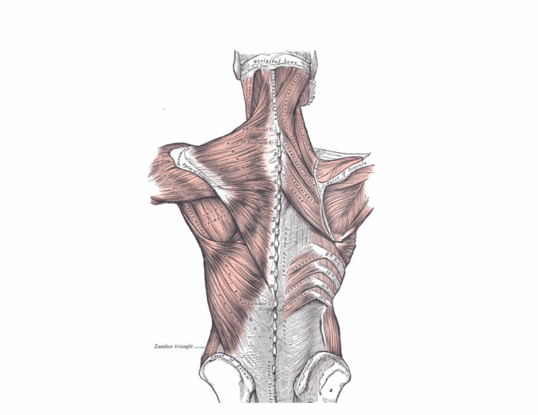
FIG. 409– Muscles connecting the upper extremity to the vertebral column. (See enlarged image)
The clavicular insertion of this muscle varies in extent; it sometimes reaches as far as the middle of the clavicle, and occasionally may blend with the posterior edge of the Sternocleidomastoideus, or overlap it.
7
The Latissimus dorsi (Fig. 409) is a triangular, flat muscle, which covers the lumbar region and the lower half of the thoracic region, and is gradually contracted into a narrow fasciculus at its insertion into the humerus. It arises by tendinous fibers from the spinous processes of the lower six thoracic vertebræ and from the posterior layer of the lumbodorsal fascia (see page 397), by which it is attached to the spines of the lumbar and sacral vertebræ, to the supraspinal ligament, and to the posterior part of the crest of the ilium. It also arises by muscular fibers from the external lip of the crest of the ilium lateral to the margin of the Sacrospinalis, and from the three or four lower ribs by fleshy digitations, which are interposed between similar processes of the Obliquus abdominis externus (Fig. 392, page 409). From this extensive origin the fibers pass in different directions, the upper ones horizontally, the middle obliquely upward, and the lower vertically upward, so as to converge and form a thick fasciculus, which crosses the inferior angle of the scapula, and usually receives a few fibers from it. The muscle curves around the lower border of the Teres major, and is twisted upon itself, so that the superior fibers become at first posterior and then inferior, and the vertical fibers at first anterior and then superior. It ends in a quadrilateral tendon, about 7 cm. long, which passes in front of the tendon of the Teres major, and is inserted into the bottom of the intertubercular groove of the humerus; its insertion extends higher on the humerus than that of the tendon of the Pectoralis major. The lower border of its tendon is united with that of the Teres major, the surfaces of the two being separated near their insertions by a bursa; another bursa is sometimes interposed between the muscle and the inferior angle of the scapula. The tendon of the muscle gives off an expansion to the deep fascia of the arm.
8
Variations.—The number of dorsal vertebræ to which it is attached vary from four to seven or eight; the number of costal attachments varies; muscle fibers may or may not reach the crest of the ilium.
9
A muscular slip, the axillary arch, varying from 7 to 10 cm. in length, and from 5 to 15 mm. in breadth, occasionally springs from the upper edge of the Latissimus dorsi about the middle of the posterior fold of the axilla, and crosses the axilla in front of the axillary vessels and nerves, to join the under surface of the tendon of the Pectoralis major, the Coracobrachialis, or the fascia over the Biceps brachii. This axillary arch crosses the axillary artery, just above the spot usually selected for the application of a ligature, and may mislead the surgeon during the operation. It is present in about 7 per cent. of subjects and may be easily recognized by the transverse direction of its fibers.
10
A fibrous slip usually passes from the lower border of the tendon of the Latissimus dorsi, near its insertion, to the long head of the Triceps brachii. This is occasionally muscular, and is the representative of the Dorsoepitrochlearis brachii of apes.
11
The lateral margin of the Latissimus dorsi is separated below from the Obliquus externus abdominis by a small triangular interval, the lumbar triangle of Petit, the base of which is formed by the iliac crest, and its floor by the Obliquus internus abdominis. Another triangle is situated behind the scapula. It is bounded above by the Trapezius, below by the Latissimus dorsi, and laterally by the vertebral border of the scapula; the floor is partly formed by the Rhomboideus major. If the scapula be drawn forward by folding the arms across the chest, and the trunk bent forward, parts of the sixth and seventh ribs and the interspace between them become subcutaneous and available for ausculation. The space is therefore known as the triangle of ausculation.
12
Nerves.—The Trapezius is supplied by the accessory nerve, and by branches from the third and fourth cervical nerves; the Latissimus dorsi by the sixth, seventh, and eighth cervical nerves through the thoracodorsal (long subscapular) nerve.
13

The Rhomboideus major (Fig. 409) arises by tendinous fibers from the spinous processes of the second, third, fourth, and fifth thoracic vertebræ and the supraspinal ligament, and is inserted into a narrow tendinous arch, attached above to the lower part of the triangular surface at the root of the spine of the scapula; below to the inferior angle, the arch being connected to the vertebral border by a thin membrane. When the arch extends, as it occasionally does, only a short distance, the muscular fibers are inserted directly into the scapula.
14
The Rhomboideus minor (Fig. 409) arises from the lower part of the ligamentum nuchæ and from the spinous processes of the seventh cervical and first thoracic vertebræ. It isinserted into the base of the triangular smooth surface at the root of the spine of the scapula, and is usually separated from the Rhomboideus major by a slight interval, but the adjacent margins of the two muscles are occasionally united.
15
Variations.—The vertebral and scapular attachments of the two muscles vary in extent. A small slip from the scapula to the occipital bone close to the minor occasionally occurs, theRhomboideus occipitalis muscle.
16
The Levator scapulæ. (Levator anguli scapulæ) (Fig. 409) is situated at the back and side of the neck. It arises by tendinous slips from the transverse processes of the atlas and axis and from the posterior tubercles of the transverse processes of the third and fourth cervical vertebræ. It is inserted into the vertebral border of the scapula, between the medial angle and the triangular smooth surface at the root of the spine.
17
Variations.—The number of vertebral attachments varies; a slip may extend to the occipital or mastoid, to the Trapezius, Scalene or Serratus anterior, or to the first or second rib. The muscle may be subdivided into several distinct parts from origin to insertion.Levator claviculæ from the transverse processes of one or two upper cervical vertebræ to the outer end of the clavicle corresponds to a muscle of lower animals. More or less union with the Serratus anterior.
18
Nerves.—The Rhomboidei are supplied by the dorsal scapular nerve from the fifth cervical; the Levator scapulæ by the third and fourth cervical nerves, and frequently by a branch from the dorsal scapular.
19
Actions.—The movements effected by the preceding muscles are numerous, as may be conceived from their extensive attachments. When the whole Trapezius is in action it retracts the scapula and braces back the shoulder; if the head be fixed, the upper part of the muscle will elevate the point of the shoulder, as in supporting weights; when the lower fibers contract they assist in depressing the scapula. The middle and lower fibers of the muscle rotate the scapula, causing elevation of the acromion. If the shoulders be fixed, the Trapezii, acting together, will draw the head directly backward; or if only one act, the head is drawn to the corresponding side.
20
When the Latissimus dorsi acts upon the humerus, it depresses and draws it backward, and at the same time rotates it inward. It is the muscle which is principally employed in giving a downward blow, as in felling a tree or in sabre practice. If the arm be fixed, the muscle may act in various ways upon the trunk; thus, it may raise the lower ribs and assist in forcible inspiration; or, if both arms be fixed, the two muscles may assist the abdominal muscles and Pectorales in suspending and drawing the trunk forward, as in climbing.
21
If the head be fixed, the Levator scapulæ raises the medial angle of the scapula; if the shoulder be fixed, the muscle inclines the neck to the corresponding side and rotates it in the same direction. The Rhomboidei carry the inferior angle backward and upward, thus producing a slight rotation of the scapula upon the side of the chest, the Rhomboideus major acting especially on the inferior angle of the scapula, through the tendinous arch by which it is inserted. The Rhomboidei, acting together with the middle and inferior fibers of the Trapezius, will retract the scapula.

The Muscles Connecting the Upper Extremity to the Anterior and Lateral Thoracic Walls The muscles of the anterior and lateral thoracic regions are: 1
Pectoralis major. Subclavius. Pectoralis minor. Serratus anterior.
Superficial Fascia.—The superficial fascia of the anterior thoracic region is continuous with that of the neck and upper extremity above, and of the abdomen below. It encloses the mamma and gives off numerous septa which pass into the gland, supporting its various lobes. From the fascia over the front of the mamma, fibrous processes pass forward to the integument and papilla; these were called by Sir A. Cooper the ligamenta suspensoria.
2
Pectoral Fascia.—The pectoral fascia is a thin lamina, covering the surface of the Pectoralis major, and sending numerous prolongations between its fasciculi: it is attached, in the middle line, to the front of the sternum; above, to the clavicle; laterally and below it is continuous with the fascia of the shoulder, axilla, and thorax. It is very thin over the upper part of the Pectoralis major, but thicker in the interval between it and the Latissimus dorsi, where it closes in the axillary space and forms the axillary fascia; it divides at the lateral margin of the Latissimus dorsi into two layers, one of which passes in front of, and the other behind it; these proceed as far as the spinous processes of the thoracic vertebræ, to which they are attached. As the fascia leaves the lower edge of the Pectoralis major to cross the floor of the axilla it sends a layer upward under cover of the muscle; this lamina splits to envelop the Pectoralis minor, at the upper edge of which it is continuous with the coracoclavicular fascia. The hollow of the armpit, seen when the arm is abducted, is produced mainly by the traction of this fascia on the axillary floor, and hence the lamina is sometimes named the suspensory ligament of the axilla. At the lower part of the thoracic region the deep fascia is well-developed, and is continuous with the fibrous sheaths of the Recti abdominis.
3

FIG. 410– —Superficial muscles of the chest and front of the arm. (See enlarged image) The Pectoralis major (Fig. 410) is a thick, fan-shaped muscle, situated at the upper and forepart of the chest. It arises from the anterior surface of the sternal half of the clavicle; from half the breadth of the anterior surface of the sternum, as low down as the attachment of the cartilage of the sixth or seventh rib; from the cartilages of all the true ribs, with the exception, frequently, of the first or seventh, or both, and from the aponeurosis of the Obliquus externus abdominis. From this extensive origin the fibers converge toward their insertion; those arising from the clavicle pass obliquely downward and lateralward, and are usually separated from the rest by a slight interval; those from the lower part of the sternum, and the cartilages of the lower true ribs, run upward and lateralward; while the middle fibers pass horizontally. They all end in a flat tendon, about 5 cm. broad, which is insertedinto the crest of the greater tubercle of the humerus. This tendon consists of two laminæ, placed one in front of the other, and usually blended together below. The anterior lamina, the thicker, receives the clavicular and the uppermost sternal
4

fibers; they are inserted in the same order as that in which they arise: that is to say, the most lateral of the clavicular fibers are inserted at the upper part of the anterior lamina; the uppermost sternal fibers pass down to the lower part of the lamina which extends as low as the tendon of the Deltoideus and joins with it. The posterior lamina of the tendon receives the attachment of the greater part of the sternal portion and the deep fibers, i. e., those from the costal cartilages. These deep fibers, and particularly those from the lower costal cartilages, ascend the higher, turning backward successively behind the superficial and upper ones, so that the tendon appears to be twisted. The posterior lamina reaches higher on the humerus than the anterior one, and from it an expansion is given off which covers the intertubercular groove and blends with the capsule of the shoulder-joint. From the deepest fibers of this lamina at its insertion an expansion is given off which lines the intertubercular groove, while from the lower border of the tendon a third expansion passes downward to the fascia of the arm. Variations.—The more frequent variations are greater or less extent of attachment to the ribs and sternum, varying size of the abdominal part or its absence, greater or less extent of separation of sternocostal and clavicular parts, fusion of clavicular part with deltoid, decussation in front of the sternum. Deficiency or absence of the sternocostal part is not uncommon. Absence of the clavicular part is less frequent. Rarely the whole muscle is wanting.
5
Costocoracoideus is a muscular band occasionally found arising from the ribs or aponeurosis of the External oblique between the Pectoralis major and Latissimus dorsi and inserted into the coracoid process.
6
Chondro-epitrochlearis is a muscular slip occasionally found arising from the costal cartilages or from the aponeurosis of the External oblique below the Pectoralis major or from the Pectoralis major itself. The insertion is variable on the inner side of the arm to fascia, intermuscular septum or internal condyle.
7
Sternalis, in front of the sternal end of the Pectoralis major parallel to the margin of the sternum. It is supplied by the anterior thoracic nerves and is probably a misplaced part of the pectoralis.
8
Coracoclavicular Fascia (fascia coracoclavicularis; costocoracoid membrane; clavipectoral fascia).—The coracoclavicular fascia is a strong fascia situated under cover of the clavicular portion of the Pectoralis major. It occupies the interval between the Pectoralis minor and Subclavius, and protects the axillary vessels and nerves. Traced upward, it splits to enclose the Subclavius, and its two layers are attached to the clavicle, one in front of and the other behind the muscle; the latter layer fuses with the deep cervical fascia and with the sheath of the axillary vessels. Medially, it blends with the fascia covering the first two intercostal spaces, and is attached also to the first rib medial to the origin of the Subclavius. Laterally, it is very thick and dense, and is attached to the coracoid process. The portion extending from the first rib to the coracoid process is often whiter and denser than the rest, and is sometimes called the costocoracoid ligament. Below this it is thin, and at the upper border of the Pectoralis minor it splits into two layers to invest the muscle; from the lower border of the Pectoralis minor it is continued downward to join the axillary fascia, and lateralward to join the fascia over the short head of the Biceps brachii. The coracoclavicular fascia is pierced by the cephalic vein, thoracoacromial artery and vein, and external anterior thoracic nerve.
9
The Pectoralis minor (Fig. 411) is a thin, triangular muscle, situated at the upper part of the thorax, beneath the Pectoralis major. It arises from the upper margins and outer surfaces of the third, fourth, and fifth ribs, near their cartilage and from the aponeuroses covering the Intercostalis; the fibers pass upward and lateralward and converge to form a flat tendon, which is inserted into the medial border and upper surface of the coracoid process of the scapula.
10
Variations.—Origin from second, third and fourth or fifth ribs. The tendon of insertion may extend over the coracoid process to the greater tubercle. May be split into several parts. Absence rare.
11

Pectoralis minimus, first rib-cartilage to coracoid process. Rare. 12
FIG. 411– Deep muscles of the chest and front of the arm, with the boundaries of the axilla. (See enlarged image)
The Subclavius (Fig. 411) is a small triangular muscle, placed between the clavicle and the first rib. It arises by a short, thick tendon from the first rib and its cartilage at their junction, in front of the costoclavicular ligament; the fleshy fibers proceed obliquely upward and lateralward, to be inserted into the groove on the under surface of the clavicle between the costoclavicular and conoid ligaments.
13
Variations.—Insertion into coracoid process instead of clavicle or into both clavicle and coracoid process. Sternoscapular fasciculus to the upper border of scapula.Sternoclavicularis from manubrium to clavicle between Pectoralis major and coracoclavicular fascia.
14
The Serratus anterior (Serratus magnus) (Fig. 411) is a thin muscular sheet, situated between the ribs and the scapula at the upper and lateral part of the chest. It arises by fleshy digitations from the outer surfaces and superior borders of the upper eight or nine ribs, and from the aponeuroses covering the intervening Intercostales. Each digitation (except the first) arises from the corresponding rib; the first springs from the first and second ribs; and from the fascia covering the first intercostal space. From this extensive attachment the fibers pass backward, closely applied to the chest-wall, and reach the vertebral border of the scapula, and are inserted into its ventral surface in the following manner. The first
15

digitation is inserted into a triangular area on the ventral surface of the medial angle. The next two digitations spread out to form a thin, triangular sheet, the base of which is directed backward and is inserted into nearly the whole length of the ventral surface of the vertebral border. The lower five or six digitations converge to form a fan-shaped mass, the apex of which is inserted, by muscular and tendinous fibers, into a triangular impression on the ventral surface of the inferior angle. The lower four slips interdigitate at their origins with the upper five slips of the Obliquus externus abdominis. Variations.—Attachment to tenth rib. Absence of attachments to first rib, to one or more of the lower ribs. Division into three parts; absence or defect of middle part. Union with Levator scapulæ, External intercostals or External oblique.
16
Nerves.—The Pectoralis major is supplied by the medial and lateral anterior thoracic nerves; through these nerves the muscle receives filaments from all the spinal nerves entering into the formation of the brachial plexus; the Pectoralis minor receives its fibers from the eighth cervical and first thoracic nerves through the medial anterior thoracic nerve. The Subclavius is suplied by a filament from the fifth and sixth cervical nerves; the Serratus anterior is supplied by the long thoracic, which is derived from the fifth, sixth, and seventh cervical nerves.
17
Actions.—If the arm has been raised by the Deltoideus, the Pectoralis major will, conjointly with the Latissimus dorsi and Teres major, depress it to the side of the chest. If acting alone, it adducts and draws forward the arm, bringing it across the front of the chest, and at the same time rotates it inward. The Pectoralis minor depresses the point of the shoulder, drawing the scapula downward and medialward toward the thorax, and throwing the inferior angle backward. The Subclavius depresses the shoulder, carrying it downward and forward. When the arms are fixed, all three of these muscles act upon the ribs; drawing them upward and expanding the chest, and thus becoming very important agents in forced inspiration. The Serratus anterior, as a whole, carries the scapula forward, and at the same time raises the vertebral border of the bone. It is therefore concerned in the action of pushing. Its lower and stronger fibers move forward the lower angle and assist the Trapezius in rotating the bone at the sternoclavicular joint, and thus assist this muscle in raising the acromion and supporting weights upon the shoulder. It is also an assistant to the Deltoideus in raising the arm, inasmuch as during the action of this latter muscle it fixes the scapula and so steadies the glenoid cavity on which the head of the humerus rotates. After the Deltoideus has raised the arm to a right angle with the trunk, the Serratus anterior and the Trapezius, by rotating the scapula, raise the arm into an almost vertical position. It is possible that when the shoulders are fixed the lower fibers of the Serratus anterior may assist in raising and everting the ribs; but it is not the important inspiratory muscle it was formerly believed to be.
7c. The Muscles and Fasciæ of the Shoulder In this group are included: 1
Deltoideus. Infraspinatus. Subscapularis. Teres minor. Supraspinatus. Teres major.
Deep Fascia.—The deep fascia covering the Deltoideus invests the muscle, and sends numerous septa between its fasciculi. In front it is continuous with the fascia covering the Pectoralis major; behind, where it is thick and strong, with that covering the Infraspinatus; above, it is
2
attached to the clavicle, the acromion, and the spine of the scapula; below, it is continuous with the deep fascia of the arm. The Deltoideus (Deltoid muscle) (Fig. 410) is a large, thick, triangular muscle, which covers the shoulder-joint in front, behind, and laterally. It arises from the anterior border and upper surface of the lateral third of the clavicle; from the lateral margin and upper surface of the acromion, and from the lower lip of the posterior border of the spine of the scapula, as far back as the triangular surface at its medial end. From this extensive origin the fibers converge toward their insertion, the middle passing vertically, the anterior obliquely backward and lateralward, the posterior obliquely forward and lateralward; they unite in a thick tendon, which is inserted into the deltoid prominence on the middle of the lateral side of the body of the humerus. At its insertion the muscle gives off an expansion to the deep fascia of the arm. This muscle is remarkably coarse in texture, and the arrangement of its fibers is somewhat peculiar; the central portion of the muscle—that is to say, the part arising from the acromion—consists of oblique fibers; these arise in a bipenniform manner from the sides of the tendinous intersections, generally four in number, which are attached above to the acromion and pass downward parallel to one another in the substance of the muscle. The oblique fibers thus formed are inserted into similar tendinous intersections, generally three in number, which pass upward from the insertion of the muscle and alternate with the descending septa. The portions of the muscle arising from the clavicle and spine of the scapula are not arranged in this manner, but are inserted into the margins of the inferior tendon.
3
Variations.—Large variations uncommon. More or less splitting common. Continuation into the Trapezius; fusion with the Pectoralis major; additional slips from the vertebral border of the scapula, infraspinous fascia and axillary border of scapula not uncommon. Insertion varies in extent or rarely is prolonged to origin of Brachioradialis.
4
Nerves.—The Deltoideus is supplied by the fifth and sixth cervical through the axillary nerve. 5 Actions.—The Deltoideus raises the arm from the side, so as to bring it at right angles with the trunk. Its anterior fibers, assisted by the Pectoralis major, draw the arm forward; and its posterior fibers, aided by the Teres major and Latissimus dorsi, draw it backward.
6
Subscapular Fascia (fascia subscapularis).—The subscapular fascia is a thin membrane attached to the entire circumference of the subscapular fossa, and affording attachment by its deep surface to some of the fibers of the Subscapularis.
7
The Subscapularis (Fig. 411) is a large triangular muscle which fills the subscapular fossa, and arises from its medial two-thirds and from the lower two-thirds of the groove on the axillary border of the bone. Some fibers arise from tendinous laminæ which intersect the muscle and are attached to ridges on the bone; others from an aponeurosis, which separates the muscle from the Teres major and the long head of the Triceps brachii. The fibers pass lateralward, and, gradually converging, end in a tendon which is inserted into the lesser tubercle of the humerus and the front of the capsule of the shoulder-joint. The tendon of the muscle is separated from the neck of the scapula by a large bursa, which communicates with the cavity of the shoulder-joint through an aperture in the capsule.
8
Nerves.—The Subscapularis is supplied by the fifth and sixth cervical nerves through the upper and lower subscapular nerves. 9 Actions.—The Subscapularis rotates the head of the humerus inward; when the arm is raised, it draws the humerus forward and downward. It is a powerful defence to the front of the shoulder-joint, preventing displacement of the head of the humerus.
10
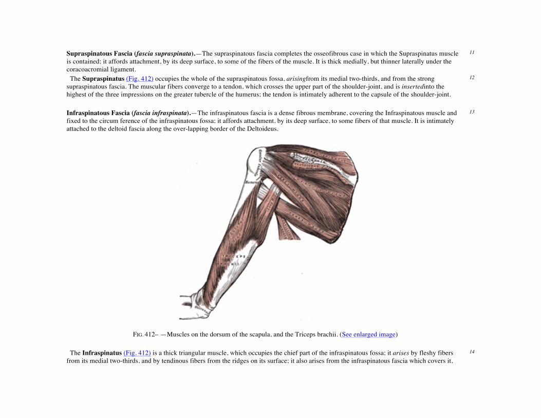
Supraspinatous Fascia (fascia supraspinata).—The supraspinatous fascia completes the osseofibrous case in which the Supraspinatus muscle is contained; it affords attachment, by its deep surface, to some of the fibers of the muscle. It is thick medially, but thinner laterally under the coracoacromial ligament.
11
The Supraspinatus (Fig. 412) occupies the whole of the supraspinatous fossa, arisingfrom its medial two-thirds, and from the strong supraspinatous fascia. The muscular fibers converge to a tendon, which crosses the upper part of the shoulder-joint, and is insertedinto the highest of the three impressions on the greater tubercle of the humerus; the tendon is intimately adherent to the capsule of the shoulder-joint.
12
Infraspinatous Fascia (fascia infraspinata).—The infraspinatous fascia is a dense fibrous membrane, covering the Infraspinatous muscle and fixed to the circum ference of the infraspinatous fossa; it affords attachment, by its deep surface, to some fibers of that muscle. It is intimately attached to the deltoid fascia along the over-lapping border of the Deltoideus.
13
FIG. 412– —Muscles on the dorsum of the scapula, and the Triceps brachii. (See enlarged image)
The Infraspinatus (Fig. 412) is a thick triangular muscle, which occupies the chief part of the infraspinatous fossa; it arises by fleshy fibers from its medial two-thirds, and by tendinous fibers from the ridges on its surface; it also arises from the infraspinatous fascia which covers it,
14

and separates it from the Teretes major and minor. The fibers converge to a tendon, which glides over the lateral border of the spine of the scapula, and, passing across the posterior part of the capsule of the shoulder-joint, is inserted into the middle impression on the greater tubercle of the humerus. The tendon of this muscle is sometimes separated from the capsule of the shoulder-joint by a bursa, which may communicate with the joint cavity. The Teres minor (Fig. 412) is a narrow, elongated muscle, which arises from the dorsal surface of the axillary border of the scapula for the upper two-thirds of its extent, and from two aponeurotic laminæ, one of which separates it from the Infraspinatus, the other from the Teres major. Its fibers run obliquely upward and lateralward; the upper ones end in a tendon which is inserted into the lowest of the three impressions on the greater tubercle of the humerus; the lowest fibers are inserted directly into the humerus immediately below this impression. The tendon of this muscle passes across, and is united with, the posterior part of the capsule of the shoulder-joint.
15
Variations.—It is sometimes inseparable from the Infraspinatus. 16 The Teres major (Fig. 412) is a thick but somewhat flattened muscle, which arises from the oval area on the dorsal surface of the inferior angle of the scapula, and from the fibrous septa interposed between the muscle and the Teres minor and Infraspinatus; the fibers are directed upward and lateralward, and end in a flat tendon, about 5 cm. long, which isinserted into the crest of the lesser tubercle of the humerus. The tendon, at its insertion, lies behind that of the Latissimus dorsi, from which it is separated by a bursa, the two tendons being, however, united along their lower borders for a short distance.
17
Nerves.—The Supraspinatus and Infraspinatus are supplied by the fifth and sixth cervical nerves through the suprascapular nerve; the Teres minor, by the fifth cervical, through the axillary; and the Teres major, by the fifth and sixth cervical, through the lowest subscapular.
18
Actions.—The Supraspinatus assists the Deltoideus in raising the arm from the side of the trunk and fixes the head of the humerus in the glenoid cavity. The Infraspinatus and Teres minor rotate the head of the humerus outward; they also assist in carrying the arm backward. One of the most important uses of these three muscles is to protect the shoulder-joint, the Supraspinatus supporting it above, and the Infraspinatus and Teres minor behind. The Teres major assists the Latissimus dorsi in drawing the previously raised humerus downward and backward, and in rotating it inward; when the arm is fixed it may assist the Pectorales and the Latissimus dorsi in drawing the trunk forward.
7d. The Muscles and Fasciæ of the Arm
The muscles of the arm are: 1
Coracobrachialis. Brachialis. Biceps brachii. Triceps brachii.
Brachial Fascia (fascia brachii; deep fascia of the arm).—The brachial fascia is continuous with that covering the Deltoideus and the Pectoralis major, by means of which it is attached, above, to the clavicle, acromion, and spine of the scapula; it forms a thin, loose, membranous sheath for the muscles of the arm, and sends septa between them; it is composed of fibers disposed in a circular or spiral direction, and connected together by vertical and oblique fibers. It differs in thickness at different parts, being thin over the Biceps brachii, but thicker where it covers the
2
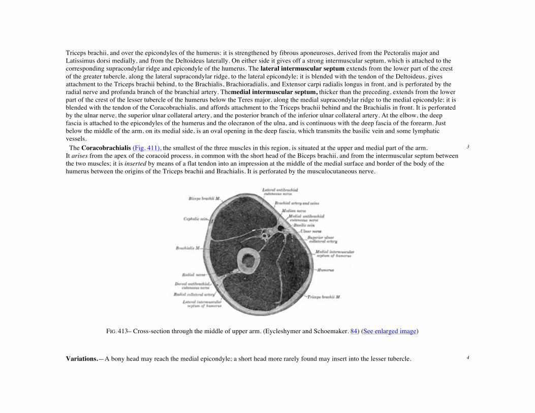
Triceps brachii, and over the epicondyles of the humerus: it is strengthened by fibrous aponeuroses, derived from the Pectoralis major and Latissimus dorsi medially, and from the Deltoideus laterally. On either side it gives off a strong intermuscular septum, which is attached to the corresponding supracondylar ridge and epicondyle of the humerus. The lateral intermuscular septum extends from the lower part of the crest of the greater tubercle, along the lateral supracondylar ridge, to the lateral epicondyle; it is blended with the tendon of the Deltoideus, gives attachment to the Triceps brachii behind, to the Brachialis, Brachioradialis, and Extensor carpi radialis longus in front, and is perforated by the radial nerve and profunda branch of the branchial artery. Themedial intermuscular septum, thicker than the preceding, extends from the lower part of the crest of the lesser tubercle of the humerus below the Teres major, along the medial supracondylar ridge to the medial epicondyle; it is blended with the tendon of the Coracobrachialis, and affords attachment to the Triceps brachii behind and the Brachialis in front. It is perforated by the ulnar nerve, the superior ulnar collateral artery, and the posterior branch of the inferior ulnar collateral artery. At the elbow, the deep fascia is attached to the epicondyles of the humerus and the olecranon of the ulna, and is continuous with the deep fascia of the forearm. Just below the middle of the arm, on its medial side, is an oval opening in the deep fascia, which transmits the basilic vein and some lymphatic vessels. The Coracobrachialis (Fig. 411), the smallest of the three muscles in this region, is situated at the upper and medial part of the arm. It arises from the apex of the coracoid process, in common with the short head of the Biceps brachii, and from the intermuscular septum between the two muscles; it is inserted by means of a flat tendon into an impression at the middle of the medial surface and border of the body of the humerus between the origins of the Triceps brachii and Brachialis. It is perforated by the musculocutaneous nerve.
3
FIG. 413– Cross-section through the middle of upper arm. (Eycleshymer and Schoemaker. 84) (See enlarged image)
Variations.—A bony head may reach the medial epicondyle; a short head more rarely found may insert into the lesser tubercle. 4

The Biceps brachii (Biceps; Biceps flexor cubiti) (Fig. 411) is a long fusiform muscle, placed on the front of the arm, and arising by two heads, from which circumstance it has received its name. The short head arises by a thick flattened tendon from the apex of the coracoid process, in common with the Coracobrachialis. The long head arises from the supraglenoid tuberosity at the upper margin of the glenoid cavity, and is continuous with the glenoidal labrum. This tendon, enclosed in a special sheath of the synovial membrane of the shoulder-joint, arches over the head of the humerus; it emerges from the capsule through an opening close to the humeral attachment of the ligament, and descends in the intertubercular groove; it is retained in the groove by the transverse humeral ligament and by a fibrous prolongation from the tendon of the Pectoralis major. Each tendon is succeeded by an elongated muscular belly, and the two bellies, although closely applied to each other, can readily be separated until within about 7.5 cm. of the elbow-joint. Here they end in a flattened tendon, which is inserted into the rough posterior portion of the tuberosity of the radius, a bursa being interposed between the tendon and the front part of the tuberosity. As the tendon of the muscle approaches the radius it is twisted upon itself, so that its anterior surface becomes lateral and is applied to the tuberosity of the radius at its insertion. Opposite the bend of the elbow the tendon gives off, from its medial side, a broad aponeurosis, the lacertus fibrosus (bicipital fascia) which passes obliquely downward and medialward across the brachial artery, and is continuous with the deep fascia covering the origins of the Flexor muscles of the forearm (Fig. 410).
5
Variations.—A third head (10 per cent.) to the Biceps brachii is occasionally found, arising at the upper and medial part of the Brachialis, with the fibers of which it is continuous, and inserted into the lacertus fibrosus and medial side of the tendon of the muscle. In most cases this additional slip lies behind the brachial artery in its coarse down the arm. In some instances the third head consists of two slips, which pass down, one in front of and the other behind the artery, concealing the vessel in the lower half of the arm. More rarely a fourth head occurs arising from the outer side of the humerus, from the intertubercular groove, or from the greater tubercle. Other heads are occasionally found. Slips sometimes pass from the inner border of the muscle over the brachial artery to the medial intermuscular septum, or the medial epicondyle; more rarely to the Pronator teres or Brachialis. The long head may be absent or arise from the intertubercular groove.
6
The Brachialis (Brachialis anticus) (Fig. 411) covers the front of the elbow-joint and the lower half of the humerus. It arises from the lower half of the front of the humerus, commencing above at the insertion of the Deltoideus, which it embraces by two angular processes. Its origin extends below to within 2.5 cm. of the margin of the articular surface. It also arises from the intermuscular septa, but more extensively from the medial than the lateral; it is separated from the lateral below by the Brachioradialis and Extensor carpi radialis longus. Its fibers converge to a thick tendon, which is inserted into the tuberosity of the ulna and the rough depression on the anterior surface of the coronoid process.
7
Variations.—Occasionally doubled; additional slips to the Supinator, Pronator teres, Biceps, lacertus fibrosus, or radius are more rarely found. 8 Nerves.—The Coracobrachialis, Biceps brachii and Brachialis are supplied by the musculocutaneous nerve; the Brachialis usually receives an additional filament from the radial. The Coracobrachialis receives its supply primarily from the seventh cervical, the Biceps brachii and Brachialis from the fifth and sixth cervical nerves.
9
Actions.—The Coracobrachialis draws the humerus forward and medialward, and at the same time assists in retaining the head of the bone in contact with the glenoid cavity. The Biceps brachii is a flexor of the elbow and, to a less extent, of the shoulder; it is also a powerful supinator, and serves to render tense the deep fascia of the forearm by means of the lacertus fibrosus given off from its tendon. The Brachialis is a flexor of the forearm, and forms an important defence to the elbow-joint. When the forearm is fixed, the Biceps brachii and Brachialis flex the arm upon the forearm, as in efforts of climbing.
10
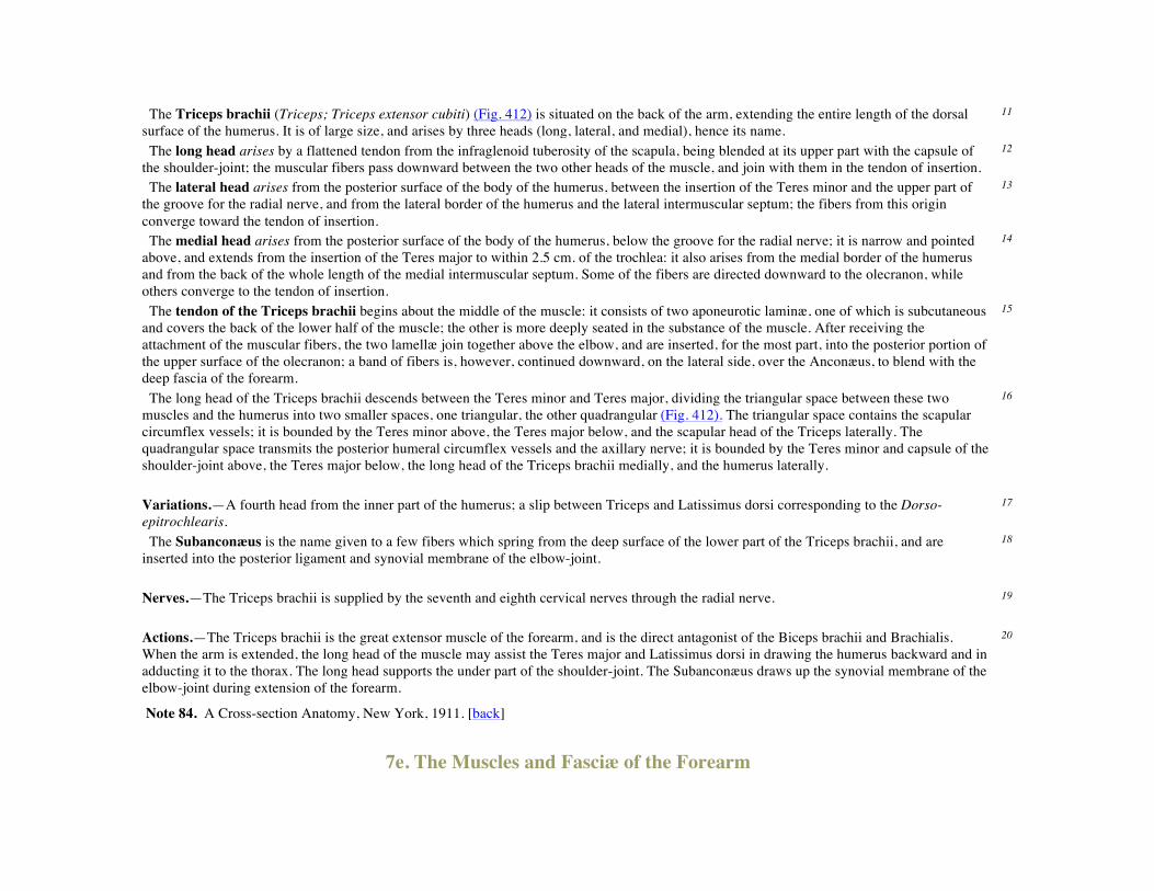
The Triceps brachii (Triceps; Triceps extensor cubiti) (Fig. 412) is situated on the back of the arm, extending the entire length of the dorsal surface of the humerus. It is of large size, and arises by three heads (long, lateral, and medial), hence its name.
11
The long head arises by a flattened tendon from the infraglenoid tuberosity of the scapula, being blended at its upper part with the capsule of the shoulder-joint; the muscular fibers pass downward between the two other heads of the muscle, and join with them in the tendon of insertion.
12
The lateral head arises from the posterior surface of the body of the humerus, between the insertion of the Teres minor and the upper part of the groove for the radial nerve, and from the lateral border of the humerus and the lateral intermuscular septum; the fibers from this origin converge toward the tendon of insertion.
13
The medial head arises from the posterior surface of the body of the humerus, below the groove for the radial nerve; it is narrow and pointed above, and extends from the insertion of the Teres major to within 2.5 cm. of the trochlea: it also arises from the medial border of the humerus and from the back of the whole length of the medial intermuscular septum. Some of the fibers are directed downward to the olecranon, while others converge to the tendon of insertion.
14
The tendon of the Triceps brachii begins about the middle of the muscle: it consists of two aponeurotic laminæ, one of which is subcutaneous and covers the back of the lower half of the muscle; the other is more deeply seated in the substance of the muscle. After receiving the attachment of the muscular fibers, the two lamellæ join together above the elbow, and are inserted, for the most part, into the posterior portion of the upper surface of the olecranon; a band of fibers is, however, continued downward, on the lateral side, over the Anconæus, to blend with the deep fascia of the forearm.
15
The long head of the Triceps brachii descends between the Teres minor and Teres major, dividing the triangular space between these two muscles and the humerus into two smaller spaces, one triangular, the other quadrangular (Fig. 412). The triangular space contains the scapular circumflex vessels; it is bounded by the Teres minor above, the Teres major below, and the scapular head of the Triceps laterally. The quadrangular space transmits the posterior humeral circumflex vessels and the axillary nerve; it is bounded by the Teres minor and capsule of the shoulder-joint above, the Teres major below, the long head of the Triceps brachii medially, and the humerus laterally.
16
Variations.—A fourth head from the inner part of the humerus; a slip between Triceps and Latissimus dorsi corresponding to the Dorso-epitrochlearis.
17
The Subanconæus is the name given to a few fibers which spring from the deep surface of the lower part of the Triceps brachii, and are inserted into the posterior ligament and synovial membrane of the elbow-joint.
18
Nerves.—The Triceps brachii is supplied by the seventh and eighth cervical nerves through the radial nerve. 19 Actions.—The Triceps brachii is the great extensor muscle of the forearm, and is the direct antagonist of the Biceps brachii and Brachialis. When the arm is extended, the long head of the muscle may assist the Teres major and Latissimus dorsi in drawing the humerus backward and in adducting it to the thorax. The long head supports the under part of the shoulder-joint. The Subanconæus draws up the synovial membrane of the elbow-joint during extension of the forearm.
20
Note 84. A Cross-section Anatomy, New York, 1911. [back]
7e. The Muscles and Fasciæ of the Forearm

Antibrachial Fascia (fascia antibrachii; deep fascia of the forearm).—The antibrachial fascia continuous above with the brachial fascia, is a dense, membranous investment, which forms a general sheath for the muscles in this region; it is attached, behind, to the olecranon and dorsal border of the ulna, and gives off from its deep surface numerous intermuscular septa, which enclose each muscle separately. Over the Flexor tendons as they approach the wrist it is especially thickened, and forms the volar carpal ligament. This is continuous with the transverse carpal ligament, and forms a sheath for the tendon of the Palmaris longus which passes over the transverse carpal ligament to be inserted into the palmar aponeurosis.Behind, near the wrist-joint, it is thickened by the addition of many transverse fibers, and forms the dorsal carpal ligament. It is much thicker on the dorsal than on the volar surface, and at the lower than at the upper part of the forearm, and is strengthened above by tendinous fibers derived from the Biceps brachii in front, and from the Triceps brachii behind. It gives origin to muscular fibers, especially at the upper part of the medial and lateral sides of the forearm, and forms the boundaries of a series of cone-shaped cavities, in which the muscles are contained. Besides the vertical septa separating the individual muscles, transverse septa are given off both on the volar and dorsal surfaces of the forearm, separating the deep from the superficial layers of muscles. Apertures exist in the fascia for the passage of vessels and nerves; one of these apertures of large size, situated at the front of the elbow, serves for the passage of a communicating branch between the superficial and deep veins.
1
The antibrachial or forearm muscles may be divided into a volar and a dorsal group. 2 1. The Volar Antibrachial Muscles—These muscles are divided for convenience of description into two groups, superficial and deep. 3 The Superficial Group (Fig. 414).
Pronator teres. Palmaris longus. Flexor carpi radialis. Flexor carpi ulnaris.
Flexor digitorum sublimis.
The muscles of this group take origin from the medial epicondyle of the humerus by a common tendon; they receive additional fibers from the deep fascia of the forearm near the elbow, and from the septa which pass from this fascia between the individual muscles.
4
The Pronator teres has two heads of origin—humeral and ulnar. The humeral head, the larger and more superficial, arises immediately above the medial epicondyle, and from the tendon common to the origin of the other muscles; also from the intermuscular septum between it and the Flexor carpi radialis and from the antibrachial fascia. The ulnar head is a thin fasciculus, which arises from the medial side of the coronoid process of the ulna, and joins the preceding at an acute angle. The median nerve enters the forearm between the two heads of the muscle, and is separated from the ulnar artery by the ulnar head. The muscle passes obliquely across the forearm, and ends in a flat tendon, which is inserted into a rough impression at the middle of the lateral surface of the body of the radius. The lateral border of the muscle forms the medial boundary of a triangular hollow situated in front of the elbow-joint and containing the brachial artery, median nerve, and tendon of the Biceps brachii.
5
Variations.—Absence of ulnar head; additional slips from the medial intermuscular septum, from the Biceps and from the Brachialis anticus occasionally occur.
6
The Flexor carpi radialis lies on the medial side of the preceding muscle. It arises from the medial epicondyle by the common tendon; from the fascia of the forearm; and from the intermuscular septa between it and the Pronator teres laterally, the Palmaris longus medially, and the
7

Flexor digitorum sublimis beneath. Slender and aponeurotic in structure at its commencement, it increases in size, and ends in a tendon which forms rather more than the lower half of its length. This tendon passes through a canal in the lateral part of the transverse carpal ligament and runs through a groove on the greater multangular bone; the groove is converted into a canal by fibrous tissue, and lined by a mucous sheath. The tendon is inserted into the base of the second metacarpal bone, and sends a slip to the base of the third metacarpal bone. The radial artery, in the lower part of the forearm, lies between the tendon of this muscle and the Brachioradialis. Variations.—Slips from the tendon of the Biceps, the lacertus fibrosus, the coronoid, and the radius have been found. Its insertion often varies and may be mostly into the annular ligament, the trapezium, or the fourth metacarpal as well as the second or third. The muscle may be absent.
8
The Palmaris longus is a slender, fusiform muscle, lying on the medial side of the preceding. It arises from the medial epicondyle of the humerus by the common tendon, from the intermuscular septa between it and the adjacent muscles, and from the antibrachial fascia. It ends in a slender, flattened tendon, which passes over the upper part of the transverse carpal ligament, and is inserted into the central part of the transverse carpal ligament and lower part of the palmar aponeurosis, frequently sending a tendinous slip to the short muscles of the thumb.
9
Variations.—One of the most variable muscles in the body. This muscle is often absent about (10 per cent.), and is subject to many variations; it may be tendinous above and muscular below; or it may be muscular in the center with a tendon above and below; or it may present two muscular bundles with a central tendon; or finally it may consist solely of a tendinous band. The muscle may be double. Slips of origin from the coronoid process or from the radius have been seen.Partial or complete insertion into the fascia of the forearm, into the tendon of the Flexor carpi ulnaris and pisiform bone, into the navicular, and into the muscles of the little finger have been observed.
10
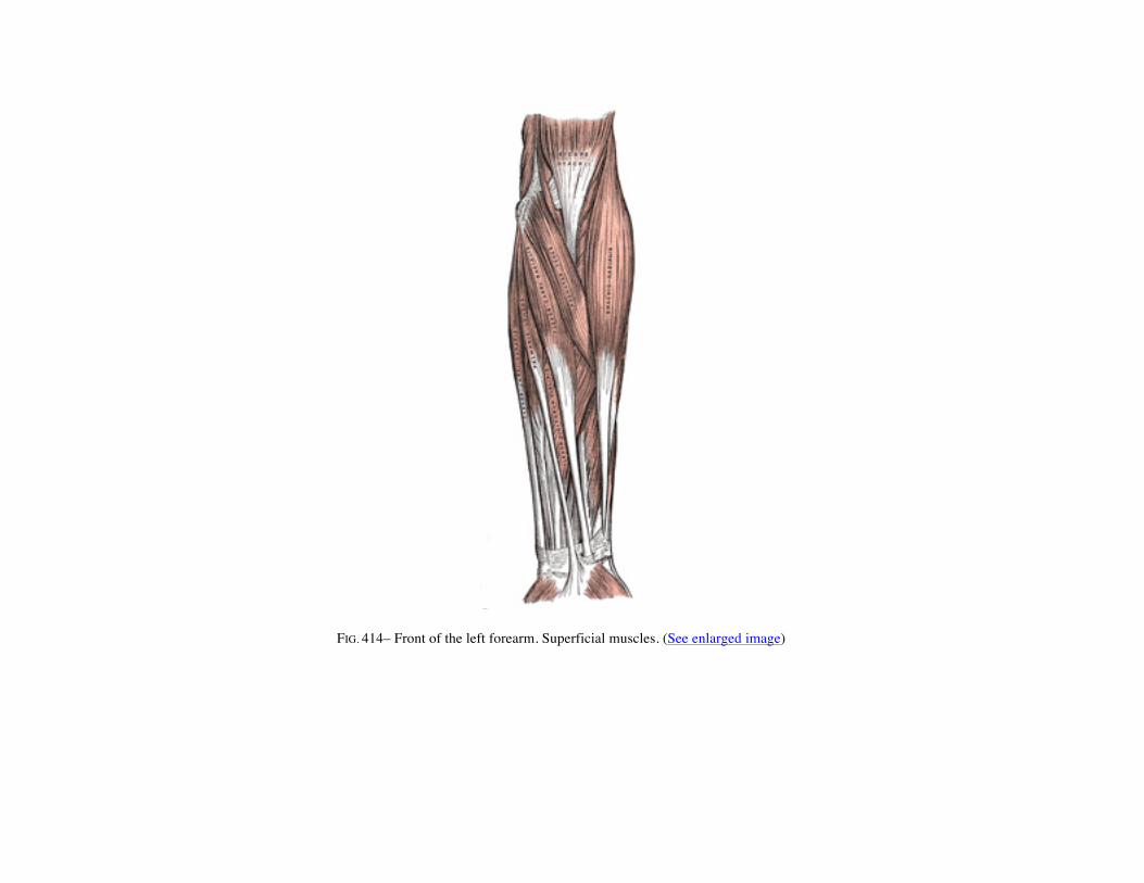
FIG. 414– Front of the left forearm. Superficial muscles. (See enlarged image)
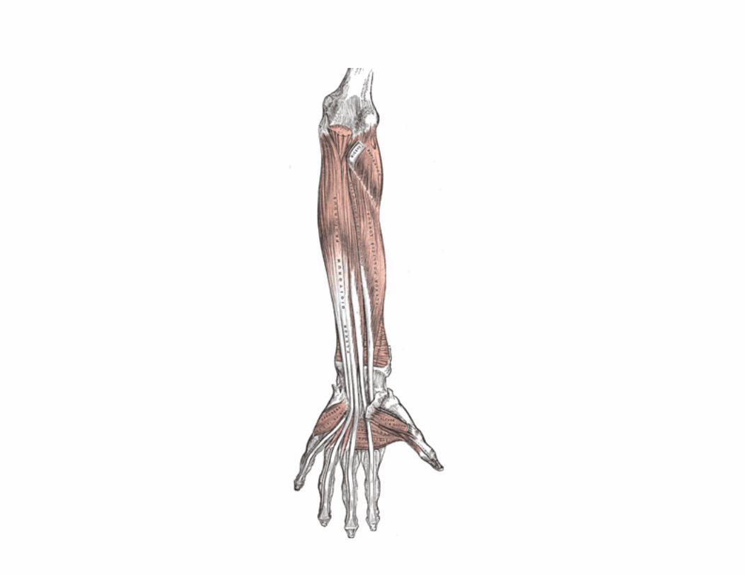
FIG. 415– Front of the left forearm. Deep muscles. (See enlarged image)
The Flexor carpi ulnaris lies along the ulnar side of the forearm. It arises by two heads, humeral and ulnar, connected by a tendinous arch, beneath which the ulnar nerve and posterior ulnar recurrent artery pass. The humeral head arises from the medial epicondyle of the humerus by the common tendon; the ulnar head arises from the medial margin of the olecranon and from the upper two-thirds of the dorsal border of the ulna by an aponeurosis, common to it and the Extensor carpi ulnaris and Flexor digitorum profundus; and from the intermuscular septum between it and the Flexor digitorum sublimis. The fibers end in a tendon, which occupies the anterior part of the lower half of the muscle and isinserted into the pisiform bone, and is prolonged from this to the hamate and fifth metacarpal bones by the pisohamate and pisometacarpal ligaments; it is also attached by a few fibers to the transverse carpal ligament. The ulnar vessels and nerve lie on the lateral side of the tendon of this muscle, in the lower two-thirds of the forearm.
11
Variations.—Slips of origin from the coronoid. The Epitrochleo-anconæus, a small muscle often present runs from the back of the inner condyle to the olecranon, over the ulnar nerve.
12
The Flexor digitorum sublimis is placed beneath the previous muscle; it is the largest of the muscles of the superficial group, and arises by three heads—humeral, ulnar, and radial. The humeral head arises from the medial epicondyle of the humerus by the common tendon, from the ulnar collateral ligament of the elbow-joint, and from the intermuscular septa between it and the preceding muscles. The ulnar head arises from the medial side of the coronoid process, above the ulnar origin of the Pronator teres (see Fig. 213, page 216). The radial head arises from the oblique line of the radius, extending from the radial tuberosity to the insertion of the Pronator teres. The muscle speedily separates into two planes of muscular fibers, superficial and deep: the superficial plane divides into two parts which end in tendons for the middle and ring fingers; the deep plane gives off a muscular slip to join the portion of the superficial plane which is associated with the tendon of the ring finger, and then divides into two parts, which end in tendons for the index and little fingers. As the four tendons thus formed pass beneath the transverse carpal ligament into the palm of the hand, they are arranged in pairs, the superficial pair going to the middle and ring fingers, the deep pair to the index and little fingers. The tendons diverge from one another in the palm and form dorsal relations to the superficial volar arch and digital branches of the median and ulnar nerves. Opposite the bases of the first phalanges each tendon divides into two slips to allow of the passage of the corresponding tendon of the Flexor digitorum profundus; the two slips then reunite and form a grooved channel for the reception of the accompanying tendon of the Flexor digitorum profundus. Finally the tendon divides and is inserted into the sides of the second phalanx about its middle.
13
Variations.—Absence of radial head, of little finger portion; accessory slips from ulnar tuberosity to the index and middle finger portions; from the inner head to the Flexor profundus; from the ulnar or annular ligament to the little finger.
14
The Deep Group (Fig. 415).
Flexor digitorum profundus. Flexor pollicis longus. Pronator quadratus.
The Flexor digitorum profundus is situated on the ulnar side of the forearm, immediately beneath the superficial Flexors. It arises from the upper three-fourths of the volar and medial surfaces of the body of the ulna, embracing the insertion of the Brachialis above, and extending
15
below to within a short distance of the Pronator quadratus. It also arises from a depression on the medial side of the coronoid process; by an aponeurosis from the upper three-fourths of the dorsal border of the ulna, in common with the Flexor and Extensor carpi ulnaris; and from the ulnar half of the interosseous membrane. The muscle ends in four tendons which run under the transverse carpal ligament dorsal to the tendons of the Flexor digitorum sublimis. Opposite the first phalanges the tendons pass through the openings in the tendons of the Flexor digitorum sublimis, and are finally inserted into the bases of the last phalanges. The portion of the muscle for the index finger is usually distinct throughout, but the tendons for the middle, ring, and little fingers are connected together by areolar tissue and tendinous slips, as far as the palm of the hand. Fibrous Sheaths of the Flexor Tendons.—After leaving the palm, the tendons of the Flexores digitorum sublimis and profundus lie in osseo-aponeurotic canals (Fig. 427),formed behind by the phalanges and in front by strong fibrous bands, which arch across the tendons, and are attached on either side to the margins of the phalanges. Opposite the middle of the proximal and second phalanges the bands (digital vaginal ligaments) are very strong, and the fibers are transverse; but opposite the joints they are much thinner, and consist of annular and cruciate ligamentous fibers. Each canal contains a mucous sheath, which is reflected on the contained tendons.
16
Within each canal the tendons of the Flexores digitorum sublimis and profundus are connected to each other, and to the phalanges, by slender, tendinous bands, called vincula tendina (Fig. 416). There are two sets of these; (a) the vincula brevia, which are two in number in each finger, and consist of triangular bands of fibers, one connecting the tendon of the Flexor digitorum sublimis to the front of the first interphalangeal joint and head of the first phalanx, and the other the tendon of the Flexor digitorum profundus to the front of the second interphalangeal joint and head of the second phalanx; (b) the vincula longa, which connect the under surfaces of the tendons of the Flexor digitorum profundus to those of the subjacent Flexor sublimis after the tendons of the former have passed through the latter.
17
Variations.—The index finger portion may arise partly from the upper part of the radius. Slips from the inner head of the Flexor sublimis, medial epicondyle, or the coronoid are found. Connection with the Flexor pollicis longus.
18
Four small muscles, the Lumbricales, are connected with the tendons of the Flexor profundus in the palm. They will be described with the muscles of the hand (page 464).
19
The Flexor pollicis longus is situated on the radial side of the forearm, lying in the same plane as the preceding. It arises from the grooved volar surface of the body of the radius, extending from immediately below the tuberosity and oblique line to within a short distance of the Pronator quadratus. It arises also from the adjacent part of the interosseous membrane, and generally by a fleshy slip from the medial border of the coronoid process, or from the medial epicondyle of the humerus. The fibers end in a flattened tendon, which passes beneath the transverse carpal ligament, is then lodged between the lateral head of the Flexor pollicis brevis and the oblique part of the Adductor pollicis, and, entering an osseoaponeurotic canal similar to those for the Flexor tendons of the fingers, is inserted into the base of the distal phalanx of the thumb. The volar interosseous nerve and vessels pass downward on the front of the interosseous membrane between the Flexor pollicis longus and Flexor digitorum profundus.
20
Variations.—Slips may connect with Flexor sublimis, or Profundus, or Pronator teres. An additional tendon to the index finger is sometimes found.
21
The Pronator quadratus is a small, flat, quadrilateral muscle, extending across the front of the lower parts of the radius and ulna. It arises from the pronator ridge on the lower part of the volar surface of the body of the ulna; from the medial part of the volar surface of the lower fourth of the ulna; and from a strong aponeurosis which covers the medial third of the muscle. The fibers pass lateralward and slightly
22

downward, to be inserted into the lower fourth of the lateral border and the volar surface of the body of the radius. The deeper fibers of the muscle are inserted into the triangular area above the ulnar notch of the radius—an attachment comparable with the origin of the Supinator from the triangular area below the radial notch of the ulna.
FIG. 416– Tendons of forefinger and vincula tendina. (See enlarged image)
Variations.—Rarely absent; split into two or three layers; increased attachment upward or downward. 23 Nerves.—All the muscles of the superficial layer are supplied by the median nerve, excepting the Flexor carpi ulnaris, which is supplied by the ulnar. The Pronator teres, the Flexor carpi radialis, and the Palmaris longus derive their supply primarily from the sixth cervical nerve; the Flexor digitorum sublimis from the seventh and eighth cervical and first thoracic nerves, and the Flexor carpi ulnaris from the eighth cervical and first thoracic. Of the deep layer, the Flexor digitorum profundus is supplied by the eighth cervical and first thoracic through the ulnar, and the volar interosseous branch of the median. The Flexor pollicis longus and Pronator quadratus are supplied by the eighth cervical and first thoracic through the volar interosseous branch of the median.
24
Actions.—These muscles act upon the forearm, the wrist, and hand. The Pronator teres rotates the radius upon the ulna, rendering the hand 25

prone; when the radius is fixed, it assists in flexing the forearm. The Flexor carpi radialis is a flexor and abductor of the wrist; it also assists in pronating the hand, and in bending the elbow. The Flexor carpi ulnaris is a flexor and adductor of the wrist; it also assists in bending the elbow. The Palmaris longus is a flexor of the wrist-joint; it also assists in flexing the elbow. The Flexor digitorum sublimis flexes first the middle and then the proximal phalanges; it also assists in flexing the wrist and elbow. The Flexor digitorum profundus is one of the flexors of the phalanges. After the Flexor sublimis has bent the second phalanx, the Flexor profundus flexes the terminal one; but it cannot do so until after the contraction of the superficial muscle. It also assists in flexing the wrist. The Flexor pollicis longus is a flexor of the phalanges of the thumb; when the thumb is fixed, it assists in flexing the wrist. The Pronator quadratus rotates the radius upon the ulna, rendering the hand prone.
FIG. 417– Cross-section through the middle of the forearm. (Eycleshymer and Schoemaker.) (See enlarged image)
2. The Dorsal Antibrachial Muscles—These muscles are divided for convenience of description into two groups, superficial and deep. 26 The Superficial Group (Fig. 418).
Brachioradialis. Extensor digitorum communis. Extensor carpi radialis longus. Extensor digiti quinti proprius. Extensor carpi radialis brevis. Extensor carpi ulnaris.
Anconæus.

The Brachioradialis (Supinator longus) is the most superficial muscle on the radial side of the forearm. It arises from the upper two-thirds of the lateral supracondylar ridge of the humerus, and from the lateral intermuscular septum, being limited above by the groove for the radial nerve. Interposed between it and the Brachialis are the radial nerve and the anastomosis between the anterior branch of the profunda artery and the radial recurrent. The fibers end above the middle of the forearm in a flat tendon, which is inserted into the lateral side of the base of the styloid process of the radius. The tendon is crossed near its insertion by the tendons of the Abductor pollicis longus and Extensor pollicis brevis; on its ulnar side is the radial artery.
27
Variations.—Fusion with the Brachialis; tendon of insertion may be divided into two or three slips; insertion partial or complete into the middle of the radius, fasciculi to the tendon of the Biceps, the tuberosity or oblique line of the radius; slips to the Extensor carpi radialis longus or Abductor pollicis longus; absence; rarely doubled.
28
The Extensor carpi radialis longus (Extensor carpi radialis longior) is placed partly beneath the Brachioradialis. It arises from the lower third of the lateral supracondylar ridge of the humerus, from the lateral intermuscular septum, and by a few fibers from the common tendon of origin of the Extensor muscles of the forearm. The fibers end at the upper third of the forearm in a flat tendon, which runs along the lateral border of the radius, beneath the Abductor pollicis longus and Extensor pollicis brevis; it then passes beneath the dorsal carpal ligament, where it lies in a groove on the back of the radius common to it and the Extensor carpi radialis brevis, immediately behind the styloid process. It is inserted into the dorsal surface of the base of the second metacarpal bone, on its radial side.
29
The Extensor carpi radialis brevis (Extensor carpi radialis brevior) is shorter and thicker than the preceding muscle, beneath which it is placed. It arises from the lateral epicondyle of the humerus, by a tendon common to it and the three following muscles; from the radial collateral ligament of the elbow-joint; from a strong aponeurosis which covers its surface; and from the intermuscular septa between it and the adjacent muscles. The fibers end about the middle of the forearm in a flat tendon, which is closely connected with that of the preceding muscle, and accompanies it to the wrist; it passes beneath the Abductor pollicis longus and Extensor pollicis brevis, then beneath the dorsal carpal ligament, and is inserted into the dorsal surface of the base of the third metacarpal bone on its radial side. Under the dorsal carpal ligament the tendon lies on the back of the radius in a shallow groove, to the ulnar side of that which lodges the tendon of the Extensor carpi radialis, longus, and separated from it by a faint ridge.
30
The tendons of the two preceding muscles pass through the same compartment of the dorsal carpal ligament in a single mucous sheath. 31 Variations.—Either muscle may split into two or three tendons of insertion to the second and third or even the fourth metacarpal. The two muscles may unite into a single belly with two tendons. Cross slips between the two muscles may occur. The Extensor carpi radialis intermedius rarely arises as a distinct muscle from the humerus, but is not uncommon as an accessory slip from one or both muscles to the second or third or both metacarpals. TheExtensor carpi radialis accessorius is occasionally found arising from the humerus with or below the Extensor carpi radialis longus and inserted into the first metacarpal, the Abductor pollicis brevis, the First dorsal interosseous, or elsewhere.
32
The Extensor digitorum communis arises from the lateral epicondyle of the humerus, by the common tendon; from the intermuscular septa between it and the adjacent muscles, and from the antibrachial fascia. It divides below into four tendons, which pass, together with that of the Extensor indicis proprius, through a separate compartment of the dorsal carpal ligament, within a mucous sheath. The tendons then diverge on the back of the hand, and are inserted into the second and third phalanges of the fingers in the following manner. Opposite the metacarpophalangeal articulation each tendon is bound by fasciculi to the collateral ligaments and serves as the dorsal ligament of this joint; after having crossed the joint, it spreads out into a broad aponeurosis, which covers the dorsal surface of the first phalanx and is reinforced, in this situation, by the tendons of the Interossei and Lumbricalis. Opposite the first interphalangeal joint this aponeurosis divides into three slips; an
33

intermediate and two collateral: the former is inserted into the base of the second phalanx; and the two collateral, which are continued onward along the sides of the second phalanx, unite by their contiguous margins, and are inserted into the dorsal surface of the last phalanx. As the tendons cross the interphalangeal joints, they furnish them with dorsal ligaments. The tendon to the index finger is accompanied by the Extensor indicis proprius, which lies on its ulnar side. On the back of the hand, the tendons to the middle, ring, and little fingers are connected by two obliquely placed bands, one from the third tendon passing downward and lateralward to the second tendon, and the other passing from the same tendon downward and medialward to the fourth. Occasionally the first tendon is connected to the second by a thin transverse band.

FIG. 418– Posterior surface of the forearm. Superficial muscles. (See enlarged image)
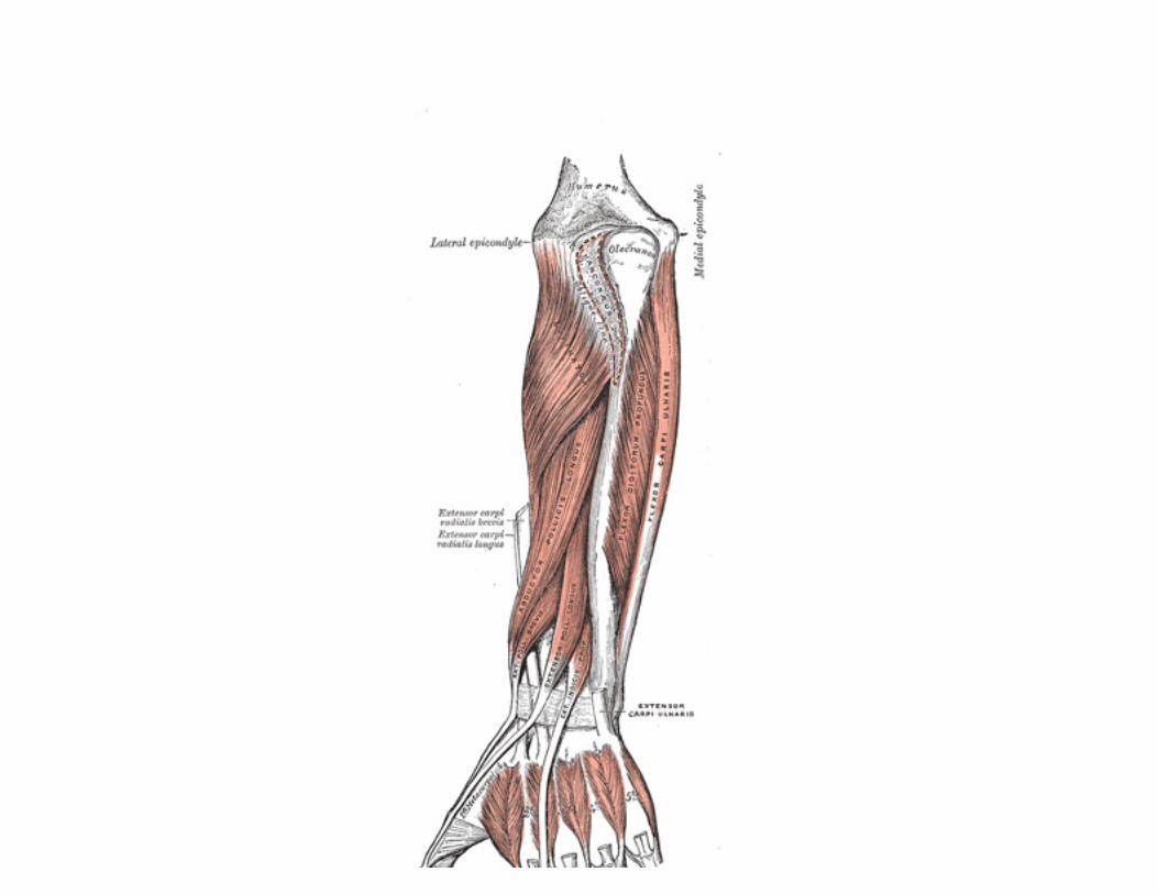
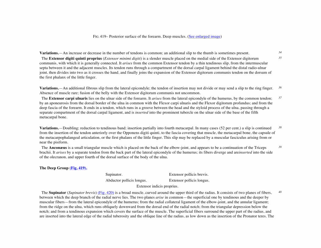
FIG. 419– Posterior surface of the forearm. Deep muscles. (See enlarged image)
Variations.—An increase or decrease in the number of tendons is common; an additional slip to the thumb is sometimes present. 34 The Extensor digiti quinti proprius (Extensor minimi digiti) is a slender muscle placed on the medial side of the Extensor digitorum communis, with which it is generally connected. It arises from the common Extensor tendon by a thin tendinous slip, from the intermuscular septa between it and the adjacent muscles. Its tendon runs through a compartment of the dorsal carpal ligament behind the distal radio-ulnar joint, then divides into two as it crosses the hand, and finally joins the expansion of the Extensor digitorum communis tendon on the dorsum of the first phalanx of the little finger.
35
Variations.—An additional fibrous slip from the lateral epicondyle; the tendon of insertion may not divide or may send a slip to the ring finger. Absence of muscle rare; fusion of the belly with the Extensor digitorum communis not uncommon.
36
The Extensor carpi ulnaris lies on the ulnar side of the forearm. It arises from the lateral epicondyle of the humerus, by the common tendon; by an aponeurosis from the dorsal border of the ulna in common with the Flexor carpi ulnaris and the Flexor digitorum profundus; and from the deep fascia of the forearm. It ends in a tendon, which runs in a groove between the head and the styloid process of the ulna, passing through a separate compartment of the dorsal carpal ligament, and is inserted into the prominent tubercle on the ulnar side of the base of the fifth metacarpal bone.
37
Variations.—Doubling; reduction to tendinous band; insertion partially into fourth metacarpal. In many cases (52 per cent.) a slip is continued from the insertion of the tendon anteriorly over the Opponens digiti quinti, to the fascia covering that muscle, the metacarpal bone, the capsule of the metacarpophalangeal articulation, or the first phalanx of the little finger. This slip may be replaced by a muscular fasciculus arising from or near the pisiform.
38
The Anconæus is a small triangular muscle which is placed on the back of the elbow-joint, and appears to be a continuation of the Triceps brachii. It arises by a separate tendon from the back part of the lateral epicondyle of the humerus; its fibers diverge and areinserted into the side of the olecranon, and upper fourth of the dorsal surface of the body of the ulna.
39
The Deep Group (Fig. 419).
Supinator. Extensor pollicis brevis. Abductor pollicis longus. Extensor pollicis longus.
Extensor indicis proprius.
The Supinator (Supinator brevis) (Fig. 420) is a broad muscle, curved around the upper third of the radius. It consists of two planes of fibers, between which the deep branch of the radial nerve lies. The two planes arise in common—the superficial one by tendinous and the deeper by muscular fibers—from the lateral epicondyle of the humerus; from the radial collateral ligament of the elbow-joint, and the annular ligament; from the ridge on the ulna, which runs obliquely downward from the dorsal end of the radial notch; from the triangular depression below the notch; and from a tendinous expansion which covers the surface of the muscle. The superficial fibers surround the upper part of the radius, and are inserted into the lateral edge of the radial tuberosity and the oblique line of the radius, as low down as the insertion of the Pronator teres. The
40

upper fibers of the deeper plane form a sling-like fasciculus, which encircles the neck of the radius above the tuberosity and is attached to the back part of its medial surface; the greater part of this portion of the muscle is inserted into the dorsal and lateral surfaces of the body of the radius, midway between the oblique line and the head of the bone. The Abductor pollicis longus (Extensor oss. metacarpi pollicis) lies immediately below the Supinator and is sometimes united with it. It arises from the lateral part of the dorsal surface of the body of the ulna below the insertion of the Anconæus, from the interosseous membrane, and from the middle third of the dorsal surface of the body of the radius. Passing obliquely downward and lateralward, it ends in a tendon, which runs through a groove on the lateral side of the lower end of the radius, accompanied by the tendon of the Extensor pollicis brevis, and is inserted into the radial side of the base of the first metacarpal bone. It occasionally gives off two slips near its insertion: one to the greater multangular bone and the other to blend with the origin of the Abductor pollicis brevis.
41
Variations.—More or less doubling of muscle and tendon with insertion of the extra tendon into the first metacarpal, the greater multangular, or into the Abductor pollicis brevis or Opponens pollicis.
42
FIG. 420– The Supinator. (See enlarged image)

The Extensor pollicis brevis (Extensor primi internodii pollicis) lies on the medial side of, and is closely connected with, the Abductor pollicis longus. It arises from the dorsal surface of the body of the radius below that muscle, and from the interosseous membrane. Its direction is similar to that of the Abductor pollicis longus, its tendon passing the same groove on the lateral side of the lower end of the radius, to be inserted into the base of the first phalanx of the thumb.
43
Variations.—Absence; fusion of tendon with that of the Extensor pollicis longus. 44 The Extensor pollicis longus (Extensor secundi internodii pollicis) is much larger than the preceding muscle, the origin of which it partly covers. It arises from the lateral part of the middle third of the dorsal surface of the body of the ulna below the origin of the Abductor pollicis longus, and from the interosseous membrane. It ends in a tendon, which passes through a separate compartment in the dorsal carpal ligament, lying in a narrow, oblique groove on the back of the lower end of the radius. It then crosses obliquely the tendons of the Extensores carpi radialis longus and brevis, and is separated from the Extensor brevis pollicis by a triangular interval, in which the radial artery is found; and is finally inserted into the base of the last phalanx of the thumb. The radial artery is crossed by the tendons of the Abductor pollicis longus and of the Extensores pollicis longus and brevis.
45
The Extensor indicis proprius (Extensor indicis) is a narrow, elongated muscle, placed medial to, and parallel with, the preceding. It arises, from the dorsal surface of the body of the ulna below the origin of the Extensor pollicis longus, and from the interosseous membrane. Its tendon passes under the dorsal carpal ligament in the same compartment as that which transmits the tendons of the Extensor digitorum communis, and opposite the head of the second metacarpal bone, joins the ulnar side of the tendon of the Extensor digitorum communis which belongs to the index finger.
46
Variations.—Doubling; the ulnar part may pass beneath the dorsal carpal ligament with the Extensor digitorum communis; a slip from the tendon may pass to the index finger.
47
Nerves.—The Brachioradialis is supplied by the fifth and sixth, the Extensores carpi radialis longus and brevis by the sixth and seventh, and the Anconæus by the seventh and eighth cervical nerves, through the radial nerve; the remaining muscles are innervated through the deep radial nerve, the Supinator being supplied by the sixth, and all the other muscles by the seventh cervical.
48
Actions.—The muscles of the lateral and dorsal aspects of the forearm, which comprise all the Extensor muscles and the Supinator, act upon the forearm, wrist, and hand; they are the direct antagonists of the Pronator and Flexor muscles. The Anconæus assists the Triceps in extending the forearm. The Brachioradialis is a flexor of the elbow-joint, but only acts as such when the movement of flexion has been initiated by the Biceps brachii and Brachialis. The action of the Supinator is suggested by its name; it assists the Biceps in bringing the hand into the supine position. The Extensor carpi radialis longus extends the wrist and abducts the hand. It may also assist in bending the elbow-joint; at all events it serves to fix or steady this articulation. The Extensor carpi radialis brevis extends the wrist, and may also act slightly as an abductor of the hand. The Extensor carpi ulnaris extends the wrist, but when acting alone inclines the hand toward the ulnar side; by its continued action it extends the elbow-joint. The Extensor digitorum communis extends the phalanges, then the wrist, and finally the elbow. It acts principally on the proximal phalanges, the middle and terminal phalanges being extended mainly by the Interossei and Lumbricales. It tends to separate the fingers as it extends them. The Extensor digiti quinti proprius extends the little finger, and by its continued action assists in extending the wrist. It is owing to this muscle that the little finger can be extended or pointed while the others are flexed. The chief action of the Abductor pollicis longus is to

carry the thumb laterally from the palm of the hand. By its continued action it helps to extend and abduct the wrist. The Extensor pollicis brevis extends the proximal phalanx, and the Extensor pollicis longus the terminal phalanx of the thumb; by their continued action they help to extend and abduct the wrist. The Extensor indicis proprius extends the index finger, and by its continued action assists in extending the wrist.
1F. The Muscles and Fasciæ of the Hand
The muscles of the hand are subdivided into three groups: (1) those of the thumb, which occupy the radial side and produce the thenar eminence; (2) those of the little finger, which occupy the ulnar side and give rise to the hypothenar eminence; (3) those in the middle of the palm and between the metacarpal bones.
1
Volar Carpal Ligament (ligamentum carpi volare).—The volar carpal ligament is the thickened band of antibrachial fascia which extends from the radius to the ulna over the Flexor tendons as they enter the wrist.
2
Transverse Carpal Ligament (ligamentum carpi transversum; anterior annular ligament) (Figs. 421, 422).—The transverse carpal ligament is a strong, fibrous band, which arches over the carpus, converting the deep groove on the front of the carpal bones into a tunnel, through which the Flexor tendons of the digits and the median nerve pass. It is attached, medially, to the pisiform and the hamulus of the hamate bone; laterally, to the tuberosity of the navicular, and to the medial part of the volar surface and the ridge of the greater multangular. It is continuous, above, with the volar carpal ligament; and below, with the palmar aponeurosis. It is crossed by the ulnar vessels and nerve, and the cutaneous branches of the median and ulnar nerves. At its lateral end is the tendon of theFlexor carpi radialis, which lies in the groove on the greater multangular between the attachments of the ligament to the bone. On its volar surface the tendons of the Palmaris longus and Flexor carpi ulnaris are partly inserted; below, it gives origin to the short muscles of the thumb and little finger
3
FIG. 421– Transverse section across distal ends of radius and ulna. (See enlarged image)

FIG. 422– Transverse section across the wrist and digits. (See enlarged image)
The Mucous Sheaths of the Tendons on the Front of the Wrist.—Two sheaths envelop the tendons as they pass beneath the transverse carpal ligament, one for the Flexores digitorum sublimis and profundus, the other for the Flexor pollicis longus (Fig. 423). They extend into the forearm for about 2.5 cm. above the transverse carpal ligament, and occasionally communicate with each other under the ligament. The sheath which surrounds the Flexores digitorum extends downward about half-way along the metacarpal bones, where it ends in blind diverticula around the tendons to the index, middle, and ring fingers. It is prolonged on the tendons to the little finger and usually communicates with the mucoussheath of these tendons. The sheath of the tendon of the Flexor pollicis longus is continued along the thumb as far as the insertion of the tendon. The mucous sheaths enveloping the terminal parts of the tendons of the Flexores digitorum have been described on page 449.
4

FIG. 423– The mucous sheaths of the tendons on the front of the wrist and digits. (See enlarged image) Dorsal Carpal Ligament (ligamentum carpi dorsale; posterior annular ligament) (Figs. 421, 422).—The dorsal carpal ligament is a strong, fibrous band, extending obliquely downward and medialward across the back of the wrist, and consisting of part of the deep fascia of the back of the forearm, strengthened by the addition of some transverse fibers. It is attached, medially, to the styloid process of the ulna and to the triangular and pisiform bones; laterally, to the lateral margin of the radius; and, in its passage across the wrist, to the ridges on the dorsal surface of the radius.
5
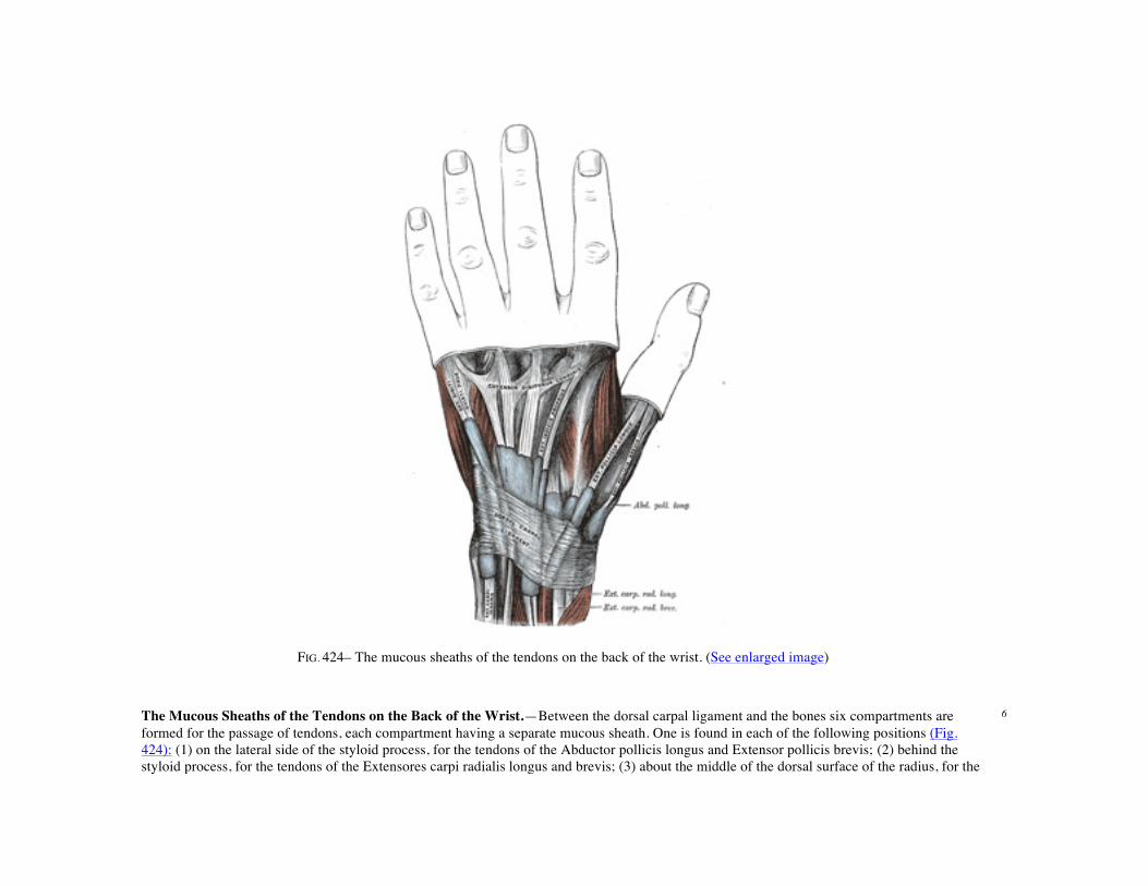
FIG. 424– The mucous sheaths of the tendons on the back of the wrist. (See enlarged image)
The Mucous Sheaths of the Tendons on the Back of the Wrist.—Between the dorsal carpal ligament and the bones six compartments are formed for the passage of tendons, each compartment having a separate mucous sheath. One is found in each of the following positions (Fig. 424): (1) on the lateral side of the styloid process, for the tendons of the Abductor pollicis longus and Extensor pollicis brevis; (2) behind the styloid process, for the tendons of the Extensores carpi radialis longus and brevis; (3) about the middle of the dorsal surface of the radius, for the
6

tendon of the Extensor pollicis longus; (4) to the medial side of the latter, for the tendons of the Extensor digitorum communis and Extensor indicis proprius; (5) opposite the interval between the radius and ulna, for the Extensor digiti quinti proprius; (6) between the head and styloid process of the ulna, for the tendon of the Extensor carpi ulnaris. The sheaths lining these compartments extends from above the dorsal carpal ligament; those for the tendons of Abductor pollicis longus, Extensor brevis pollicis, Extensores carpi radialis, and Extensor carpi ulnaris stop immediately proximal to the bases of the metacarpal bones, while the sheaths for Extensor communis digitorum, Extensor indicis proprius, and Extensor digiti quinti proprius are prolonged to the junction of the proximal and intermediate thirds of the metacarpus.
FIG. 425– The palmar aponeurosis. (See enlarged image)
Palmar Aponeurosis (aponeurosis palmaris; palmar fascia) (Fig. 425).—The palmar aponeurosis invests the muscles of the palm, and consists of central, lateral, and medial portions.
7
The central portion occupies the middle of the palm, is triangular in shape, and of great strength and thickness. Its apex is continuous with the lower margin of the transverse carpal ligament, and receives the expanded tendon of the Palmaris longus. Its base divides below into four slips, one for each finger. Each slip gives off superficial fibers to the skin of the palm and finger, those to the palm joining the skin at the furrow corresponding to the metacarpophalangeal articulations, and those to the fingers passing into the skin at the transverse fold at the bases of the fingers. The deeper part of each slip subdivides into two processes, which are inserted into the fibrous sheaths of the Flexor tendons. From the sides of these processes offsets are attached to the transverse metacarpal ligament. By this arrangement short channels are formed on the front of the heads of the metacarpal bones; through these the Flexor tendons pass. The intervals between the four slips transmit the digital vessels and nerves, and the tendons of the Lumbricales. At the points of division into the slips mentioned, numerous strong, transverse fasciculi bind the separate processes together. The central part of the palmar aponeurosis is intimately bound to the integument by dense fibroareolar tissue forming the superficial palmar fascia, and gives origin by its medial margin to the Palmaris brevis. It covers the superficial volar arch, the tendons of the Flexor muscles, and the branches of the median and ulnar nerves; and on either side it gives off a septum, which is continuous with the interosseous aponeurosis, and separates the intermediate from the collateral groups of muscles.
8
The lateral and medial portions of the palmar aponeurosis are thin, fibrous layers, which cover, on the radial side, the muscles of the ball of the thumb, and, on the ulnar side, the muscles of the little finger; they are continuous with the central portion and with the fascia on the dorsum of the hand.
9
The Superficial Transverse Ligament of the Fingers is a thin band of transverse fasciculi (Fig. 425); it stretches across the roots of the four fingers, and is closely attached to the skin of the clefts, and medially to the fifth metacarpal bone, forming a sort of rudimentary web. Beneath it the digital vessels and nerves pass to their destinations.
10
1. The Lateral Volar Muscles (Figs. 426, 427)
Abductor pollicis brevis. Flexor pollicis brevis. Opponens pollicis. Adductor pollicis (obliquus).
Adductor pollicis (transversus).
The Abductor pollicis brevis (Abductor pollicis) is a thin, flat muscle, placed immediately beneath the integument. It arises from the transverse carpal ligament, the tuberosity of the navicular, and the ridge of the greater multangular, frequently by two distinct slips. Running lateralward and downward, it is inserted by a thin, flat tendon into the radial side of the base of the first phalanx of the thumb and the capsule of the metacarpophalangeal articulation.
11
The Opponens pollicis is a small, triangular muscle, placed beneath the preceding. Itarises from the ridge on the greater multangular and from the transverse carpal ligament, passes downward and lateralward, and is inserted into the whole length of the metacarpal bone of the thumb on its radial side.
12
The Flexor pollicis brevis consists of two portions, lateral and medial. The lateral and more superficial portion arises from the lower border of the transverse carpal ligament and the lower part of the ridge on the greater multangular bone; it passes along the radial side of the tendon of the Flexor pollicis longus, and, becoming tendinous, is inserted into the radial side of the base of the first phalanx of the thumb; in its tendon of
13
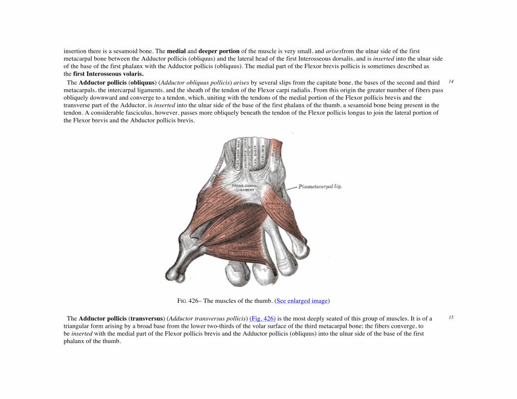
insertion there is a sesamoid bone. The medial and deeper portion of the muscle is very small, and arisesfrom the ulnar side of the first metacarpal bone between the Adductor pollicis (obliquus) and the lateral head of the first Interosseous dorsalis, and is inserted into the ulnar side of the base of the first phalanx with the Adductor pollicis (obliquus). The medial part of the Flexor brevis pollicis is sometimes described as the first Interosseous volaris. The Adductor pollicis (obliquus) (Adductor obliquus pollicis) arises by several slips from the capitate bone, the bases of the second and third metacarpals, the intercarpal ligaments, and the sheath of the tendon of the Flexor carpi radialis. From this origin the greater number of fibers pass obliquely downward and converge to a tendon, which, uniting with the tendons of the medial portion of the Flexor pollicis brevis and the transverse part of the Adductor, is inserted into the ulnar side of the base of the first phalanx of the thumb, a sesamoid bone being present in the tendon. A considerable fasciculus, however, passes more obliquely beneath the tendon of the Flexor pollicis longus to join the lateral portion of the Flexor brevis and the Abductor pollicis brevis.
14
FIG. 426– The muscles of the thumb. (See enlarged image)
The Adductor pollicis (transversus) (Adductor transversus pollicis) (Fig. 426) is the most deeply seated of this group of muscles. It is of a triangular form arising by a broad base from the lower two-thirds of the volar surface of the third metacarpal bone; the fibers converge, to be inserted with the medial part of the Flexor pollicis brevis and the Adductor pollicis (obliquus) into the ulnar side of the base of the first phalanx of the thumb.
15

Variations.—The Abductor pollicis brevis is often divided into an outer and an inner part; accessory slips from the tendon of the Abductor pollicis longus or Palmaris longus, more rarely from the Extensor carpi radialis longus, from the styloid process or Opponens pollicis or from the skin over the thenar eminence. The deep head of the Flexor pollicis brevis may be absent or enlarged. The two adductors vary in their relative extent and in the closeness of their connection. The Adductor obliquus may receive a slip from the transverse metacarpal ligament.
16
Nerves.—The Abductor brevis, Opponens, and lateral head of the Flexor pollicis brevis are supplied by the sixth and seventh cervical nerves through the median nerve; the medial head of the Flexor brevis, and the Adductor, by the eighth cervical through the ulnar nerve.
17
Actions.—The Abductor pollicis brevis draws the thumb forward in a plane at right angles to that of the palm of the hand. The Adductor pollicis is the opponent of this muscle, and approximates the thumb to the palm. The Opponens pollicis flexes the metacarpal bone, i. e., draws it medialward over the palm; the Flexor pollicis brevis flexes and adducts the proximal phalanx.
18
2. The Medial Volar Muscles (Figs. 426, 427)
Palmaris brevis. Flexor digiti quinti brevis. Abductor digiti quinti. Opponens digiti quinti.
The Palmaris brevis is a thin, quadrilateral muscle, placed beneath the integument of the ulnar side of the hand. It arises by tendinous fasciculi from the transverse carpal ligament and palmar aponeurosis; the fleshy fibers are inserted into the skin on the ulnar border of the palm of the hand.
19

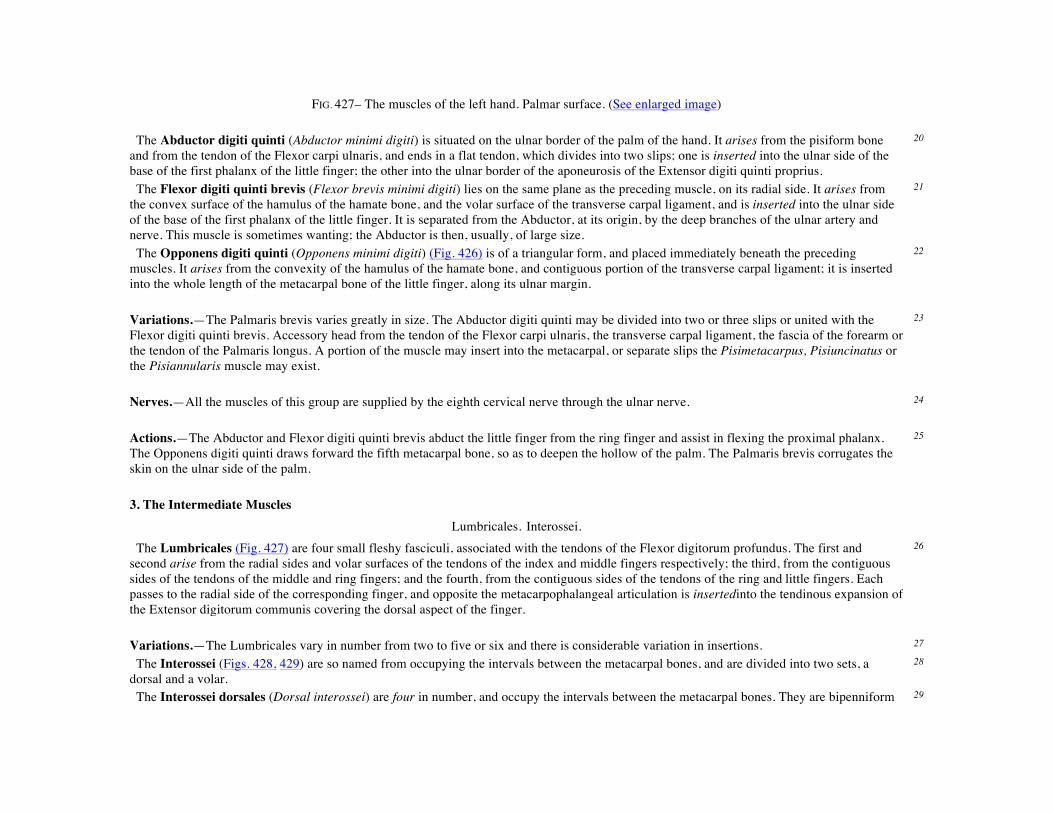
FIG. 427– The muscles of the left hand. Palmar surface. (See enlarged image) The Abductor digiti quinti (Abductor minimi digiti) is situated on the ulnar border of the palm of the hand. It arises from the pisiform bone and from the tendon of the Flexor carpi ulnaris, and ends in a flat tendon, which divides into two slips; one is inserted into the ulnar side of the base of the first phalanx of the little finger; the other into the ulnar border of the aponeurosis of the Extensor digiti quinti proprius.
20
The Flexor digiti quinti brevis (Flexor brevis minimi digiti) lies on the same plane as the preceding muscle, on its radial side. It arises from the convex surface of the hamulus of the hamate bone, and the volar surface of the transverse carpal ligament, and is inserted into the ulnar side of the base of the first phalanx of the little finger. It is separated from the Abductor, at its origin, by the deep branches of the ulnar artery and nerve. This muscle is sometimes wanting; the Abductor is then, usually, of large size.
21
The Opponens digiti quinti (Opponens minimi digiti) (Fig. 426) is of a triangular form, and placed immediately beneath the preceding muscles. It arises from the convexity of the hamulus of the hamate bone, and contiguous portion of the transverse carpal ligament; it is inserted into the whole length of the metacarpal bone of the little finger, along its ulnar margin.
22
Variations.—The Palmaris brevis varies greatly in size. The Abductor digiti quinti may be divided into two or three slips or united with the Flexor digiti quinti brevis. Accessory head from the tendon of the Flexor carpi ulnaris, the transverse carpal ligament, the fascia of the forearm or the tendon of the Palmaris longus. A portion of the muscle may insert into the metacarpal, or separate slips the Pisimetacarpus, Pisiuncinatus or the Pisiannularis muscle may exist.
23
Nerves.—All the muscles of this group are supplied by the eighth cervical nerve through the ulnar nerve. 24 Actions.—The Abductor and Flexor digiti quinti brevis abduct the little finger from the ring finger and assist in flexing the proximal phalanx. The Opponens digiti quinti draws forward the fifth metacarpal bone, so as to deepen the hollow of the palm. The Palmaris brevis corrugates the skin on the ulnar side of the palm.
25
3. The Intermediate Muscles
Lumbricales. Interossei. The Lumbricales (Fig. 427) are four small fleshy fasciculi, associated with the tendons of the Flexor digitorum profundus. The first and second arise from the radial sides and volar surfaces of the tendons of the index and middle fingers respectively; the third, from the contiguous sides of the tendons of the middle and ring fingers; and the fourth, from the contiguous sides of the tendons of the ring and little fingers. Each passes to the radial side of the corresponding finger, and opposite the metacarpophalangeal articulation is insertedinto the tendinous expansion of the Extensor digitorum communis covering the dorsal aspect of the finger.
26
Variations.—The Lumbricales vary in number from two to five or six and there is considerable variation in insertions. 27 The Interossei (Figs. 428, 429) are so named from occupying the intervals between the metacarpal bones, and are divided into two sets, a dorsal and a volar.
28
The Interossei dorsales (Dorsal interossei) are four in number, and occupy the intervals between the metacarpal bones. They are bipenniform 29
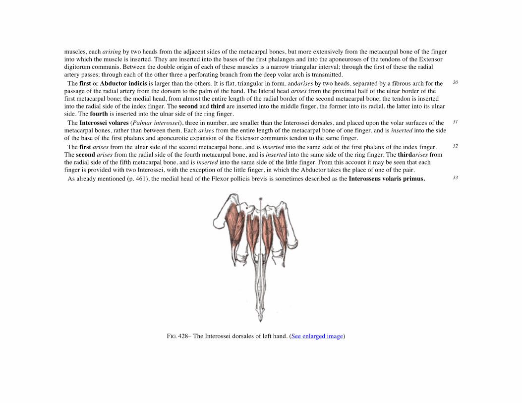
muscles, each arising by two heads from the adjacent sides of the metacarpal bones, but more extensively from the metacarpal bone of the finger into which the muscle is inserted. They are inserted into the bases of the first phalanges and into the aponeuroses of the tendons of the Extensor digitorum communis. Between the double origin of each of these muscles is a narrow triangular interval; through the first of these the radial artery passes; through each of the other three a perforating branch from the deep volar arch is transmitted. The first or Abductor indicis is larger than the others. It is flat, triangular in form, andarises by two heads, separated by a fibrous arch for the passage of the radial artery from the dorsum to the palm of the hand. The lateral head arises from the proximal half of the ulnar border of the first metacarpal bone; the medial head, from almost the entire length of the radial border of the second metacarpal bone; the tendon is inserted into the radial side of the index finger. The second and third are inserted into the middle finger, the former into its radial, the latter into its ulnar side. The fourth is inserted into the ulnar side of the ring finger.
30
The Interossei volares (Palmar interossei), three in number, are smaller than the Interossei dorsales, and placed upon the volar surfaces of the metacarpal bones, rather than between them. Each arises from the entire length of the metacarpal bone of one finger, and is inserted into the side of the base of the first phalanx and aponeurotic expansion of the Extensor communis tendon to the same finger.
31
The first arises from the ulnar side of the second metacarpal bone, and is inserted into the same side of the first phalanx of the index finger. The second arises from the radial side of the fourth metacarpal bone, and is inserted into the same side of the ring finger. The thirdarises from the radial side of the fifth metacarpal bone, and is inserted into the same side of the little finger. From this account it may be seen that each finger is provided with two Interossei, with the exception of the little finger, in which the Abductor takes the place of one of the pair.
32
As already mentioned (p. 461), the medial head of the Flexor pollicis brevis is sometimes described as the Interosseus volaris primus. 33
FIG. 428– The Interossei dorsales of left hand. (See enlarged image)
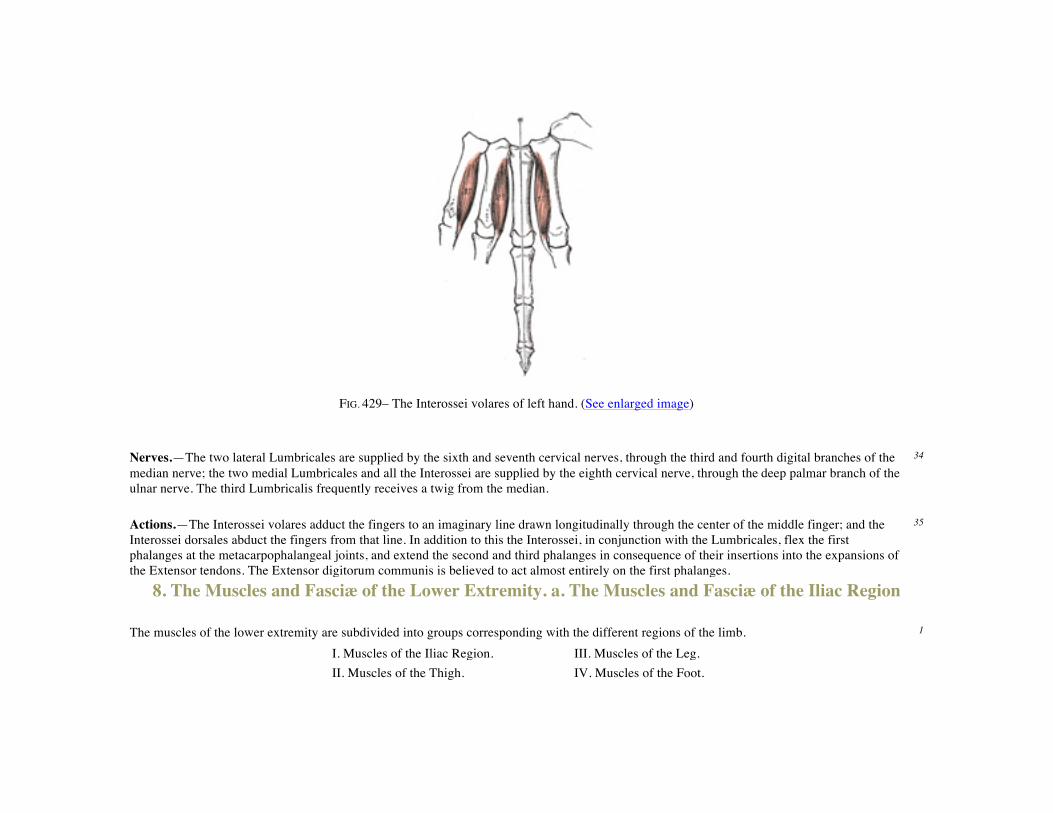
FIG. 429– The Interossei volares of left hand. (See enlarged image)
Nerves.—The two lateral Lumbricales are supplied by the sixth and seventh cervical nerves, through the third and fourth digital branches of the median nerve; the two medial Lumbricales and all the Interossei are supplied by the eighth cervical nerve, through the deep palmar branch of the ulnar nerve. The third Lumbricalis frequently receives a twig from the median.
34
Actions.—The Interossei volares adduct the fingers to an imaginary line drawn longitudinally through the center of the middle finger; and the Interossei dorsales abduct the fingers from that line. In addition to this the Interossei, in conjunction with the Lumbricales, flex the first phalanges at the metacarpophalangeal joints, and extend the second and third phalanges in consequence of their insertions into the expansions of the Extensor tendons. The Extensor digitorum communis is believed to act almost entirely on the first phalanges.
35
8. The Muscles and Fasciæ of the Lower Extremity. a. The Muscles and Fasciæ of the Iliac Region The muscles of the lower extremity are subdivided into groups corresponding with the different regions of the limb. 1
I. Muscles of the Iliac Region. III. Muscles of the Leg. II. Muscles of the Thigh. IV. Muscles of the Foot.

The Muscles and Fasciæ of the Iliac Region (Fig. 430). Psoas major. Psoas minor. Iliacus.
The Fascia Covering the Psoas and Iliacus is thin above, and becomes gradually thicker below as it approaches the inguinal ligament. 2

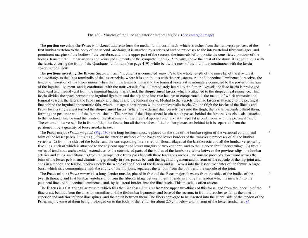
FIG. 430– Muscles of the iliac and anterior femoral regions. (See enlarged image)
The portion covering the Psoas is thickened above to form the medial lumbocostal arch, which stretches from the transverse process of the first lumbar vertebra to the body of the second. Medially, it is attached by a series of arched processes to the intervertebral fibrocartilages, and prominent margins of the bodies of the vertebræ, and to the upper part of the sacrum; the intervals left, opposite the constricted portions of the bodies, transmit the lumbar arteries and veins and filaments of the sympathetic trunk. Laterally, above the crest of the ilium, it is continuous with the fascia covering the front of the Quadratus lumborum (see page 419), while below the crest of the ilium it is continuous with the fascia covering the Iliacus.
3
The portions investing the Iliacus (fascia iliaca; iliac fascia) is connected, laterally to the whole length of the inner lip of the iliac crest; and medially, to the linea terminalis of the lesser pelvis, where it is continuous with the periosteum. At the iliopectineal eminence it receives the tendon of insertion of the Psoas minor, when that muscle exists. Lateral to the femoral vessels it is intimately connected to the posterior margin of the inguinal ligament, and is continuous with the transversalis fascia. Immediately lateral to the femoral vessels the iliac fascia is prolonged backward and medialward from the inguinal ligament as a band, the iliopectineal fascia, which is attached to the iliopectineal eminence. This fascia divides the space between the inguinal ligament and the hip bone into two lacunæ or compartments, the medial of which transmits the femoral vessels, the lateral the Psoas major and Iliacus and the femoral nerve. Medial to the vessels the iliac fascia is attached to the pectineal line behind the inguinal aponeurotic falx, where it is again continuous with the transversalis fascia. On the thigh the fasciæ of the Iliacus and Psoas form a single sheet termed the iliopectineal fascia. Where the external iliac vessels pass into the thigh, the fascia descends behind them, forming the posterior wall of the femoral sheath. The portion of the iliopectineal fascia which passes behind the femoral vessels is also attached to the pectineal line beyond the limits of the attachment of the inguinal aponeurotic falx; at this part it is continuous with the pectineal fascia. The external iliac vessels lie in front of the iliac fascia, but all the branches of the lumbar plexus are behind it; it is separated from the peritoneum by a quantity of loose areolar tissue.
4
The Psoas major (Psoas magnus) (Fig. 430) is a long fusiform muscle placed on the side of the lumbar region of the vertebral column and brim of the lesser pelvis. It arises (1) from the anterior surfaces of the bases and lower borders of the transverse processes of all the lumbar vertebræ (2) from the sides of the bodies and the corresponding intervertebral fibrocartilages of the last thoracic and all the lumbar vertebræ by five slips, each of which is attached to the adjacent upper and lower margins of two vertebræ, and to the intervertebral fibrocartilage; (3) from a series of tendinous arches which extend across the constricted parts of the bodies of the lumbar vertebræ between the previous slips; the lumbar arteries and veins, and filaments from the sympathetic trunk pass beneath these tendinous arches. The muscle proceeds downward across the brim of the lesser pelvis, and diminishing gradually in size, passes beneath the inguinal ligament and in front of the capsule of the hip-joint and ends in a tendon; the tendon receives nearly the whole of the fibers of the Iliacus and is inserted into the lesser trochanter of the femur. A large bursa which may communicate with the cavity of the hip-joint, separates the tendon from the pubis and the capsule of the joint.
5
The Psoas minor (Psoas parvus) is a long slender muscle, placed in front of the Psoas major. It arises from the sides of the bodies of the twelfth thoracic and first lumbar vertebræ and from the fibrocartilage between them. It ends in a long flat tendon which is insertedinto the pectineal line and iliopectineal eminence, and, by its lateral border, into the iliac fascia. This muscle is often absent.
6
The Iliacus is a flat, triangular muscle, which fills the iliac fossa. It arises from the upper two-thirds of this fossa, and from the inner lip of the iliac crest; behind, from the anterior sacroiliac and the iliolumbar ligaments, and base of the sacrum; in front, it reaches as far as the anterior superior and anterior inferior iliac spines, and the notch between them. The fibers converge to be inserted into the lateral side of the tendon of the Psoas major, some of them being prolonged on to the body of the femur for about 2.5 cm. below and in front of the lesser trochanter. 85
7

Variations.—The Iliacus minor or Iliocapsularis, a small detached part of the Iliacus is frequently present. It arises from the anterior inferior spine of the ilium and is inserted into the lower part of the intertrochanteric line of the femur or into the iliofemoral ligament.
8
Nerves.—The Psoas major is supplied by branches of the second and third lumbar nerve; the Psoas minor by a branch of the first lumbar nerve; and the Iliacus by branches of the second and third lumbar nerves through the femoral nerve.
9
Actions.—The Psoas major, acting from above, flexes the thigh upon the pelvis, being assisted by the Iliacus; acting from below, with the femur fixed, it bends the lumbar portion of the vertebral column forward and to its own side, and then, in conjunction with the Iliacus, tilts the pelvis forward. When the muscles of both sides are acting from below, they serve to maintain the erect posture by supporting the vertebral column and pelvis upon the femora, or in continued action bend the trunk and pelvis forward, as in raising the trunk from the recumbent posture.
10
The Psoas minor is a tensor of the iliac fascia. 11
Note 85. The Psoas major and iliacus are sometimes regarded as a single muscle named theIliopsoas. [back]
8b. The Muscles and Fasciæ of the Thigh 1. The Anterior Femoral Muscles (Fig. 430).
Sartorius. Quadriceps femoris.
Rectus femoris.
Vastus lateralis.
Vastus medialis.
Vastus intermedius.
Articularis genu.
Superficial Fascia.—The superficial fascia forms a continuous layer over the whole of the thigh; it consists of areolar tissue containing in its meshes much fat, and may be separated into two or more layers, between which are found the superficial vessels and nerves. It varies in thickness in different parts of the limb; in the groin it is thick, and the two layers are separated from one another by the superficial inguinal lymph glands, the great saphenous vein, and several smaller vessels. The superficial layer is continuous above with the superficial fascia of the abdomen. The deep layer of the superficial fascia is a very thin, fibrous stratum, best marked on the medial side of the great saphenous vein and below the
1
inguinal ligament. It is placed beneath the subcutaneous vessels and nerves and upon the surface of the fascia lata. It is intimately adherent to the fascia lata a little below the inguinal ligament. It covers the fossa ovalis (saphenous opening), being closely united to its circumference, and is connected to the sheath of the femoral vessels. The portion of fascia covering this fossa is perforated by the great saphenous vein and by numerous blood and lymphatic vessels, hence it has been termed the fascia cribrosa, the openings for these vessels having been likened to the holes in a sieve. A large subcutaneous bursa is found in the superficial fascia over the patella. Deep Fascia.—The deep fascia of the thigh is named, from its great extent, the fascia lata;it constitutes an investment for the whole of this region of the limb, but varies in thickness in different parts. Thus, it is thicker in the upper and lateral part of the thigh, where it receives a fibrous expansion from the Glutæus maximus, and where the Tensor fasciæ latæ is inserted between its layers; it is very thin behind and at the upper and medial part, where it covers the Adductor muscles, and again becomes stronger around the knee, receiving fibrous expansions from the tendon of the Biceps femoris laterally, from the Sartorius medially, and from the Quadriceps femoris in front. The fascia lata is attached, above and behind, to the back of the sacrum and coccyx; laterally, to the iliac crest; in front, to the inguinal ligament, and to the superior ramus of the pubis; and medially, to the inferior ramus of the pubis, to the inferior ramus and tuberosity of the ischium, and to the lower border of the sacrotuberous ligament. From its attachment to the iliac crest it passes down over the Glutæus medius to the upper border of the Glutæus maximus, where it splits into two layers, one passing superficial to and the other beneath this muscle; at the lower border of the muscle the two layers reunite. Laterally, the fascia lata receives the greater part of the tendon of insertion of the Glutæus maximus, and becomes proportionately thickened. The portion of the fascia lata attached to the front part of the iliac crest, and corresponding to the origin of the Tensor fasciæ latæ, extends down the lateral side of the thigh as two layers, one superficial to and the other beneath this muscle; at the lower end of the muscle these two layers unite and form a strong band, having first received the insertion of the muscle. This band is continued downward, under the name of the iliotibial band (tractus iliotibialis) and is attached to the lateral condyle of the tibia. The part of the iliotibial band which lies beneath the Tensor fasciæ latæ is prolonged upward to join the lateral part of the capsule of the hip-joint. Below, the fasciæ lata is attached to all the prominent points around the knee-joint, viz., the condyles of the femur and tibia, and the head of the fibula. On either side of the patella it is strengthened by transverse fibers from the lower parts of the Vasti, which are attached to and support this bone. Of these the lateral are the stronger, and are continuous with the iliotibial band. The deep surface of the fascia lata gives off two strong intermuscular septa, which are attached to the whole length of the linea aspera and its prolongations above and below; the lateral and stronger one, which extends from the insertion of the Glutæus maximus to the lateral condyle, separates the Vastus lateralis in front from the short head of the Biceps femoris behind, and gives partial origin to these muscles; the medial and thinner one separates the Vastus medialis from the Adductores and Pectineus. Besides these there are numerous smaller septa, separating the individual muscles, and enclosing each in a distinct sheath.
2
The Fossa Ovalis (saphenous opening) (Fig. 431).—At the upper and medial part of the thigh, a little below the medial end of the inguinal ligament, is a large oval-shaped aperture in the fascia lata; it transmits the great saphenous vein, and other, smaller vessels, and is termed the fossa ovalis. The fascia cribrosa, which is pierced by the structures passing through the opening, closes the aperture and must be removed to expose it. The fascia lata in this part of the thigh is described as consisting of a superficial and a deep portion.
3
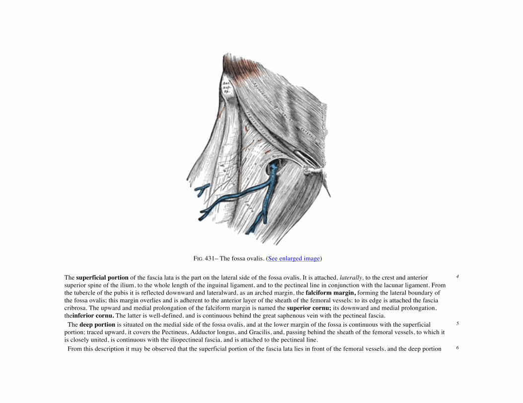
FIG. 431– The fossa ovalis. (See enlarged image) The superficial portion of the fascia lata is the part on the lateral side of the fossa ovalis. It is attached, laterally, to the crest and anterior superior spine of the ilium, to the whole length of the inguinal ligament, and to the pectineal line in conjunction with the lacunar ligament. From the tubercle of the pubis it is reflected downward and lateralward, as an arched margin, the falciform margin, forming the lateral boundary of the fossa ovalis; this margin overlies and is adherent to the anterior layer of the sheath of the femoral vessels: to its edge is attached the fascia cribrosa. The upward and medial prolongation of the falciform margin is named the superior cornu; its downward and medial prolongation, theinferior cornu. The latter is well-defined, and is continuous behind the great saphenous vein with the pectineal fascia.
4
The deep portion is situated on the medial side of the fossa ovalis, and at the lower margin of the fossa is continuous with the superficial portion; traced upward, it covers the Pectineus, Adductor longus, and Gracilis, and, passing behind the sheath of the femoral vessels, to which it is closely united, is continuous with the iliopectineal fascia, and is attached to the pectineal line.
5
From this description it may be observed that the superficial portion of the fascia lata lies in front of the femoral vessels, and the deep portion 6
behind them, so that an apparent aperture exists between the two, through which the great saphenous passes to join the femoral vein. The Sartorius, the longest muscle in the body, is narrow and ribbon-like; it arises by tendinous fibers from the anterior superior iliac spine and the upper half of the notch below it. It passes obliquely across the upper and anterior part of the thigh, from the lateral to the medial side of the limb, then descends vertically, as far as the medial side of the knee, passing behind the medial condyle of the femur to end in a tendon. This curves obliquely forward and expands into a broad aponeurosis, which is inserted, in front of the Gracilis and Semitendinous, into the upper part of the medial surface of the body of the tibia, nearly as far forward as the anterior crest. The upper part of the aponeurosis is curved backward over the upper edge of the tendon of the Gracilis so as to be inserted behind it. An offset, from its upper margin, blends with the capsule of the knee-joint, and another from its lower border, with the fascia on the medial side of the leg.
7
Variations.—Slips of origin from the outer end of the inguinal ligament, the notch of the ilium, the ilio-pectineal line or the pubis occur. The muscle may be split into two parts, and one part may be inserted into the fascia lata, the femur, the ligament of the patella or the tendon of the Semitendinosus. The tendon of insertion may end in the fascia lata, the capsule of the knee-joint, or the fascia of the leg. The muscle may be absent.
8
The Quadriceps femoris (Quadriceps extensor) includes the four remaining muscles on the front of the thigh. It is the great extensor muscle of the leg, forming a large fleshy mass which covers the front and sides of the femur. It is subdivided into separate portions, which have received distinctive names. One occupying the middle of the thigh, and connected above with the ilium, is called from its straight course the Rectus femoris. The other three lie in immediate connection with the body of the femur, which they cover from the trochanters to the condyles. The portion on the lateral side of the femur is termed theVastus lateralis; that covering the medial side, the Vastus medialis; and that in front, theVastus intermedius.
9
The Rectus femoris is situated in the middle of the front of the thigh; it is fusiform in shape, and its superficial fibers are arranged in a bipenniform manner, the deep fibers running straight down to the deep aponeurosis. It arises by two tendons: one, the anterior or straight, from the anterior inferior iliac spine; the other, the posterior or reflected, from a groove above the brim of the acetabulum. The two unite at an acute angle, and spread into an aponeurosis which is prolonged downward on the anterior surface of the muscle, and from this the muscular fibers arise. The muscle ends in a broad and thick aponeurosis which occupies the lower two-thirds of its posterior surface, and, gradually becoming narrowed into a flattened tendon, is inserted into the base of the patella.
10
The Vastus lateralis (Vastus externus) is the largest part of the Quadriceps femoris. Itarises by a broad aponeurosis, which is attached to the upper part of the intertrochanteric line, to the anterior and inferior borders of the greater trochanter, to the lateral lip of the gluteal tuberosity, and to the upper half of the lateral lip of the linea aspera; this aponeurosis covers the upper three-fourths of the muscle, and from its deep surface many fibers take origin. A few additional fibers arise from the tendon of the Glutæus maximus, and from the lateral intermuscular septum between the Vastus lateralis and short head of the Biceps femoris. The fibers form a large fleshy mass, which is attached to a strong aponeurosis, placed on the deep surface of the lower part of the muscle: this aponeurosis becomescontracted and thickened into a flat tendon inserted into the lateral border of the patella, blending with the Quadriceps femoris tendon, and giving an expansion to the capsule of the knee-joint.
11
The Vastus medialis and Vastus intermedius appear to be inseparably united, but when the Rectus femoris has been reflected a narrow interval will be observed extending upward from the medial border of the patella between the two muscles, and the separation may be continued as far as the lower part of the intertrochanteric line, where, however, the two muscles are frequently continuous.
12
The Vastus medialis (Vastus internus) arises from the lower half of the intertrochanteric line, the medial lip of the linea aspera, the upper part of the medial supracondylar line, the tendons of the Adductor longus and the Adductor magnus and the medial intermuscular septum. Its fibers are directed downward and forward, and are chiefly attached to an aponeurosis which lies on the deep surface of the muscle and is inserted into
13
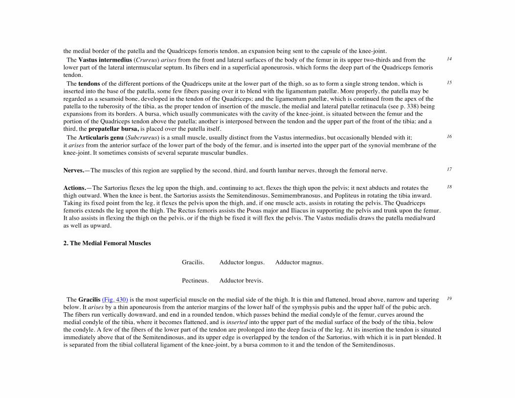
the medial border of the patella and the Quadriceps femoris tendon, an expansion being sent to the capsule of the knee-joint. The Vastus intermedius (Crureus) arises from the front and lateral surfaces of the body of the femur in its upper two-thirds and from the lower part of the lateral intermuscular septum. Its fibers end in a superficial aponeurosis, which forms the deep part of the Quadriceps femoris tendon.
14
The tendons of the different portions of the Quadriceps unite at the lower part of the thigh, so as to form a single strong tendon, which is inserted into the base of the patella, some few fibers passing over it to blend with the ligamentum patellæ. More properly, the patella may be regarded as a sesamoid bone, developed in the tendon of the Quadriceps; and the ligamentum patellæ, which is continued from the apex of the patella to the tuberosity of the tibia, as the proper tendon of insertion of the muscle, the medial and lateral patellar retinacula (see p. 338) being expansions from its borders. A bursa, which usually communicates with the cavity of the knee-joint, is situated between the femur and the portion of the Quadriceps tendon above the patella; another is interposed between the tendon and the upper part of the front of the tibia; and a third, the prepatellar bursa, is placed over the patella itself.
15
The Articularis genu (Subcrureus) is a small muscle, usually distinct from the Vastus intermedius, but occasionally blended with it; it arises from the anterior surface of the lower part of the body of the femur, and is inserted into the upper part of the synovial membrane of the knee-joint. It sometimes consists of several separate muscular bundles.
16
Nerves.—The muscles of this region are supplied by the second, third, and fourth lumbar nerves, through the femoral nerve. 17 Actions.—The Sartorius flexes the leg upon the thigh, and, continuing to act, flexes the thigh upon the pelvis; it next abducts and rotates the thigh outward. When the knee is bent, the Sartorius assists the Semitendinosus, Semimembranosus, and Popliteus in rotating the tibia inward. Taking its fixed point from the leg, it flexes the pelvis upon the thigh, and, if one muscle acts, assists in rotating the pelvis. The Quadriceps femoris extends the leg upon the thigh. The Rectus femoris assists the Psoas major and Iliacus in supporting the pelvis and trunk upon the femur. It also assists in flexing the thigh on the pelvis, or if the thigh be fixed it will flex the pelvis. The Vastus medialis draws the patella medialward as well as upward.
18
2. The Medial Femoral Muscles
Gracilis. Adductor longus. Adductor magnus.
Pectineus. Adductor brevis.
The Gracilis (Fig. 430) is the most superficial muscle on the medial side of the thigh. It is thin and flattened, broad above, narrow and tapering below. It arises by a thin aponeurosis from the anterior margins of the lower half of the symphysis pubis and the upper half of the pubic arch. The fibers run vertically downward, and end in a rounded tendon, which passes behind the medial condyle of the femur, curves around the medial condyle of the tibia, where it becomes flattened, and is inserted into the upper part of the medial surface of the body of the tibia, below the condyle. A few of the fibers of the lower part of the tendon are prolonged into the deep fascia of the leg. At its insertion the tendon is situated immediately above that of the Semitendinosus, and its upper edge is overlapped by the tendon of the Sartorius, with which it is in part blended. It is separated from the tibial collateral ligament of the knee-joint, by a bursa common to it and the tendon of the Semitendinosus.
19
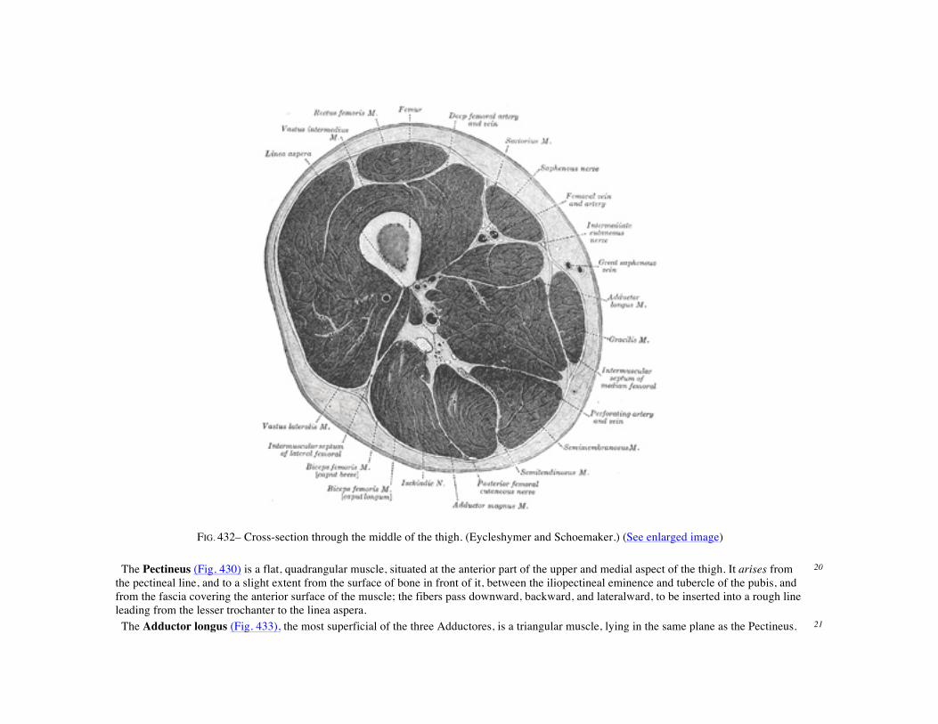
FIG. 432– Cross-section through the middle of the thigh. (Eycleshymer and Schoemaker.) (See enlarged image)
The Pectineus (Fig. 430) is a flat, quadrangular muscle, situated at the anterior part of the upper and medial aspect of the thigh. It arises from the pectineal line, and to a slight extent from the surface of bone in front of it, between the iliopectineal eminence and tubercle of the pubis, and from the fascia covering the anterior surface of the muscle; the fibers pass downward, backward, and lateralward, to be inserted into a rough line leading from the lesser trochanter to the linea aspera.
20
The Adductor longus (Fig. 433), the most superficial of the three Adductores, is a triangular muscle, lying in the same plane as the Pectineus. 21

It arises by a flat, narrow tendon, from the front of the pubis, at the angle of junction of the crest with the symphysis; and soon expands into a broad fleshy belly. This passes downward, backward, and lateralward, and is inserted, by an aponeurosis, into the linea aspera, between the Vastus medialis and the Adductor magnus, with both of which it is usually blended. The Adductor brevis (Fig. 433) is situated immediately behind the two preceding muscles. It is somewhat triangular in form, and arises by a narrow origin from the outer surfaces of the superior and inferior rami of the pubis, between the Gracilis and Obturator externus. Its fibers, passing backward, lateralward, and downward, are inserted, by an aponeurosis, into the line leading from the lesser trochanter to the linea aspera and into the upper part of the linea aspera, immediately behind the Pectineus and upper part of the Adductor longus.
22
The Adductor magnus (Fig. 433) is a large triangular muscle, situated on the medial side of the thigh. It arises from a small part of the inferior ramus of the pubis, from the inferior ramus of the ischium, and from the outer margin of the inferior part of the tuberosity of the ischium. Those fibers which arise from the ramus of the pubis are short, horizontal in direction, and are inserted into the rough line leading from the greater trochanter to the linea aspera, medial to the Glutæus maximus; those from the ramus of the ischium are directed downward and lateralward with different degrees of obliquity, to be inserted, by means of a broad aponeurosis, into the linea aspera and the upper part of its medial prolongation below. The medial portion of the muscle, composed principally of the fibers arising from the tuberosity of the ischium, forms a thick fleshy mass consisting of coarse bundles which descend almost vertically, and end about the lower third of the thigh in a rounded tendon which is inserted into the adductor tubercle on the medial condyle of the femur, and is connected by a fibrous expansion to the line leading upward from the tubercle to the linea aspera. At the insertion of the muscle, there is a series of osseoaponeurotic openings, formed by tendinous arches attached to the bone. The upper four openings are small, and give passage to the perforating branches of the profunda femoris artery. The lowest is of large size, and transmits the femoral vessels to the popliteal fossa.
23
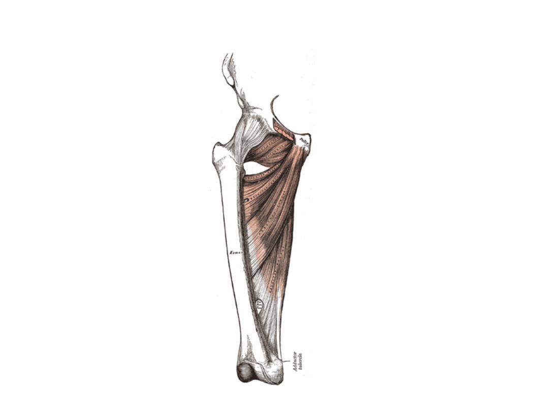

FIG. 433– Deep muscles of the medial femoral region. (See enlarged image)
Variations.—The Pectineus is sometimes divided into an outer part supplied by the femoral nerve and an inner part supplied by the obturator nerve. The muscle may be attached to or inserted into the capsule of the hip-joint. The Adductor longus may be double, may extend to the knee, or be more or less united with the Pectineus. The Adductor brevis may be divided into two or three parts, or it may be united to the Adductor magnus. The Adductor magnus may be more or less segmented, the anterior and superior portion is often described as a separate muscle, the Adductor minimus. The muscle may be fused with the Quadratus femoris.
24
Nerves.—The three Adductores and the Gracilis are supplied by the third and fourth lumbar nerves through the obturator nerve; the Adductor magnus receiving an additional branch from the sacral plexus through the sciatic. The Pectineus is supplied by the second, third, and fourth lumbar nerves through the femoral nerve, and by the third lumbar through the accessory obturator when this latter exists. Occasionally it receives a branch from the obturator nerve. 86
25
Actions.—The Pectineus and three Adductores adduct the thigh powerfully; they are especially used in horse exercise, the sides of the saddle being grasped between the knees by the contraction of these muscles. In consequence of the obliquity of their insertions into the linea aspera, they rotate the thigh outward, assisting the external Rotators, and when the limb has been abducted, they draw it medialward, carrying the thigh across that of the opposite side. The Pectineus and Adductores brevis and longus assist the Psoas major and Iliacus in flexing the thigh upon the pelvis. In progression, all these muscles assist in drawing forward the lower limb. The Gracilis assists the Sartorius in flexing the leg and rotating it inward; it is also an adductor of the thigh. If the lower extremities be fixed, these muscles, taking their fixed points below, may act upon the pelvis, serving to maintain the body in an erect posture; or, if their action be continued, flex the pelvis forward upon the femur.
26
3. The Muscles of the Gluteal Region (Fig. 434).
Glutæus maximus. Obturator internus. Glutæus medius. Gemellus superior. Glutæus minimus. Gemellus inferior. Tensor fasciæ latæ. Quadratus femoris. Piriformis. Obturator externus.
The Glutæus maximus, the most superficial muscle in the gluteal region, is a broad and thick fleshy mass of a quadrilateral shape, and forms the prominence of the nates. Its large size is one of the most characteristic features of the muscular system in man, connected as it is with the power he has of maintaining the trunk in the erect posture. The muscle is remarkably coarse in structure, being made up of fasciculi lying parallel with one another and collected together into large bundles separated by fibrous septa. It arises from the posterior gluteal line of the ilium, and the rough portion of bone including the crest, immediately above and behind it; from the posterior surface of the lower part of the sacrum and the side of the coccyx; from the aponeurosis of the Sacrospinalis, the sacrotuberous ligament, and the fascia (gluteal aponeurosis) covering the Glutæus medius. The fibers are directed obliquely downward and lateralward; those forming the upper and larger portion of the muscle, together with the superficial fibers of the lower portion, end in a thick tendinous lamina, which passes across the greater trochanter, and
27
is inserted into the iliotibial band of the fascia lata; the deeper fibers of the lower portion of the muscle are inserted into the gluteal tuberosity between the Vastus lateralis and Adductor magnus. Bursæ—Three bursæ are usually found in relation with the deep surface of this muscle. One of these, of large size, and generally multilocular, separates it from the greater trochanter; a second, often wanting, is situated on the tuberosity of the ischium; a third is found between the tendon of the muscle and that of the Vastus lateralis.
28
The Glutæus medius is a broad, thick, radiating muscle, situated on the outer surface of the pelvis. Its posterior third is covered by the Glutæus maximus, its anterior two-thirds by the gluteal aponeurosis, which separates it from the superficial fascia and integument. Itarises from the outer surface of the ilium between the iliac crest and posterior gluteal line above, and the anterior gluteal line below; it also arises from the gluteal aponeurosis covering its outer surface. The fibers converge to a strong flattened tendon, which isinserted into the oblique ridge which runs downward and forward on the lateral surface of the greater trochanter. A bursa separates the tendon of the muscle from the surface of the trochanter over which it glides.
29
Variations.—The posterior border may be more or less closely united to the Piriformis, or some of the fibers end on its tendon. 30 The Glutæus minimus, the smallest of the three Glutæi, is placed immediately beneath the preceding. It is fan-shaped, arising from the outer surface of the ilium, between the anterior and inferior gluteal lines, and behind, from the margin of the greater sciatic notch. The fibers converge to the deep surface of a radiated aponeurosis, and this ends in a tendon which is inserted into an impression on the anterior border of the greater trochanter, and gives an expansion to the capsule of the hip-joint. A bursa is interposed between the tendon and the greater trochanter. Between the Glutæus medius and Glutæus minimus are the deep branches of the superior gluteal vessels and the superior gluteal nerve. The deep surface of the Glutæus minimus is in relation with the reflected tendon of the Rectus femoris and the capsule of the hip-joint.
31
Variations.—The muscle may be divided into an anterior and a posterior part, or it may send slips to the Piriformis, the Gemellus superior or the outer part of the origin of the Vastus lateralis.
32


FIG. 434– Muscles of the gluteal and posterior femoral regions. (See enlarged image)
The Tensor fasciæ latæ (Tensor fasciæ femoris) arises from the anterior part of the outer lip of the iliac crest; from the outer surface of the anterior superior iliac spine, and part of the outer border of the notch below it, between the Glutæus medius and Sartorius; and from the deep surface of the fascia lata. It is inserted between the two layers of the iliotibial band of the fascia lata about the junction of the middle and upper thirds of the thigh.
33
The Piriformis is a flat muscle, pyramidal in shape, lying almost parallel with the posterior margin of the Glutæus medius. It is situated partly within the pelvis against its posterior wall, and partly at the back of the hip-joint. It arises from the front of the sacrum by three fleshy digitations, attached to the portions of bone between the first, second, third, and fourth anterior sacral foramina, and to the grooves leading from the foramina: a few fibers also arise from the margin of the greater sciatic foramen, and from the anterior surface of the sacrotuberous ligament. The muscle passes out of the pelvis through the greater sciatic foramen, the upper part of which it fills, and is inserted by a rounded tendon into the upper border of the greater trochanter behind, but often partly blended with, the common tendon of the Obturator internus and Gemelli.
34
Variations.—It is frequently pierced by the common peroneal nerve and thus divided more or less into two parts. It may be united with the Glutæus medius, or send fibers to the Glutæus minimus or receive fibers from the Gemellus superior. It may have only one or two sacral attachments or be inserted in to the capsule of the hip-joint. It may be absent.
35

FIG. 435– The obturator membrane. (See enlarged image)
Obturator Membrane (Fig. 435).—The obturator membrane is a thin fibrous sheet, which almost completely closes the obturator foramen. Its fibers are arranged in interlacing bundles mainly transverse in direction; the uppermost bundle is attached to the obturator tubercles and completes the obturator canal for the passage of the obturator vessels and nerve. The membrane is attached to the sharp margin of the obturator foramen except at its lower lateral angle, where it is fixed to the pelvic surface of the inferior ramus of the ischium, i. e., within the margin. Both obturator muscles are connected with this membrane.
36
The Obturator internus is situated partly within the lesser pelvis, and partly at the back of the hip-joint. It arises from the inner surface of the antero-lateral wall of the pelvis, where it surrounds the greater part of the obturator foramen, being attached to the inferior rami of the pubis and ischium, and at the side to the inner surface of the hip bone below and behind the pelvic brim, reaching from the upper part of the greater sciatic foramen above and behind to the obturator foramen below and in front. It also arises from the pelvic surface of the obturator membrane except in the posterior part, from the tendinous arch which completes the canal for the passage of the obturator vessels and nerve, and to a slight extent from the obturator fascia, which covers the muscle. The fibers converge rapidly toward the lesser sciatic foramen, and end in four or five tendinous bands, which are found on the deep surface of the muscle; these bands are reflected at a right angle over the grooved surface of the ischium between its spine and tuberosity. This bony surface is covered by smooth cartilage, which is separated from the tendon by a bursa, and presents one or more ridges corresponding with the furrows between the tendinous bands. These bands leave the pelvis through the lesser sciatic foramen and unite into a single flattened tendon, which passes horizontally across the capsule of the hip-joint, and, after receiving the attachments of the Gemelli, is inserted into the forepart of the medial surface of the greater trochanter above the trochanteric fossa. A bursa, narrow and elongated in form, is usually found between the tendon and the capsule of the hip-joint; it occasionally communicates with the bursa between the tendon and the ischium.
37
The Gemelli are two small muscular fasciculi, accessories to the tendon of the Obturator internus which is received into a groove between them.
38
The Gemellus superior, the smaller of the two, arises from the outer surface of the spine of the ischium, blends with the upper part of the tendon of the Obturator internus, and isinserted with it into the medial surface of the greater trochanter. It is sometimes wanting.
39
The Gemellus inferior arises from the upper part of the tuberosity of the ischium, immediately below the groove for the Obturator internus tendon. It blends with the lower part of the tendon of the Obturator internus, and is inserted with it it into the medial surface of the greater trochanter. Rarely absent.
40
The Quadratus femoris is a flat, quadrilateral muscle, between the Gemellus inferior and the upper margin of the Adductor magnus; it is separated from the latter by the terminal branches of the medial femoral circumflex vessels. It arises from the upper part of the external border of the tuberosity of the ischium, and is inserted into the upper part of the linea quadrata—that is, the line which extends vertically downward from the intertrochanteric crest. A bursa is often found between the front of this muscle and the lesser trochanter. Sometimes absent.
41
The Obturator externus (Fig. 436) is a flat, triangular muscle, which covers the outer surface of the anterior wall of the pelvis. It arises from the margin of bone immediately around the medial side of the obturator foramen, viz., from the rami of the pubis, and the inferior ramus of the ischium; it also arises from the medial two-thirds of the outer surface of the obturator membrane, and from the tendinous arch which completes the canal for the passage of the obturator vessels and nerves. The fibers springing from the pubic arch extend on to the inner surface of the bone, where they obtain a narrow origin between the margin of the foramen and the attachment of the obturator membrane. The fibers converge and
42

pass backward, lateralward, and upward, and end in a tendon which runs across the back of the neck of the femur and lower part of the capsule of the hipjoint and is insertedinto the trochanteric fossa of the femur. The obturator vessels lie between the muscle and the obturator membrane; the anterior branch of the obturator nerve reaches the thigh by passing in front of the muscle, and the posterior branch by piercing it. Nerves.—The Glutæus maximus is supplied by the fifth lumbar and first and second sacra nerves through the inferior gluteal nerve; the Glutæi medius and minimus and the Tensor fasciæ latæ by the fourth and fifth lumbar and first sacral nerves through the superior gluteal; the Piriformis is supplied by the first and second sacral nerves; the Gemellus inferior and Quadratus femoris by the last lumbar and first sacral nerves; the Gemellus superior and Obturator internus by the first, second, and third sacral nerves, and the Obturator externus by the third and fourth lumbar nerves through the obturator.
43
FIG. 436– The Obturator externus. (See enlarged image)
Actions.—When the Glutæus maximus takes its fixed point from the pelvis, it extends the femur and brings the bent thigh into a line with the body. Taking its fixed point from below, it acts upon the pelvis, supporting it and the trunk upon the head of the femur; this is especially obvious in standing on one leg. Its most powerful action is to cause the body to regain the erect position after stooping, by drawing the pelvis backward, being assisted in this action by the Biceps femoris, Semitendinosus, and Semimembranosus. The Glutæus maximus is a tensor of the fascia lata, and by its connection with the iliotibial band steadies the femur on the articular surfaces of the tibia during standing, when the Extensor muscles are relaxed. The lower part of the muscle also acts as an adductor and external rotator of the limb. The Glutæi medius and minimus abduct the thigh, when the limb is extended, and are principally called into action in supporting the body on one limb, in conjunction with the Tensor fasciæ
44

latæ. Their anterior fibers, by drawing the greater trochanter forward, rotate the thigh inward, in which action they are also assisted by the Tensor fasciæ latæ. The Tensor fasciæ latæ is a tensor of the fascia lata; continuing its action, the oblique direction of its fibers enables it to abduct the thigh and to rotate it inward. In the erect posture, acting from below, it will serve to steady the pelvis upon the head of the femur; and by means of the iliotibial band it steadies the condyles of the femur on the articular surfaces of the tibia, and assists the Glutæus maximus in supporting the knee in the extended position. The remaining muscles are powerful external rotators of the thigh. In the sitting posture, when the thigh is flexed upon the pelvis, their action as rotators ceases, and they become abductors, with the exception of the Obturator externus, which still rotates the femur outward. 4. The Posterior Femoral Muscles (Hamstring Muscles) (Fig. 434). 45
Biceps femoris. Semitendinosus. Semimembranosus.
The Biceps femoris (Biceps) is situated on the posterior and lateral aspect of the thigh. It has two heads of origin; one, the long head, arises from the lower and inner impression on the back part of the tuberosity of the ischium, by a tendon common to it and the Semitendinosus, and from the lower part of the sacrotuberous ligament; the other, the short head, arises from the lateral lip of the linea aspera, between the Adductor magnus and Vastus lateralis, extending up almost as high as the insertion of the Glutæus maximus; from the lateral prolongation of the linea aspera to within 5 cm. of the lateral condyle; and from the lateral intermuscular septum. The fibers of the long head form a fusiform belly, which passes obliquely downward and lateralward across the sciatic nerve to end in an aponeurosis which covers the posterior surface of the muscle, and receives the fibers of the short head; this aponeurosis becomes gradually contracted into a tendon, which is insertedinto the lateral side of the head of the fibula, and by a small slip into the lateral condyle of the tibia. At its insertion the tendon divides into two portions, which embrace the fibular collateral ligament of the knee-joint. From the posterior border of the tendon a thin expansion is given off to the fascia of the leg. The tendon of insertion of this muscle forms the lateral hamstring; the common personeal nerve descends along its medial border.
46
Variations.—The short head may be absent; additional heads may arise from the ischial tuberosity, the linea aspera, the medial supracondylar ridge of the femur or from various other parts. A slip may pass to the Gastrocnemius.
47
The Semitendinosus, remarkable for the great length of its tendon of insertion, is situated at the posterior and medial aspect of the thigh. It arises from the lower and medial impression on the tuberosity of the ischium, by a tendon common to it and the long head of the Biceps femoris; it also arises from an aponeurosis which connects the adjacent surfaces of the two muscles to the extent of about 7.5 cm. from their origin. The muscle is fusiform and ends a little below the middle of the thigh in a long round tendon which lies along the medial side of the popliteal fossa; it then curves around the medial condyle of the tibia and passes over the tibial collateral ligament of the knee-joint, from which it is separated by a bursa, and is inserted into the upper part of the medial surface of the body of the tibia, nearly as far forward as its anterior crest. At its insertion it gives off from its lower border a prolongation to the deep fascia of the leg and lies behind the tendon of the Sartorius, and below that of the Gracilis, to which it is united. A tendinous intersection is usually observed about the middle of the muscle.
48
The Semimembranosus, so called from its membranous tendon of origin, is situated at the back and medial side of the thigh. It arises by a thick tendon from the upper and outer impression on the tuberosity of the ischium, above and lateral to the Biceps femoris and Semitendinosus. The tendon of origin expands into an aponeurosis, which covers the upper part of the anterior surface of the muscle; from this aponeurosis muscular fibers arise, and converge to another aponeurosis which covers the lower part of the posterior surface of the muscle and contracts into the tendon of insertion. It is inserted mainly into the horizontal groove on the posterior medial aspect of the medial condyle of the tibia. The
49

tendon of insertion gives off certain fibrous expansions: one, of considerable size, passes upward and lateralward to be inserted into the back part of the lateral condyle of the femur, forming part of the oblique popliteal ligament of the knee-joint; a second is continued downward to the fascia which covers the Popliteus muscle; while a few fibers join the tibial collateral ligament of the joint and the fascia of the leg. The muscle overlaps the upper part of the popliteal vessels. Variations.—It may be reduced or absent, or double, arising mainly from the sacrotuberous ligament and giving a slip to the femur or Adductor magnus.
50
The tendons of insertion of the two preceding muscles form the medial hamstrings. 51 Nerves.—The muscles of this region are supplied by the fourth and fifth lumbar and the first, second, and third sacral nerves; the nerve to the short head of the Biceps femoris is derived from the common peroneal, the other muscles are supplied through the tibial nerve.
52
Actions.—The hamstring muscles flex the leg upon the thigh. When the knee is semiflexed, the Biceps femoris in consequence of its oblique direction rotates the leg slightly outward; and the Semitendinosus, and to a slight extent the Semimembranosus, rotate the leg inward, assisting the Popliteus. Taking their fixed point from below, these muscles serve to support the pelvis upon the head of the femur, and to draw the trunk directly backward, as in raising it from the stooping position or in feats of strength, when the body is thrown backward in the form of an arch. As already indicated on page 285, complete flexion of the hip cannot be effected unless the knee-joint is also flexed, on account of the shortness of the hamstring muscles.
53
Note 86. The Pectineus may consist of two incompletely separated strata; the lateral or dorsal stratum, which is constant, is supplied by a branch from the femoral nerve, or in the absence of this branch by the accessory obturator nerve; the medial or ventral stratum, when present, is supplied by the obturator nerve.—A. M. Paterson, Journal of Anatomy and Physiology, xxvi, 43. [back]
8c. The Muscles and Fasciæ of the Leg
The muscles of the leg may be divided into three groups: anterior, posterior, and lateral. 1 1. The Anterior Crural Muscles (Fig. 437).
Tibialis anterior. Extensor digitorum longus. Extensor hallucis longus. Peronæus tertius.
Deep Fascia (fascia cruris).—The deep fascia of the leg forms a complete investment to the muscles, and is fused with the periosteum over the subcutaneous surfaces of the bones. It is continuous above with the fascia lata, and is attached around the knee to the patella, the ligamentum patellæ, the tuberosity and condyles of the tibia, and the head of the tibula.Behind, it forms the popliteal fascia, covering in the popliteal fossa; here it is strengthened by transverse fibers, and perforated by the small saphenous vein. It receives an expansion from the tendon of the Biceps femoris laterally, and from the tendons of the Sartorius, Gracilis, Semitendinosus, and Semimembranosus medially; in front, it blends with the
2

periosteum covering the subcutaneous surface of the tibia, and with that covering the head and malleolus of the fibula; below, it is continuous with the transverse crural and laciniate ligaments. It is thick and dense in the upper and anterior part of the leg, and gives attachment, by its deep surface, to the Tibialis anterior and Extensor digitorum longus; but thinner behind, where it covers the Gastrocnemius and Soleus. It gives off from its deep surface, on the lateral side of the leg, two strong intermuscular septa, the anterior andposterior peroneal septa, which enclose the Peronæi longus and brevis, and separate them from the muscles of the anterior and posterior crural regions, and several more slender processes which enclose the individual muscles in each region. A broad transverse intermuscular septum, called the deep transverse fascia of the leg, intervenes between the superficial and deep posterior crural muscles. The Tibialis anterior (Tibialis anticus) is situated on the lateral side of the tibia; it is thick and fleshy above, tendinous below. It arises from the lateral condyle and upper half or two-thirds of the lateral surface of the body of the tibia; from the adjoining part of the interosseous membrane; from the deep surface of the fascia; and from the intermuscular septum between it and the Extensor digitorum longus. The fibers run vertically downward, and end in a tendon, which is apparent on the anterior surface of the muscle at the lower third of the leg. After passing through the most medial compartments of the transverse and cruciate crural ligaments, it is inserted into the medial and under surface of the first cuneiform bone, and the base of the first metatarsal bone. This muscle overlaps the anterior tibial vessels and deep peroneal nerve in the upper part of the leg.
3
Variations.—A deep portion of the muscle is rarely inserted into the talus, or a tendinous slip may pass to the head of the first metatarsal bone or the base of the first phalanx of the great toe. The Tibiofascialis anterior, a small muscle from the lower part of the tibia to the transverse or cruciate crural ligaments or deep fascia.
4
The Extensor hallucis longus (Extensor proprius hallucis) is a thin muscle, situated between the Tibialis anterior and the Extensor digitorum longus. It arises from the anterior surface of the fibula for about the middle two-fourths of its extent, medial to the origin of the Extensor digitorum longus; it also arises from the interosseous membrane to a similar extent. The anterior tibial vessels and deep peroneal nerve lie between it and the Tibialis anterior. The fibers pass downward, and end in a tendon, which occupies the anterior border of the muscle, passes through a distinct compartment in the cruciate crural ligament, crosses from the lateral to the medial side of the anterior tibial vessels near the bend of the ankle, and is inserted into the base of the distal phalanx of the great toe. Opposite the metatarsophalangeal articulation, the tendon gives off a thin prolongation on either side, to cover the surface of the joint. An expansion from the medial side of the tendon is usually inserted into the base of the proximal phalanx.
5
Variations.—Occasionally united at its origin with the Extensor digitorum longus.Extensor ossis metatarsi hallucis, a small muscle, sometimes found as a slip from the Extensor hallucis longus, or from the Tibialis anterior, or from the Extensor digitorum longus, or as a distinct muscle; it traverses the same compartment of the transverse ligament with the Extensor hallucis longus.
6
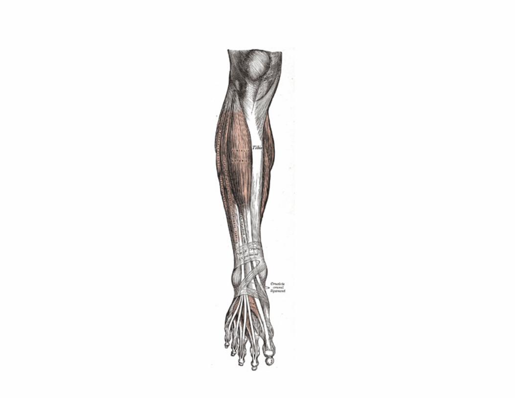
FIG. 437– Muscles of the front of the leg. (See enlarged image)
The Extensor digitorum longus is a penniform muscle, situated at the lateral part of the front of the leg. It arises from the lateral condyle of the tibia; from the upper three-fourths of the anterior surface of the body of the fibula; from the upper part of the interosseous membrane; from the deep surface of the fascia; and from the intermuscular septa between it and the Tibialis anterior on the medial, and the Peronæi on the lateral side. Between it and the Tibialis anterior are the upper portions of the anterior tibial vessels and deep peroneal nerve. The tendon passes under the transverse and cruciate crural ligaments in company with the Peronæus tertius, and divides into four slips, which run forward on the dorsum of the foot, and are inserted into the second and third phalanges of the four lesser toes. The tendons to the second, third, and fourth toes are each joined, opposite the metatarsophalangeal articulation, on the lateral side by a tendon of the Extensor digitorum brevis. The tendons are inserted in the following manner: each receives a fibrous expansion from the Interossei and Lumbricalis, and then spreads out into a broad aponeurosis, which covers the dorsal surface of the first phalanx: this aponeurosis, at the articulation of the first with the second phalanx, divides into three slips—an intermediate, which is inserted into the base of the second phalanx; and two collateral slips, which, after uniting on the dorsal surface of the second phalanx, are continued onward, to be inserted into the base of the third phalanx.
7
Variations.—This muscle varies considerably in the modes of origin and the arrangement of its various tendons. The tendons to the second and fifth toes may be found doubled, or extra slips are given off from one or more tendons to their corresponding metatarsal bones, or to the short extensor, or to one of the interosseous muscles. A slip to the great toe from the innermost tendon has been found.
8
The Peronæus tertius is a part of the Extensor digitorum longus, and might be described as its fifth tendon. The fibers belonging to this tendon arise from the lower third or more of the anterior surface of the fibula; from the lower part of the interosseous membrane; and from an intermuscular septum between it and the Peronæus brevis. The tendon, after passing under the transverse and cruciate crural ligaments in the same canal as the Extensor digitorum longus, is inserted into the dorsal surface of the base of the metatarsal bone of the little toe. This muscle is sometimes wanting.
9
Nerves.—These muscles are supplied by the fourth and fifth lumbar and first sacral nerves through the deep peroneal nerve. 10 Actions.—The Tibialis anterior and Peronæus tertius are the direct flexors of the foot at the ankle-joint; the former muscle, when acting in conjunction with the Tibialis posterior, raises the medial border of the foot, i. e., inverts the foot; and the latter, acting with the Peronæi brevis and longus, raises the lateral border of the foot, i. e., everts the foot. The Extensor digitorum longus and Extensor hallucis longus extend the phalanges of the toes, and, continuing their action, flex the foot upon the leg. Taking their fixed points from below, in the erect posture, all these muscles serve to fix the bones of the leg in the perpendicular position, and give increased strength to the ankle-joint.
11
2. The Posterior Crural Muscles—The muscles of the back of the leg are subdivided into two groups—superficial and deep. Those of the superficial group constitute a powerful muscular mass, forming the calf of the leg. Their large size is one of the most characteristic features of the muscular apparatus in man, and bears a direct relation to his erect attitude and his mode of progression.
12
The Superficial Group (Fig. 438).

Gastrocnemius. Soleus. Plantaris.
The Gastrocnemius is the most superficial muscle, and forms the greater part of the calf. It arises by two heads, which are connected to the condyles of the femur by strong, flat tendons. The medial and larger head takes its origin from a depression at the upper and back part of the medial condyle and from the adjacent part of the femur. The lateral headarises from an impression on the side of the lateral condyle and from the posterior surface of the femur immediately above the lateral part of the condyle. Both heads, also, arise from the subjacent part of the capsule of the knee. Each tendon spreads out into an aponeurosis, which covers the posterior surface of that portion of the muscle to which it belongs. From the anterior surfaces of these tendinous expansions, muscular fibers are given off; those of the medial head being thicker and extending lower than those of the lateral. The fibers unite at an angle in the middle line of the muscle in a tendinous raphé, which expands into a broad aponeurosis on the anterior surface of the muscle, and into this the remaining fibers are inserted. The aponeurosis, gradually contracting, unites with the tendon of the Soleus, and forms with it the tendo calcaneus.
13
Variations.—Absence of the outer head or of the entire muscle. Extra slips from the popliteal surface of the femur. 14 The Soleus is a broad flat muscle situated immediately in front of the Gastrocnemius. Itarises by tendinous fibers from the back of the head of the fibula, and from the upper third of the posterior surface of the body of the bone; from the popliteal line, and the middle third of the medial border of the tibia; some fibers also arise from a tendinous arch placed between the tibial and fibular origins of the muscle, in front of which the popliteal vessels and tibial nerve run. The fibers end in an aponeurosis which covers the posterior surface of the muscle, and, gradually becoming thicker and narrower, joins with the tendon of the Gastrocnemius, and forms with it the tendo calcaneus.
15
Variations.—Accessory head to its lower and inner part usually ending in the tendocalcaneus, or the calcaneus, or the laciniate ligament. 16 The Gastrocnemius and Soleus together form a muscular mass which is occasionally described as the Triceps suræ; its tendon of insertion is the tendo calcaneus.
17
Tendo Calcaneus (tendo Achillis).—The tendo calcaneus, the common tendon of the Gastrocnemius and Soleus, is the thickest and strongest in the body. It is about 15 cm. long, and begins near the middle of the leg, but receives fleshy fibers on its anterior surface, almost to its lower end. Gradually becoming contracted below, it is inserted into the middle part of the posterior surface of the calcaneus, a bursa being interposed between the tendon and the upper part of this surface. The tendon spreads out somewhat at its lower end, so that its narrowest part is about 4 cm. above its insertion. It is covered by the fascia and the integument, and is separated from the deep muscles and vessels by a considerable interval filled up with areolar and adipose tissue. Along its lateral side, but superficial to it, is the small saphenous vein.
18
The Plantaris is placed between the Gastrocnemius and Soleus. It arises from the lower part of the lateral prolongation of the linea aspera, and from the oblique popliteal ligament of the knee-joint. It forms a small fusiform belly, from 7 to 10 cm. long, ending in a long slender tendon which crosses obliquely between the two muscles of the calf, and runs along the medial border of the tendo calcaneus, to be inserted with it into the posterior part of the calcaneus. This muscle is sometimes double, and at other times wanting. Occasionally, its tendon is lost in the laciniate ligament, or in the fascia of the leg.
19
Nerves.—The Gastrocnemius and Soleus are supplied by the first and second sacral nerves, and the Plantaris by the fourth and fifth lumbar and 20

first sacral nerves, through the tibial nerve. Actions.—The muscles of the calf are the chief extensors of the foot at the ankle-joint. They possess considerable power, and are constantly called into use in standing, walking, dancing, and leaping; hence the large size they usually present. In walking, these muscles raise the heel from the ground; the body being thus supported on the raised foot, the opposite limb can be carried forward. In standing, the Soleus, taking its fixed point from below, steadies the leg upon the foot and prevents the body from falling forward. The Gastrocnemius, acting from below, serves to flex the femur upon the tibia, assisted by the Popliteus. The Plantaris is the rudiment of a large muscle which in some of the lower animals is continued over the calcaneus to be inserted into the plantar aponeurosis. In man it is an accessory to the Gastrocnemius, extending the ankle if the foot be free, or bending the knee if the foot be fixed.
21
The Deep Group (Fig. 439).
Popliteus. Flexor digitorum longus. Flexor hallucis longus. Tibialis posterior.
Deep Transverse Fascia.—The deep transverse fascia of the leg is a transversely placed, intermuscular septum, between the superficial and deep muscles of the back of the leg. At the sides it is connected to the margins of the tibia and fibula. Above, where it covers the Popliteus, it is thick and dense, and receives an expansion from the tendon of the Semimembranosus; it is thinner in the middle of the leg; but below, where it covers the tendons passing behind the malleoli, it is thickened and continuous with the laciniate ligament.
22


FIG. 438– Muscles of the back of the leg. Superficial layer. (See enlarged image)
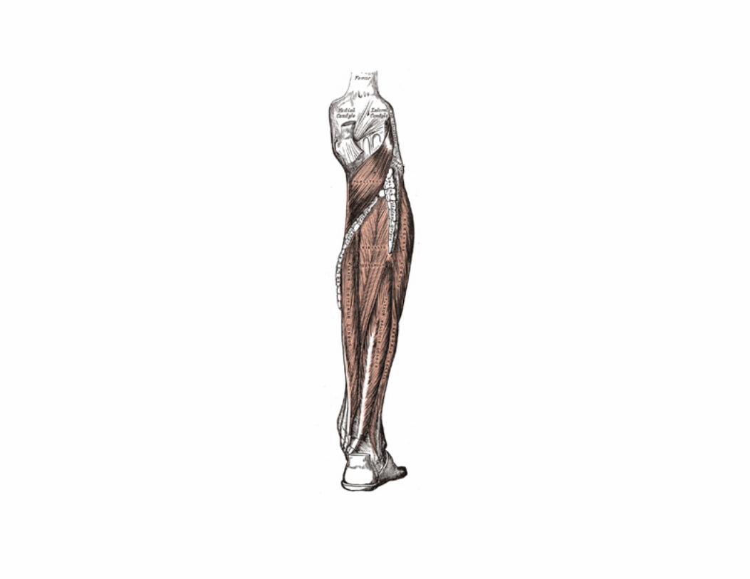
FIG. 439– Muscles of the back of the leg. Deep layer. (See enlarged image) The Popliteus is a thin, flat, triangular muscle, which forms the lower part of the floor of the popliteal fossa. It arises by a strong tendon about 2.5 cm. long, from a depression at the anterior part of the groove on the lateral condyle of the femur, and to a small extent from the oblique popliteal ligament of the knee-joint; and is inserted into the medial two-thirds of the triangular surface above the popliteal line on the posterior surface of the body of the tibia, and into the tendinous expansion covering the surface of the muscle.
23
Variations.—Additional head from the sesamoid bone in the outer head of the Gastrocnemius. Popliteus minor, rare, origin from femur on the inner side of the Plantaris, insertion into the posterior ligament of the knee-joint. Peroneotibialis, 14 per cent., origin inner side of the head of the fibula, insertion into the upper end of the oblique line of the tibia, it lies beneath the Popliteus.
24
The Flexor hallucis longus is situated on the fibular side of the leg. It arises from the inferior two-thirds of the posterior surface of the body of the fibula, with the exception of 2.5 cm. at its lowest part; from the lower part of the interosseous membrane; from an intermuscular septum between it and the Peronæi, laterally, and from the fascia covering the Tibialis posterior, medially. The fibers pass obliquely downward and backward, and end in a tendon which occupies nearly the whole length of the posterior surface of the muscle. This tendon lies in a groove which crosses the posterior surface of the lower end of the tibia, the posterior surface of the talus, and the under surface of the sustentaculum tali of the calcaneus; in the sole of the foot it runs forward between the two heads of the Flexor hallucis brevis, and is inserted into the base of the last phalanx of the great toe. The grooves on the talus and calcaneus, which contain the tendon of the muscle, are converted by tendinous fibers into distinct canals, lined by a mucous sheath. As the tendon passes forward in the sole of the foot, it is situated above, and crosses from the lateral to the medial side of the tendon of the Flexor digitorum longus, to which it is connected by a fibrous slip.
25
Variations.—Usually a slip runs to the Flexor digitorum and frequently an additional slip runs from the Flexor digitorum to the Flexor hallucis. Peroneocalcaneus internus, rare, origin below or outside the Flexor hallucis from the back of the fibula, passes over the sustentaculum tali with the Flexor hallucis and is inserted into the calcaneum.
26
The Flexor digitorum longus is situated on the tibial side of the leg. At its origin it is thin and pointed, but it gradually increases in size as it descends. It arises from the posterior surface of the body of the tibia, from immediately below the popliteal line to within 7 or 8 cm. of its lower extremity, medial to the tibial origin of the Tibialis posterior; it also arisesfrom the fascia covering the Tibialis posterior. The fibers end in a tendon, which runs nearly the whole length of the posterior surface of the muscle. This tendon passes behind the medial malleolus, in a groove, common to it and the Tibialis posterior, but separated from the latter by a fibrous septum, each tendon being contained in a special compartment lined by a separate mucous sheath. It passes obliquely forward and lateralward, superficial to the deltoid ligament of the ankle-joint, into the sole of the foot (Fig. 444), where it crosses below the tendon of the Flexor hallucis longus, and receives from it a strong tendinous slip. It then expands and is joined by the Quadratus plantæ, and finally divides into four tendons, which are inserted into the bases of the last phalanges of the second, third, fourth, and fifth toes, each tendon passing through an opening in the corresponding tendon of the Flexor digitorum brevis opposite the base of the first phalanx.
27
Variations.—Flexor accessorius longus digitorum, not infrequent, origin from fibula, or tibia, or the deep fascia and ending in a tendon which, after passing beneath the laciniate ligament, joins the tendon of the long flexor or the Quadratus plantæ.
28
The Tibialis posterior (Tibialis posticus) lies between the two preceding muscles, and is the most deeply seated of the muscles on the back of the leg. It begins above by two pointed processes, separated by an angular interval through which the anterior tibial vessels pass forward to the
29

front of the leg. It arises from the whole of the posterior surface of the interosseous membrane, excepting its lowest part; from the lateral portion of the posterior surface of the body of the tibia, between the commencement of the popliteal line above and the junction of the middle and lower thirds of the body below; and from the upper two-thirds of the medial surface of the fibula; some fibers also arise from the deep transverse fascia, and from the intermuscular septa separating it from the adjacent muscles. In the lower fourth of the leg its tendon passes in front of that of the Flexor digitorum longus and lies with it in a groove behind the medial malleolus, but enclosed in a separate sheath; it next passes under the laciniate and over the deltoid ligament into the foot, and then beneath the plantar calcaneonavicular ligament. The tendon contains a sesamoid fibrocartilage, as it runs under the plantar calcaneonavicular ligament. It is inserted into the tuberosity of the navicular bone, and gives off fibrous expansions, one of which passes backward to the sustentaculum tali of the calcaneus, others forward and lateralward to the three cuneiforms, the cuboid, and the bases of the second, third, and fourth metatarsal bones. Nerves.—The Popliteus is supplied by the fourth and fifth lumbar and first sacral nerves, the Flexor digitorum longus and Tibialis posterior by the fifth lumbar and first sacral, and the Flexor hallucis longus by the fifth lumbar and the first and second sacral nerves, through the tibial nerve.
30
Actions.—The Popliteus assists in flexing the leg upon the thigh; when the leg is flexed, it will rotate the tibia inward. It is especially called into action at the beginning of the act of bending the knee, inasmuch as it produces the slight inward rotation of the tibia which is essential in the early stage of this movement. The Tibialis posterior is a direct extensor of the foot at the ankle-joint; acting in conjunction with the Tibialis anterior, it turns the sole of the foot upward and medialward, i.e., inverts the foot, antagonizing the Peronæi, which turn it upward and lateralward (evert it). In the sole of the foot the tendon of the Tibialis posterior lies directly below the plantar calcaneonavicular ligament, and is therefore an important factor in maintaining the arch of the foot. The Flexor digitorum longus and Flexor hallucis longus are the direct flexors of the phalanges, and, continuing their action, extend the foot upon the leg; they assist the Gastrocnemius and Soleus in extending the foot, as in the act of walking, or in standing on tiptoe. In consequence of the oblique direction of its tendons the Flexor digitorum longus would draw the toes medialward, were it not for the Quadratus plantæ, which is inserted into the lateral side of the tendon, and draws it to the middle line of the foot. Taking their fixed point from the foot, these muscles serve to maintain the upright posture by steadying the tibia and fibula perpendicularly upon the talus.
31
3. The Lateral Crural Muscles (Fig. 439).
Peronæus longus. Peronæus brevis. The Peronæus longus is situated at the upper part of the lateral side of the leg, and is the more superficial of the two muscles. It arises from the head and upper two-thirds of the lateral surface of the body of the fibula, from the deep surface of the fascia, and from the intermuscular septa between it and the muscles on the front and back of the leg; occasionally also by a few fibers from the lateral condyle of the tibia. Between its attachments to the head and to the body of the fibula there is a gap through which the common peroneal nerve passes to the front of the leg. It ends in a long tendon, which runs behind the lateral malleolus, in a groove common to it and the tendon of the Peronæus brevis, behind which it lies; the groove is converted into a canal by the superior peroneal retinaculum, and the tendons in it are contained in a common mucous sheath. The tendon then extends obliquely forward across the lateral side of the calcaneus, below the trochlear process, and the tendon of the Peronæus brevis, and under cover of the inferior peroneal retinaculum. It crosses the lateral side of the cuboid, and then runs on the under surface of that bone in a groove which is converted into a canal by the long plantar ligament; the tendon then crosses the sole of the foot obliquely, and is inserted into the lateral side of the base of the first metatarsal bone and the lateral side of the first cuneiform. Occasionally it sends a slip to the base of the second metatarsal bone. The tendon changes its direction at two points: first, behind the lateral malleolus; secondly, on the cuboid
32

bone; in both of these situations the tendon is thickened, and, in the latter, a sesamoid fibrocartilage (sometimes a bone), is usually developed in its substance.
FIG. 440– Cross-section through middle of leg. (Eycleshymer and Schoemaker.) (See enlarged image)
The Peronæus brevis lies under cover of the Peronæus longus, and is a shorter and smaller muscle. It arises from the lower two-thirds of the lateral surface of the body of the fibula; medial to the Peronæus longus; and from the intermuscular septa separating it from the adjacent muscles on the front and back of the leg. The fibers pass vertically downward, and end in a tendon which runs behind the lateral malleolus along with but in front of that of the preceding muscle, the two tendons being enclosed in the same compartment, and lubricated by a common mucous sheath. It then runs forward on the lateral side of the calcaneus, above the trochlear process and the tendon of the Peronæus longus, and isinserted into the tuberosity at the base of the fifth metatarsal bone, on its lateral side.
33
On the lateral surface of the calcaneus the tendons of the Peronæi longus and brevis occupy separate osseoaponeurotic canals formed by the calcaneus and the perineal retinacula; each tendon is enveloped by a forward prolongation of the common mucous sheath.
34
Variations.—Fusion of the two peronæi is rare. A slip from the Peronæus longus to the base of the third, fourth or fifth metatarsal bone, or to the Adductor hallucis is occasionally seen.
35
Peronæus accessorius, origin from the fibula between the longus and brevis, joins the tendon of the longus in the sole of the foot. 36 Peronæus quinti digiti, rare, origin lower fourth of the fibula under the brevis, insertion into the Extensor aponeurosis of the little toe. More common as a slip of the tendon of the Peronæus brevis.
37
Peronæus quartus, 13 per cent. (Gruber), origin back of fibula between the brevis and the Flexor hallucis, insertion into the peroneal spine of the calcaneum, (peroneocalcaneus externum), or less frequently into the tuberosity of the cuboid (peroneocuboideus).
38
Nerves.—The Peronæi longus and brevis are supplied by the fourth and fifth lumbar and first sacral nerves through the superficial peroneal nerve.
39
Actions.—The Peronæi longus and brevis extend the foot upon the leg, in conjunction with the Tibialis posterior, antagonizing the Tibialis anterior and Peronæus tertius, which are flexors of the foot. The Peronæus longus also everts the sole of the foot, and from the oblique direction of the tendon across the sole of the foot is an important agent in the maintenance of the transverse arch. Taking their fixed points below, the Peronæi serve to steady the leg upon the foot. This is especially the case in standing upon one leg, when the tendency of the superincumbent weight is to throw the leg medialward; the Peronæus longus overcomes this tendency by drawing on the lateral side of the leg.
40
8d. The Fasciæ Around the Ankle
Fibrous bands, or thickened portions of the fascia, bind down the tendons in front of and behind the ankle in their passage to the foot. They comprise three ligaments, viz., thetransverse crural, the cruciate crural and the laciniate; and the superior and inferior peroneal retinacula.
1

FIG. 441– The mucous sheaths of the tendons around the ankle. Lateral aspect. (See enlarged image) Transverse Crural Ligament (ligamentum transversum cruris; upper part of anterior annular ligament) (Fig. 441).—The transverse crural ligament binds down the tendons of Extensor digitorum longus, Extensor hallucis longus, Peronæus tertius, and Tibialis anterior as they descend on the front of the tibia and fibula; under it are found also the anterior tibial vessels and deep peroneal nerve. It is attached laterally to the lower end of the fibula, and medially to the tibia; above it is continuous with the fascia of the leg.
2
Cruciate Crural Ligament (ligamentum cruciatum cruris; lower part of anterior annular ligament) (Figs. 441, 442).—The cruciate crural ligament is a Y-shaped band placed in front of the ankle-joint, the stem of the Y being attached laterally to the upper surface of the calcaneus, in front of the depression for the interosseous talocalcanean ligament; it is directed medialward as a double layer, one lamina passing in front of, and the other behind, the tendons of the Peronæus tertius and Extensor digitorum longus. At the medial border of the latter tendon these two layers join together, forming a compartment in which the tendons are enclosed. From the medial extremity of this sheath the two limbs of the Y diverge: one is directed upward and medialward, to be attached to the tibial malleolus, passing over the Extensor hallucis longus and the vessels and nerves, but enclosing the Tibialis anterior by a splitting of its fibers. The other limb extends downward and medialward, to be attached to the border of the plantar aponeurosis, and passes over the tendons of the Extensor hallucis longus and Tibialis anterior and also the vessels and nerves.
3

Laciniate Ligament (ligamentum laciniatum; internal annular ligament).—The laciniate ligament is a strong fibrous band, extending from the tibial malleolus above to the margin of the calcaneus below, converting a series of bony grooves in this situation into canals for the passage of the tendons of the Flexor muscles and the posterior tibial vessels and tibial nerve into the sole of the foot. It is continuous by its upper border with the deep fascia of the leg, and by its lower border with the plantar aponeurosis and the fibers of origin of the Abductor hallucis muscle. Enumerated from the medial side, the four canals which it forms transmit the tendon of the Tibialis posterior; the tendon of the Flexor digitorum longus; the posterior tibial vessels and tibial nerve, which run through a broad space beneath the ligament; and lastly, in a canal formed partly by the talus, the tendon of the Flexor hallucis longus.
4
FIG. 442– The mucous sheaths of the tendons around the ankle. Medial aspect. (See enlarged image)
Peroneal Retinacula.—The peroneal retinacula are fibrous bands which bind down the tendons of the Peronæi longus and brevis as they run across the lateral side of the ankle. The fibers of the superior retinaculum (external annular ligament) are attached above to the lateral malleolus and below to the lateral surface of the calcaneus. The fibers of the inferior retinaculum are continuous in front with those of the cruciate crural ligament; behind they are attached to the lateral surface of the calcaneus; some of the fibers are fixed to the peroneal trochlea, forming a septum between the tendons of the Peronæi longus and brevis.
5
The Mucous Sheaths of the Tendons Around the Ankle.—All the tendons crossing the ankle-joint are enclosed for part of their length in 6

mucous sheaths which have an almost uniform length of about 8 cm. each. On the front of the ankle (Fig. 441) the sheath for the Tibialis anterior extends from the upper margin of the transverse crural ligament to the interval between the diverging limbs of the cruciate ligament; those for the Extensor digitorum longus and Extensor hallucis longus reach upward to just above the level of the tips of the malleoli, the former being the higher. The sheath of the Extensor hallucis longus is prolonged on to the base of the first metatarsal bone, while that of the Extensor digitorum longus reaches only to the level of the base of the fifth metatarsal. On the medial side of the ankle (Fig. 442) the sheath for the Tibialis posterior extends highest up—to about 4 cm. above the tip of the malleolus—while below it stops just short of the tuberosity of the navicular. The sheath for Flexor hallucis longus reaches up to the level of the tip of the malleolus, while that for the Flexor digitorum longus is slightly higher; the former is continued to the base of the first metatarsal, but the latter stops opposite the first cuneiform bone. On the lateral side of the ankle (Fig. 441) a sheath which is single for the greater part of its extent encloses the Peronæi longus and brevis. It extends upward for about 4 cm. above the tip of the malleolus and downward and forward for about the same distance.
8e. The Muscles and Fasciæ of the Foot
1. The Dorsal Muscle of the Foot Extensor digitorum brevis—The fascia on the dorsum of the foot is a thin membranous layer, continuous above with the transverse and cruciate crural ligaments; on either side it blends with the plantar aponeurosis; anteriorly it forms a sheath for the tendons on the dorsum of the foot.
1
The Extensor digitorum brevis (Fig. 441) is a broad, thin muscle, which arises from the forepart of the upper and lateral surfaces of the calcaneus, in front of the groove for the Peronæus brevis; from the lateral talocalcanean ligament; and from the common limb of the cruciate crural ligament. It passes obliquely across the dorsum of the foot, and ends in four tendons. The most medial, which is the largest, is inserted into the dorsal surface of the base of the first phalanx of the great toe, crossing the dorsalis pedis artery; it is frequently described as a separate muscle—the Extensor hallucis brevis. The other three are insertedinto the lateral sides of the tendons of the Extensor digitorum longus of the second, third, and fourth toes.
2
Variations.—Accessory slips of origin from the talus and navicular, or from the external cunei-form and third metatarsal bones to the second slip of the muscle, and one from the cuboid to the third slip have been observed. The tendons vary in number and position; they may be reduced to two, or one of them may be doubled, or an additional slip may pass to the little toe. A supernumerary slip ending on one of the metatarsophalangeal articulations, or joining a dorsal interosseous muscle is not uncommon. Deep slips between this muscle and the Dorsal interossei occur.
3
Nerves.—It is supplied by the deep peroneal nerve. 4 Actions.—The Extensor digitorum brevis extends the phalanges of the four toes into which it is inserted, but in the great toe acts only on the first phalanx. The obliquity of its direction counteracts the oblique movement given to the toes by the long Extensor, so that when both muscles act, the toes are evenly extended.
5
2. The Plantar Muscles of the Foot Plantar Aponeurosis (aponeurosis plantaris; plantar fascia).—The plantar aponeurosis is of great strength, and consists of pearly white glistening fibers, disposed, for the most part, longitudinally: it is divided into central, lateral, and medial portions.
6
The central portion, the thickest, is narrow behind and attached to the medial process of the tuberosity of the calcaneus, posterior to the origin of the Flexor digitorum brevis; and becoming broader and thinner in front, divides near the heads of the metatarsal bones into five processes, one for each of the toes. Each of these processes divides opposite the metatarsophalangeal articulation into two strata, superficial and deep. The superficial stratum is inserted into the skin of the transverse sulcus which separates the toes from the sole. The deeper stratum divides into two slips which embrace the side of the Flexor tendons of the toes, and blend with the sheaths of the tendons, and with the transverse metatarsal ligament, thus forming a series of arches through which the tendons of the short and long Flexors pass to the toes. The intervals left between the five processes allow the digital vessels and nerves and the tendons of the Lumbricales to become superficial. At the point of division of the aponeurosis, numerous transverse fasciculi are superadded; these serve to increase the strength of the aponeurosis at this part by binding the processes together, and connecting them with the integument. The central portion of the plantar aponeurosis is continuous with the lateral and medial portions and sends upward into the foot, at the lines of junction, two strong vertical intermuscular septa, broader in front than behind, which separate the intermediate from the lateral and medial plantar groups of muscles; from these again are derived thinner transverse septa which separate the various layers of muscles in this region. The upper surface of this aponeurosis gives origin behind to the Flexor digitorum brevis.
7
The lateral and medial portions of the plantar aponeurosis are thinner than the central piece, and cover the sides of the sole of the foot. 8 The lateral portion covers the under surface of the Abductor digiti quinti; it is thin in front and thick behind, where it forms a strong band between the lateral process of the tuberosity of the calcaneus and the base of the fifth metatarsal bone; it is continuous medially with the central portion of the plantar aponeurosis, and laterally with the dorsal fascia.
9
The medial portion is thin, and covers the under surface of the Abductor hallucis; it isattached behind to the laciniate ligament, and is continuous around the side of the foot with the dorsal fascia, and laterally with the central portion of the plantar aponeurosis.
10
The muscles in the plantar region of the foot may be divided into three groups, in a similar manner to those in the hand. Those of the medial plantar region are connected with the great toe, and corrrespond with those of the thumb; those of the lateral plantar region are connected with the little toe, and correspond with those of the little finger; and those of the intermediate plantar region are connected with the tendons intervening between the two former groups. But in order to facilitate the description of these muscles, it is more convenient to divide them into four layers, in the order in which they are successively exposed.
11
The First Layer (Fig. 443).
Abductor hallucis. Flexor digitorum brevis. Abductor digiti quinti.
The Abductor hallucis lies along the medial border of the foot and covers the origins of the plantar vessels and nerves. It arises from the medial process of the tuberosity of the calcaneus, from the laciniate ligament, from the plantar aponeurosis, and from the intermuscular septum between it and the Flexor digitorum brevis. The fibers end in a tendon, which is inserted, together with the medial tendon of the Flexor hallucis brevis, into the tibial side of the base of the first phalanx of the great toe.
12

Variations.—Slip to the base of the first phalanx of the second toe. 13 The Flexor digitorum brevis lies in the middle of the sole of the foot, immediately above the central part of the plantar aponeurosis, with which it is firmly united. Its deep surface is separated from the lateral plantar vessels and nerves by a thin layer of fascia. It arises by a narrow tendon, from the medial process of the tuberosity of the calcaneus, from the central part of the plantar aponeurosis, and from the intermuscular septa between it and the adjacent muscles. It passes forward, and divides into four tendons, one for each of the four lesser toes. Opposite the bases of the first phalanges, each tendon divides into two slips, to allow of the passage of the corresponding tendon of the Flexor digitorum longus; the two portions of the tendon then unite and form a grooved channel for the reception of the accompanying long Flexor tendon. Finally, it divides a second time, and is inserted into the sides of the second phalanx about its middle. The mode of division of the tendons of the Flexor digitorum brevis, and of their insertion into the phalanges, is analogous to that of the tendons of the Flexor digitorum sublimis in the hand.
14
Variations.—Slip to the little toe frequently wanting, 23 per cent.; or it may be replaced by a small fusiform muscle arising from the long flexor tendon or from the Quadratus plantæ.
15
Fibrous Sheaths of the Flexor Tendons.—The terminal portions of the tendons of the long and short Flexor muscles are contained in osseoaponeurotic canals similar in their arrangement to those in the fingers. These canals are formed above by the phalanges and below by fibrous bands, which arch across the tendons, and are attached on either side to the margins of the phalanges. Opposite the bodies of the proximal and second phalanges the fibrous bands are strong, and the fibers are transverse; but opposite the joints they are much thinner, and the fibers are directed obliquely. Each canal contains a mucous sheath, which is reflected on the contained tendons.
16

FIG. 443– Muscles of the sole of the foot. First layer. (See enlarged image) The Abductor digiti quinti (Abductor minimi digiti) lies along the lateral border of the foot, and is in relation by its medial margin with the lateral plantar vessels and nerves. Itarises, by a broad origin, from the lateral process of the tuberosity of the calcaneus, from the under surface of
17
the calcaneus between the two processes of the tuberosity, from the forepart of the medial process, from the plantar aponeurosis, and from the intermuscular septum between it and the Flexor digitorum brevis. Its tendon, after gliding over a smooth facet on the under surface of the base of the fifth metatarsal bone, is inserted, with the Flexor digiti quinti brevis, into the fibular side of the base of the first phalanx of the fifth toe. Variations.—Slips of origin from the tuberosity at the base of the fifth metatarsal.Abductor ossis metatarsi quinti, origin external tubercle of the calcaneus, insertion into tuberosity of the fifth metatarsal bone in common with or beneath the outer margin of the plantar fascia.
18
The Second Layer (Fig. 444).
Quadratus plantæ. Lumbricales. The Quadratus plantæ (Flexor accessorius) is separated from the muscles of the first layer by the lateral plantar vessels and nerve. It arises by two heads, which are separated from each other by the long plantar ligament: the medial or larger head is muscular, and is attached to the medial concave surface of the calcaneus, below the groove which lodges the tendon of the Flexor hallucis longus; the lateral head, flat and tendinous, arises from the lateral border of the inferior surface of the calcaneus, in front of the lateral process of its tuberosity, and from the long plantar ligament. The two portions join at an acute angle, and end in a flattened band which is inserted into the lateral margin and upper and under surfaces of the tendon of the Flexor digitorum longus, forming a kind of groove, in which the tendon is lodged. It usually sends slips to those tendons of the Flexor digitorum longus which pass to the second, third, and fourth toes.
19
Variations.—Lateral head often wanting; entire muscle absent. Variation in the number of digital tendons to which fibers can be traced. Most frequent offsets are sent to the second, third and fourth toes; in many cases to the fifth as well; occasionally to two toes only.
20
The Lumbricales are four small muscles, accessory to the tendons of the Flexor digitorum longus and numbered from the medial side of the foot; they arise from these tendons, as far back as their angles of division, each springing from two tendons, except the first. The muscles end in tendons, which pass forward on the medial sides of the four lesser toes, and are inserted into the expansions of the tendons of the Extensor digitorum longus on the dorsal surfaces of the first phalanges.
21
Variations.—Absence of one or more; doubling of the third or fourth. Insertion partly or wholly into the first phalanges. 22 The Third Layer (Fig. 445).
Flexor hallucis brevis. Adductor hallucis. Flexor digiti quinti brevis.
The Flexor hallucis brevis arises, by a pointed tendinous process, from the medial part of the under surface of the cuboid bone, from the contiguous portion of the third cuneiform, and from the prolongation of the tendon of the Tibialis posterior which is attached to that bone. It divides in front into two portions, which are inserted into the medial and lateral sides of the base of the first phalanx of the great toe, a sesamoid bone being present in each tendon at its insertion. The medial portion is blended with the Abductor hallucis previous to its insertion; the lateral portion with the Adductor hallucis; the tendon of the Flexor hallucis longus lies in a groove between them; the lateral portion is sometimes described as the first Interosseous plantaris.
23

Variations.—Origin subject to considerable variation; it often receives fibers from the calcaneus or long plantar ligament. Attachment to the cuboid sometimes wanting. Slip to first phalanx of the second toe.
24
The Adductor hallucis (Adductor obliquus hallucis) arises by two heads—oblique and transverse. The oblique head is a large, thick, fleshy mass, crossing the foot obliquely and occupying the hollow space under the first second, third, and fourth metatarsal bones. Itarises from the bases of the second, third, and fourth metatarsal bones, and from the sheath of the tendon of the Peronæus longus, and is inserted, together with the lateral portion of the Flexor hallucis brevis, into the lateral side of the base of the first phalanx of the great toe. The transverse head (Transversus pedis) is a narrow, flat fasciculus which arisesfrom the plantar metatarsophalangeal ligaments of the third, fourth, and fifth toes (sometimes only from the third and fourth), and from the transverse ligament of the metatarsus. It is inserted into the lateral side of the base of the first phalanx of the great toe, its fibers blending with the tendon of insertion of the oblique head.
25

FIG. 444– Muscles of the sole of the foot. Second layer. (See enlarged image)
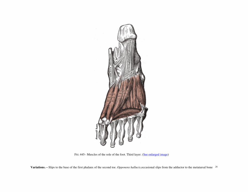
FIG. 445– Muscles of the sole of the foot. Third layer. (See enlarged image) Variations.—Slips to the base of the first phalanx of the second toe. Opponens hallucis,occasional slips from the adductor to the metatarsal bone 26

of the great toe. The Abductor, Flexor brevis, and Adductor of the great toe, like the similar muscles of the thumb, give off, at their insertions, fibrous expansions to blend with the tendons of the Extensor digitorum longus.
27
The Flexor digiti quinti brevis (Flexor brevis minimi digiti) lies under the metatarsal bone of the little toe, and resembles one of the Interossei. It arises from the base of the fifth metatarsal bone, and from the sheath of the Peronæus longus; its tendon is inserted into the lateral side of the base of the first phalanx of the fifth toe. Occasionally a few of the deeper fibers are inserted into the lateral part of the distal half of the fifth metatarsal bone; these are described by some as a distinct muscle, the Opponens digiti quinti.
28
The Fourth Layer Interossei—The Interossei in the foot are similar to those in the hand, with this exception, that they are grouped around the middle line of the second digit, instead of that of the third.They are seven in number, and consist of two groups, dorsal and plantar.
29
The Interossei dorsales (Dorsal interossei) (Fig. 446), four in number, are situated between the metatarsal bones. They are bipenniform muscles, each arising by two heads from the adjacent sides of the metatarsal bones between which it is placed; their tendons areinserted into the bases of the first phalanges, and into the aponeurosis of the tendons of the Extensor digitorum longus. In the angular interval left between the heads of each of the three lateral muscles, one of the perforating arteries passes to the dorsum of the foot; through the space between the heads of the first muscle the deep plantar branch of the dorsalis pedis artery enters the sole of the foot. The first is inserted into the medial side of the second toe; the other three are inserted into the lateral sides of the second, third, and fourth toes.
30
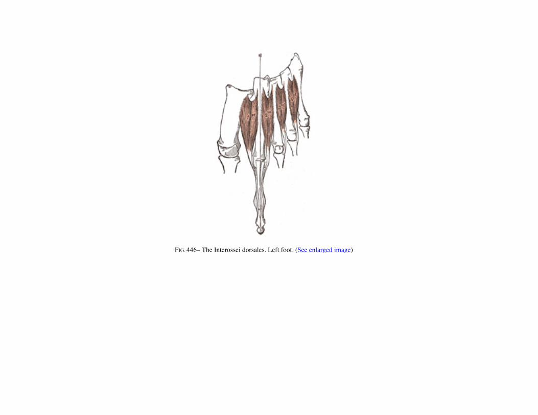
FIG. 446– The Interossei dorsales. Left foot. (See enlarged image)
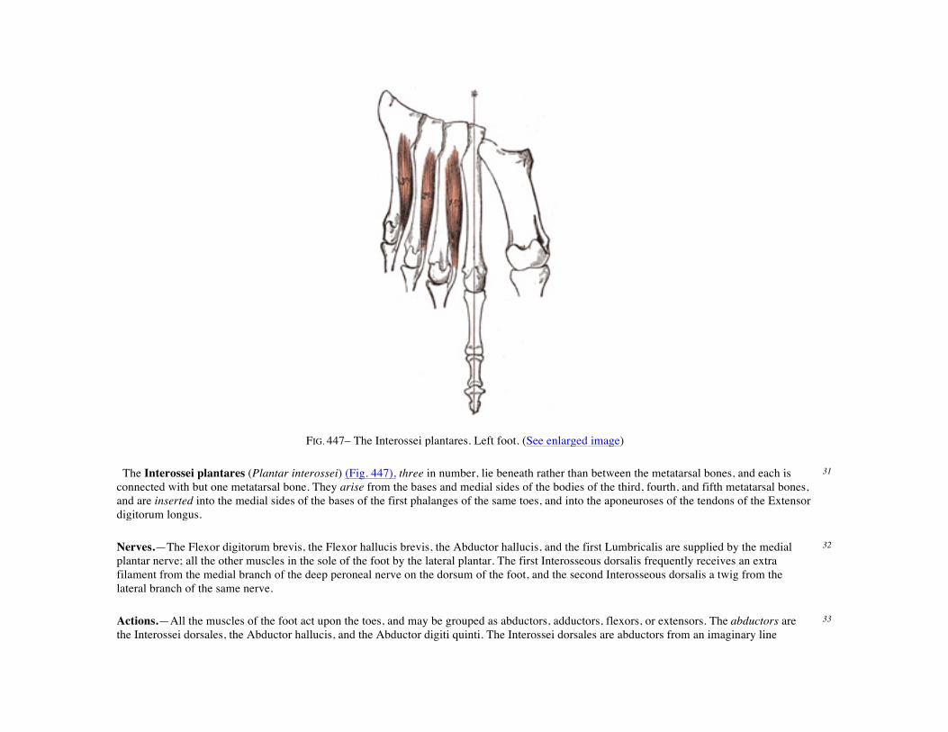
FIG. 447– The Interossei plantares. Left foot. (See enlarged image) The Interossei plantares (Plantar interossei) (Fig. 447), three in number, lie beneath rather than between the metatarsal bones, and each is connected with but one metatarsal bone. They arise from the bases and medial sides of the bodies of the third, fourth, and fifth metatarsal bones, and are inserted into the medial sides of the bases of the first phalanges of the same toes, and into the aponeuroses of the tendons of the Extensor digitorum longus.
31
Nerves.—The Flexor digitorum brevis, the Flexor hallucis brevis, the Abductor hallucis, and the first Lumbricalis are supplied by the medial plantar nerve; all the other muscles in the sole of the foot by the lateral plantar. The first Interosseous dorsalis frequently receives an extra filament from the medial branch of the deep peroneal nerve on the dorsum of the foot, and the second Interosseous dorsalis a twig from the lateral branch of the same nerve.
32
Actions.—All the muscles of the foot act upon the toes, and may be grouped as abductors, adductors, flexors, or extensors. The abductors are the Interossei dorsales, the Abductor hallucis, and the Abductor digiti quinti. The Interossei dorsales are abductors from an imaginary line
33

passing through the axis of the second toe, so that the first muscle draws the second toe medialward, toward the great toe, the second muscle draws the same toe lateralward, and the third and fourth draw the third and fourth toes in the same direction. Like the Interossei in the hand, each assists in flexing the first phalanx and extending the second and third phalanges. The Abductor hallucis abducts the great toe from the second, and also flexes its proximal phalanx. In the same way the action of the Abductor digiti quinti is twofold, as an abductor of this toe from the fourth, and also as a flexor of its proximal phalanx. The adductors are the Interossei plantares and the Adductor hallucis. The Interossei plantares adduct the third, fourth, and fifth toes toward the imaginary line passing through the second toe, and by means of their insertions into the aponeuroses of the Extensor tendons they assist in flexing the proximal phalanges and extending the middle and terminal phalanges. The oblique head of the Adductor hallucis is chiefly concerned in adducting the great toe toward the second one, but also assists in flexing this toe; the transverse head approximates all the toes and thus increases the curve of the transverse arch of the metatarsus. The flexors are the Flexor digitorum brevis, the Quadratus plantæ, the Flexor hallucis brevis, the Flexor digiti quinti brevis, and the Lumbricales. The Flexor digitorum brevis flexes the second phalanges upon the first, and, continuing its action, flexes the first phalanges also, and brings the toes together. The Quadratus plantæ assists the Flexor digitorum longus and converts the oblique pull of the tendons of that muscle into a direct backward pull upon the toes. The Flexor digiti quinti brevis flexes the little toe and draws its metatarsal bone downward and medialward. The Lumbricales, like the corresponding muscles in the hand, assist in flexing the proximal phalanges, and by their insertions into the tendons of the Extensor digitorum longus aid that muscle in straightening the middle and terminal phalanges. The Extensor digitorum brevis extends the first phalanx of the great toe and assists the long Extensor in extending the next three toes, and at the same time gives to the toes a lateral direction when they are extended.
Bibliography
BARDEEN, C. R.: Development and Variation, etc., of the Inferior Extremity, etc., Am. Jour. Anat., 1907, vi. 1 BARDEEN and LEWIS: Development of the Back, Body Wall and Limbs in Man, Am. Jour. Anat., 1901, i. 2 EISLER, P.: Die Muskeln des Stammes, v. Bardeleben’s Handbuch der Anatomie des Menschen, Bd. ii, Abt. ii, Teil 1. 3 FICK, R.: Anatomie und Mechanik der Gelenke unter Berücksichtigung der bewegenden Muskeln, v. Bardeleben’s Handbuch der Anatomie des Menschen, Bd. ii, Abt. i, Teil 2 and 3.
4
FROHSE and FRÄNKEL: Die Muskeln des Menschlichen Beines; Die Muskeln des Menschlichen Armes, Handbuch der Anatomie des Menschen, von Bardeleben, Bd. ii, Abt. ii, Teil 2, A and B.
5
HENLE, J.: Handbuch der Systematischen Anatomie des Menschen, 1871–79. 6 KOCH, J. C.: The Laws of Bone Architecture, Am. Jour. Anat., 1917, xxi. 7 LE. DOUBLE: Traité des Variations du Systeme Musculaire de L’Homme, 1897. 8 LEWIS, W. H.: Development of the Arm in Man, Am. Jour. Anat., 1901, i. 9 LEWIS, W. H.: Development of the Muscular System, Keibel and Mall, Manual of Human Embryology. 10 POIRIER, P., et CHARPY, A.: Traité d’Anatomie, 1899–1901. 11 TESTUT, L.: Traité d’Anatomie Humaine, 1893–94. 12 WOLFF, J.: Das Gesetz der Transformation der Knochen, Berlin, 1892. 13