Embed Size (px)
Citation preview
IVH in Preterm Infants
Sue Ann Smith
Preterm Neonates - IVH
• Gestation usually less than 32 weeks, but may occur in more mature preterm infants
• May rarely occur in the term neonate
• The primary site of hemorrhage is the germinal matrix.
• A secondary site is the choroid plexus - especially in the more mature infant
IVH timing
• Set up for IVH usually present at birth
• Bleeding usually not present before 24 hours, 75% of all IVH seen by 72 hours, and > 95% by 1 week of age.
• Risk of new IVH after 1 weeks is very low.
• Still need to follow for PVL, which is not usually detectable for 4-6 weeks by US.
Grading IVH by Ultrasound
• Grade I - Germinal matrix hemorrhage (GMH) without extension into the ventricle
• Grade II - GMH with extension of blood into ventricle, but without distension of ventricle.
• Grade III - GMH with distension of ventricle.
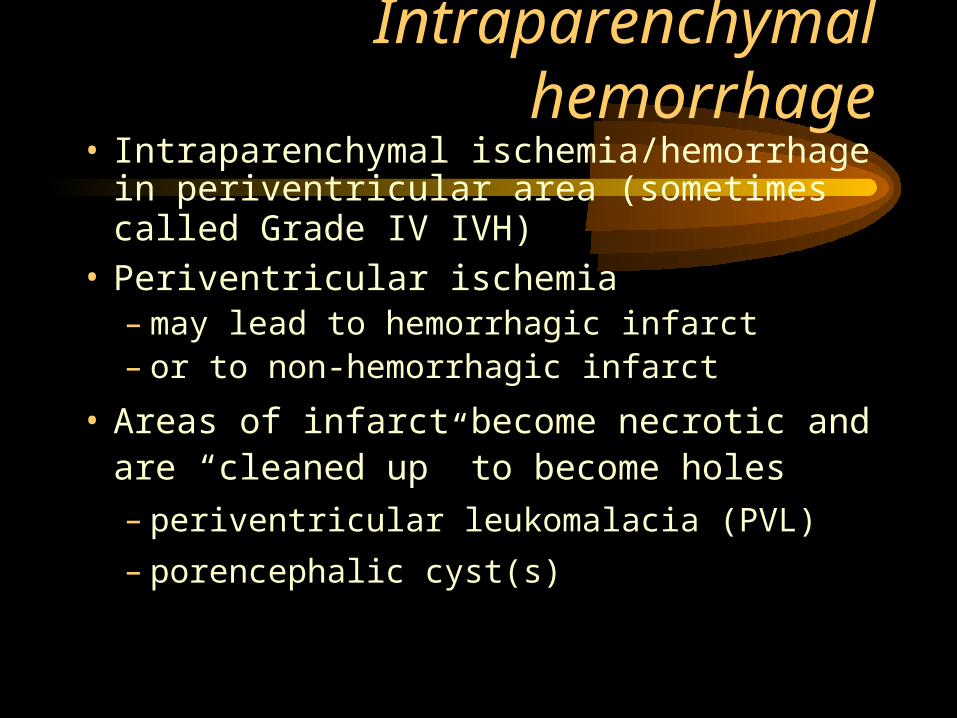
Intraparenchymal hemorrhage• Intraparenchymal ischemia/hemorrhage in
periventricular area (sometimes called Grade IV IVH)
• Periventricular ischemia – may lead to hemorrhagic infarct – or to non-hemorrhagic infarct
• Areas of infarct become necrotic and are “cleaned up” to become holes
– periventricular leukomalacia (PVL)
– porencephalic cyst(s)
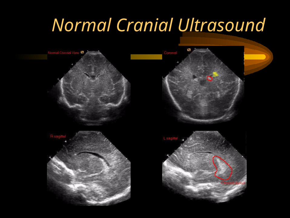
Normal Cranial Ultrasound
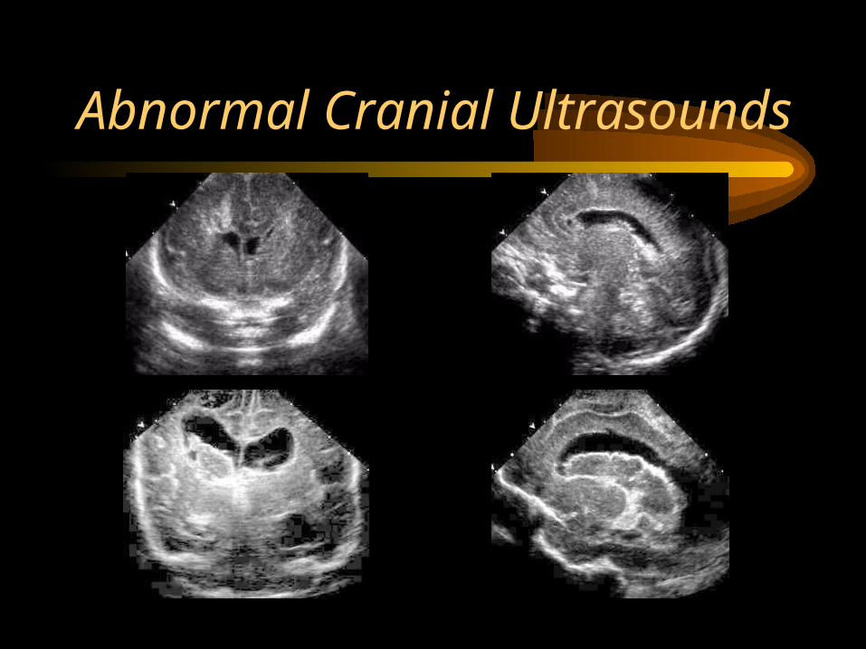
Abnormal Cranial Ultrasounds
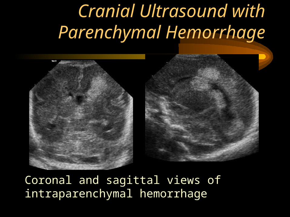
Cranial Ultrasound with Parenchymal Hemorrhage
Coronal and sagittal views of intraparenchymal hemorrhage
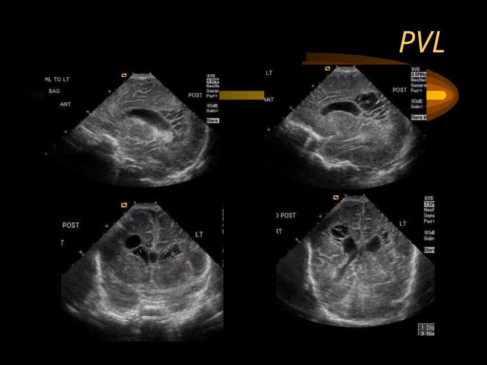
PVL


![Whitelaw Prevention of IVH.pptx [Somente leitura] · 2016. 6. 27. · Vitamin E and IVH in preterm infants Reduced IVH IV vit E increased Sepsis IV vit E increased parenchymal infarction](https://img.pdfslide.net/doc/110x75/609837e00ff34b46217b7516/whitelaw-prevention-of-ivhpptx-somente-leitura-2016-6-27-vitamin-e-and-ivh.jpg)