Embed Size (px)
Citation preview
Izolacija i karakterizacija proteolitičkih sojevabakterija i kvasaca iz ovčjeg i kozjeg sira
Zadro, Dragana
Master's thesis / Diplomski rad
2016
Degree Grantor / Ustanova koja je dodijelila akademski / stručni stupanj: University of Zagreb, Faculty of Food Technology and Biotechnology / Sveučilište u Zagrebu, Prehrambeno-biotehnološki fakultet
Permanent link / Trajna poveznica: https://urn.nsk.hr/urn:nbn:hr:159:664242
Rights / Prava: In copyright
Download date / Datum preuzimanja: 2022-02-15
Repository / Repozitorij:
Repository of the Faculty of Food Technology and Biotechnology
SVEUČILIŠTE U ZAGREBU
PREHRAMBENO-BIOTEHNOLOŠKI FAKULTET
DIPLOMSKI RAD
Zagreb, rujan 2016. Dragana Zadro
642/PI
ISOLATION
AND CHARACTERIZATION OF
PROTEOLYTIC BACTERIA AND
YEAST STRAINS FROM SHEEP
AND GOAT CHEESES

This work was done under supervision of dr. sc. Monika Kovacs at Department of Microbiology
and Biotechnology, on the Faculty of Food science at Szent Istvan University, Budapest, and
prof. dr. sc. Vladimir Mrša on the Faculty of Food Technology and Biotechnology, University of
Zagreb.

I would like to express my sincere gratitude to my supervisors PhD. Mónika Kovács, Assistent
professor, and PhD. Vladimir Mrša, Full professor for the continuous support, patience and
motivation during this work.
Also, I am grateful to my family for providing me support and encouragement throughout my
years of study. This accomplishment would not have been possible without them.
TEMELJNA DOKUMENTACIJSKA KARTICA
Diplomski rad
Sveučilište u Zagrebu
Prehrambeno-biotehnološki fakultet
Zavod za kemiju i biokemiju
Laboratorij za biokemiju
Znanstveno podrucje: Biotehničke znanosti
Znanstveno polje: Prehrambena tehnologija
IZOLACIJA I KARAKTERIZACIJA PROTEOLITIČKIH SOJEVA BAKTERIJA I
KVASACA IZ OVČJEG I KOZJEG SIRA
Dragana Zadro, 642/PI
Sažetak: Cilj ovog rada bila je izolacija i karakterizacija mikroorganizama iz ovčjeg i kozjeg feta sira.
Provedena je selektivna izolacija i prebrojavanje kvasaca i bakterija te izolacija pojedinačnih kolonija i
njihova karakterizacija na WL hranjivoj podlozi. Ukupno je izolirano 17 kolonija, od toga 4 kvasca s
površine ovčjeg sira i 13 bakterija s površine i unutarnjih dijelova obje vrste sira. Provedena je
karakterizacija izoliranih spojeva promatranjem morfologije stanica i kolonija, rasta na različitim
temperaturama, bojanja po Gramu i provedbom katalaza, oksidaza i ureaza testa. Proteolitička aktivnost
nije detektirana u slučaju izoliranih sojeva kvasca, dok su bakterijski izolati pokazali značajnu
proteolitičku aktivnosti detektiranu na SM agar podlozi. Nadalje, testiranjem proteolitičke aktivnosti u
različitim medijima, pokazalo se da određene komponente hranjivog medija, poput peptona i natrijevog
klorida mogu utjecati na proteolitičku aktivnost izoliranih sojeva. Spektrofotometrijskim mjerenjem
aktivnosti sojeva pri različitim temperaturama, pH vrijednostima i načinima uzgoja, soj G-2b je pokazao
najveću aktivnost pri pH 9, 25°C i uzgojem na tresilici.
Ključne riječi: proteaze, izolacija, bakterije, kvasci, karakterizacija, proteoltička aktivnost
Rad sadrži: 60 stranica, 14 slika, 23 tablice, 85 literaturnih navoda Jezik izvornika: engleski
Rad je u tiskanom i elektroničkom (pdf format) obliku pohranjen u: Knjižnica Prehrambeno-
biotehnološkog fakulteta, Kačićeva 23, Zagreb
Mentor: prof. dr. sc. Vladimir Mrša
Stručno povjerenstvo za ocjenu i obranu:
1. Doc. dr.sc. Igor Stuparević
2. Prof.dr.sc. Vladimir Mrša
3. Izv. prof. dr.sc. Ksenija Markov
4. Prof. dr. sc. Blaženka Kos (zamjena)
Datum obrane: 23. rujna 2016.
BASIC DOCUMENTATION CARD
Graduate Thesis
University of Zagreb
Faculty of Food Technology and Biotechnology
Department of Chemistry abd Biochemistry
Laboratory for Biochemistry
Scientific area: Biotechnical Sciences
Scientific field: Food Technology
ISOLATION AND CHARACTERIZATION OF PROTEOLYTIC BACTERIA AND YEAST
STRAINS FROM SHEEP AND GOAT CHEESES
Dragana Zadro, 642/PI
Abstract: Aim of this work was to isolate and characterize microorganisms from two types of Feta cheese
made from sheep and goat milk. Selective isolation and enumeration of yeasts and bacteria were
performed, which was followed by obtaining and characterizing single colonies on WL nutrient medium.
Altogether 17 colonies were isolated, 4 yeasts from surface of sheep cheese, and 13 bacteria from surface
and inside parts of both cheeses. Isolated strains were characterized by observing their cell and colony
morphology, growth at different temperatures, Gram staining, and catalase, oxidase and urease tests.
Proteolytic activity was not detected in case of yeast strains, while bacterial isolates showed considerable
proteolytic activity detected on SM agar. Furthermore, testing the proteolytic activity in different media, it
has been shown that certain components of growth media, like peptone and sodium chloride, could affect
ability of bacterial strains to produce proteolytic enzymes. Quantifying the proteolytic activity with
spectrophotometric measurements at different temperatures, pH values and way of culturing, strain G-2b
showed the highest activity at pH 9 and 25°C in agitating cultivation conditions.
Keywords: Proteases, isolation, bacteria, yeasts, characterization, proteolytic activity
Thesis contains: 60 pages, 14 figures, 23 tables, 85 references
Original in: English
Graduate Thesis in printed and electronic (pdf format) version is deposited in: Library of the
Faculty of Food Technology and Biotechnology, Kačićeva 23, Zagreb.
Mentor: PhD. Vladimir Mrša, Full Professor
Reviewers:
1. PhD. Igor Stuparević, Assistent professor
2. PhD. Vladimir Mrša, Full professor
3. PhD. Ksenija Markov, Associate professor
4. PhD. Blaženka Kos, Full professor (substitute)
Thesis defended: September 23th
2016
Table of Contents
1. INTRODUCTION 1
2. LITERATURE REVIEW 2
2.1. Sources of proteases 2
2.1.1. Plant Proteases 2
2.1.2. Animal Proteases 2
2.1.3. Microbial Proteases 3
2.1.3.1. Bacterial proteases 3
2.1.3.2. Fungal proteases 4
2.2. Classification of proteases 4
2.2.1. Endopeptidases (proteinases) (E.C. 3.4.21-99) 5
2.2.1.1. Serine Proteases (E.C. 3.4.21) 6
2.2.1.2. Cysteine/Thiol Proteases (E.C. 3.4.22) 6
2.2.1.3. Aspartic Proteases (Acidic Proteases) (E.C. 3.4.23) 7
2.2.1.4. Metalloproteases (E.C. 3.4.24) 7
2.2.2. Exopeptidases (peptidases) (E.C. 3.4.11-19) 7
2.3. Industrial Applications of Proteases 8
2.3.1. Detergent Industry 9
2.3.2. Food Industry 10
2.3.2.1. Dairy industry 10
2.3.2.2. Baking industry 10
2.3.3. Other applications 10
2.4. Bacteria and yeasts in cheese 10
3. MATERIALS AND METHODS 13
3.1.1. Cheese samples for isolation 13
3.1.2. Growth media and solutions 13
3.1.2.1. RBC (Rose Bengal Chloramphenicol) agar 13
3.1.2.2. Milk PC (Milk Plate Count) agar 14
3.1.2.3. WL Nutrient Agar 15
3.1.2.4. YEPD (Yeast Extract Peptone Dextrose) and TGE (Tryptone Glucose
Extract) agar 15
3.1.2.5. SM (Skim Milk) agar 16
3.1.2.6. MM (Minimal Media) 17
3.1.2.7. Urea R Broth (Urea Rapid Broth) 17
3.1.2.8. Urea broth base 18
3.1.3. Solutions used for serial dilution and sample processing 19
3.1.4. Chemicals and components for growth media and solutions 19
3.1.5. Equipment and apparatus 19
3.1.5.1. Densitometer 19
3.1.5.2. Laboratory centrifuge 19
3.1.5.3. Spectrophotometer and Multiskan machine 19
3.1.6. Other equipment and apparatus 19
3.2. Methods 20
3.2.1. Processing of cheese samples 20
3.2.2. Preparation of pure cultures and characterization of isolates 21
3.2.3. Strain maintenance 21
3.2.4. Microscopic investigation of isolates 21
3.2.4.1. Preparation of native dissection in case of yeast isolates 21
3.2.4.2. Gram-staining of bacterial isolates 21
3.2.5. KOH test 22
3.2.6. Testing the growth of isolates at different temperatures 22
3.2.7. Testing the proteolytic activity of isolates on SM Agar 22
3.2.8. Extracellular proteolytic activity of strains in different media 22
3.2.9. Multi scanning analysis 22
3.2.10. Influence of NaCl on proteolytic activity of S-5b strain 23
3.2.11. Urea R Broth test 23
3.2.12. Urea Broth Base 23
3.2.13. Catalase test and Oxidase test 23
3.2.14. Quantification of produced extracellular enzyme with spectrophotometric
measurement 24
3.2.14.1. Shaked and static bacterial cultures 24
3.2.14.2. Different temperatures and pH values on enzyme activity 25
4. RESULTS AND DISCUSSION 26
4.1. Enumeration of microbes in different cheeses 26
4.2. Preparation of pure cultures and investigation of colony morphology on WL
Nutrient Agar 28
4.3. Cell morphology, Gram properties, oxidase and catalase test of isolates 30
4.3.1. Cell morphology 30
4.3.2. Gram properties, oxidase and catalase test of bacterial isolates 32
4.3.3. Urease test of yeasts isolates 35
4.4. Growth of isolates at different temperatures 36
4.5. Proteolytic activity of isolated strains 38
4.5.1. Testing the proteolytic activity of isolates on SM Agar 38
4.5.2. Extracellular proteolytic activity of strains in different media 40
4.6. Influence of NaCl on growth and proteolytic enzyme production 43
4.6.1. Influence on growth 43
4.6.2. Influence on proteolytic activity 44
4.7. Quantification of produced extracellular enzyme with spectorphotometric
measurement 47
4.7.1. Spectrophotometric measurements of shaked and static bacterial cultures 47
4.7.2. Effect of temperature and pH on enzyme activity 49
5. CONCLUSIONS 51
6. LITERATURE 52
1
1. INTRODUCTION
Proteolytic enzymes, also called proteases, proteinases, or peptidases, are group of
enzymes, whose catalytic function is to hydrolyze peptide bonds of proteins and break them down
into polypeptides or free amino acids. They possess catalytic activity in wide temperature (5-
100°C) and pH (0-14) range. Predominantly, they are extracellular and secreted into the
fermentation medium. There are thousands of different protease molecules that have been isolated
and characterized, and also several hundred proteases that have commercial relevance.
Proteolytic enzymes can be classified by source organism (animal, plant, bacterial,
fungal), proteolytic mechanism (serine, threonine, cysteine, aspartic, metalloproteases and
glutamic acid proteases), active pH range (acid, neutral, alkaline and high-alkaline), and peptide
bond specificity (endopeptidases and exopeptidases). The protease enzymes, from the hydrolytic
group of enzyems, are the most important groups in the industrial enzymes with wide applications
in several industrial sectors, particularly in the food, pharmaceutical, chemicals, detergent and
leather processing industries.
Two thirds of the industrially produced proteases are from microbial sources. In order to
obtain commercial quantities, microbial strains that produce the desired enzyme are being
isolated and characterized. Microorganisms are an essential component of all natural cheese
varieties, what makes cheese desirable source of potential microbial proteases.
The aim of this work is to isolate different microorganisms from two types of Feta cheese
made from sheep and goat milk, characterize isolated strains by obtaining cell and colony
morphology, and to characterize various characteristic of isolated strains. Furthermore, to test and
quantify proteolytic activity at different conditions.
2
2. LITERATURE REVIEW
2.1. Sources of proteases
For living organisms proteolytic enzymes are physiologically necessary and have
degradative and synthetic functions. Proteolytic enzymes can be found in a wide diversity of
sources, from prokaryotes, eukaryotes to viruses.
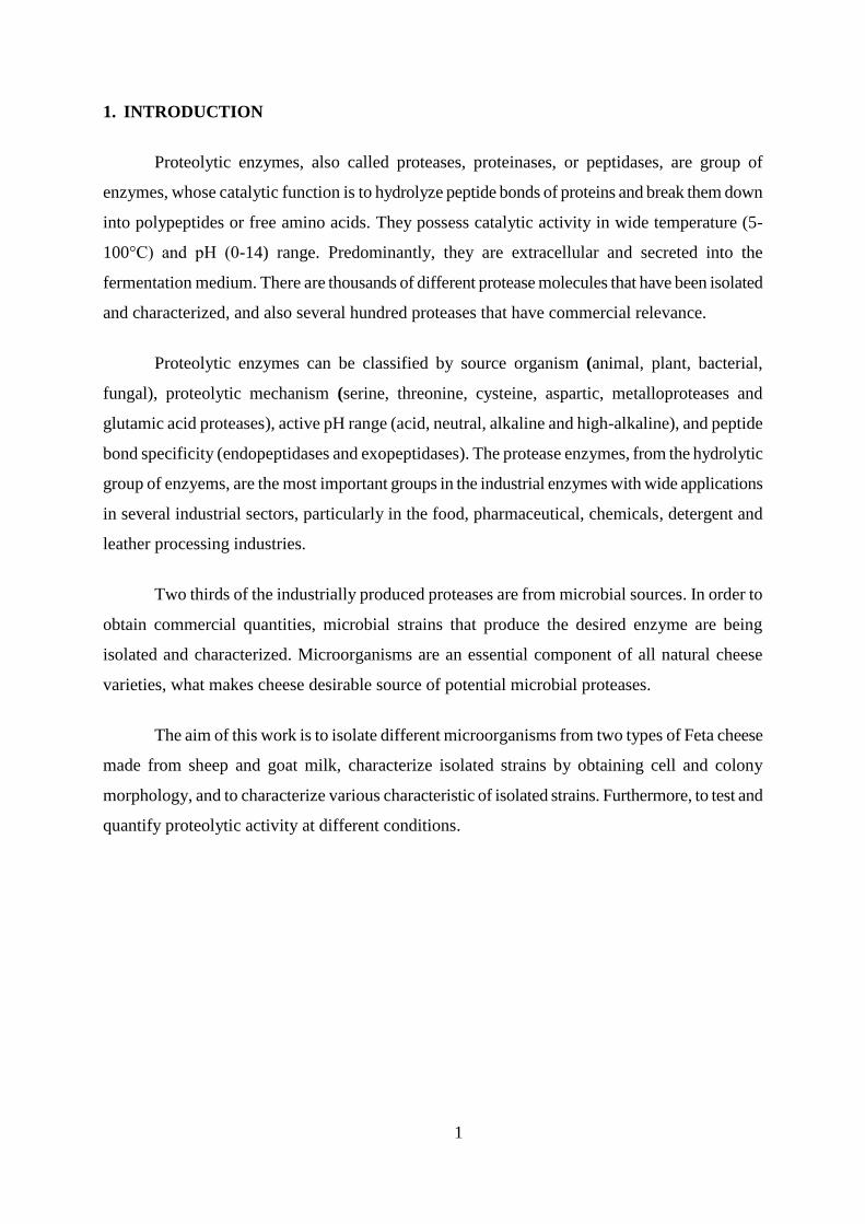
Fig. 1. The mechanism of protease hydrolysis (Source: Güracar, 2011)
2.1.1. Plant Proteases
Production of proteases from plants is limited by climatic conditions and the availability
of land for cultivation, what makes production a time-consuming process. Also, processing of
large amounts of plant material is necessary since the plant tissue contains low concentration of
enzyme (Veloorvalappil et al., 2013). Papain, bromelain, keratinases, and ficin represent some of
the well-known proteases of plant origin. The performance of the enzyme depends on the plant
source, the climatic conditions for growth, and the methods used for its extraction and
purification (Pandey et al., 2006). Plant proteases contain sulfhydryl groups in the active site
which are responsible for the catalytic activity and used as meat tenderizers and in other food
applications (Uhlig, 1998).
2.1.2. Animal Proteases
The main characteristic of animal proteases is their ability to occur as inactive precursor in
producing organs until they are secreted into the digestive tract where they activate to the active
form. The most familiar proteases of animal origin are pancreatic trypsin, chymotrypsin, pepsin,
and rennin. However, availability of livestock for slaughter determinate their production (Pandey
et al., 2006). They are used in the manufacture of protein hydrolysates, processing meat and fish
3
residues, in medicine as part of digestive medical aid preparations and also in leather
manufacturing industry (Uhlig, 1998).
2.1.3. Microbial Proteases
Microorganisms are the most common sources of commercial enzymes due to their
physiological and biochemical properties, wide biochemical diversity, ability of genetic
manipulation, the rapid growth of the microorganisms and limited space required for cell
cultivation. Among microbial enzymes, proteases are the most important for the industry, and
constitute approximately 60% of the total industrial enzyme market (Rodarte et al., 2011).
Proteolytic enzymes not only play a critical role in cellular metabolic processes but have
also gained considerable attention in the industrial community which leads to increasing interests
in the study of proteolytic enzymes. In order to obtain commercial quantities, microbial strains
that produce the desired enzyme are being isolated and characterized by using optimal conditions
for growth. Proteases can be secreted inside or outside of cell. Intracellular proteases playing an
important role in different cells and metabolism processes, while extracellular hydrolyze proteins
in fermented media and enable the cell to absorb and utilize hydrolytic products (Veloorvalappil
et al., 2013).
2.1.3.1. Bacterial proteases
Mostly, they are produced by genus Bacillus due to their vital importance in the industry
(Priest, 1977). Bacterial proteases are mainly neutral and alkaline. The bacterial neutral proteases
have low thermotolerance what represents advantageous in controlling their reactivity during the
production of food hydrolysates with a low degree of hydrolysis. They are active in pH range 5-8,
not sensitive to the natural plant proteinases and are, therefore, useful in the brewing industry.
One of the main characteristics is their high affinity for hydrophobic amino acids. Bacterial
alkaline proteases have wide substrate specificity, activity at alkaline pH (e.g. pH 10) and optimal
temperature around 60°C what makes them suitable for use in the detergent industry (Mala et. al.,
1998).
4
2.1.3.2. Fungal proteases
One of the many advantages of fungal proteases is the ability of fungi to grow on low cost
materials and secret enzymes into the culture medium, making them easily recoverable (Anitha
and Palanivelu, 2013). They are active over a wide pH range (pH 4 to 11), but have lower heat
tolerance than the bacterial enzymes what makes them useful in the cheese-making industry.
Fungal proteases are recognized as GRAS (generally regarded as safe) and are, therefore, safer
for usage than bacteria (Germano et al., 2003).
2.2. Classification of proteases
Proteases are divided into three groups, intracellular, periplasmic, and extracellular
proteases (Kohlmann et al., 1991). Proteases are of relevant importance in cellular, metabolic and
regulatory processes (intracellular) and in the hydrolysis of proteins in cell-free environments
(extracellular). Many factors influence production of intracellular or extracellular enzymes, such
as pH, temperature, nitrogen and carbon sources, inorganic salts, and dissolved oxygen
concentration (Gupta and Beg, 2002).
Depending on optimum pH, proteases are divided in acidic proteases, neutral proteases,
alkaline proteases and high-alkaline proteases. Acidic proteases require alkaline conditions for
their production and because of their catalytic nature they have wide range of application. They
are found in animal cells, moulds, yeasts and rarely bacteria (Asokan, 2010). Neutral proteases
comprise cystein proteases (papain, bromelain, ficin) isolated from botanical origin and the
metalloproteases. They require neutral and low thermal conditions (Rao et al., 1998;
Siddalingeshwara et. al., 2010).
Depending on their site of action, proteases are divided into two groups: exopeptidases or
endopeptidases, according to the Enzyme Commission (EC) classification (García-Carreńo,
1993). If the peptide bond is cleaved closer to the amino or carboxy terminus of the substrate,
enzymes are classified as exopeptidases. If the enzyme cleaves peptide bonds in the inner regions
of peptide chains, distant from the termini, they are classified as endopeptidases (also known as
proteinases) (Güracar, 2011).
5
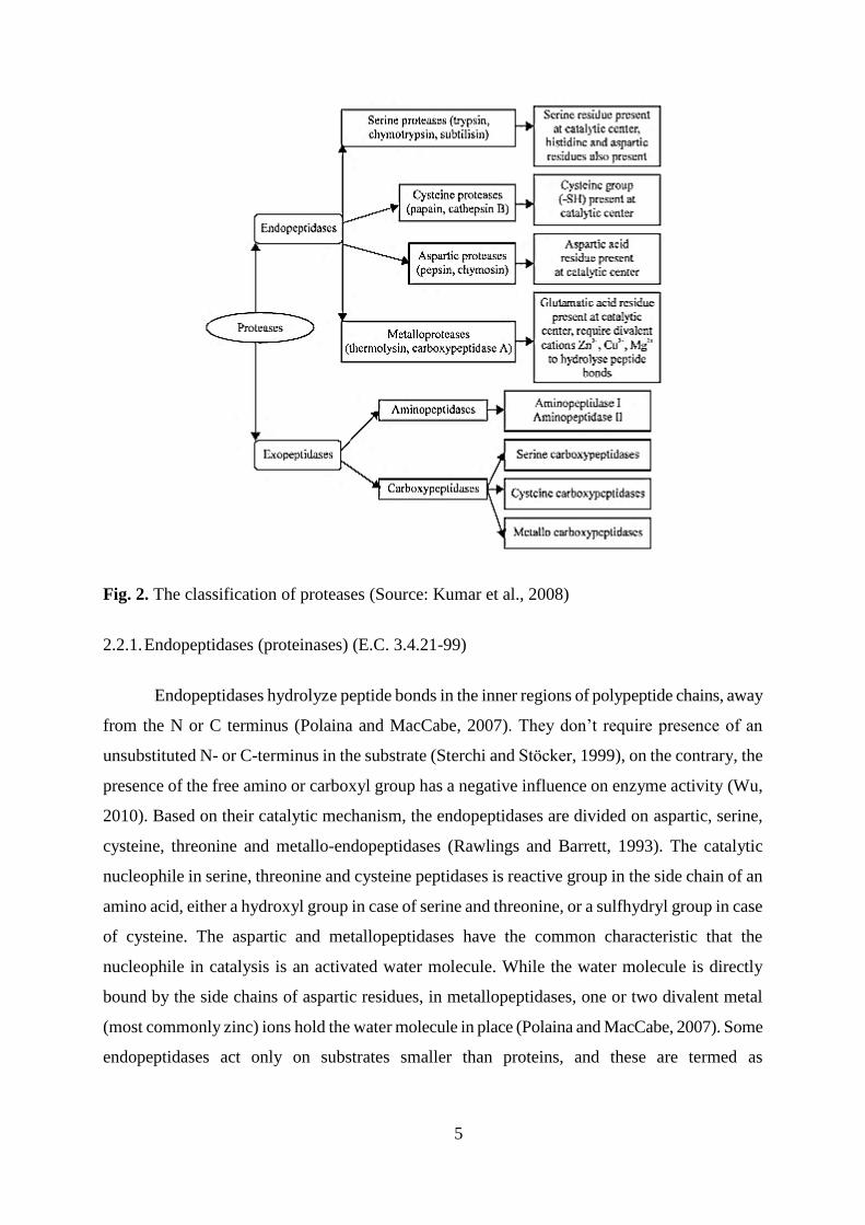
Fig. 2. The classification of proteases (Source: Kumar et al., 2008)
2.2.1. Endopeptidases (proteinases) (E.C. 3.4.21-99)
Endopeptidases hydrolyze peptide bonds in the inner regions of polypeptide chains, away
from the N or C terminus (Polaina and MacCabe, 2007). They don’t require presence of an
unsubstituted N- or C-terminus in the substrate (Sterchi and Stöcker, 1999), on the contrary, the
presence of the free amino or carboxyl group has a negative influence on enzyme activity (Wu,
2010). Based on their catalytic mechanism, the endopeptidases are divided on aspartic, serine,
cysteine, threonine and metallo-endopeptidases (Rawlings and Barrett, 1993). The catalytic
nucleophile in serine, threonine and cysteine peptidases is reactive group in the side chain of an
amino acid, either a hydroxyl group in case of serine and threonine, or a sulfhydryl group in case
of cysteine. The aspartic and metallopeptidases have the common characteristic that the
nucleophile in catalysis is an activated water molecule. While the water molecule is directly
bound by the side chains of aspartic residues, in metallopeptidases, one or two divalent metal
(most commonly zinc) ions hold the water molecule in place (Polaina and MacCabe, 2007). Some
endopeptidases act only on substrates smaller than proteins, and these are termed as
6
oligopeptidases. An example of an oligopeptidase is thimet oligopeptidase (Rawlings and Barrett,
1993). Endopeptidases initiate the digestion of food proteins, generating new N- and C-termini
that are substrates for the exopeptidases that complete the process (Polaina and MacCabe, 2007).
2.2.1.1. Serine Proteases (E.C. 3.4.21)
Serine proteases are the most researched and understood group of proteolytic enzymes
characterized by the essential serine residue that acts as a nucleophile in the active site. They are
widely distributed and spread among viruses, bacteria and eukaryotes and can be found among
exopeptidases, endopeptidases, oligopeptidases and omega peptidases (Güracar, 2011). Based on
structure, they can be divided into a series of subfamilies. In comparison with other
microorganisms, bacteria are the best known producers of serine proteases, where bacterial
subtilisins have wide application as detergent additives (Maheshwari et al., 2000). The production
of serine proteases usually occurs at neutral (pH 7) and alkaline (pH 11) conditions, while their
isoelectric points rang between pH 4 and 6. They generally have low molecular weight between
18,5-35 kDa, and can be recognized by irreversible inhibition by di-isopropyl fluorophosphates
(DFP) and phenyl methyl sulfonyl fluoride (PMSF) (Ellaiah et al., 2002).
2.2.1.2. Cysteine/Thiol Proteases (E.C. 3.4.22)
Cysteine proteases are characterized by the presence of a cysteine (SH-) and histidine
residues at the active site (Garcia-Carreno, 1992) and produced by prokaryotes and eukaryotes
where they occur in only a few fungi (Kalisz, 1988). They are divided into twenty families
depending on the differences in the order of cysteine and histidine residues (Barett, 1994), and in
four groups based on their side chain specificity: papain-like, trypsin-like, glutamic acid-like and
others (Güracar, 2011). Cysteine proteases have neutral pH-optima and they are active only in the
presence of reducing agents such as potassium cyanide or cysteine, EDTA or dithiothreitol
(Maheshwari et al., 2000). They have molecular masses between 30 and 40 kDa, isoelectric
points from pH 4,9 to pH 8,4 (Rao et al., 1998), and are inhibited by thiol reagents (heavy metals,
alkylating-oxidizing agents), as well as by sulphydryl reagents (chloromercuribenzoate and
iodoacetamide) (Zeigler, 2001).
7
2.2.1.3. Aspartic Proteases (Acidic Proteases) (E.C. 3.4.23)
Aspartic proteases are a group of proteases which contain an aspartic acid residue in their
active sites. Acidic conditions are required for their production and they show maximum activity
at acidic pH, between 3 and 4. Mostly, they are found in fungi, rarely in bacteria (Ellaiah et al.,
2002). They can be divided into three groups, pepsin, retropepsin and enzymes from
pararetroviruses (Güracar, 2011). Their molecular masses range between 30 and 40 kDa, while
isoelectric points are between pH 3,4 and 4,6. Aspartic proteases are not sensitive to inhibitors of
other three groups of enzymes, but are sensitive to diazo-ketone and epoxy compounds in the
presence of copper cations, such as diazoacetyl norleucine methyl ester (DAN), and 1, 2-
epoxypnitrophenoxy propane (EPNP) (Rao et al., 1998; Zeigler, 2001).
2.2.1.4. Metalloproteases (E.C. 3.4.24)
Metalloproteases require divalent metal ions (catalytic zinc, manganese, cobalt, nickel or
copper) for biological activity. The metal ion is complexed by three conserved amino acid
residues that can be glutamic acid (Glu), aspartic acid (Asp), histidin (His) or lysin (Lys) in their
active sites. They are widespread and the most diverse of the catalytic types of proteases (Barett,
1995). Zinc is essential for their activity (one atom of zinc per molecule of enzyme), while
calcium is important for structure stability. According to the amino acid sequences and the
relation between the amino acids and the metal binding sites, they are divided into thirty families.
According to the catalytic action they are neutral, alkaline, Myxobacter I and Myxobacter II
(Ellaiah et al., 2002). Metalloproteases have maximum activity at neutral to alkaline pH, in the
range of 5-9, with molecular masses between 19 and 37 kDa They are sensitive to metal-chelating
reagents, for example EDTA (Ziegler, 2001).
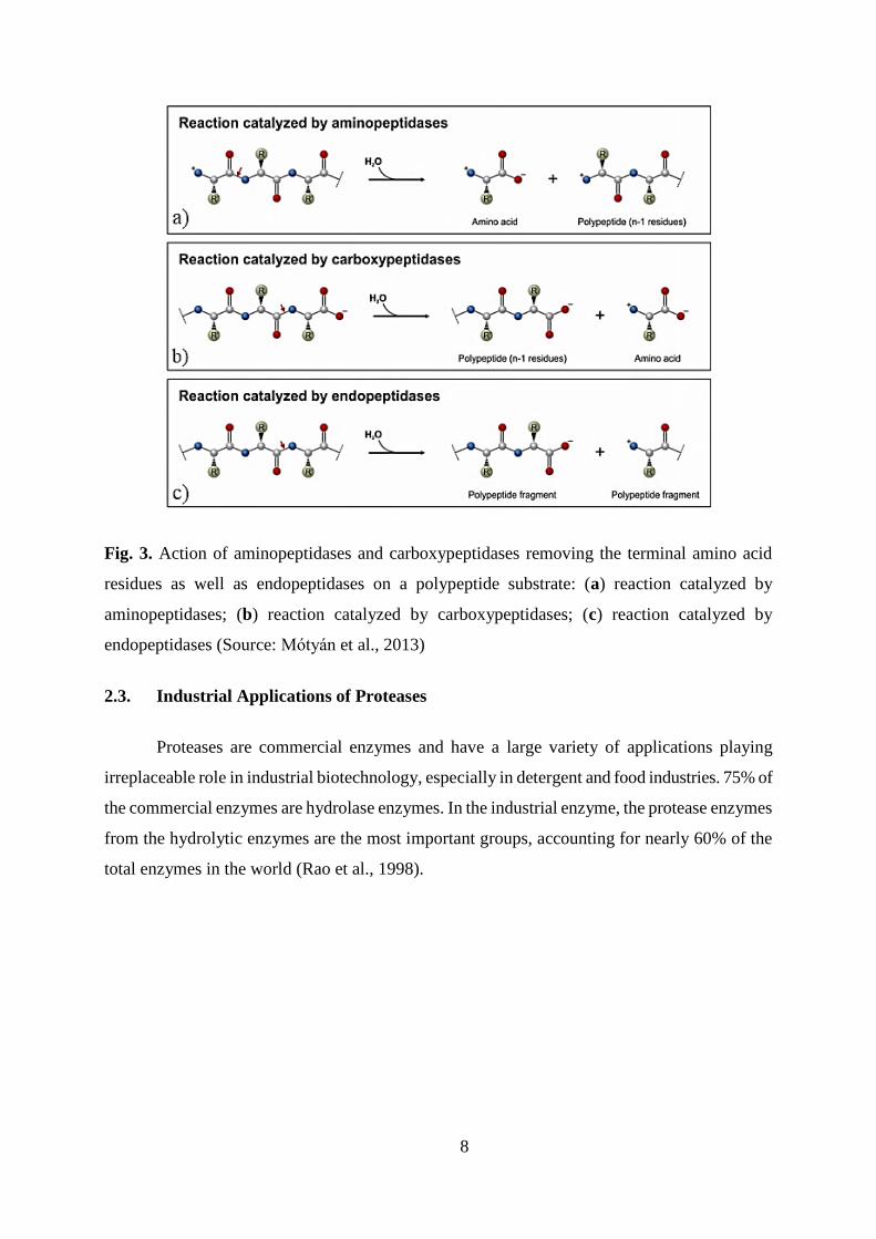
2.2.2. Exopeptidases (peptidases) (E.C. 3.4.11-19)
Exopeptidases are the enzymes which cleave peptide bonds in the terminal amino end or
carboxylic end of the substrate (Garcia-Carreno, 1992). They remove single amino acid, dipeptide
or tripeptide from the N- or C-terminus. The exopeptidases that act at a free N-terminus are
named aminopeptidases. They are generally intracellular enzymes, widespread among microbial
species. Exopeptidases acting at a free C-terminus are called carboxypeptidases. Based on the
nature of the amino acid residues at the active site of the proteases, they are divided into serine,
metal and cistein carboxypeptidases (Mekashwari et al., 2010).
8
Fig. 3. Action of aminopeptidases and carboxypeptidases removing the terminal amino acid
residues as well as endopeptidases on a polypeptide substrate: (a) reaction catalyzed by
aminopeptidases; (b) reaction catalyzed by carboxypeptidases; (c) reaction catalyzed by
endopeptidases (Source: Mótyán et al., 2013)
2.3. Industrial Applications of Proteases
Proteases are commercial enzymes and have a large variety of applications playing
irreplaceable role in industrial biotechnology, especially in detergent and food industries. 75% of
the commercial enzymes are hydrolase enzymes. In the industrial enzyme, the protease enzymes
from the hydrolytic enzymes are the most important groups, accounting for nearly 60% of the
total enzymes in the world (Rao et al., 1998).
9
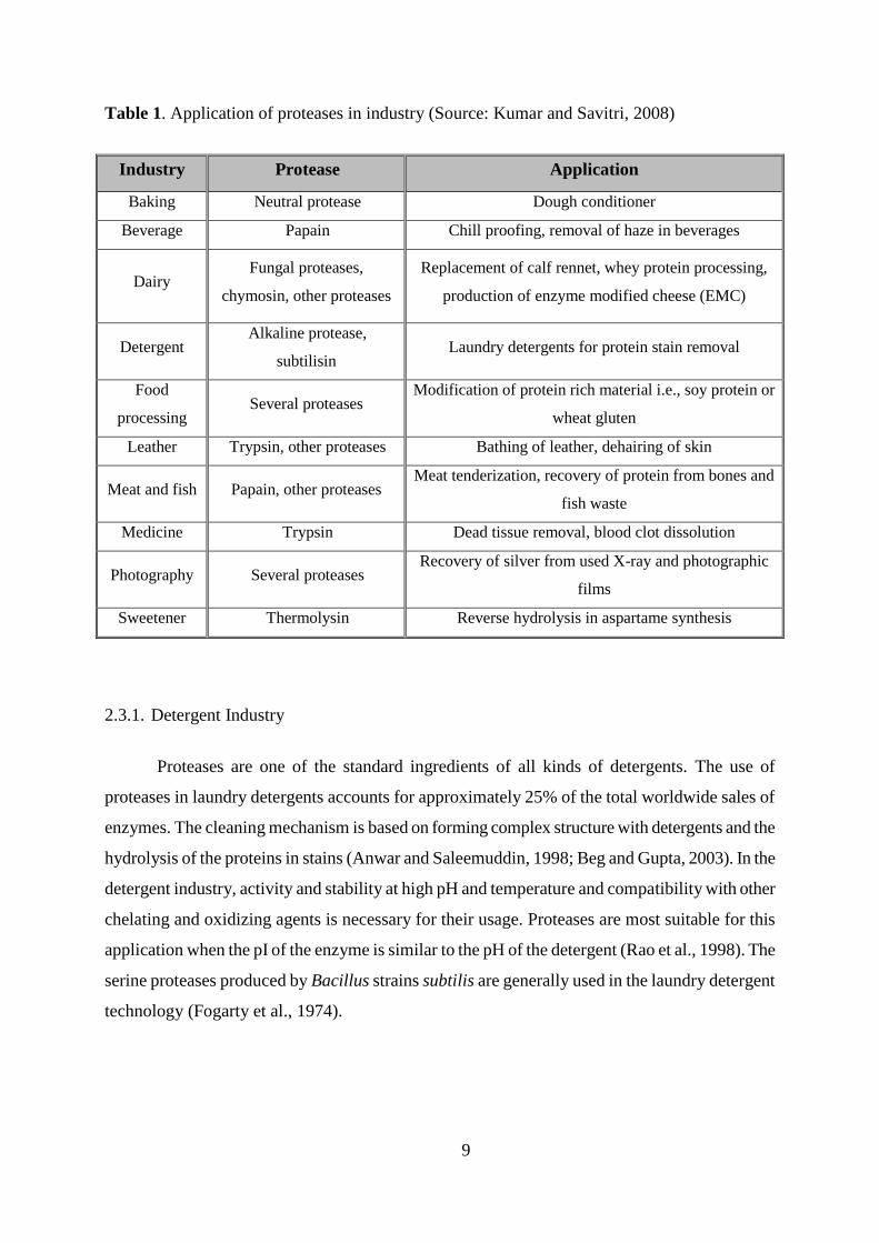
Table 1. Application of proteases in industry (Source: Kumar and Savitri, 2008)
Industry Protease Application
Baking Neutral protease Dough conditioner
Beverage Papain Chill proofing, removal of haze in beverages
Dairy Fungal proteases,
chymosin, other proteases
Replacement of calf rennet, whey protein processing,
production of enzyme modified cheese (EMC)
Detergent Alkaline protease,
subtilisin Laundry detergents for protein stain removal
Food
processing Several proteases
Modification of protein rich material i.e., soy protein or
wheat gluten
Leather Trypsin, other proteases Bathing of leather, dehairing of skin
Meat and fish Papain, other proteases Meat tenderization, recovery of protein from bones and
fish waste
Medicine Trypsin Dead tissue removal, blood clot dissolution
Photography Several proteases Recovery of silver from used X-ray and photographic
films
Sweetener Thermolysin Reverse hydrolysis in aspartame synthesis
2.3.1. Detergent Industry
Proteases are one of the standard ingredients of all kinds of detergents. The use of
proteases in laundry detergents accounts for approximately 25% of the total worldwide sales of
enzymes. The cleaning mechanism is based on forming complex structure with detergents and the
hydrolysis of the proteins in stains (Anwar and Saleemuddin, 1998; Beg and Gupta, 2003). In the
detergent industry, activity and stability at high pH and temperature and compatibility with other
chelating and oxidizing agents is necessary for their usage. Proteases are most suitable for this
application when the pI of the enzyme is similar to the pH of the detergent (Rao et al., 1998). The
serine proteases produced by Bacillus strains subtilis are generally used in the laundry detergent
technology (Fogarty et al., 1974).
10
2.3.2. Food Industry
The food industries are the major protease using industries. They have been routinely used
for various purposes such as cheese making, baking, preparation of soya hydrolysates, and meat
tenderization (Cheong et al., 1993).
2.3.2.1. Dairy industry
The major application of proteases in the dairy industry is in the manufacture of
cheese. The proteases are used as milk-coagulation agents for cheese production. Advantages of
using proteases in dairy industry is ability of staying at higher temperatures in the pasteurization
and obtaining high quality of milk in the dairy industry (Meer et al., 1991).
2.3.2.2. Baking industry
Endo- and exoproteinases from Aspergillus oryzae have been used to modify wheat gluten
by limited proteolysis. Enzymatic treatment of the dough improves handling and machining and
permits the production of a wider range of products. The addition of proteases reduces the mixing
time, improve the extensibility and strength of the dough (Argos, 1987).
2.3.3. Other applications
Proteases from the Bacillus species are generally used in brewing industry in order to
cleave peptide bonds in proteins. In the leather industry they are used instead of hazardous
chemicals (sodium sulfide) to prevent the pollution problems, obtain high quality, easy control,
speed up dehairing and reduce the waste materials. The usage of these enzymes is also common
in developing therapeutic agents, preparation of medicines, medical diagnosis, biopharmaceutical
products (contact-lens enzyme cleaners, enzymatic debriders) and cosmetics (skin care ointments)
(Anwar and Saleemuddin, 2000 ).
2.4. Bacteria and yeasts in cheese
Microorganisms are an essential component of all natural cheese varieties as the major
contributor to cheese flavor, aroma, texture, and appearance (Beresford et al., 2001; Irlinger and
Mounier, 2009; Ogier et al., 2002). The variety and number of specific types of organisms present
in cheese depend on the microbial quality of the milk, heat treatment of the milk, manufacturing
11
conditions, temperature and humidity during ripening, amount of salting, and exposure of the
cheese to exogenous microorganisms during and after manufacture (Torkar and Teger, 2006).
In cheese making, two types of cultures are used. The primary cultures include all the
starter lactic acid bacteria responsible for acid production during manufacture and in cheese
ripening. The secondary cultures do not contribute to acid production during manufacture, but
generally play a significant role during ripening. The secondary culture is composed of non-
starter lactic acid bacteria which grow internally in most cheese varieties and other bacteria,
yeasts and molds, which grow internally or externally and are usually unique to specific cheese
types. During cheese ripening, the starter culture, along with the secondary flora promotes a
complex series of biochemical reactions which are vital for proper development of flavor and
texture (Beresford et al., 2001).
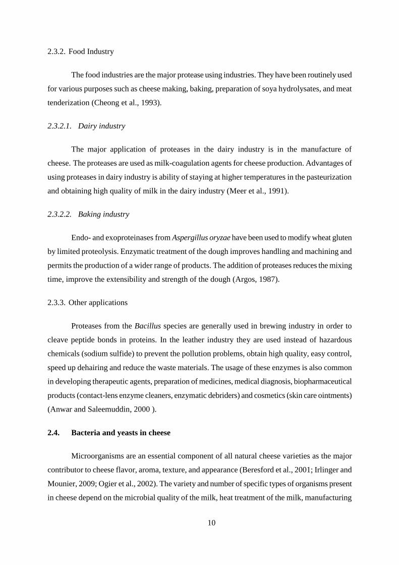
During ripening, proteolysis in cheese is catalyzed by enzymes from coagulant (e.g.,
chymosin, pepsin, microbial or plant acid proteinases), milk (plasmin), enzymes from the starter,
nonstarter, or secondary cultures and exogenous proteinases or peptidases (fig 4). Proteolysis
contributes to the taste of cheese by the production of peptides and free amino acids. Large
peptides do not contribute directly to cheese flavor, but are important for the development of the
correct texture. Engels and Visser (1994) analyzed water-soluble fractions from Cheddar, Edam,
Gouda, Gruyere, Maasdam, Parmesan and Proosdij cheeses and suggested that low-molecular
compounds (small peptides, amino acids, free fatty acids or their breakdown products) were
responsible for the basic taste of cheese.
Fig. 4. Proteolytic agents in cheese during ripening (Source: Sousa et al., 2001)
12
The starter is not only responsible for acid metabolism but also for metabolic activities in
sensory properties of cheese. They are often used in cheese because of their proteolytic
characteristics which contributes flavor development. However, compared to yeasts and molds,
starter bacteria are very weakly proteolytic (Donnelly, 2014). The starter cultures commonly used
in cheese manufacture include mesophilic Lactococcus and Leuconostoc species, thermophilic
Lactobacillus species and Streptococcus thermophilus. Although lactic acid bacteria (LAB) are
weakly proteolytic, they possess proteinase/peptidase system capable of hydrolyzing
oligopeptides to small peptides and amino acids (Pederson and Steele, 1999).
The secondary and adjunct cultures include yeasts (e.g., Geotrichum candidum), moulds
(e.g., Penicilium camemberti) and bacteria (e.g. Micrococcus), and grow mainly on the cheese
surface. Yeasts are used mainly in mould and bacterial surface-ripened cheeses because they
promote the growth of other microorganisms. They colonise numerous cheeses, particulary their
surface and show a large diversity in proteolytic activity between species and strains of the same
species. They have caseinolytic, aminopeptidase and carboxypeptidase activities (Fox et al.,
2004).
Bacteria cause fermentation of milk and are therefore considered to be of major
importance during cheese making (Cousin, 1982). Yeasts, however, possess the ability to grow
under conditions unfavorable to many bacteria and play an important role in the spoilage of dairy
products (Fleet and Mian, 1987; Seiler and Busse, 1990). Role of yeasts as spoilage organisms is
related to their nutritional requirements, growth at low temperatures, low pH values, low water
activities and high salt concentration. However, in some cases yeasts may contribute positively to
the fermentation and maturation process of cheeses by inhibiting undesired microorganisms,
supporting the function of the starter culture by proteolytic activity, lipolytic activity and
metabolizing lactic acid leading to an increase in pH (Welthagen and Viljoen, 1998).
13
3. MATERIALS AND METHODS
3.1. Materials
3.1.1. Cheese samples for isolation
For the purpose of this work, two types of Feta cheese made from sheep and goat milk
were used. They were originated from a Újpest Marketplace in Budapest, Hungary. Cheeses were
refrigerated at 4ºC.
3.1.2. Growth media and solutions
Different media and solutions were sterilized at 121 °C for 15 minutes (if otherwise not
stated). Media with agar were cooled down to 40-50 °C in water bath and were poured in sterile
Petri dishes (20-25 ml into each dish). Media were left to solidify on flat surface and after
solidification they were turned upside down to prevent condense water forming. Isolated strains
were maintained on agar slops and stored at 4 ºC.
Main components of growth media are peptone (provides nitrogen, vitamins, minerals
and amino acids essential for growth), glucose (fermentable carbohydrate providing carbon and
energy), yeast extract (source of vitamins, particularly of the B-group; minerals) and
bacteriological agar (solidifying agent).
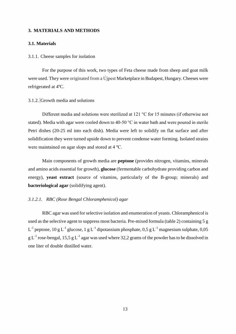
3.1.2.1. RBC (Rose Bengal Chloramphenicol) agar
RBC agar was used for selective isolation and enumeration of yeasts. Chloramphenicol is
used as the selective agent to suppress most bacteria. Pre-mixed formula (table 2) containing 5 g
L-1
peptone, 10 g L-1
glucose, 1 g L-1
dipotassium phosphate, 0,5 g L-1
magnesium sulphate, 0,05
g L-1
rose-bengal, 15,5 g L-1
agar was used where 32,2 grams of the powder has to be dissolved in
one liter of double distilled water.
14
Table 2. Composition of RBC agar for selective isolation and enumeration of yeasts
Ingredients Concentration [g L-1
]
Mycological peptone 5,0
Glucose 10,0
Dipotassium phosphate 1,0
Magnesium sulphate 0,5
Rose-Bengal 0,05
Chloramphenicol 0,1
Agar 15,5
pH 7,2 ± 0,2 (at 25°C)
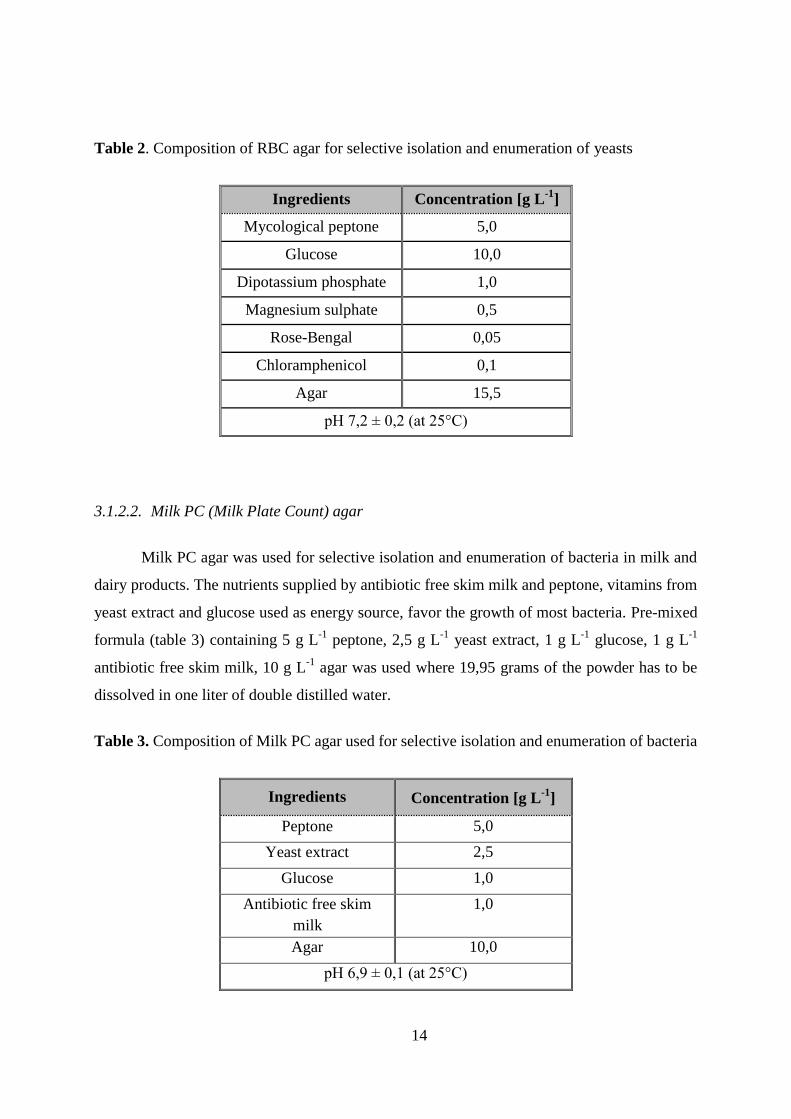
3.1.2.2. Milk PC (Milk Plate Count) agar
Milk PC agar was used for selective isolation and enumeration of bacteria in milk and
dairy products. The nutrients supplied by antibiotic free skim milk and peptone, vitamins from
yeast extract and glucose used as energy source, favor the growth of most bacteria. Pre-mixed
formula (table 3) containing 5 g L-1
peptone, 2,5 g L-1
yeast extract, 1 g L-1
glucose, 1 g L-1
antibiotic free skim milk, 10 g L-1
agar was used where 19,95 grams of the powder has to be
dissolved in one liter of double distilled water.
Table 3. Composition of Milk PC agar used for selective isolation and enumeration of bacteria
Ingredients Concentration [g L-1
]
Peptone 5,0
Yeast extract 2,5
Glucose 1,0
Antibiotic free skim
milk
1,0
Agar 10,0
pH 6,9 ± 0,1 (at 25°C)
15
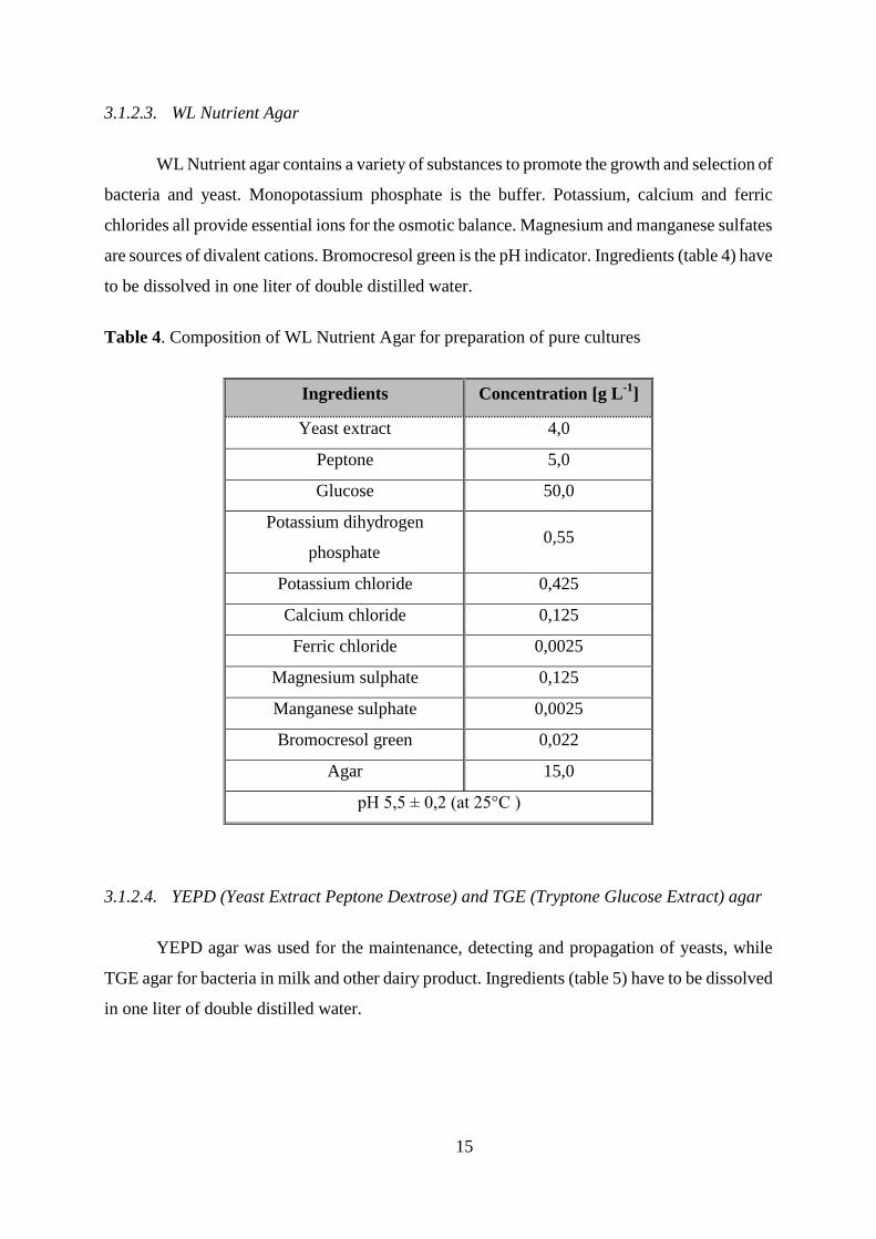
3.1.2.3. WL Nutrient Agar
WL Nutrient agar contains a variety of substances to promote the growth and selection of
bacteria and yeast. Monopotassium phosphate is the buffer. Potassium, calcium and ferric
chlorides all provide essential ions for the osmotic balance. Magnesium and manganese sulfates
are sources of divalent cations. Bromocresol green is the pH indicator. Ingredients (table 4) have
to be dissolved in one liter of double distilled water.
Table 4. Composition of WL Nutrient Agar for preparation of pure cultures
Ingredients Concentration [g L-1
]
Yeast extract 4,0
Peptone 5,0
Glucose 50,0
Potassium dihydrogen
phosphate 0,55
Potassium chloride 0,425
Calcium chloride 0,125
Ferric chloride 0,0025
Magnesium sulphate 0,125
Manganese sulphate 0,0025
Bromocresol green 0,022
Agar 15,0
pH 5,5 ± 0,2 (at 25°C )
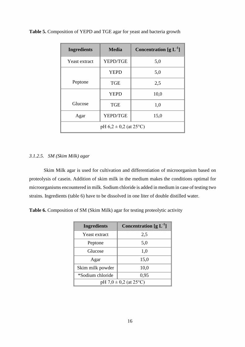
3.1.2.4. YEPD (Yeast Extract Peptone Dextrose) and TGE (Tryptone Glucose Extract) agar
YEPD agar was used for the maintenance, detecting and propagation of yeasts, while
TGE agar for bacteria in milk and other dairy product. Ingredients (table 5) have to be dissolved
in one liter of double distilled water.
16
Table 5. Composition of YEPD and TGE agar for yeast and bacteria growth
Ingredients Media Concentration [g L-1
]
Yeast extract YEPD/TGE 5,0
Peptone
YEPD 5,0
TGE 2,5
Glucose
YEPD 10,0
TGE 1,0
Agar YEPD/TGE 15,0
pH 6,2 ± 0,2 (at 25°C)
3.1.2.5. SM (Skim Milk) agar
Skim Milk agar is used for cultivation and differentiation of microorganism based on
proteolysis of casein. Addition of skim milk in the medium makes the conditions optimal for
microorganisms encountered in milk. Sodium chloride is added in medium in case of testing two
strains. Ingredients (table 6) have to be dissolved in one liter of double distilled water.
Table 6. Composition of SM (Skim Milk) agar for testing proteolytic activity
Ingredients Concentration [g L-1
]
Yeast extract 2,5
Peptone 5,0
Glucose 1,0
Agar 15,0
Skim milk powder 10,0
*Sodium chloride 0,95
pH 7,0 ± 0,2 (at 25°C)
17
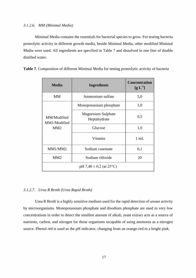
3.1.2.6. MM (Minimal Media)
Minimal Media contains the essentials for bacterial species to grow. For testing bacteria
proteolytic activity in different growth media, beside Minimal Media, other modified Minimal
Media were used. All ingredients are specified in Table 7 and dissolved in one liter of double
distilled water.
Table 7. Composition of different Minimal Media for testing proteolytic activity of bacteria
Media Ingredients Concentration
[g L-1
]
MM Ammonium sulfate 5,0
MM/Modified
MM1/Modified
MM2
Monopotassium phosphate 1,0
Magnesium Sulphate
Heptahydrate 0,5
Glucose 1,0
Vitamin 1 mL
MM1/MM2 Sodium caseinate 0,1
MM2 Sodium chloride 20
pH 7,40 ± 0,2 (at 25°C)
3.1.2.7. Urea R Broth (Urea Rapid Broth)
Urea R Broth is a highly sensitive medium used for the rapid detection of urease activity
by microorganisms. Monopotassium phosphate and disodium phosphate are used in very low
concentrations in order to detect the smallest amount of alkali, yeast extract acts as a source of
nutrients, carbon, and nitrogen for those organisms incapable of using ammonia as a nitrogen
source. Phenol red is used as the pH indicator, changing from an orange-red to a bright pink.
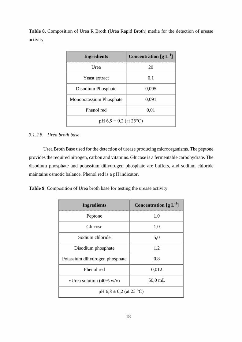
18
Table 8. Composition of Urea R Broth (Urea Rapid Broth) media for the detection of urease
activity
Ingredients Concentration [g L-1
]
Urea 20
Yeast extract 0,1
Disodium Phosphate 0,095
Monopotassium Phosphate 0,091
Phenol red 0,01
pH 6,9 ± 0,2 (at 25°C)
3.1.2.8. Urea broth base
Urea Broth Base used for the detection of urease producing microorganisms. The peptone
provides the required nitrogen, carbon and vitamins. Glucose is a fermentable carbohydrate. The
disodium phosphate and potassium dihydrogen phosphate are buffers, and sodium chloride
maintains osmotic balance. Phenol red is a pH indicator.
Table 9. Composition of Urea broth base for testing the urease activity
Ingredients Concentration [g L-1
]
Peptone 1,0
Glucose 1,0
Sodium chloride 5,0
Disodium phosphate 1,2
Potassium dihydrogen phosphate 0,8
Phenol red 0,012
Urea solution (40% w/v) 50,0 mL
pH 6,8 ± 0,2 (at 25 °C)
19
3.1.3. Solutions used for serial dilution and sample processing
Solution used for serial dilution and sample processing contained 1,0 g L-1
casein peptone
and 8,5 g L-1
NaCl. For serial dilution 9 ml of prepared solution was distributed into tubes before
sterilization.
3.1.4. Chemicals and components for growth media and solutions
All used chemicals were of the laboratory grade of purity.
3.1.5. Equipment and apparatus
3.1.5.1. Densitometer
McFarland Densitometer DEN-1B (bioSan, Latvia) (suspension turbidity meter) was used
for measuring turbidity of cell suspensions. It is calibrated to measure turbidity in the range of
0,5 to 4,0 McFarland units (0,5 McFarland Standard for bacteria and 2,5 McFarland Standard for
yeasts) with a small standard deviation.
3.1.5.2. Laboratory centrifuge
Laboratory centrifuge Jouan BR4i (Thermo Scientific Labor, Hungary) was used for
preparing extracellular cells samples for testing proteolytic activity of strains in different media
and for spectrophotometric measurements.
3.1.5.3. Spectrophotometer and Multiskan machine
Using UV/Vis Spectrophotometer SPECORD 200 PLUS (Analytik Jena, Germany)
apsorbance at 280 and 366 nm were determined for measuring concentration of proteins.
Thermo Multiskan Ascent is a microplate photometer (Labexchange, Germany) used for
measuring concentrations of proteins in different growth media chapter.
3.1.6. Other equipment and apparatus
Table 10 shows the list of equipment and apparatus used in this research. Also, other
equipment was used, including basic laboratory equipment, Erlenmeyer flask, Eppendorf tubes,
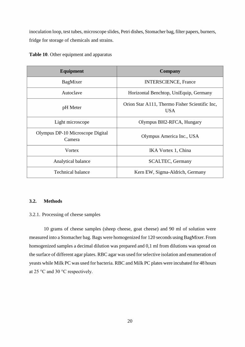
20
inoculation loop, test tubes, microscope slides, Petri dishes, Stomacher bag, filter papers, burners,
fridge for storage of chemicals and strains.
Table 10. Other equipment and apparatus
Equipment Company
BagMixer INTERSCIENCE, France
Autoclave Horizontal Benchtop, UniEquip, Germany
pH Meter Orion Star A111, Thermo Fisher Scientific Inc,
USA
Light microscope Olympus BH2-RFCA, Hungary
Olympus DP-10 Microscope Digital
Camera Olympus America Inc., USA
Vortex IKA Vortex 1, China
Analytical balance SCALTEC, Germany
Technical balance Kern EW, Sigma-Aldrich, Germany
3.2. Methods
3.2.1. Processing of cheese samples
10 grams of cheese samples (sheep cheese, goat cheese) and 90 ml of solution were
measured into a Stomacher bag. Bags were homogenized for 120 seconds using BagMixer. From
homogenized samples a decimal dilution was prepared and 0,1 ml from dilutions was spread on
the surface of different agar plates. RBC agar was used for selective isolation and enumeration of
yeasts while Milk PC was used for bacteria. RBC and Milk PC plates were incubated for 48 hours
at 25 °C and 30 °C respectively.
21
3.2.2. Preparation of pure cultures and characterization of isolates
Single colonies from RBC and Milk PC agar were streaked using a loop on WL nutrient
media to obtain single colonies. Plates were incubated for 3 days at 25 °C.
3.2.3. Strain maintenance
Yeast and bacterial isolates were inoculated on YEPD and TGE slopes (5 ml of agar was
distributed into tubes before sterilization). After autoclaving, agar in tubes was left to solidify
slanting the tubes to obtain agar slopes, respectively and incubated at 25 °C. After growth, slopes
were stored at 4 °C.
3.2.4. Microscopic investigation of isolates
3.2.4.1. Preparation of native dissection in case of yeast isolates
Yeast suspensions were prepared from one day old yeast cultures (one loopful of cells was
transferred into 0,5 ml sterile double distilled water) from which 10 µl were pipetted onto
microscope slides. Cover-slides were laid on the smears and investigated under light-microscope.
3.2.4.2. Gram-staining of bacterial isolates
Smears were prepared on microscope slides using 10 µl of sterile double distilled water
and a small amount of one day old bacteria colonies. Smears were air dried and heat fixed before
staining. Cells were stained with crystal violet dye for two minutes and rinsed with distilled
water. Lugol solution (iodine and potassium iodide) was added as mordant. After one minute
slides were rinsed with distilled water. Ethanol was dropped onto tilted slides for a few seconds to
decolorize Gram-negative bacteria. After differentiation slides were rinsed with distilled water
immediately. Safranin was used for counterstaining. Cells were stained for two minutes and
rinsed with distilled water. Slides were air dried and investigated by light-microscope.
22
3.2.5. KOH test
One drop of 3% KOH was pipetted onto a microscope slide. A loopful of one day old
bacteria culture was transferred into the KOH drop and stirred. Microbiological loop was raised
up and down to detect mucoid string formed by Gram-negative strains.
3.2.6. Testing the growth of isolates at different temperatures
Cell suspensions were prepared from one day old yeast and bacteria cultures (one loopful
of cells were transferred into 0,5 ml sterile double distilled water) from which 10 µl was pipetted
onto YEPD and TGE agar plates, respectively. Plates were incubated for three days at 10 ºC, 15 º
C, 20 ºC, 25 ºC, 30 ºC and 37 ºC.
3.2.7. Testing the proteolytic activity of isolates on SM Agar
Cell suspensions with adjusted density (0,5 McFarland Standard for bacteria and 2,5
McFarland Standard for yeasts) were prepared from one day old yeast and bacteria cultures. 10 µl
of each strain sample was inoculated on SM Agar plates. Plates were incubated for 3 days on 25
ºC.
3.2.8. Extracellular proteolytic activity of strains in different media
Strains who showed proteolytic activity were inoculated in different growth media to test
their proteolytic activity after 24, 48 and 72 h of incubation at 25 ºC in rotator. Media used for
this experiment were YEPD, YEPD+2% NaCl, MM, MM+2% NaCl, mMM1 and mMM2. 100 µl
of one day old cell suspension with adjusted density was incubated in 3 ml of each media. First,
second and third day 500 µl from each tube was mixed with 5 µl gentamicin and centrifuged at
14000 rpm, 5 min. 100 µl of supernatant was distributed in drilled circle holes in SM agar and
incubated at 25 ºC 72 h.
3.2.9. Multi scanning analysis
Cell suspensions of S-5a and S-5b bacteria strains with adjusted density were prepared
from one day old bacteria cultures and inoculated in TGE media with different concentration of
NaCl (0%, 0,5%, 1%, 1,5%, 2%) to test effect of NaCl on bacteria growth ability. 100 µl of
bacteria, 400 µl double distilled water and 500 µl TGE media with different concentration of

23
NaCl were prepared in Eppendorf tubes from which 300 µl was pipetted in kits and putted on
multi scanning analysis for 48 h.
3.2.10. Influence of NaCl on proteolytic activity of S-5b strain
One day old cell suspension of S-5b with adjusted density was inoculated in five tubes
with TGE media containing different concentration of NaCl (0%, 0,5%, 1%, 1,5%, 2%) and
incubated 24 h in rotator at 25º. 700 µl from each tube was distributed in Eppendorf tubes and
centrifuged at 14 000 rpm for 5 min after adding 7 µl of gentamicin. 100 µl of supernatant was
distributed in drilled circle holes in SM agar plates containing different concentration of NaCl
(0%, 0,5%, 1%, 1,5%, 2%). SM agar plates were incubated 48 h at 25 ºC.
3.2.11. Urea R Broth test
Components were added to distilled water and mixed thoroughly. Media was filtered and
distributed to Eppendorf tubes. One loop of one day old yeasts cells were aseptically distributed
into tubes. After incubation at 35 ºC, change of color in inoculated media was checked after 6 to 8
and 12 to 18 hours.
3.2.12. Urea Broth Base
Components were suspended in distilled water and sterilized by autoclaving for 15 min at
121°C. 40% sterile urea solution was added to media and mixed. 2 mL of prepared media was
distributed in tubes and inoculated with one loop of one day old yeasts cells while one tube was
left as control sample (without yeast cells). Tubes were incubated 24 h at 25°C in rotator. The
release of ammonia during urea hydrolysis will change the pH of the medium to alkaline,
converting the light-orange phenol red indicator to pink.
3.2.13. Catalase test and Oxidase test
Catalase test demonstrates the presence of catalase, an enzyme that catalyzes the release of
oxygen from hydrogen peroxide (H2O2). Small amount of one day old bacteria culture was
transferred with toothpick to 20 µl of 3% H2O2 on the surface of glass slide and mixed. The
presence of the enzyme in a bacterial isolate is evident when a small inoculum is introduced into
24
hydrogen peroxide, and the rapid elaboration of oxygen bubbles occurs. The lack of catalase is
evident by a lack or weak bubble production.
The oxidase test is used to determine if an organism possesses the cytochrome c oxidase
enzyme. Small amount of one day old bacteria culture was rubbed with toothpick on 10 µl of
Kovacs Oxidase Reagent (5% tetra-methyl-p-phenylenediamine dihydrochloride) dropped on
filter paper. Organisms which contain cytochrome c as part of their respiratory chain are oxidase-
positive and turn the reagent blue/purple appearing within 5-10 seconds. When the enzyme is not
present, the reagent remains reduced and is colorless.
3.2.14. Quantification of produced extracellular enzyme with spectrophotometric
measurement
3.2.14.1. Shaked and static bacterial cultures
Absorbance was measured with two different methods. Six bacteria strains (G-1, G-2b, G-
3, G-4, S-5b, S-7) were inoculated in 6 Erlenmeyer flask containing YEPD medium. Flasks were
incubated at 25 °C with and without slow rotation. After incubation, 1 mL from each culture was
centrifuged at 14000 rpm for 5 min and supernatant was used for measuring in
spectrophotometer. Absorbance was measured after 24, 48 and 72 h.
Method 1 – Sodium caseinat
1% casein sodium salt was prepared with 50mM Sorensen’s phosphate buffer (pH 7),
250μl of each supernatant was added in 500μl of casein solution and incubated for two hours at
30°C. Reaction was stopped with 375μl TCA (20% trichloroacetic acid). The tubes were placed
in an ice bath for 30 minutes and then centrifuged at 6000 rpm for 15 minutes at 4 °C. Blank
samples were prepared for each strain using 500μl of casein solution, 250μl of supernatant and
375μl TCA without previous incubation. Blank samples were placed in an ice bath for 30
minutes. Absorbance was measured at 280nm.
Method 2 – Azocasein
0,5% azocasein was dissolved in 0,1mol citrate buffer (pH 6) by boiling and then filtered.
100μl of culture supernatant was added to 500μl azocasein dissolvent and 500μl of citrate buffer.
25
After incubation at 30 ºC for 30 min, the reaction was stopped with 1,1 ml 10% TCA and left on
ice for 15 min. The samples were centrifuged at 6000 rpm 15 min and the absorbance of the
supernatant was measured at 366 nm in a spectrophotometer.
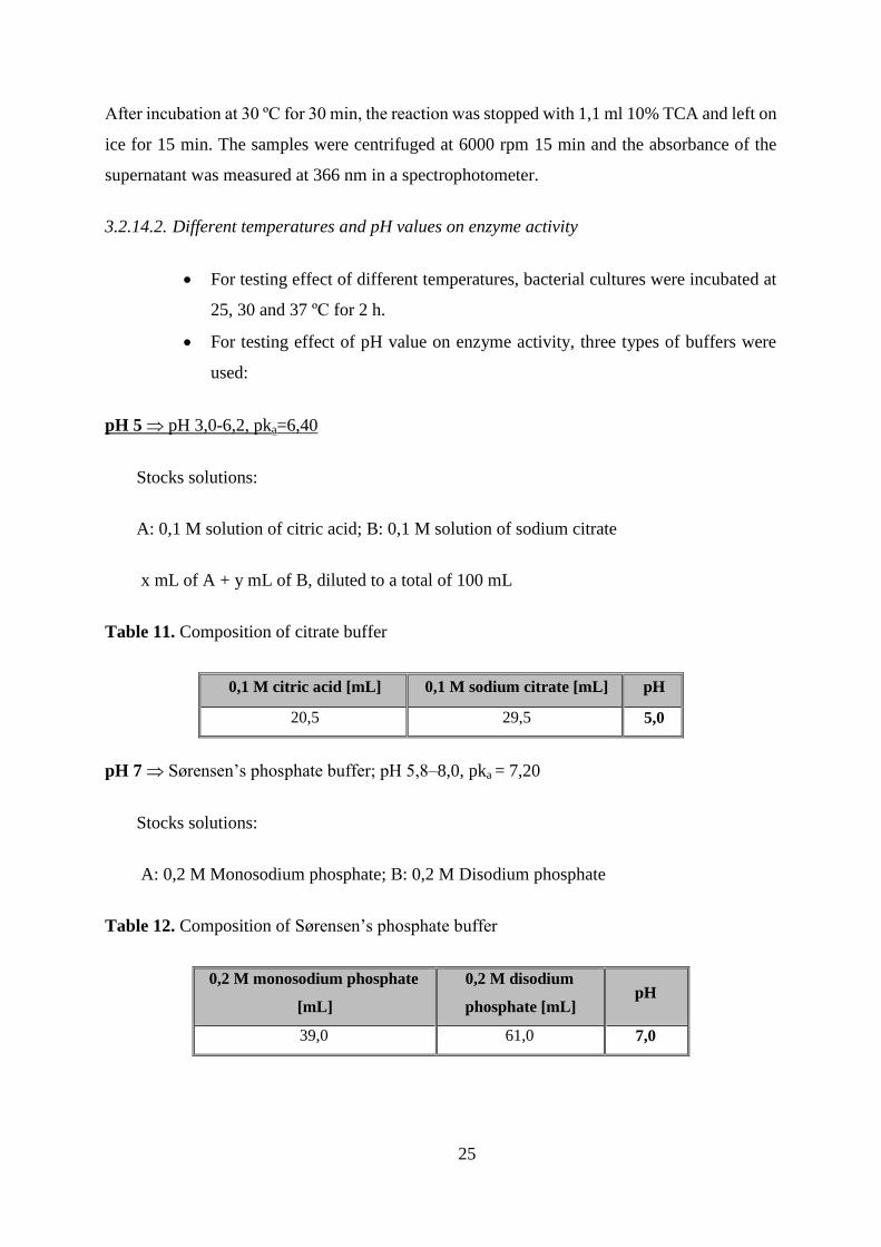
3.2.14.2. Different temperatures and pH values on enzyme activity
For testing effect of different temperatures, bacterial cultures were incubated at
25, 30 and 37 ºC for 2 h.
For testing effect of pH value on enzyme activity, three types of buffers were
used:
pH 5 pH 3,0-6,2, pka=6,40
Stocks solutions:
A: 0,1 M solution of citric acid; B: 0,1 M solution of sodium citrate
x mL of A + y mL of B, diluted to a total of 100 mL
Table 11. Composition of citrate buffer
0,1 M citric acid [mL] 0,1 M sodium citrate [mL] pH
20,5 29,5 5,0
pH 7 Sørensen’s phosphate buffer; pH 5,8–8,0, pka = 7,20
Stocks solutions:
A: 0,2 M Monosodium phosphate; B: 0,2 M Disodium phosphate
Table 12. Composition of Sørensen’s phosphate buffer
0,2 M monosodium phosphate
[mL]
0,2 M disodium
phosphate [mL] pH
39,0 61,0 7,0
26
pH 9 Tris-HCl buffer; pH 7,0- 9,2; pka = 8,0
Stocks solutions: 0,2 M Tris (hydroxymethyl) aminomethane (Tris base) and HCl for
adjusting pH value
4. RESULTS AND DISCUSSION
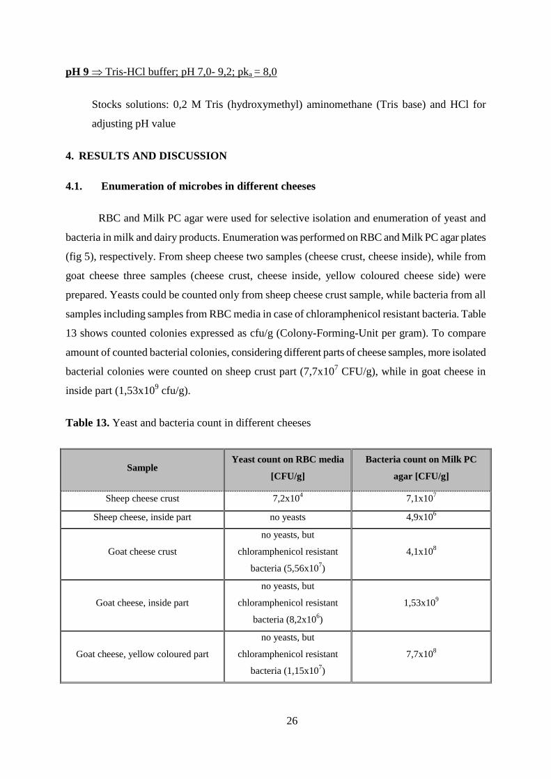
4.1. Enumeration of microbes in different cheeses
RBC and Milk PC agar were used for selective isolation and enumeration of yeast and
bacteria in milk and dairy products. Enumeration was performed on RBC and Milk PC agar plates
(fig 5), respectively. From sheep cheese two samples (cheese crust, cheese inside), while from
goat cheese three samples (cheese crust, cheese inside, yellow coloured cheese side) were
prepared. Yeasts could be counted only from sheep cheese crust sample, while bacteria from all
samples including samples from RBC media in case of chloramphenicol resistant bacteria. Table
13 shows counted colonies expressed as cfu/g (Colony-Forming-Unit per gram). To compare
amount of counted bacterial colonies, considering different parts of cheese samples, more isolated
bacterial colonies were counted on sheep crust part (7,7x10⁷ CFU/g), while in goat cheese in
inside part (1,53x10⁹ cfu/g).
Table 13. Yeast and bacteria count in different cheeses
Sample Yeast count on RBC media
[CFU/g]
Bacteria count on Milk PC
agar [CFU/g]
Sheep cheese crust 7,2x10⁴ 7,1x10
7
Sheep cheese, inside part no yeasts 4,9x10⁶
Goat cheese crust
no yeasts, but
chloramphenicol resistant
bacteria (5,56x10⁷)
4,1x10⁸
Goat cheese, inside part
no yeasts, but
chloramphenicol resistant
bacteria (8,2x10⁶)
1,53x10⁹
Goat cheese, yellow coloured part
no yeasts, but
chloramphenicol resistant
bacteria (1,15x10⁷)
7,7x10⁸
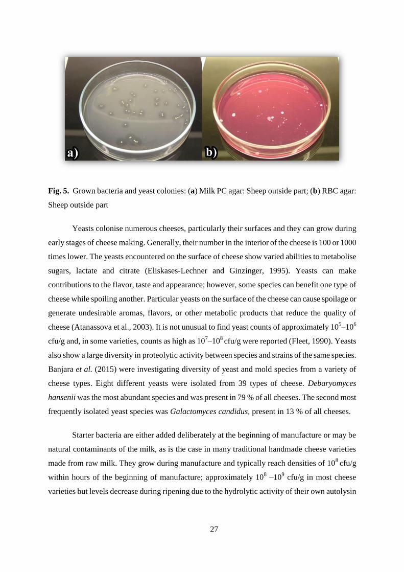
27
Fig. 5. Grown bacteria and yeast colonies: (a) Milk PC agar: Sheep outside part; (b) RBC agar:
Sheep outside part
Yeasts colonise numerous cheeses, particularly their surfaces and they can grow during
early stages of cheese making. Generally, their number in the interior of the cheese is 100 or 1000
times lower. The yeasts encountered on the surface of cheese show varied abilities to metabolise
sugars, lactate and citrate (Eliskases-Lechner and Ginzinger, 1995). Yeasts can make
contributions to the flavor, taste and appearance; however, some species can benefit one type of
cheese while spoiling another. Particular yeasts on the surface of the cheese can cause spoilage or
generate undesirable aromas, flavors, or other metabolic products that reduce the quality of
cheese (Atanassova et al., 2003). It is not unusual to find yeast counts of approximately 105–10
6
cfu/g and, in some varieties, counts as high as 107–10
8 cfu/g were reported (Fleet, 1990). Yeasts
also show a large diversity in proteolytic activity between species and strains of the same species.
Banjara et al. (2015) were investigating diversity of yeast and mold species from a variety of
cheese types. Eight different yeasts were isolated from 39 types of cheese. Debaryomyces
hansenii was the most abundant species and was present in 79 % of all cheeses. The second most
frequently isolated yeast species was Galactomyces candidus, present in 13 % of all cheeses.
Starter bacteria are either added deliberately at the beginning of manufacture or may be
natural contaminants of the milk, as is the case in many traditional handmade cheese varieties
made from raw milk. They grow during manufacture and typically reach densities of 108
cfu/g
within hours of the beginning of manufacture; approximately 108 –10
9 cfu/g in most cheese
varieties but levels decrease during ripening due to the hydrolytic activity of their own autolysin
28
enzymes (Thomas and Batt, 1969). The primary function of starter bacteria in cheese is to
produce acid during the fermentation process; however, they also contribute to cheese ripening
while their enzymes are involved in proteolysis and conversion of amino acids into flavor
compounds (Fox and Wallace, 1997).
The predominant surface microflora of fresh feta cheese is composed of LAB, yeasts, and
salt-tolerant microbes. Leuconostoc, Lactococcus lactis, and Lactobacillus plantarum constitue
the predominant LAB microflora develop in the beginning of ripening, while at the end the LAB
microflora is composed of lactobacilli, with L. plantarum being the predominant species (Fang
and O’Toole, 2009). The predominant yeasts species in the beginning of ripening are
Saccharomyces cerevisiae and Debaryomyces hansenii, while at the end of ripening at room
temperature, Saccharomyces cerevisiae predominate (Raes and Bork, 2008). The halotolerant
surface microflora is composed of staphylococci, micrococci, enterococci, and coryneform
bacteria. Staphylococcus saprohyticus predominates the halotolerant bacteria content on the
surface of cheese in the beginning, while coryneforms in further stages of ripening. As indicated
by their enzyme systems, lactobacilli of the cheese surface may affect proteolysis, yeasts
participate in both proteolysis and lipolysis, and staphylococci contribute to cheese lipolysis.
Lactococci are the most frequently group among the NSLAB found in the fresh cheese, with L.
Lactis subsp. lactis predominating. Lactobacilli are also isolated more frequently than enterococii,
and leuconostocs are equal to lactobacilli (Bassler, 2002).
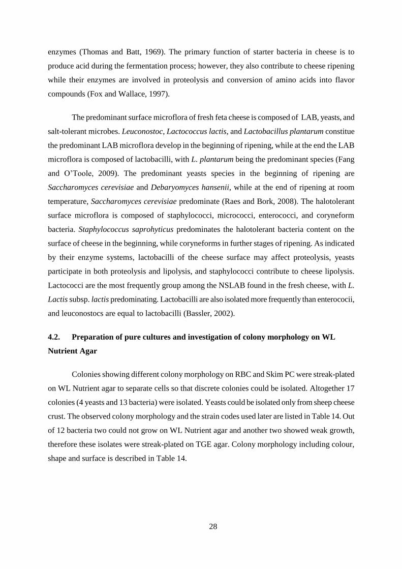
4.2. Preparation of pure cultures and investigation of colony morphology on WL
Nutrient Agar
Colonies showing different colony morphology on RBC and Skim PC were streak-plated
on WL Nutrient agar to separate cells so that discrete colonies could be isolated. Altogether 17
colonies (4 yeasts and 13 bacteria) were isolated. Yeasts could be isolated only from sheep cheese
crust. The observed colony morphology and the strain codes used later are listed in Table 14. Out
of 12 bacteria two could not grow on WL Nutrient agar and another two showed weak growth,
therefore these isolates were streak-plated on TGE agar. Colony morphology including colour,
shape and surface is described in Table 14.
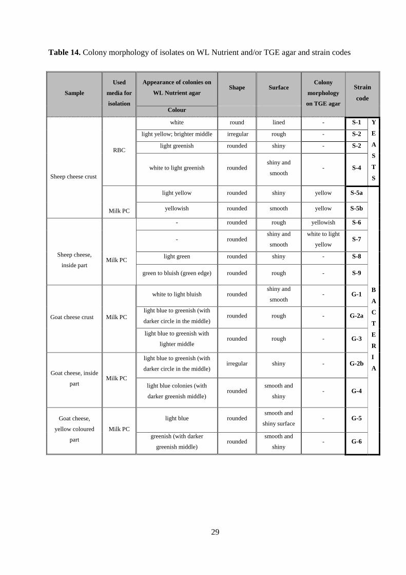
29
Table 14. Colony morphology of isolates on WL Nutrient and/or TGE agar and strain codes
Sample
Used
media for
isolation
Appearance of colonies on
WL Nutrient agar Shape Surface
Colony
morphology
on TGE agar
Strain
code
Colour
Sheep cheese crust
RBC
white round lined - S-1 Y
E
A
S
T
S
light yellow; brighter middle irregular rough - S-2
light greenish rounded shiny - S-2
white to light greenish rounded shiny and
smooth - S-4
Milk PC
light yellow rounded shiny yellow S-5a
B
A
C
T
E
R
I
A
yellowish rounded smooth yellow S-5b
Sheep cheese,
inside part
Milk PC
- rounded rough yellowish S-6
- rounded shiny and
smooth
white to light
yellow S-7
light green rounded shiny - S-8
green to bluish (green edge) rounded rough - S-9
Goat cheese crust Milk PC
white to light bluish rounded shiny and
smooth - G-1
light blue to greenish (with
darker circle in the middle) rounded rough - G-2a
light blue to greenish with
lighter middle rounded rough - G-3
Goat cheese, inside
part Milk PC
light blue to greenish (with
darker circle in the middle) irregular shiny - G-2b
light blue colonies (with
darker greenish middle) rounded
smooth and
shiny - G-4
Goat cheese,
yellow coloured
part
Milk PC
light blue rounded smooth and
shiny surface - G-5
greenish (with darker
greenish middle) rounded
smooth and
shiny - G-6
30
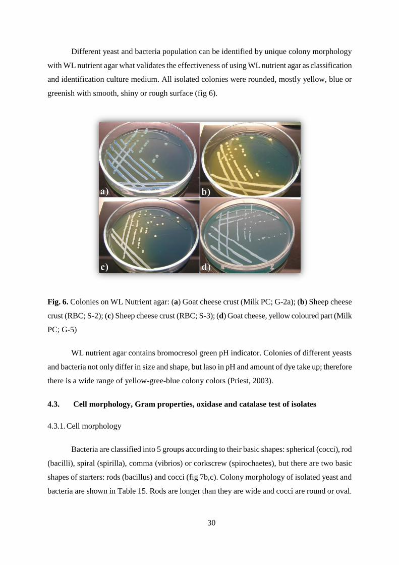
Different yeast and bacteria population can be identified by unique colony morphology
with WL nutrient agar what validates the effectiveness of using WL nutrient agar as classification
and identification culture medium. All isolated colonies were rounded, mostly yellow, blue or
greenish with smooth, shiny or rough surface (fig 6).
Fig. 6. Colonies on WL Nutrient agar: (a) Goat cheese crust (Milk PC; G-2a); (b) Sheep cheese
crust (RBC; S-2); (c) Sheep cheese crust (RBC; S-3); (d) Goat cheese, yellow coloured part (Milk
PC; G-5)
WL nutrient agar contains bromocresol green pH indicator. Colonies of different yeasts
and bacteria not only differ in size and shape, but laso in pH and amount of dye take up; therefore
there is a wide range of yellow-gree-blue colony colors (Priest, 2003).
4.3. Cell morphology, Gram properties, oxidase and catalase test of isolates
4.3.1. Cell morphology
Bacteria are classified into 5 groups according to their basic shapes: spherical (cocci), rod
(bacilli), spiral (spirilla), comma (vibrios) or corkscrew (spirochaetes), but there are two basic
shapes of starters: rods (bacillus) and cocci (fig 7b,c). Colony morphology of isolated yeast and
bacteria are shown in Table 15. Rods are longer than they are wide and cocci are round or oval.
31
They occur as single cells or pairs, but most commonly, they are connected end to end as short
chains. Bacteria isolated from sheep chesse were all cocci, except strain S-8 who was rods. In
case of isolates from goat cheese, all were rod rods, except coccus strains G-2a and G-2b.
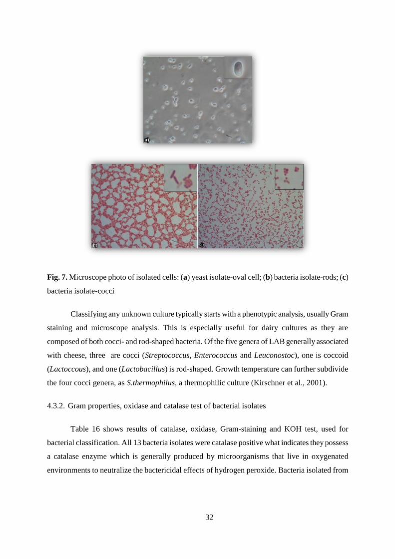
The size and shape of cells within a strain are the same when the yeast is propagated under
identical conditions; they may change with the nutrient and the environment. Some strains
produce one cell form, while others propagate in several shapes (dimorphism- two shapes
predominate; polymorphism- more than two cell forms occur). According to cell morphology
observed under light microscope, all isolated yeast cells were oval shaped (fig 7a).
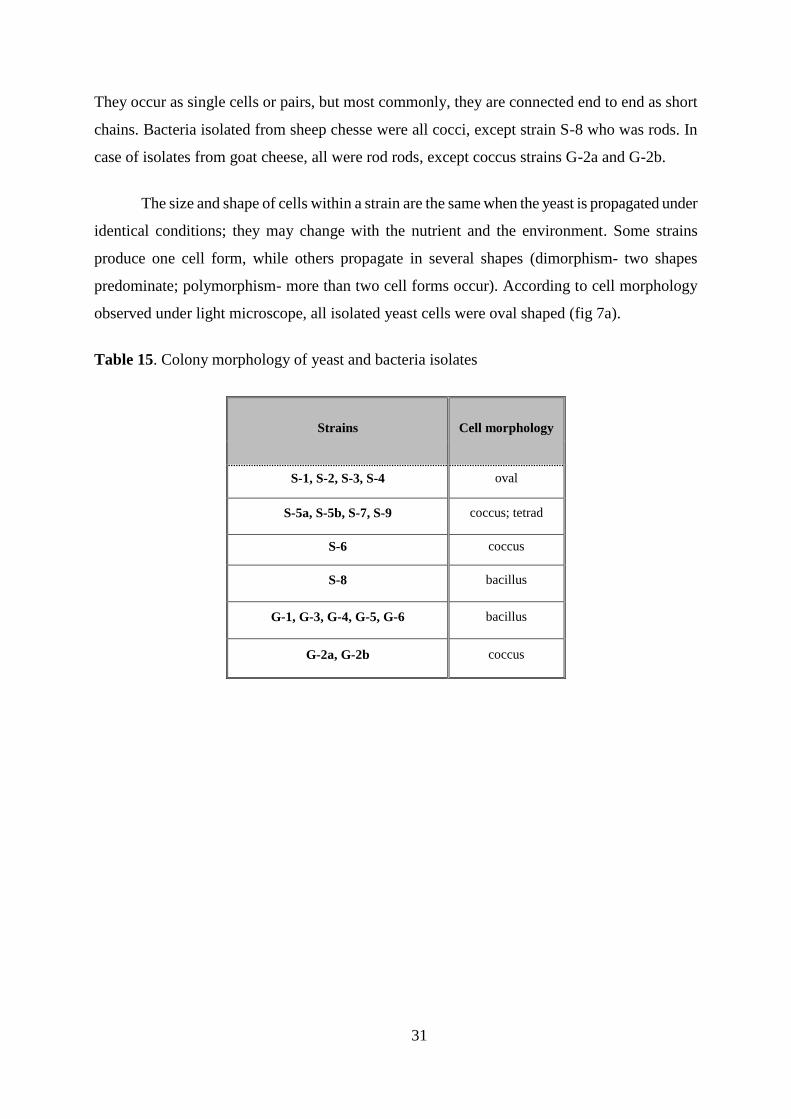
Table 15. Colony morphology of yeast and bacteria isolates
Strains Cell morphology
S-1, S-2, S-3, S-4 oval
S-5a, S-5b, S-7, S-9 coccus; tetrad
S-6 coccus
S-8 bacillus
G-1, G-3, G-4, G-5, G-6 bacillus
G-2a, G-2b coccus
32
Fig. 7. Microscope photo of isolated cells: (a) yeast isolate-oval cell; (b) bacteria isolate-rods; (c)
bacteria isolate-cocci
Classifying any unknown culture typically starts with a phenotypic analysis, usually Gram
staining and microscope analysis. This is especially useful for dairy cultures as they are
composed of both cocci- and rod-shaped bacteria. Of the five genera of LAB generally associated
with cheese, three are cocci (Streptococcus, Enterococcus and Leuconostoc), one is coccoid
(Lactoccous), and one (Lactobacillus) is rod-shaped. Growth temperature can further subdivide
the four cocci genera, as S.thermophilus, a thermophilic culture (Kirschner et al., 2001).
4.3.2. Gram properties, oxidase and catalase test of bacterial isolates
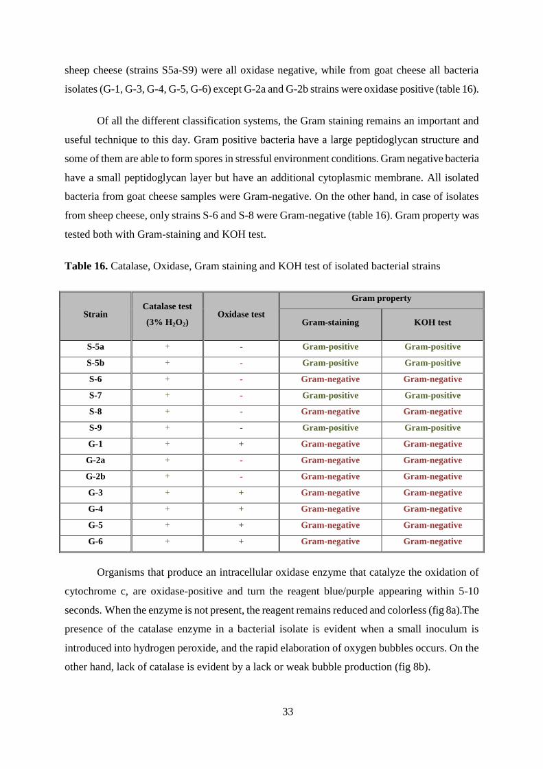
Table 16 shows results of catalase, oxidase, Gram-staining and KOH test, used for
bacterial classification. All 13 bacteria isolates were catalase positive what indicates they possess
a catalase enzyme which is generally produced by microorganisms that live in oxygenated
environments to neutralize the bactericidal effects of hydrogen peroxide. Bacteria isolated from
33
sheep cheese (strains S5a-S9) were all oxidase negative, while from goat cheese all bacteria
isolates (G-1, G-3, G-4, G-5, G-6) except G-2a and G-2b strains were oxidase positive (table 16).
Of all the different classification systems, the Gram staining remains an important and
useful technique to this day. Gram positive bacteria have a large peptidoglycan structure and
some of them are able to form spores in stressful environment conditions. Gram negative bacteria
have a small peptidoglycan layer but have an additional cytoplasmic membrane. All isolated
bacteria from goat cheese samples were Gram-negative. On the other hand, in case of isolates
from sheep cheese, only strains S-6 and S-8 were Gram-negative (table 16). Gram property was
tested both with Gram-staining and KOH test.
Table 16. Catalase, Oxidase, Gram staining and KOH test of isolated bacterial strains
Strain Catalase test
(3% H2O2) Oxidase test
Gram property
Gram-staining KOH test
S-5a + - Gram-positive Gram-positive
S-5b + - Gram-positive Gram-positive
S-6 + - Gram-negative Gram-negative
S-7 + - Gram-positive Gram-positive
S-8 + - Gram-negative Gram-negative
S-9 + - Gram-positive Gram-positive
G-1 + + Gram-negative Gram-negative
G-2a + - Gram-negative Gram-negative
G-2b + - Gram-negative Gram-negative
G-3 + + Gram-negative Gram-negative
G-4 + + Gram-negative Gram-negative
G-5 + + Gram-negative Gram-negative
G-6 + + Gram-negative Gram-negative
Organisms that produce an intracellular oxidase enzyme that catalyze the oxidation of
cytochrome c, are oxidase-positive and turn the reagent blue/purple appearing within 5-10
seconds. When the enzyme is not present, the reagent remains reduced and colorless (fig 8a).The
presence of the catalase enzyme in a bacterial isolate is evident when a small inoculum is
introduced into hydrogen peroxide, and the rapid elaboration of oxygen bubbles occurs. On the
other hand, lack of catalase is evident by a lack or weak bubble production (fig 8b).
34
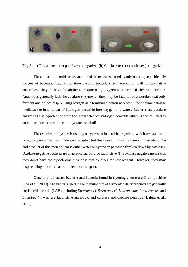
Fig. 8. (a) Oxidase test: (+) positive; (-) negative; (b) Catalase test: (+) positive; (-) negative
The catalase and oxidase test are one of the main tests used by microbiologists to identify
species of bacteria. Catalase-positive bacteria include strict aerobes as well as facultative
anaerobes. They all have the ability to respire using oxygen as a terminal electron acceptor.
Anaerobes generally lack the catalase enzyme, or they may be facultative anaerobes that only
ferment and do not respire using oxygen as a terminal electron acceptor. The enzyme catalase
mediates the breakdown of hydrogen peroxide into oxygen and water. Bacteria use catalase
enzyme as a self-protection from the lethal effect of hydrogen peroxide which is accumulated as
an end product of aerobic carbohydrate metabolism.
The cytochrome system is usually only present in aerobic organisms which are capable of
using oxygen as the final hydrogen receptor, but this doesn’t mean they are strict aerobes. The
end product of this metabolism is either water or hydrogen peroxide (broken down by catalase).
Oxidase-negative bacteria are anaerobic, aerobic, or facultative. The oxidase negative means that
they don’t have the cytochrome c oxidase that oxidizes the test reagent. However, they may
respire using other oxidases in electron transport.
Generally, all starter bacteria and bacteria found in ripening cheese are Gram-positive
(Fox et al., 2000). The bacteria used in the manufacture of fermented dairy products are generally
lactic acid bacteria (LAB) including Enterococci, Streptococci, Leuconostoc, Lactococcus, and
Lactobacilli, who are facultative anaerobic and catalase and oxidase negative (Baluja et al.,
2011).
35
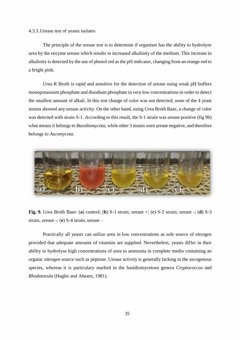
4.3.3. Urease test of yeasts isolates
The principle of the urease test is to determine if organism has the ability to hydrolyze
urea by the enzyme urease which results in increased alkalinity of the medium. This increase in
alkalinity is detected by the use of phenol red as the pH indicator, changing from an orange-red to
a bright pink.
Urea R Broth is rapid and sensitive for the detection of urease using weak pH buffers
monopotassium phosphate and disodium phosphate in very low concentrations in order to detect
the smallest amount of alkali. In this test change of color was not detected; none of the 4 yeast
strains showed any urease activity. On the other hand, using Urea Broth Base, a change of color
was detected with strain S-1. According to this result, the S-1 strain was urease positive (fig 9b)
what means it belongs to Basidiomycota, while other 3 strains were urease negative, and therefore
belongs to Ascomycota.
Fig. 9. Urea Broth Base: (a) control; (b) S-1 strain, urease +; (c) S-2 strain; urease -; (d) S-3
strain, urease -; (e) S-4 strain, urease –
Practically all yeasts can utilize urea in low concentrations as sole source of nitrogen
provided that adequate amounts of vitamins are supplied. Nevertheless, yeasts differ in their
ability to hydrolyse high concentrations of urea to ammonia in complete media containing an
organic nitrogen source such as peptone. Urease activity is generally lacking in the ascogenous
species, whereas it is particulary marked in the basidiomycetous genera Cryptococcus and
Rhodotorula (Hagler and Ahearn, 1981).
36
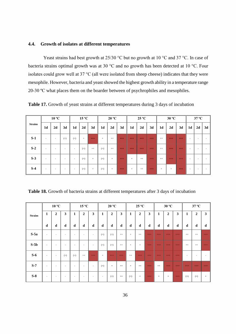
4.4. Growth of isolates at different temperatures
Yeast strains had best growth at 25/30 °C but no growth at 10 °C and 37 °C. In case of
bacteria strains optimal growth was at 30 °C and no growth has been detected at 10 °C. Four
isolates could grow well at 37 °C (all were isolated from sheep cheese) indicates that they were
mesophile. However, bacteria and yeast showed the highest growth ability in a temperature range
20-30 ºC what places them on the boarder between of psychrophiles and mesophiles.
Table 17. Growth of yeast strains at different temperatures during 3 days of incubation
Strains
10 ºC 15 ºC 20 ºC 25 ºC 30 ºC 37 ºC
1d 2d 3d 1d 2d 3d 1d 2d 3d 1d 2d 3d 1d 2d 3d 1d 2d 3d
S-1 - - (+) (+) + +++ + ++ +++ +++ +++ +++ ++ +++ +++ - - -
S-2 - - - - (+) ++ (+) ++ +++ +++ +++ +++ ++ +++ +++ - - -
S-3 - - - - (+) + (+) + +++ + ++ +++ ++ +++ +++ - - -
S-4 - - - - (+) + (+) + +++ + ++ +++ + + +++ - - -
Table 18. Growth of bacteria strains at different temperatures after 3 days of incubation
Strains
10 ºC 15 ºC 20 ºC 25 ºC 30 ºC 37 ºC
1
d
2
d
3
d
1
d
2
d
3
d
1
d
2
d
3
d
1
d
2
d
3
d
1
d
2
d
3
d
1
d
2
d
3
d
S-5a - - - - - - (+) (+) ++ + ++ +++ +++ +++ +++ ++ ++ +++
S-5b - - - - - - (+) (+) ++ + + +++ +++ +++ +++ ++ ++ +++
S-6 - - (+) (+) ++ +++ + +++ +++ ++ +++ +++ +++ +++ +++ - - -
S-7 - - - - - - (+) + ++ + ++ +++ ++ +++ +++ +++ +++ +++
S-8 - - - - - - - (+) ++ (+) + +++ + + +++ (+) (+) +
37
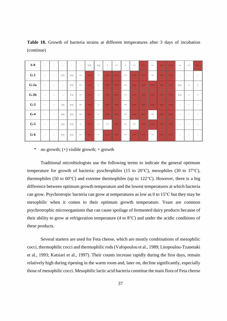
Table 18. Growth of bacteria strains at different temperatures after 3 days of incubation
(continue)
S-9 - - - - - (+) (+) + ++ + ++ +++ ++ +++ +++ ++ ++ +++
G-1 - - (+) (+) ++ +++ + +++ +++ ++ +++ +++ ++ +++ +++ - - -
G-2a - - - (+) ++ +++ + +++ +++ ++ +++ +++ +++ +++ +++ (+) + +
G-2b - - - (+) ++ +++ + +++ +++ ++ +++ +++ +++ +++ +++ (+) + +
G-3 - - (+) (+) ++ +++ + +++ +++ ++ +++ +++ +++ +++ +++ - - -
G-4 - - (+) (+) ++ +++ + +++ +++ ++ +++ +++ ++ +++ +++ - - -
G-5 - - (+) (+) + +++ + ++ +++ ++ ++ +++ +++ +++ +++ - - -
G-6 - - (+) (+) ++ +++ + +++ +++ ++ +++ +++ ++ +++ +++ - - -
* no growth; (+) visible growth; + growth
Traditional microbiologists use the following terms to indicate the general optimum
temperature for growth of bacteria: pyschrophiles (15 to 20°C), mesophiles (30 to 37°C),
thermophiles (50 to 60°C) and extreme thermophiles (up to 122°C). However, there is a big
difference between optimum growth temperature and the lowest temperatures at which bacteria
can grow. Psychrotropic bacteria can grow at temperatures as low as 0 to 15°C but they may be
mesophilic when it comes to their optimum growth temperature. Yeast are common
psychrotrophic microorganisms that can cause spoilage of fermented dairy products because of
their ability to grow at refrigeration temperature (4 to 8°C) and under the acidic conditions of
these products.
Several starters are used for Feta cheese, which are mostly combinations of mesophilic
cocci, thermophilic cocci and thermophilic rods (Vafopoulou et al., 1989; Litopoulou-Tzanetaki
et al., 1993; Katsiari et al., 1997). Their counts increase rapidly during the first days, remain
relatively high during ripening in the warm room and, later on, decline significantly, especially
those of mesophilic cocci. Mesophilic lactic acid bacteria constitue the main flora of Feta cheese
38
(Tzanetakis and Litopoulou, 1992; Sarantinopoulos, 2002). Their number significantly increases
during the first 15 days in the warm room and remains high throughout the ripening time. This
explains the high proteolytic activity observed at the beginning of the ripening period. Proteolytic
activity of the extracellular proteinases from psychrotrophic bacteria in raw milk reduces cheese
yield and increase the levels of nitrogenous compounds in whey. The loss in cheese yield directly
relates to the storage time of the raw milk. The proteolytic activity in cheese by heat stable
proteinases can cause flavour quality and higher texture problems (Erkmen and Bozoglu, 2016)
4.5. Proteolytic activity of isolated strains
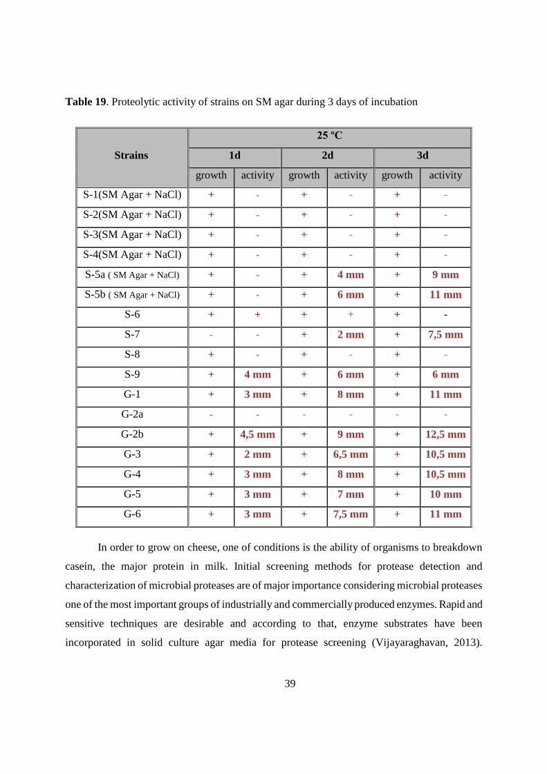
4.5.1. Testing the proteolytic activity of isolates on SM Agar
A clear zone of skim hydrolysis gave an indication of protease producing organisms. The
proteolytic activity was assayed using SM agar and expressed as a radius of clear zone in mm
(table 19; fig 10a,b). Strain G-2b exhibited the highest proteolytic activity during all three days of
incubation, with a clear zone radius of 12,5 mm measured on third day of incubation. G-2b strain
is followed by strains S-5b, G-1 and G-6 with clear zone of 11 mm . Strains S-8 and G-2a didn’t
show proteolytic activity, while S-6 had low, not measurable activity. The surface of the cheese
has a relatively high salt content and therefore the microorganisms that grow on it are salt tolerant
and can require salt for growth and activity. Strains isolated from sheep cheese crust part, S-5a
and S-5b were not growing, and S-1, S-2, S-3 and S-4 didn’t show any activity. Therefore, they
have been tested on 0,85% NaCl containing SM agar. Depending on the existing zone of
clearance, strains S-5a, S-5b, S-7, S-9, G-1, G-2b, G-3, G-4, G-5, and G-6 were selected for
further experimental studies as isolated proteolytic bacterial strains.
39
Table 19. Proteolytic activity of strains on SM agar during 3 days of incubation
Strains
25 ºC
1d 2d 3d
growth activity growth activity growth activity
S-1(SM Agar + NaCl) + - + - + -
S-2(SM Agar + NaCl) + - + - + -
S-3(SM Agar + NaCl) + - + - + -
S-4(SM Agar + NaCl) + - + - + -
S-5a ( SM Agar + NaCl) + - + 4 mm + 9 mm
S-5b ( SM Agar + NaCl) + - + 6 mm + 11 mm
S-6 + + + + + -
S-7 - - + 2 mm + 7,5 mm
S-8 + - + - + -
S-9 + 4 mm + 6 mm + 6 mm
G-1 + 3 mm + 8 mm + 11 mm
G-2a - - - - - -
G-2b + 4,5 mm + 9 mm + 12,5 mm
G-3 + 2 mm + 6,5 mm + 10,5 mm
G-4 + 3 mm + 8 mm + 10,5 mm
G-5 + 3 mm + 7 mm + 10 mm
G-6 + 3 mm + 7,5 mm + 11 mm
In order to grow on cheese, one of conditions is the ability of organisms to breakdown
casein, the major protein in milk. Initial screening methods for protease detection and
characterization of microbial proteases are of major importance considering microbial proteases
one of the most important groups of industrially and commercially produced enzymes. Rapid and
sensitive techniques are desirable and according to that, enzyme substrates have been
incorporated in solid culture agar media for protease screening (Vijayaraghavan, 2013).
40
Degradation of casein contributes to the texture of the cheese and about 50% of the flavour
components in cheese are amino acids, short peptides and products of their metabolism
(Kieronczyk, 2006). Amino acids and short peptides can typically be bitter, but di-, tri- and short
peptides can be bioactive contributing to cheese as a functional food (Korhonen and Pihlanto,
2003).
4.5.2. Extracellular proteolytic activity of strains in different media
Two nutrient media were used to test effect of growth media compositiont on proteolytic
activity of isolated bacteria strains: YEPD and Minimal Media. Both media provides all required
components for bacteria growth, but results indicated that bacteria for proteolytic activity require
certain components that were not included in growth media. Sodium caseinate is the major
protein source for the proteolytic organism while casein enzymic hydrolysates and, also yeast
extract, provide nitrogenous nutrients to the proteolytic organisms. Nitrogen source in Minimal
Media was replaced with sodium caseinate as a new source, and NaCl is added to test his
influence on activity. To test extracellular proteolytic activity of isolated bacterial strains, after
cultivation in different media, samples were treated with antibiotic gentamicin and centrifuged to
get extracellular proteolytic enzymes in supernatant. Well diffusion method on SM agar plates
was used for determination of protease activity. After incubation in SM agar, strains S-5b, S-7, G-
1, G-2b, G-3, and G-4 showed proteolytic activity detected as the clear zone around drilled holes.
Proteolytic activity increased during three days of incubation, showing the biggest radius of
clearing zone in case of strains S-7 (11mm) and G-2b (10,5mm) (table 20). Strains S-5a, S-9, G-5,
and G-6 didn’t show extracellular proteolytic activity.
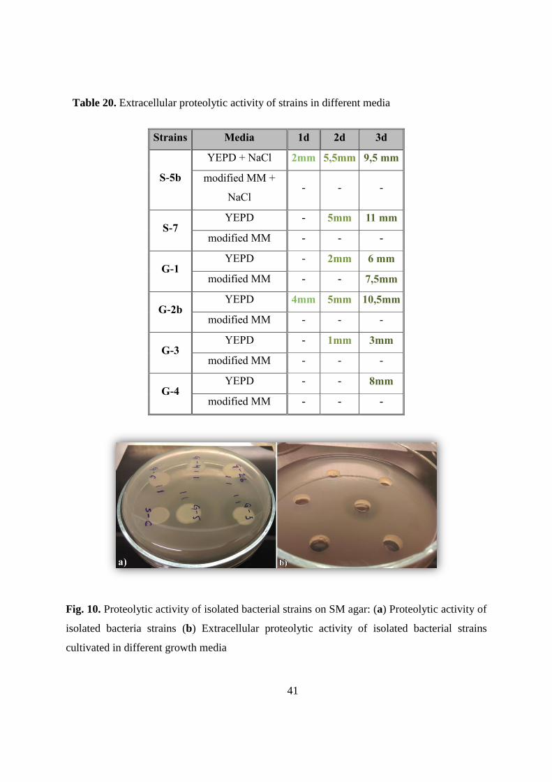
41
Table 20. Extracellular proteolytic activity of strains in different media
Strains Media 1d 2d 3d
S-5b
YEPD + NaCl 2mm 5,5mm 9,5 mm
modified MM +
NaCl - - -
S-7 YEPD - 5mm 11 mm
modified MM - - -
G-1 YEPD - 2mm 6 mm
modified MM - - 7,5mm
G-2b YEPD 4mm 5mm 10,5mm
modified MM - - -
G-3 YEPD - 1mm 3mm
modified MM - - -
G-4 YEPD - - 8mm
modified MM - - -
Fig. 10. Proteolytic activity of isolated bacterial strains on SM agar: (a) Proteolytic activity of
isolated bacteria strains (b) Extracellular proteolytic activity of isolated bacterial strains
cultivated in different growth media
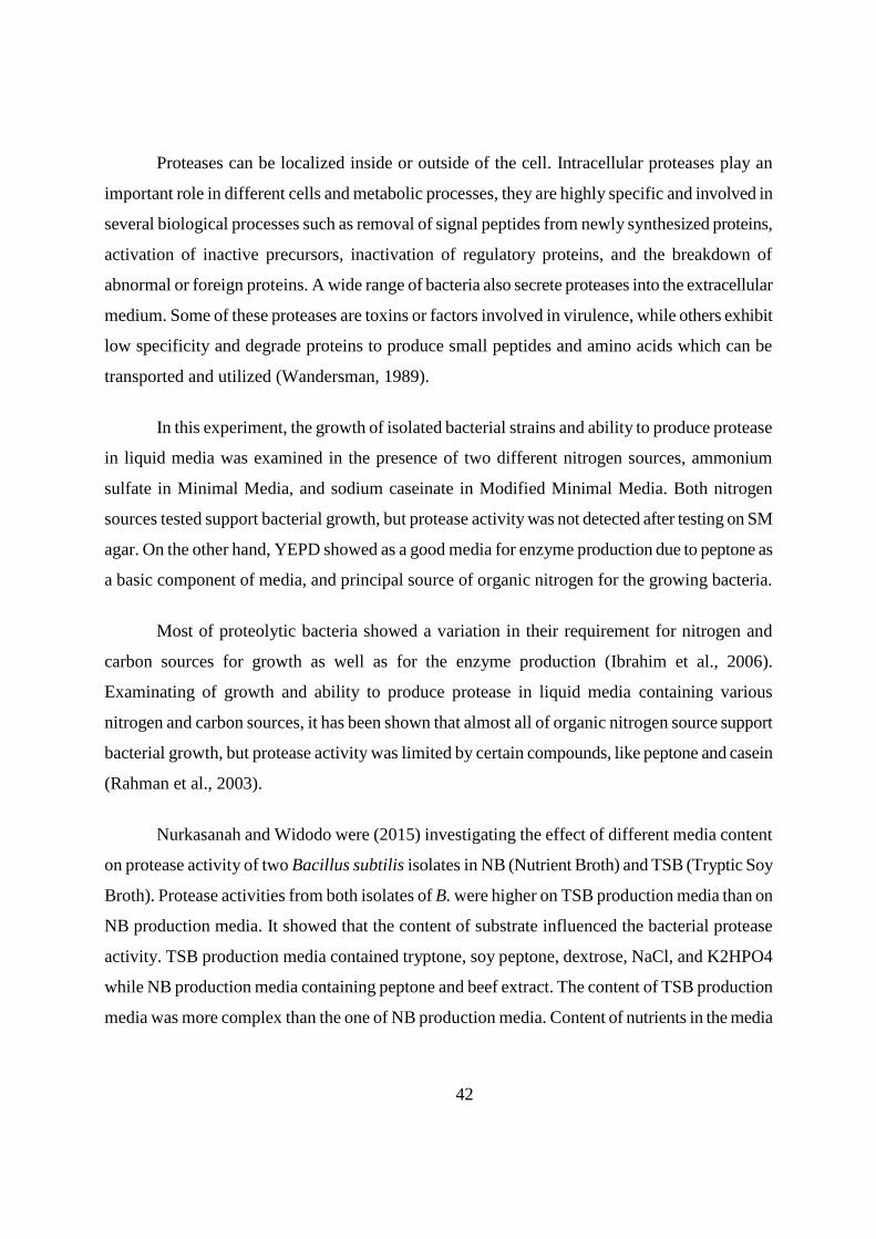
42
Proteases can be localized inside or outside of the cell. Intracellular proteases play an
important role in different cells and metabolic processes, they are highly specific and involved in
several biological processes such as removal of signal peptides from newly synthesized proteins,
activation of inactive precursors, inactivation of regulatory proteins, and the breakdown of
abnormal or foreign proteins. A wide range of bacteria also secrete proteases into the extracellular
medium. Some of these proteases are toxins or factors involved in virulence, while others exhibit
low specificity and degrade proteins to produce small peptides and amino acids which can be
transported and utilized (Wandersman, 1989).
In this experiment, the growth of isolated bacterial strains and ability to produce protease
in liquid media was examined in the presence of two different nitrogen sources, ammonium
sulfate in Minimal Media, and sodium caseinate in Modified Minimal Media. Both nitrogen
sources tested support bacterial growth, but protease activity was not detected after testing on SM
agar. On the other hand, YEPD showed as a good media for enzyme production due to peptone as
a basic component of media, and principal source of organic nitrogen for the growing bacteria.
Most of proteolytic bacteria showed a variation in their requirement for nitrogen and
carbon sources for growth as well as for the enzyme production (Ibrahim et al., 2006).
Examinating of growth and ability to produce protease in liquid media containing various
nitrogen and carbon sources, it has been shown that almost all of organic nitrogen source support
bacterial growth, but protease activity was limited by certain compounds, like peptone and casein
(Rahman et al., 2003).
Nurkasanah and Widodo were (2015) investigating the effect of different media content
on protease activity of two Bacillus subtilis isolates in NB (Nutrient Broth) and TSB (Tryptic Soy
Broth). Protease activities from both isolates of B. were higher on TSB production media than on
NB production media. It showed that the content of substrate influenced the bacterial protease
activity. TSB production media contained tryptone, soy peptone, dextrose, NaCl, and K2HPO4
while NB production media containing peptone and beef extract. The content of TSB production
media was more complex than the one of NB production media. Content of nutrients in the media
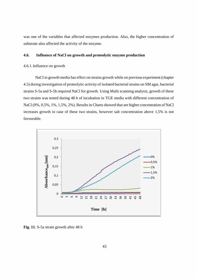
43
was one of the variables that affected enzymes production. Also, the higher concentration of
substrate also affected the activity of the enzyme.
4.6. Influence of NaCl on growth and proteolytic enzyme production
4.6.1. Influence on growth
NaCl in growth media has effect on strains growth while on previous experiment (chapter
4.5) during investigation of proteolytic activity of isolated bacterial strains on SM agar, bacterial
strains S-5a and S-5b required NaCl for growth. Using Multi scanning analysis, growth of these
two strains was tested during 48 h of incubation in TGE media with different concentration of
NaCl (0%, 0,5%, 1%, 1,5%, 2%). Results in Charts showed that are higher concentration of NaCl
increases growth in case of these two strains, however salt concentration above 1,5% is not
favourable.
Fig. 11. S-5a strain growth after 48 h
0
0,05
0,1
0,15
0,2
0,25
0,3
0 3 6 9
12
15
18
21
24
27
30
33
36
39
42
45
48
0%
0,5%
1%
1,5%
2%
Time [h]
Ab
sorb
an
ce6
00
[n
m]
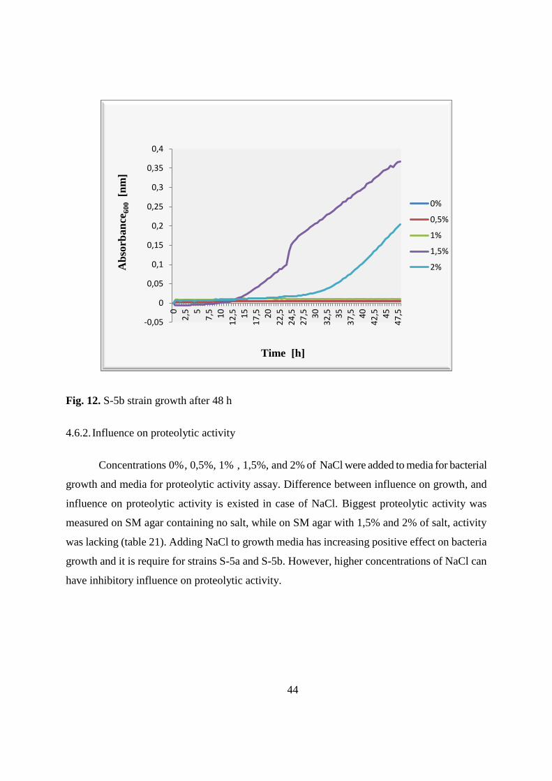
44
Fig. 12. S-5b strain growth after 48 h
4.6.2. Influence on proteolytic activity
Concentrations 0% , 0,5%, 1% , 1,5%, and 2% of NaCl were added to media for bacterial
growth and media for proteolytic activity assay. Difference between influence on growth, and
influence on proteolytic activity is existed in case of NaCl. Biggest proteolytic activity was
measured on SM agar containing no salt, while on SM agar with 1,5% and 2% of salt, activity
was lacking (table 21). Adding NaCl to growth media has increasing positive effect on bacteria
growth and it is require for strains S-5a and S-5b. However, higher concentrations of NaCl can
have inhibitory influence on proteolytic activity.
-0,05
0
0,05
0,1
0,15
0,2
0,25
0,3
0,35
0,4
0
2,5 5
7,5 10
12
,5 15
17
,5 20
22
,5
24
,5
27
,5 30
32
,5 35
37
,5 40
42
,5 45
47
,5
0%
0,5%
1%
1,5%
2%
Time [h]
Ab
sorb
an
ce6
00 [n
m]
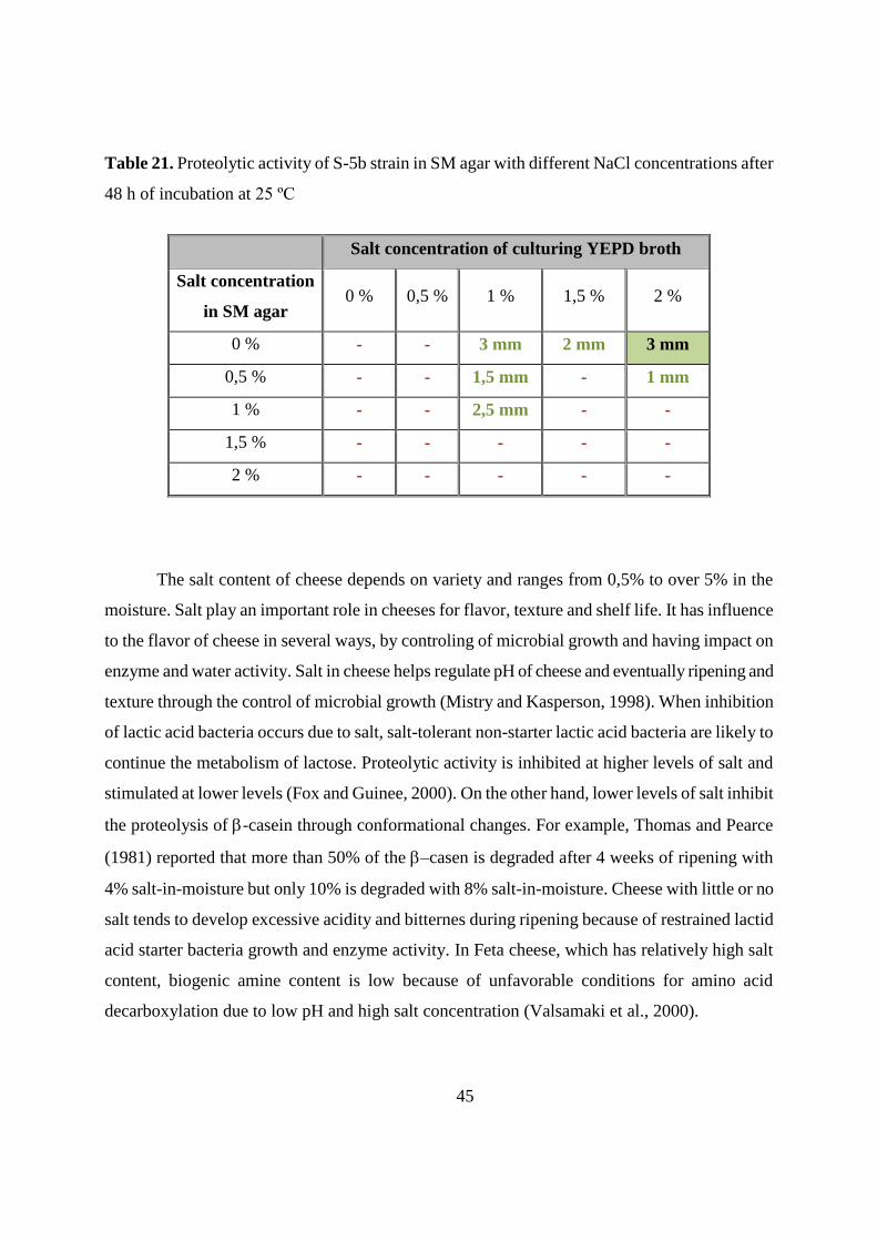
45
Table 21. Proteolytic activity of S-5b strain in SM agar with different NaCl concentrations after
48 h of incubation at 25 ºC
Salt concentration of culturing YEPD broth
Salt concentration
in SM agar 0 % 0,5 % 1 % 1,5 % 2 %
0 % - - 3 mm 2 mm 3 mm
0,5 % - - 1,5 mm - 1 mm
1 % - - 2,5 mm - -
1,5 % - - - - -
2 % - - - - -
The salt content of cheese depends on variety and ranges from 0,5% to over 5% in the
moisture. Salt play an important role in cheeses for flavor, texture and shelf life. It has influence
to the flavor of cheese in several ways, by controling of microbial growth and having impact on
enzyme and water activity. Salt in cheese helps regulate pH of cheese and eventually ripening and
texture through the control of microbial growth (Mistry and Kasperson, 1998). When inhibition
of lactic acid bacteria occurs due to salt, salt-tolerant non-starter lactic acid bacteria are likely to
continue the metabolism of lactose. Proteolytic activity is inhibited at higher levels of salt and
stimulated at lower levels (Fox and Guinee, 2000). On the other hand, lower levels of salt inhibit
the proteolysis of -casein through conformational changes. For example, Thomas and Pearce
(1981) reported that more than 50% of the –casen is degraded after 4 weeks of ripening with
4% salt-in-moisture but only 10% is degraded with 8% salt-in-moisture. Cheese with little or no
salt tends to develop excessive acidity and bitternes during ripening because of restrained lactid
acid starter bacteria growth and enzyme activity. In Feta cheese, which has relatively high salt
content, biogenic amine content is low because of unfavorable conditions for amino acid
decarboxylation due to low pH and high salt concentration (Valsamaki et al., 2000).
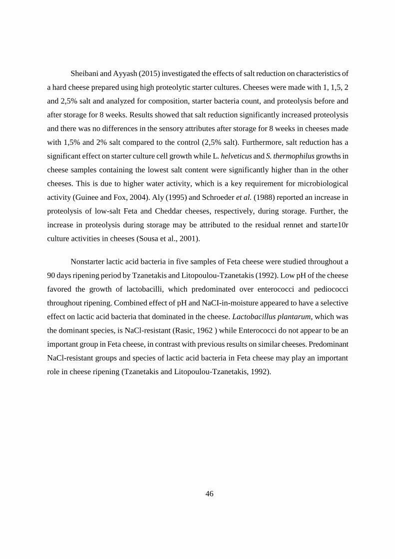
46
Sheibani and Ayyash (2015) investigated the effects of salt reduction on characteristics of
a hard cheese prepared using high proteolytic starter cultures. Cheeses were made with 1, 1,5, 2
and 2,5% salt and analyzed for composition, starter bacteria count, and proteolysis before and
after storage for 8 weeks. Results showed that salt reduction significantly increased proteolysis
and there was no differences in the sensory attributes after storage for 8 weeks in cheeses made
with 1,5% and 2% salt compared to the control (2,5% salt). Furthermore, salt reduction has a
significant effect on starter culture cell growth while L. helveticus and S. thermophilus growths in
cheese samples containing the lowest salt content were significantly higher than in the other
cheeses. This is due to higher water activity, which is a key requirement for microbiological
activity (Guinee and Fox, 2004). Aly (1995) and Schroeder et al. (1988) reported an increase in
proteolysis of low-salt Feta and Cheddar cheeses, respectively, during storage. Further, the
increase in proteolysis during storage may be attributed to the residual rennet and starte10r
culture activities in cheeses (Sousa et al., 2001).
Nonstarter lactic acid bacteria in five samples of Feta cheese were studied throughout a
90 days ripening period by Tzanetakis and Litopoulou-Tzanetakis (1992). Low pH of the cheese
favored the growth of lactobacilli, which predominated over enterococci and pediococci
throughout ripening. Combined effect of pH and NaCI-in-moisture appeared to have a selective
effect on lactic acid bacteria that dominated in the cheese. Lactobacillus plantarum, which was
the dominant species, is NaCl-resistant (Rasic, 1962 ) while Enterococci do not appear to be an
important group in Feta cheese, in contrast with previous results on similar cheeses. Predominant
NaCl-resistant groups and species of lactic acid bacteria in Feta cheese may play an important
role in cheese ripening (Tzanetakis and Litopoulou-Tzanetakis, 1992).
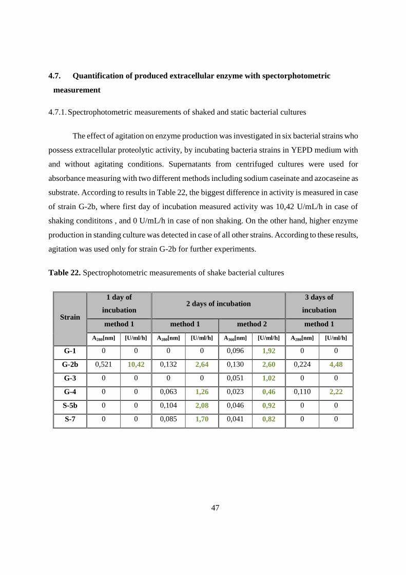
47
4.7. Quantification of produced extracellular enzyme with spectorphotometric
measurement
4.7.1. Spectrophotometric measurements of shaked and static bacterial cultures
The effect of agitation on enzyme production was investigated in six bacterial strains who
possess extracellular proteolytic activity, by incubating bacteria strains in YEPD medium with
and without agitating conditions. Supernatants from centrifuged cultures were used for
absorbance measuring with two different methods including sodium caseinate and azocaseine as
substrate. According to results in Table 22, the biggest difference in activity is measured in case
of strain G-2b, where first day of incubation measured activity was 10,42 U/mL/h in case of
shaking condititons , and 0 U/mL/h in case of non shaking. On the other hand, higher enzyme
production in standing culture was detected in case of all other strains. According to these results,
agitation was used only for strain G-2b for further experiments.
Table 22. Spectrophotometric measurements of shake bacterial cultures
Strain
1 day of
incubation 2 days of incubation
3 days of
incubation
method 1 method 1 method 2 method 1
A280[nm] [U/ml/h] A280[nm] [U/ml/h] A366[nm] [U/ml/h] A280[nm] [U/ml/h]
G-1 0 0 0 0 0,096 1,92 0 0
G-2b 0,521 10,42 0,132 2,64 0,130 2,60 0,224 4,48
G-3 0 0 0 0 0,051 1,02 0 0
G-4 0 0 0,063 1,26 0,023 0,46 0,110 2,22
S-5b 0 0 0,104 2,08 0,046 0,92 0 0
S-7 0 0 0,085 1,70 0,041 0,82 0 0
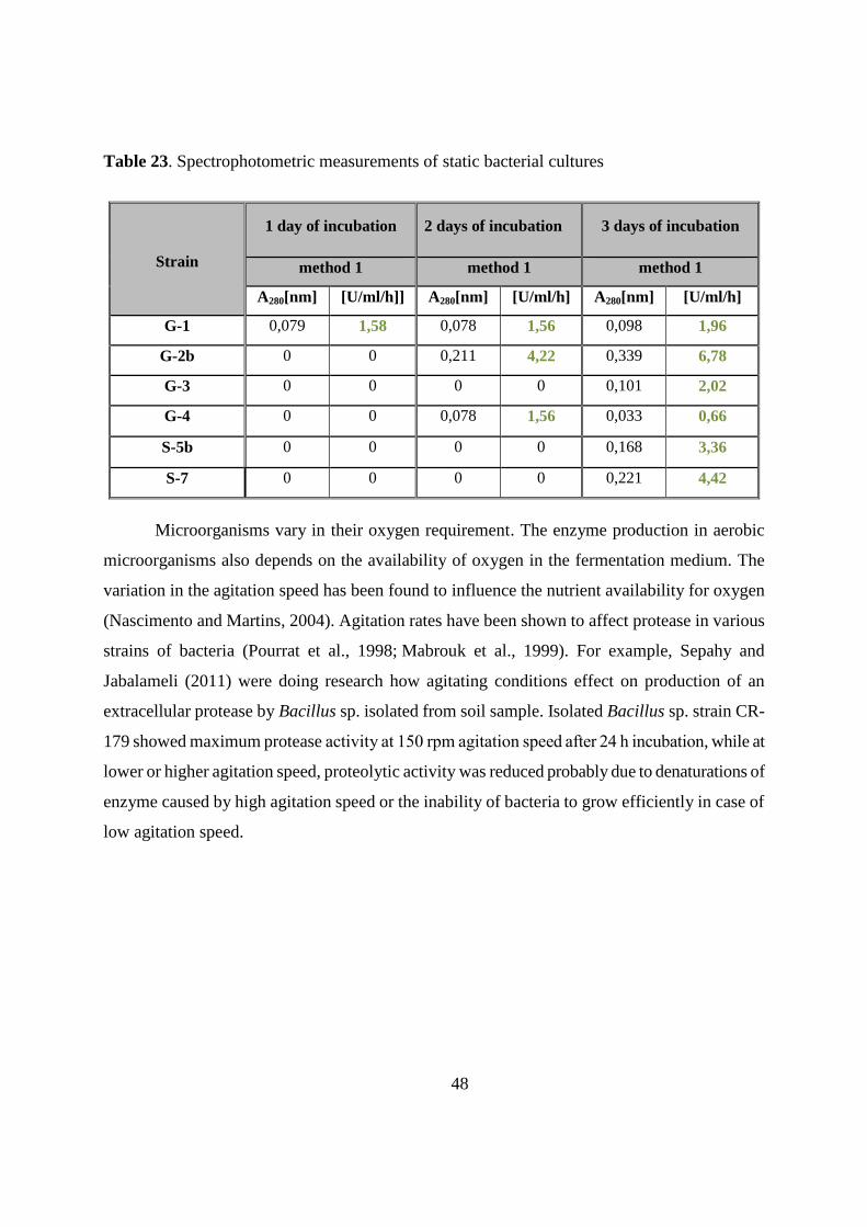
48
Table 23. Spectrophotometric measurements of static bacterial cultures
Strain
1 day of incubation 2 days of incubation 3 days of incubation
method 1 method 1 method 1
A280[nm] [U/ml/h]] A280[nm] [U/ml/h] A280[nm] [U/ml/h]
G-1 0,079 1,58 0,078 1,56 0,098 1,96
G-2b 0 0 0,211 4,22 0,339 6,78
G-3 0 0 0 0 0,101 2,02
G-4 0 0 0,078 1,56 0,033 0,66
S-5b 0 0 0 0 0,168 3,36
S-7 0 0 0 0 0,221 4,42
Microorganisms vary in their oxygen requirement. The enzyme production in aerobic
microorganisms also depends on the availability of oxygen in the fermentation medium. The
variation in the agitation speed has been found to influence the nutrient availability for oxygen
(Nascimento and Martins, 2004). Agitation rates have been shown to affect protease in various
strains of bacteria (Pourrat et al., 1998; Mabrouk et al., 1999). For example, Sepahy and
Jabalameli (2011) were doing research how agitating conditions effect on production of an
extracellular protease by Bacillus sp. isolated from soil sample. Isolated Bacillus sp. strain CR-
179 showed maximum protease activity at 150 rpm agitation speed after 24 h incubation, while at
lower or higher agitation speed, proteolytic activity was reduced probably due to denaturations of
enzyme caused by high agitation speed or the inability of bacteria to grow efficiently in case of
low agitation speed.
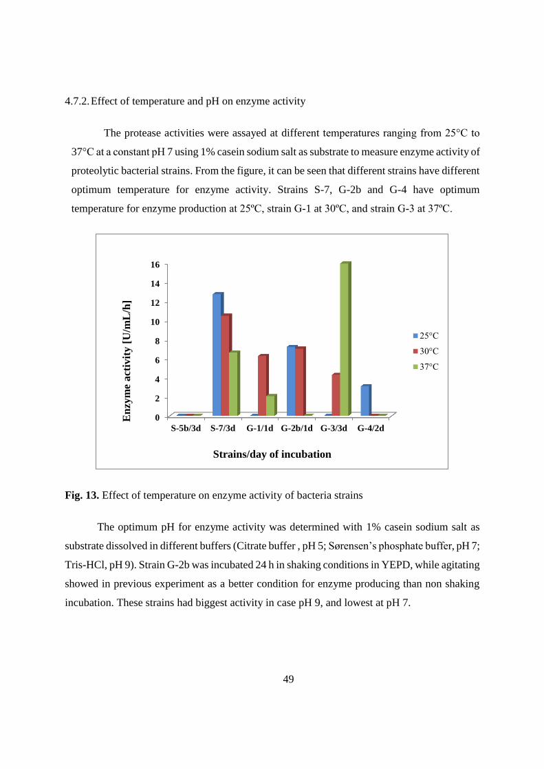
49
4.7.2. Effect of temperature and pH on enzyme activity
The protease activities were assayed at different temperatures ranging from 25°C to
37°C at a constant pH 7 using 1% casein sodium salt as substrate to measure enzyme activity of
proteolytic bacterial strains. From the figure, it can be seen that different strains have different
optimum temperature for enzyme activity. Strains S-7, G-2b and G-4 have optimum
temperature for enzyme production at 25ºC, strain G-1 at 30ºC, and strain G-3 at 37ºC.
Fig. 13. Effect of temperature on enzyme activity of bacteria strains
The optimum pH for enzyme activity was determined with 1% casein sodium salt as
substrate dissolved in different buffers (Citrate buffer , pH 5; Sørensen’s phosphate buffer, pH 7;
Tris-HCl, pH 9). Strain G-2b was incubated 24 h in shaking conditions in YEPD, while agitating
showed in previous experiment as a better condition for enzyme producing than non shaking
incubation. These strains had biggest activity in case pH 9, and lowest at pH 7.
0
2
4
6
8
10
12
14
16
S-5b/3d S-7/3d G-1/1d G-2b/1d G-3/3d G-4/2d
25°C
30°C
37°C
En
zym
e act
ivit
y [
U/m
L/h
]
Strains/day of incubation
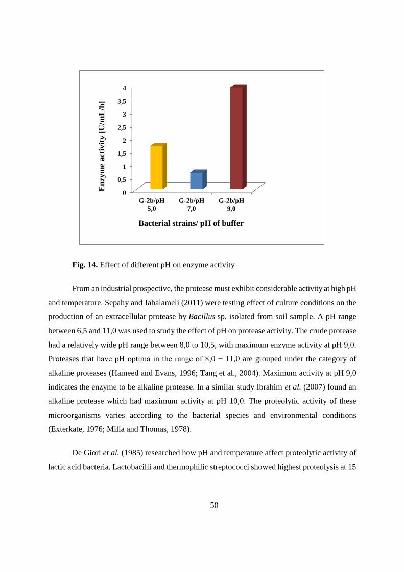
50
Fig. 14. Effect of different pH on enzyme activity
From an industrial prospective, the protease must exhibit considerable activity at high pH
and temperature. Sepahy and Jabalameli (2011) were testing effect of culture conditions on the
production of an extracellular protease by Bacillus sp. isolated from soil sample. A pH range
between 6,5 and 11,0 was used to study the effect of pH on protease activity. The crude protease
had a relatively wide pH range between 8,0 to 10,5, with maximum enzyme activity at pH 9,0.
Proteases that have pH optima in the range of 8,0 − 11,0 are grouped under the category of
alkaline proteases (Hameed and Evans, 1996; Tang et al., 2004). Maximum activity at pH 9,0
indicates the enzyme to be alkaline protease. In a similar study Ibrahim et al. (2007) found an
alkaline protease which had maximum activity at pH 10,0. The proteolytic activity of these
microorganisms varies according to the bacterial species and environmental conditions
(Exterkate, 1976; Milla and Thomas, 1978).
De Giori et al. (1985) researched how pH and temperature affect proteolytic activity of
lactic acid bacteria. Lactobacilli and thermophilic streptococci showed highest proteolysis at 15
0
0,5
1
1,5
2
2,5
3
3,5
4
G-2b/pH
5,0
G-2b/pH
7,0
G-2b/pH
9,0
En
zym
e act
ivit
y [
U/m
L/h
]
Bacterial strains/ pH of buffer

51
and 45°C, and mesophilic streptococci showed only a slight proteolysis at all temperatures,
except for S. cremoris strains at 50°C and S. lactis at 45°C. Variations in pH also had a strong
influence on casein hydrolysis; this effect was more evident in streptococci than in lactobacilli.
5. CONCLUSIONS
1. Temperature needed for growth was not always the same as the temperature optimal for
developing enzymatic activity.
2. One of the factors that influenced the proteolytic activity of bacteria was the composition
of the media used as nutritional sources. Different nitrogen sources, ammonium sulfate,
sodium caseinate, and peptone, support bacterial growth, but protease activity on SM
agar was not detected when growth media did not contain peptone.
3. YEPD media showed good properties for enzyme production, while modified MM was
not appropiate due to the abscence of peptone as a basic component.
4. Sodium chloride has a positive effect on bacteria growth but inhibitory influence on
proteolytic activity.
5. The growth and production rates could be correlated to each other, but good growth did
not necessarily mean a high enzyme production.
6. Skimmed milk agar plate assays allows qualitative determination of protease activity and
provides a sensitive, convenient, and inexpensive method for screening protease
production.
52
6. LITERATURE
Aly, M. E. (1995) An attempt for producing low-sodium feta type cheese. Food Chem. 52, 295-
299.
Anitha, T. S, Palanivelu, P. (2013) Purification and characterization of an extracellular
keratinolytic protease from a new isolate of Aspergillus parasiticus. Protein Expr Purif. 88, 214–
220.
Anwar, A., Saleemuddin, M. (1998) Alkaline proteases: A review. Bioresource Technol. 64, 175-
183.
Argos, P. (1987) A sensitive procedure to compare amino acid sequences. J. Mol. Biol. 193, 385–
396.
Asokan, J. S. C. (2010). Alkalıne protease production by Bacillus licheniformis and Bacillus
coagulans. Cell Tissue Res. 10, 2119-2123.
Atanassova M, Choise Y., Dalgalarrondo, M., Chobert, J.M., Dousset, X., Ivanova, I., Haertle, T.
(2003) Isolation and partial biochemical characterization of a proteinaceous anti-bacteria and
anti-yeast compound produced by Lactobacillus paracasei subsp. paracasei strain M3. Int. J. Food
Microbiol. 87, 63-73.
Baluja, M., Q., Bohme, K., Fernandez-No, J.C., Morandi, S., Franco, C., Gallardo, J. M., Barros-
Velazquez, J., Calo-Mato, P. (2011) Rapid differentiation of Enterococcus species by MALDI-
TOF mass spectrometry. In: Microbes in Applied Research: Current Advances and Challenges
(Mendes-Vilas, A. ed.), World Scientific Publishing, Singapore, pp. 294-298.
Banjara, N., Suhr, M. J., Hallen-Adams, H. E. (2015) Diversity of Yeast and Mold Species from a
Variety of Cheese Types. Curr Microbiol. 70, 792-800.
Bassler, B. L. (2002) Small talk. Cell-to-cell communication in bacteria. Cell 109, 421-424.
53
Beg, Q. K., Gupta, R. (2003) Purification and characterization of an oxidation-stable,
thioldependent serine alkaline protease from Bacillus mojavensis. Enzyme Microb. Techn. 32,
294–304.
Beresford, T.P., Fitzsimons, N. A., Brennan, N. L., Cogan, T. M. (2001) Recent advances in
cheese microbiology. Int. Dairy J. 11, 259-274.
Cheong, C., Chun, S.S., Kim, Y. H. (1993) Production and properties of an alkaline protease from
Pseudomonas sp. SJ-320. Korea. Bid. J. 26, 479-484.
Cousin, M.A., 1982. Presence and activity of psychrotrophic microorganisms in milk and dairy
products: A review. J. Food Prot. 45, 172–207.
De Giori, G. S., de Valdez, G. F., de Holgado, R. A. P., de Oliver, G. (1985) Effect of pH and
Temperature on the Proteolytic Activity of Lactic Acid Bacteria. J Dairy Sci. 68, 2160-2164.
Donnelly, C. W. (2014) Cheese and Microbes, American Society for Microbiology, Washington,
DC.
Eliskases-Lechner, F., Ginzinger, W. (1995) The bacterial flora of surface-ripened cheeses with
special regard to coryneforms. Lait 75, 571-584.
Ellaiah, P., Adinarayana, K., Bhavani, Y., Padmaja, P., Srinivasulu, B. (2002) Optimization of
process parameters for glucoamylase production under solid state fermentation by a newly
isolated Aspergillus species. Process. Biochem. 38, 315-620.
Engels, W. J. M., Visser, S. (1994) Isolation and comparative characterization of compounds that
contribute to the flavor of different cheese types. Neth. Milk Dairy J. 48, 127-140.
Erkmen, O., Bozoglu, T. F. (2016) Food Microbiology: principles into principles, John Wiley &
Sons, New Jersey.
54
Fang, F., O’Toole, P. W. (2009) Genetic tools for investigation the biology of commensal
lactobacilli. Front. Biosci. 14, 3111-3127.
Fleet, G., Mian, M.A., (1987) The occurrence and growth of yeasts in dairy products. Int. J. Food
Microbiol. 4, 145–155.
Fleet, G.H. (1990) Yeasts in dairy products: A review. J. Appl. Bacteriol. 68, 199–211.
Fogarty, W.M., Grilffin, P. H., Joyce, A. M. (1974) Enzymes of Bacillus species- Part 2. Process
Biochem. 9, 27-35.
Fox, P. F., Guinee, P. T., Cogan, M. T., McSweeney, P. L. H. (2000) Fundamentals of Cheese
Science, Aspen Publishers, Gaithersburg.
Fox, P. F., Wallace, J. M. (1997) Formation of flavor compounds. Adv. Appl. Microbiol. 45, 17-
85.
Garcica-Carreno, F. L. (1992) Protease inhibition in theory and practice. Biotechnol. Educ. 3,
145-150.
Germano, S, Pandey, A, Osaku, C. A (2003) Characterization and stability of proteases
from Penicilliumsp. produced by solid-state fermentation. Enzym Microb Technol. 32, 246–251.
Gupta, R., Beg, Q. K. (2002) Bacterial alkaline proteases: molecular approaches and industrial
applications. Appl. Microbiol. Biot. 59, 15-32.
Güracar, S. (2011), production, purification and characterization of thermostable protease from
alkaliphilic and thermophilic Geobacillus sp., Graduate Thesis, Graduate School of Engineering
and Sciences, İzmir Institute of Technology.
Hagler, A., N., Ahearn, D. G. (1981) A rapid DBB test to detect basidiomycetous affinity of
yeasts. Int. J. Syst. Bacteriol. 31, 204-208.
55
Hameed, A., Natt, M. A., Evans C. S. (1996) Short communication: production of alkaline
protease by a new Bacillus subtilis isolate for use as a bating enzyme in leather treatment. World
J. Microb. Biot. 12, 289–291.
Hase, C. C., Finkelstein, R. A. (1993) Bacterial extracellular zinc-containing metalloproteases.
Microbiol. Rev. 57, 823-837.
Ibrahim, A. S. S., A EI-Shayeb, N. M., Mabrouk, S. (2007) Isolation and identification of
Alkaline protease Producing Alkaliphilic bacteria from an Egyptian soda lake. J. appl. sci. res. 3,
1363–1368.
Ibrahim, N. A., Leow., T., C., Rahman, A. R. N. Z. R. Salleh, A., B., Basri, M. (2006)
Thermostable proteases. In: New Lipases and Proteases (Salleh, A.B., Rahman, R. N. Z. R. A.,
Basri, M., eds.), Nova Science, New York, pp. 77-93.
Irlinger, F., Mounier, J. (2009) Microbial interactions in cheese: Implications for cheese quality
and safety. Curr. Opin. Biotechnol, 20, 142–148.
Kalisz, H. M. (1988) Microbial proteinases. Adv. Biochem. Eng. Biotechnol. 36, 1–65.
Katsiari, M., C., Voutsianas, L., P., Alichanidis, E., Roussis, I., G. (1997) Reduction of sodium
content in Feta cheese by partial substitution of NaCl by KCl. Int. Dairy J. 7, 465-472.
Kieronczyk, A., Cachon, R., Feron, G., Yvon, M. (2006) Addition of oxidizing or reducing agents
to the reaction medium influences amino acid conversion to aroma compounds by Lactococcus
lactis. J. Appl. Microbiol. 101, 1114-1122.
Kirschner, C., Maquelin, K., Pina, P., Ngo Thi, N. A., Choo-Smith, L. P., Sockalingum, G.
D., Sandt, C., Ami, D., Orsini, F., Doglia, S.M., Allouch, P., Mainfait, M., Puppels, G.J.,
Naumann, D. (2001) Classification and identification of Enterococci: a Comparative Phenotypic,
Genotypic, and Vibrational Spectroscopic Study. J. Clin. Microbiol. 39, 1763-1770.
56
Kohlmann, K. L., Nielsen, S. S., Steenson, L. R., Ladisch, M. R. (1991) Production of proteases
by psychrotrophic microorganisms. J. Dairy Sci. 74, 3275-3283.
Korhonen H, Pihlanto A. (2006) Bioactive peptides: production and functionality. Int Dairy J 16,
945-960.
Kumar, D., Savitri, T. N., Verma, R. Bhalla, T. C. (2008) Microbial proteases and application as
Laundry Detergent additive. Res. J. Microb. 3, 661-672.
Litopoulou-Tzanetaki, E., Tzanetakis, N., Vafopoulou-Mastrojiannaki, A. (1993) Effect of the
type of lactic starter on microbiological, chemical and sensory characteristic of Feta cheese. Food
Microbiol. 10, 31-41.
Mabrouk, S. S., Hashem, A. M., El-Shayeb, N. M. A., Ismail, A. M. S., Abdel-Fattah, A. F.
(1999) Optimization of alkaline protease productivity by Bacillus licheniformis ATCC 21415.
Bioresource Technol. 69, 155–159.
Maheshwari, R., Bharadwaj, G., Bhat, M. K. (2000) Thermophilic fungi: their physiology and
enzymes. Microbiol. Mol. Biol. Rev. 64, 461–488.
Mala, B.R., Aparna, M.T., Mohinis, G., Vasanti, V.D. (1998) Molecular and Biotechnological
aspect of microbial proteases. Microbiol. Mole. Biol. rev. 62, 597-635.
Matsubara, H., Feder, J. (1971) Other Bacterial, Molad, and Yeasts Proteases. In: The Enzymes
(Boyer, P., ed.) Academic Press, New York, pp 721-792.
Meer, R. R., Baker, J., Bodyfelt, F.W., Griffiths, M.W. (1991) Psychrotrophic Bacillus spp. in
fluid milk products: A review. J. Food Protect. 54, 969-979.
Mistry, V.V., Kasperson, K.M. (1998) Influence of salt on the quality of reduced fat Cheddar
cheese. J. Dairy Sci. 81, 1214-1221.
57
Mótyán, J. A., Tóth, F., Tőzsér, J. (2013) Research Applications of Proteolytic Enzymes in
Molecular Biology: A review. Biomolecules 3, 923-942.
Nascimento, W. C. A., Martins, M. L. L. (2004) Production and properties of an extracellular
protease from thermophilic Bacillus sp. Braz. J. Microbiol. 35, 91–96.
Nurkasanah, S., Widodo, N. (2015) The effect of Different Media Content on Protease Activity
Bacillus suptillis. Biotropica 3, 104-106.
Ogier, J.C., Son, O., Gruss, A., Tailliez, P., Delacroix-Buchet, A. (2002) Identification of the
bacterial microflora in dairy products by temporal temperature gradient gel electrophoresis. Appl.
Environ. Microbiol. 68, 3691–3701.
Pandey, A, Nigamp, V. T., Socco, L., Singh, D., Mohan, R. (2006) Advances in microbial
amylases. Biochem, 31, 35-152.
Polaina, J., MacCabe, A. P. (2007) Industrial Enzymes: Structure, Function and Applications,
Springer, Dordrecht.
Pourrat, H., Barthomeuf, C., Texier, O., Pourrat, A. (1988) Production of semi-alkaline protease
by Aspergillus niger. J. Ferment. Technol. 66, 383–388.
Priest, F.G. (1977) Extracellular enzyme-synthesis in genus Bacillus. Bacteriol. Rev. 41, 711-753.
Priest, F. G. (2003) Brewing Microbiology, third ed., Kluwer Academic, New York.
Raes, J., Bork, P. (2008) Molecular eco-system biology: towards and understanding of
community function. Nat. Rev. Microbiol. 6, 693-699.
Rahman, R., N., Z., A., Basri, M., Salleh, A. B. (2003) Thermostable alkaline protease from
Bacillus stearothermophilus F1; nutritional factors affecting protease production. Ann. Microbiol.
53, 199-210.
58
Rao, M. B., Tanksale, A. M., Ghatge, M. S., Deshpande, V. V. (1998) Molecular and
biotechnological aspects of microbial proteases. Microb. Mol. Biol. R. 62, 597-635.
Rodarte, M. P, Dias, D.R., Vilela, D.M., Schwan, R.F. (2011) Proteolytic activities of bacteria,
yeasts and filamentous fungi isolated from coffee fruit (Coffea arabica L). Acta. Sci. Agron. 33,
457–464.
Rawlings, N.D., Barrett, A.J. (1993) Evolutionary families of peptidases. Biochem. J. 290, 205-
218.
Sarantinopoulos, P., Kalautzopoulos, G., Tsakalidou, E. (2002) Effect of Enterococcus faccium
on microbiological, physicochemical and sensory characteristic of Greek Feta cheese. Int. J. Food
Microbiol. 76, 93-105.
Schroeder, L. R., Bodyfelt, F. W., Wyatt, C. J., McDaniel, M. R. (1988) Reduction of Sodium
Chloride in Cheddar Cheese: Effect on Sensory, Microbiological and Chemical Properties. J.
Dairy. Sci. 71, 2010-2020.
Seiler, H., Busse, M., (1990) The yeasts of cheese brines. Int. J. Food Microbiol. 11, 289–304.
Sepahy, A. A., Jabalameli, L. (2011) Effect of Culture Conditions on the Production of an
Extracellular Protease by Bacillus sp. Isolated from Soil Sample of Lavizan Jungle Park. Enzyme
Res. 20, 1-7.
Sheibani, A., Ayyash, M. M., Shah, N. P., Mishra, V. K. (2015) The effects of salt reduction on
characteristics of hard type cheese made using high proteolytic starter culture. Int. Food Res. J.
22, 2452-2459.
Siddalingeshwara, K. G, Uday, J., Huchesh, C. H., Puttaraju, H.P., Karthic, J., Sudipta, K. M.,
Pramod, T., Vishwanatha, T. (2010) Screening And Characterization Of Protease From Bacıllus
Sp. IJBT 1, 575-581.
59
Sousa, M. J., Ardö, Y., McSweeney, P. L. H. (2001) Advances in the study of proteolysis during
cheese ripening. Int. Dairy J. 11, 327- 345.
Sterchi, E. E., Stöcker, W. (1999) Proteolytic enzymes: Tools and Targets, Springer, Berlin.
Tang, X. M., Shen, W., Lakay (2004) Cloning and overexpression of an alkaline protease from
Bacillus licheniformis. Biotechnol. Lett. 26, 975–979.
Thomas, T, D., Pearce, K. N. (1981) Influence of salt on lactose fermentation and proteolysis in
Cheddar cheese. NZ J Dairy Sci. Technol. 16, 253-259.
Thomas, T. D., Batt, R. D. (1969) Degradation of cell constituents by starved Streptococcus lactis
in relation to survival. J. Gen. Microbiol. 58, 347-362.
Torkar, K.G., Teger S..G (2006) The presence of some pathogen microorganisms, yeasts and
moulds in cheese samples produced at small dairy-processing plants. Acta. Agric. Slo. 88, 37–51.
Tzanetakis, N., Litopoulou-Tzanetakis, E. (1992) Changes in Numbers and Kinds of Lactic Acid
Bacteria in Feta and Teleme, Two Greek Cheeses from Ewes' Milk. J. Dairy. Sci. 75, 1389-1393.
Uhlig, H. (1998) Industrial Enzyme and Their Application, John Wiley & Sons, New York.
Vafopoulou, A., Alichanidis, E., Zerfiridis, G. (1989) Accelerated ripening of Feta cheese, with
heat-shocked cultures or microbial proteases. J. Dairy Res. 56, 285-296.
Valsamaki, K., Michaelidou, A., Polychroniadou, A. (2000) Biogenic amine production in Feta
cheese. Food Chem. 71, 259-266.
Veloorvalappil, N. J., Robinson, B. S., Selvanesan, P., Sasidharan, S., Kizhakkepawothail, N. U.,
Sreedharan, S, Prakasan P., Moolakkariyil, S. J., Sailas B. (2013) Versatility of microbial
proteases. Adv.Enzyme Res. 1, 39-51.
60
Vijayaraghavan, P., Vincent, S. G. P. (2013) A simple method for the detection of protease
activity on agar plates using bromocresolgreen dye. J. Biochem. Tech. 4, 628-630.
Wandersman, C. (1989) Secretion, processing and activation of bacterial extracellular proteases.
Mol. Microbiol. 3, 1825-1831.
Welthagen, J. J., Viljoen, B. C. (1998) Yeast profile in Gouda cheese during processing and
ripening. Int. J. Food Microbiol. 75, 571-584.
Wu, G. (2010) Amino acids: Biochemistry and Nutrition, Taylor & Francis Group, Boca Raton.
Zeigler, D. R. (2001) The Genus Geobacillus. BGSC. 3,1-25.


![KARAKTERISTIKE SOJEVA VEROCITOTOKSIN PRODUKUJU]IH … · 2015-02-06 · udk 616-022.09:579.842.11 karakteristike sojeva verocitotoksin produkuju]ih escherichia coli izolovanih iz](https://img.pdfslide.net/doc/110x75/5e27ac1d029886576407d060/karakteristike-sojeva-verocitotoksin-produkujuih-2015-02-06-udk-616-0220957984211.jpg)